Article

TPX2 Is a Prognostic Marker and Contributes to Growth and Metastasis of Human Hepatocellular Carcinoma

1
Department of Hepatobiliary Surgery, Nanfang Hospital, Southern Medical University, Guangzhou 510515, China
2
Department of Urology, the Third Affiliated Hospital, Southern Medical University, Guangzhou 510000, China
*
Author to whom correspondence should be addressed.
Int. J. Mol. Sci. 2014, 15(10), 18148-18161; https://doi.org/10.3390/ijms151018148
Submission received: 2 September 2014 / Revised: 16 September 2014 / Accepted: 26 September 2014 / Published: 9 October 2014
(This article belongs to the Special Issue Advances in Molecular Oncology 2014)

Abstract

:
Targeting protein for Xenopus kinesin-like protein 2 (TPX2), a microtubule-associated protein, impacts spindle assembly in human cells. Several studies have demonstrated that TPX2 is overexpressed in different types of human cancers and promotes tumor growth and metastasis. In this study, we found that the expression level of TPX2 was obviously higher in hepatocellular carcinoma (HCC) tissues than in matched nontumor tissues. Elevated expressions of TPX2 mRNA were observed in all HCC cell lines (HepG2, Hep3B, SMMC-7721, Bel-7402 and Huh7) as compared with that in a non-transformed hepatic cell line (LO2). Clinical analysis indicated that the positive expression of TPX2 was significantly correlated with venous infiltration, high Edmondson-Steiner grading and advanced TNM tumor stage in HCC. Furthermore, TPX2 was a novel prognostic marker for predicting 5-year overall survival (OS) and disease-free survival (DFS) of HCC patients. In vitro studies found that TPX2 knockdown significantly inhibited cell proliferation and viability in both Hep3B and HepG2 cells. Moreover, TPX2 knockdown obviously slowed down tumor growth in a nude mouse xenograft model. Otherwise, TPX2 knockdown prominently suppressed HCC cell invasion and migration. In conclusion, these results indicate that TPX2 may serve as a prognostic marker and promotes tumorigenesis and metastasis of HCC.

1. Introduction

More than 600,000 hepatocellular carcinoma (HCC) patients were newly diagnosed per year, and the incidence of HCC-related death exceeded 500,000 cases worldwide in 2008 [1,2]. At present, liver resection is the main curative therapy for HCC. However, the prognosis of HCC patients remains poor due to the high recurrence rate and early metastasis [3]. Therefore, it is important to investigate the molecular mechanisms involved in the recurrence and metastasis of HCC and identify novel prognostic biomarkers of HCC. Accordingly, these will contribute to a better prognosis and provide potential therapeutic targets for HCC.
Targeting protein for Xenopus kinesin-like protein 2 (TPX2), a microtubule-associated protein, was initially found by Heidebrecht et al. in 1997 [4]. Subsequent researches have explored that TPX2 is essential for spindle formation and microtubule nucleation around the chromosomes [5]. Notably, The TPX2 protein is a nuclear proliferation-related protein and is implicated in the regulation of the cell mitosis, which is adjusted by the cell cycle [6]. During the period of cell mitosis, TPX2 is evidently associated with the mitotic spindle and targets Xklp2 to the spindle microtubule for the stability of spindle pole. The overexpression of TPX2 induces the amplification of centrosome and leads to DNA polyploidy [7]. Recently, a variety of studies have paid attention to the relationship between TPX2 and human malignancies [8,9]. Increasing evidences indicate that the aberrant expression of TPX2 may play an important role in the invasion and progression of human cancers [10]. TPX2 has verified to be overexpressed in various human cancers including lung, colon, and bladder cancer [11,12,13]. Elevated expression of TPX2 promotes tumor growth in colon cancer, cervical cancer, pancreatic cancer and esophageal squamous cell carcinoma [12,14,15,16]. High levels of TPX2 expression are correlated with the aggressiveness of ovarian cancer and salivary gland cancer [17,18]. Importantly, TPX2 acts as an oncogenic protein and upregulates the expression of matrix metalloproteases (MMPs) through activation of the phosphatidylinositol 3-kinase (PI3K)/Akt signaling pathway in colon cancer [12]. Recently, Satow et al. [19] have demonstrated an aberrant overexpression of TPX2 in HCC through combined functional genome survey, but the clinical significance of TPX2 and its role in HCC are poorly understood.
In this study, we demonstrate that elevated expression of TPX2 is observed in the HCC tissues and cells. The positive expression of TPX2 is significantly correlated with poor clinicopathological features of HCC. Moreover, the positive expression of TPX2 confers a worse 5-year survival of HCC patients. TPX2 knockdown inhibits tumor growth in vitro and in vivo. Furthermore, TPX2 knockdown suppresses HCC cell migration and invasion. Our results demonstrate that TPX2 may act as a potent prognostic marker and contribute to tumor growth and metastasis of HCC.

2. Results and Discussion

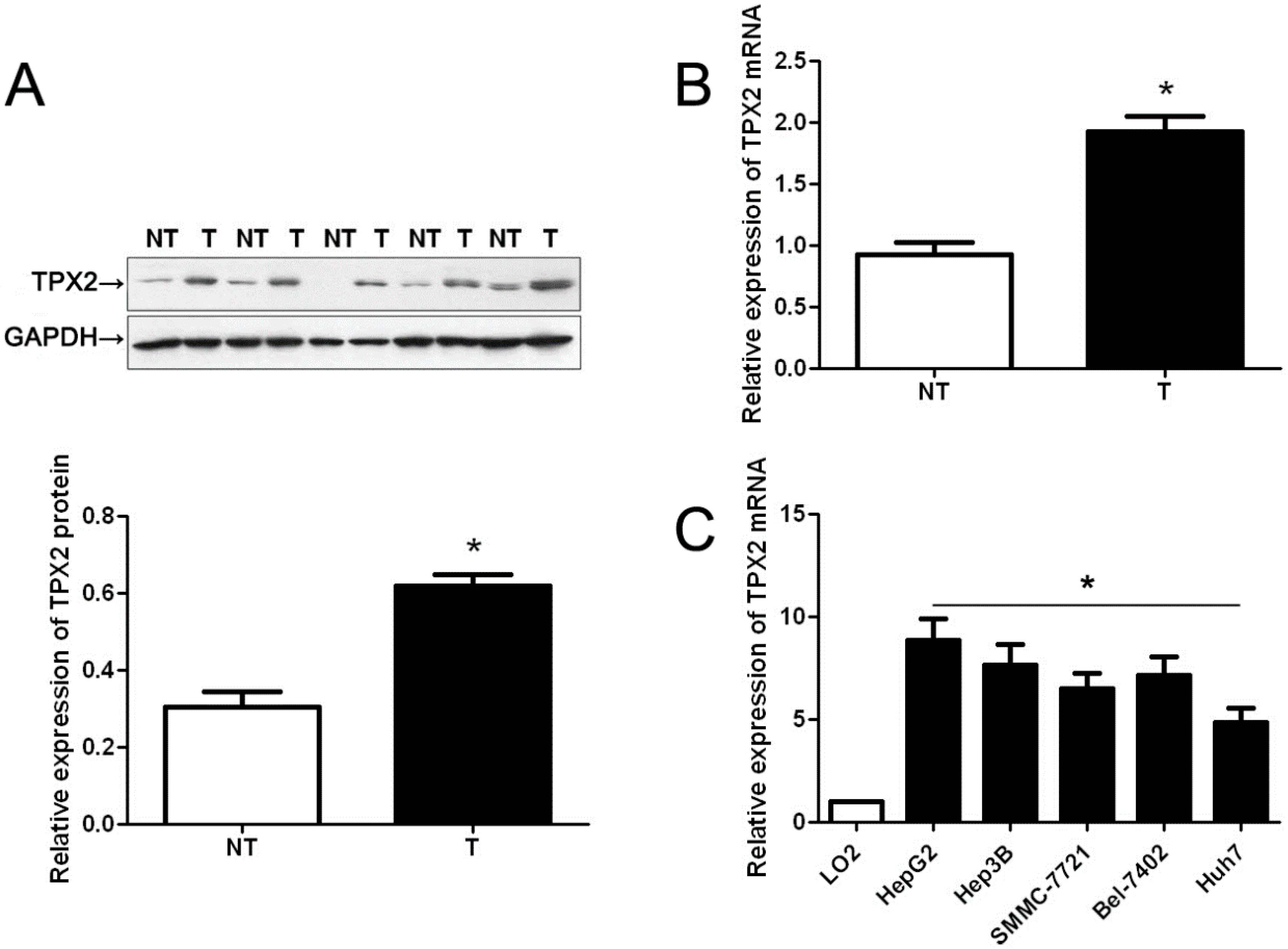
2.1. Clinical Significance of TPX2 (Targeting Protein for Xenopus Kinesin-Like Protein 2) in HCC (Hepatocellular Carcinoma) Specimens

Initially, we tested TPX2 expression in a retrospective cohort of 20 HCC samples using immunoblotting and qRT-PCR. In these cases, we found that the levels of TPX2 protein and mRNA in HCC tissues were prominently higher than those in matched tumor-adjacent tissues (p < 0.05, Figure 1A,B). Furthermore, TPX2 mRNA expressions were up-regulated in HCC cell lines, (Hep3B, HepG2, SMMC-7721, Bel-7402 and Huh7) as compared with that in normal hepatocyte cell line, LO2 (p < 0.05, Figure 1C). 86 pairs of tumor tissues and matched adjacent nontumor tissues were subjected to immunostaining for TPX2. TPX2 protein expression was considered as either negative (scores 0) or positive (scores 1–3). The positive expression of TPX2 protein was detected in 65.1% (56/86) of the HCC specimens, whereas only 27.9% (24/86) of the noncancerous tissues showed a positive TPX2 signal (p < 0.05, Figure 2A). As shown in Table 1, the positive expression of TPX2 protein was prominently associated with venous infiltration (p = 0.004), high Edmondson-Steiner grading (p = 0.019) and advanced TNM tumor stage (p = 0.004). Thus, our results indicate that elevated expression of TPX2 is correlated with malignant clinicopathologic parameters of HCC.
Figure 1. The expression levels of TPX2 in hepatocellular carcinoma (HCC) tissues and cells. Comparing differences in the expression levels of TPX2 between (A) and (B) HCC (T) and matched nontumor tissues (NT) (n = 20), and (C) HCC cell lines and the immortalized hepatic cell line LO2 (n = 6). Values are depicted as Mean ± SEM; * p < 0.05 by t test.
Ijms 15 18148 g001
Table 1. Clinical correlation of TPX2 (Targeting protein for Xenopus kinesin-like protein 2) protein expression in HCC (hepatocellular carcinoma) (n = 86).
Clinicopathologic Features Total No. of Patients, n = 86No. of Patientsp
TPX2 positiveTPX2 negative
Age (year)<502715120.208
≥50594118
SexMale6944250.597
Female17125
HBVAbsent3019110.800
Present563719
Serum AFP level (ng/mL)<4003424100.389
≥400523220
Tumor size (cm)<5302280.242
≥5563422
No. of tumor nodules16636170.489
≥2202013
CirrhosisAbsent3726110.384
Present493019
Venous infiltrationAbsent4221210.004 *
Present44359
Edmondson-Steiner gradingI + II2914150.019 *
III + IV574215
TNM tumor stageI + II6134270.004 *
III + IV25223
HCC, hepatocellular carcinoma; HBV, hepatitis B virus; AFP, alpha-fetoprotein; TNM, tumor-node-metastasis. * Statistically significant.

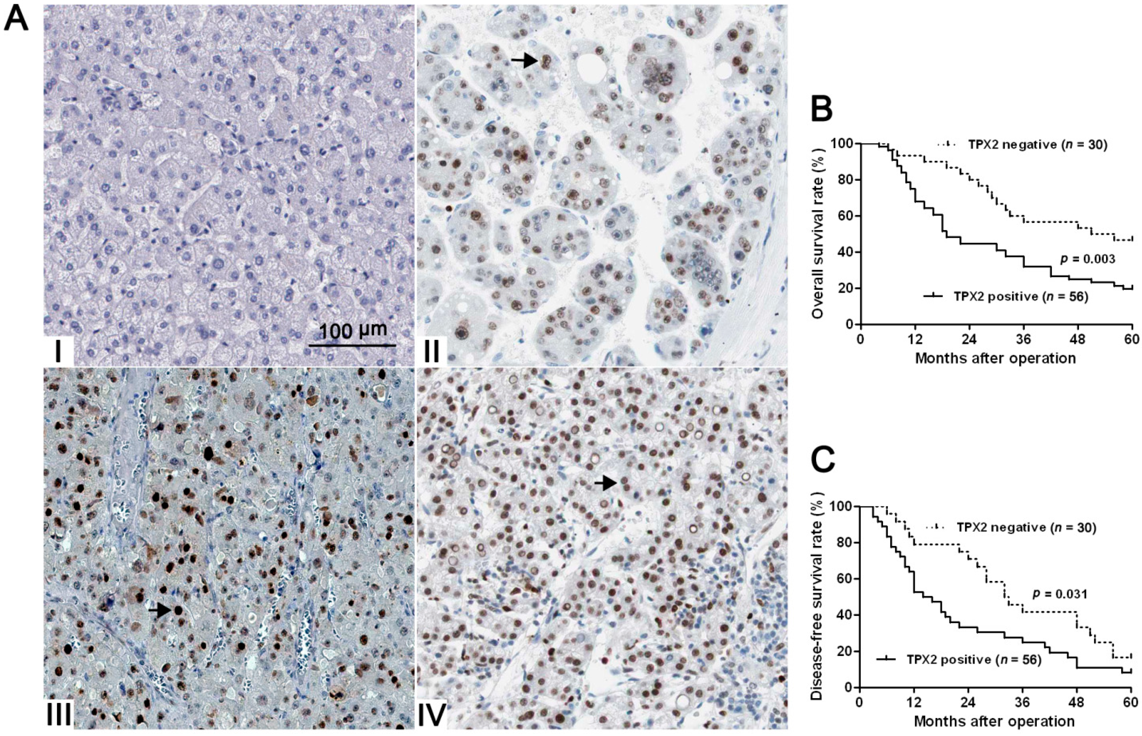
2.2. Positive TPX2 Expression Confers a Worse 5-Year Survival for HCC Patients

Next, 86 HCC patients with clinical survival information (with a median follow-up time of 35.5 months) were analyzed by Kaplan Meier estimation. Tumors with TPX2 positive expression indeed associated with worse overall survival (OS) and disease-free survival (DFS) of HCC patients (p < 0.05, respectively, Figure 2B,C). Furthermore, TPX2 expression was an independent factor for predicting both 5-year OS and DFS of HCC patients (p = 0.001 and 0.006, respectively, Table 2). These data indicate that TPX2 may act as a potent biomarker for predicting prognosis of HCC patients.
Figure 2. The immunostaining of TPX2 and its prognostic significance in HCC specimens. (A) Immunohistochemical staining of TPX2 in HCC. TPX2 was localized within the nuclei. Elevated expression of TPX2 (the arrows) in the tumor cells of HCC tissue (II, III, and IV) compared to normal tumor-adjacent tissues with negative staining (I) (scale bar: 100 μm); (B) The overall survival (OS) and (C) disease-free survival rates (DFS) were estimated by the Kaplan-Meier method. Both the OS rate and DFS rate of patients with TPX2 positive primary tumor were significantly lower than that of patients with TPX2 negative primary tumor (log-rank test, p < 0.05).
Ijms 15 18148 g002
Table 2. Multivariate Cox regression analysis of 5-year OS and DFS of 86 HCC patients.
VariablesOSDFS
HR95% CIpHR95% CIp
Venous infiltration (no vs. yes)2.91.1–7.40.022 *1.50.8–3.10.176
Edmondson-Steiner grading (I/II vs. III/IV)1.60.7–3.70.2563.01.7–5.3<0.001 *
TNM tumor stage (I/II vs. III/IV)3.71.8–7.5<0.001 *4.62.5–8.4<0.001 *
TPX2 (negative vs. positive)3.71.7–8.10.001 *2.21.2–3.90.006 *
OS, overall survival; DFS, disease-free survival; TNM, tumor-node-metastasis; HR, hazard ratio; CI, confidence interval. * Statistically significant.

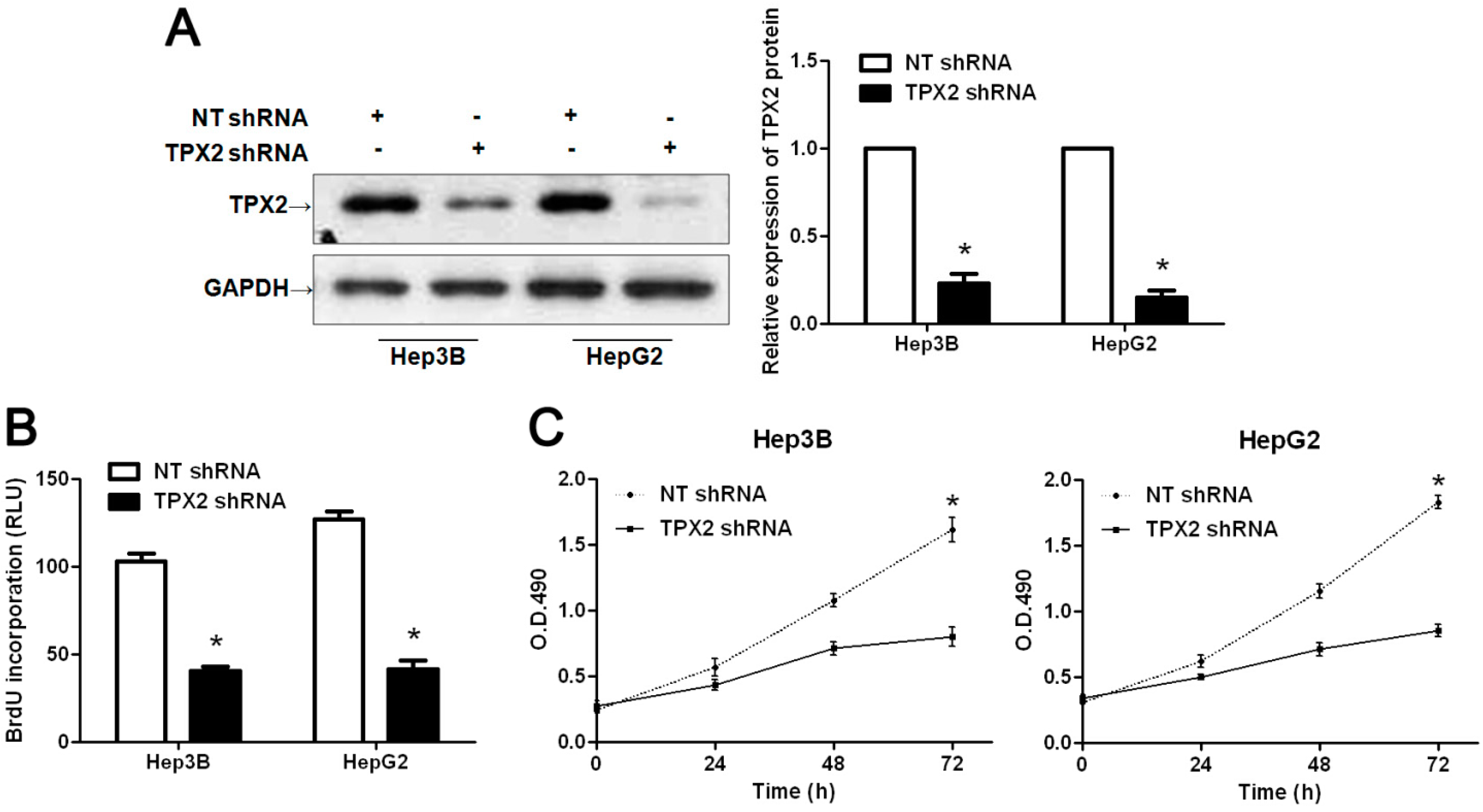
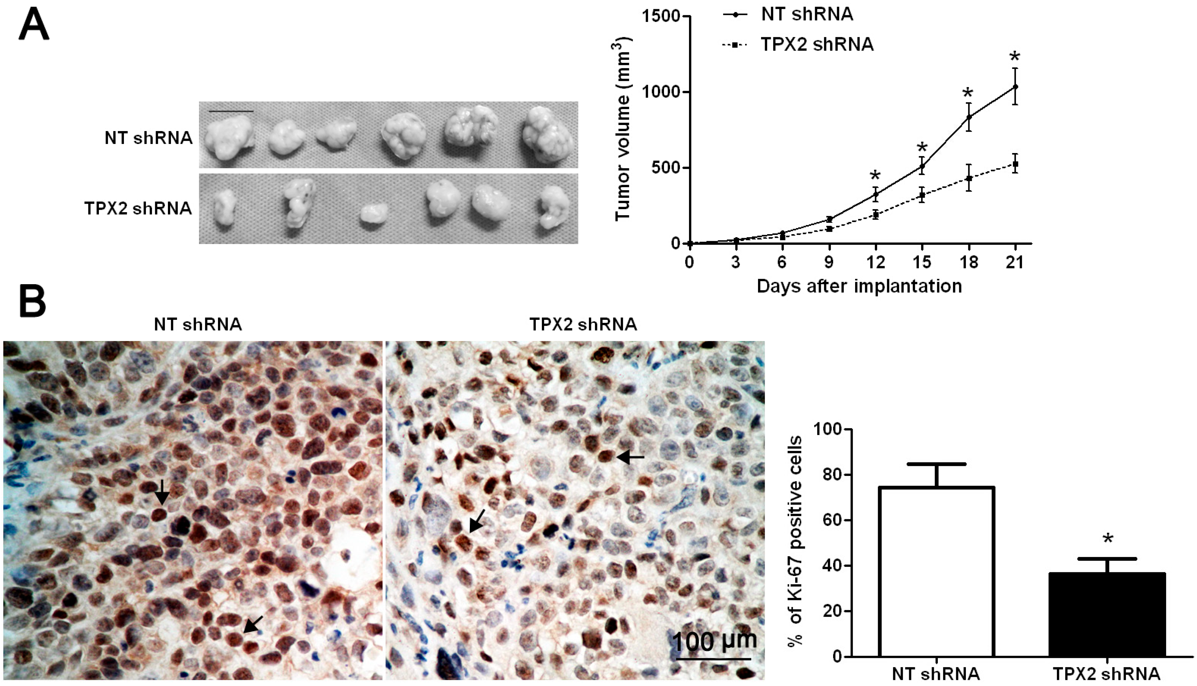
2.3. TPX2 Promotes Tumor Growth in Vitro and in Vivo

Previous studies have demonstrated that TPX2 plays an important role in promoting tumorigenesis and metastasis of human cancer [8,10,12,13,14,15,16,18]. To identify the effect of TPX2 on HCC, TPX2 was knocked down by shRNA in two HCC cell lines, Hep3B and HepG2. As assessed by WB, the TPX2 protein expression could be obviously down-regulated in both cell lines (p < 0.05, respectively, Figure 3A). BrdU assays were performed to test the effect of altering TPX2 levels on tumor cell proliferation. We found that TPX2 knockdown led to a significant reduction of cell proliferation in both Hep3B and HepG2 cells (p < 0.05, respectively, Figure 3B). Furthermore, as determined by MTT, the viability of Hep3B and HepG2 cells were significantly decreased after TPX2 knockdown (p < 0.05, respectively, Figure 3C). We next sought to determine whether TPX2 affects tumor growth using a nude mouse xenograft model. Hep3B cells that were transfected with non-targeting (NT) shRNA or TPX2 shRNA were implanted into nude mice though subcutaneous injection. Tumor growth curves revealed that TPX2 knockdown significantly slowed down tumor growth in mice (p < 0.05, Figure 4A). Furthermore, we performed immunohistochemistry for Ki-67 in the xenografted tissues. Consistent with our in intro data, TPX2 knockdown inhibited Hep3B cell proliferation in vivo (p < 0.05, Figure 4B). Thus, TPX2 may act as an oncogene by promoting tumor growth in HCC.
Figure 3. TPX2 knockdown inhibits cell proliferation and viability in HCC. (A) Hep3B and HepG2 cells that were transfected with non-targeting (NT) shRNA and TPX2 shRNA, respectively, were subjected to WB for TPX2. n = 6; * p < 0.05 by t test; (B) Cell proliferation as measured by BrdU incorporation was inhibited by TPX2 knockdown in Hep3B and HepG2 cells. * p < 0.05 by t test; n = 3 repeats with similar results; (C) As assessed by MTT assays, TPX2 knockdown was found to reduce the viability of Hep3B and HepG2 cells. * p < 0.05 by two-way ANOVA; n = 3 repeats with similar results. Values are depicted as Mean ± SEM.
Ijms 15 18148 g003
Figure 4. TPX2 knockdown suppresses tumor growth in mice. (A) Hep3B cells that were transfected with NT shRNA or TPX2 shRNA were implanted into nude mice through subcutaneous injection. Tumor growth curves indicated that TPX2 knockdown Hep3B cells (n = 6) exhibited a greater tumor-inhibiting effect compared with control cells (n = 6). Scale bar: 100 μm; * p < 0.05 by two-way ANOVA; (B) Tumor nodules were subjected to immunohistochemical staining for Ki-67 and quantitative analysis. Representative immunostaining of Ki-67 (the arrows) revealed that TPX2 knockdown significantly reduced the number of Ki-67 positive cells. Scale bar: 100 μm; n = 6; * p < 0.05 by t test.
Ijms 15 18148 g004

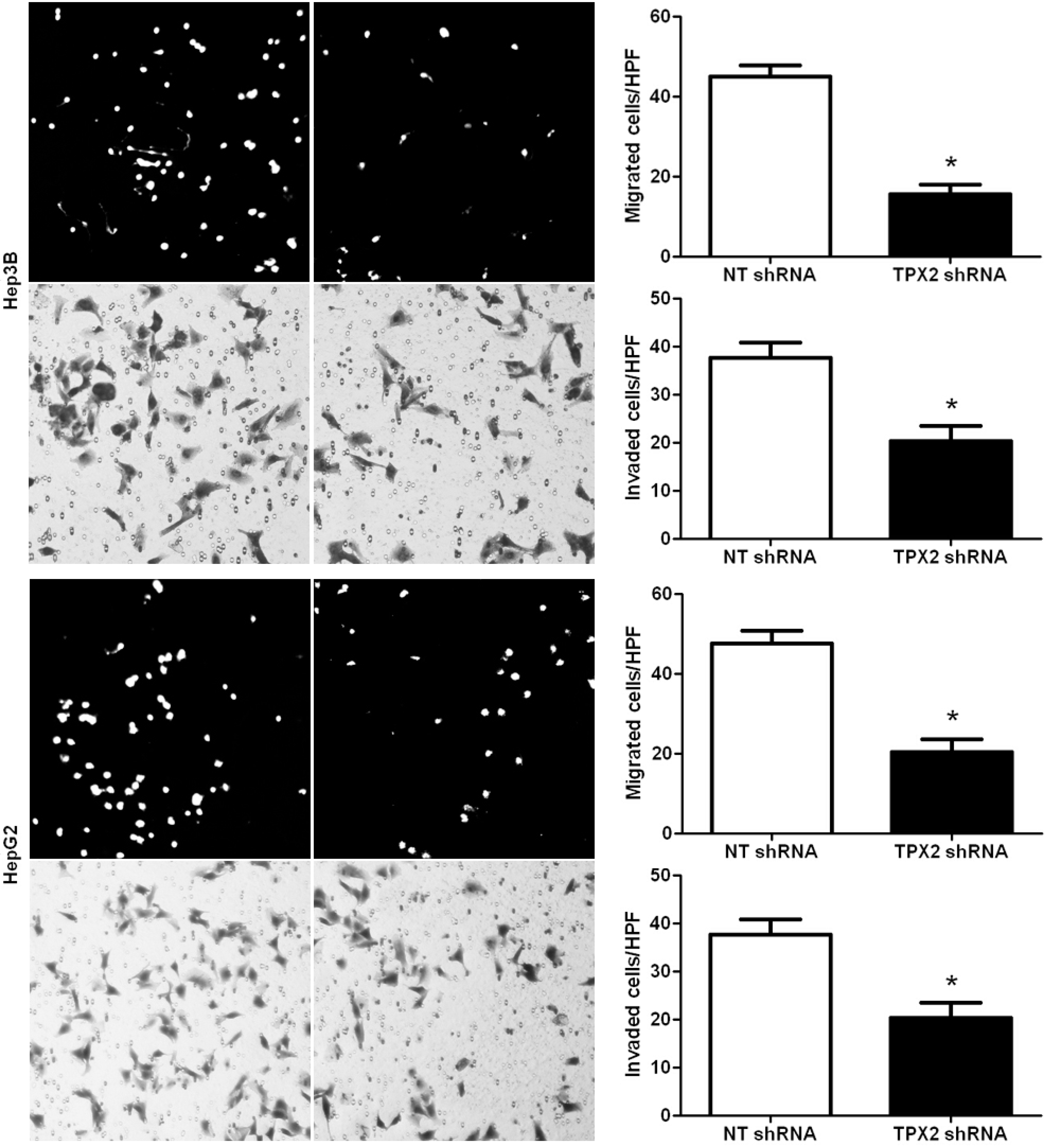
2.4. Promoting Effect of TPX2 on HCC Cell Migration and Invasion

To investigate the role of TPX2 in HCC cell migration and invasion, we down-regulated the expression level of TPX2 in two HCC cell lines, Hep3B and HepG2. Boyden chamber assays were performed to test the effect of altering TPX2 levels on HCC cell migration. We found that TPX2 knockdown led to a significantly less number of migrated Hep3B and HepG2 cells (p < 0.05, respectively, Figure 5). Furthermore, as determined by Transwell assays, the numbers of invaded Hep3B and HepG2 cells were significantly reduced after TPX2 knockdown (p < 0.05, respectively, Figure 5). Thus, TPX2 may exert a pro-metastatic effect on HCC.
Figure 5. TPX2 knockdown inhibits HCC cell migration and invasion. Cell migration as measured by Boyden chamber assays was inhibited by TPX2 knockdown in Hep3B and HepG2 cells as compared with control cells. TPX2 down-regulating Hep3B and HepG2 cells conferred a less number of invaded cells as compared with control cells. n = 6 repeats with similar results; * p < 0.05 by t test. Values are depicted as Mean ± SEM.
Ijms 15 18148 g005
TPX2, a microtubule-associated protein, is located in chromosome 20q11 and promotes nuclear proliferation [8]. TPX2 has been considered to be implicated in the regulation of mitotic spindle and chromosome isolation [8]. Importantly, more and more evidences have demonstrated that the aberrant overexpression of TPX2 is commonly detected in multiple kinds of malignant tumor including ovarian carcinoma, colon cancer and prostate cancer [12,20,21]. However, few works have been done to explore the clinical significance and role of TPX2 in HCC. Satow et al. [19] reported that TPX2 was highly expressed in HCC in comparison with corresponding nontumorous liver. However, the clinical significance of TPX2 and its role in HCC are poorly investigated. In this study, we initially detected the expression status of TPX2 in samples of surgical resected HCC tissues. Our data indicated that the expression levels of TPX2 in HCC were significantly higher than those in normal tumor-adjacent tissues. Elevated expressions of TPX2 mRNA were observed in HCC cell lines as compared with that in normal hepatocyte cell line. Furthermore, TPX2 protein was expressed at significantly higher levels in HCC patients with venous infiltration, high Edmondson-Steiner grading and advanced TNM tumor stage. These results suggest that elevated expression of TPX2 is obviously correlated with poor clinicopathologic features in HCC. Importantly, our data demonstrated that TPX2 positive expression was correlated with a significant worse 5-year survival for HCC patients. Multivariate Cox repression analysis indicated that TPX2 was an independent prognostic factor for predicting survival of HCC patients. Altogether, these results suggest that TPX2 expression is critical for prognosis determination in HCC patients.
Several studies have identified TPX2 as a driving oncogene in human cancers [8,10,11,12,13,14,15,16,17,18,19,20,21]. Mechanistically, TPX2 exerts protumorigenic functions by promoting tumor growth and metastasis [8,10,11,12,13,14,15,16,17,18,19,20,21]. Previous studies have demonstrated that TPX2 is important in the regulation of tumor growth and is a promising diagnostic and therapeutic target for cervical cancer [10,14]. Wei P et al. [12] suggest that TPX2 plays an important role in promoting tumorigenesis and metastasis of human colon cancer, and may represent a novel prognostic biomarker and therapeutic target for the disease. In esophageal squamous cell carcinoma, TPX2 expression is associated with cell proliferation and patient outcome [16]. It has been reported that TPX2 knockdown effectively inhibits pancreatic cancer cell growth in tissue culture, induces apoptosis, and suppresses growth in soft agar and in nude mice [15]. In our study, we found that TPX2 knockdown significantly inhibited cell proliferation and viability in both Hep3B and HepG2 cells. In tumor bearing mice, TPX2 knockdown prominently slowed down tumor growth in vivo. Importantly, TPX2 knockdown reduced the number of migrated and invaded HCC cells. Taken together, these data suggest that TPX2 promotes tumorigenesis and metastasis of human HCC.
In conclusion, we find that TPX2 is elevated in HCC and its high expression is evidently correlated with malignant clinicopathologic characteristics. Moreover, TPX2 expression is an independent prognostic marker for predicting 5-year survival of HCC patients. In vitro and in vivo studies demonstrate that TPX2 knockdown inhibits HCC cell growth, migration and invasion. Altogether, we consider that TPX2 may potentially act as a clinical biomarker, and may also be a therapeutic target, in HCC.

3. Experimental Section

3.1. Ethical Review

The Southern Medical University Ethics Committee approved all protocols according to the Helsinki Declaration (as revised in Edinburgh 2000) and the informed consent was signed by each patient. All animal protocols were approved by the Institutional Animal Care and Use Committee of Southern Medical University.

3.2. Clinical Samples

Eighty-six HCC samples were collected from patients including 69 males and 17 females, who underwent the resection of their primary HCC in the Department of Hepatobiliary Surgery at Nanfang Hospital of Southern Medical University during January 2006 to December 2008. All samples were used after obtaining informed consent. Patients did not receive preoperative chemotherapy or embolization. The demographic features and clinicopathologic data were shown in Table 1.

3.3. Immunohistochemical Staining

Immunohistochemistry was performed on paraformaldehyde-fixed paraffin sections. TPX2 (sc-376812; Santa Cruz, CA, USA) (1:200) and Ki-67 (#9027, Cell Signaling, Danvers, MA, USA) (1:400) antibodies were used in immunohistochemistry with streptavidin peroxidase conjugated (SP-IHC) method. Immunohistochemistry was performed as previous reported [22]. The percentage of positive tumor cells or hepatocytes was graded as per the following criteria: 0, less than 10%; 1, 10%–30%; 2, 31%–50%; 3, more than 50%.

3.4. Cell Lines and Transfection

The human immortalized normal hepatocyte cell line, LO2, and five HCC cell lines, HepG2, Hep3B, SMMC-7721, Bel-7402 and Huh7 (the Institute of Biochemistry and Cell Biology, Chinese Academy of Sciences, Shanghai, China), were cultured in complete Dulbecco’s modified Eagle medium (DMEM, Gibco, Grand Island, NY, USA) containing 10% fetal bovine serum (FBS, Gibco) with 100 units/mL penicillin and 100 μg/mL streptomycin (Sigma, St-Louis, MO, USA) in a humidified containing of 5% CO2 incubator at 37 °C.
TPX2 shRNA and non-targeting (NT) shRNA were generated by inserting the respective sequence into the pSilencer™ 2.1-U6 puro Vector (Ambion, Austin, TX, USA) according to manufacturer’s protocol. The target and negative control sequences were 5'-AAGAATGGAACTGGAGGGCTT-3' and 5'-GTACCGCACGTCATTCGTATC-3', respectively. Transfection of TPX2 shRNA or NT shRNA plasmids were performed using the Lipofectamine 2000 reagent (Invitrogen, Carlsbad, CA, USA) according to the manufacturer’s instructions. Transfected cells were selected with 0.5 μg/mL puromycin (Gibco) and maintained with 0.3 μg/mL puromycin.

3.5. Western Blot

The following primary antibodies were used in the immunoblotting assays: TPX2 (1:1000) and GAPDH (G8140; US Biological, Swampscott, MA, USA) (1:5000). Horseradish peroxidase-conjugated goat anti-mouse secondary antibody (Bio-Rad, Hercules, CA, USA) were used at a 1:1000–1:5000 dilution and detected using a Western Blotting Luminol Reagent (sc-2048; Santa Cruz, MA, USA), as described in previous study [23].

3.6. BrdU Cell Proliferation Assay and MTT Assays

For the proliferation assay, HCC cells were seeded into 96-well plates at 5000 cells per well for 24 h and assessed using a Cell Proliferation ELISA, BrdU (5-bromodeoxyuridine) (chemiluminescent) (Roche, Indianapolis, IN, USA). The 3-(4,5-dimethylthiazol-2-yl)2,5-diphenyl tetrazolium bromide (MTT, Roche, IN, USA) assay was used to assess cell viability at 24, 48 and 72 h.

3.7. In Vivo Experiments

Hep3B cells with stably silenced TPX2 (TPX2 shRNA) or control (NT shRNA) (1 × 10⁶ cells) were subcutaneously injected into the flanks of BALB/c nude mice as previously described [24]. The tumor volume for each mouse was determined by measuring two of its dimensions and then calculated as tumor volume = length × width × width/2. The immunostaining of Ki-67 was performed in the isolated tumor tissues.

3.8. Real Time Quantitative Reverse Transcription-PCR (qRT-PCR)

The following primers were used: TPX2 sense primer 5'-AGGGGCCCTTTGAACTCTTA-3' and antisense primer 5'-TGCTCTAAACAAGCCCCATT-3' and GAPDH sense primer 5'-CGGATTTGGTCGTATTGG-3' and antisense primer 5'-TCCTGGAAGATGGTGATG-3'. The PCR amplification for the quantification of the TPX2 and GAPDH mRNAs was performed using an ABI PRISM 7300 Sequence Detection System (Applied Biosystems, Foster City, CA, USA) and a SYBR® Premix Ex Taq™ ii (Perfect Real Time) Kit (Takara Bio, Shiga, Japan), as previously reported [24].

3.9. Boyden Chamber and Transwell Assays

A Boyden chamber assay (NeuroProbe, Gaithersburg, MD, USA) was used to analyze HCC cell migration as previously described [25]. Transwell assays were done in 6 well plates with Transwell inserts equipped with 8-μm pores (Nalge Nunc International Corp, Naperville, IL, USA) coated with Matrigel at 1:6 dilution (Becton Dickinson Labware, Bedford, MA, USA) as previously described [26].

3.10. Statistical Analysis

Results are expressed as Mean ± SEM. Significance was established, with the SPSS statistical package for Windows Version 13 (SPSS, Chicago, IL, USA) and GraphPad Prism 5 software (GraphPad Software, Inc., San Diego, CA, USA), using the Pearson chi-squared tests, the multi-variant Cox regression analysis, a two-tailed Student’s t test, a Kaplan-Meier plot, a log-rank test or an ANOVA when appropriate. Difference were considered significant when p < 0.05.

4. Conclusions

In conclusion, our studies find that TPX2 is overexpressed in HCC cases and cells. TPX2 positive expression is associated with malignant clinicopathologic characteristics of HCC. Moreover, we demonstrate that the positive expression of TPX2 confers a worse 5-year survival rate of HCC patients after surgery. Multivariate Cox repression analysis indicates TPX2 expression is an independent prognostic marker for HCC. Our in vitro data prove that TPX2 knockdown by a specific shRNA inhibits cell proliferation and viability in HCC cell lines, Hep3B and HepG2. Furthermore, in tumor bearing mice, TPX2 knockdown slows down tumor growth. Otherwise, TPX2 knockdown reduced the number of migrated and invaded HCC cells. Taken together, we suggest that TPX2 may be a potent prognostic factor and a potential therapeutic target for HCC.

Acknowledgments

This study was supported by grants from the Natural Science Foundation of Guangdong Province [no.S2012020010942 and no.8151051501000029].

Author Contributions

Conceived and designed the experiments: Heping Kan. Performed the experiments: Yuqi Huang, Wenbin Guo. Analyzed the data: Yuqi Huang, Heping Kan. Contributed reagents/materials/analysis tools: Yuqi Huang, Wenbin Guo. Wrote the paper: Yuqi Huang, Heping Kan.

Conflicts of Interest

The authors declare no conflict of interest.

References

  1. Ferlay, J.; Shin, H.R.; Bray, F.; Forman, D.; Mathers, C.; Parkin, D.M. Estimates of worldwide burden of cancer in 2008: GLOBOCAN 2008. Int. J. Cancer 2010, 127, 2893–2917. [Google Scholar] [CrossRef]
  2. Bruix, J.; Sherman, M. Management of hepatocellular carcinoma: An update. Hepatology 2011, 53, 1020–1022. [Google Scholar] [CrossRef]
  3. Tu, K.; Li, C.; Zheng, X.; Yang, W.; Yao, Y.; Liu, Q. Prognostic significance of miR-218 in human hepatocellular carcinoma and its role in cell growth. Oncol. Rep. 2014, 32, 1571–1577. [Google Scholar]
  4. Heidebrecht, H.J.; Buck, F.; Steinmann, J.; Sprenger, R.; Wacker, H.H.; Parwaresch, R. p100: A novel proliferation-associated nuclear protein specifically restricted to cell cycle phases S, G2, and M. Blood 1997, 90, 226–233. [Google Scholar]
  5. Wittmann, T.; Wilm, M.; Karsenti, E.; Vernos, I. TPX2, A novel xenopus MAP involved in spindle pole organization. J. Cell Biol. 2000, 149, 1405–1418. [Google Scholar] [CrossRef]
  6. Kufer, T.A.; Sillje, H.H.; Korner, R.; Gruss, O.J.; Meraldi, P.; Nigg, E.A. Human TPX2 is required for targeting Aurora-A kinase to the spindle. J. Cell Biol. 2002, 158, 617–623. [Google Scholar] [CrossRef] [Green Version]
  7. Gruss, O.J.; Wittmann, M.; Yokoyama, H.; Pepperkok, R.; Kufer, T.; Sillje, H.; Karsenti, E.; Mattaj, I.W.; Vernos, I. Chromosome-induced microtubule assembly mediated by TPX2 is required for spindle formation in HeLa cells. Nat. Cell Biol. 2002, 4, 871–879. [Google Scholar] [CrossRef]
  8. Liu, H.C.; Zhang, Y.; Wang, X.L.; Qin, W.S.; Liu, Y.H.; Zhang, L.; Zhu, C.L. Upregulation of the TPX2 gene is associated with enhanced tumor malignance of esophageal squamous cell carcinoma. Biomed. Pharmacother. 2013, 67, 751–755. [Google Scholar] [CrossRef]
  9. Neumayer, G.; Belzil, C.; Gruss, O.J.; Nguyen, M.D. TPX2: Of spindle assembly, DNA damage response, and cancer. Cell Mol. Life Sci. 2014, 71, 3027–3047. [Google Scholar] [CrossRef]
  10. Chang, H.; Wang, J.; Tian, Y.; Xu, J.; Gou, X.; Cheng, J. The TPX2 gene is a promising diagnostic and therapeutic target for cervical cancer. Oncol. Rep. 2012, 27, 1353–1359. [Google Scholar]
  11. Li, Y.; Tang, H.; Sun, Z.; Bungum, A.O.; Edell, E.S.; Lingle, W.L.; Stoddard, S.M.; Zhang, M.; Jen, J.; Yang, P.; et al. Network-based approach identified cell cycle genes as predictor of overall survival in lung adenocarcinoma patients. Lung. Cancer 2013, 80, 91–98. [Google Scholar] [CrossRef]
  12. Wei, P.; Zhang, N.; Xu, Y.; Li, X.; Shi, D.; Wang, Y.; Li, D.; Cai, S. TPX2 is a novel prognostic marker for the growth and metastasis of colon cancer. J. Transl. Med. 2013, 11. [Google Scholar] [CrossRef]
  13. Yan, L.; Li, S.; Xu, C.; Zhao, X.; Hao, B.; Li, H.; Qiao, B. Target protein for Xklp2 (TPX2), a microtubule-related protein, contributes to malignant phenotype in bladder carcinoma. Tumour Biol. 2013, 34, 4089–4100. [Google Scholar] [CrossRef]
  14. Jiang, P.; Shen, K.; Wang, X.; Song, H.; Yue, Y.; Liu, T. TPX2 regulates tumor growth in human cervical carcinoma cells. Mol. Med. Rep. 2014, 9, 2347–2351. [Google Scholar]
  15. Warner, S.L.; Stephens, B.J.; Nwokenkwo, S.; Hostetter, G.; Sugeng, A.; Hidalgo, M.; Trent, J.M.; Han, H.; von Hoff, D.D. Validation of TPX2 as a potential therapeutic target in pancreatic cancer cells. Clin. Cancer Res. 2009, 15, 6519–6528. [Google Scholar]
  16. Hsu, P.K.; Chen, H.Y.; Yeh, Y.C.; Yen, C.C.; Wu, Y.C.; Hsu, C.P.; Hsu, W.H.; Chou, T.Y. TPX2 expression is associated with cell proliferation and patient outcome in esophageal squamous cell carcinoma. J. Gastroenterol. 2014, 49, 1231–1240. [Google Scholar] [CrossRef]
  17. Caceres-Gorriti, K.Y.; Carmona, E.; Barres, V.; Rahimi, K.; Letourneau, I.J.; Tonin, P.N.; Provencher, D.; Mes-Masson, A.M. RAN nucleo-cytoplasmic transport and mitotic spindle assembly partners XPO7 and TPX2 are new prognostic biomarkers in serous epithelial ovarian cancer. PLoS One 2014, 9, e91000. [Google Scholar]
  18. Shigeishi, H.; Ohta, K.; Hiraoka, M.; Fujimoto, S.; Minami, M.; Higashikawa, K.; Kamata, N. Expression of TPX2 in salivary gland carcinomas. Oncol. Rep. 2009, 21, 341–344. [Google Scholar]
  19. Satow, R.; Shitashige, M.; Kanai, Y.; Takeshita, F.; Ojima, H.; Jigami, T.; Honda, K.; Kosuge, T.; Ochiya, T.; Hirohashi, S.; et al. Combined functional genome survey of therapeutic targets for hepatocellular carcinoma. Clin. Cancer Res. 2010, 16, 2518–2528. [Google Scholar]
  20. Aguirre-Portoles, C.; Bird, A.W.; Hyman, A.; Canamero, M.; Perez de Castro, I.; Malumbres, M. Tpx2 controls spindle integrity, genome stability, and tumor development. Cancer Res. 2012, 72, 1518–1528. [Google Scholar] [CrossRef]
  21. Vainio, P.; Mpindi, J.P.; Kohonen, P.; Fey, V.; Mirtti, T.; Alanen, K.A.; Perala, M.; Kallioniemi, O.; Iljin, K. High-throughput transcriptomic and RNAi analysis identifies AIM1, ERGIC1, TMED3 and TPX2 as potential drug targets in prostate cancer. PLoS One 2012, 7, e39801. [Google Scholar] [CrossRef]
  22. Tu, K.; Zheng, X.; Zan, X.; Han, S.; Yao, Y.; Liu, Q. Evaluation of Fbxw7 expression and its correlation with the expression of c-Myc, cyclin E and p53 in human hepatocellular carcinoma. Hepatol. Res. 2012, 42, 904–910. [Google Scholar] [CrossRef]
  23. Tu, K.; Yang, W.; Li, C.; Zheng, X.; Lu, Z.; Guo, C.; Yao, Y.; Liu, Q. Fbxw7 is an independent prognostic marker and induces apoptosis and growth arrest by regulating YAP abundance in hepatocellular carcinoma. Mol. Cancer 2014, 13. [Google Scholar] [CrossRef]
  24. Tu, K.; Zheng, X.; Zhou, Z.; Li, C.; Zhang, J.; Gao, J.; Yao, Y.; Liu, Q. Recombinant human adenovirus-p53 injection induced apoptosis in hepatocellular carcinoma cell lines mediated by p53-Fbxw7 pathway, which controls c-Myc and cyclin E. PLoS One 2013, 8, e68574. [Google Scholar] [CrossRef]
  25. Friedman, S.L. Mechanisms of disease: Mechanisms of hepatic fibrosis and therapeutic implications. Nat. Clin. Pract. Gastroenterol. Hepatol. 2004, 1, 98–105. [Google Scholar] [CrossRef]
  26. Li, C.; Yang, W.; Zhang, J.; Zheng, X.; Yao, Y.; Tu, K.; Liu, Q. SREBP-1 has a prognostic role and contributes to invasion and metastasis in human hepatocellular carcinoma. Int. J. Mol. Sci. 2014, 15, 7124–7138. [Google Scholar] [CrossRef]

