Article

Resistin Stimulates Expression of Chemokine Genes in Chondrocytes via Combinatorial Regulation of C/EBPβ and NF-κB

1
Department of Orthopaedic Surgery, the First Affiliated Hospital of Sun Yat-sen University, Guangzhou 510080, China
2
Department of Orthopaedic Surgery, Washington University School of Medicine at Barnes-Jewish Hospital, St. Louis, MO 63110, USA
*
Authors to whom correspondence should be addressed.
These authors contributed equally to this work.
Int. J. Mol. Sci. 2014, 15(10), 17242-17255; https://doi.org/10.3390/ijms151017242
Submission received: 12 July 2014 / Revised: 2 September 2014 / Accepted: 19 September 2014 / Published: 26 September 2014
(This article belongs to the Section Biochemistry)

Abstract

:
To further investigate the regulation role of two chemokine genes CCL3 and CCL4 in chondrocytes in response to resistin, human primary chondrocytes and T/C-28a2 cells were cultured. The function of resistin on the chemokine genes, and the expression of C/EBPβ, NF-κB isoforms were tested using qPCR. The methods used to investigate timed co-regulation of C/EBPβ and NF-κBwere NF-κB inhibitor (IKK-NBD) and C/EBPβ inhibitor (SB303580) treatments, and subcellular localization, with or without resistin stimulation. Results showed that resistin could increase the up-regulation of chemokine genes independently. Resistin increased the expression of C/EBPβ and NF-κB isoforms. C/EBPβ regulated basal activity and steadily increased over time up to 24h with resistin. NF-κB was up-regulated upon induction with resistin, peaking at 4 h. C/EBPβ and NF-κB co-enhanced the chemokines expression; inhibition of their activity was additive. The timing of activation in chondrocytes was confirmed by subcellular localization of C/EBPβ and c-rel. Chondrocytes react to resistin in a non-restricted cell-specific manner, utilizing C/EBPβ and NF-κB in a combinatorial regulation of chemokine gene expression. The activity of C/EBPβ is augmented by a transient increase in activity of NF-κB, and both transcription factors act independently on the chemokine genes, CCL3 and CCL4. Thus, resistin stimulates CCL3 and CCL4 through combinatorial regulation of C/EBPβ and NF-κB in chondrocytes.

1. Introduction

Resistin, an adipokine, is expressed in rheumatoid arthritis (RA) or osteoarthritis (OA) patients and in synovial joints after injury [1,2,3,4], and the plasma resistin level is significantly correlated with erythrocyte sedimentation rate (ESR) and C reactive protein (CRP) [3]. As a 12.5 kDa cysteine-rich polypeptide, resistin is expressed not only by human adipocytes, but also in high levels by macrophages [5,6]. Resistin can up-regulate interleukin (IL)-1, IL-6 and tumor necrosis factor α (TNF-α) and is present in blood samples and the synovial fluid of patients with RA. Intra-articular injection of resistin induces arthritis in healthy mouse joints [7]. Although many aspects of the biological effects and the regulation of resistin remain controversial, our previous study indicated that resistin stimulates a large set of chemokines in chondrocytes that are known to be important in inflammatory diseases, including RA and OA [8,9,10,11].
Chemokines are a specific class of cytokines that typically mediate chemoattraction (chemotaxis) between cells. There are two major subclasses having conserved cysteine residues either adjacent (CC) or separated by one amino acid (CXC) [12]. In adult normal cartilage and OA patients, we have shown that a large set of chemokine genes is up-regulated by the pro-inflammatory cytokine IL-1β and resistin [8,13]. It can be expected that this increase in a wide range of chemokines will have a significant impact on cartilage cells and other related joint tissues; it should be considered in the pathophysiology of OA.
As we reported, resistin increased expression of IL-1β, CCL3, CCL3L1, CCL4, CCL5, CCL8, CCL20, CXCL1, CXCL2, CXCL3, CXCL5, CXCL6, and CXCL8 (IL-8) over 10-fold in human articular chondrocytes [8]. A computational analysis of these co-regulated genes identified NF-κB, C/EBPβ and myocyte enhancer binding factor 3 (MEF-3), as candidate transcriptional regulators. We have shown that C/EBPβ and NF-κB are associated with IL-1β-induced up-regulation of chemokine genes in chondrocytes [14]. Furthermore, we demonstrated that C/EBPβ, NF-κB and mRNA stability are involved the up-regulation of chemokines by human chondrocytes in response to resistin and that resistin increases chemokine genes expression at both the transcriptional and post-transcriptional levels in temporal patterns similar to the IL-1β patterns [13].
In the present study, we further investigated the mechanism of the co-regulatory roles of transcription (C/EBPβ and NF-κB) in the up-regulation of two chemokine genes CCL3 (macrophage inflammatory protein 1α, MIP-1α) and CCL4 (MIP-1β) in chondrocytes in response to resistin.

2. Results and Discussion

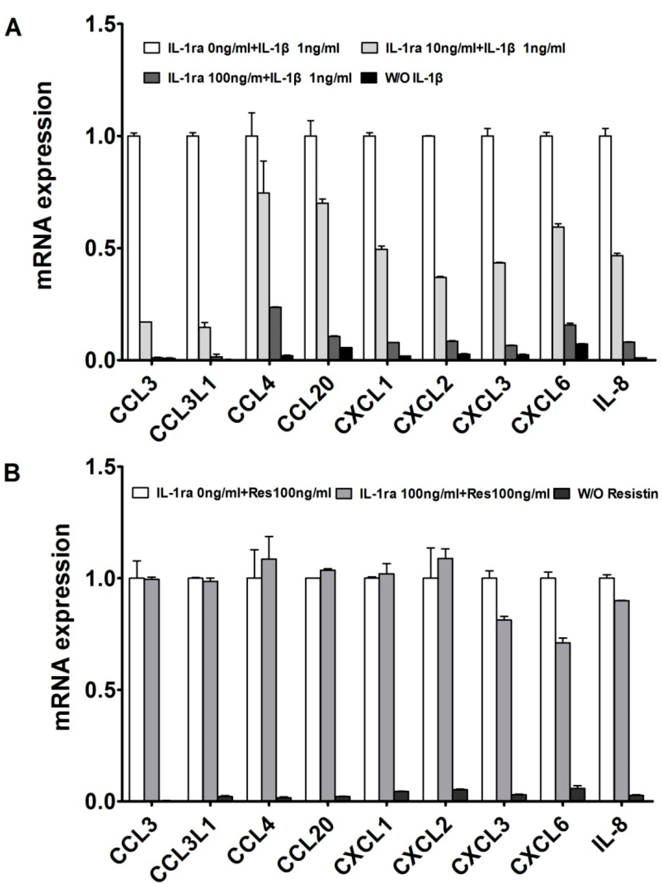
2.1. Resistin Independently Stimulates the Expression of Chemokine Genes in Normal Chondrocytes

We reported that in adult normal cartilage and OA patients, a large set of chemokines are up-regulated by the resistin or IL-1β; and resistin can stimulated the expression of IL-1β [8,13,14]. Figure 1 illustrates our treatments with effective IL-1 receptor antagonist Recombinant Human IL-1ra. The lower concentration (10 ng/mL) of IL-1ra only partially inhibited the IL-1-induced responses, whereas the more effective concentration (100 ng/mL) almost suppressed all tested cytokine and chemokine genes. Meanwhile, the higher concentration (100 ng/mL) of IL-1ra was tested against resistin in panel 1B. In this experiment, cytokine and chemokine genes were still significantly increased in human articular chondrocytes in response to resistin (Figure 1B). This further suggests that the effects of resistinare not dependent on stimulation of IL-1.
Figure 1. Resistin independently stimulates the expression of chemokines in normal human articular chondrocytes (HACs). (A) HACs were treated with 1 ng/mL IL-1β with various doses of IL-1ra for 24 h, as indicated. W/O IL-1β means blank control without IL-1β; (B) HACs were treated with 100 ng/mL resistin with or without IL-1ra for 24 h, as indicated. W/O resistin means blank control without resistin. The relative expression levels were examined by quantitative real time PCR method. Each bar represents the mean ± S.D. from three experiments. W/O resistin means blank control without resistin.
Ijms 15 17242 g001

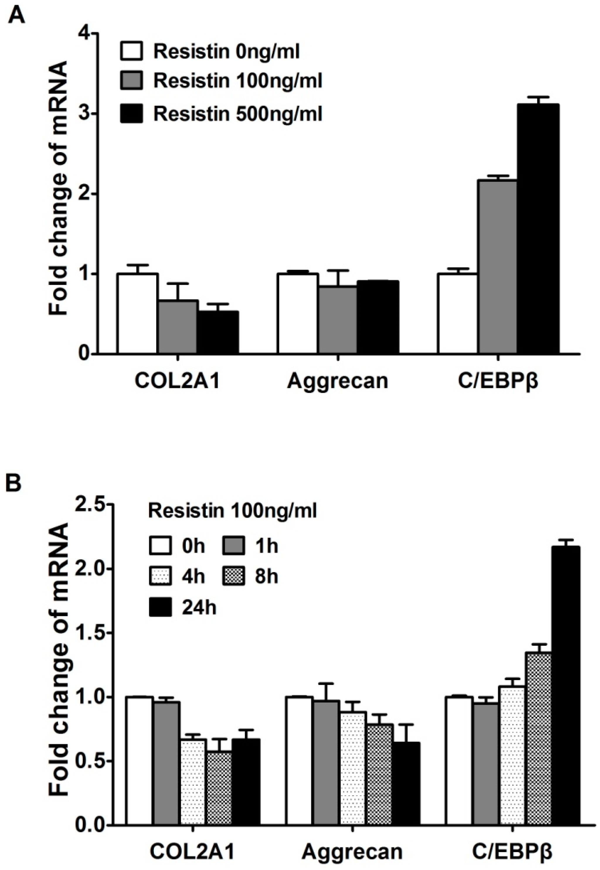
2.2. Resistin Stimulates the Expression of C/EBPβ in Chondrocytes

Computational analysis and co-transfection of C/EBPβ previously suggested that C/EBPβ was involved in the resistin-induced up-regulation of chemokine genes [14]. The down-regulation of COL2A1 and aggrecan genes and up-regulation of C/EBPβ showed a dose response (Figure 2A). There is no statistic difference (p > 0.05) referring to the changes of COL2A1 and aggrecan, but with opposite trend to CEBPβ. When cells were stimulated with 100 ng/mL resistin over 24 h, the expression level of C/EBPβ mRNA was gradually increased to about 3-fold (Figure 2B). C/EBPβ repressed expression COL2A1 mRNA to about 60% within 24 h in human chondrocytes as expected (Figure 2B) as we have shown previously for rat chondrosarcoma cells [15] and human articular chondrocytes [8,13,14]. We referred the results of COL2A1 and ACAN to illuminate that they can be restrained by C/EBPβ, and its tendency is opposite to C/EBPβ, providing another aspect to prove the function of resistin on C/EBPβ.
Figure 2. Resistin stimulates the expression of C/EBPβ in normal human articular chondrocytes (HACs). The relative expression levels were examined in normal chondrocytes from human articular cartilage treated with resistin for various doses and times using quantitative real time PCR method. (A) Cells were treated with various concentrations of resistin for 24 h as indicated in HACs; (B) Cells were treated with 100 ng/mL resistin for various times as indicated in HACs. Each bar represents the mean ± S.D. from three experiments.
Ijms 15 17242 g002

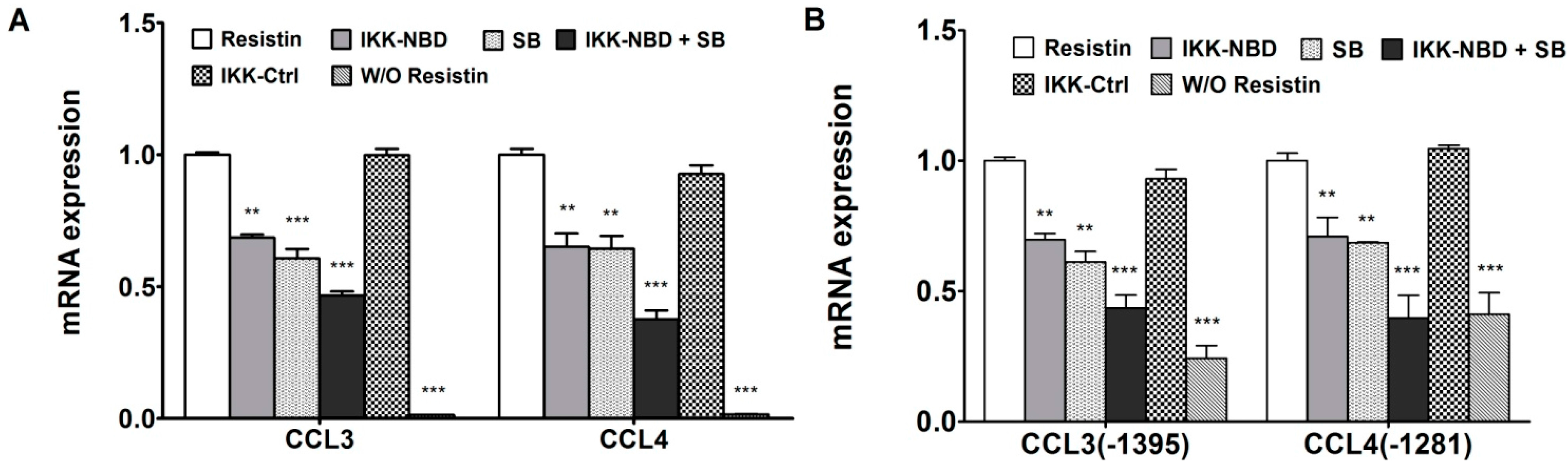
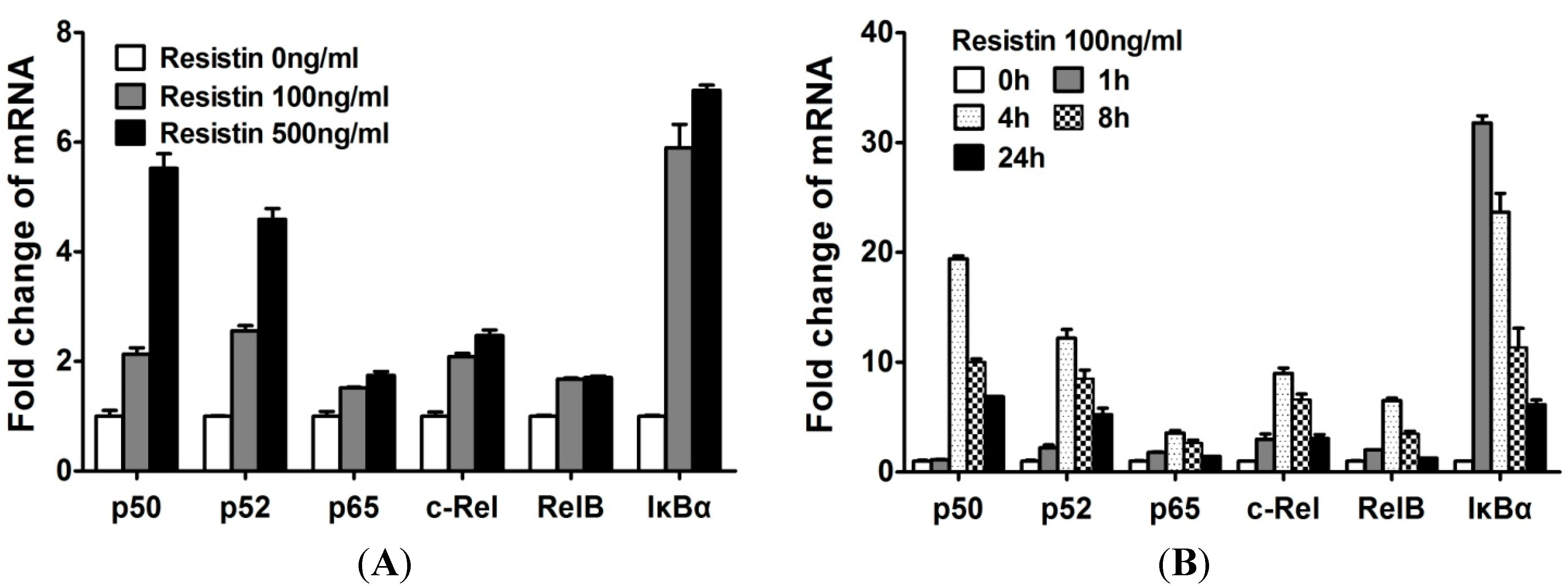
2.3. NF-κB Is Involved in the Up-Regulation of Chemokine Genes by Chondrocytes in Response to Resistin

As the transcription complex NF-κB has been shown to be involved in the resistin-induced up-regulation of multiple cytokine and chemokine genes by the co-transfection of NF-κB and C/EBPβ, and by IKK-NBD inhibition, we sought to determine the relative roles of C/EBPβ and NF-κB. The expression of NF-κB in normal human articular chondrocytes was examined. The levels of NF-κB1 (p50), NF-κB2 (p52), RelA (p65), c-Rel, RelB, IκBα mRNAs were investigated in resistin-treated chondrocytes at various doses and times. The levels of mRNA encoding NF-κB isoforms increased in a dose-dependent manner in response to resistin (Figure 3A). NF-κB isoforms were rapidly increased, but they decreased after 4 h (Figure 3B), which was the same kinetics as we previously reported for NF-κB function by using pNF-κB luciferase reporter over 24 h in response to resistin [8]. When compared to the concentration of C/EBPβ in cells, we show that the NF-κB isoforms are lower. In human chondrocytes, ∆C of C/EBPβ is average 1.73 and thus lower than ∆C of NF-κB1 (5.33), NF-κB2 (5.46), RelA (18.23), c-Rel (6.89) and RelB (7.04). However, these studies also provided insight into the likely NF-κB subunits used in chondrocytes in response to resistin. p50, p52 and c-Rel showed the highest relative increase. Therefore, these results indicated that NF-κB is involved in the expression of chemokines, particularly at 4 to 8 h of exposure.
Figure 3. Resistin stimulates the expression of NF-κB isoforms in normal human articular chondrocytes (HACs). The relative expression levels were examined in normal chondrocytes from human articular cartilage treated with resistin for various doses and times using quantitative real time PCR method. (A) Cells were treated with various concentrations of resistin for 24 h as indicated in HACs; (B) Cells were treated with 100 ng/mL resistin for various times as indicated in HACs. Each bar represents the mean ± S.D. from three experiments.
Ijms 15 17242 g003

2.4. C/EBPβ and NF-κB Co-Enhance the Expression of CCL3 and CCL4 in Human Chondrocytes in Response to Resistin

The previous results indicated that both C/EBPβ and NF-κB contributed to the transcriptional up-regulation of CCL3 and CCL4 [14]. To further investigate the C/EBPβ and NF-κB function on CCL3 and CCL4 in detail, the effect of inhibition of C/EBPβ and NF-κB was tested via inhibition of each transcription factor, separately and together. SB303580, an inhibitor of C/EBPβ via inhibition of p38MAPK [16,17], and IKK-NBD, a specific NF-κB inhibitor, reduced the expression of CCL3 and CCL4 with resistin stimulation (Figure 4A). Combining IKK-NBD and SB303580 further decreased mRNA expression of CCL3 and CCL4 (Figure 4A).
Figure 4. IKK-NBD and SB303580 (SB) co-enhance the inhibition of chemokines by human chondrocytes with resistin treatment. (A) Human articular chondrocytes (HAC) were pretreated with vehicle (DMSO), IKK-NBD peptide (100 μM), SB303580 (100 μM), or IKK-NBD control peptide (100 μM) for 1 h and then exposed to resistin (100 ng/mL) for 4 h. After resistin treatment, total RNA was isolated, and real-time quantitative-PCR was performed; (B) The CCL3 (−1395) and CCL4 (−1281) promoter constructs were transfected into T/C-28a2 cells and incubated for 24 h, then were pretreated with vehicle (DMSO), IKK-NBD peptide (100 μM), SB303580 (100 μM), and IKK-NBD control peptide (100 μM) for 1 h and then exposed to resistin (100 ng/mL) for 4 h. Relative luciferase activity indicates the fold expression relative to the activity of the construct co-transfected with empty vector (set as 1) in the presence of resistin (100 ng/mL). W/O resistin means blank control without resistin. The p value of IKK-NBD (100 μM) or SB303580 (100 μM) was compared with resistin alone based on student’s t-test (** p < 0.01; *** p < 0.001). Each bar represents the mean ± S.D. of at least three independent experiments.
Ijms 15 17242 g004
We have indicated that the CCL3 (−1395) and CCL4 (−1281) constructs contain several high probability candidate C/EBPβ and NF-κB binding sites as assessed by our computational database [14]. To confirm that both transcription factors are functional, CCL3 (−1395) and CCL4 (−1281) promoter activities were determined after inhibition. The result showed that the promoter activities of CCL3 and CCL4 were significantly decreased by the inhibition of both factors, and IKK-NBD and SB303580 co-enhanced the decrease of promoter activities of CCL3 and CCL4 (Figure 4B).

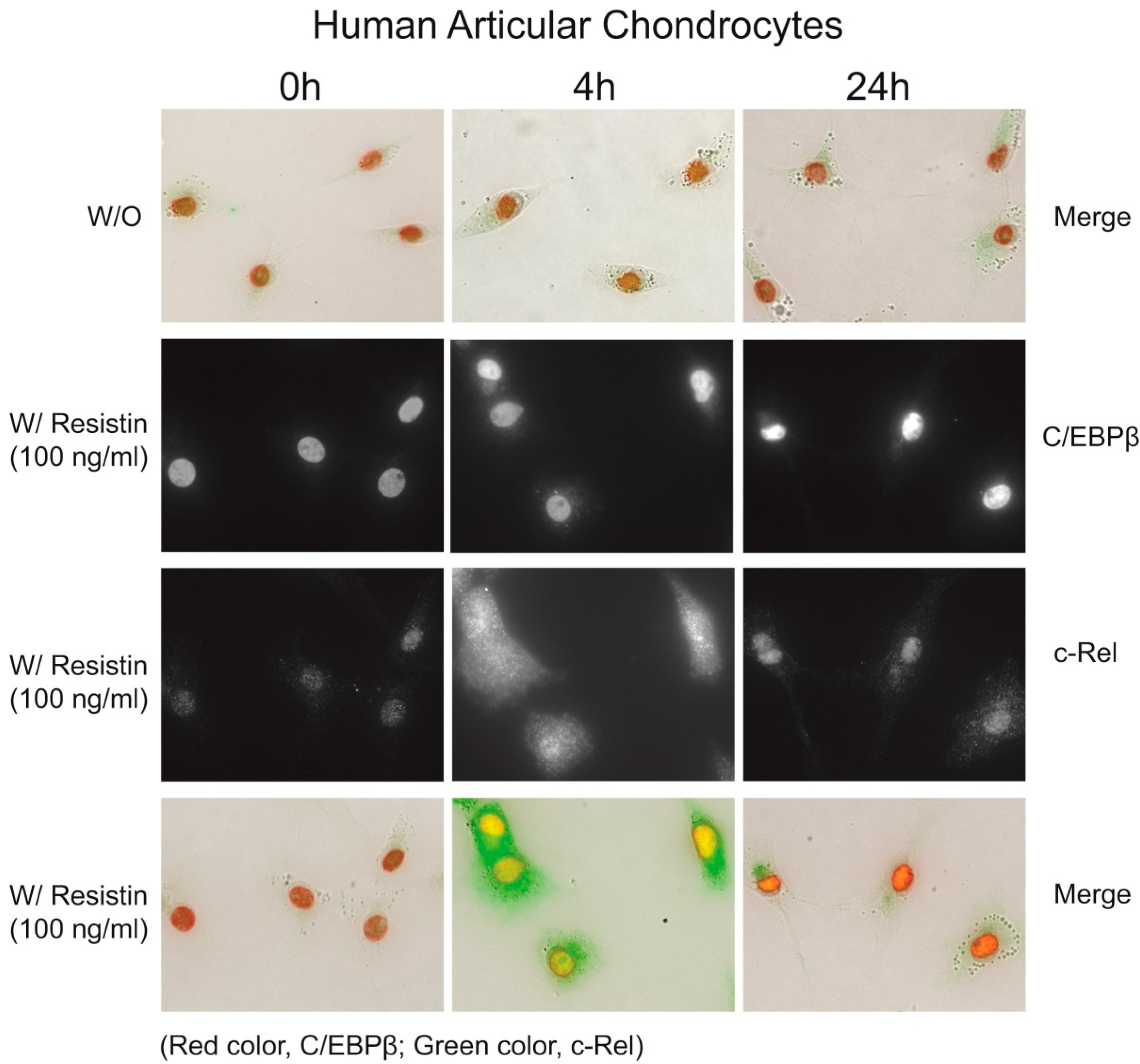
2.5. Immunohistochemistry of C/EBPβ and NF-κB

Our biochemical studies provided evidence that the regulation of CCL3 and CCL4 is coordinated early in time with the NF-κB response, and the C/EBPβ response increased and sustained. To support this conclusion, we traced the location of these transcription factors over time by immunohistochemistry. We knew from Figure 3 that p50, p52 and c-Rel exhibited the highest changes upon increasing dose of resistin. As we know, p50 and c-Rel are involved in the canonical pathway while p52 involved in the non-canonical pathway. However, we showed in chondrocytes that, of the NF-κB subunits, c-Rel mRNA was increased more with resistin stimulation, and the computational analysis in chemokine genes CCL3 and CCL4 showed that c-Rel binding sites, at −210 to −206 bp of the CCL3 promotor (site S5) and −174 to −169 bp of the CCL4 promotor (site S13), were predicted in the resistin-responsive elements [8]. Lu and colleagues reported that c-Rel was an important transcription factor in the regulation of the induction of proinflammatory cytokines [16]. Therefore, c-Rel would be an interesting target for further investigation in the immunohistochemistry experiment. Figure 5 demonstrated by double immunohistochemistry with antibodies to C/EBPβ and c-Rel after stimulation with resistin. At 0 h time, c-Rel was localized in the cytoplasm, then was localized in the nucleus at 4 h and was greatly reduced by 24 h. C/EBPβ was present at the beginning and was increased at 4 h and at 24 h.
Figure 5. Subcellular localization of C/EBPβ and c-Rel in response to resistin (200×). The top panel shows a merged image of immunohistochemistry for C/EBPβ and the NF-κB subunit c-Rel: C/EBPβ is present in the nucleus of cells even without resistin exposure, while c-Rel is located diffusely throughout the cell. With addition of resistin, C/EBPβ is increased in the nucleus and c-Rel is increased and translocates to the nucleus. At 24 h, C/EBPβ remains high in the nucleus and c-Rel is significantly reduced.
Ijms 15 17242 g005

2.6. Discussion

Resistin, a member of the adipocyte-derived cytokines, may be a potent link between adipokines and inflammatory diseases, including OA and RA [3,5,7]. Many cytokines and chemokines are up-regulated by resistin in human articular chondrocytes, including IL-1β [8]. As an important cytokine in inflammatory joint disease, IL-1β can: induce enzymes that degrade the extracellular matrix (ECM), reduce synthesis of the primary cartilage components COL2A1 and aggrecan, and increase cytokines and chemokines [13,14]. Here, we showed that resistin also increased the expression of cytokines and chemokines after effective inhibition of IL-1 receptor. Therefore, resistin and IL-1β both play a role in induction of chemokine synthesis, and resistin and IL-1β could be additive in the up-regulation of chemokine genes.
In this work, we demonstrate a crucial role of C/EBPβ in the increased expression of chemokines and cytokines in response to resistin in chondrocytes. We showed that C/EBPβ increased gradually in 24 h under the effect of resistin. We conclude from this data that C/EBPβ is involved in constitutive regulation of chemokine genes in response to resistin. From our previous work on chemokine gene stimulation and on extracellular matrix gene suppression by C/EBPβ [14], we hypothesize that C/EBPβ is one of the most important regulators of gene activity in chondrocytes in response to resistin. In fact, Kawaguchi and colleagues have shown that C/EBPβ is critical for chondrocyte hypertrophy, where it increases type X collagen and MMP-13, and suppresses type II collagen synthesis [18]. They also showed that the removal of C/EBPβ protects against osteoarthritis.
Like C/EBPβ, the classic pro-inflammatory mediator, NF-κB activity is increased by resistin; however, in contrast to C/EBPβ, the increase in NF-κB activity is transient. Considering the time course of expression of CCL3 and the gradual increase of C/EBPβ, we suggest that C/EBPβ is involved in the resistin-induced up-regulation of chemokine genes over time. This is especially true for the regulation of chemokine genes like CCL3, which are induced more slowly, and where the mRNA was gradually and steadily increased, not reaching peak expression even in the 24 h observation period. In contrast, NF-κB appears to be involved in the resistin-induced up-regulation of chemokines in the early stage (reach the peak at 4 h), with the concentration gradually decreasing in the late stage. Co-regulation by NF-κB and C/EBPβ was recently shown in several genes in different cells [14,19,20,21]. Here, we proved C/EBPβ and NF-κB were both involved in the up-regulation of chemokine genes in response to resistin in human chondrocytes in a time-ordered manner: NF-κB provides an initial up regulation and C/EBPβ sustains increased gene expression.
In summary, the data presented here show that chondrocytes react in a non- restricted, cell-specific manner to resistin, utilizing C/EBPβ, NF-κB and some chondrogenic specific miRNAs in a combinatorial regulation of chemokine gene expression. The activity of C/EBPβ is augmented by a transient increase in activity of NF-κB, and both transcription factors act independently on the chemokine genes, CCL3 and CCL4. These studies reinforce the evidence of our previous work on resistin and provide the foundation for the control of chemokine gene expression in chronic joint disease.

3. Experimental Section

3.1. Materials

The materials used in this work were purchased as follows: Dulbecco’s modified Eagle’s medium (DMEM), Ham’s F-12 medium, were from Mediatech Inc. (Herndon, VA, USA). Fetal bovine serum, pfx polymerase, SuperScript® II Reverse Transcriptase, restriction enzymes, Alexa fluor® 488 goat anti-mouse IgG, and Alexa fluor® 594 goat anti-rabbit IgG from Invitrogen (Carlsbad, CA, USA); Penicillin/streptomycin solution, ascorbic acid, Actinomycin D, Tween 20 from Sigma (St. Louis, MO, USA); 16% paraformaldehyde from Electron Microscopy Science (Hatfield, PA, USA); Recombinant human IL-1β, and Recombinant Human IL-1ra/IL-1F3 from R&D Systems, Inc. (Minneapolis, MN, USA); RNeasy Mini Kit, QIAshredder, DNase I, from Qiagen, Inc. (Valencia, CA, USA); FuGENE® 6 Transfection Reagent, Pronase and Collagenase P from Roche (Indianapolis, IN, USA); pGL3-basic vector, Reporter Lysis Buffer, Luciferase Assay Reagent, β-galactosidase from Promega (Madison, WI, USA); anti-C/EBPβ, anti-c-Rel, normal rabbit IgG and Actin antibodies from Santa Cruz Biotechnology (Santa Cruz, CA, USA); SYBR Green PCR Master Mix was from Applied Biosystens (Foster City, CA, USA); Cell-permeable NEMO binding domain (NBD) synthetic peptides (IKK-NBD peptide and IKK-NBD control peptide), were from BIOMOL (Plymouth Meeting, PA, USA); SB203580 was from Calbiochem (Gibbstown, NJ, USA).

3.2. Cell Culture

Human primary chondrocytes were obtained from articular cartilage obtained at the time of total joint replacement or from above the knee amputation, with approval of the Washington University and Sun Yat-sen University Human Studies Review Board and permission of the patient (IRB No. 05-0279, 1 July 2005 and No. 2011-029, 12 January 2011). Chondrocytes were isolated following previously published procedures [13] and plated at a density of 2.5 × 10⁵ cells/cm2 in DMEM/F12 media plus 10% fetal bovine serum (FBS), 50 μg/mL ascorbate and antibiotics (50 U/mL penicillin and 50 μg/mL streptomycin). T/C-28a2 human chondrocyte cell line were also used (a gift from Dr. Mary B. Goldring, Cornell University), and cultured like human articular chondrocytes. Cells were allowed to rest for 24 h and resistin was added at the concentrations and times indicated. Resistin was reconstituted in sterile water.

3.3. RNA Isolation and Real Time Quantitative PCR

Total RNA was isolated from human primary articular chondrocytes and T/C-28 cells with RNeasy Mini Kit with DNase I treatment, following the protocol recommended by the manufacturer (Qiagen). Total RNA (1 μg) was reverse-transcribed with a SuperScriptTM II Reverse Transcriptase to synthesize cDNA. The cDNA was then used for real-time quantitative PCR (qPCR). Real-time quantitative PCR was performed in a total volume of 20 μL reaction mixture containing 10 μL of SYBR Green PCR Master Mix, 2.5 μL of cDNA, and 200 nM of primers using a 7300 Real-Time PCR System (Applied Biosystems, Foster City, CA, USA), and done in triplicate. Primers used for qPCR were optimized for each gene, and the dissociation curve was determined by the Real-Time PCR System. The parameters of primer design included a primer size of 18 to 21 bp, a product size of 80 to 150 bp, a primer annealing temperature of 59° to 61°, and a primer GC content of 45% to 55%. Results were normalized to glyceraldehyde-3-phosphate dehydrogenase (GAPDH). The primer sequences are summarized in Table 1. The cycle threshold (Ct) values for GAPDH and those of genes of interest was measured for each sample, and the relative transcript levels were calculated as χ = 2−ΔΔCt, in which ∆∆Ct = ∆Treatment − ∆C and ∆Treatment = Ct(treatment) − Ct(GAPDH); ∆C = Ct(control) − Ct(GAPDH).
Table 1. Primers for real-time quantitative PCR.
GenePrimer Sequence
GAPDHF(5'-ACCCAGAAGACTGTGGATGG-3'), R(5'-GAGGCAGGGATGATGTTCTG-3')
COL2A1F(5'-CCCAGAGGTGACAAAGGAGA-3'), R(5'-CACCTTGGTCTCCAGAAGGA-3')
AggrecanF(5'-GGCACTAGTCAACCCTTTGG-3'), R(5'-CTGAACCCTGGTAACCCTGA-3')
C/EBPβF(5'-CTCGCAGGTCAAGAGCAAGG-3'), R(5'-TCGTCGCTGTGCTTGTCC-3')
CCL3 (MIP-1α)F(5'-GCAACCAGTTCTCTGCATCA-3'), R(5'-TGGCTGCTCGTCTCAAAGTA-3')
CCL3L1 (LD78β)F(5'-GTCCTCTCTGCACCACTTGC-3'), R(5'-GGAAGATGACACTGGGCTTG-3')
CCL4 (MIP-1β)F(5'-GCTTTTCTTACACTGCGAGGA-3'), R(5'-CCAGGATTCACTGGGATCAG-3')
CCL20 (MIP-3α)F(5'-GCTTTTCTTACACTGCGAGGA-3'), R(5'-CCAGGATTCACTGGGATCAG-3')
CXCL1 (GRO-α)F(5'-GGGAATTCACCCCAAGAAC-3'), R(5'-GATGCAGGATTGAGGCAAG-3')
CXCL2 (GRO-β)F(5'-TGCAGGGAATTCACCTCAAG-3'), R(5'-TCTTAACCATGGGCGATGC-3')
CXCL3 (GRO-γ)F(5'-ACCGAAGTCATAGCCACACTC-3'), R(5'-GGTGCTCCCCTTGTTCAGTA-3')
CXCL6 (GCP-2)F(5'-GTTTACGCGTTACGCTGAGAG-3'), R(5'-ACTTCCACCTTGGAGCACTG-3')
IL-8F(5'-GAAGGTGCAGTTTTGCCAAG-3'), R(5'-TGTGGTCCACTCTCAATCACTC-3')
IL-1αF(5'-TGCCTGAGATACCCAAAACC-3'), R(5'-AACAAGTTTGGATGGGCAAC-3')
IL-1βF(5'-TCCAGGAGAATGACCTGAGC-3'), R(5'-GTGATCGTACAGGTGCATCG-3')
NF-κB1 (p50)F(5'-CCTGGATGACTCTTGGGAAA-3'), R(5'-TCAGCCAGCTGTTTCATGTC-3')
NF-κB2 (p52)F(5'-GAACAGCCTTGCATCTAGCC-3'), R(5'-TTTTCAGCAT GGATGTCAGC-3')
p65 (RelA)F(5'-TCTGCTTCCAGGTGACAGTG-3'), R(5'-GCCAGAGTTTCGGTTCACTC-3')
c-RelF(5'-CGAACCCAATTTATGACAAC-3'), R(5'-TTTTGTTTCTTTGCTTTATTGC-3')
RelBF(5'-CTGCTTCCAGGCCTCATATC-3'), R(5'-CGCAGCTCTGATGTGTTTGT-3')
IκBαF(5'-GATCCGCCAGGTGAAGGG-3'), R(5'-GCAATTTCTGGCTGGTTGG-3')

3.4. Plasmid Constructs

A series of CCL3 and CCL4 promoter 5'-deletion constructs were made by PCR and subcloned into pGL3-basic vector using the pGL2-CCL3 (−1972/+75) and pGL3-CCL4 (−1281/+12) as previously described [22,23]. The CCL3 and CCL4 promoter constructs, C/EBPβ and IkappaB kinase 2 (IKK2) expression vector, and pNF-κB luciferase reporter were provided by the following: the human pGL2-CCL3 (−1972/+75) was from Dr. G. David Roodman (University of Pittsburgh) [15]; human pGL3-CCL4 (−1281/+12) was obtained from Dr. Sheau-Farn Yeh (National Yang-Ming University, Taipei, Taiwan) [17]; human IKK2 in the pCDNA3 vector and pNF-κB luciferase reporter were from Dr. Yousef Abu-Amer (Washington University) [24]; human C/EBP-full-length in the pCDNA3 vector was from Dr. Erika Crouch (Washington University, Washinton, DC, USA) [25]. The empty expression vectors were made by excision of cDNAs from the corresponding C/EBP expression vectors [15]. To facilitate subcloning of the amplified fragments, the antisense primer contained a HindIII restriction site adaptor, and the sense primer contained an XhoI or SmaI site. The PCR fragments and the luciferase expression vector pGL3-basic vector were digested with XhoI or SmaI and HindIII before ligation. All constructs were confirmed by DNA sequence analysis using a GL2 primer and RV3 primer.

3.5. Transient Transfection and Luciferase Assay

DNA transfections of T/C-28a2 cells were performed using FuGENE 6TM transfection reagent. One hundred and five × two of T/C-28a2 cells were cultured in a 6-well plate overnight. The transfection mixture containing 3–9 μL of transfection reagent (6:1 ratio of trasfection reagent μL to DNA μg), 500 ng of various promoter constructs, and 100 or 200 ng of pCMV-β-gal was then added, and the cells were cultured for 4 h with or without resistin as indicated. For co-transfection assay, 500 ng of C/EBPβ expression vectors or empty vector were added to the 100 μL transfection mixture as indicated. Due to low translation efficiency of C/EBPβ, we used higher amounts of plasmid (500 ng) for transfection, as shown previously [9]. FBS was added to transfection medium 4 h later to a final concentration of 10%. After 24 h of incubation, cells were replaced with fresh complete medium and incubated for an additional 4 h with or without resistin. The cells were then harvested with Reporter Lysis Buffer™ and the lysate was analyzed for luciferase activity using Promega Luciferase Assay Reagent™. The β-galactosidase activities were also measured to normalize variations in transfection efficiency. Each transfection experiment was performed in duplicate or triplicate and repeated at least three times.

3.6. Immunofluorescence

Five × one hundred and four human articular chondrocytes were cultured in each well of 8-well chamber slides (from Lab Tek, Hatfield, PA, USA). Cells were allowed to rest for 24 h and resistin was then added at the times indicated. Cells were incubated in 4% paraformaldehyde in PBS for 10 min, 0.2% Triton-X-100 in PBS for 5 min, and 10% normal goat serum in PBS for 2 h at room temperature. Cells were reacted with rabbit anti-C/EBPβ and mouse anti-c-Rel antibodies diluted to 1:400 in 2% normal goat serum in PBS for overnight at 4 °C. The secondary antibodies, Alexa fluor 488 dye-labeled goat anti-mouse IgG diluted to 1:250 and Alexa fluor 594 dye-labeled goat anti-rabbit IgG diluted to 1:400 in 2% normal goat serum in PBS were then added to the cells for 1 h at room temperature. Immunoreactivity was detected by fluorescence microscopy.

3.7. Statistical Analysis

Data were expressed as the mean ± S.E.M. from at least three independent experiments. The Student’s t-test was used to compare the differences between two groups. p < 0.05 was considered statistically significant.

4. Conclusions

Chondrocytes react in a non-restrictedly cell-specific manner to resistin, utilizing C/EBPβ and NF-κB in a combinatorial regulation of chemokine gene expression. The activity of C/EBPβ is augmented by a transient increase in activity of NF-κB, and both transcription factors act independently on the chemokine genes, CCL3 and CCL4.

Acknowledgments

We are grateful to Mary B. Goldring from Cornell University (Ithaca, NY, USA) for her selfless contribution of T/C-28a2 human chondrocyte cell line. The authors would like to thank G. David Roodman from University of Pittsburgh (Pittsburgh, PA, USA), Sheau-Farn Yeh from National Yang-Ming University (Taipei, Taiwan), Yousef Abu-Amer and Erika Crouch from Washington University (Washington, DC, USA) for their valuable assistance in constructing plasmids. The authors would also like to thank Aishan He and Dongliang Xu at the Department of Orthopaedic Surgery, the First Affiliated Hospital of Sun Yat-sen University (Guangzhou, China) and Lora L. Smallman from Southern Illinois University (Carbondale, IL, USA) for their advice in revision of the article. These studies were funded by The National Institute for Arthritis, Musculoskeletal and Skin Diseases (R01 AR050847 and R01 AR036994), National Natural Science Foundation of China (No. 81301558, 81201388, 81371941 and 81171709), Doctoral Scientific Fund Project of the Ministry of Education of China (No. 20130171120074), Medical research Foudation of Guangdong Province (No. A2012174) and Natural Science Foundation of Guangdong Province, China (No.s 2013040016269).

Author Contributions

Ziji Zhang, Zhiqi Zhang, Weiming Liao, Linda J. Sandell carried out the work. Ziji Zhang, Zhiqi Zhang, Yan Kang, Changhe Hou helped collection and assembly of data. Ziji Zhang, Zhiqi Zhang and Xin Duan conducted the data analysis and wrote the manuscript. Puyi Sheng, Weiming Liao, Linda J. Sandell designed the study and coordinated the research team. Linda J. Sandell corrected English of this article. All authors had read and approved the final manuscript.

Conflicts of Interest

The authors declare no conflict of interest.

References

  1. Lee, J.H.; Ort, T.; Ma, K.; Picha, K.; Carton, J.; Marsters, P.A.; Lohmander, L.S.; Baribaud, F.; Song, X.Y.; Blake, S. Resistin is elevated following traumatic joint injury and causes matrix degradation and release of inflammatory cytokines from articular cartilage in vitro. Osteoarthr. Cartil. 2009, 17, 613–620. [Google Scholar]
  2. Staikos, C.; Ververidis, A.; Drosos, G.; Manolopoulos, V.G.; Verettas, D.A.; Tavridou, A. The association of adipokine levels in plasma and synovial fluid with the severity of knee osteoarthritis. Rheumatology 2013, 52, 1077–1083. [Google Scholar]
  3. Senolt, L.; Housa, D.; Vernerova, Z.; Jirasek, T.; Svobodova, R.; Veigl, D.; Anderlova, K.; Muller-Ladner, U.; Pavelka, K.; Haluzik, M. Resistin in rheumatoid arthritis synovial tissue, synovial fluid and serum. Ann. Rheum. Dis. 2007, 66, 458–463. [Google Scholar]
  4. Presle, N.; Pottie, P.; Dumond, H.; Guillaume, C.; Lapicque, F.; Pallu, S.; Mainard, D.; Netter, P.; Terlain, B. Differential distribution of adipokines between serum and synovial fluid in patients with osteoarthritis. Contribution of joint tissues to their articular production. Osteoarthr. Cartil. 2006, 14, 690–695. [Google Scholar]
  5. Tilg, H.; Moschen, A.R. Adipocytokines: Mediators linking adipose tissue, inflammation and immunity. Nat. Rev. Immunol. 2006, 6, 772–783. [Google Scholar]
  6. Koerner, A.; Kratzsch, J.; Kiess, W. Adipocytokines: Leptin—The classical, resistin—The controversical, adiponectin—The promising, and more to come. Best Pract. Res. Clin. Endocrinol. Metab. 2005, 19, 525–546. [Google Scholar]
  7. Bokarewa, M.; Nagaev, I.; Dahlberg, L.; Smith, U.; Tarkowski, A. Resistin, an adipokine with potent proinflammatory properties. J. Immunol. 2005, 174, 5789–5795. [Google Scholar]
  8. Zhang, Z.; Xing, X.; Hensley, G.; Chang, L.W.; Liao, W.; Abu-Amer, Y.; Sandell, L.J. Resistin induces expression of proinflammatory cytokines and chemokines in human articular chondrocytes via transcription and messenger RNA stabilization. Arthritis Rheumatol. 2010, 62, 1993–2003. [Google Scholar]
  9. Imamura, T.; Imamura, C.; McAlinden, A.; Davies, S.R.; Iwamoto, Y.; Sandell, L.J. A novel tumor necrosis factor alpha-responsive CCAAT/enhancer binding protein site regulates expression of the cartilage-derived retinoic acid-sensitive protein gene in cartilage. Arthritis Rheumatol. 2008, 58, 1366–1376. [Google Scholar]
  10. Brass, A.; Brenndorfer, E.D. The role of chemokines in hepatitis C virus-mediated liver disease. Int. J. Mol.Sci. 2014, 15, 4747–4779. [Google Scholar]
  11. Van der Kraan, P.M. Age-related alterations in TGFβ signaling as a causal factor of cartilage degeneration in osteoarthritis. Biomed. Mater. Eng. 2014, 24, 75–80. [Google Scholar]
  12. Huang, J.; Zhu, M.; Tao, Y.; Wang, S.; Chen, J.; Sun, W.; Li, S. Therapeutic properties of quercetin on monosodium urate crystal-induced inflammation in rat. J. Pharm. Pharmacol. 2012, 64, 1119–1127. [Google Scholar]
  13. Sandell, L.J.; Xing, X.; Franz, C.; Davies, S.; Chang, L.W.; Patra, D. Exuberant expression of chemokine genes by adult human articular chondrocytes in response to IL-1β. Osteoarthr. Cartil. 2008, 16, 1560–1571. [Google Scholar]
  14. Zhang, Z.; Bryan, J.L.; DeLassus, E.; Chang, L.W.; Liao, W.; Sandell, L.J. CCAAT/enhancer-binding protein beta and NF-κB mediate high level expression of chemokine genes CCL3 and CCL4 by human chondrocytes in response to IL-1β. J. Biol. Chem. 2010, 285, 33092–33103. [Google Scholar]
  15. Okazaki, K.; Yu, H.; Davies, S.R.; Imamura, T.; Sandell, L.J. A promoter element of the CD-RAP gene is required for repression of gene expression in non-cartilage tissues in vitro and in vivo. J. Cell Biochem. 2006, 97, 857–868. [Google Scholar]
  16. Lu, Y.C.; Kim, I.; Lye, E.; Shen, F.; Suzuki, N.; Suzuki, S.; Gerondakis, S.; Akira, S.; Gaffen, S.L.; Yeh, W.C.; et al. Differential role for c-Rel and C/EBPβ/δ in TLR-mediated induction of proinflammatory cytokines. J. Immunol. 2009, 182, 7212–7221. [Google Scholar]
  17. Venza, I.; Cucinotta, M.; Visalli, M.; de Grazia, G.; Oliva, S.; Teti, D. Pseudomonas aeruginosa induces interleukin-8 (IL-8) gene expression in human conjunctiva through the recruitment of both RelA and CCAAT/enhancer-binding protein beta to the IL-8 promoter. J. Biol. Chem. 2009, 284, 4191–4199. [Google Scholar]
  18. Hirata, M.; Kugimiya, F.; Fukai, A.; Ohba, S.; Kawamura, N.; Ogasawara, T.; Kawasaki, Y.; Saito, T.; Yano, F.; Ikeda, T.; et al. C/EBPβ promotes transition from proliferation to hypertrophic differentiation of chondrocytes through transactivation of p57. PLoS One 2009, 4, e4543. [Google Scholar]
  19. Basak, C.; Pathak, S.K.; Bhattacharyya, A.; Mandal, D.; Pathak, S.; Kundu, M. NF-κB- and C/EBPβ-driven interleukin-1β gene expression and PAK1-mediated caspase-1 activation play essential roles in interleukin-1β release from Helicobacter pylori lipopolysaccharide-stimulated macrophages. J. Biol. Chem. 2005, 280, 4279–4288. [Google Scholar]
  20. Esteves, C.L.; Kelly, V.; Breton, A.; Taylor, A.I.; West, C.C.; Donadeu, F.X.; Peault, B.; Seckl, J.R.; Chapman, K.E. Proinflammatory cytokine induction of 11β-hydroxysteroid dehydrogenase type 1 (11β-HSD1) in human adipocytes is mediated by MEK.; C/EBPβ.; and NF-κB/RelA. J. Clin. Endocrinol. Metab. 2014, 99, E160–E168. [Google Scholar]
  21. Rahman, S.M.; Janssen, R.C.; Choudhury, M.; Baquero, K.C.; Aikens, R.M.; de la Houssaye, B.A.; Friedman, J.E. CCAAT/enhancer-binding protein beta (C/EBPβ) expression regulates dietary-induced inflammation in macrophages and adipose tissue in mice. J. Biol. Chem. 2012, 287, 34349–34360. [Google Scholar]
  22. Choi, S.J.; Oba, T.; Callander, N.S.; Jelinek, D.F.; Roodman, G.D. AML-1A and AML-1B regulation of MIP-1α expression in multiple myeloma. Blood 2003, 101, 3778–3783. [Google Scholar]
  23. Tseng, P.C.; Hsu, H.C.; Janmanchi, D.; Lin, C.H.; Kuo, Y.H.; Chou, C.K.; Yeh, S.F. Helioxanthin inhibits interleukin-1β-induced MIP-1 beta production by reduction of c-jun expression and binding of the c-jun/CREB1 complex to the AP-1/CRE site of the MIP-1 beta promoter in Huh7 cells. Biochem. Pharmacol. 2008, 76, 1121–1133. [Google Scholar]
  24. Otero, J.E.; Dai, S.; Alhawagri, M.A.; Darwech, I.; Abu-Amer, Y. IKKβ activation is sufficient for RANK-independent osteoclast differentiation and osteolysis. J. Bone Miner. Res. 2010, 25, 1282–1294. [Google Scholar]
  25. Maier, P.J.; Zemoura, K.; Acuna, M.A.; Yevenes, G.E.; Zeilhofer, H.U.; Benke, D. Ischemia-like oxygen and glucose deprivation mediates down-regulation of cell surface γ-aminobutyric acid B receptors via the endoplasmic reticulum (ER) stress-induced transcription factor CCAAT/enhancer-binding protein (C/EBP)-homologous protein (CHOP). J. Biol. Chem. 2014, 289, 12896–12907. [Google Scholar]

