Article

Q Fever Endocarditis in Romania: The First Cases Confirmed by Direct Sequencing

1
National Institute for Research in Microbiology and Immunology, Cantacuzino, Spl. Independentei 103, 050096, Bucharest, Romania
2
Institute for Emergency Cardiovascular Diseases Prof. C.C. Iliescu, Sos. Fundeni 258, 022328, Bucharest, Romania
3
Institute for Cardiovascular Diseases and Transplant Targu Mures, Str. Gheorghe Marinescu 50, 540136, Targu Mures, Mures, Romania
4
University of Medicine and Pharmacy Targu Mures, Str. Gheorghe Marinescu 38, 540139, Targu Mures, Mures, Romania
5
Institute for Cardiovascular Diseases Timisoara, Str. Gheorghe Adam, 13A, 300310, Timişoara, Timis, Romania
6
Clinical Hospital for Infectious Diseases Cluj-Napoca, Str. Iuliu Moldovan 23, 400348, Cluj-Napoca, Cluj, Romania
*
Author to whom correspondence should be addressed.
Int. J. Mol. Sci. 2011, 12(12), 9504-9513; https://doi.org/10.3390/ijms12129504
Submission received: 18 November 2011 / Revised: 9 December 2011 / Accepted: 12 December 2011 / Published: 20 December 2011
(This article belongs to the Section Molecular Pathology, Diagnostics, and Therapeutics)

Abstract

:
Infective endocarditis (IE) is a serious, life-threatening disease with highly variable clinical signs, making its diagnostic a real challenge. A diagnosis is readily made if blood cultures are positive, but in 2.5 to 31% of all infective endocarditis cases, routine blood cultures are negative. In such situations, alternative diagnostic approaches are necessary. Coxiella burnetii and Bartonella spp. are the etiological agents of blood culture-negative endocarditis (BCNE) most frequently identified by serology. The purpose of this study is to investigate the usefulness of molecular assays, as complementary methods to the conventional serologic methods for the rapid confirmatory diagnostic of Q fever endocarditis in patients with BCNE. Currently, detection of C. burnetii by culture or an antiphase I IgG antibody titers >800 represents a major Duke criterion for defining IE, while a titers of >800 for IgG antibodies to either B. henselae or B. quintana is used for the diagnosis of endocarditis due to Bartonella spp. We used indirect immunofluorescence assays for the detection of IgG titers for C. burnetii, B. henselae and B. quintana in 57 serum samples from patients with clinical suspicion of IE. Thirty three samples originated from BCNE patients, whereas 24 were tested before obtaining the blood cultures results, which finally were positive. The results of serologic testing showed that nine out of 33 BCNE cases exhibited antiphase I C. burnetii IgG antibody titer >800, whereas none has IgG for B. henselae or B. quintana. Subsequently, we used nested-PCR assay for the amplification of C. burnetii DNA in the nine positive serum samples, and we obtained positive PCR results for all analyzed cases. Afterwards we used the DNA sequencing of amplicons for the repetitive element associated to htpAB gene to confirm the results of nested-PCR. The results of sequencing allowed us to confirm that C. burnetii is the causative microorganism responsible for BCNE. In conclusion, the nested PCR amplification followed by direct sequencing is a reliable and accurate method when applied to serum samples, and it may be used as an additional test to the serological methods for the confirmatory diagnosis of BCNE cases determined by C. burnetii.

1. Introduction

Infective endocarditis is a serious, life-threatening disease with highly variable clinical signs that are making the condition a diagnostic challenge. A diagnosis is readily made if blood cultures are positive, but in 2.5 to 31% of all infective endocarditis cases, routine blood cultures are negative [13]. This variation in incidence could be explained by several factors, including: (i) differences in the diagnostic criteria used; (ii) specific epidemiological factors, as for fastidious zoonotic agents; (iii) variations in the early use of antibiotics before the blood sampling; (iv) differences in sampling strategies; or (v) involvement of unknown pathogens [4,5].
Blood culture negative endocarditis (BCNE) was recognized by Osler at the beginning of last century [6,7]. Recently, many publications in European countries have demonstrated a significant involvement of Coxiella burnetti, Bartonella henselae, and B. quintana in patients with BCNE [8,9]. C. burnetii is one of the most encountered fastidious agents in BCNE. Q fever is characterized by its clinical polymorphism and the presentation of the disease is variable, with both acute and chronic manifestations [10,11]. Following acute infection, 1 to 5% of patients progress to chronic infection, which can develop after months to several years after acute Q fever infection, the longest interval being 20 years after infection [12,13].
Endocarditis is the main form of chronic Q fever (78% of all chronic Q fever cases) [14]. The most exposed persons are patients with preexistent valvular disease or vascular defects, especially aortic aneurysm and aortic stents and prostheses, immunocompromised patients, and pregnant women [1518]. The estimated risk of transformation from acute infection to Q fever endocarditis in patients with preexisting valvulopathy is approximately 40% [17]. Because symptoms of Q fever endocarditis are protean and not specific, diagnosis is often delayed, only after significant valvular damage has occurred, resulting in an increasing mortality rate. Some authors proposed that all patients with acute Q fever be investigated by a transthoracic echocardiography [19].
The diagnosis of Q fever endocarditis requires both clinical endocarditis and isolation or serologic evidence of C burnetii. Because Q fever endocarditis is a chronic illness, a single serum specimen is sufficient for diagnosis. A phase I IgG titers of 800 or greater is one of the major modified Duke criteria [20]. Previously studies showed that PCR with serum samples may be helpful in establishing an early diagnosis of chronic Q fever [21].
Currently there is no data concerning the incidence of Q fever endocarditis cases among Romanian population. The first Q fever cases in Romania were registered in 1947 in Constanta County [22]. The most recent data about Q fever in Romania were represented by urban sporadic cases reported in the period 1981–1987 [23].

2. Results and Discussion

2.1. Case Definitions

Patients were considered to have definite blood culture-negative endocarditis if the results of standard blood cultures were negative, and if clinical and echocardiographic findings met the Duke criteria for infective endocarditis. All serum samples tested by serological methods for detection of IgG to C. burnetii, B. quintana and B. henselae originated from patients with clinical suspicion of BCNE. According to the modified Duke criteria a single positive blood culture for Coxiella burnetii or antiphase I IgG antibody titers >800 represents a major criterion for definite infective endocarditis [24]. The results of serological testing showed that 9 out of 33 serum samples exhibited antiphase I C. burnetii IgG antibody titers >800, while none of samples has IgG for B. henselae or B. quintana. The modified Duke criteria used for definite infective endocarditis (IE) diagnosis of analyzed patients with clinical suspicions of BCNE are presented in Table 1.
Diagnosis of IE is definite if there are 2 major criteria or 1 major and 3 minor criteria or 5 minor criteria [25,26]. Eight out of nine investigated cases fulfilled 2 major criteria (antiphase I C. burnetii IgG antibody titer >800 and when there is a vegetation or a new valvular regurgitation) for defining IE, while the remaining case fulfilled one major criterion (antiphase I C. burnetii IgG antibody titers >800) and one minor criterion (fever ≥ 38 °C), being classified as a possible case.
It is well-known that endocarditis is the most common presentation of chronic Q fever. Initially thought to be a rare disorder, later it has been estimated to account for up to 5% of all endocarditis cases worldwide [27]. It occurs almost exclusively in patients who have pre-existing valvular disease or who are immunocompromised. Unlike typical cases of endocarditis, the clinical presentation of endocarditis from chronic Q fever is often nonspecific and lacks many of the typical features of subacute, bacterial endocarditis, such as usual clinical and echocardiographic features common to typical cases of endocarditis [28]. Thus, the diagnosis is often significantly delayed or even missed, resulting in significant morbidity and mortality. Despite increasing awareness, recent studies show a mean delay of seven months from symptom onset to diagnosis [29,30]. Without prompt recognition and adequate antimicrobial therapy, the course of Q fever endocarditis is severe and potentially fatal [30].
The diagnosis of Q fever endocarditis is hampered by the inability to culture C. burnetii using routine media. As a strict obligate intracellular bacterium, it can only be cultured in living cell lines, or embryonated chicken eggs, but the cultures cannot be easily performed in most laboratories, and the technique is restricted to biosafety level 3 laboratories. Thus, the diagnosis of chronic Q fever, therefore, relies on serological testing, being characterized by increased titres against the phase I antigen.

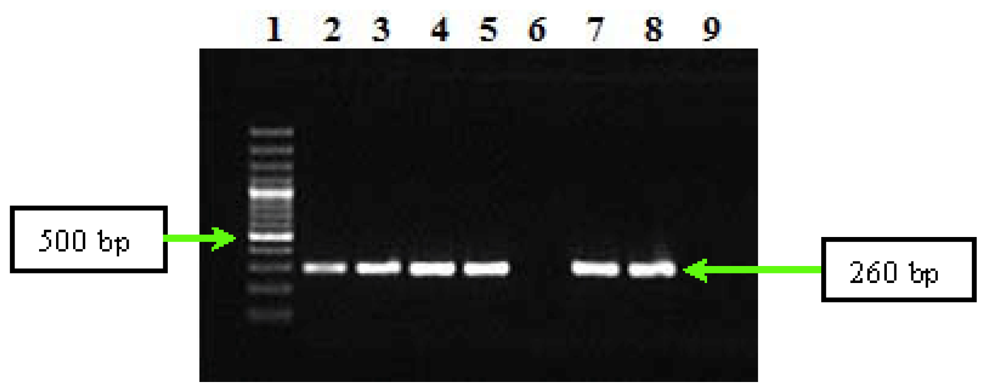
2.2. PCR and Sequencing Results

Detection of C. burnetii DNA by PCR is an important diagnostic method that could be used on different types of clinical specimens (blood, serum, infected heart valves) [31]. During the last years, several PCR based diagnostic assays have been developed to detect C. burnetii DNA in cell cultures and clinical samples. These assays used conventional PCR [32], nested PCR [3336] or real-time PCR conditions with LightCycler [37,38], SYBR Green [39] or TaqMan chemistry. The target sequences of the assays originated from single chromosomal genes like com1, on plasmids (QpH1, QpRS) or within the transposase gene of insertion element IS1111 that is present in 20 copies in the genome of the C. burnetii Nine Mile RSA493 strain [40]. Due to the multicopy number of the IS1111 element, the corresponding PCR is very sensitive.
The results of PCR assays performed on DNA from serum samples positive for antiphase I C. burnetii IgG showed that nested-PCR assay permitted the amplification of C. burnetii DNA. Thus, nested-PCR led to obtaining the amplification products of the repetitive element IS1111a associated to htpAB transposase gene in the second round of amplification for all analyzed DNAs (Figure 1).

3. Experimental Section

3.1. Patients

In this study we analyzed the role of C. burnetii as causative agent of blood culture negative infective endocarditis among patients with clinical diagnostic of infective endocarditis. The blood-cultures were performed for 102 patients hospitalized in three Institutes for Cardiovascular Diseases from Bucharest, Timisoara and Targu-Mures, and in Clinical Hospital for Infectious Diseases from Cluj. For each patient, a standardized questionnaire was filled by the physician in charge and logged into a database. The information filled in the questionnaire were consisted of: known preexisting valvular defect, type of valve involved (native/bioprosthetic/mechanical valve, and its position: aortic, mitral, tricuspid, pulmonary); previous antibiotic therapy; clinical symptoms; and laboratory results. Patients were considered to have possible or definite endocarditis according to the modified Duke criteria.

3.2. Indirect Immunofluorescence Assays

57 serum samples were tested by indirect immunofluorescence assay (IFA) for detection of IgG antibodies to C. burnetii, B. henselae and B. quintana. From these samples 33 originated from patients with BCNE, and the rest of samples were tested before obtaining the blood cultures results, which finally were positive. We used IFA kits (Vircell, Spain) for detection of IgG antiphase I and antiphase II C. burnetii, and for IgG to B. quintana and IgG to B. henseale. The presence of IgG titers >800 to C. burnetii or B. quintana or B. henselae were considered positive for endocarditis diagnosis. Furthermore, we analyzed the positive serum samples for C. burnetii with antiphase I IgG antibody titers >800 using molecular methods for the confirmation of serological results.

3.3. Molecular Methods

3.3.1. DNA Extraction

The serum samples from nine patients with antiphase I C. burnetii IgG antibody titer >800, were used for DNA extraction. Total genomic DNA was extracted from 200 microliters of serum using the QIAamp blood kit (Qiagen, Hilden, Germany) according to the manufacturer instructions. DNA was resuspended in fifty microliters of elution buffer. Genomic DNAs were stored at 4 °C until their use as templates in PCR assays and subsequently at −20 °C. DNA samples have been handled carefully to avoid the risk of cross-contamination. DNA extraction, mix preparation, and PCR were performed in different rooms to prevent PCR carryover contamination. No positive control was used to prevent lateral contamination (i.e., contamination caused by PCR products amplified in other tubes in the same assay). DNA extracted from serum specimens of blood donors was used every 4 specimens as a negative control.

3.3.2. PCR Assay

We used a nested-PCR assay for detection of the repetitive element IS1111 associated to htpAB transposase gene (GenBank accession number M80806). This repetitive element is present in multiple copies in the genome of C. burnetii strains (e.g., there are 20 copies of this element in the C. burnetii Nine Mile I genome), which increase the detection sensitivity of this pathogen in serum samples [21]. In the first round of amplification we used IS111F1 and IS111R1 primers, which were designed to amplify a 485-bp fragment of the repetitive element IS1111, while the second round of amplification was performed using the IS111F2 and IS111R2 primers, which amplify an internal 260-bp fragment from the same target [27]. The sequence of specific primers used in nested-PCR reactions, and the molecular size of the amplicons are presented in Table 2.
In the nested-PCR assay, each gene fragment amplified separately on 2700 Applied Biosystems instrument using necessary components provided by Promega. The components used in each type of PCR reaction are described in Table 3. The parameters for the amplification cycles used in each PCR experiment are presented in Table 4. PCR products from nested-PCR assay were separated in a 1.5% agarose gel for 1 h at 100 V, stained with ethidium bromide and detected by UV transillumination.

3.3.3. Sequencing of PCR Products and Sequence Analysis

The sequencing of the amplicons from nested-PCR has been used to confirm the PCR results. The amplicons from the second round of nested-PCR were sequenced in both directions using the BigDye V3.1 kit as described by the manufacturer. Sequencing products have been resolved using an ABI 3100 automated Avant Genetic Analyzer (Applied Biosystems). Sequence analysis was performed with BioEdit program, which permitted obtaining of the consensus sequences that were compared with similar sequences from BLAST. The sequences obtained showed a sequence similarity of 100% with that of the GenBank prototype strain sequence. Thus, sequencing of the amplicons from the second round of PCR reaction has permitted to confirm that amplification products belong to C. burnetii. These two molecular tests were used together for the first time to investigate the BCNE cases with C. burnetii in Romania.

4. Conclusions

We propose that all patients with clinical suspicion of IE be tested serologically for evidence of infection with other agents such as C. burnetii in parallel with performing blood cultures.
This is the first report in this country for using the molecular methods to confirm Q fever endocarditis cases on the serum samples from eight confirmed cases and from one possible case of Q fever endocarditis tested by nested-PCR, based on repetitive element IS1111a of the transposase gene. This assay exhibited high sensitivity and specificity and led us to obtain specific amplification products, which were subsequently confirmed by direct sequencing to belong to C. burnetii, confirming previous studies showing that this nested-PCR assay presents a specificity of 100% and a sensitivity of one C. burnetii DNA copy [27]. In conclusion, our results have demonstrated that nested-PCR amplification, followed by direct sequencing, is a reliable and accurate method when applied to serum samples and can be used as a supplementary diagnosis tool for BCNE cases.

Acknowledgments

This work was supported by a UEFISCDI grant awarded to the project “Bacterial infective endocarditis (BIE)—development of a functional model for surveillance and characterization of etiological organisms involved in BIE based on molecular and immunological methods” (2009–2011), No. 42119/1.10.2008.

References

  1. Rolain, J.M.; Lecam, C.; Raoult, D. Simplified serological diagnosis of endocarditis due to Coxiella burnetii and Bartonella. Clin. Diagn. Lab. Immunol 2003, 10, 1147–1148. [Google Scholar]
  2. Brouqui, P.; Raoult, D. Endocarditis due to rare and fastidious bacteria. Clin. Microbiol. Rev 2001, 14, 177–207. [Google Scholar]
  3. Houpikian, P.; Raoult, D. Diagnostic methods. Current best practices and guidelines for identification of difficult-to-culture pathogens ininfective endocarditis. Cardiol. Clin 2003, 21, 207–217. [Google Scholar]
  4. Fournier, P.-E.; Thuny, F.; Richet, H.; Lepidi, H.; Casalta, J.-P.; Arzouni, J.-P.; Maurin, M.; Celard, M.; Mainardi, J.-L.; Caus, T.; Collart, F.; Habib, G.; Raoult, D. Comprehensive diagnostic strategy for blood culture-negative endocarditis, a prospective study of 819 new cases. Clin. Infect. Dis 2010, 51, 131–140. [Google Scholar]
  5. Raoult, D.; Casalta, J.P.; Richet, H.; Khan, M.; Bernit, E.; Rovery, C.; Branger, S.; Gouriet, F.; Imbert, G.; Bothello, E.; Collart, F.; Habib, G. Contribution of systematic serological testing in diagnosis of infective endocarditis. J. Clin. Microbiol 2005, 43, 5238–5242. [Google Scholar]
  6. Lamas, C.C.; Eykyn, S.J. Blood culture negative endocarditis, analysis of 63 cases presenting over 25 years. Heart 2003, 89, 258–262. [Google Scholar]
  7. Osler, W. Chronic infectious endocarditis. QJM: Int. J. Med 1909, 2, 219–230. [Google Scholar]
  8. Siciliano, R.F.; Strabelli, T.M.; Zeigler, R.; Rodrigues, C.; Castelli, J.B.; Grinberg, M.; Colombo, S.; da Silva, L.J.; Mendes do Nascimento, E.M.; Pereira dos Santos, F.C.; Uip, D.E. Infective endocarditis due to Bartonella spp. and Coxiella burnetii experience at a cardiology hospital in Sao Paulo, Brazil. Ann. N.Y. Acad. Sci 2006, 1078, 215–222. [Google Scholar]
  9. Houpikian, P.; Raoult, D. Blood culture-negative endocarditis in a reference center, etiologic diagnosis of 348 cases. Medicine (Baltimore) 2005, 84, 162–173. [Google Scholar]
  10. Botelho-Nevers, E.; Fournier, P.-E.; Richet, H.; Fenollar, F.; Lepidi, H.; Foucault, C.; Branchereau, A.; Piquet, P.; Maurin, M.; Raoult, D. Coxiella burnetii infection of aortic aneurysms or vascular grafts, report of 30 new cases and evaluation of outcome. Eur. J. Clin. Microbiol. Infect. Dis 2007, 26, 635–640. [Google Scholar]
  11. Lepidi, H.; Houpikian, P.; Liang, Z.; Raoult, D. Cardiac valves in patients with Q fever endocarditis, microbiological, molecular, and histologic studies. J. Infect. Dis 2003, 187, 1097–1106. [Google Scholar]
  12. Landais, C.; Fenollar, F.; Thuny, F.; Raoult, D. From acute Q fever to endocarditis, serological follow-up strategy. Clin. Infect. Dis 2007, 44, 1337–1340. [Google Scholar]
  13. Healy, B.; Llewelyn, M.; Westmoreland, D.; Lloyd, G.; Brown, N. The value of follow-up after acute Q fever infection. J. Infect 2006, 52, e109–e112. [Google Scholar]
  14. Raoult, D.; Marrie, T.; Mege, J. Natural history and pathophysiology of Q fever. Lancet Infect. Dis 2005, 5, 219–226. [Google Scholar]
  15. Kampschreur, L.M.; Oosterheert, J.J.; de Vries Feyens, C.A.; Delsing, C.E.; Hermans, M.H.; van Sluisveld, I.L.; Lestrade, P.J.; Renders, N.H.; Elsman, P.; Wever, P.C. Chronic Q fever-related dual-pathogen endocarditis, case series of three patients. J. Clin. Microbiol 2011, 49, 1692–1694. [Google Scholar]
  16. Brouqui, P.; Dupont, H.T.; Drancourt, M.; Berland, Y.; Etienne, J.; Leport, C.; Goldstein, F.; Massip, P.; Micoud, M.; Bertrand, A.; et al. Chronic Q fever. Ninety-two cases from France, including 27 cases without endocarditis. Arch. Intern. Med 1993, 153, 642–648. [Google Scholar]
  17. Fenollar, F.; Fournier, P.E.; Carrieri, M.P.; Habib, G.; Messana, T.; Raoult, D. Risks factors and prevention of Q fever endocarditis. Clin. Infect. Dis 2001, 33, 312–316. [Google Scholar]
  18. Parker, N.R.; Barralet, J.H.; Bell, A.M. Q fever. Lancet 2006, 367, 679–688. [Google Scholar]
  19. Fenollar, F.; Thuny, F.; Xeridat, B.; Lepidi, H.; Raoult, D. Endocarditis after acute Q fever in patients with previously undiagnosed valvulopathies. Clin. Infect. Dis 2006, 42, 818–821. [Google Scholar]
  20. Hartzell, J.D.; Wood-Morris, R.N.; Martinez, L.J.; Trotta, R.F. Q fever, epidemiology, diagnosis, and treatment. Mayo Clin. Proc 2008, 83, 574–579. [Google Scholar]
  21. Fenollar, F.; Raoult, D. Molecular genetic methods for the diagnosis of fastidious microorganisms. APMIS 2004, 112, 785–807. [Google Scholar]
  22. Cracea, E. Q fever epidemiology in Roumania. Zentralbl. Bakteriol. Mikrobiol. Hyg. A 1987, 267, 7–9. [Google Scholar]
  23. Crăcea, E.; Constantinescu, S.; Tofan, N.; Căruntu, F.; Dogaru, D. Q fever urban cases in Romania. Arch. Roum. Pathol. Exp. Microbiol 1989, 48, 13–17. [Google Scholar]
  24. Li, J.S.; Sexton, D.J.; Mick, N.; Nettles, R.; Fowler, V.G., Jr; Ryan, T.; Bashore, T.; Corey, G.R. Proposed modifications to the duke criteria for the diagnosis of infective endocarditis. Clin. Infect. Dis. 2000, 30, 633–638. [Google Scholar]
  25. Habib, G.; Hoen, B.; Tornos, P.; Thuny, F.; Prendergast, B.; Vilacosta, I.; Moreillon, P.; de Jesus Antunes, M.; Thilen, U.; Lekakis, J.; et al. ESC Committee for Practice Guidelines. Guidelines on the prevention, diagnosis, and treatment of infective endocarditis (new version 2009): The task force on the prevention, diagnosis, and treatment of infective endocarditis of the European Society of Cardiology (ESC). Endorsed by the European Society of Clinical Microbiology and Infectious Diseases (ESCMID) and the International Society of Chemotherapy (ISC) for infection and cancer. Eur. Heart J 2009, 30, 2369–2413. [Google Scholar]
  26. Murdoch, D.R.; Corey, G.R.; Hoen, B.; Miro, J.M.; Fowler, V.G., Jr; Bayer, A.S.; Karchmer, A.W.; Olaison, L.; Pappas, P.A.; Moreillon, P.; et al. Clinical presentation, etiology, and outcome of infective endocarditis in the 21st century the international collaboration on endocarditis-prospective cohort study. Arch. Intern. Med. 2009, 169, 463–473. [Google Scholar]
  27. Fenollar, F.; Fournier Fenollar, F.; Fournier, P.E.; Raoult, D. Molecular detection of Coxiella burnetii in the sera of patients with Q fever endocarditis or vascular infection. J. Clin. Microbiol 2004, 42, 4919–4924. [Google Scholar]
  28. Deyell, M.W.; Chiu, B.; Ross, D.B.; Alvarez, N. Q fever endocarditis, A case report and review of the literature. Can. J. Cardiol. Vol 2006, 22, 781–785. [Google Scholar]
  29. Houpikian, P.; Habib, G.; Mesana, T.; Raoult, D. Changing clinical presentation of Q fever endocarditis. Clin. Infect. Dis 2002, 34, E28–E31. [Google Scholar]
  30. Mesana, T.G.; Collart, F.; Caus, T.; Salamand, A. Q fever endocarditis, A surgical view and a word of caution. J. Thorac. Cardiovasc. Surg 2003, 125, 217–218. [Google Scholar]
  31. Klee, S.R.; Tyczka, J.; Ellerbrok, H.; Franz, T.; Linke, S.; Baljer, G.; Appel, B. Highly sensitive real-time PCR for specific detection and quantification of. Coxiella burnetii. BMC Microbiol 2006, 6. [Google Scholar] [CrossRef]
  32. Jhartzell, D.; Wood-Morris, R.N.; Martinez, L.; Trotta, R.F. Q fever, epidemiology, diagnosis, and treatment. Mayo Clin. Proc 2008, 83, 574–579. [Google Scholar]
  33. Willems, H.; Thiele, D.; Krauss, H. Plasmid based differentiation and detection of Coxiella burnetii in clinical samples. Eur. J. Epidemiol 1993, 9, 411–418. [Google Scholar]
  34. Kato, K.; Arashima, Y.; Asai, S.; Furuya, Y.; Yoshida, Y.; Murakami, M.; Takahashi, Y.; Hayashi, K.; Katayama, T.; Kumasaka, K.; et al. Detection of Coxiella burnetii specific DNA in blood samples from Japanese patients with chronic nonspecific symptoms by nested polymerase chain reaction. FEMS Immunol. Med. Microbiol 1998, 21, 139–144. [Google Scholar]
  35. Zhang, G.Q.; Nguyen, S.V.; To, H.; Ogawa, M.; Hotta, A.; Yamaguchi, T.; Kim, H.J.; Fukushi, H.; Hirai, K. Clinical evaluation of a new PCR assay for detection of Coxiella burnetii in human serum samples. J. Clin. Microbiol 1998, 36, 77–80. [Google Scholar]
  36. Zhang, G.Q.; Hotta, A.; Mizutani, M.; Ho, T.; Yamaguchi, T.; Fukushi, H.; Hirai, K. Direct identification of Coxiella burnetii plasmids in human sera by nested PCR. J. Clin. Microbiol 1998, 36, 2210–2213. [Google Scholar]
  37. Fournier, P.E.; Raoult, D. Comparison of PCR and serology assays for early diagnosis of acute Q fever. J. Clin. Microbiol 2003, 41, 5094–5098. [Google Scholar]
  38. Fenollar, F.; Fournier, P.E.; Raoult, D. Molecular detection of Coxiella burnetii in the sera of patients with Q fever endocarditis or vascular infection. J. Clin. Microbiol 2004, 42, 4919–4924. [Google Scholar]
  39. Boulos, A.; Rolain, J.M.; Maurin, M.; Raoult, D. Measurement of the antibiotic susceptibility of Coxiella burnetii using real time PCR. Int. J. Antimicrob. Agents 2004, 23, 169–174. [Google Scholar]
  40. Seshadri, R.; Paulsen, I.T.; Eisen, J.A.; Read, T.D.; Nelson, K.E.; Nelson, W.C.; Ward, N.L.; Tettelin, H.; Davidsen, T.M.; Beanan, M.J.; et al. Complete genome sequence of the Q-fever pathogen Coxiella burnetii. Proc. Natl. Acad. Sci. USA 2003, 100, 5455–5460. [Google Scholar]
Figure 1. Gel electrophoresis of amplification products from the second round of repetitive element associated to htpAB gene in analyzed DNA samples of Q fever endocarditis cases. Line 1—Gene Ruler 100 bp Plus DNA Ladder (Fermentas); 25 and 78—DNA samples from Q fever endocarditis cases, 6—negative serum for C. burnetii; 9—negative control (pure water).
Ijms 12 09504f1
Table 1. The modified Duke criteria used for defining infective endocarditis (IE) diagnosis applied to the nine patients.
Major criteria
CaseAge (years)/sexAntiphase I C. burnetii IgG antibody titer >800Evidence of endocardial involvement— Echocardiography positive for IE-vegetationEvidence of endocardial involvement—New valvular regurgitationMinor criteria
151/MPresentMVMV; AVFever ≥ 38 °C
262/FPresent-MVFever ≥ 38 °C
358/MPresentAVAVFever ≥ 38 °C
464/MPresentMVAV; MVFever ≥ 38 °C
560/MPresentAV; MVAV; MVFever ≥ 38 °C
657/MPresent--Fever ≥ 38 °C
770/FPresentMVAV; MVFever ≥ 38 °C
864/MPresentAVAVFever ≥ 38 °C
960/MPresentMVAV; MVFever ≥ 38 °C
Note: MV—mitral valve; AV—aortic valve.
Table 2. The primer sequences used in nested-PCR assay for the repetitive element IS1111a of htpAB transposase.
The genePrimerNucleotide sequenceAmplicon size (bp)
IS1111IS111F15′-TACTGGGTGTTGATATTGC-3′485
IS111R15′-CCGTTTCATCCGCGGTG-3′
IS111F25′-GTAAAGTGATCTACACGA-3′260
IS111R25′-TTAACAGCGCTTGAACGT-3′
Table 3. The components used in nested-PCR reactions.
The components used in nested-PCR
Primers conc.MgCl2 conc.dNTP mix conc.DNA TaqPol conc.TaqPol buffer conc.DNA conc.Volume/reaction
0.3 μM2.5 mM0.2 mM0.025 U/μL50 ng/μL50 μL
Table 4. The conditions used for the amplification of repetitive element IS1111 of htpAB transposase gene.
The amplification program for nested PCR assay
GeneInitial denaturationNo. of cyclesDenaturation in each cycleAnnealingPrimers extensionFinal extension
IS1111—first round94 °C, 5 min3594 °C, 1 min52 °C, 1 min72 °C, 1 min72 °C, 5 min
IS1111—second round48 °C, 1 min

