NMR-Based Fragment Screening for RNA-Targeted Drug Discovery

Abstract
1. Introduction
2. Pre-Screening Preparation for RNA-Targeted Fragment Screening
2.1. RNA Sample Preparation
2.2. Fragment Library Construction
2.3. Fragment Pooling
2.4. Screening Conditions
3. NMR Screening
3.1. Integrity of Fragments and Fragment Pools
3.2. The Integrity of RNA
3.3. Ligand-Observed Binding Assay
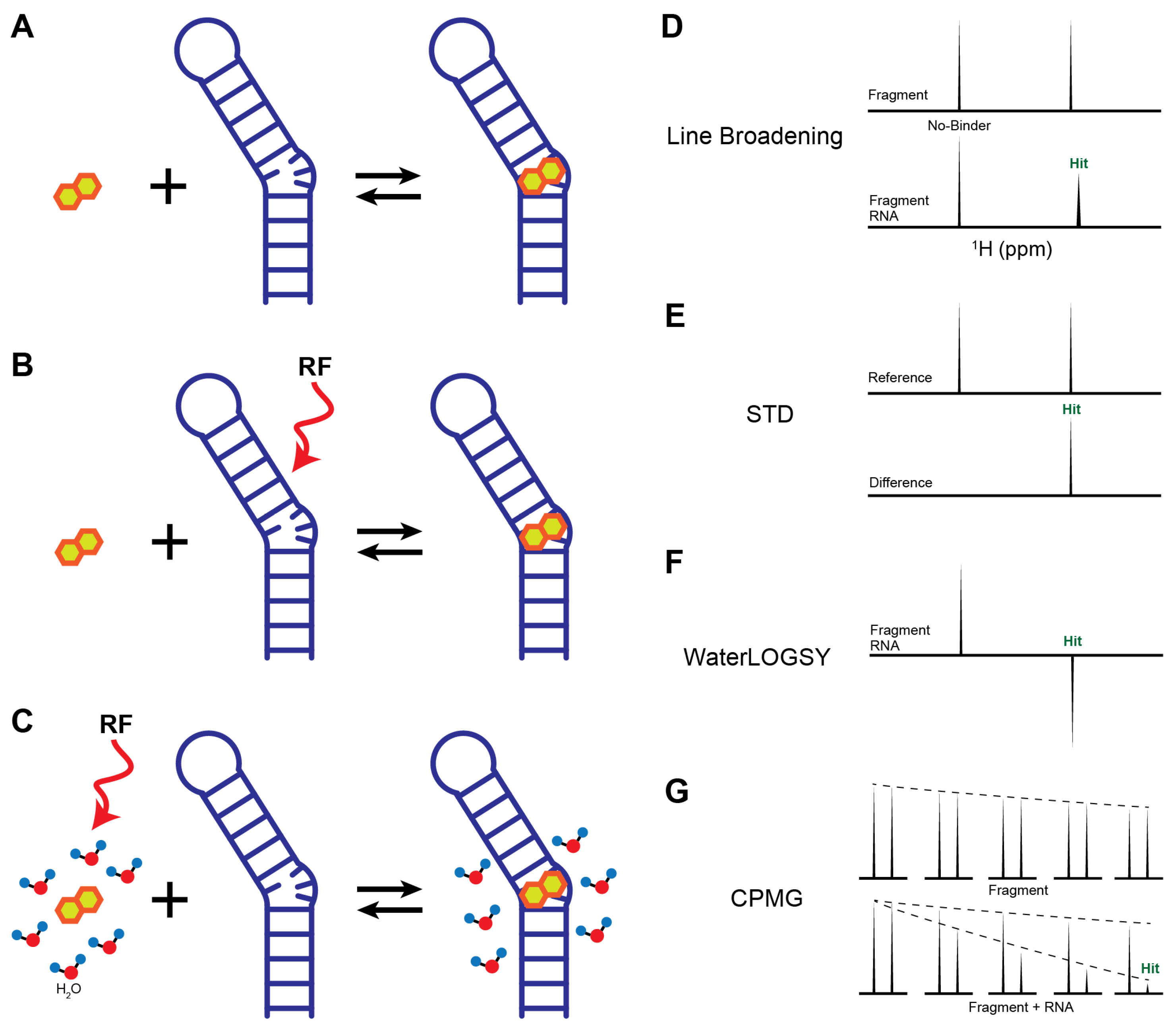
- Line Broadening: Line broadening experiments directly compare ligand peak shape, width, or intensity in the absence and presence of RNA (Figure 2A). If interactions occur, the influence of a large RNA molecule on a ligand causes the peak to shift or broaden (Figure 2D). This change can be considered binding and allow the ligands to be identified as initial hits. The extent of line broadening often correlates qualitatively with binding strength and can be used to prioritize hits [73,74]. Therefore, the absence of chemical shift perturbation (CSP) or peak intensity reduction indicates a lack of binding event between RNA and ligands.
- Saturation Transfer Difference (STD): STD is one of the most widely used ligand-observed techniques in drug discovery and has been successfully applied to RNA targets [75,76,77]. In an STD experiment, RNA resonances (e.g., imino or ribose protons) are selectively saturated through a radio frequency pulse, and then the saturation is rapidly transferred to the entire molecule through spin diffusion (Figure 2B) [78,79]. If binding occurs between RNA and ligand, their proximity causes nuclear Overhauser effect (NOE) transfer from the saturated RNA to the binding site of the ligand. The fast exchange between bound and free ligands results in a reduction in the bulk magnetization of the ligand, while any ligands that do not bind with RNA are minimally affected. However, for high-affinity ligands in the slow exchange regime, the STD effect is markedly reduced or lost. This occurs because the ligands remain trapped in the binding pocket for a duration longer than the saturation period, preventing the accumulation of a detectable signal in the bulk free ligand population. In the optimal exchange regime (typically KD is in the range of μM to mM), the difference in peak intensities between spectra collected with and without saturation only shows the proton signals from the ligand that binds with the RNA (Figure 2E). The relevant reduction in peak intensity also gives additional details on the distance of that proton to the targets as protons that are in closest contact with the RNA surface exhibit the most intense STD signals. Thus, STD not only confirms binding but also provides epitope mapping information on ligand protons proximal to the RNA surface.
- Water-Ligand Observed via Gradient Spectroscopy (WaterLOGSY): WaterLOGSY is another popular NMR screening method that detects ligand-RNA interactions through water-mediated NOEs [80,81]. In a WaterLOGSY experiment, bulk water magnetization is selectively excited and subsequently transferred to ligands in solution (Figure 2C) [80]. Upon binding, water magnetization is further transferred to RNA-bound ligands via intermolecular NOEs. The tumbling rate of free ligands is much faster than the rate of RNA-bound ligands. Due to the slower tumbling rate of RNA–ligand complex, the NOE sign of bound ligands is inverted relative to that of free ligands, resulting in opposite signal phases in the spectrum (Figure 2F) [80]. Similar to STD experiments, WaterLOGSY is insensitive to the tight binding events, but is often more sensitive than STD for detecting weak fragment binding [82]. As a water-mediated technique, WaterLOGSY is not suitable for the hydrophobic pockets where water is absent, but it can provide useful information on solvent-exposed regions of ligands [21]. Given the intrinsically hydrophilic nature of RNA surfaces, WaterLOGSY is particularly well suited for RNA-targeted fragment screening [83].
- Carr–Purcell–Meiboom–Gill (CPMG): CPMG probes binding interactions by exploiting chemical exchange and differences in transverse relaxation rates between the free and bound states of a ligand. Free ligands typically exhibit long transverse relaxation times (T2) due to rapid molecular tumbling, whereas ligands bound to RNA experience substantially shortened T2 values as a consequence of the slower tumbling of the RNA–ligand complex. As a result, NMR signals from RNA-bound ligands decay more rapidly than those from free ligands under a CPMG pulse train, providing a sensitive readout of binding (Figure 2G). In addition to 1H detection, CPMG experiments can be implemented using other nuclei, such as 19F, which offers high sensitivity, a wide chemical shift dispersion, and minimal background signals in biological samples. 19F-CPMG has been successfully applied to identify fluorinated fragments that bind selectively to telomeric repeat-containing RNA (TERRA) G4 and SARS-CoV2 RNAs, highlighting its utility for RNA-targeted fragment screening [84,85].
3.4. RNA-Observed Binding Assay
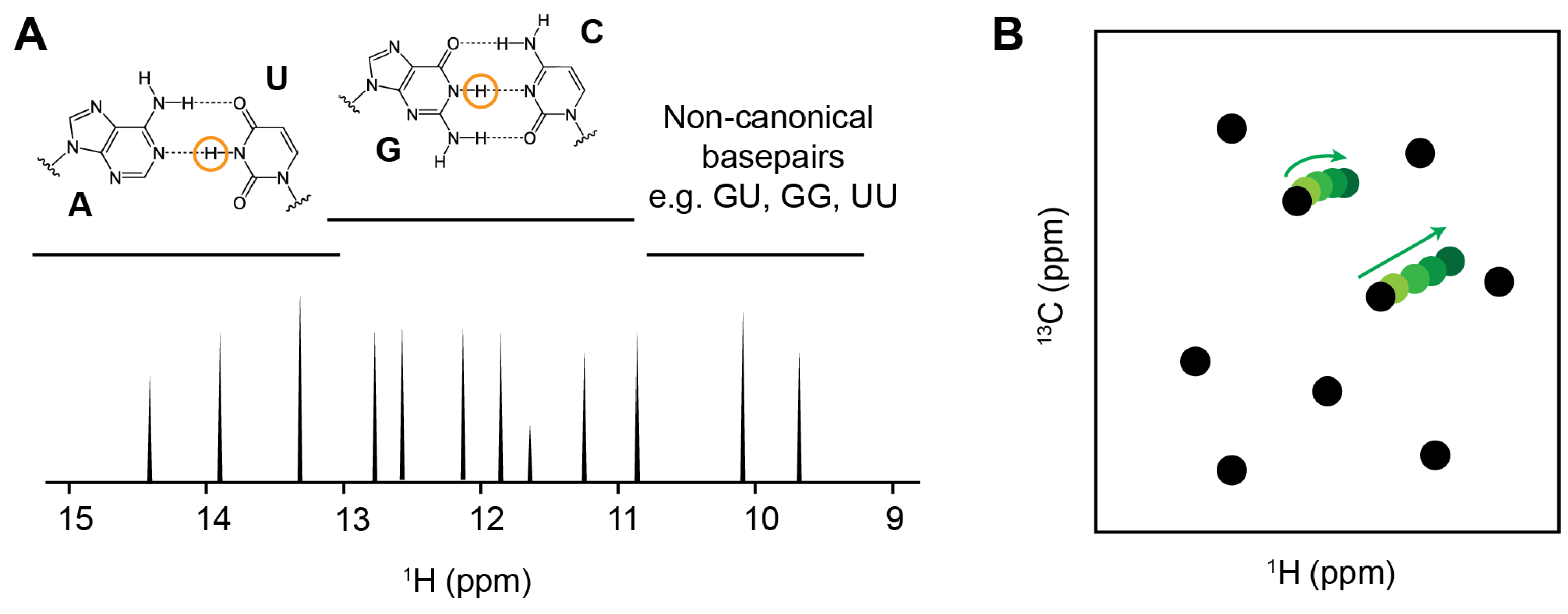
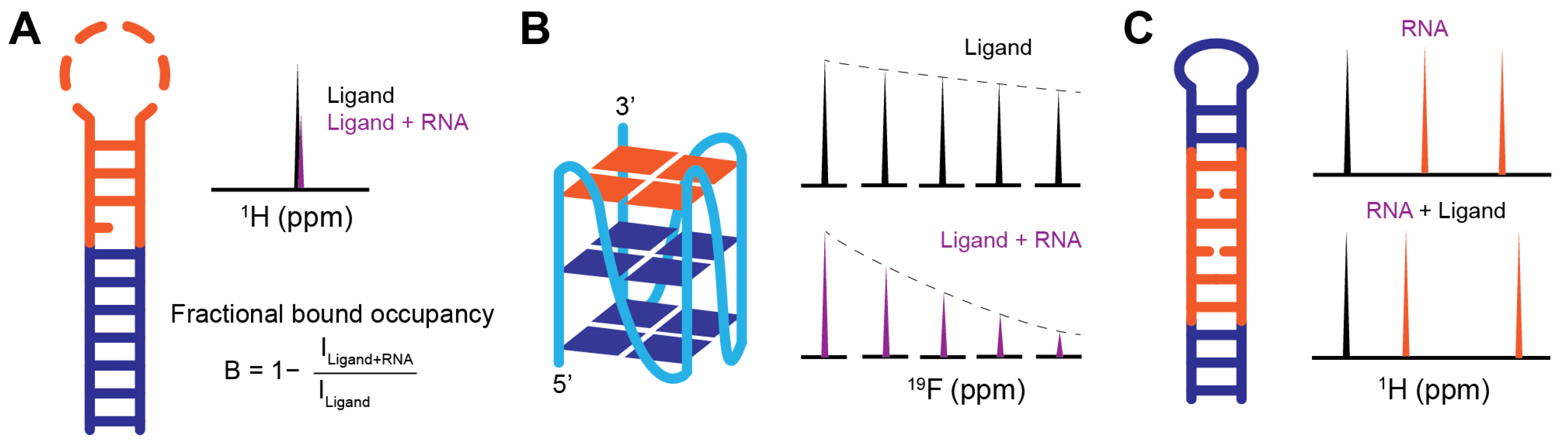
- 1D Imino Proton Spectroscopy: Imino proton spectroscopy, which detects the H1 proton of guanosine and the H3 proton of uridine, provides a sensitive means to monitor any binding at the base-pair level with the advantage of no isotopic labeling requirement [84]. Imino proton signals arise from only the hydrogen-bonded base pairs and typically resonate in a well-dispersed chemical shift region between ~9 to 15 ppm (Figure 3A). This is largely away from the overlap with other proton peaks that come from small-molecule fragments [15]. With the assignment of the imino peaks, which is commonly achieved through routine 2D imino 1H-1H nuclear Overhauser effect spectroscopy (NOESY), the binding site on the RNA can be mapped based on the ligand-induced CSPs or intensity changes. However, since imino proton peaks come from stable hydrogen bonding within the base pairs, unstructured or dynamic elements, such as loops, bulges, and single-stranded regions, are absent in the imino proton spectra. Therefore, 1D imino proton experiment may fail to detect ligand interactions that occur primarily within flexible or non-base-paired RNA regions.
- Total Correlation Spectroscopy (TOCSY): TOCSY is a common RNA NMR experiment that does not require isotopic labeling and fits well with monitoring RNA folding and ligand-induced conformational changes. Through scalar coupling networks, TOCSY detects through-bond correlations between H5 and H6 protons of pyrimidine residues (uridine and cytidine) with high sensitivity [15]. These crosspeaks are in a distinct chemical shift range (H5 protons generally resonate between 5.0 and 6.0 ppm, while H6 protons resonate from 7.0 to 8.5 ppm) and are typically well resolved in folded RNAs, which allow detection of subtle interactions related to pyrimidines at the residue level [86,87]. Complementary to the imino proton spectroscopy, TOCSY can observe interactions that involve pyrimidines located in unstructured or dynamic regions such as loops and bulges, which are common RNA structural motifs and frequently serves as small-molecule binding pockets. Notably, the Varani group has recently employed TOCSY-based screening to identify fragment hits that bind the unstructured apical loop of the precursor microRNA-21 (pre-miR-21), demonstrating the utility of this approach for probing flexible RNA elements [74].
- Heteronuclear Single Quantum Correlation Spectroscopy (HSQC): HSQC is a widely used in protein-targeted drug discovery to monitor ligand interactions via amide proton resonances. In RNA, however, aromatic (H2, H5, H6, and H8), ribose (H1′, H2′, H3′, H4′, H5′, and H5″), and imino (H1 and H3) protons could be severely overlapped in 1D proton spectra along with the increasing of their size or structural complexity. This overlap can be effectively resolved with the additional heteronuclear dimension, making high-resolution analysis of RNA–ligand interactions possible (Figure 3B) [15]. Although HSQC experiments typically require isotopically labeled RNA, the spectra are able to provide valuable binding information at the atomic level. CSP or titration experiments using 1H-13C HSQC spectra allow the characterization of ligand interactions involving RNA bases and ribose moieties, while 1H-31P HSQC experiments are sensitive to perturbations of the phosphate backbone. With the recent development of new pulse sequences, such as SOFAST and other fast-pulsing experiments, together with nonuniform sampling strategies, HSQC-based approaches, in some cases, have the ability to directly observe non-labeled RNA [88,89].
4. Hit Ranking and Optimization
5. RNA–Ligand Structural Modeling
6. Case Studies in NMR-Based RNA Fragment Screenings
6.1. Discovery of Fragment Binders to Pre-miR-21
6.2. 19F NMR Screening Against TERRA G-Quadruplexes
6.3. Targeting the Myotonic Dystrophy CUG Repeats
7. Summary and Future Perspectives
Author Contributions
Funding
Institutional Review Board Statement
Informed Consent Statement
Data Availability Statement
Acknowledgments
Conflicts of Interest
Abbreviations
| 1D | one-dimension |
| CF | monofluoride |
| CF3 | trifluoromethyl |
| CPMG | Carr–Purcell–Meiboom–Gill |
| Cryo-EM | cryo-electron microscopy |
| CSP | chemical shift perturbations |
| DEEP-STD | differential epitope mapping by STD NMR |
| DMSO-d6 | deuterated dimethyl sulfoxide |
| FBDD | fragment-based drug discovery |
| G4 | G-quadruplex |
| HSQC | heteronuclear single quantum correlation spectroscopy |
| HTS | high-throughput screening |
| KD | dissociation constants |
| LB | Line broadening |
| MBNL1 | Muscleblind-like 1 |
| NMR | nuclear magnetic resonance |
| NOE | nuclear Overhauser effect |
| NOESY | nuclear Overhauser effect spectroscopy |
| PAGE | polyacrylamide gel electrophoresis |
| R-BIND | RNA-targeted bioactive ligand database |
| rNTP | ribonucleotides |
| SALMON | solvent accessibility, ligand binding, and mapping of ligand orientation by NMR spectroscopy |
| SAR | structure–activity relationship |
| SPR | surface plasmon resonance |
| STD | saturation transfer difference |
| TERRA | telomeric repeat-containing RNA |
| TOCSY | total correlation spectroscopy |
| WaterLOGSY | water-ligand observed via gradient spectroscopy |
References
- Shuker, S.B.; Hajduk, P.J.; Meadows, R.P.; Fesik, S.W. Discovering high-affinity ligands for proteins: SAR by NMR. Science 1996, 274, 1531–1534. [Google Scholar] [CrossRef] [Scilit] [PubMed]
- Erlanson, D.A.; Fesik, S.W.; Hubbard, R.E.; Jahnke, W.; Jhoti, H. Twenty years on: The impact of fragments on drug discovery. Nat. Rev. Drug Discov. 2016, 15, 605–619. [Google Scholar] [CrossRef] [Scilit]
- Schoepfer, J.; Jahnke, W.; Berellini, G.; Buonamici, S.; Cotesta, S.; Cowan-Jacob, S.W.; Dodd, S.; Drueckes, P.; Fabbro, D.; Gabriel, T.; et al. Discovery of Asciminib (ABL001), an Allosteric Inhibitor of the Tyrosine Kinase Activity of BCR-ABL1. J. Med. Chem. 2018, 61, 8120–8135. [Google Scholar] [CrossRef] [Scilit]
- Addie, M.; Ballard, P.; Buttar, D.; Crafter, C.; Currie, G.; Davies, B.R.; Debreczeni, J.; Dry, H.; Dudley, P.; Greenwood, R.; et al. Discovery of 4-amino-N-[(1S)-1-(4-chlorophenyl)-3-hydroxypropyl]-1-(7H-pyrrolo[2,3-d]pyrimidin-4-yl)piperidine-4-carboxamide (AZD5363), an orally bioavailable, potent inhibitor of Akt kinases. J. Med. Chem. 2013, 56, 2059–2073. [Google Scholar] [CrossRef] [Scilit]
- Perera, T.P.S.; Jovcheva, E.; Mevellec, L.; Vialard, J.; De Lange, D.; Verhulst, T.; Paulussen, C.; Van De Ven, K.; King, P.; Freyne, E.; et al. Discovery and Pharmacological Characterization of JNJ-42756493 (Erdafitinib), a Functionally Selective Small-Molecule FGFR Family Inhibitor. Mol. Cancer Ther. 2017, 16, 1010–1020. [Google Scholar] [CrossRef] [Scilit]
- Tap, W.D.; Wainberg, Z.A.; Anthony, S.P.; Ibrahim, P.N.; Zhang, C.; Healey, J.H.; Chmielowski, B.; Staddon, A.P.; Cohn, A.L.; Shapiro, G.I.; et al. Structure-Guided Blockade of CSF1R Kinase in Tenosynovial Giant-Cell Tumor. N. Engl. J. Med. 2015, 373, 428–437. [Google Scholar] [CrossRef] [Scilit] [PubMed]
- Lanman, B.A.; Allen, J.R.; Allen, J.G.; Amegadzie, A.K.; Ashton, K.S.; Booker, S.K.; Chen, J.J.; Chen, N.; Frohn, M.J.; Goodman, G.; et al. Discovery of a Covalent Inhibitor of KRAS(G12C) (AMG 510) for the Treatment of Solid Tumors. J. Med. Chem. 2020, 63, 52–65. [Google Scholar] [CrossRef] [Scilit] [PubMed]
- Bollag, G.; Tsai, J.; Zhang, J.; Zhang, C.; Ibrahim, P.; Nolop, K.; Hirth, P. Vemurafenib: The first drug approved for BRAF-mutant cancer. Nat. Rev. Drug Discov. 2012, 11, 873–886. [Google Scholar] [CrossRef] [Scilit] [PubMed]
- Souers, A.J.; Leverson, J.D.; Boghaert, E.R.; Ackler, S.L.; Catron, N.D.; Chen, J.; Dayton, B.D.; Ding, H.; Enschede, S.H.; Fairbrother, W.J.; et al. ABT-199, a potent and selective BCL-2 inhibitor, achieves antitumor activity while sparing platelets. Nat. Med. 2013, 19, 202–208. [Google Scholar] [CrossRef] [Scilit]
- Neumann, T.; Junker, H.D.; Schmidt, K.; Sekul, R. SPR-based fragment screening: Advantages and applications. Curr. Top. Med. Chem. 2007, 7, 1630–1642. [Google Scholar] [CrossRef] [Scilit]
- Gossert, A.D.; Jahnke, W. NMR in drug discovery: A practical guide to identification and validation of ligands interacting with biological macromolecules. Prog. Nucl. Magn. Reson. Spectrosc. 2016, 97, 82–125. [Google Scholar] [CrossRef] [Scilit] [PubMed]
- Hartshorn, M.J.; Murray, C.W.; Cleasby, A.; Frederickson, M.; Tickle, I.J.; Jhoti, H. Fragment-based lead discovery using X-ray crystallography. J. Med. Chem. 2005, 48, 403–413. [Google Scholar] [CrossRef] [Scilit] [PubMed]
- Lea, W.A.; Simeonov, A. Fluorescence polarization assays in small molecule screening. Expert Opin. Drug Discov. 2011, 6, 17–32. [Google Scholar] [CrossRef] [Scilit] [PubMed]
- Saur, M.; Hartshorn, M.J.; Dong, J.; Reeks, J.; Bunkoczi, G.; Jhoti, H.; Williams, P.A. Fragment-based drug discovery using cryo-EM. Drug Discov. Today 2020, 25, 485–490. [Google Scholar] [CrossRef] [Scilit]
- Furtig, B.; Richter, C.; Wohnert, J.; Schwalbe, H. NMR spectroscopy of RNA. ChemBioChem 2003, 4, 936–962. [Google Scholar] [CrossRef] [Scilit]
- Bothe, J.R.; Nikolova, E.N.; Eichhorn, C.D.; Chugh, J.; Hansen, A.L.; Al-Hashimi, H.M. Characterizing RNA dynamics at atomic resolution using solution-state NMR spectroscopy. Nat. Methods 2011, 8, 919–931. [Google Scholar] [CrossRef] [Scilit]
- Sugiki, T.; Furuita, K.; Fujiwara, T.; Kojima, C. Current NMR Techniques for Structure-Based Drug Discovery. Molecules 2018, 23, 148. [Google Scholar] [CrossRef] [Scilit]
- Norton, R.S.; Jahnke, W. NMR in pharmaceutical discovery and development. J. Biomol. NMR 2020, 74, 473–476. [Google Scholar] [CrossRef] [Scilit]
- Mureddu, L.G.; Vuister, G.W. Fragment-Based Drug Discovery by NMR. Where Are the Successes and Where can It Be Improved? Front. Mol. Biosci. 2022, 9, 834453. [Google Scholar] [CrossRef] [Scilit]
- Janela, T.; Bajorath, J. Simple nearest-neighbour analysis meets the accuracy of compound potency predictions using complex machine learning models. Nat. Mach. Intell. 2022, 4, 1246–1255. [Google Scholar] [CrossRef] [Scilit]
- Raingeval, C.; Cala, O.; Brion, B.; Le Borgne, M.; Hubbard, R.E.; Krimm, I. 1D NMR WaterLOGSY as an efficient method for fragment-based lead discovery. J. Enzym. Inhib. Med. Chem. 2019, 34, 1218–1225. [Google Scholar] [CrossRef] [Scilit]
- Jiang, H.; Wang, J.; Cong, W.; Huang, Y.; Ramezani, M.; Sarma, A.; Dokholyan, N.V.; Mahdavi, M.; Kandemir, M.T. Predicting Protein-Ligand Docking Structure with Graph Neural Network. J. Chem. Inf. Model. 2022, 62, 2923–2932. [Google Scholar] [CrossRef] [Scilit]
- Cooper, T.A.; Wan, L.; Dreyfuss, G. RNA and disease. Cell 2009, 136, 777–793. [Google Scholar] [CrossRef] [Scilit]
- Mortimer, S.A.; Kidwell, M.A.; Doudna, J.A. Insights into RNA structure and function from genome-wide studies. Nat. Rev. Genet. 2014, 15, 469–479. [Google Scholar] [CrossRef] [Scilit]
- Sharp, P.A. The centrality of RNA. Cell 2009, 136, 577–580. [Google Scholar] [CrossRef] [Scilit] [PubMed]
- Wapinski, O.; Chang, H.Y. Long noncoding RNAs and human disease. Trends Cell Biol. 2011, 21, 354–361. [Google Scholar] [CrossRef] [Scilit] [PubMed]
- Warner, K.D.; Hajdin, C.E.; Weeks, K.M. Principles for targeting RNA with drug-like small molecules. Nat. Rev. Drug Discov. 2018, 17, 547–558. [Google Scholar] [CrossRef] [Scilit] [PubMed]
- Thomas, J.R.; Hergenrother, P.J. Targeting RNA with small molecules. Chem. Rev. 2008, 108, 1171–1224. [Google Scholar] [CrossRef] [Scilit]
- Childs-Disney, J.L.; Yang, X.; Gibaut, Q.M.R.; Tong, Y.; Batey, R.T.; Disney, M.D. Targeting RNA structures with small molecules. Nat. Rev. Drug Discov. 2022, 21, 736–762. [Google Scholar] [CrossRef] [Scilit]
- Disney, M.D.; Dwyer, B.G.; Childs-Disney, J.L. Drugging the RNA World. Cold Spring Harb. Perspect. Biol. 2018, 10, a034769. [Google Scholar] [CrossRef] [Scilit]
- Malone, B.; Urakova, N.; Snijder, E.J.; Campbell, E.A. Structures and functions of coronavirus replication-transcription complexes and their relevance for SARS-CoV-2 drug design. Nat. Rev. Mol. Cell Biol. 2022, 23, 21–39. [Google Scholar] [CrossRef] [Scilit]
- Zafferani, M.; Haddad, C.; Luo, L.; Davila-Calderon, J.; Chiu, L.Y.; Mugisha, C.S.; Monaghan, A.G.; Kennedy, A.A.; Yesselman, J.D.; Gifford, R.J.; et al. Amilorides inhibit SARS-CoV-2 replication in vitro by targeting RNA structures. Sci. Adv. 2021, 7, eabl6096. [Google Scholar] [CrossRef] [Scilit]
- Sreeramulu, S.; Richter, C.; Berg, H.; Wirtz Martin, M.A.; Ceylan, B.; Matzel, T.; Adam, J.; Altincekic, N.; Azzaoui, K.; Bains, J.K.; et al. Exploring the Druggability of Conserved RNA Regulatory Elements in the SARS-CoV-2 Genome. Angew. Chem. Int. Ed. Engl. 2021, 60, 19191–19200. [Google Scholar] [CrossRef] [Scilit]
- Shao, Y.; Zhang, Q.C. Targeting RNA structures in diseases with small molecules. Essays Biochem. 2020, 64, 955–966. [Google Scholar] [CrossRef] [Scilit]
- Meyer, S.M.; Williams, C.C.; Akahori, Y.; Tanaka, T.; Aikawa, H.; Tong, Y.; Childs-Disney, J.L.; Disney, M.D. Small molecule recognition of disease-relevant RNA structures. Chem. Soc. Rev. 2020, 49, 7167–7199. [Google Scholar] [CrossRef] [Scilit] [PubMed]
- Hargrove, A.E. Small molecule-RNA targeting: Starting with the fundamentals. Chem. Commun. 2020, 56, 14744–14756. [Google Scholar] [CrossRef] [Scilit] [PubMed]
- Walker, M.J.; Varani, G. Design of RNA-targeting macrocyclic peptides. Methods Enzymol. 2019, 623, 339–372. [Google Scholar] [CrossRef] [Scilit]
- Gallego, J.; Varani, G. Targeting RNA with small-molecule drugs: Therapeutic promise and chemical challenges. Acc. Chem. Res. 2001, 34, 836–843. [Google Scholar] [CrossRef] [Scilit] [PubMed]
- Petrone, J.; DeFrancesco, L. Small molecules get the message. Nat. Biotechnol. 2018, 36, 787–790. [Google Scholar] [CrossRef] [Scilit]
- Cai, Z.; Ma, H.; Ye, F.; Lei, D.; Deng, Z.; Li, Y.; Gu, R.; Wen, H. Discovery of RNA-Targeting Small Molecules: Challenges and Future Directions. MedComm 2025, 6, e70342. [Google Scholar] [CrossRef] [Scilit]
- Wang, L.; Gao, J.; Ma, R.; Liu, Y.; Liu, M.; Zhong, F.; Hu, J.; Li, S.; Wu, J.; Jiang, H.; et al. Recent progress in fragment-based drug discovery facilitated by NMR spectroscopy. Magn. Reson. Lett. 2022, 2, 107–118. [Google Scholar] [CrossRef] [Scilit]
- Lundquist, K.P.; Panchal, V.; Gotfredsen, C.H.; Brenk, R.; Clausen, M.H. Fragment-Based Drug Discovery for RNA Targets. ChemMedChem 2021, 16, 2588–2603. [Google Scholar] [CrossRef] [Scilit]
- Suresh, B.M.; Taghavi, A.; Childs-Disney, J.L.; Disney, M.D. Fragment-based approaches to identify RNA binders. ChemRxiv 2023. [Google Scholar] [CrossRef] [Scilit]
- Veenbaas, S.D.; Felder, S.; Weeks, K.M. fpocketR: A Platform for Identification and Analysis of Ligand-Binding Pockets in RNA. ACS Chem. Biol. 2026, 21, 151–159. [Google Scholar] [CrossRef] [Scilit]
- Veenbaas, S.D.; Koehn, J.T.; Irving, P.S.; Lama, N.N.; Weeks, K.M. Ligand-binding pockets in RNA and where to find them. Proc. Natl. Acad. Sci. USA 2025, 122, e2422346122. [Google Scholar] [CrossRef] [Scilit]
- Krishnan, S.R.; Roy, A.; Wong, L.; Gromiha, M.M. DRLiPS: A novel method for prediction of druggable RNA-small molecule binding pockets using machine learning. Nucleic Acids Res. 2025, 53, gkaf239. [Google Scholar] [CrossRef] [Scilit] [PubMed]
- Dominguez, C.; Schubert, M.; Duss, O.; Ravindranathan, S.; Allain, F.H. Structure determination and dynamics of protein-RNA complexes by NMR spectroscopy. Prog. Nucl. Magn. Reson. Spectrosc. 2011, 58, 1–61. [Google Scholar] [CrossRef] [Scilit]
- Longhini, A.P.; LeBlanc, R.M.; Dayie, T.K. Chemo-enzymatic labeling for rapid assignment of RNA molecules. Methods 2016, 103, 11–17. [Google Scholar] [CrossRef] [Scilit] [PubMed]
- Ferre-D’Amare, A.R.; Scott, W.G. Small self-cleaving ribozymes. Cold Spring Harb. Perspect. Biol. 2010, 2, a003574. [Google Scholar] [CrossRef] [Scilit] [PubMed]
- Shields, T.P.; Mollova, E.; Ste Marie, L.; Hansen, M.R.; Pardi, A. High-performance liquid chromatography purification of homogenous-length RNA produced by trans cleavage with a hammerhead ribozyme. RNA 1999, 5, 1259–1267. [Google Scholar] [CrossRef] [Scilit]
- Kirsch, P.; Hartman, A.M.; Hirsch, A.K.H.; Empting, M. Concepts and Core Principles of Fragment-Based Drug Design. Molecules 2019, 24, 4309. [Google Scholar] [CrossRef] [Scilit]
- Congreve, M.; Carr, R.; Murray, C.; Jhoti, H. A ‘rule of three’ for fragment-based lead discovery? Drug Discov. Today 2003, 8, 876–877. [Google Scholar] [CrossRef] [Scilit] [PubMed]
- Hewitt, W.M.; Calabrese, D.R.; Schneekloth, J.S., Jr. Evidence for ligandable sites in structured RNA throughout the Protein Data Bank. Bioorganic Med. Chem. 2019, 27, 2253–2260. [Google Scholar] [CrossRef] [Scilit] [PubMed]
- Padroni, G.; Patwardhan, N.N.; Schapira, M.; Hargrove, A.E. Systematic analysis of the interactions driving small molecule-RNA recognition. RSC Med. Chem. 2020, 11, 802–813. [Google Scholar] [CrossRef] [Scilit]
- Rizvi, N.F.; Santa Maria, J.P., Jr.; Nahvi, A.; Klappenbach, J.; Klein, D.J.; Curran, P.J.; Richards, M.P.; Chamberlin, C.; Saradjian, P.; Burchard, J.; et al. Targeting RNA with Small Molecules: Identification of Selective, RNA-Binding Small Molecules Occupying Drug-Like Chemical Space. SLAS Discov. 2020, 25, 384–396. [Google Scholar] [CrossRef] [Scilit]
- Haniff, H.S.; Knerr, L.; Liu, X.; Crynen, G.; Bostrom, J.; Abegg, D.; Adibekian, A.; Lekah, E.; Wang, K.W.; Cameron, M.D.; et al. Design of a small molecule that stimulates vascular endothelial growth factor A enabled by screening RNA fold-small molecule interactions. Nat. Chem. 2020, 12, 952–961. [Google Scholar] [CrossRef] [Scilit]
- Donlic, A.; Swanson, E.G.; Chiu, L.Y.; Wicks, S.L.; Juru, A.U.; Cai, Z.; Kassam, K.; Laudeman, C.; Sanaba, B.G.; Sugarman, A.; et al. R-BIND 2.0: An Updated Database of Bioactive RNA-Targeting Small Molecules and Associated RNA Secondary Structures. ACS Chem. Biol. 2022, 17, 1556–1566. [Google Scholar] [CrossRef] [Scilit] [PubMed]
- Sun, S.; Yang, J.; Zhang, Z. RNALigands: A database and web server for RNA-ligand interactions. RNA 2022, 28, 115–122. [Google Scholar] [CrossRef] [Scilit]
- Disney, M.D.; Winkelsas, A.M.; Velagapudi, S.P.; Southern, M.; Fallahi, M.; Childs-Disney, J.L. Inforna 2.0: A Platform for the Sequence-Based Design of Small Molecules Targeting Structured RNAs. ACS Chem. Biol. 2016, 11, 1720–1728. [Google Scholar] [CrossRef] [Scilit]
- Morgan, B.S.; Forte, J.E.; Culver, R.N.; Zhang, Y.; Hargrove, A.E. Discovery of Key Physicochemical, Structural, and Spatial Properties of RNA-Targeted Bioactive Ligands. Angew. Chem. Int. Ed. Engl. 2017, 56, 13498–13502. [Google Scholar] [CrossRef] [Scilit]
- Morgan, B.S.; Sanaba, B.G.; Donlic, A.; Karloff, D.B.; Forte, J.E.; Zhang, Y.; Hargrove, A.E. R-BIND: An Interactive Database for Exploring and Developing RNA-Targeted Chemical Probes. ACS Chem. Biol. 2019, 14, 2691–2700. [Google Scholar] [CrossRef] [Scilit]
- Morgan, B.S.; Forte, J.E.; Hargrove, A.E. Insights into the development of chemical probes for RNA. Nucleic Acids Res. 2018, 46, 8025–8037. [Google Scholar] [CrossRef] [Scilit]
- Rzuczek, S.G.; Southern, M.R.; Disney, M.D. Studying a Drug-like, RNA-Focused Small Molecule Library Identifies Compounds That Inhibit RNA Toxicity in Myotonic Dystrophy. ACS Chem. Biol. 2015, 10, 2706–2715. [Google Scholar] [CrossRef] [Scilit]
- Lepre, C.A.; Moore, J.M.; Peng, J.W. Theory and applications of NMR-based screening in pharmaceutical research. Chem. Rev. 2004, 104, 3641–3676. [Google Scholar] [CrossRef] [Scilit]
- Arroyo, X.; Goldflam, M.; Feliz, M.; Belda, I.; Giralt, E. Computer-aided design of fragment mixtures for NMR-based screening. PLoS ONE 2013, 8, e58571. [Google Scholar] [CrossRef] [Scilit] [PubMed]
- Stark, J.L.; Eghbalnia, H.R.; Lee, W.; Westler, W.M.; Markley, J.L. NMRmix: A Tool for the Optimization of Compound Mixtures in 1D (1)H NMR Ligand Affinity Screens. J. Proteome Res. 2016, 15, 1360–1368. [Google Scholar] [CrossRef] [Scilit] [PubMed]
- Dalvit, C.; Fagerness, P.E.; Hadden, D.T.; Sarver, R.W.; Stockman, B.J. Fluorine-NMR experiments for high-throughput screening: Theoretical aspects, practical considerations, and range of applicability. J. Am. Chem. Soc. 2003, 125, 7696–7703. [Google Scholar] [CrossRef] [Scilit] [PubMed]
- Gronenborn, A.M. Small, but powerful and attractive: 19F in biomolecular NMR. Structure 2022, 30, 6–14. [Google Scholar] [CrossRef] [Scilit]
- Norton, R.S.; Leung, E.W.; Chandrashekaran, I.R.; MacRaild, C.A. Applications of 19F-NMR in Fragment-Based Drug Discovery. Molecules 2016, 21, 860. [Google Scholar] [CrossRef] [Scilit]
- Jordan, J.B.; Poppe, L.; Xia, X.; Cheng, A.C.; Sun, Y.; Michelsen, K.; Eastwood, H.; Schnier, P.D.; Nixey, T.; Zhong, W. Fragment based drug discovery: Practical implementation based on 19F NMR spectroscopy. J. Med. Chem. 2012, 55, 678–687. [Google Scholar] [CrossRef] [Scilit]
- Lee, J.; Vogt, C.E.; McBrairty, M.; Al-Hashimi, H.M. Influence of dimethylsulfoxide on RNA structure and ligand binding. Anal. Chem. 2013, 85, 9692–9698. [Google Scholar] [CrossRef] [Scilit]
- Pellecchia, M.; Bertini, I.; Cowburn, D.; Dalvit, C.; Giralt, E.; Jahnke, W.; James, T.L.; Homans, S.W.; Kessler, H.; Luchinat, C.; et al. Perspectives on NMR in drug discovery: A technique comes of age. Nat. Rev. Drug Discov. 2008, 7, 738–745. [Google Scholar] [CrossRef] [Scilit]
- Shortridge, M.D.; Hage, D.S.; Harbison, G.S.; Powers, R. Estimating protein-ligand binding affinity using high-throughput screening by NMR. J. Comb. Chem. 2008, 10, 948–958. [Google Scholar] [CrossRef] [Scilit]
- Shortridge, M.D.; Varani, G. Efficient NMR Screening Approach to Discover Small Molecule Fragments Binding Structured RNA. Acs Med. Chem. Lett. 2021, 12, 1253–1260. [Google Scholar] [CrossRef] [Scilit]
- Walpole, S.; Monaco, S.; Nepravishta, R.; Angulo, J. STD NMR as a Technique for Ligand Screening and Structural Studies. Methods Enzymol. 2019, 615, 423–451. [Google Scholar] [CrossRef] [Scilit]
- Mayer, M.; Meyer, B. Characterization of Ligand Binding by Saturation Transfer Difference NMR Spectroscopy. Angew. Chem. Int. Ed. Engl. 1999, 38, 1784–1788. [Google Scholar] [CrossRef]
- Davidson, A.; Begley, D.W.; Lau, C.; Varani, G. A small-molecule probe induces a conformation in HIV TAR RNA capable of binding drug-like fragments. J. Mol. Biol. 2011, 410, 984–996. [Google Scholar] [CrossRef] [Scilit] [PubMed]
- Abulwerdi, F.A.; Xu, W.; Ageeli, A.A.; Yonkunas, M.J.; Arun, G.; Nam, H.; Schneekloth, J.S., Jr.; Dayie, T.K.; Spector, D.; Baird, N.; et al. Selective Small-Molecule Targeting of a Triple Helix Encoded by the Long Noncoding RNA, MALAT1. ACS Chem. Biol. 2019, 14, 223–235. [Google Scholar] [CrossRef] [Scilit] [PubMed]
- Mayer, M.; James, T.L. Detecting ligand binding to a small RNA target via saturation transfer difference NMR experiments in D2O and H2O. J. Am. Chem. Soc. 2002, 124, 13376–13377. [Google Scholar] [CrossRef] [Scilit]
- Dalvit, C.; Fogliatto, G.; Stewart, A.; Veronesi, M.; Stockman, B. WaterLOGSY as a method for primary NMR screening: Practical aspects and range of applicability. J. Biomol. NMR 2001, 21, 349–359. [Google Scholar] [CrossRef] [Scilit] [PubMed]
- Huang, R.; Leung, I.K.H. Protein-Small Molecule Interactions by WaterLOGSY. Methods Enzymol. 2019, 615, 477–500. [Google Scholar] [CrossRef] [Scilit]
- Antanasijevic, A.; Ramirez, B.; Caffrey, M. Comparison of the sensitivities of WaterLOGSY and saturation transfer difference NMR experiments. J. Biomol. NMR 2014, 60, 37–44. [Google Scholar] [CrossRef] [Scilit]
- Johnson, E.C.; Feher, V.A.; Peng, J.W.; Moore, J.M.; Williamson, J.R. Application of NMR SHAPES screening to an RNA target. J. Am. Chem. Soc. 2003, 125, 15724–15725. [Google Scholar] [CrossRef] [Scilit] [PubMed]
- Garavis, M.; Lopez-Mendez, B.; Somoza, A.; Oyarzabal, J.; Dalvit, C.; Villasante, A.; Campos-Olivas, R.; Gonzalez, C. Discovery of selective ligands for telomeric RNA G-quadruplexes (TERRA) through 19F-NMR based fragment screening. ACS Chem. Biol. 2014, 9, 1559–1566. [Google Scholar] [CrossRef] [Scilit]
- Hymon, D.; Martins, J.; Richter, C.; Sreeramulu, S.; Wacker, A.; Ferner, J.; Patwardhan, N.N.; Hargrove, A.E.; Schwalbe, H. NMR 1H,19F-based screening of the four stem-looped structure 5_SL1-SL4 located in the 5′-untranslated region of SARS-CoV 2 RNA. RSC Med. Chem. 2024, 15, 165–177. [Google Scholar] [CrossRef] [Scilit] [PubMed]
- Abulwerdi, F.A.; Shortridge, M.D.; Sztuba-Solinska, J.; Wilson, R.; Le Grice, S.F.; Varani, G.; Schneekloth, J.S., Jr. Development of Small Molecules with a Noncanonical Binding Mode to HIV-1 Trans Activation Response (TAR) RNA. J. Med. Chem. 2016, 59, 11148–11160. [Google Scholar] [CrossRef] [Scilit]
- Murchie, A.I.; Davis, B.; Isel, C.; Afshar, M.; Drysdale, M.J.; Bower, J.; Potter, A.J.; Starkey, I.D.; Swarbrick, T.M.; Mirza, S.; et al. Structure-based drug design targeting an inactive RNA conformation: Exploiting the flexibility of HIV-1 TAR RNA. J. Mol. Biol. 2004, 336, 625–638. [Google Scholar] [CrossRef] [Scilit]
- Farjon, J.; Boisbouvier, J.; Schanda, P.; Pardi, A.; Simorre, J.P.; Brutscher, B. Longitudinal-relaxation-enhanced NMR experiments for the study of nucleic acids in solution. J. Am. Chem. Soc. 2009, 131, 8571–8577. [Google Scholar] [CrossRef] [Scilit]
- Sathyamoorthy, B.; Lee, J.; Kimsey, I.; Ganser, L.R.; Al-Hashimi, H. Development and application of aromatic [13C, 1H] SOFAST-HMQC NMR experiment for nucleic acids. J. Biomol. NMR 2014, 60, 77–83. [Google Scholar] [CrossRef] [Scilit] [PubMed]
- Schulz, M.N.; Hubbard, R.E. Recent progress in fragment-based lead discovery. Curr. Opin. Pharmacol. 2009, 9, 615–621. [Google Scholar] [CrossRef] [Scilit]
- Friberg, A.; Vigil, D.; Zhao, B.; Daniels, R.N.; Burke, J.P.; Garcia-Barrantes, P.M.; Camper, D.; Chauder, B.A.; Lee, T.; Olejniczak, E.T.; et al. Discovery of potent myeloid cell leukemia 1 (Mcl-1) inhibitors using fragment-based methods and structure-based design. J. Med. Chem. 2013, 56, 15–30. [Google Scholar] [CrossRef] [Scilit]
- Szczepankiewicz, B.G.; Liu, G.; Hajduk, P.J.; Abad-Zapatero, C.; Pei, Z.; Xin, Z.; Lubben, T.H.; Trevillyan, J.M.; Stashko, M.A.; Ballaron, S.J.; et al. Discovery of a potent, selective protein tyrosine phosphatase 1B inhibitor using a linked-fragment strategy. J. Am. Chem. Soc. 2003, 125, 4087–4096. [Google Scholar] [CrossRef] [Scilit]
- de Esch, I.J.P.; Erlanson, D.A.; Jahnke, W.; Johnson, C.N.; Walsh, L. Fragment-to-Lead Medicinal Chemistry Publications in 2020. J. Med. Chem. 2022, 65, 84–99. [Google Scholar] [CrossRef] [Scilit]
- Jahnke, W.; Erlanson, D.A.; de Esch, I.J.P.; Johnson, C.N.; Mortenson, P.N.; Ochi, Y.; Urushima, T. Fragment-to-Lead Medicinal Chemistry Publications in 2019. J. Med. Chem. 2020, 63, 15494–15507. [Google Scholar] [CrossRef] [Scilit]
- Li, S.; Zhang, K.; Chiu, W. Near-Atomic Resolution Cryo-EM Image Reconstruction of RNA. Methods Mol. Biol. 2023, 2568, 179–192. [Google Scholar] [CrossRef] [Scilit] [PubMed]
- Zhang, K.; Zheludev, I.N.; Hagey, R.J.; Haslecker, R.; Hou, Y.J.; Kretsch, R.; Pintilie, G.D.; Rangan, R.; Kladwang, W.; Li, S.; et al. Cryo-EM and antisense targeting of the 28-kDa frameshift stimulation element from the SARS-CoV-2 RNA genome. Nat. Struct. Mol. Biol. 2021, 28, 747–754. [Google Scholar] [CrossRef] [Scilit] [PubMed]
- Ma, H.; Pham, P.; Luo, B.; Rangan, R.; Kappel, K.; Su, Z.; Das, R. Auto-DRRAFTER: Automated RNA Modeling Based on Cryo-EM Density. Methods Mol. Biol. 2023, 2568, 193–211. [Google Scholar] [CrossRef] [Scilit] [PubMed]
- Kappel, K.; Zhang, K.; Su, Z.; Watkins, A.M.; Kladwang, W.; Li, S.; Pintilie, G.; Topkar, V.V.; Rangan, R.; Zheludev, I.N.; et al. Accelerated cryo-EM-guided determination of three-dimensional RNA-only structures. Nat. Methods 2020, 17, 699–707. [Google Scholar] [CrossRef] [Scilit]
- Shortridge, M.D.; Wille, P.T.; Jones, A.N.; Davidson, A.; Bogdanovic, J.; Arts, E.; Karn, J.; Robinson, J.A.; Varani, G. An ultra-high affinity ligand of HIV-1 TAR reveals the RNA structure recognized by P-TEFb. Nucleic Acids Res. 2019, 47, 1523–1531. [Google Scholar] [CrossRef] [Scilit]
- Kang, M.; Eichhorn, C.D.; Feigon, J. Structural determinants for ligand capture by a class II preQ1 riboswitch. Proc. Natl. Acad. Sci. USA 2014, 111, E663–E671. [Google Scholar] [CrossRef] [Scilit]
- Tolbert, M.; Morgan, C.E.; Pollum, M.; Crespo-Hernandez, C.E.; Li, M.L.; Brewer, G.; Tolbert, B.S. HnRNP A1 Alters the Structure of a Conserved Enterovirus IRES Domain to Stimulate Viral Translation. J. Mol. Biol. 2017, 429, 2841–2858. [Google Scholar] [CrossRef] [Scilit]
- Williams, B., 2nd; Zhao, B.; Tandon, A.; Ding, F.; Weeks, K.M.; Zhang, Q.; Dokholyan, N.V. Structure modeling of RNA using sparse NMR constraints. Nucleic Acids Res. 2017, 45, 12638–12647. [Google Scholar] [CrossRef] [Scilit] [PubMed]
- Peterson, R.D.; Theimer, C.A.; Wu, H.; Feigon, J. New applications of 2D filtered/edited NOESY for assignment and structure elucidation of RNA and RNA-protein complexes. J. Biomol. NMR 2004, 28, 59–67. [Google Scholar] [CrossRef] [Scilit] [PubMed]
- van Zundert, G.C.P.; Rodrigues, J.; Trellet, M.; Schmitz, C.; Kastritis, P.L.; Karaca, E.; Melquiond, A.S.J.; van Dijk, M.; de Vries, S.J.; Bonvin, A. The HADDOCK2.2 Web Server: User-Friendly Integrative Modeling of Biomolecular Complexes. J. Mol. Biol. 2016, 428, 720–725. [Google Scholar] [CrossRef] [Scilit] [PubMed]
- Schwieters, C.D.; Kuszewski, J.J.; Tjandra, N.; Clore, G.M. The Xplor-NIH NMR molecular structure determination package. J. Magn. Reson. 2003, 160, 65–73. [Google Scholar] [CrossRef] [Scilit]
- Case, D.A.; Cerutti, D.S.; Cruzeiro, V.W.D.; Darden, T.A.; Duke, R.E.; Ghazimirsaeed, M.; Giambasu, G.M.; Giese, T.J.; Gotz, A.W.; Harris, J.A.; et al. Recent Developments in Amber Biomolecular Simulations. J. Chem. Inf. Model. 2025, 65, 7835–7843. [Google Scholar] [CrossRef] [Scilit] [PubMed]
- Ludwig, C.; Michiels, P.J.; Wu, X.; Kavanagh, K.L.; Pilka, E.; Jansson, A.; Oppermann, U.; Gunther, U.L. SALMON: Solvent accessibility, ligand binding, and mapping of ligand orientation by NMR spectroscopy. J. Med. Chem. 2008, 51, 1–3. [Google Scholar] [CrossRef] [Scilit]
- Ma, X.; Kumar, M.; Choudhury, S.N.; Becker Buscaglia, L.E.; Barker, J.R.; Kanakamedala, K.; Liu, M.F.; Li, Y. Loss of the miR-21 allele elevates the expression of its target genes and reduces tumorigenesis. Proc. Natl. Acad. Sci. USA 2011, 108, 10144–10149. [Google Scholar] [CrossRef] [Scilit]
- Schoeftner, S.; Blasco, M.A. Developmentally regulated transcription of mammalian telomeres by DNA-dependent RNA polymerase II. Nat. Cell Biol. 2008, 10, 228–236. [Google Scholar] [CrossRef] [Scilit]
- Azzalin, C.M.; Reichenbach, P.; Khoriauli, L.; Giulotto, E.; Lingner, J. Telomeric repeat containing RNA and RNA surveillance factors at mammalian chromosome ends. Science 2007, 318, 798–801. [Google Scholar] [CrossRef] [Scilit]
- Xu, Y.; Suzuki, Y.; Ito, K.; Komiyama, M. Telomeric repeat-containing RNA structure in living cells. Proc. Natl. Acad. Sci. USA 2010, 107, 14579–14584. [Google Scholar] [CrossRef] [Scilit] [PubMed]
- Martadinata, H.; Phan, A.T. Structure of propeller-type parallel-stranded RNA G-quadruplexes, formed by human telomeric RNA sequences in K+ solution. J. Am. Chem. Soc. 2009, 131, 2570–2578. [Google Scholar] [CrossRef] [Scilit]
- Collie, G.W.; Haider, S.M.; Neidle, S.; Parkinson, G.N. A crystallographic and modelling study of a human telomeric RNA (TERRA) quadruplex. Nucleic Acids Res. 2010, 38, 5569–5580. [Google Scholar] [CrossRef] [Scilit]
- Brook, J.D.; McCurrach, M.E.; Harley, H.G.; Buckler, A.J.; Church, D.; Aburatani, H.; Hunter, K.; Stanton, V.P.; Thirion, J.P.; Hudson, T.; et al. Molecular basis of myotonic dystrophy: Expansion of a trinucleotide (CTG) repeat at the 3′ end of a transcript encoding a protein kinase family member. Cell 1992, 68, 799–808. [Google Scholar] [CrossRef] [Scilit]
- Miller, J.W.; Urbinati, C.R.; Teng-Umnuay, P.; Stenberg, M.G.; Byrne, B.J.; Thornton, C.A.; Swanson, M.S. Recruitment of human muscleblind proteins to (CUG)n expansions associated with myotonic dystrophy. EMBO J. 2000, 19, 4439–4448. [Google Scholar] [CrossRef] [Scilit] [PubMed]
- Jia, Y.; Taghavi, A.; Zanon, P.R.A.; Disney, M.D. Streamlined Fragment-Based Discovery Platform for Targeting Structured RNAs. ACS Chem. Biol. 2025, 20, 2243–2254. [Google Scholar] [CrossRef] [Scilit]
- Sheridan, C. First small-molecule drug targeting RNA gains momentum. Nat. Biotechnol. 2021, 39, 6–8. [Google Scholar] [CrossRef] [Scilit]
- Ratni, H.; Ebeling, M.; Baird, J.; Bendels, S.; Bylund, J.; Chen, K.S.; Denk, N.; Feng, Z.; Green, L.; Guerard, M.; et al. Discovery of Risdiplam, a Selective Survival of Motor Neuron-2 (SMN2) Gene Splicing Modifier for the Treatment of Spinal Muscular Atrophy (SMA). J. Med. Chem. 2018, 61, 6501–6517. [Google Scholar] [CrossRef] [Scilit]
- Menichelli, E.; Lam, B.J.; Wang, Y.; Wang, V.S.; Shaffer, J.; Tjhung, K.F.; Bursulaya, B.; Nguyen, T.N.; Vo, T.; Alper, P.B.; et al. Discovery of small molecules that target a tertiary-structured RNA. Proc. Natl. Acad. Sci. USA 2022, 119, e2213117119. [Google Scholar] [CrossRef] [Scilit] [PubMed]
- Sun, Y.T.; Shortridge, M.D.; Varani, G. A Small Cyclic beta-Hairpin Peptide Mimics the Rbfox2 RNA Recognition Motif and Binds to the Precursor miRNA 20b. ChemBioChem 2019, 20, 931–939. [Google Scholar] [CrossRef] [Scilit] [PubMed]
- Suresh, B.M.; Akahori, Y.; Taghavi, A.; Crynen, G.; Gibaut, Q.M.R.; Li, Y.; Disney, M.D. Low-Molecular Weight Small Molecules Can Potently Bind RNA and Affect Oncogenic Pathways in Cells. J. Am. Chem. Soc. 2022, 144, 20815–20824. [Google Scholar] [CrossRef] [Scilit]
- Kognole, A.A.; Hazel, A.; MacKerell, A.D., Jr. SILCS-RNA: Toward a Structure-Based Drug Design Approach for Targeting RNAs with Small Molecules. J. Chem. Theory Comput. 2022, 18, 5672–5691. [Google Scholar] [CrossRef] [Scilit] [PubMed]
- Vicens, Q.; Kieft, J.S. Thoughts on how to think (and talk) about RNA structure. Proc. Natl. Acad. Sci. USA 2022, 119, e2112677119. [Google Scholar] [CrossRef] [Scilit] [PubMed]
- Bodoor, K.; Boyapati, V.; Gopu, V.; Boisdore, M.; Allam, K.; Miller, J.; Treleaven, W.D.; Weldeghiorghis, T.; Aboul-ela, F. Design and implementation of an ribonucleic acid (RNA) directed fragment library. J. Med. Chem. 2009, 52, 3753–3761. [Google Scholar] [CrossRef] [Scilit] [PubMed]
- Cressina, E.; Chen, L.H.; Abell, C.; Leeper, F.J.; Smith, A.G. Fragment screening against the thiamine pyrophosphate riboswitch. Chem. Sci. 2011, 2, 157–165. [Google Scholar] [CrossRef] [Scilit]
- Lee, M.K.; Bottini, A.; Kim, M.; Bardaro, M.F., Jr.; Zhang, Z.; Pellecchia, M.; Choi, B.S.; Varani, G. A novel small-molecule binds to the influenza A virus RNA promoter and inhibits viral replication. Chem. Commun. 2014, 50, 368–370. [Google Scholar] [CrossRef] [Scilit]
- Tam, B.; Sherf, D.; Cohen, S.; Eisdorfer, S.A.; Perez, M.; Soffer, A.; Vilenchik, D.; Akabayov, S.R.; Wagner, G.; Akabayov, B. Discovery of small-molecule inhibitors targeting the ribosomal peptidyl transferase center (PTC) of M. tuberculosis. Chem. Sci. 2019, 10, 8764–8767. [Google Scholar] [CrossRef] [Scilit]
- Binas, O.; de Jesus, V.; Landgraf, T.; Volklein, A.E.; Martins, J.; Hymon, D.; Kaur Bains, J.; Berg, H.; Biedenbander, T.; Furtig, B.; et al. 19F NMR-Based Fragment Screening for 14 Different Biologically Active RNAs and 10 DNA and Protein Counter-Screens. ChemBioChem 2021, 22, 423–433. [Google Scholar] [CrossRef] [Scilit]
- Kwai, B.X.C.; Chandrashekaran, I.R.; Mohanty, B.; Gunzburg, M.J.; Doak, B.C.; Sethi, A.; Pillay, S.; Lok, D.; Harrison, S.J.; Serrano, P.; et al. NMR and SPR Fragment-Based Screening Can Produce Novel High Affinity Small Molecule Hits against Structured RNAs. ACS Chem. Biol. 2025, 20, 2863–2872. [Google Scholar] [CrossRef] [Scilit]
- Lundquist, K.P.; Romeo, I.; Puglielli, R.B.; Pestalozzi, M.; Gram, M.L.; Hudson, E.S.; Levi, O.; Arava, Y.S.; Gotfredsen, C.H.; Clausen, M.H. Design, synthesis, and screening of an RNA optimized fluorinated fragment library. SLAS Discov. 2025, 31, 100215. [Google Scholar] [CrossRef] [Scilit]
- Panchal, V.N.; Husmann, J.A.; Gunther, K.; Zeeshan, M.; Haug, B.E.; Brenk, R. Discovery of RNA-binding fragments using biolayer interferometry. RSC Med. Chem. 2025, 16, 5629–5640. [Google Scholar] [CrossRef] [Scilit]
- Toews, S.; Dona, F.; Keller, M.; Krauss, J.; Bracher, F.; Lopez-Garcia, U.; Pabel, J.; Merk, D.; Blommers, M.J.J.; Ferner, J.; et al. Targeting the SARS-CoV-2 RNA Translation Initiation Element SL1 by Molecules of Low Molecular Weight. J. Am. Chem. Soc. 2025, 147, 28783–28798. [Google Scholar] [CrossRef] [Scilit]
- Zhou, Y.; Chen, S.J. Harnessing Computational Approaches for RNA-Targeted Drug Discovery. RNA Nanomed 2024, 1, 1–15. [Google Scholar] [CrossRef] [Scilit] [PubMed]
- Arney, J.W.; Weeks, K.M. RNA-Ligand Interactions Quantified by Surface Plasmon Resonance with Reference Subtraction. Biochemistry 2022, 61, 1625–1632. [Google Scholar] [CrossRef] [Scilit] [PubMed]
- Chen, X.; Wang, L.; Xie, J.; Nowak, J.S.; Luo, B.; Zhang, C.; Jia, G.; Zou, J.; Huang, D.; Glatt, S.; et al. RNA sample optimization for cryo-EM analysis. Nat. Protoc. 2024, 20, 1114–1157. [Google Scholar] [CrossRef] [Scilit] [PubMed]
- Kang, C.B. 19F-NMR in target-based drug discovery. Curr. Med. Chem. 2019, 26, 4964–4983. [Google Scholar] [CrossRef] [Scilit]
- Diethelm-Varela, B. Using NMR Spectroscopy in the Fragment-Based Drug Discovery of Small-Molecule Anticancer Targeted Therapies. ChemMedChem 2021, 16, 725–742. [Google Scholar] [CrossRef] [Scilit]




| Parameter | Typical Range | Notes/Best Practice |
|---|---|---|
| RNA concentration | 1–100 μM | Lower for ligand-observed; higher for RNA-observed |
| Fragment concentration | 50–200 μM | Avoid aggregation; confirm solubility |
| Fragment:RNA ratio | 20:1–100:1 | Keep detectable bound fraction |
| DMSO (%) | ≤5% | Check if DMSO concentration affects RNA integrity |
| Mg2+ | 0–5 mM | Required for tertiary structure |
| Buffer | 10 mM phosphate, pH~6.4 | Optimizes imino visibility |
| Temperature | 278–298 K | Balance stability vs. dynamics |
| Observation Type | NMR Method | RNA Size | Sensitivity (KD Range) | Labeling Required? | RNA Amount Required | Strengths | Key Limitations |
|---|---|---|---|---|---|---|---|
| Ligand | LB | Any | μM–mM | No | Low (~2–20 μM) | Simple, fast, low sample | False positives from aggregation |
| STD | Any | μM–mM | No | Low (~2–20 μM) | Epitope mapping | Insensitive to tight binders | |
| WaterLOGSY | Any | μM–mM | No | Low (~2–20 μM) | High sensitivity | Requires solvent-exposed sites | |
| CPMG | Any | μM–mM | No | Low (~2–20 μM) | High sensitivity | Sensitive to exchange regime | |
| 19F NMR | Any | μM–mM | No | Very low (~0.5–10 μM) | Clean spectra | Requires fluorinated library | |
| RNA | 1D imino | ≤~100 nt | nM–mM | No | Moderate (~10–50 μM) | Base-pair level mapping | Misses unpaired regions |
| TOCSY | ≤~80 nt | nM–mM | No | Moderate (>50 μM) | Loop/bulge sensitivity | Pyrimidine-only | |
| HSQC | ≤~70 nt * | nM–mM | Yes | High (>50 μM) | Atomic resolution | Sample preparation cost |
| RNA Target | Disease/Application | Fragment Library | NMR Methods | Hit Ranking and Triage Approach | Reference |
|---|---|---|---|---|---|
| Bacterial ribosomal A-site model RNA | Antibiotics (translation inhibition) | RNA-directed fragment library (102 compounds) | WaterLOGSY and 1H CPMG | 5 hits were confirmed by RNA-observed imino proton | Bodoor et al., J. Med. Chem. (2009) [124] |
| Thiamine pyrophosphate riboswitch thiM | Riboswitch | Fragment library of 1300 compounds | WaterLOGSY and 1H CPMG | 17 primary hits; further validated by other biophysics assay: equilibrium dialysis and ITC | Cressina et al., Chem. Sci. (2011) [125] |
| HIV-1 TAR RNA | Antiviral (Tat–TAR interaction) | Maybridge “Rule of 3” collection (250 compounds) | STD, interligand NOEs (ILOE) | 20 primary hits; ranked by STD signal intensity; 6 hits were confirmed by ILOE | Davidson et al., Chem. Biol. (2011) [77] |
| TERRA G-quadruplex | Cancer/telomere biology | Fluorinated fragment library (355 compounds) | 19F CPMG | 20 primary hits; 6 were confirmed by STD and RNA-observed imino proton | Garavís et al., ACS Chem. Biol. (2014) [84] |
| Influenza A promoter RNA | Antiviral (influenza) | Fragment library of 4279 compounds | 1D imino proton | 7 hits; the one that has the most dramatic CSP was advanced to structural determination | Lee et al., Chem. Commun. (2014) [126] |
| M. tuberculosis rRNA PTC/hairpin 91 | Tuberculosis antibiotics | Maybridge “Rule of 3” collection (1000 compounds) | 1H CPMG | 9 primary hits; hit expansion with virtual library; ranked on the basis of the binding energy | Tam et al., Chem. Sci. (2019) [127] |
| 14 structured RNAs (incl. riboswitches) | Multi-disease | Fluorinated fragment library (102 compounds) | 19F CPMG | Hit rate up to 26% for some targets; ranked by the CPMG signal | Binas et al., ChemBioChem (2021) [128] |
| Pre-miR-21 apical loop | Cancer/miRNA dysregulation | Maybridge “Rule of 3” collection (420 compounds) | Line broadening | 17 primary hits; ranked by fractional bound occupancy; 4 hits were confirmed by NOESY | Shortridge and Varani, ACS Med. Chem. Lett. (2021) [74] |
| 15 conserved SARS-CoV-2 RNA elements | Antiviral (COVID-19) | The DSI-poised library (768 compounds) | CSP, WaterLOGSY, line broadening, 1H CPMG | 69 primary hits across targets; ranked by consistent binding across multiple methods | Sreeramulu et al., Angew. Chem. Int. Ed. (2021) [33] |
| SARS-CoV-2 5′UTR stem loops | Antiviral (COVID-19) | RNA-dedicated fluorinated DRTL-F library (49 compounds) and non-RNA-dedicated DSI-PL (768 compounds) | Parallel 1H/19F CSP and 19F CPMG | 10 primary hits from DRTL-F library and 5 primary hits from DSI-PL library. | Hymon et al., RSC Med. Chem. (2024) [85] |
| Myotonic dystrophy type 1 CUG repeats | Muscular dystrophy | Fully functionalized fragment library (187 compounds) | CSP/line broadening, WaterLOGSY, 1H CPMG | 14 primary hits from primary fluorescent binding screening; 4 were confirmed by NMR assay; 1 was picked based on consistent binding across multiple NMR methods | Jia et al., ACS Chem. Biol. (2025) [116] |
| Theophylline aptamer | Riboswitch | Fragment library (1975 compounds) | 1D imino proton | 28 primary hits; 4 hits were confirmed by SPR | Kwai et al., ACS Chem. Biol. (2025) [129] |
| Human cytoplasmic A-site and the S. cerevisiae tRNA anticodon stem loop with and without modification | Translation | RNA-optimized fluorinated fragment library + 2 non-optimized libraries (149 to 529 compounds depend on the targets) | WaterLOGSY and 19F CPMG | 24, 31, and 20 primary hits against the respective targets from 19F screening. Secondary WaterLOGSY screening verifies a few positive binders | Lundquist et al., SLAS Discov. (2025) [130] |
| Riboswitches (FMN, SAM-I, and TPP) | Riboswitch | Fragment library (651 compounds) | WaterLOGSY, CSP, 1H CPMG | 35 primary hits from biolayer interferometry; 7 verified by secondary NMR screening were used to confirm the specificity | Panchal et al., RSC Med. Chem. (2025) [131] |
| SARS-CoV-2 5′UTR stem-loop 1 | Antiviral (COVID-19) | Lead-derived library (41 compounds) | CSP, WaterLOGSY, line broadening, 1H CPMG, TOCSY | Ranked by consistent binding across multiple methods; confirmed by a counterscreen; further confirmed by TOCSY | Toews et al., JACS (2025) [132] |
Disclaimer/Publisher’s Note: The statements, opinions and data contained in all publications are solely those of the individual author(s) and contributor(s) and not of MDPI and/or the editor(s). MDPI and/or the editor(s) disclaim responsibility for any injury to people or property resulting from any ideas, methods, instructions or products referred to in the content. |
© 2026 by the authors. Licensee MDPI, Basel, Switzerland. This article is an open access article distributed under the terms and conditions of the Creative Commons Attribution (CC BY) license.
Share and Cite
Petersen, R.J.; Wang, Y. NMR-Based Fragment Screening for RNA-Targeted Drug Discovery. Molecules 2026, 31, 916. https://doi.org/10.3390/molecules31060916
Petersen RJ, Wang Y. NMR-Based Fragment Screening for RNA-Targeted Drug Discovery. Molecules. 2026; 31(6):916. https://doi.org/10.3390/molecules31060916
Chicago/Turabian StylePetersen, Riley J., and Yaqiang Wang. 2026. "NMR-Based Fragment Screening for RNA-Targeted Drug Discovery" Molecules 31, no. 6: 916. https://doi.org/10.3390/molecules31060916
APA StylePetersen, R. J., & Wang, Y. (2026). NMR-Based Fragment Screening for RNA-Targeted Drug Discovery. Molecules, 31(6), 916. https://doi.org/10.3390/molecules31060916