Article

Molecular Structure, Theoretical NBO Analysis, Vibrational Spectrum of CO2-Responsive Hydroxyamidine-Based Ionic Liquid: A Combined Computational and Experimental Approach


by
Lyazzat Abulyaissova
1,*,
Nikolay Barashkov
2,
Irina Irgibaeva
3 and
Yerbolat Tashenov
3
1
Chemistry Department, Buketov National Research University, Karaganda 100028, Kazakhstan
2
Micro-Tracers, Inc., 1370 Van Dyke Avenue, San Francisco, CA 94124, USA
3
Department of Chemistry, L.N. Gumilyov Eurasian National University, Astana 010008, Kazakhstan
*
Author to whom correspondence should be addressed.
Molecules 2026, 31(6), 1055; https://doi.org/10.3390/molecules31061055
Submission received: 7 February 2026 / Revised: 8 March 2026 / Accepted: 15 March 2026 / Published: 23 March 2026

Abstract

The utilization and chemical transformation of carbon dioxide remains a pressing problem in modern chemistry. Numerous experimental and theoretical studies have focused on the interaction of CO2 with amines. In this work, quantum chemical density functional theory (DFT) calculations of equilibrium geometries, energies, electronic and vibrational characteristics of CO2-sensitive mono-, di-, tris-hydroxyamidines and their associates were carried out by the B3LYP/6-31G(d, p) method. The harmonic vibrational frequencies were scaled and compared with the experimental FTIR spectra for supporting wavenumber assignments. Natural bond orbital (NBO) analysis of the atomic charges and charge delocalization was employed to investigate the nature of hydrogen bonding in hydroxyamidine associates. We also used the intrinsically polarizable continuum model (IEFPCM), and the DFT-D3 method was applied to account for dispersion effects during associate formation. Using the 6-311+G(2d, p) basis set for tris-hydroxyamidine, and its adducts, a comparative analysis of the experimental and calculated 1H NMR spectra was performed. Here, we considered non-trivial sites of carbon dioxide absorption and hydroxyamidine protonation, which, to our knowledge, have hardly been considered by other authors. Present DFT results agree rather well with the experimental data and support new insight into the formation of the PIL structure.

1. Introduction

Carbon capture, utilization, and storage (CCUS), including carbon dioxide capture and storage (CCS), has been and remains a key scientific and technical challenge. High atmospheric CO2 concentrations pose global environmental challenges.
However, through continuous photosynthesis and carbon dioxide absorption in plants, carbon dioxide levels in the atmosphere are self-regulated. So, increasing plant cover is one of the most effective ways to maintain ecological balance. Another promising approach is the incorporation of carbon dioxide into closed technological cycles, which should also contribute to reducing the greenhouse effect.
Advanced carbon dioxide capture technologies have been described in the literature, but they have both advantages and disadvantages from a technical standpoint [1,2,3,4,5,6,7,8]. One traditional and accessible method of CO2 capture is chemical absorption with aqueous amine solutions, which has been the subject of numerous applied and theoretical studies [9,10,11,12,13,14,15,16,17,18,19,20,21,22]. However, the use of amines is not without its drawbacks: high energy consumption, the formation of hazardous nitrosamines, amine losses, etc. Therefore, the search for new types of more effective absorbents/adsorbents continues.
Some promising candidates are amidines [23,24,25,26,27]. In [23], a study of the interaction of the polyamidine-polyethylene glycol binary system with carbon dioxide showed that 66% of the amidine groups bind CO2 molecules. The authors of [24] obtained new polyamidine polymers capable of selective and reversible carbon dioxide capture at room temperature and under atmospheric pressure. The monomer 4-vinylbenzyl amidine, which changes its hydrophobicity/hydrophilicity under the influence of CO2, was developed and synthesized [25]. We previously reported on the possibility of using carbon dioxide gas to obtain polymeric ionic liquids (PILs) by interaction with mono- and tris-amidines [26,27].
The mechanism of interaction of carbon dioxide with the listed and other compounds is studied both experimentally and using computer modeling methods. In order to fundamentally understand the mechanism of CO2 capture by unhindered and hindered amines, the authors of [9] performed density functional theory calculations that revealed opposite kinetic results for different amines, namely, that the formation of bicarbonate for 2-amino-2-methyl-1-propanol (AMP) is more favorable than the formation of carbamate. However, the situation is reversed for monoethanolamine (MEA). This explains well the experimental observation of different product distributions in the reactions of AMP and MEA with CO2. According to the authors, the difference in the electrostatic potential distribution of AMP and MEA is a possible reason leading to the different mechanisms of their reactions.
The study [10] discusses the formation of the nitrogen (monoethanolamine)–carbon (CO2) bond under solvent conditions based on molecular theory using RISM-SCF-SEDD, a hybrid method of quantum chemistry of the solute and statistical mechanics of the solvent. Authors clarified that the role of amine as a base is crucial in the proton transfer step. In [14], the thermochemistry of carbamate and bicarbonate formation was calculated using the composite CCSD(T) and DFT methods. Carbon dioxide chemisorption was shown to occur via the carbamate intermediate for all primary and secondary amines. To comprehensively understand the mechanism of carbon dioxide absorption by aqueous amine solutions, the authors of [15] performed ab initio molecular orbital calculations in combination with the solvation model (PCM). Based on the calculated activation energies, an efficient pathway for the direct interconversion of carbamate and bicarbonate without the participation of free CO2 was proposed.
DFT-B3LYP modeling [18] was used to characterize the products and energetics of reactions of carbon dioxide with a number of substituted amines at amine/CO2 stoichiometry of 1:1 and 2:1. The results showed that by controlling both the nature and arrangement of functional groups, the reaction energies can be tuned over a significant range. A quantum chemical analysis of carbon dioxide absorption by aqueous amine solutions using the continuum solvation model (SMD/IEF-PCM) in combination with density functional theory (DFT) was performed in [19]. Based on the obtained results, it was suggested that the carbamate anion forms by a two-step reaction via a zwitterion intermediate, and this occurs faster than the formation of the bicarbonate anion.
Unique and unexpected reaction mechanisms involved in CO2 absorption by aqueous hydrazine were revealed using 1H, 13C and 15N NMR spectroscopy in combination with first-principles quantum mechanical modeling [20]. In [21], the reaction pathways of carbamate as well as bicarbonate formations were analyzed by DFT modeling. The authors of [22] considered the formation of carbamate, carbamic acid and bicarbonate in the reaction of carbon dioxide with a sterically hindered amine and an aromatic amine based on molecular dynamics calculations.
A literature survey reveals that many of them are devoted to the interaction of carbon dioxide with amines, particularly under aqueous conditions. As an alternative to amines, amidines have also attracted the attention of researchers, as discussed above. Take our previous work as an example; we also studied the interaction of CO2 with mono- and tris-amidines to obtain polymeric ionic liquids (PILs) and proposed the structure of the PILs [26,27].
So, conventional CO2 capture systems are typically based on amine–CO2 chemistry, where absorption proceeds via carbamate or bicarbonate formation in aqueous media and is governed by carbonic acid equilibria [28,29,30]. Alcohol-based systems may additionally form alkylcarbonate species upon reaction with CO2 [31]. In contrast, the hydroxyamidine systems studied here exhibit a distinct binding mechanism: CO2 uptake occurs through hydroxyl-containing amidines, forming alkylcarbonate-type zwitterionic structures stabilized by hydrogen bonding. Moreover, methanol present in the reaction medium may participate in a proton-relay mechanism facilitating proton transfer between the carbonate fragment and the imino nitrogen atom. This pathway differs from classical carbonic-acid-mediated CO2 capture and provides new insight into CO2 fixation in alkylhydroxy-functionalized amidine systems.
In this work, which continues our research, we synthesized dihydroxylamidine with subsequent CO2 treatment, then modeled and investigated the structure of three hydroxyamidine-based PILs containing mono-, di-, tris-hydroxyl groups and the interaction of CO2 with hydroxyamidines using a combined experimental and computational approach. Here we (a) use quantum chemical calculations using the density functional theory (DFT) method, and conduct a comparative analysis of previously obtained and present experimental infrared absorption and 1H NMR spectra with the calculated spectra; (b) consider associates in the absence of water molecules, but in the presence of methanol as a medium.

2. Results and Discussion

2.1. Molecular Geometry of Hydroxyamidines

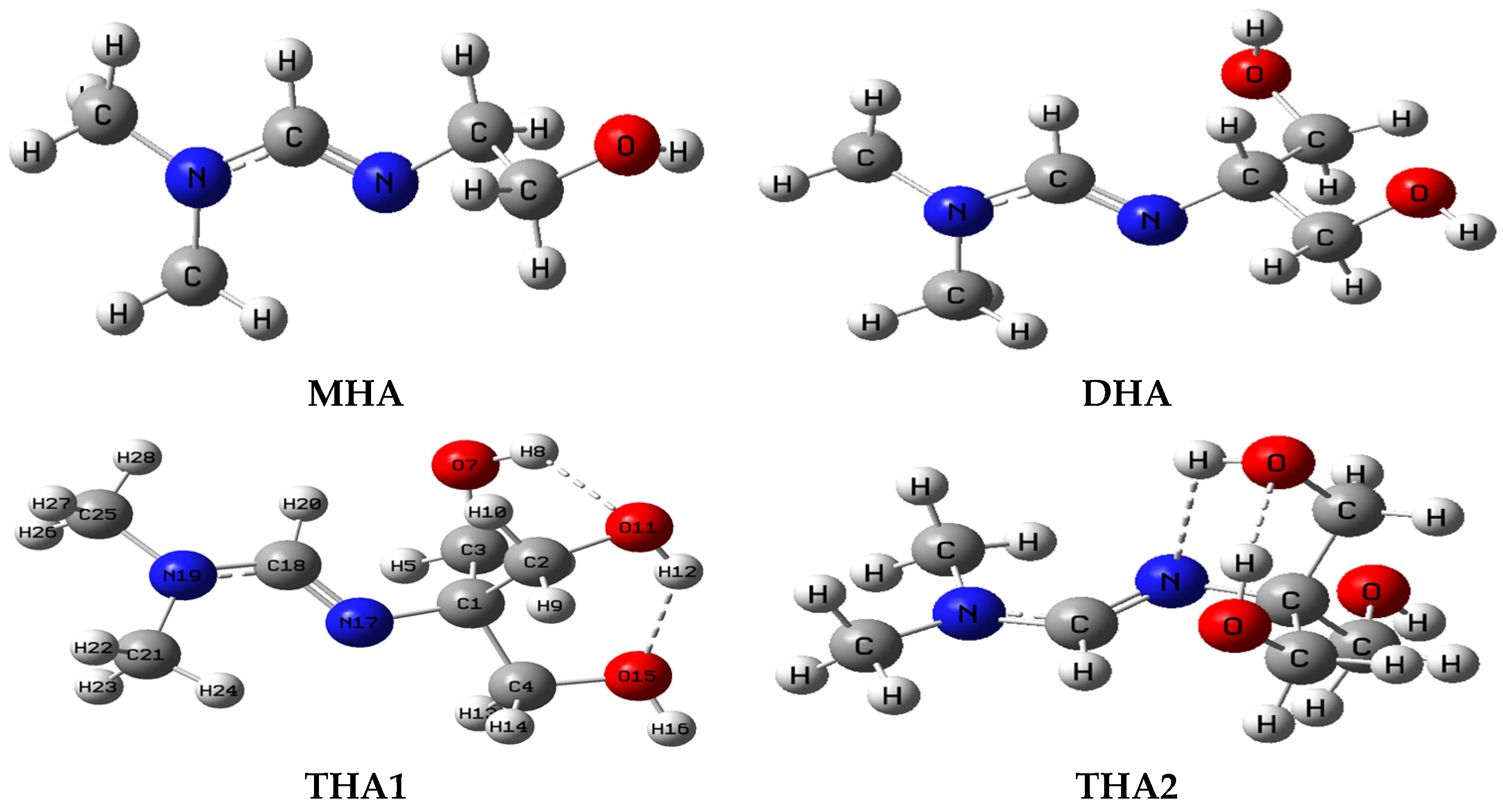
Stable conformations of hydroxyamidine molecules (Figure 1) contain the functional group >N-C(R=H)=N- (amino and imino nitrogen) and hydroxyl groups: from one (monohydroxyamidine, MHA) to three (tris-hydroxyamidine, THA). Dihydroxyamidine (DHA) contains two hydroxyl groups. Optimized molecular structures of hydroxyamidines are shown in Figure 1; their energies taking into account zero-point vibrations are presented in Table 1. The structural parameters calculated for the equilibrium geometry of hydroxyamidines are given in Supplementary Table S1.
Figure 1 shows two conformations of tris-hydroxyamidine, THA1 and THA2, which differ in mutual arrangement of hydroxyl groups, the number of hydrogen bonds and the possibility of forming an intramolecular H-bond with the nitrogen atom. According to calculations, THA1 is 3.25 kcal/mol more energetically stable than THA2. In the tris-hydroxyamidine THA1 molecule, three OH groups form two intramolecular hydrogen bonds with internuclear O…H distances of 2.168 and 1.989 Å, which contribute to the energetic stabilization of the molecule. The latter are analogous to the distances in diols [32].
In the THA2 conformation, the only hydrogen O-H…O bond with an oxygen-hydrogen distance of 1.946 Å occurs between the two OH groups, but there is also the possibility of intramolecular H-bonding with the imino nitrogen. The consideration of a less stable conformation, THA2, can be justified by the fact that thermal fluctuations result in a statistical distribution of molecular conformations. Therefore, at a given temperature, molecules in various conformations are always present in the bulk of a substance, not just those in the conformation with the lowest energy. It should also be noted that molecules of the same compound in different phase states can differ in conformation; i.e., an energetically favorable conformation in one phase may be unstable in another. Biphenyl is noteworthy in this regard. Its molecule is planar in the crystal, but non-planar in the gas phase and isotropic solution—the phenyl rings are arranged at angles of 40–45° and 30°, respectively [33].

2.2. Correlation of IR Spectra with the Structure of Amidines

The molecular structures presented above contain several functional groups, which leads to the occurrence of absorption of radiation of the characteristic frequency. The influence of the rest of the molecule usually does not exceed 5%. The calculation of vibrational IR spectra was carried out only for structures minimized by potential energy in order to avoid forces of atom–atom interactions that are too large, leading to the appearance of negative frequencies in the spectrum. Quantum chemical calculations made it possible to obtain spectral characteristics of molecules, such as frequencies and forms of normal vibrations, as well as the intensities of IR bands for the entire spectrum region, including the low-frequency region (˂400 cm–1). However, due to the introduction of the adiabatic approximation in quantum chemical methods, a discrepancy is always observed in the calculated and experimental frequencies: the first, harmonic frequencies are usually overestimated compared to the experiment. In this regard, a scaling factor (0.95) was applied to adjust the calculated data to the frequencies calculated at the B3LYP/6-31G(d, p) level [34]. The scaled wavenumbers of the IR spectra of the structures are presented in Table 2.
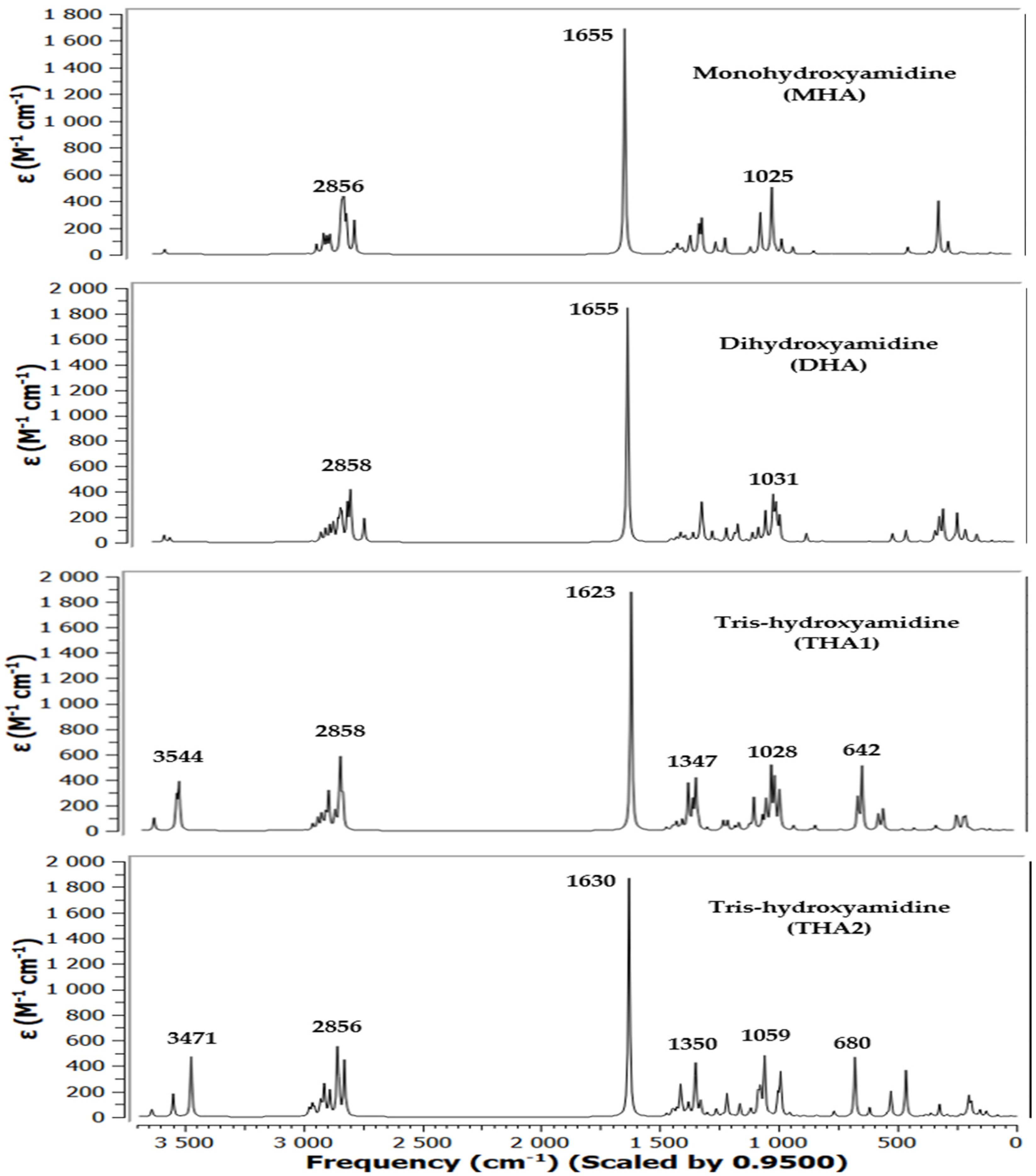
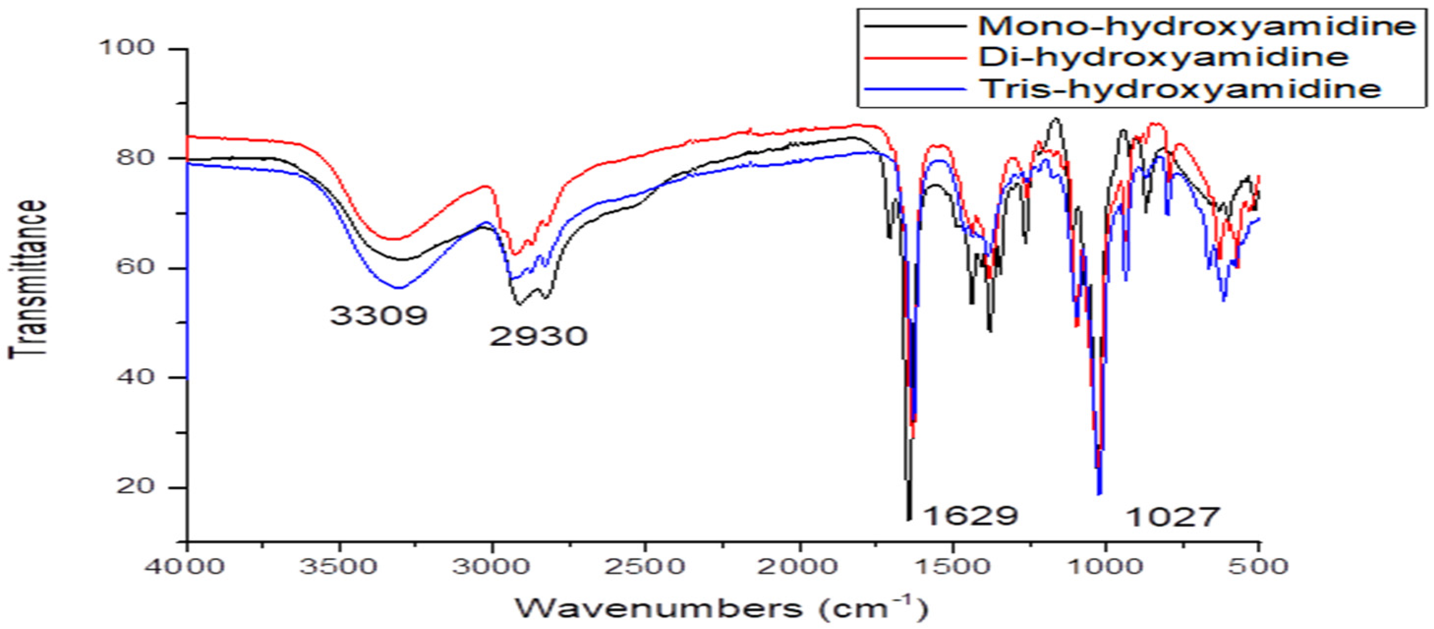
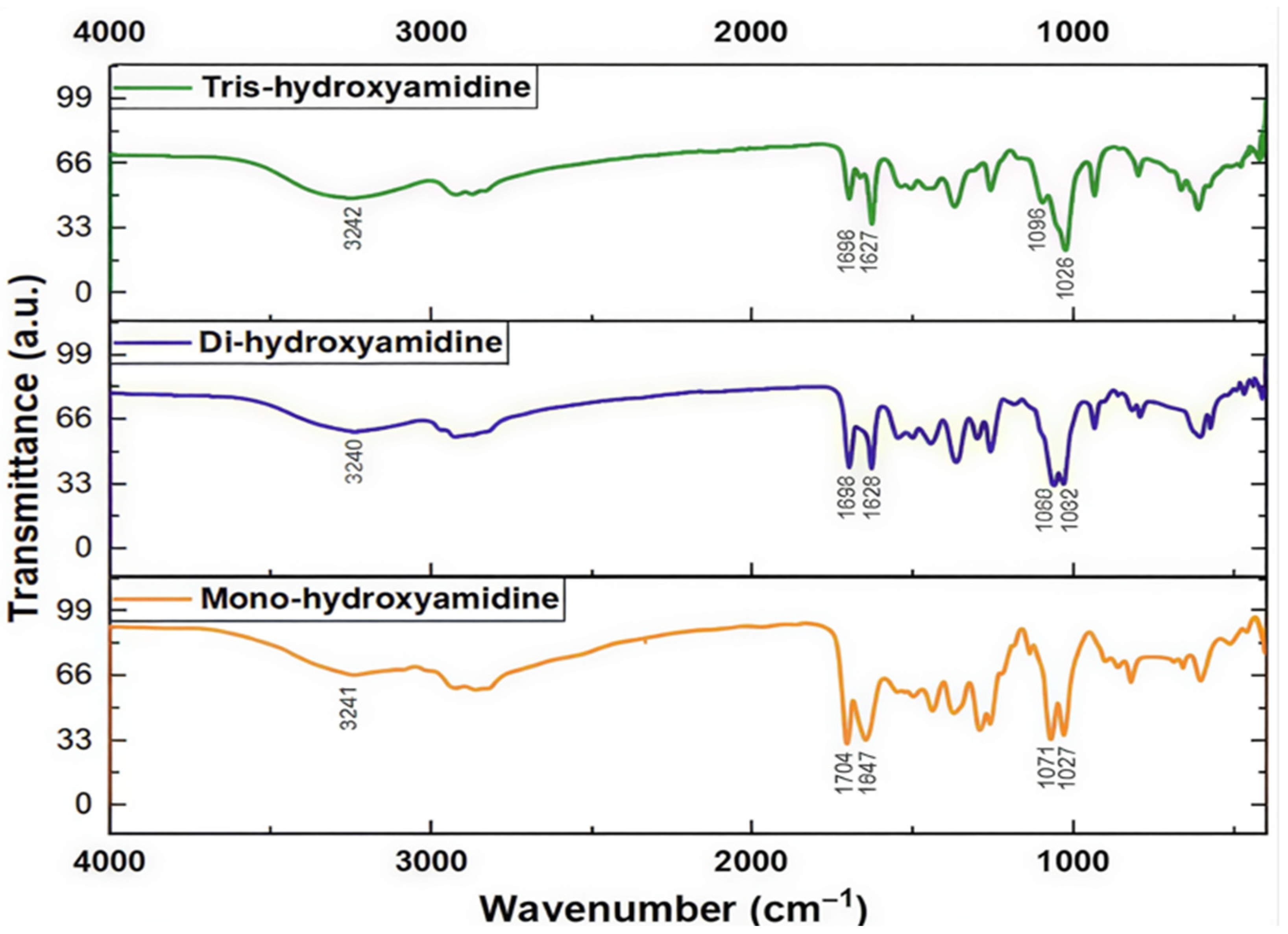
To assign all bands in the IR spectrum of hydroxyamidines, a comparative analysis of the results of quantum chemical calculation of the vibrational absorption spectra of the molecules and the experimental spectra of the MHA, DHA and THA was performed. Figure 2 and Figure 3 show the theoretical spectra (in coordinates absorption (transmittance) coefficient, M−1 × cm−1wave number, or frequency, cm−1) of four molecules and experimental spectra (in coordinates transmission intensity, %—wave number, cm−1) [27] and the present work’s IR spectra of three molecules, respectively.

2.2.1. O-H Bonds

In the experimental IR spectra of mono-, tris- [27] and dihydroxyamidine (this work) compounds, vibration signals are observed in the region of 3300–400 cm−1.
The absorption bands of hydroxyl groups bound by hydrogen bonds usually lie in the region below 3550 cm−1, while free, unassociated ones are in the region of 3580–3650 cm−1. In the theoretically calculated spectra of three hydroxyamidine molecules, weak bands with frequencies in the region of 3619–3650 cm−1 and 3540–3555 cm−1 can be assigned to the stretching vibrations of unassociated OH groups and of intramolecular hydrogen bonded hydroxyl groups in the THA1 molecule, respectively (Table 2).
The absence of such signals in the experimental spectra indicates that there are no free OH groups in the molecules, and all the hydroxyl groups of hydroxyamidines participate in the formation of hydrogen bonds: intra- and/or intermolecular, it is also possible that some of the vibrations in the experimental spectra have low IR intensity. However, in the observed spectra of mono-, di- and tris-hydroxyamidines, bands of medium intensity are observed at 3294, 3319 and 3309 cm−1, respectively, which were attributed to the OH stretching vibration ([27] and this work). As has already been stated above, it is obvious that these are the associated OH groups.

2.2.2. C-H Bonds

The absorption bands νexp 2912 and 2827 cm−1, 2930 cm−1, 2936 and 2828 cm−1 of MHA, DHA and THA, respectively, belong to the stretching vibrations of C-H bonds of methylene and/or methyl, methine groups ([27] and this work).
In the theoretical spectra of the molecules, the absorption region in the range of 3010–2800 cm−1 corresponds to asymmetric and symmetric vibrations of C–H of the indicated groups (Table 2).

2.2.3. C=N Bonds

One of the most intense bands in the experimental IR spectra of mono- (νexp 1644.89 cm−1), di- (ν϶ĸcn 1644.89 cm−1), and tris-hydroxyamidine (νexp 1629.93 cm−1) is due to the stretching vibrations of the C=N bond, which are observed in the theoretical spectra at 1654.70 and 1622.78 cm−1, respectively. In the spectrum of dihydroxyamidine, the corresponding vibrations appear at νtheor 1655.21 cm−1.

2.2.4. C-O Bonds

Asymmetric and symmetric stretching vibrations of C-O bonds appear with very high intensity in the region of νtheor 1025 cm−1 for mono- (νexp 1030 cm−1), νtheor 1019, 1031 cm−1 for di- (ν϶ĸcn 1028 cm−1), and 1028–993 cm−1 (νexp 1025 cm−1) for tris-hydroxyamidine (THA1).
As a rule, deformation vibrations are observed below 1500 cm−1. The vibration frequencies δCOH 1114 cm−1 (mono-) and 1097 cm−1 (tris-hydroxyamidine) [27] correspond to the deformation vibrations of COH and methylene groups in the regions of the mono-, di- and tris-hydroxyamidine theoretical spectra.

2.2.5. Other Vibrations

The vibration of medium intensity is observed at the experimental frequencies νCC 1380.23 cm−1 (mono-), 1381 cm−1 (di-), and 1384.70 cm−1 (tris-), which, however, in the theoretical spectra corresponds to the region of mixed deformation vibrations of groups and the stretching vibration of the C-N bond (1385–1299 cm−1) (Table 2).
The frequency range of 849 (MHA), 930–820 (DHA) and 932–841 cm−1 (THA1) of the calculated spectra of hydroxyamidines corresponds to the stretching vibrations of the N-C(CH3) bonds and the deformation vibrations of the CH2 groups (νexp 924, 871 cm−1 (mono-), 936 cm−1 (di-), and 937 cm−1, 869 cm−1 (tris-)). The last vibration νexp 800 cm−1 is attributed by the authors [27] to the vibration δCNC.
In the calculated spectra, the region of medium frequencies 770 (mono-), 619 (di-) and 734–642 cm−1 (tris-) corresponds to C-C stretching vibrations and deformation vibrations of NCN and OH groups. Below 600 cm−1, mixed scissor vibrations of methylene groups, symmetric and asymmetric deformation vibrations of methyl groups, and skeletal rocking are observed.

2.3. Geometric Architecture of the Associates of Hydroxyamidine with CO2

Known mechanisms of the interaction of amines and amidines with CO2 involve the formation of carbamates or bicarbonates [1,2,3,4,5,6,7,8,9,10,11,12,13,14,15,16,17,18,19,20,21,22,23,24,25,26,27,28,29,30].
The interaction of hydroxyamidine with CO2 is possible in several directions: heteroatoms (nitrogen, oxygen) can be the main reaction centers. As follows from our previous experimental work [27], hydroxyamidines (mono- and tris-), interacting with carbon dioxide, form an ionic liquid (IL), the structure of which was proposed in the form of repeating links RNH+…RCOO. The specified ionic associate of the carbonate type corresponds to protonated imino nitrogen =N- and the deprotonated carbonate group, which can be schematically depicted as follows (using the example of monohydroxyamidine):
2R=N(CH2)2OH + CO2 → RN[(CH2)2]H+…ROCOO (or RNH+…RCOO)
In this paper, we tried to model the IL structure in the form of associates of protonated MHA, DHA and THA1 (MHAH+, DHAH+, THA1H+) with MHA-, DHA- and THA1-carbonate (MHACOO, DHACOO and THA1COO) in different ratios of the initial molecules.
It should be noted that in [27] the capture of carbon dioxide by the oxygen atom of one of the hydroxyl groups of the amidine was assumed. As far as we know, this reaction channel has not been considered by anyone before. Proton transfer is possible in the presence of a proton donor, which can be methanol formed during the synthesis of hydroxyamidine and remaining in the reaction medium. In our recent work [35], methanol was intentionally retained in the system to investigate its influence on CO2 fixation. The presence of methanol was confirmed by characteristic signals in the 1H and 13C NMR spectra.
According to our assumption, the transfer does not occur directly from the carbonate group to the imino nitrogen atom, but by a relay mechanism: first to methanol, then from methanol to the imino nitrogen of the hydroxyamidine. In the present study, methanol is considered within the computational model as an implicit solvent (IEFPCM), to account for its stabilizing hydrogen-bonding effect. No explicit methanol-derived carbonate species were modeled.
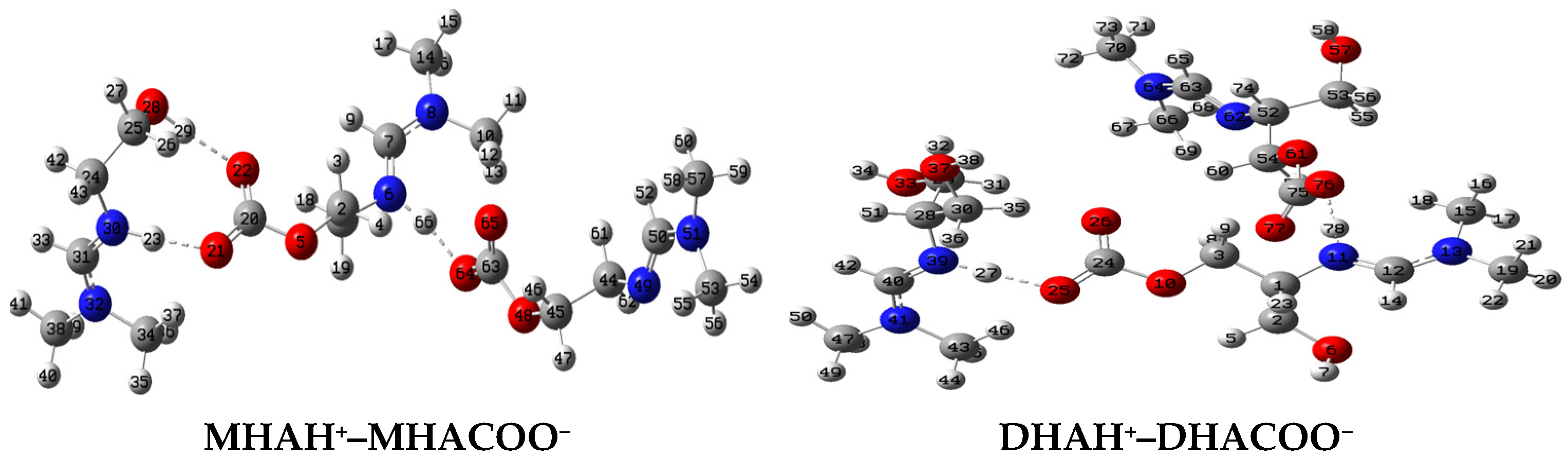
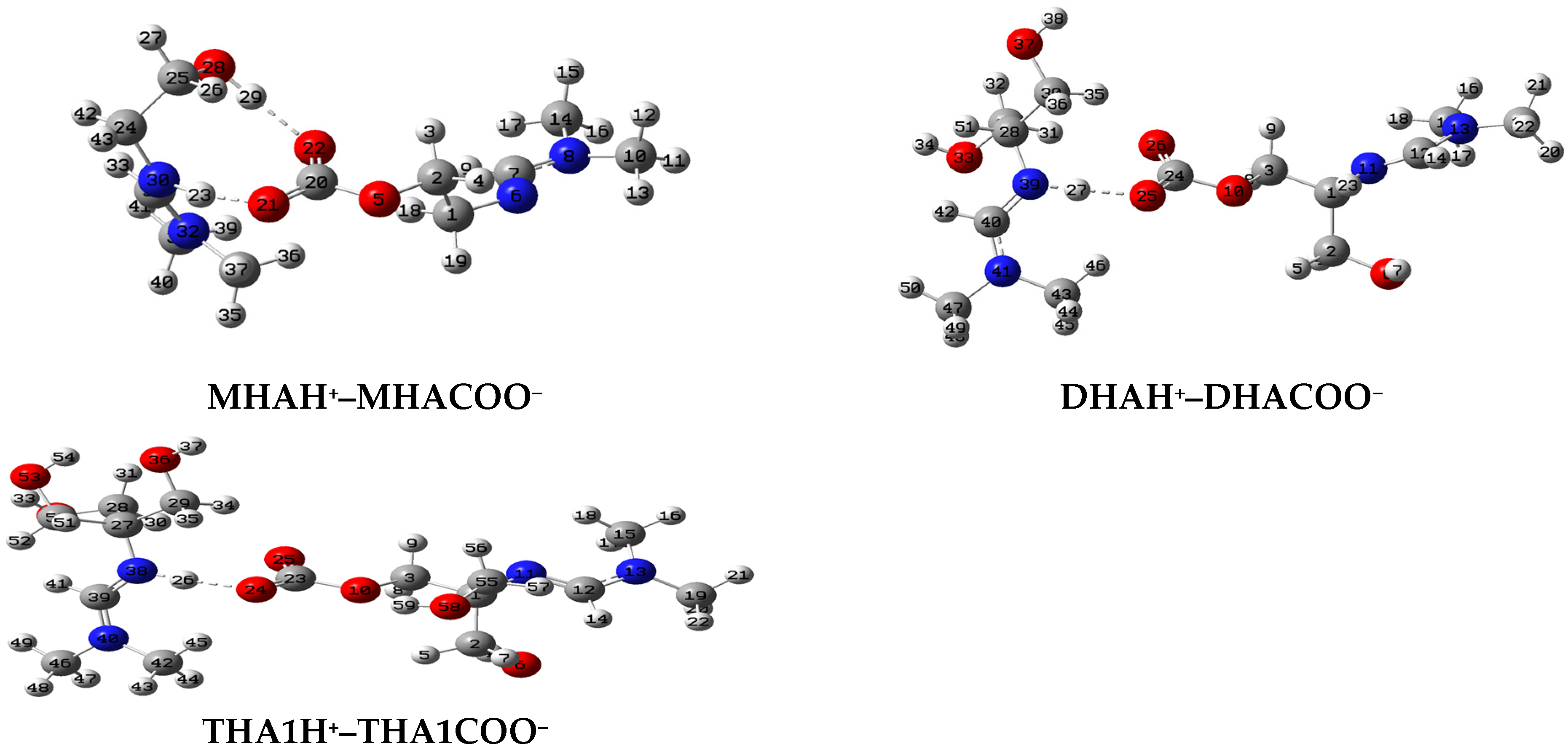
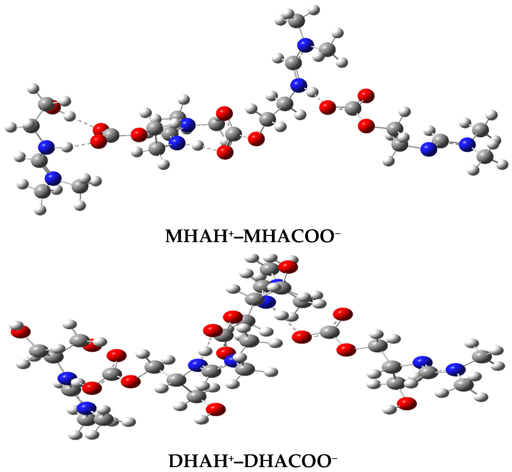
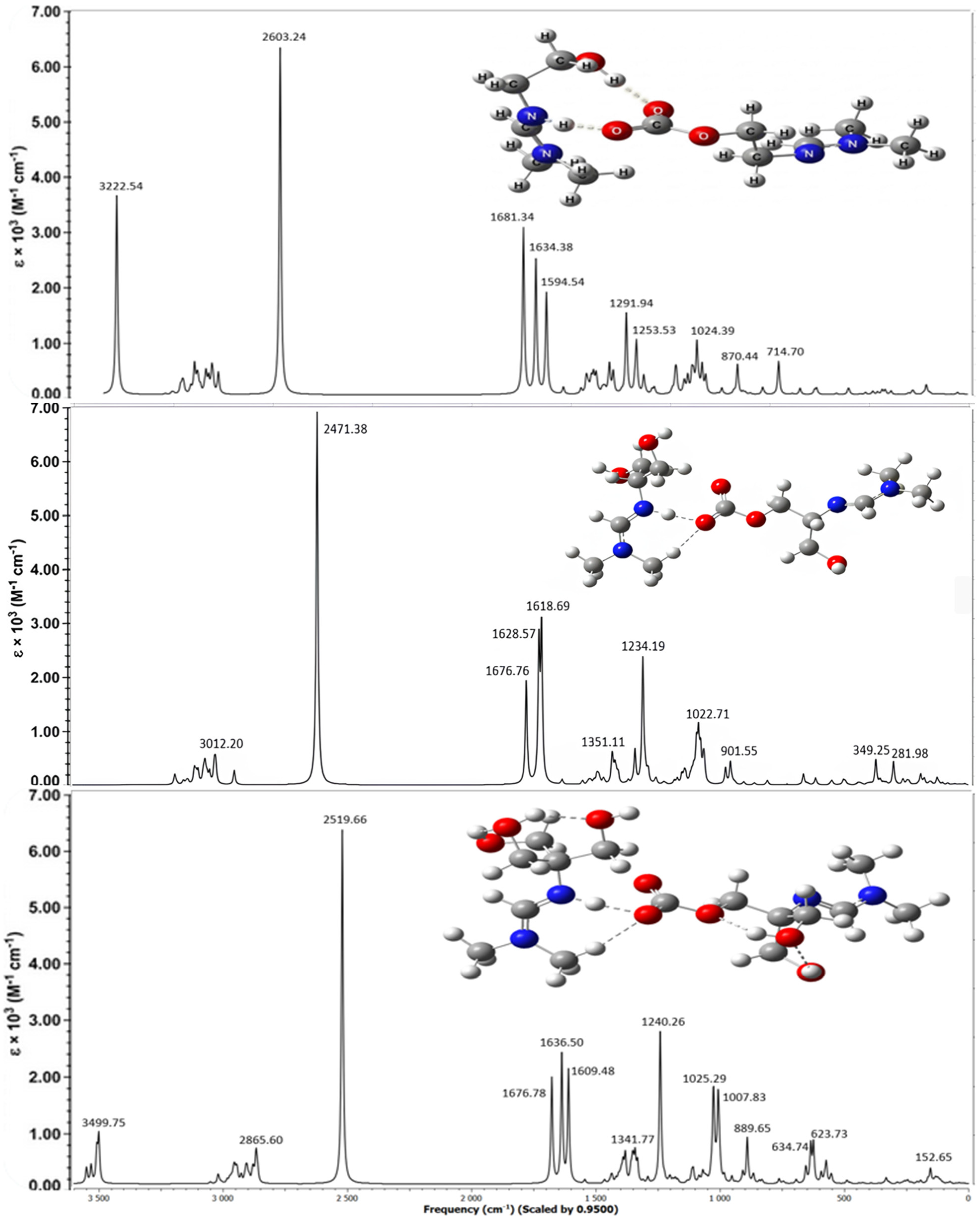
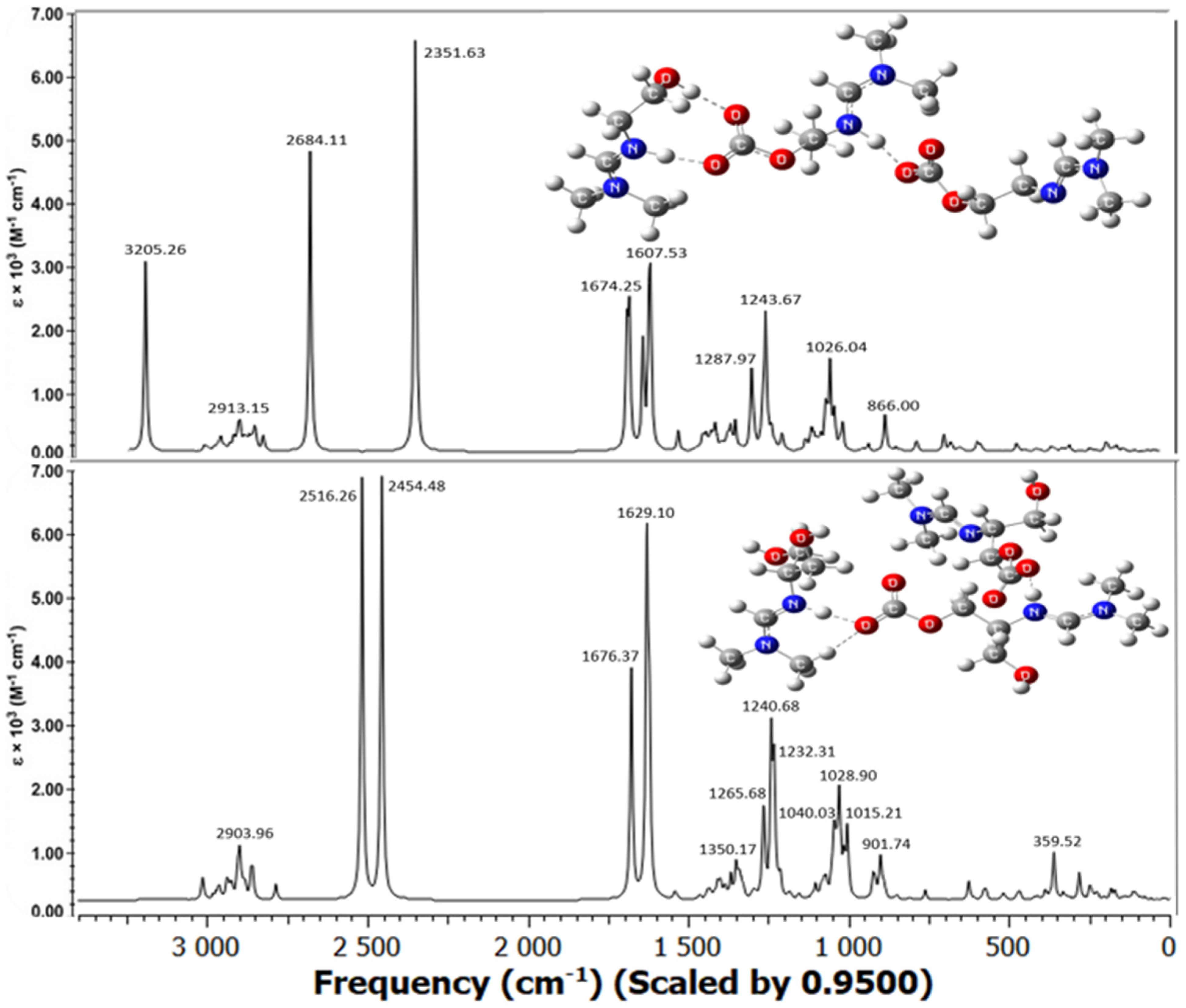
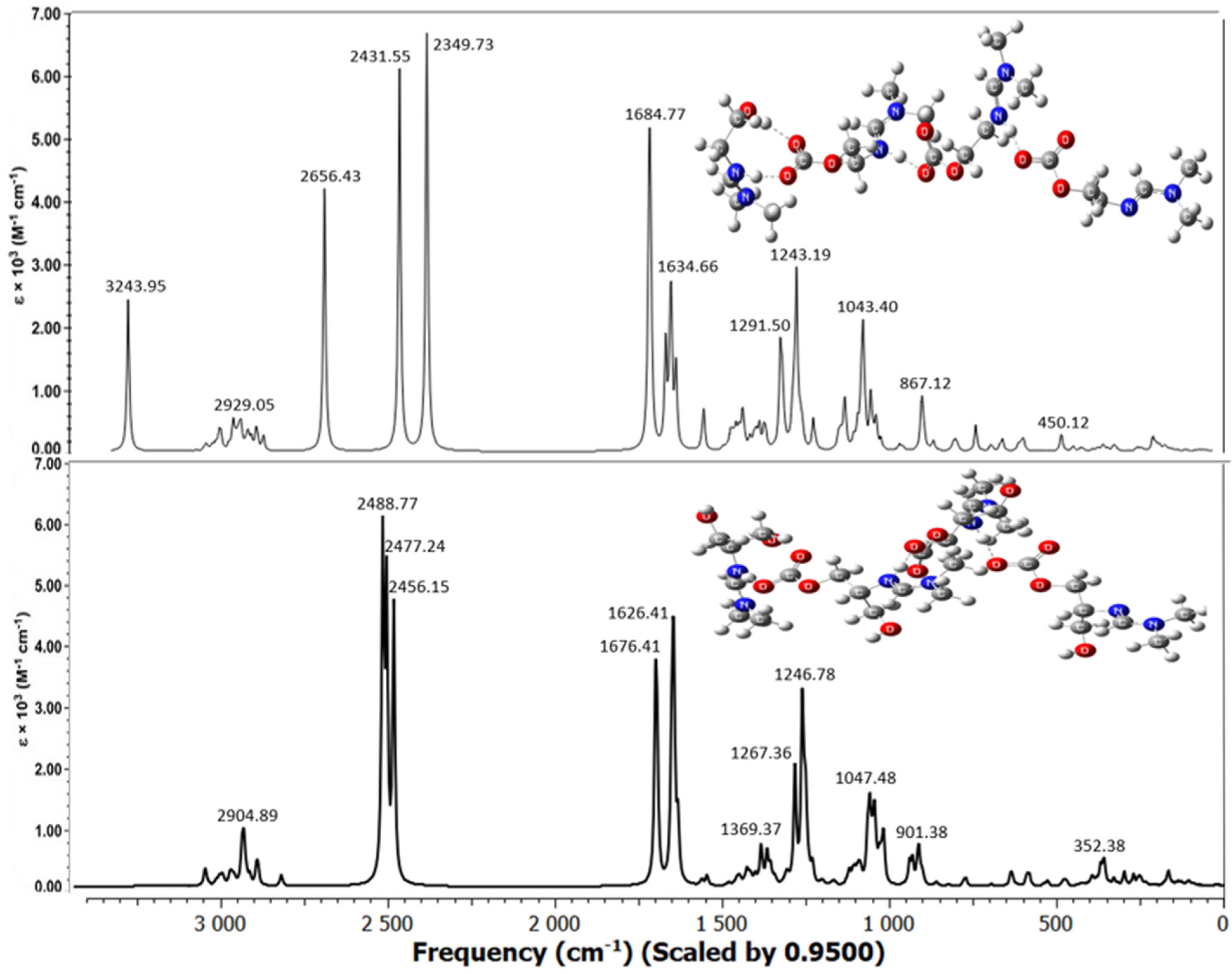
The molecular structures of the associates are shown in Figure 4, Figure 5 and Figure 6. Hydrogen bridges are indicated by dotted lines. The internal fragments of the adducts with more than three molecules are zwitterions (Figure 5 and Figure 6). The total energies and binding energies of the associated systems are given in Supplementary Table S2. As the table shows, with an increase in the number of fragments in the associate, the binding energy, which characterizes the strength of the interaction, increases. As expected, this directly depends on the number of hydrogen bonds formed. The energy is negative, which indicates the exothermic nature of the associate formation process.
Not all of the systems studied contain a true H-bond, i.e., an X-H…Y hydrogen bridge. Most of the bridges have the form X…H…Y, where the hydrogen atom is not chemically bonded to any of the electronegative atoms, in our case, to the nitrogen and oxygen atoms: N…H…O. Table 3 shows the internuclear distances and angles between pairs of N…H and H…O atoms, which indicate the linear geometry of the hydrogen bridges (angles are close to 180°) and the presence of one hydrogen bond with an N-H bond length less than 1.06 Å only in the monohydroxyamidine associates (Table 3 and Figure 4, Figure 5 and Figure 6). As expected, all hydrogen bridges are asymmetric; however, the hydrogen atom is shifted to nitrogen (Table 3).
In the absence of a solvent, i.e., in the gas phase, according to electrostatic forces, the proton should shift toward the more electronegative oxygen atom rather than the less electronegative nitrogen atom. In contrast, the presence of methanol provokes interactions between the hydroxyamidine associate and methanol molecules. Such solvent–solute interactions prevail over the electrostatic factor and lead to the displacement of the proton towards the N atom. It should be noted that the distance between the N and O atoms in the systems corresponds to the length of a true H-bond of average strength. There is also a noticeable tendency for the N…O and O…H distances to be shortened and, on the contrary, for r(N…H) to be lengthened with an increase in the number of molecules in the associate (Table 3). In the MHAH+–MHACOO system, an intermolecular hydrogen bond O-H…O is also formed with lengths O-H 0.987 Å, H…O 1.732 Å (Figure 4), O-H 0.988 Å, H…O 1.727 Å (Figure 5), O-H 0.986 Å, H…O 1.742 Å (Figure 6) and angles of 171–172°.
Analysis of the C=N, C=O, and O-CCO2 bonds showed that the length of the bond formed between the chemisorbed CO2 molecule and the oxygen atom of the hydroxyamidine is in the range of 1.380–1.397 (depending on the amidine type (mono-, di-, tris-), the size of the associate, and the number of sorbed CO2), being shorter than a regular O–C bond due to the influence of electron-rich double C=O bonds. An external influence, such as the formation of a hydrogen bond, increases the distance between the atoms, so the C=O double bonds themselves are stretched compared to the bonds in a free carbon dioxide molecule (RC=O = 1.169 Å). In this case, as expected, the C=O bonds involved in the hydrogen bond experience greater stretching (1.262–1.271 Å), while those not involved are less stretched (1.242–1.255 Å). The carbon–nitrogen double bond is also lengthened by 0.03–0.04 Å compared to the original amidine molecules.
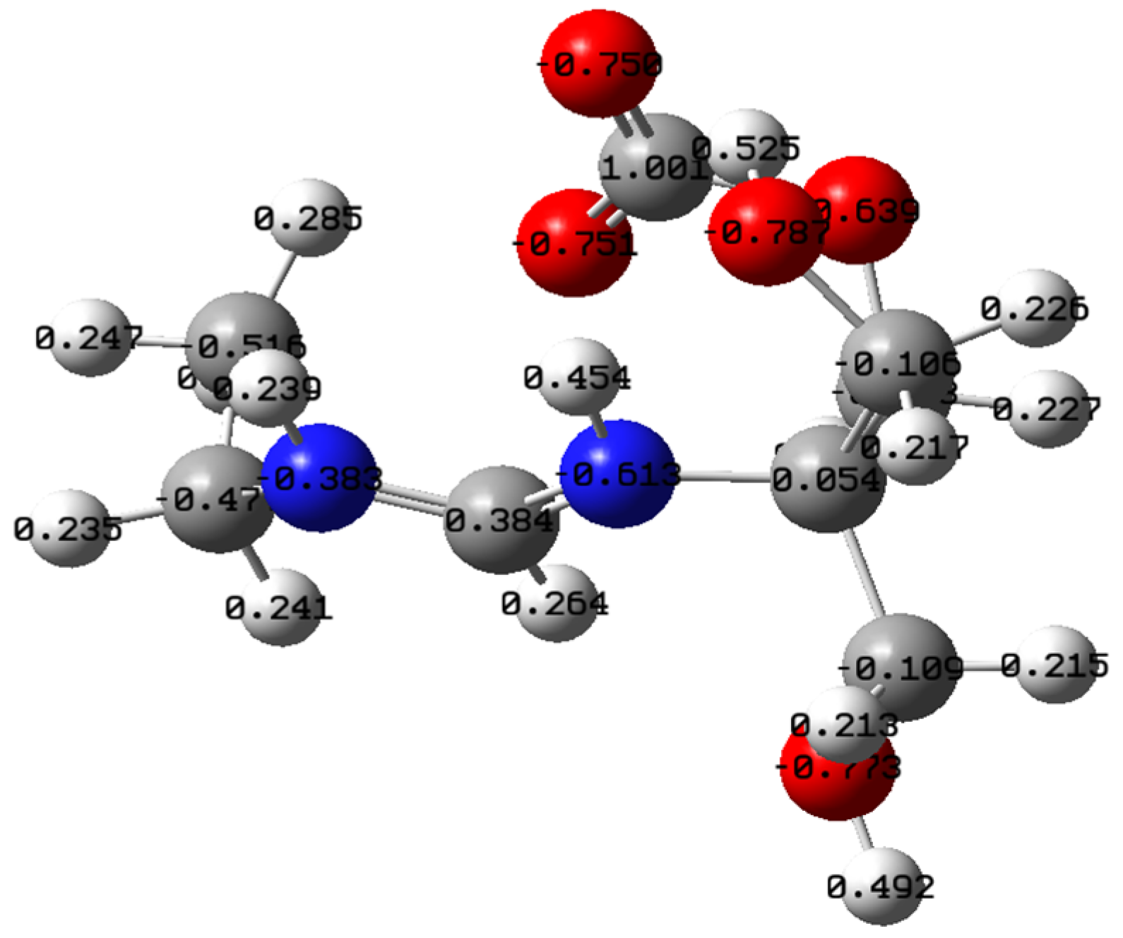
The above-considered conformation of tris-amidine THA2 can also absorb carbon dioxide, for example, as follows (Figure 7). The resulting structure resembles a zwitterion (the model is shown with charges on the atoms according to NPA). The geometric parameters of the THA2–CO2 system are reported in Supplementary Table S4.
As the calculation results showed, the stretching vibrations of the CO2 group in the THA2–CO2 system are observed at νcalc 1709 cm−1, and for the C=N bond at 1643 cm−1. Unlike the intermolecular associates, there are no additional bands here.

2.3.1. NBO/NPA

Natural population analysis (NPA) allowed us to conclude that the formation of a hydrogen bridge causes a significant redistribution of electron density, polarization of charges on the bridge atoms, and a decrease in density on the nitrogen (the charge becomes more positive), i.e., charge transference between fragments of the associate, leading to the structure of an intermolecular ion pair RNH+...RCOO or RN+H…RCOO, which is confirmed by the calculated data in Table 3.
If the isolated fragments have formal −1 and +1 charges, the net charges of each fragment in a hydrogen-bonded associate, for example, MHAH+–MHACOO (2:1) (Figure 4), are, according to NPA, +0.804 and −0.804, respectively (Table 4). A similar charge redistribution is observed for other systems of different stoichiometries (Table 4).
High dipole moments indicate significant delocalization of electron density, as well as the contribution of lone electron pairs. As the number of molecules in the associates increases, the magnitude of the dipole moment increases, but when moving from mono- to di- and tris-hydroxyamidine, the dipole moment decreases, indicating vector compensation.
To assess the degree of electron density delocalization and understand the nature of hydrogen bonding, an NBO analysis of the delocalization energies determined using second-order perturbation theory was performed at the B3LYP/6-31G(d, p) level of theory. As can be seen from Table 5, the highest delocalization energies are observed for the LP(2)O64 → σ*(N6…H66) and LP(2)O87 → σ*(N49…H89) interactions in monohydroxyamidine associates. These are the most intense interactions with energies of 50 kJ/mol between the electron donor and electron acceptor at monohydroxyamidine to CO2 ratios of 3:2 and 4:3.
In the case of dihydroxyamidine, significant electron delocalization, but with lower energy, occurs for the transitions LP(2)O76 → σ*(N11…H78) and LP(2)O76 → σ*(N11…H78). It should be noted that with the appearance of additional hydrogen bonding N…H…O in the systems, subsequent interactions are strengthened. For tris-hydroxyamidine, the electron density is transferred from the lone pair of LP(2)O24 to the adjacent antibonding orbital σ*(H26…N38). Almost twice lower stabilization energies (14–20 kJ/mol) were determined for interactions within O-H…O hydrogen bonds, for example, LP(2)O22 → σ*(O28-H29). All energy values correlate with the H-bonding geometry (Table 3).

2.3.2. Frontier Molecular Orbitals

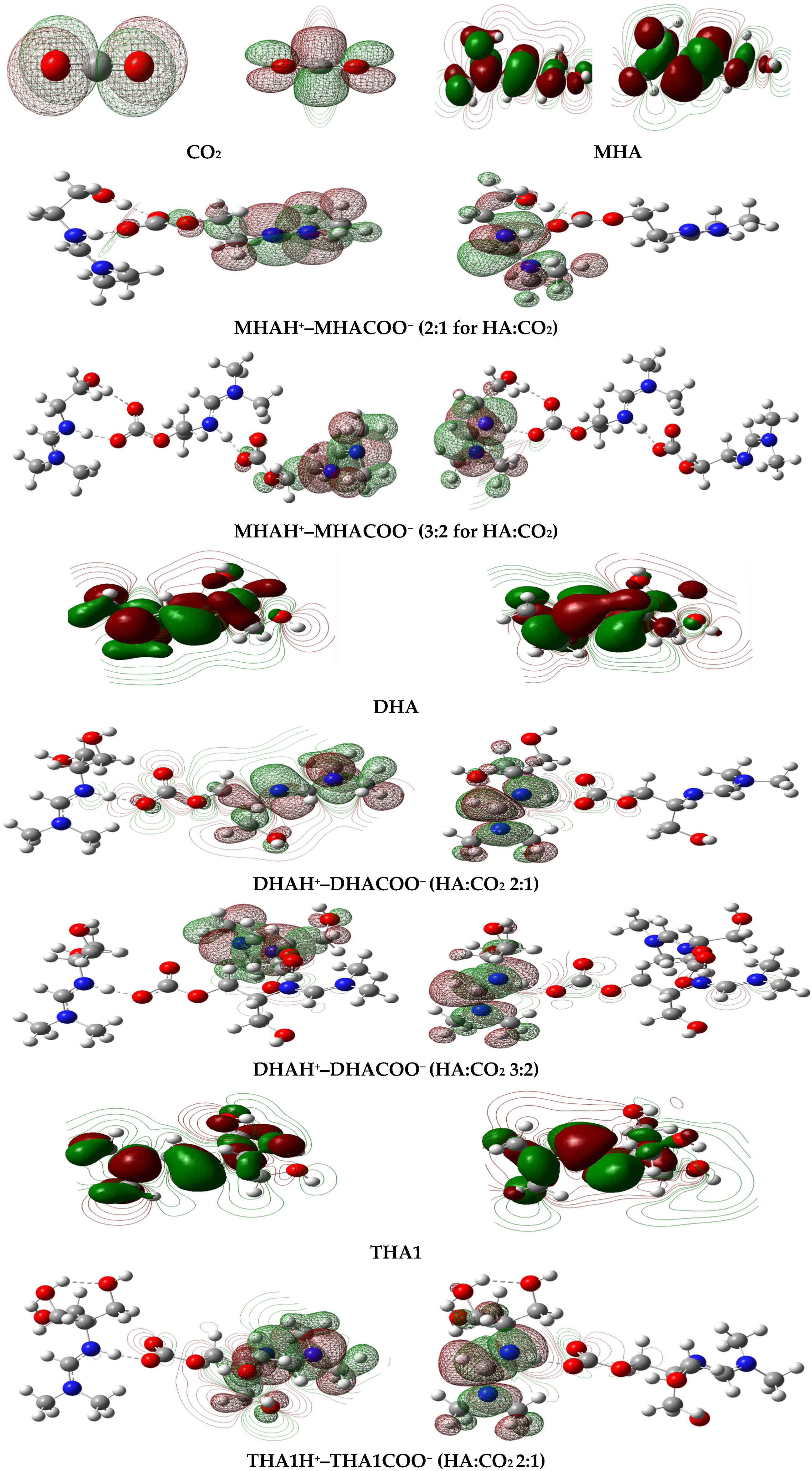
It is known that the indicators of electron transfer in molecular systems are the frontier molecular orbitals HOMO (highest occupied molecular orbital) and LUMO (lowest unoccupied molecular orbital). Generated via Gaussian 16, electron density surface plots for these molecular orbitals of isolated molecules and associates are shown in Figure 8. The HOMO of monohydroxyamidine (MHA) is mainly localized on the pz orbitals of the amino nitrogen, and the LUMO is located on the imino nitrogen atom and the carbon atoms linked to it by a double bond. The HOMO of carbon dioxide is a π-type orbital localized on the oxygen atoms, while the LUMO is π*-characterized and delocalized.
The HOMO and LUMO of the MHAH+–MHACOO complex occupy different regions of the associate, which leads to a significant dipole moment (Table 4). The same is true for di- and tris-hydroxyamidines (Figure 8, Table 4). The calculated energies of the frontier orbitals are given in Supplementary Table S5. The HOMO-LUMO energy gaps of the amidine systems at a HA:CO2 ratio of 2:1 are 5.31 eV for MHAH+–MHACOO, 5.31 eV for DHAH+–DHACOO and 5.17 eV for THA1H+–THA1COO. The smallest energy gap is observed for the tris-hydroxyamidine associate, which favors the formation of the most stable system. A stabilizing factor is the presence of several intra- and intermolecular hydrogen bonds compared to mono- and dihydroxyamidine associates.

2.3.3. Vibrational Analysis of Amidine Systems with CO2

As is known, changes in chemical composition and particle association lead to anisotropy of the electron charge distribution in molecular systems and, consequently, to changes in the dipole moments of bonds and molecules, which, in turn, are associated with molecular vibrations.
Below, the experimental IR spectra of mono-, di-, and tris-hydroxyamidines obtained after treating the latter with gaseous carbon dioxide are compared with the theoretically calculated vibrational spectra of amidine systems with CO2 (in a methanol medium) (Figure 9, Figure 10, Figure 11 and Figure 12).
The absorption spectra of hydroxyamidine molecules chemically bound to carbon dioxide, calculated by the B3LYP-D3/6-31G(d, p) method taking into account dispersion effects and using the integral equation formalism (IEF) of the polarizable continuum model (PCM), are shown in Figure 10, Figure 11 and Figure 12. Within the framework of the density functional and basis set used by us, all calculated vibrational spectra are scaled by 0.95.
Figure 10 shows the spectra of mono-, di-, and tris-hydroxyl-containing amidines at a HA:CO2 ratio of 2:1, while Figure 11 and Figure 12 show the spectra of mono- and dihydroxyl-containing amidines at HA:CO2 ratios of 3:2 and 4:3, respectively. A comparison of experimental and calculated frequencies for hydroxyamidine-carbon dioxide systems showed satisfactory agreement between them. Scaled values of vibrational frequencies and IR band intensities are presented in Table 6.
As noted above, the lack of signals in the region > 3550 cm−1 in the experimental spectra indicates that there are no free OH groups in the IL, and all hydroxyl groups of the MHA, DHA, and THA ILs participate in the formation of hydrogen bonds, both intra- and/or intermolecular ones. An intense broad band is observed in the region of 3240–3242 cm−1 in the experimental spectra of mono-, di-, and tris-hydroxyamidines, which corresponds to the intermolecular O-H…O hydrogen bond formed between the hydroxyl group of the amidine molecule and the COO carbonate group. The theoretical frequency of 3244 cm−1 for the tris-hydroxyamidine associate is closest to these values. The absorption bands νexp 2859 cm−1, 2928 cm−1 and 2927 cm−1 of the MHA, DHA and THA ILs, respectively, belong to the stretching vibrations of the C–H bonds of the methylene and/or methyl groups ([27] and the present work). In the theoretical spectra of the systems, the absorption region in the range of 3012–2860 cm−1 also corresponds to the asymmetric and symmetric vibrations of the C–H bonds of the indicated groups (Table 6).
Some of the most intense bands in the experimental IR spectra of the IL MHA (νexp 1704 and 1647 cm−1), DHA (νexp 1698 and 1628 cm−1) and THA (νexp 1698 and 1627 cm−1) are due to the stretching vibrations of double bonds: C=O and C=N, respectively. In the theoretical spectra, these vibrations appear at 1595, 1677, 1677 (C=O) and 1634, 1629, 1637 (C=N) cm−1 for the MHA, DHA, and THA1 associates, respectively.
It should be noted that our systems simulate the binding of the oxygen of the carbonate group to the hydrogen located near the imino nitrogen. The formation of hydrogen bonding weakens and lengthens the double bonds, and changes the frequency of their vibrations; therefore, the absorption bands are shifted to the low-frequency region.
The calculated spectra also contain a very intense band corresponding to the N+…H vibration (νtheor 2350–2603 cm−1 in the MHA, DHA, and THA1 associates). This can be compared, for example, to the IR spectrum of the zwitterionic form of the amino acid glycine, where N+–H stretching vibrations are observed in the region above 2000 cm−1 [36]. However, the experimental spectra of mono-, di-, and tris-hydroxyamidines do not exhibit a similar band. We assume that the N+–H stretching vibrations (2350–2600 cm−1) do not appear in the experimental spectra because the bond between the nitrogen atom and the proton is not a true chemical bond, i.e., the N+–H bond. As we noted above, the proton is indeed shifted toward the imino nitrogen atom and is quite close (1.05–1.06 Å), but is practically not chemically bound.
Mixed vibrations of single and double carbon–nitrogen bonds are observed in the region of νtheor 1609–1681 cm−1, and those of carbonate C-O bonds are observed in the region of νtheor 1232–1292 cm−1 in mono-, di-, and tris-hydroxyamidine associates. Vibration frequencies below 1050 cm−1 of the theoretical spectra characterize deformation vibrations of OH and methylene groups, and changes in the valence angles δ(OCO), δ(HCH), δ(NCN), δ(COH).
Previously [27] we observed the appearance of a new band between 1700 and 1500 cm−1 in the spectra of mono- and tris-hydroxyamidines treated with CO2 and attributed this vibration to the formation of the amidinium group C=N+. A similar band was also recorded in the spectrum of di-hydroxyamidine (this work, Figure 9). The results of vibrational calculations are consistent with these facts: additional bands in the indicated region appear in the spectra of the MHA, DHA, and THA associates (Figure 10, Figure 11 and Figure 12, Table 6).

2.3.4. Comparison of Chemical Shifts in the Spectra of the Tris-Hydroxyamidine Associate

1H NMR spectra of some tris-hydroxyamidine systems were also calculated in the work. Table 7 presents the experimental and theoretical (B3LYP/6-311+G(2d, p) method) chemical shifts of 1H nuclei for tris-hydroxyamidine, ionic liquid and model systems. The signals of protons at the CN double bond in the calculated spectra of tris-hydroxyamidine and associates are strongly shifted to a weak field compared to the experimental spectra of tris-hydroxyamidine and IL.
Downfield shifts are observed for the signal of OH-group protons in methanol molecules. On the contrary, an upfield shift is manifested for the signal of OH-group protons of hydroxyamidine molecules. In the experimental 1H NMR spectrum of IL [27], an assignment was made for the chemical shift of 5.20 ppm as belonging to the 1H nucleus of the intermolecular hydrogen bond between the oxygen atoms of the CO2 group and the iminonitrogen of the hydroxyamidine. In the calculated systems with CO2, the corresponding signals are extremely strongly shifted to a weak field.
Calculations of amidine molecule systems with bridging CO2 groups in methanol improved the results for the H-bond proton: they showed an upfield shift in the aforementioned signal compared to gas-phase calculations, which better matches the experimental results.

3. Materials and Methods

3.1. Synthesis Method for Di-Hydroxyamidine

Chemical Reagents: The following reagents were used for the synthesis of amidine: 2-amino-2-methyl-1,3-propanediol (C4H11NO2, M = 105.14 g/mol, purity ≥ 99%, Sigma-Aldrich, St. Louis, MO, USA) and N,N-dimethylformamide dimethyl acetal (C5H13NO2, M = 119.16 g/mol, purity ≥ 97%, Thermo Fisher Scientific, Waltham, MA, USA). All reagents were used without further purification.
General information on the synthesis of amidine based on hydroxy-containing amine: amidine with hydroxyl substituents is obtained by reacting the corresponding amino alcohol with N,N-dimethylformamide dimethyl acetal (DFDA). The process is carried out at a 1:1 molar ratio of the reagents using a magnetic stirrer. The reaction is carried out in two stages: initial stirring at 40–50 °C until the starting amino alcohol is completely dissolved (3 h), then raising the temperature to 60 °C, followed by additional stirring for 30 min. Next, the amidine is treated with CO2 gas.
Synthesis of dihydroxyamidine:
2-Amino-2-methyl-1,3-propanediol (5.26 g, 0.05 mol) was gradually (0.3–0.4 g at a time) added to a solution of dimethylformamide dimethyl acetal (6.90 mL, 0.05 mol). After complete dissolution of the reagent, the mixture was maintained under standard synthesis conditions. The target product was isolated in 97% yield.
The synthesis method for mono- and tris-hydroxyamidines was described in [27].
Fourier transform infrared spectroscopy (FTIR) was employed to examine the characteristic functional groups of dihydroxyamidine and PIL. Spectra were recorded using a Nicolet™ iS10 FTIR spectrometer (Thermo Scientific, Waltham, MA, USA) fitted with a diamond attenuated total reflectance (ATR) accessory. Each sample was analyzed directly in ATR mode without additional preparation. The spectra were acquired in the range of 4000–500 cm−1, with a spectral resolution of 4 cm−1, and 32 individual scans were averaged for each run to improve the signal-to-noise ratio. The raw data was processed and interpreted using Omnic 5.2 and OriginPro-2024 softwares. Spectra of mono- (FTIR) and tris-hydroxyamidines (FTIR, 1H NMR and 13C NMR recorded in CDCl3) from our work [27] were used for comparative analysis.
FTIR and NMR analysis coupled with theoretical modeling.

3.2. Computational Details

Quantum chemical calculations of molecular structure and properties of the studied systems were carried out by the density functional theory (DFT) using the closed-shell three-parameter hybrid exchange-correlation functional Becke 3 Lee Yang Parr (B3LYP). The valence-split 6-31G(d, p) basis set augmented by d polarization functions on heavy atoms and p polarization functions on hydrogen atoms [37].
Full geometry optimization was performed for all systems under consideration without imposing symmetry constraints, regardless of the local symmetry of the alkyl groups comprising the systems. The environment was accounted for at the B3LYP/6-31G(d, p) level using the intrinsically independent polarizable continuum model (IEFPCM). As expected, the addition of methanol as a medium lowered the overall energy of the systems. The DFT-D3 method [38] was used to account for dispersion effects during complex formation.
Vibrational frequencies were computed for the optimized structures using the same method in order to assess the nature of stationary points and to obtain spectroscopic characteristics. The resulting structures were identified as stationary points (minima) on the potential energy surface (all vibrational frequencies of the optimized structures are valid). To ensure consistency between the calculated spectra and the experimental ones, a scaling factor of 0.95 was applied, which is within the range of expected values for the density functional and basis set used in our calculations [34].
Natural population analysis (NPA), based on NBO (Natural Bond Orbital) analysis [39,40], was used for a comparative study of the charge distribution over atoms in isolated components and complexes. NPA was chosen for the following reasons: it provides a better description of the electron distribution, especially for systems with an ionic or polar character. NBO analysis was also performed to determine orbital contributions to charge transfer between fragments of the complexes. The electronic state of all systems corresponds to the ground singlet state.
NMR spectra of tris-hydroxyamidine and its associates were calculated at DFT level employing B3LYP/6-311+G(2d, p). All calculations were performed using the Gaussian16 software package [37]. The visualizations of the calculated spectra were done using the GaussView 6.0.16, Chemcraft 1.8, Multiwfn 3.8 programs [41,42,43].

4. Conclusions

Structural, electron-energetic, and spectral data from DFT calculations for mono-, di-, and tris-hydroxyamidines and their associates with CO2 are presented. Optimal minimum-energy conformations of the molecules are identified. Another possible conformation with the probability of forming an intramolecular O-H…N hydrogen bond is discussed for tris-hydroxyamidine. Experimental and theoretical IR absorption spectra correlate with the structure of hydroxyamidines and are consistent with each other. The previously proposed structure of ionic liquids in the form of repeating RNH+…RCOO- units is modeled in this work as associates of protonated mono-, di-, and tris-hydroxyamidines with carbonate counterions in different ratios of the initial molecules. The formation of these units is facilitated by nontrivial sites of CO2 absorption and hydroxyamidine protonation, namely, the oxygen atom of one of the hydroxyl groups of the amidine (CO2 chemisorption) and the imino nitrogen as a proton acceptor. The internal fragments of the associates for molecules greater than three are zwitterions. The present DFT results are in fairly good agreement with the experimental data: they confirm the appearance of a new band between 1700 and 1500 cm−1 in the spectra of model PILs of mono-, di-, and tris-hydroxyamidenes, previously attributed to the formation of the amidinium group C=N+, and support new insight into the formation of the PIL structure.

Supplementary Materials

The following supporting information can be downloaded at: https://www.mdpi.com/article/10.3390/molecules31061055/s1, Table S1: The structural parameters for the equilibrium geometry of hydroxyamidines; Table S2: The total and binding energies of the hydroxyamidine associates; Table S3: The key structural parameters of hydroxyamidine associates; Table S4: The theoretical values of geometric parameters for the THA2–CO2 system; Table S5: The calculated values of the HOMO-LUMO energies for the systems studied.

Author Contributions

Conceptualization, I.I. and N.B.; methodology, I.I., Y.T. and L.A.; software, L.A.; validation, I.I., N.B. and L.A.; formal analysis, Y.T.; investigation, Y.T. and L.A.; resources, I.I. and L.A.; data curation, I.I., L.A. and Y.T.; writing—original draft preparation, L.A.; writing—review and editing, L.A. and I.I.; visualization, Y.T. and L.A.; supervision, I.I. and L.A.; project administration, I.I.; funding acquisition, I.I. All authors have read and agreed to the published version of the manuscript.

Funding

This research was funded by the Science Committee of the Ministry of Science and Higher Education of the Republic of Kazakhstan, grant number AP23488935, project title “Synthesis of Hydroxyl-Containing Amidines for Carbon Dioxide Capture and Use of the Resulting Polymeric Ionic Liquids to Obtain Biodegradable Composites” (Project PI—I. Irgibaeva).

Institutional Review Board Statement

Not applicable.

Informed Consent Statement

Not applicable.

Data Availability Statement

The original contributions presented in the study are included in the article/Supplementary Material; further inquiries can be directed to the corresponding author.

Conflicts of Interest

Author Nikolay Barashkov was employed by the company Micro-Tracers, Inc., San Francisco, CA, USA. The remaining authors declare that the research was conducted in the absence of any commercial or financial relationships that could be construed as a potential conflict of interest.

References

  1. Rathi, A.K.A.; Rathi, J.A. CO2 capture: A concise, comprehensive overview of recent research trends. Acad. Environ. Sci. Sustain. 2025, 2, 1–15. [Google Scholar] [CrossRef]
  2. D’Alessandro, D.M.; Smit, B.; Long, J.R. Carbon dioxide capture: Prospects for new materials. Angew. Chem. Int. Ed. 2010, 49, 6058–6082. [Google Scholar] [CrossRef]
  3. Rubin, E.S.; Mantripragada, H.; Marks, A.; Versteeg, P.; Kitchin, J. The outlook for improved carbon capture technology. Prog. Energy Combust. Sci. 2012, 38, 630–671. [Google Scholar] [CrossRef]
  4. Yu, C.-H.; Huang, C.-H.; Tan, C.-S. A review of CO2 capture by absorption and adsorption. Aerosol Air Qual. Res. 2012, 12, 745–769. [Google Scholar] [CrossRef]
  5. Markewitz, P.; Kuckshinrichs, W.; Leitner, W.; Linssen, J.; Zapp, P.; Bongartz, R.; Schreiber, A.; Muller, T.E. Worldwide innovations in the development of carbon capture technologies and the utilization of CO2. Energy Environ. Sci. 2012, 5, 7281–7305. [Google Scholar] [CrossRef]
  6. Kenarsari, S.D.; Yang, D.; Jiang, G.; Zhang, S.; Wang, J.; Russell, A.G.; Wei, Q.; Fan, M. Review of recent advances in carbon dioxide separation and capture. RSC Adv. 2013, 3, 22739–22773. [Google Scholar] [CrossRef]
  7. Shakerian, F.; Kim, K.-H.; Szulejko, J.E.; Park, J.-W. A comparative review between amines and ammonia as sorptive media for post-combustion CO2 capture. Appl. Energy 2015, 148, 10–22. [Google Scholar] [CrossRef]
  8. Makul, N. Towards Computational CO2 Capture and Storage Models: A Review. Glob. Environ. Eng. 2021, 8, 55–69. [Google Scholar] [CrossRef]
  9. Xie, H.-B.; He, N.; Song, Z.; Chen, J.; Li, X. Theoretical Investigation on the Different Reaction Mechanisms of Aqueous 2-Amino-2-methyl-1-propanol and Monoethanolamine with CO2. Ind. Eng. Chem. Res. 2014, 53, 3363–3372. [Google Scholar] [CrossRef]
  10. Iida, K.; Sato, H. Proton Transfer Step in the Carbon Dioxide Capture by Monoethanol Amine: A Theoretical Study at the Molecular Level. Phys. Chem. B 2012, 116, 2244–2248. [Google Scholar] [CrossRef]
  11. Amirkhosrow, M.; Pérez-Calvo, J.-F.; Gazzani, M.; Mazzotti, M.; Lay, E.N. Rigorous rate-based model for CO2 capture via monoethanolamine-based solutions: Effect of kinetic models, masstransfer, and holdup correlations on prediction accuracy. Sep. Sci. Technol. 2020, 56, 1491–1509. [Google Scholar] [CrossRef]
  12. Dutcher, B.; Fan, M.; Russell, A.G. Amine-based CO2 capture technology development from the beginning of 2013: A review. ACS Appl. Mater. Interfaces 2015, 7, 2137–2148. [Google Scholar] [CrossRef]
  13. Arshad, M.W.; Svendsen, H.F.; Fosbøl, P.L.; von Solms, N.; Thomsen, K. Equilibrium total pressure and CO2 solubility in binary and ternary aqueous solutions of 2-(diethylamino) ethanol (DEEA) and 3-(methylamino) propylamine (MAPA). J. Chem. Eng. Data 2014, 59, 764–774. [Google Scholar] [CrossRef]
  14. Jackson, P. Experimental and theoretical evidence suggests carbamate intermediates play a key role in CO2 sequestration catalysed by sterically hindered amines. Struct. Chem. 2014, 25, 1535–1546. [Google Scholar] [CrossRef]
  15. Matsuzaki, Y.; Yamada, H.; Chowdhury, F.A.; Higashii, T.; Onoda, M. Ab initio study of CO2 capture mechanisms in aqueous monoethanolamine: Reaction pathways for the direct interconversion of carbamate and bicarbonate. J. Phys. Chem. A 2013, 117, 9274–9281. [Google Scholar] [CrossRef]
  16. Bougie, F.; Iliuta, M.C. Sterically hindered amine-based absorbents for the removal of CO2 from gas streams. J. Chem. Eng. Data 2012, 57, 635–669. [Google Scholar] [CrossRef]
  17. Zhu, D.; Fang, M.; Lv, Z.; Wang, Z.; Luo, Z. Selection of blended solvents for CO2 absorption from coal-fired flue gas. Part 1: Monoethanolamine (MEA)-based solvents. Energy Fuels 2012, 26, 147–153. [Google Scholar] [CrossRef]
  18. Mindrup, E.M.; Schneider, W.F. Computational comparison of the reactions of substituted amines with CO2. ChemSusChem 2010, 3, 931–938. [Google Scholar] [CrossRef] [PubMed]
  19. Yamada, H.; Matsuzaki, Y.; Higashii, T.; Kazama, S. Density Functional Theory Study on Carbon Dioxide Absorption into Aqueous Solutions of 2-Amino-2-methyl-1-propanol Using a Continuum Solvation Model. J. Phys. Chem. A 2011, 115, 3079–3086. [Google Scholar] [CrossRef]
  20. Lee, B.; Stowe, H.M.; Lee, K.H.; Hur, N.H.; Hwang, S.-J.; Paek, E.; Hwang, G.S. Understanding CO2 capture mechanisms in aqueous hydrazine via combined NMR and first-principles studies. Phys. Chem. Chem. Phys. 2017, 19, 24067. [Google Scholar] [CrossRef]
  21. McCann, N.; Phan, D.; Wang, X.; Conway, W.; Burns, R.; Attalla, M.; Puxty, G.; Maeder, M. Kinetics and mechanism of carbamate formation from CO2 (aq), carbonate species, and monoethanolamine in aqueous solution. J. Phys. Chem. A 2009, 113, 5022–5029. [Google Scholar] [CrossRef] [PubMed]
  22. Ma, C.; Pietrucci, F.; Andreoni, W. CO2 Capture and Release in Amine Solutions: To What Extent Can Molecular Simulations Help Understand the Trends? Molecules 2023, 28, 6447. [Google Scholar] [CrossRef] [PubMed]
  23. Furusho, Y.; Endo, T. Capture and release of CO2 by polyamidine. J. Polym. Sci. Part A Polym. Chem. 2013, 51, 3404–3411. [Google Scholar] [CrossRef]
  24. Barkakaty, B.; Browning, K.; Sumpter, B.; Uhrig, D.M.; Karpisova, I.; Harman, K.; Ivanov, I.; Hensley, D.; Messman, J.M.; Kilbey, S.M.; et al. Amidinefunctionalized Poly(2-vinyl-4,4-dimethylazlactone) for selective and efficient CO2. Fixing Macromol. 2016, 49, 1523–1531. [Google Scholar] [CrossRef]
  25. Liu, H.; Yin, H.; Feng, Y. A CO2-switchable amidine monomer: Synthesis and characterization. Des. Monomers Polym. 2017, 20, 363–367. [Google Scholar] [CrossRef]
  26. Barashkov, N.; Irgibayeva, I.; Zhapakova, A.; Eralinov, A.; Sakhno, T. FTIR and fluorescent control of interaction between hydroxy-containing amidines and carbon dioxide. In Proceedings of the ACS Meeting, San Diego, CA, USA, 20–24 March 2022; Available online: https://acs.digitellinc.com/acs/sessions/463141/view (accessed on 1 February 2026).
  27. Irgibaeva, I.; Barashkov, N.; Aldongarov, A.; Zhapakova, A.; Eralinov, A.; Sakhno, T.; Seralin, A.; Sakhno, Y. FTIR and NMR spectra of polymeric ionic liquids—Products of reaction between hydroxycontaining amidines and carbon dioxide. J. CO2 Util. 2023, 77, 102594. [Google Scholar] [CrossRef]
  28. Calva, V.; Játiva, N.; Ricaurte, M. CO2 Capture Using Amine-Based Solvents: Identification of Additives to Improve the Kinetics and Thermodynamics of CO2 Sorption at High-Pressure Conditions. Gases 2025, 5, 4. [Google Scholar] [CrossRef]
  29. Zhao, L.; Hu, H.-Y.; Wu, A.-G.; Terent’ev, A.O.; He, L.-N.; Li, H.-R. CO2 capture and in-situ conversion to organic molecules. J. CO2 Util. 2024, 82, 102753. [Google Scholar] [CrossRef]
  30. Nguyen, T.D.; Chen, X.; Sharma, R. Steering amine–CO2 chemistry: A molecular insight into the amino site relationship of carbamate and protonated amine. ACS Omega 2025, 10, 46648–46658. [Google Scholar] [CrossRef]
  31. Delibas, B.; Cotton, D.; Sudhakaran, K.P.; Dawlaty, J.M. Evaluation of Aliphatic Alcohols for CO2 Capture Using the Characteristic Carbonate Frequency. ChemSusChem 2025, 18, e202402288. [Google Scholar] [CrossRef]
  32. Plass, M.; Kolbe, A. Intramolecular Hydrogen Bond Interaction in Selected Diols. Z. Phys. Chem. 2003, 217, 1085–1096. [Google Scholar] [CrossRef]
  33. Göller, A.; Grummt, U.-W. Torsional barriers in biphenyl, 2,2′-bipyridine and 2-phenylpyridine. Chem. Phys. Lett. 2000, 321, 399–405. [Google Scholar] [CrossRef]
  34. Scott, A.P.; Radom, L. Harmonic vibrational frequencies: An evaluation of Hartree−Fock, Møller−Plesset, quadratic con-figuration interaction, density functional theory, and Semiempirical scale factors. J. Phys. Chem. 1996, 100, 16502–16513. [Google Scholar] [CrossRef]
  35. Irgibaeva, I.; Barashkov, N.; Tarikhov, F.; Aldongarov, A.; Salkeeva, L.; Dzhardimalieva, G.; Tashenov, Y. Methanol-Assisted CO2 Fixation by Hydroxyl-Containing Amidine Leading to Polymeric Ionic Liquid and Cross-Linked Network Formation. Polymers 2025, 17, 3306. [Google Scholar] [CrossRef] [PubMed]
  36. Pretsch, E.; Buhlmann, P.; Affolter, C. Structure Determination of Organic Compounds, 3rd ed.; Springer: Berlin, Germany, 2000; p. 309. [Google Scholar]
  37. Frisch, M.J.; Trucks, G.W.; Schlegel, H.B.; Scuseria, G.E.; Robb, M.A.; Cheeseman, J.R.; Scalmani, G.; Barone, V.; Petersson, G.A.; Nakatsuji, H.; et al. Gaussian 16, Revision A.03; Gaussian Inc.: Wallingford, CT, USA, 2016. Available online: https://gaussian.com/ (accessed on 10 January 2026).
  38. Grimme, S.; Hansen, A.; Brandenburg, J.G.; Bannwarth, C. Dispersion-Corrected Mean-Field Electronic Structure Methods. Chem. Rev. 2016, 116, 5105–5154. [Google Scholar] [CrossRef] [PubMed]
  39. Reed, A.E.; Curtiss, L.A.; Weinhold, F. Intermolecular interactions from a natural bond orbital, donor-acceptor view-point. Chem. Rev. 1988, 88, 899–926. [Google Scholar] [CrossRef]
  40. Foster, J.P.; Weinhold, F. Natural hybrid orbitals. J. Am. Chem. Soc. 1980, 102, 7211–7218. [Google Scholar] [CrossRef]
  41. Dennington, R.D., II; Keith, T.A.; Millam, J.M. GaussView, Version 6.0; Gaussian Inc.: Wallingford, CT, USA, 2016. Available online: https://gaussian.com/gaussview6/ (accessed on 28 December 2025).
  42. Chemcraft—Graphical Software for Visualization of Quantum Chemistry Computations, Version 1.8, build 682. Available online: https://www.chemcraftprog.com/ (accessed on 15 January 2026).
  43. Lu, T. A comprehensive electron wavefunction analysis toolbox for chemists, Multiwfn. J. Chem. Phys. 2024, 161, 082503. [Google Scholar] [CrossRef]
Figure 1. Geometric structures of mono-, di- and tris-hydroxyamidine molecules in the ground electronic state according to the B3LYP/6-31G(d, p) method (atom numbering according to the GaussView program).
Molecules 31 01055 g001
Figure 2. Calculated IR spectra of hydroxyamidine molecules (B3LYP/6-31G(d, p) method).
Molecules 31 01055 g002
Figure 3. Experimental IR spectra of hydroxyamidines ([27] and this work).
Molecules 31 01055 g003
Figure 4. Optimized molecular systems in stoichiometry 2:1 (HA:CO2).
Molecules 31 01055 g004
Figure 5. Optimized molecular systems in stoichiometry 3:2 (HA:CO2).
Molecules 31 01055 g005
Figure 6. Optimized molecular systems in stoichiometry 4:3 (HA:CO2).
Molecules 31 01055 g006
Figure 7. THA2–CO2 structure after optimization by B3LYP/6-31G(d, p) method. Etot = −800.105334 a.u.
Molecules 31 01055 g007
Figure 8. HOMO (left) and LUMO (right) of isolated molecules and the mono-, di- and tris-hydroxyamidine associates.
Molecules 31 01055 g008
Figure 9. Observed IR spectra of hydroxyamidines treated with CO2 gas ([27] and the present work).
Molecules 31 01055 g009
Figure 10. Simulated vibrational spectra of mono-, di-, and tris-hydroxyamidine associates at a H A:CO2 ratio of 2:1.
Molecules 31 01055 g010
Figure 11. Calculated vibrational spectra of mono-, di-hydroxyamidine associates at a HA:CO2 ratio of 3:2.
Molecules 31 01055 g011
Figure 12. Simulated vibrational spectra of mono-, di-hydroxyamidine associates at a HA:CO2 ratio of 4:3.
Molecules 31 01055 g012
Table 1. Total electronic energies of amidine molecules (B3LYP/6-31G(d, p) method).
Molecule−Et, a.u.E0, a.u.−(Et + Eo), a.u.
MHA382.4723340.175994382.296340
DHA497.0341460.209152496.824994
THA1611.5352040.243583611.291621
THA2611.5300280.243116611.286912
Table 2. Observed and calculated (B3LYP/6-31G(d, p) method) scaled frequencies (ν) and intensities (I) of stretching and mid-frequency vibrations in mono-, di- and tris-hydroxyamidine molecules.
Vibration TypeMonohydroxyamidineDihydroxyamidineTris-Hydroxyamidine
Calc.
(ν),
cm–1
Exp. [27] (ν),
cm–1
I
(calc.)
Calc.
(ν),
cm–1
Exp.
[This Work] (ν),
cm–1
I
(calc.)
Calc.
(ν),
cm–1
Exp. [27]
(ν),
cm–1
I
(calc.)
ν(O-H) *36193294133643
3619
331918
11
3650
3555
3544
(3471) **
330931
77
109
144
νas,s(CH3, CH2, CH)
(out of phase, in phase)
2970
2941
2927
2913
2868
2862
2856
2853
2843
2809
2912
2827
22
45
35
42
46
76
89
49
74
75
2972
2951
2933
2918
2898
2888
2881
2858
2845
2786
293024
31
38
44
45
60
53
85
71
57
2975
2955
2938
2934
2921
2909
2881
2863
2858
(2856)
2848
2936
2828
16
30
34
8
36
52
42
53
128
113
69
ν(C=N)16551645515165516295621623
(1630)
1629
(53) ***
573
569
δ(CH3), δ(CH2)1471–14071438
1406
8
14
1470–140814388
16
1472–1406-7
24
ν(C-N), ρ(CH), δ(OH),
δ(CH3), w(CH2), τ(CH2), δ(COH)
1374
1337
1325
1266
1226
1380
1345
(76)
1264
(78)
1114
(69)
30
62
78
28
38
1374
1344
1338
1292
1232
1184
1118
1094
1381
1259
17
22
83
27
34
43
23
34
1380
1361
1347
(1350)
1338
1299
1231
1166
1101
1384
(76)
1096
(66)
106
64
117
107
19
6
24
19
77
ν(C(CH3)-N), δ(CCC), ρ(CH), skeleton swing10751063
(66)
761063
1041
1037
109673
9
10
1065
1049
(1059)
1036
-31
69
127
6
νas,s(C-O), skeleton swing10251030
(41)
1621031
1019
1004
1028101
83
59
1028
1013
993
(992)
1025
(38)
145
118
92
103
νs(C-N), δ(HCH), ρ(CH2), τ(CH), δ(OH),
skeleton swing
984
937
849
924
(83)
871
(78)
18
8
930
889
858
820
9362
22
3
4
983
932
859
841
937
(71)
869
(78)
6
13
5
14
ρ(OH), νs(C-C), ρ(OH), δ(NCN), skeleton swing770-2619
518
794
630
573
4
22
734
660
642
(680)
800
(74)
4
77
152
143
* ν—stretch vibration, νas—asymmetric stretch vibration, νs—symmetric stretch vibration, ν(C-C)—stretch vibrations of the carbon skeleton; deformation vibrations: δ—change in the valence angle; ρ—pendulum; w—fan; τ—torsional. ** Vibrational mode in THA2. *** Vibration intensity in the experimental spectrum.
Table 3. Hydrogen bond geometry (B3LYP/6-31G(d, p) method) in MHA, DHA, and THA1 associates (Figure 4, Figure 5 and Figure 6).
Parameterr(N…O), År(N…H), År(O…H), Åφ(N… H…O), °
HA:CO2MHAH+–MHACOO
2:12.663 a1.0611.614168.94
3:22.7021.0551.648175.88
(2.619) b(1.079)(1.556)(167.02)
4:32.6741.0571.630168.46
(2.619(1.073(1.562(166.98
2.598)1.079)1.539)165.50)
DHAH+–DHACOO
2:12.6621.0691.594176.85
3:22.6691.0661.604176.43
(2.659)(1.070)(1.590)(175.82)
4:32.6461.0681.580176.59
(2.646(1.068(1.579(175.26
2.636)1.071)1.581)167.01)
THA1H+–THA1COO
2:12.6541.0651.592174.33
a Internuclear distances N…O, N…H, and O…H, and the N…H…O angle for the first hydrogen bridge in the associates. b Internuclear distances and angle for other hydrogen bridges in the associates.
Table 4. Electronic charges of hydrogen bond atoms (B3LYP/6-31G(d, p), NBO method) in mono-, di- and tris-hydroxyamidine associates.
Natural Atomic Chargeµ, D
NHOHAH+HACOO
HA:CO2MHAH+–MHACOO
-−0.5820.447−0.774+1−12.463 a
(16.866)
2:1−0.5810.466−0.7730.804−0.8048.918
3:2−0.570
(−0.570) b
0.470
(0.475)
−0.786
(−0.788)
0.838−0.83815.163
4:3−0.580
(−0.569, −0.568)
0.466
(0.474,
0.474)
−0.766
(−0.774,
−0.792)
0.841−0.84123.429
DHAH+–DHACOO
-−0.5840.455−0.807+1−15.878
(21.229)
2:1−0.5780.473−0.8090.836−0.8368.497
3:2−0.578
(−0.571)
0.473
(0.473)
−0.808
(−0.807)
0.837−0.83711.963
4:3−0.580
(−0.574,
−0.574)
0.471
(0.471,
0.472)
−0.802
(−0.800,
−0.786)
0.839−0.83912.639
THA1H+–THA1COO
-−0.5920.456−0.785+1−17.981
(17.558)
2:1−0.5880.470−0.7990.845−0.8456.418
a Dipole moments of the HAH+ and HACOO systems, respectively. b Charges on analogous atoms.
Table 5. Delocalization energies of mono-, di-, and tris-hydroxyamidine associates at the B3LYP/6-31G(d, p) level of theory.
HA:CO2DelocalizationEnergy, kJ/mol
MHAH+
MHACOO
DHAH+
DHACOO
THA1H+
THA1COO
2:1LP(2)O21 → σ*(H23…N30)37.52--
LP(2)O22 → σ*(O28-H29)15.44--
LP(2)O25 → σ*(H27…N39)-42.19-
LP(2)O24 → σ*(H26…N38)--42.45
3:2LP(2)O21 → σ*(H23-N30)33.08--
LP(2)O22 → σ*(O28-H29)19.57--
LP(2)O64 → σ*(N6…H66)49.87--
LP(2)O25 → σ*(H27…N39)-40.04-
LP(2)O76 → σ*(N11…H78)-44.23-
4:3LP(2)O21 → σ*(H23-N30)35.74--
LP(2)O22 → σ*(O28-H29)14.24--
LP(2)O64 → σ*(N6…H66)47.96--
LP(2)O87 → σ*(N49…H89)49.80--
LP(2)O25 → σ*(H27…N39)-41.08-
LP(2)O76 → σ*(N11…H78)-45.10-
LP(2)O103 → σ*(N62…H105)-42.08-
Table 6. Experimental and theoretical (B3LYP/6-31G(d, p) method) scaled frequencies (ν) and intensities (I) of stretching and mid-frequency vibrations in the MHA, DHA, and THA systems with CO2 at different HA:CO2 ratios.
Type of Vibrations MHA-CO2DHA-CO2THA1-CO2
Calc.
(ν), cm–1
Exp. [27]
(ν), cm–1
I
(calc.),
km/mol
Calc. (ν),
cm–1
Exp. [Present Work]
(ν), cm–1
I
(calc.), km/mol
Calc. (ν),
cm–1
Exp. [27] (ν), cm–1I
(calc.), km/mol
ν(O-H) *3223
(3205 **,
3244)
32411114
1345
1072
-3240-35003242323
νas,s(CH3, CH2) (out of phase, in phase)2862
(2861,
2929)
2859121
129
100
2862
(2861,
2905)
2928149
151
72
28662927144
ν(N…H)2603
(2352,
2350)
-1941
2903
2944
2471
(2454,
2477)
-2726
2694
2497
2520-2487
νas(CO2)1595
(1608,
1621)
1704565
1014
721
1619
(1621,
1626)
16981087
1028
1180
16771698741
ν(C=N), ν(C-N)1681
(1674,
1685)
1647930
916
1155
1629
(1629,
1676)
1628981
1021
1034
1637
1609
1627899
789
Mid frequencies
δ(COH), τ(CH2),
δ(HCH)
1416
(1415,
1415)
1497
1439
97
90
104
1541
(1542,
1529)
1544
1500
1444
36
38
90
1544
1437
1505
1440
27
27
ρ(CH), w(CH2)1355
(1354,
1354)
1370159
159
165
1351
(1350,
1369)
1364155
172
271
13421371184
ν(C(O2)-O), w(CH2), skeleton swing1292
1254
(1288
1244,
1292
1243)
1290
1259
453
284
545
919
647
1182
1264
1234
(1241
1232,
1247
1236)
1298
1258
248
921
994
784
1443
652
124012581022
ν(C(H2)-O),
δ(COH), ρ(CH2), skeleton swing
1024
(1040,
1043)
1137
1071
276
574
595
1023
(1029,
1047)
1060
1032
330
540
290
1025
1007
1096
1026
429
514
δ(OCO), ρ(CH2), δ(HCH), δ(NCN), skeleton swing870
(866,
867)
865
822
169
212
247
902
(902,
901)
935
793
163
191
173
890935
799
322
* ν—stretch vibration, νas—asymmetric stretch vibration, νs—symmetric stretch vibration, ν(C-C)—stretch vibrations of the carbon skeleton; deformation vibrations: δ—change in valence angle; ρ—pendulum vibration; w—fan vibration; τ—torsional vibration. ** Frequencies at other HA:CO2 ratios.
Table 7. Experimental and calculated (B3LYP/6-311+G(2d, p) method) chemical shifts of 1H nuclei in some tris-hydroxyamidine systems.
SystemChemical Shift δ, ppm
1H(CH3)1H(CH2)1H(OH)1H(C=N)1H(N)
Exp. [27]Calc.Exp.
[27]
Calc.Exp. [27]Exp.Exp.
[27]
Calc.Exp. [27]Calc.
THA13.602.013.073.451.260.316.968.81--
3.622.064.193.58 2.66
3.672.724.203.67 3.14
3.702.73 4.02
3.733.07 4.04
4.23 4.82
PIL3.28-3.48-4.06-6.86-5.20-
THA1H+- THA1COO 1.92 3.01 2.64 8.45 15.11
2.123.263.038.75
2.343.433.77
2.493.495.90
2.703.526.47
2.813.71
3.003.80
3.014.29
3.0174.53
3.024.71
4.315.05
5.515.13
THA1H+- THA1COO with implicit methanol solvation 2.18 3.12 2.67 8.28 11.37
2.333.382.778.70
2.633.513.77
2.793.716.09
2.823.7296.92
2.963.731
3.043.82
3.184.15
3.224.50
3.274.64
3.754.66
4.534.94
Disclaimer/Publisher’s Note: The statements, opinions and data contained in all publications are solely those of the individual author(s) and contributor(s) and not of MDPI and/or the editor(s). MDPI and/or the editor(s) disclaim responsibility for any injury to people or property resulting from any ideas, methods, instructions or products referred to in the content.