Article

Influence of Pr Content on Structural Evolution of Doped Ceria-Based High-Entropy Oxides

1
Department of Chemistry, Josip Juraj Strossmayer University of Osijek, Cara Hadrijana 8/A, HR-31000 Osijek, Croatia
2
Materials Research Laboratory, University of Nova Gorica, Vipavska 13, SI-5000 Nova Gorica, Slovenia
3
Department of Applied and Environmental Chemistry, University of Szeged, Rerrich Béla Sq. 1, H-6720 Szeged, Hungary
*
Author to whom correspondence should be addressed.
Molecules 2026, 31(4), 598; https://doi.org/10.3390/molecules31040598

Submission received: 14 January 2026 / Revised: 29 January 2026 / Accepted: 1 February 2026 / Published: 9 February 2026

Abstract

High-entropy fluorite oxides offer exceptional tunability of structure and functionality through controlled multi-cation substitution. In this work, Ce-Zr-Pr-Sm-Eu-based high-entropy oxides, with systematically varied Pr content, were synthesized using a modified sol–gel citrate method to investigate the influence of Pr incorporation on lattice structure, defect formation, and photocatalytic performance. All compositions crystallized in a single-phase cubic fluorite structure, where increasing Pr concentration induced gradual lattice expansion and microstrain due to the substitution of larger Pr3+ ions. Morphological and surface analyses revealed porous nanostructures at moderate Pr levels, while excessive Pr promoted densification and reduced surface accessibility. Spectroscopic studies confirmed the coexistence of Pr3+/Pr4+ and Ce3+/Ce4+ redox couples, strong 4f–2p orbital hybridization, and enhanced defect-related electronic states that narrowed the optical bandgap. The optimized Pr-doped composition exhibited almost 100% degradation of methylene blue under UV light over 30 min, untypical for semiconductors with a narrower bandgap, and is enabled by efficient charge separation and redox cycling between Ce and Pr centers.

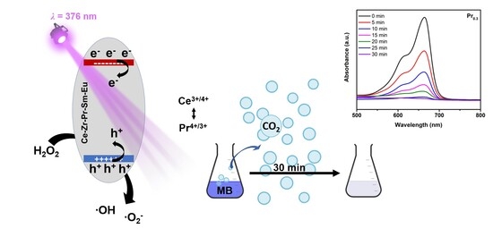
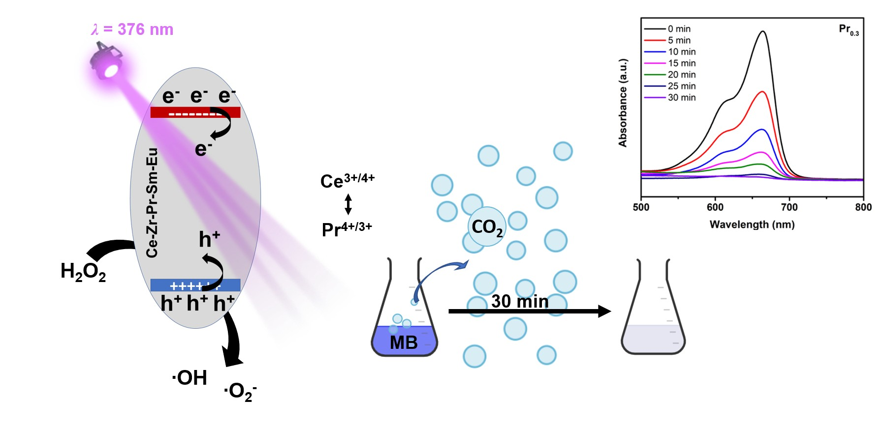
Graphical Abstract

1. Introduction

Advanced functional materials, defined by their multi-component compositions, represent a rapidly developing field in material science. They are well known as high-entropy materials defined by their multicomponent composition, typically incorporating five or more cations in near-equimolar ratios within a single crystallographic position [1,2,3]. Their stability is primarily regulated by a large configurational entropy, which reduces the Gibbs free energy and suppresses phase segregation at higher temperatures [4]. This concept, initially adapted from high-entropy alloys to oxide systems by Rost et al. [5], in 2015 enabled the formation of single-phase solid solutions from typically incompatible binary oxides through entropy-driven stabilization [6]. The intrinsic structural disorder resulting from random cation distribution in high entropy oxides gives rise to properties unattainable in conventional ordered oxides [7,8]. Higher configuration entropy, combined with lattice distortion and charge compensation, produces complex defect chemistry that directly influences ionic transport, redox activity, and thermal stability [9,10]. These disorder-driven effects grant high-entropy oxides enhanced tolerance to thermal cycling and superior oxygen mobility as compared to pure oxides, making them promising materials for different applications [11,12,13,14]. Among them, fluorite-type oxides, typically derived from CeO2, ZrO2, or mixed rare-earth oxides exhibits outstanding chemical and thermal stability. Their ability to accommodate large cationic size and valence mismatches without structural collapse enables a disordered but coherent oxygen sublattice, which supports defect mediated oxygen transport with tunable redox behavior [15,16].
Cerium oxide serves as an ideal structural foundation for high-entropy design due to its cubic fluorite type lattice and exceptional redox flexibility [17]. The reversible Ce4+/Ce3+ redox couple enables oxygen storage and release under changing oxygen partial pressures [18]. When Ce4+ is partially reduced to Ce3+, charge neutrality is maintained through oxygen vacancy formation, which controls oxygen migration and surface reactivity [19]. Such vacancies improve ionic conductivity, catalytic performance, and oxygen storage capacity, all of which can be further tuned through controlled doping strategies. Aliovalent doping with rare-earth cations, such as Sm3+, Gd3+, Nd3+, Y3+, introduces charge compensating oxygen vacancies and lattice distortion, reducing defect formation energy and enhancing ion mobility [20]. The high substitutional tolerance of CeO2 and its ability to maintain the crystal structure even under severe redox cycling make it ideal “host” for entropy-stabilized solid solutions [15]. Among various dopants, Pr4+/3+ is known as particularly effective dopant, since its substitution generates oxygen vacancies [21]. At moderate dopant concentrations, Pr lower vacancy formation energies and increase surface reducibility, due to mixed valence state enables redox cycling and enhances oxygen storage capacity [22]. However, excessive doping often leads to enhanced cation mobility and sintering, resulting in grain coalescence and reduced surface area [23].
Despite progress in understanding the defect chemistry of rare-earth doped ceria, the relationship between dopant composition, oxygen non-stoichiometry, and surface textural evolution, particularly within high-entropy frameworks, remains insufficiently understood [14,24,25,26]. While most research has focused on the electronic and catalytic properties of rare-earth doped ceria, comprehensive insights into how these dopants influence surface area, porosity, and sintering kinetics are still lacking [27,28,29,30]. Notably, the unique redox flexibility of Pr4+/3+ introduces fundamentally distinct pathways for oxygen retention, vacancy clustering, and structural compaction. In response, this study seeks to elucidate the interconnected effects of Pr doping on the structural, surface, and compositional characteristics of CeO2-based high-entropy oxides. Analyzed compounds were systematically synthesized and tested, with varying Pr content and thoroughly analyzed to establish quantitative correlations between dopant-induced lattice distortion, oxygen vacancy formation, and surface changes, further identifying optimal dopant levels that maximize active sites while minimizing densification. These findings advance the understanding of the defect–structure–property relationship in high-entropy oxides and their suitability for advanced applications. Furthermore, the synthesized compounds were evaluated as photocatalysts for methylene blue degradation to provide deeper insights into the structure–property–application relationship.
Photocatalysis emerged as a powerful advanced oxidation process for environmental applications, enabling the degradation of persistent organic pollutants through light-driven redox reaction on semiconductor surface [31]. Upon irradiation with photons possessing energy equal or greater than the bandgap of the semiconductor, electron-hole pairs are generated, initiating a cascade of surface reactions that leads to the formation of highly reactive oxygen species, which are capable of mineralizing organic contaminants [32]. Among various organic dyes, methylene blue is widely employed as a model pollutant in photocatalytic research, due to its well defined molecular structure, strong visible-light adsorption, and chemical stability [33]. Recent studies demonstrated that defect engineering, introduction of oxygen vacancies and mixed valence cationic states are factors that can precisely tune the properties of semiconductor material to enhance photocatalytic activity [34,35]. Oxygen vacancies introduce localized electronic states within a bandgap, which facilitates charge trapping and transfer processes, simultaneously alongside promoting surface adsorption and activation of molecular oxygen [35]. In ceria-based photocatalysts, the main advantage is the reversible Ce3+/4+ redox pair, which enables efficient oxygen storage and release as well as enhanced ROS generation under irradiation. When combined with additional redox-active dopants, synergistic effects can arise through coupled redox cycling and defect stabilization [36].

2. Results and Discussion

2.1. Structural Characterization of the Material

2.1.1. Powder X-Ray Diffraction

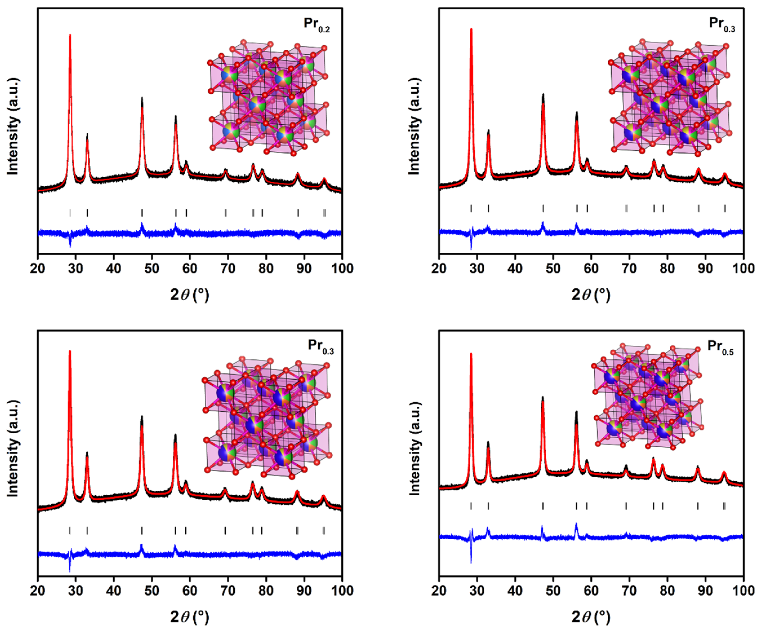
PXRD combined with Rietveld refinement (Figure 1) confirmed that all Pr-substituted high-entropy oxides crystallize in a single-phase cubic fluorite structure (Fm–3m), as evidenced by the excellent fitting quality (Rwp = 10–17%, GoF < 1.8) and absence of any secondary impurities. The refinement results are summarized in Table S1.
The lattice parameters increase slightly from a = 5.4205 Å for Pr0.2 to a = 5.4376 Å for Pr0.5, accompanied by a monotonic expansion of the unit cell volume from 159.26 to 160.78 Å3. This systematic lattice dilation indicates the successful incorporation of Pr ions into the cationic sublattice and correlates with the larger ionic radius of Pr3+ (1.126 Å) compared to Ce4+ (0.97 Å) and Zr4+ (0.84 Å). The trend follows Vegard’s law, suggesting the formation of a homogeneous solid solution without phase segregation. The average microstrain and average apparent crystallite size derived from peak broadening reveal a nanoscale size (10–12 nm) and moderate lattice strain (0.28–0.35%). The strain reflects local distortions induced by multivalent Pr species (Pr3+/4+) and the resulting formation of oxygen vacancies. These intrinsic defects and local charge imbalances contribute to static lattice disorder, a structural feature typical for high-entropy fluorite oxides. Such disorder increases with configurational complexity and enhances the defect tolerance of the lattice, stabilizing the cubic fluorite structure even at higher Pr concentrations. The gradual lattice expansion and strain modulation with increasing Pr content suggests competition between the substitutional incorporation of larger Pr3+ ions and the formation of charge-compensating oxygen vacancies.

2.1.2. Scanning Electron Microscopy

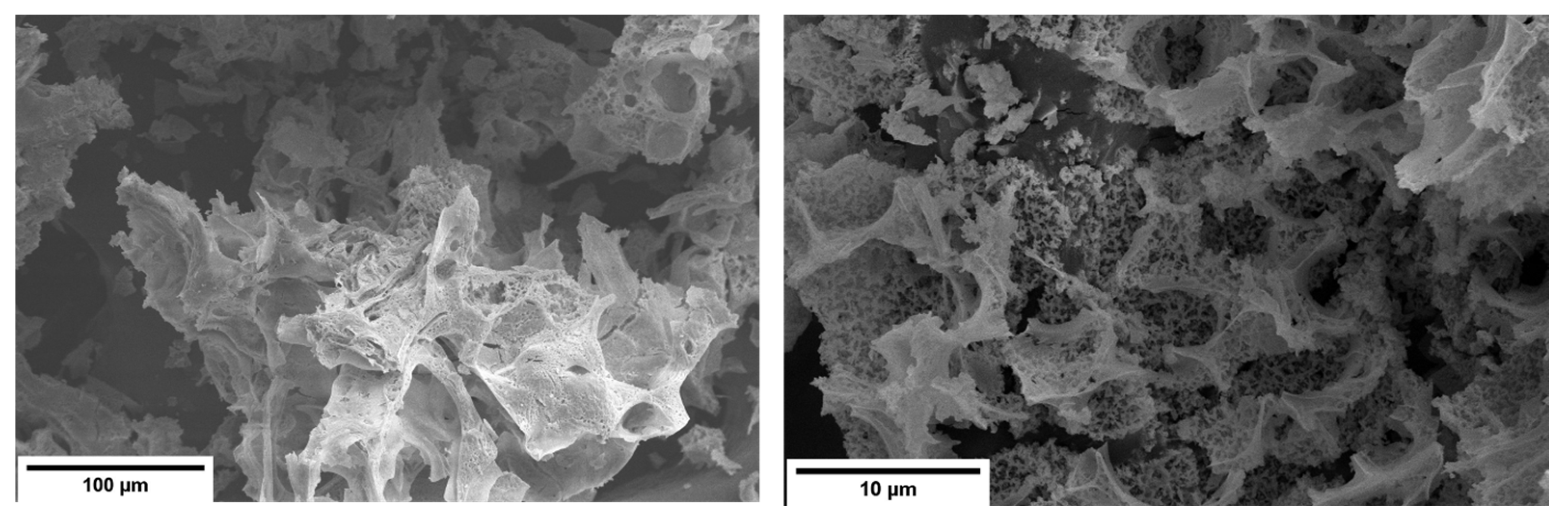
The SEM images reveal porous, sponge or sheet-like microstructure, consisting of interconnected, irregular agglomerates and open macropores, as seen in Figure 2. Such morphology is characteristic of materials obtained using sol–gel citrate-nitrate methods, where the decomposition of organic precursors and the evolution of gaseous species during heating and calcination generates a lightweight, foam like oxide network. At higher magnifications, the samples display rough, fragmented surfaces composed of thin sheet-like structures and open intergranular cavities. This morphology indicates incomplete densification and limited particle coalescence, even after calcination, which can be attributed to the presence of different mixed-valence species and oxygen-vacancy-induced lattice distortions. These defects reduce cation diffusivity and impede grain boundary mobility, thereby stabilizing a fine and highly porous microstructure [37]. The hierarchical porosity and rough surface topology are expected to increase the accessible surface area and enhance oxygen transport within the oxide matrix [38,39].

2.1.3. Physisorption Analysis

Introduction of trivalent rare earth cations into the CeO2 fluorite type lattice typically influences the materials structural, textural and redox characteristics. In our research, Pr possesses larger ionic radius than Ce4+, which induces local lattice distortions and generates compensating oxygen vacancies. These structural changes increase defect-related surface activity, directly enhancing adsorption–desorption performance, which is responsible for the catalytic properties of the materials. Experimental results from the present work confirm that all synthesized compounds exhibit type IV nitrogen adsorption–desorption isotherms with H3 hysteresis loops [40], classifying them as mesoporous materials with slit-like pore geometries (Figure S1). Measured specific surface areas of the Pr-doped compounds range between 25.9 m2g−1 for Pr0.5 and 33.8 m2g−1 for Pr0.2, in contrast to 21.5 m2g−1 for pure CeO2, as shown in Table S2. The enhancement at lower Pr molar concentration can be attributed to the formation of additional surface defects (e.g., oxygen vacancies) and disordered domains, thereby increasing the density of surface-active sites. This aligns with prior studies on Pr-doped ceria, which reported that moderate Pr incorporation promotes oxygen mobility and surface redox activity without significant structural collapse [41]. However, a higher dopant concentration usually induces sintering and particle agglomeration during calcination, reducing accessible surface area and pore volume. The enhanced diffusivity of Pr3+ ions at higher concentration is known to facilitate early-stage sintering by grain boundary diffusion, consistent with observations from Shajahan et al. [23]. The reduction in pore size at higher dopant concentration, particularly Pr0.5 is also consistent with well-known behavior of rare-earth modified systems, where the dopant cations occupy both bulk and near-surface sites, reducing the interconnectivity of the pores. The densification effect suppresses mesopore formation, shifting pore distribution towards smaller diameters and limiting gas diffusion through solid matrix [42].

2.2. Spectroscopic Characterization of the Material

2.2.1. Energy Dispersive X-Ray Spectroscopy

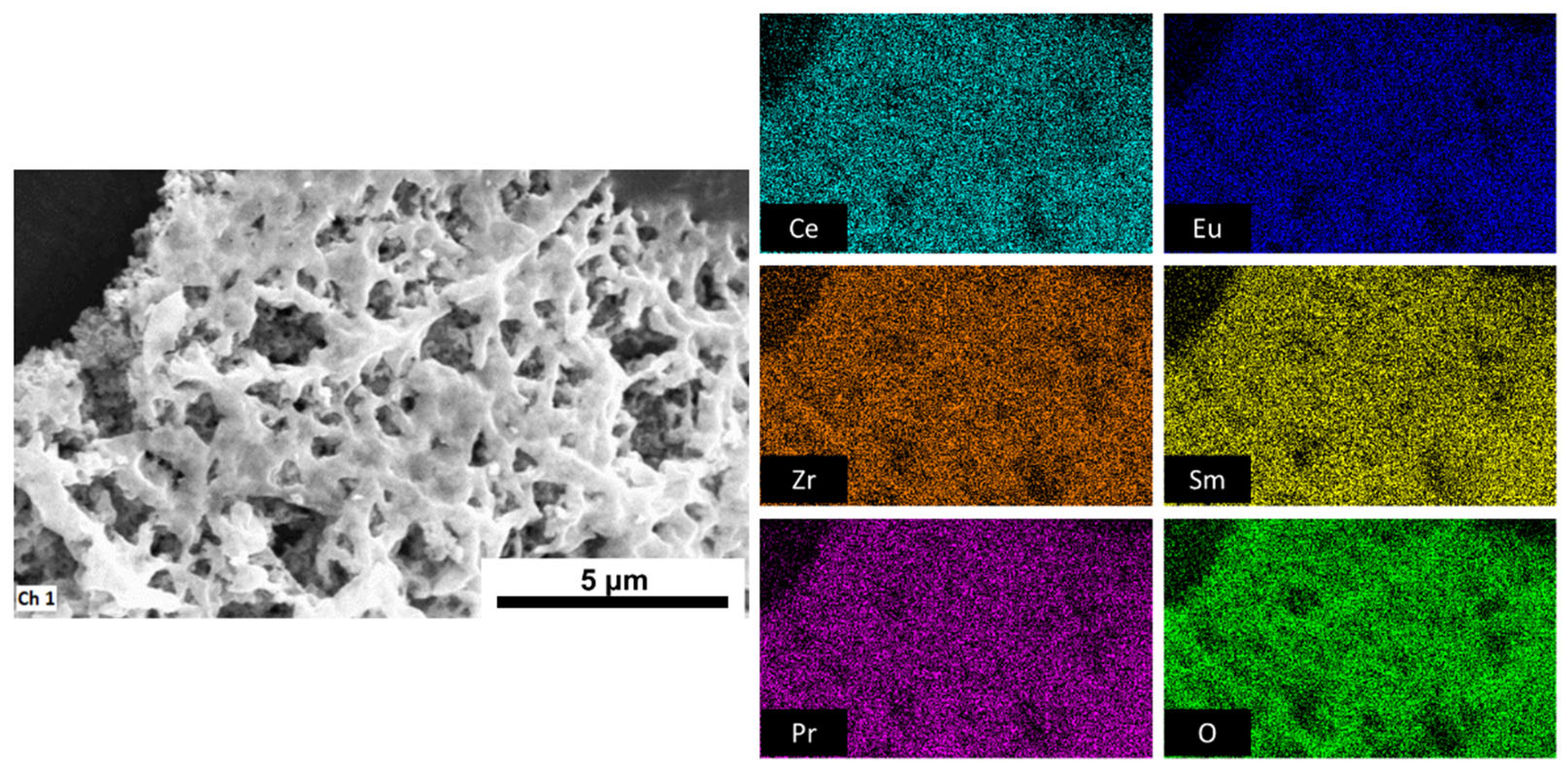
EDS confirmed the successful incorporation of all intended cations (Ce, Zr, Sm, Eu, and Pr) into a single-phase structure without any evidence of elemental segregation or phase separation. The elemental mapping demonstrated a homogeneous distribution of all constituents across the analyzed regions (Figure 3), indicating that the compounds exhibit compositional homogeneity characteristics of HEOs. Quantitative EDS results from Table S3 revealed that experimentally determined cation ratios closely matched their theoretical stoichiometries, with only minor deviations, which are due to energy overlap, and additionally confirmed uniform dopant incorporation within the ceria lattice. The small discrepancies in oxygen content are consistent with the presence of oxygen vacancies formed to compensate for charge introduced by trivalent cations substituting for Ce4+ [43]. Compounds with (nearly) equimolar ratios (0.2–0.3) showed nearly ideal cation ratios and slightly oxygen-deficient composition, suggesting well-balanced defect chemistry and efficient charge neutrality. At higher dopant content (0.4–0.5), the oxygen stoichiometry deviated further, possibly due to enhanced vacancy formation and potential defect clustering [26]. Although trivalent dopant substitution in ceria generates oxygen vacancies, the experimental EDS data reveal a slight increase in oxygen stoichiometry at higher Pr fractions. This deviation arises from a combination of physical and analytical effects. At higher Pr concentrations, oxygen vacancies tend to associate with dopant cations, forming defect clusters that effectively saturate vacancy sites and suppress further vacancy generation [26,44]. Simultaneously, lattice distortion at higher Pr content enhances Ce3+ oxidation back to Ce4+ and promotes oxygen re-absorption from the ambient atmosphere, which produces an apparent oxygen enrichment [24,45]. Even though EDS analysis tends to overestimate oxygen due to matrix effect and incomplete corrections for X-ray absorption [46], which means the observed oxygen stoichiometries >2.00 do not indicate true over-stoichiometry but rather deflect clustering and partial reoxidation phenomena, which is consistent with defect saturation behavior reported in similar systems. EDS sum spectra are shown in Figure S2.

2.2.2. Raman Spectroscopy

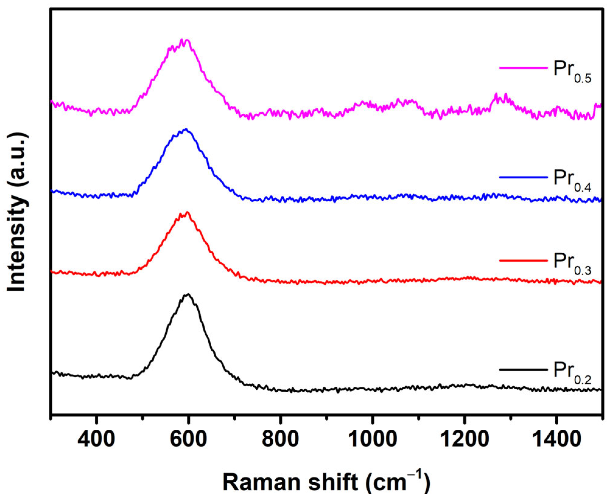
When discussing structural evolution, Raman spectroscopy is a powerful tool for comparative analysis. Typically, cubic CeO2 possesses one active, F2g, Raman mode, which significantly decreases with the introduction of defects in crystal lattice, while simultaneously, a new defect band appears at approx. 600 cm−1 [47]. In ceria-based high-entropy oxides, specifically in nanocrystalline form with increased defects, F2g almost disappears, which is also visible in Figure 4 for Pr0.2 compound, where slight maximum is visible in the range of 420–460 cm−1, but this feature lowers at higher Pr concentrations. The disappearance of strong attenuation of characteristic F2g band of the fluorite structure can be attributed to extensive defect formation and local symmetry breaking induced by Pr substitution. In Pr-doped ceria, the introduction of Pr3+/Pr4+ mixed valence cations lead to a higher concentration of oxygen vacancies. As the concentration of defects increases, a broad, defect-related band develops around 550–600 cm−1, often overlapping and effectively masking the F2g feature [39]. In addition, resonance enhancement of defect-related modes involving Pr4+ near 530 nm excitation significantly amplify the defect band relative to F2g, leading to its apparent disappearance [48]. Total apparent absence of F2g band at Pr0.4 and Pr0.5 can be explained by transformation of F2g band into a broad background feature, which is characteristic of disordered or reduced Ce-Pr-Zr-O lattices [49]. This absence typically reflects the dominance of defect related vibrations and stronger local disorder, rather than the loss of the fluorite framework. The emergence of broad and weak Raman band between 1000 and 1600 cm−1 in the Pr0.4 and Pr0.5 compounds can be attributed to second order and vibronic processes associated with the strong defect–electron–phonon coupling in highly disordered lattices [50]. In defective ceria-based oxides, the principal defect-induced modes give rise to higher-frequency Raman features in the range 1000–1300 cm−1 [51]. Loridant [50] demonstrated that in redox active doped ceria, particularly systems with Tb or Pr, these higher frequency bands are intensified by the strong vibronic coupling between localized 4f localized electronic states, and lattice phonons, resulting from 4f–2p charge–transfer transitions. The appearance of these bands signifies the dominance of localized electronic transitions and small polaron like excitations, rather than simple lattice vibrations, consistent with previous reports [49]. Consequently, this region above 1000 cm−1 in Pr0.4 and Pr0.5 represents a spectroscopic fingerprint of extensive electronic-lattice coupling and short range structural disorder arising from the increased Pr content and oxygen vacancy concentration.

2.2.3. UV–VIS Spectroscopy

Diffuse reflectance UV–Vis spectroscopy of synthesized compounds reveals direct bandgap energies in the narrow range of 2.63–2.66 eV, as determined from Tauc plot extrapolation (Figure S3). This bandgap is notably smaller than that of pure CeO2, additionally confirming the possibility to alter bandgap value by different dopants. As shown by Poggio-Fraccari et al. [29], Pr substitution lowers the oxygen vacancy formation energy and enhances the reducibility of Ce-Pr oxides, directly modifying their electronic density of states. Similarly, Xu et al. [38] demonstrated that Pr3+ ions substitute Ce4+ in the fluorite lattice, producing charge-compensating vacancies and intermediate 4f defect levels that extend absorption into the visible region. Furthermore, the coexistence of mixed oxidation states, such as Pr3+/4+ and Ce3+/4+, give rise to multiple optical transitions, including O 2p to Ce 4f, O 2p to Pr 4f and Pr3+ to Pr4+ charge transfers excitations [38,52,53]. These overlapping electronic transitions usually create sub-bandgap absorption tails and can appear experimentally as an apparently single, but narrowed band gap. Similar effects have been observed in high-entropy fluorite oxides containing Pr, where strong 4f–2p hybridization and oxygen-vacancy disorder progressively reduced the optical gap from 3.15 eV to 1.87 eV [53].

2.2.4. X-Ray Photoelectron Spectroscopy

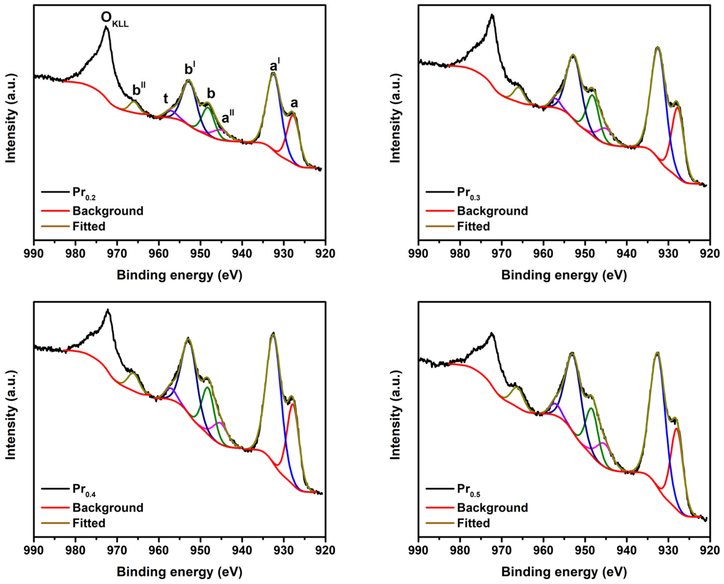
To investigate the surface chemical composition and oxidation states of the elements present in the Pr-substituted high-entropy fluorite oxides, XPS measurements were performed. The survey spectra confirm the presence of Ce, Zr, Pr, Sm, Eu, and O, indicating the successful incorporation of all cations into the surface region (Figure S4). The C 1s peak at 284.8 eV, assigned to adventitious carbon, was used as an internal standard for binding-energy calibration (Figure S5). The Ce 3d spectra (Figure S6) display the characteristic multiplet structure composed of ten peaks associated with Ce4+ (u, uII, uIII, v, vII, vIII) and Ce3+ (uI, vI, u0, v0) species, consistent with the CeO2 fluorite structure [54,55]. Deconvolution reveals a predominant Ce4+ component accompanied by a minor Ce3+ contribution (Table 1), confirming the partial reduction in Ce4+ to Ce3+. The relative Ce3+ concentration increases slightly from 3.1% for Pr0.2 to 5.8% for Pr0.4, followed by a decrease to 3.2% for Pr0.5, suggesting that Pr incorporation promotes Ce4+ to Ce3+ reduction up to an optimal concentration. Such redox coupling between Ce and Pr is already reported in Ce-Pr mixed oxides, where Pr3+/4+ redox transitions facilitate oxygen-vacancy formation and alter the local electronic environment [55]. The Pr 3d region in Figure 5 exhibits overlapping doublets characteristic of mixed-valence Pr3+/Pr4+ species. The features centered around 933.1 eV and 936.2 eV correspond to Pr3+ 3d5/2 and Pr4+ 3d5/2 states, respectively, along with associated shake-up satellites [55]. Quantitative fitting indicates a progressive decrease in the relative surface Pr3+ fraction from 47.9% (Pr0.2) to 36.1% (Pr0.5), consistent with increasing oxidation and partial stabilization of Pr4+ species at higher Pr contents [55].
The O 1s spectra were deconvoluted into the following two main components: the lattice oxygen peak (OL) at approx. 529.4 eV and a higher-binding energy component (Oads) at 531.0–532.0 eV, attributed to surface hydroxyl groups, oxygen vacancies, or adsorbed oxygen species, as seen in Figure S7 [56]. The proportion of Oads increases notably with Pr content, reaching 45.7% for Pr0.5, confirming that Pr incorporation promotes surface defect formation and enhances oxygen mobility. The Zr 3d spectrum in Figure S8 shows a single doublet (Zr4+ 3d5/2 at approx. 182.3 eV and Zr4+ 3d3/2 at approx. 184.7 eV) [2], confirming that Zr remains fully oxidized and chemically stable across all compositions. The Eu 3d and Sm 4d spectra were recorded to verify the presence of these elements at the surface and are presented in Figure S9. However, due to extensive multiplet splitting and overlapping satellite structures, reliable deconvolution proved challenging [57]. Therefore, these spectra provide qualitative confirmation of Eu and Sm incorporation at the surface, consistent with their expected trivalent states. Valence-band analysis from Figure S10 reveals that the top of the valence band lies between 0.7 and 1.5 eV below the Fermi level, indicative of a narrow band gap and defect-assisted electronic structure, consistent with the UV–Vis DRS results. The valence edge broadening with increasing Pr content reflects enhanced 4f–2p hybridization and oxygen-vacancy-related states.

2.3. Methylene Blue Degradation Analysis

The photocatalytic degradation performance of the synthesized high-entropy oxides was evaluated under UV illumination (λ = 376 nm) using methylene blue (MB) as a model organic pollutant. Although the synthesized materials possess narrow optical bandgaps (2.63–2.66 eV) that typically favor visible-light photocatalysis, they were deliberately tested under ultraviolet light to examine their intrinsic charge-transfer capability and photochemical stability under higher photon energy conditions, while pushing its limits as UV light photocatalysts, even though they originally have properties to be successful visible-light-driven photocatalysts. Visible-light-driven photocatalysis is additionally performed to confirm their photocatalytic efficiency under visible light, and without co-catalyst addition, as shown in Figure S11.
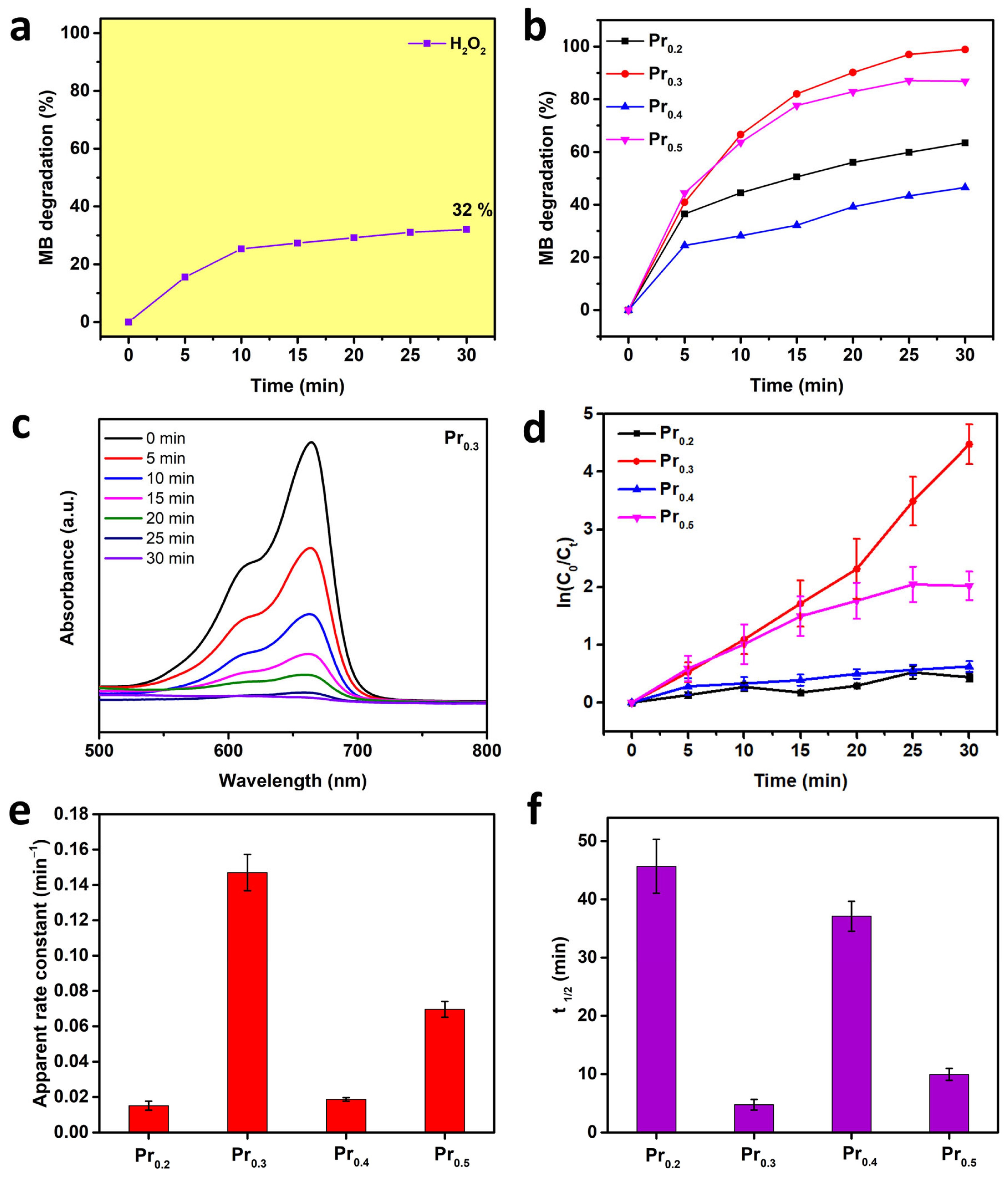
Prior to UV-light-driven photocatalysis testing, a control experiment containing only MB and H2O2 was performed to quantify the contribution of homogeneous photolysis and direct peroxide activation (Figure 6a). Only about 32% of MB degradation was achieved after 30 min, demonstrating that light irradiation and peroxide alone are insufficient for efficient dye removal. The photocatalytic performance of all Pr-substituted samples is presented in Figure 6b. The degradation efficiency increased sharply within the first 10 min of irradiation for all catalysts, followed by a slower approach toward saturation, indicating a typical pseudo-first-order kinetic behavior [58]. Among the tested compositions, Pr0.3 exhibited the highest degradation efficiency (98.9%), followed by Pr0.5 (86.8%), Pr0.2 (63.4%), and Pr0.4 (46.5%) after 30 min of illumination. The total duration time was selected as per the first compound to reach nearly 100% degradation. This performance trend suggests that an optimal Pr concentration provides the most effective balance between oxygen vacancy density and the coexistence of Pr3+/Pr4+ and Ce3+/Ce4+ redox pairs, thereby maximizing the rate of electron-hole separation and interfacial ROS generation [59,60]. The progressive spectral evolution of MB degradation over the most active Pr0.3 catalyst is shown in Figure 6c. The characteristic absorption peak at 664 nm gradually decreased in intensity with increasing irradiation time, accompanied by a visible color fading of the solution from deep blue to nearly colorless. The absence of significant spectral shifts indicates that the degradation process primarily involves stepwise demethylation and ring-opening oxidation, typical of MB mineralization under oxidative conditions [61].
To gain deeper insight into reaction dynamics and quantify relative catalytic activities, the kinetics of MB degradation were analyzed using the pseudo-first-order Langmuir–Hinshelwood model [58]. The linear relationships between ln(C0/Ct) and irradiation time for all samples are presented in Figure 6d. All curves exhibited excellent linearity, confirming that the degradation process follows pseudo-first-order kinetics, with the reaction rate directly proportional to the surface concentration of adsorbed MB molecules [2]. The apparent rate constants (kapp), obtained from the slope of the linear fits, are compared in Figure 6e. The rate constants correspond to the observed degradation efficiencies [59]. The Pr0.3 compound reached the highest apparent rate constant (0.15 min−1), nearly an order of magnitude higher than that of Pr0.2, indicating that the optimum Pr substitution promotes rapid electron-hole separation and more efficient formation of reactive oxygen species. The calculated half-life times (t1/2 = ln(2)/kapp) for each composition are displayed in Figure 6f. The shortest t1/2 was recorded for Pr0.3, whereas Pr0.2 and Pr0.4 required substantially longer times, in agreement with their lower rate constants.
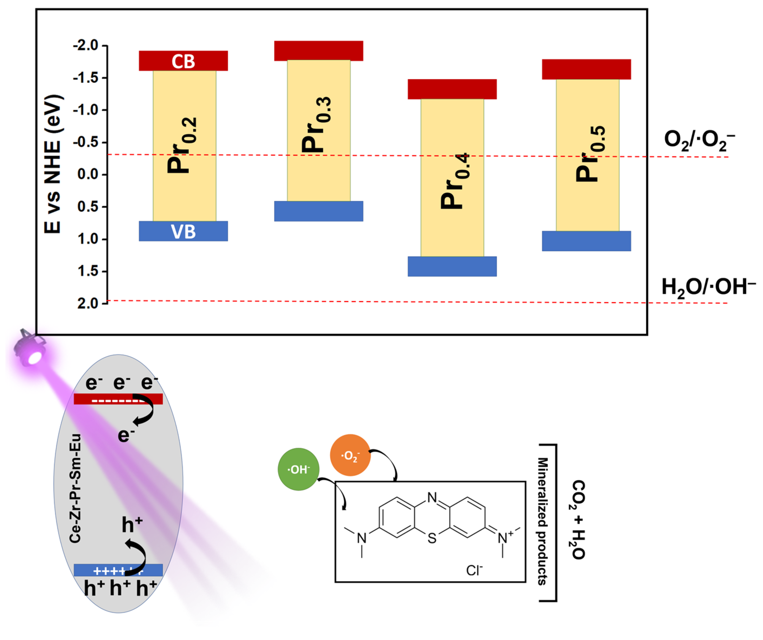
Based on the optical, structural, and photocatalytic results, as shown in Table 2, a mechanism for MB degradation over the synthesized high-entropy fluorite oxides has been proposed (Figure 7). The measured optical bandgaps and valence band maxima indicate that these oxides possess narrowed electronic structures, as compared to pure CeO2 [2], primarily due to the incorporation ions with variable oxidation states.
This mixed valence introduces intermediate 4f defect levels and oxygen-vacancy-related subbands, thereby promoting visible-light absorption and facilitating charge transfer across the lattice [38]. Upon UV illumination, electrons in the valence band (VB) are excited to the conduction band (CB), leaving behind photogenerated holes [62]. The CB edge positions (−1.72, −1.96, −1.15, and −1.58 eV for Pr0.2, Pr0.3, Pr0.4, and Pr0.5, respectively) are sufficiently negative to thermodynamically enable the reduction in dissolved or adsorbed molecular oxygen to superoxide radicals (∙O2). In contrast, the VB maxima (0.69–1.51 eV vs. NHE) are not positive enough to directly oxidize water or hydrogen peroxide to hydroxyl radicals (∙OH) [63,64]. Therefore, direct ∙OH formation from water oxidation is unlikely in these systems. Instead, hydroxyl radicals can predominantly be generated via photo-assisted Fenton-like activation of H2O2. The photogenerated electrons are rapidly trapped by Ce4+ or Pr4+ species, forming Ce3+ and Pr3+ ions, while suppressing electron-hole recombination. These reduced metal centers react with H2O2 to generate hydroxyl radicals and regenerate the higher oxidation states, establishing a catalytic redox cycle. Simultaneously, electrons from CB can participate in reductive activation of H2O2, further contributing to ROS formation. This dual redox cycling, sustained by the coexistence of 4+/3+ pairs, enhances carrier separation and continuously regenerates active species on the catalyst surface [65,66,67].
The redox coupling between adjacent Pr and Ce centers, acts as an internal electron relay, minimizing charge recombination and supporting a quasi-Fenton cycle that promotes continuous ROS production [48,68]. The superoxide radicals produced via oxygen reduction can further evolve through secondary reactions [69]. The combined action of superoxide and H2O2 derived hydroxyl radicals constitutes the dominant oxidative pathway responsible for the degradation. The generated radicals attack the conjugated chromophore of MB, causing progressive demethylation, ring-opening, and eventual mineralization to CO2, NH4+, and SO42− [69,70,71,72]. In this mechanism, the oxygen vacancies introduced by Pr substitution serve as active adsorption sites for O2 and H2O2 molecules, lower the activation barrier for radical formation, and promote fast electron transfer through localized defect bands. The structural flexibility of the high-entropy fluorite lattice allows for reversible redox transitions without loss of crystallinity or catalytic performance, ensuring sustained activity during repeated photocatalytic cycles. In general, mechanism can be summarized as a multi-step photo-Fenton-like process driven by mixed-valence rare-earth redox couples in the next order: (i) generation of e/h+ pairs in the HEO under UV irradiation; (ii) electron transfer through Ce4+/3+ and Pr3+/4+ cycles enhances separation; (iii) surface reactions with O2 and H2O2 produce ∙O2 and ∙OH radicals; (iv) radicals attack MB molecules, leading to chromophore cleavage and mineralization; and (v) redox couples are continuously restored through electron exchange with oxygen and peroxide species.

3. Materials and Methods

3.1. Synthesis and Materials

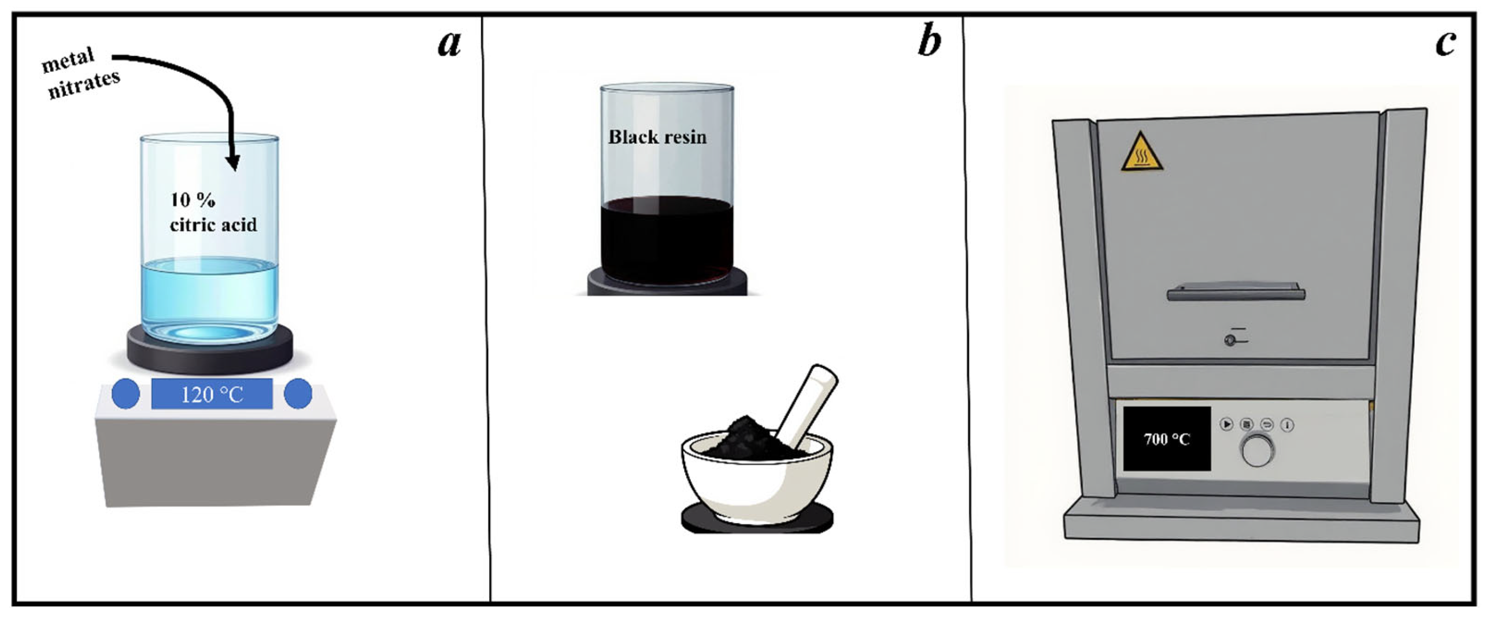
High-entropy oxides were synthesized via a modified aqueous sol–gel citrate route, adapted from previously established procedures for complex oxide systems, and shown in Figure 8 [73]. Stoichiometric amounts of metal nitrates corresponding to the targeted compositions presented in Table 3 (Ce, Zr, Sm, Eu, and Pr ratios with the total cationic sum fixed at 1 mmol) were used to achieve high configurational entropy in the system. In a typical synthesis, a total of 1 mmol of mixed metal precursors was dissolved in a 10% aqueous citric acid solution (prepared by dissolving 10 g of citric acid in 100 mL of Milli-Q water) under constant magnetic stirring. The resulting solution was stirred for 30 min to ensure complete dissolution of all precursors, after which the pH value was adjusted to approximately 5 by the slow addition of concentrated ammonia solution, monitored with a pH meter (HANNA Instruments, Zagreb, Croatia). The reaction mixture was then stirred and heated at 120 °C until solvent evaporation produced a black resin. This resin was dried overnight at 120 °C in an oven (Instrumentaria ST-01/02, Sesvete, Croatia) to remove residual moisture. The dried mass was ground manually using a mortar and pestle and then subjected to calcination in a muffle furnace (Nabertherm GmbH, Lilienthal, Germany) at 700 °C for 8 h, with a controlled heating rate of 240 °C per hour. The calcination process yielded single-phase, high-entropy oxide powders with homogeneous distribution of constituent cations. Chemical used for the synthesis were used as purchased: Ce(NO3)3 ∙ 6H2O, Eu(NO3)3 ∙ 5H2O, Pr(NO3)3 ∙ 6H2O, ZrO(NO3)2nH2O, purchased from Sigma Aldrich, Taufkirchen, Germany; Sm(NO3)3 ∙ 6H2O purchased from ThermoFisher, Waltham, MA, USA; Citric acid monohydrate purchased from TTT, Sveta Nedelja, Croatia; and concentrated ammonia solution from Gram-Mol, Zagreb, Croatia.

3.2. Structural Characterization

Powder X-ray diffraction (PXRD) analysis was employed to examine the crystal structure and microstructure of the synthesized materials. Measurements were conducted using a PANalytical Aeris Research Diffractometer (PANalytical, Malvern, UK) operating in θ-θ geometry with Cu Kα radiation (λ = 1.5406 Å) generated at 40 kV and 15 mA. Data were collected over a 2θ range of 20–100° at 25 °C, with a step size of 0.02° and a counting time of 20.4 s/step. Instrumental parameters included a divergence slit of 1° and a fixed mask of 13 mm. The obtained diffraction patterns were refined using the Rietveld method implemented in the FULLPROF software suite (version: January-2021) [74], applying the Thomson–Cox–Hastings pseudo-Voigt profile function to ensure accurate phase quantification and structural modeling. Visualization of the refined structures were performed using the VESTA software package (version: 3.4.8) [75].
The surface microstructure of the samples was characterized using a Thermo Fisher Scientific Apreo C field-emission scanning electron microscope (Thermo Fisher Scientific, Waltham, MA, USA).

3.3. Spectroscopy Analysis

Elemental distribution analysis was carried out using an Oxford Instruments X-Max50 microanalysis system (Oxford Instruments plc, Tubney Woods, Abingdon, UK) equipped with a 50 mm2 silicon drift detector (SDD). The corresponding elemental maps were acquired and processed using INCA software (INCA V7.5, Oxford Instruments plc, Tubney Woods, Abingdon, UK), enabling visualization of the spatial distribution and homogeneity of the constituent elements within the samples.
Raman spectra were acquired using a Bruker Senterra II Raman spectrometer (Bruker, Billerica, MA, USA) equipped with a 532 nm excitation laser. All samples were measured under identical conditions with a laser power of 2.5 mW, an integration time of 10,000 ms and averaged over three co-additions to improve the signal-to-noise ratio. The spectrometer has a spectral resolution of 4 cm−1.
X-ray photoelectron spectroscopy (XPS) measurements were performed on a Kratos Supra+ spectrometer (Kratos Analytical, Manchester, UK) equipped with a monochromatic Al Kα radiation source ( = 1486.6 eV). To prevent surface charging during analysis, a low-energy charge neutralizer was employed throughout all measurements. Spectra were collected at a take-off angle of 90° relative to the sample surface, using a pass energy of 20 eV for high-resolution scans. Powder samples were pressed onto conductive carbon adhesive tape and mounted on a silicon wafer for analysis. Data acquisition and processing were performed using ESCApe 1.5 software (Kratos Analytical). All binding energies were calibrated with respect to the C 1s hydrocarbon peak (C–C/C–H) set at 284.8 eV to correct for any residual surface charging effects.
The specific surface area and pore characteristics of the synthesized oxides were determined using a gas sorption analyzer Autosorb iQ (Quantachrome Instruments, Boynton Beach, FL, USA). Each sample was degassed at 250 °C under vacuum for 5 h using a degassing station to remove adsorbed moisture and residual impurities from the pores. Nitrogen adsorption–desorption isotherms were recorded at −196 °C. The obtained isotherms were used to calculate the specific surface area of the mesoporous materials according to the Brunauer–Emmett–Teller (BET) method.

3.4. Methylene Blue Degradation Analysis

Experiments on the photocatalytic degradation of organic dyes were carried out at room temperature under a UV lamp, which exposed the samples to light with a wavelength of 376 nm. The lamp was kept at a constant distance from the samples. A stock solution of methylene blue (MB) with a concentration of 2 × 10⁻⁵ mol/L was prepared in ultrapure water. All reactions were conducted in a 50 mL laboratory beaker, and the suspensions were stirred during light exposure using a magnetic stirrer. To 20 mL of the stock MB solution, 2 mg of the synthesized compounds and 4 drops of the cocatalyst H2O2 were added, and the resulting suspensions were subjected to constant stirring and illumination. Control experiments were performed by adding only H2O2 to the MB solution in order to analyze MB degradation exclusively under illumination and in the presence of the cocatalyst. Afterwards, the degradation of MB was monitored using a UV–Vis spectrophotometer (UV-3600Plus, Shimadzu, Kyoto, Japan) by taking aliquots of the reaction mixture at regular 5 min intervals. Absorbance was measured at 664 nm, which corresponds to the maximum absorption wavelength of MB, and a total reaction time was used to be the time the first compound reached a nearly 100% degradation. Each photocatalytic degradation experiment was performed in triplicate under identical conditions, and the reported values represent the average of three independent measurements, while the corresponding standard deviations are shown as error bars where applicable.
Additionally, visible light-driven photodegradation of methylene blue was conducted under the same conditions as described above, but without co-catalyst addition, and under 20 W tungsten-halogen lamp to confirm the photocatalytic efficiency under visible light.
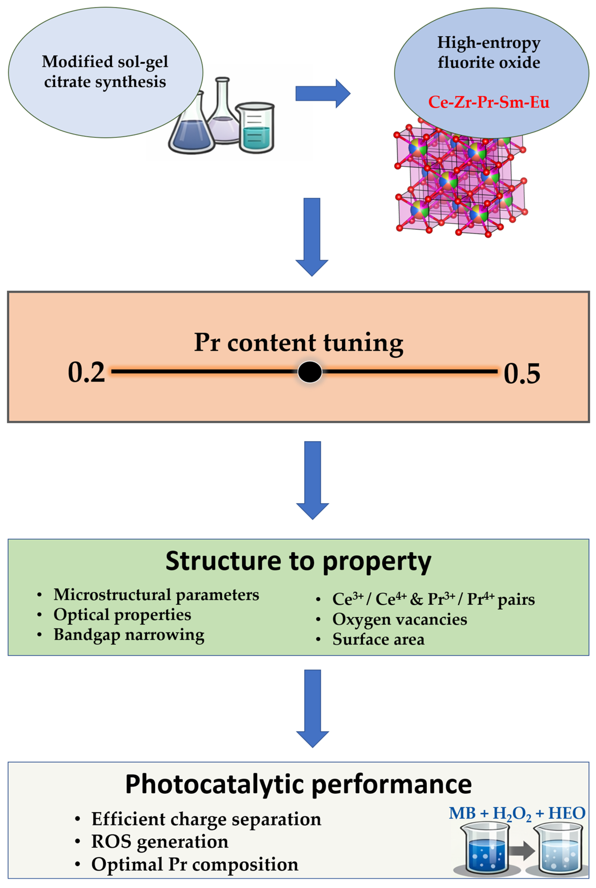
The overall experimental logic of this research is summarized in Figure 9.

4. Conclusions

This study systematically elucidates the influence of praseodymium incorporation on the structural evolution, defect chemistry, and photocatalytic activity of Ce-Zr-Pr-Sm-Eu-based high-entropy fluorite oxides. Through careful compositional control and comprehensive structural characterization, it was demonstrated that Pr substitution profoundly alters the balance between lattice disorder, oxygen vacancy formation, and redox flexibility. All synthesized compositions crystallized in a single-phase cubic fluorite structure, confirming that high configurational entropy successfully stabilizes complex multicomponent solid solutions without phase segregation. Progressive lattice expansion and increased microstrain with rising Pr content were directly correlated with the incorporation of larger Pr3+ ions and the generation of compensating oxygen vacancies. Morphological and physisorption analyses revealed that moderate Pr concentrations favored the formation of highly porous nanostructures with abundant surface-active sites, whereas excessive doping led to partial densification due to defect clustering and enhanced cation diffusion during calcination. Spectroscopic characterization provided deeper insight into the defect-redox behavior. Raman and XPS analyses confirmed the coexistence of Pr3+/Pr4+ and Ce3+/Ce4+ species, illustrating strong redox coupling between the two cationic sublattices. This coupling enhances electronic communication through hybridization, creating intermediate defect-related electronic states that facilitate charge delocalization and bandgap narrowing. The presence of abundant surface oxygen vacancies, confirmed by O 1s XPS deconvolution, enhances improved oxygen mobility and redox reversibility. The UV–Vis spectra corroborated these findings, showing reduced optical bandgaps indicative of defect-mediated electronic transitions and strong hybridization effects. Photocatalytic degradation experiments using methylene blue as a model organic pollutant revealed that Pr concentration determines the efficiency of light-driven reactions. The optimal composition, with moderate Pr substitution, exhibited the highest degradation efficiency, demonstrating that a balanced defect population maximizes charge-carrier separation and reactive oxygen species generation. The photocatalytic process proceeds via a photo-Fenton-like mechanism driven by redox cycling between Ce4+/3+ and Pr3+/4+ redox couples, leading to efficient dye degradation and mineralization. In general, prior highlight of this research is the fact that typically UV inactive photocatalysts can achieve higher photocatalytic efficiency by altering their properties and/or constituent ratio, remaining stable in its high-entropy form.

Supplementary Materials

The following supporting information can be downloaded at https://www.mdpi.com/article/10.3390/molecules31040598/s1, Table S1: Crystallographic data obtained by Rietveld refinement; Figure S1: Physisorption isotherms of synthesized Pr0.2–0.5 compounds; Table S2: Data obtained by physisorption analysis; Table S3: Quantitative composition of each synthesized compound by EDS; Figure S2: EDS sum spectra of synthesized compounds; Figure S3: Tauc plots of synthesized compound for bandgap approximation; Figure S4: XPS survey spectra of as-synthesized compounds; Figure S5: Deconvoluted C 1s XPS spectra; Figure S6: Deconvoluted Ce 3d XPS spectra; Figure S7: Deconvoluted O 1s XPS spectra; Figure S8: Deconvoluted Zr 3d spectra; Figure S9: Eu 3d (left) and Sm 4d (right) XPS spectra; Figure S10: Determination of Valence band position by XPS; Figure S11: Visible light driven photocatalysis over synthesized compounds, using a 20 W halogen lamp as a light source.

Author Contributions

Conceptualization, D.T., L.M. and S.Š.; Methodology, D.T., J.B. and J.K.; Validation, D.T., J.B. and P.Š.; Formal analysis: J.B., L.M. and A.S.; Investigation, D.T., C.D., G.B. and I.S.; Writing—original draft preparation, D.T., S.Š., P.Š. and J.K.; writing—review and editing, A.S. and I.D.; Supervision, Á.K., A.M. and I.D.; Funding acquisition, I.D. All authors have read and agreed to the published version of the manuscript.

Funding

This research was funded by the European Union—Next Generation EU, project number 581-UNIOS-105.

Institutional Review Board Statement

Not applicable.

Informed Consent Statement

Not applicable.

Data Availability Statement

The data presented in this study are available on request from the corresponding author.

Acknowledgments

The project is co-financed by the Republic of Slovenia, the Ministry of Education, Science and Sport, and the European Union under the European Regional Development Fund.

Conflicts of Interest

The authors declare no conflicts of interest. The funders had no role in the design of the study; in the collection, analyses, or interpretation of data; in the writing of the manuscript; nor in the decision to publish the results. The views and opinions expressed are solely those of the author(s) and do not necessarily reflect those of the European Union or the European Commission. Neither the European Union nor the European Commission can be held responsible for them.

References

  1. Wright, A.J.; Wang, Q.; Hu, C.; Yeh, Y.-T.; Chen, R.; Luo, J. Single-Phase Duodenary High-Entropy Fluorite/Pyrochlore Oxides with an Order-Disorder Transition. Acta Mater. 2021, 211, 116858. [Google Scholar] [CrossRef]
  2. Nundy, S.; Tatar, D.; Kojčinović, J.; Ullah, H.; Ghosh, A.; Mallick, T.K.; Meinusch, R.; Smarsly, B.M.; Tahir, A.A.; Djerdj, I. Bandgap Engineering in Novel Fluorite-Type Rare Earth High-Entropy Oxides (RE-HEOs) with Computational and Experimental Validation for Photocatalytic Water Splitting Applications. Adv. Sustain. Syst. 2022, 6, 2200067. [Google Scholar] [CrossRef]
  3. Tatar, D.; Ullah, H.; Yadav, M.; Kojčinović, J.; Šarić, S.; Szenti, I.; Skalar, T.; Finšgar, M.; Tian, M.; Kukovecz, Á.; et al. High-Entropy Oxides: A New Frontier in Photocatalytic CO2 Hydrogenation. ACS Appl. Mater. Interfaces 2024, 16, 29946–29962. [Google Scholar] [CrossRef] [PubMed]
  4. Jiao, Y.; Dai, J.; Fan, Z.; Cheng, J.; Zheng, G.; Grema, L.; Zhong, J.; Li, H.-F.; Wang, D. Overview of High-Entropy Oxide Ceramics. Mater. Today 2024, 77, 92–117. [Google Scholar] [CrossRef]
  5. Rost, C.M.; Sachet, E.; Borman, T.; Moballegh, A.; Dickey, E.C.; Hou, D.; Jones, J.L.; Curtarolo, S.; Maria, J.-P. Entropy-Stabilized Oxides. Nat. Commun. 2015, 6, 8485. [Google Scholar] [CrossRef] [PubMed]
  6. Fracchia, M.; Coduri, M.; Ghigna, P.; Anselmi-Tamburini, U. Phase Stability of High Entropy Oxides: A Critical Review. J. Eur. Ceram. Soc. 2024, 44, 585–594. [Google Scholar] [CrossRef]
  7. Jiang, B.; Bridges, C.A.; Unocic, R.R.; Pitike, K.C.; Cooper, V.R.; Zhang, Y.; Lin, D.-Y.; Page, K. Probing the Local Site Disorder and Distortion in Pyrochlore High-Entropy Oxides. J. Am. Chem. Soc. 2021, 143, 4193–4204. [Google Scholar] [CrossRef]
  8. Sarkar, A.; Kruk, R.; Hahn, H. Magnetic Properties of High Entropy Oxides. Dalt. Trans. 2021, 50, 1973–1982. [Google Scholar] [CrossRef]
  9. Kumar, A.; Bérardan, D.; Brisset, F.; Dragoe, D.; Dragoe, N. Novel Entropy-Stabilized Fluorite Oxides with Multifunctional Properties. J. Mater. Chem. A 2023, 11, 14320–14332. [Google Scholar] [CrossRef]
  10. Kotsonis, G.N.; Almishal, S.S.I.; dos Santos Vieira, F.; Crespi, V.H.; Dabo, I.; Rost, C.M.; Maria, J.-P. High-Entropy Oxides: Harnessing Crystalline Disorder for Emergent Functionality. J. Am. Ceram. Soc. 2023, 106, 5587–5611. [Google Scholar] [CrossRef]
  11. Mao, H.R.; Guo, R.F.; Cao, Y.; Jin, S.B.; Qiu, X.M.; Shen, P. Ultrafast Densification of High-Entropy Oxide (La0.2Nd0.2Sm0.2Eu0.2Gd0.2)2Zr2O7 by Reactive Flash Sintering. J. Eur. Ceram. Soc. 2021, 41, 2855–2860. [Google Scholar] [CrossRef]
  12. Corey, Z.J.; Lu, P.; Zhang, G.; Sharma, Y.; Rutherford, B.X.; Dhole, S.; Roy, P.; Wang, Z.; Wu, Y.; Wang, H.; et al. Structural and Optical Properties of High Entropy (La,Lu,Y,Gd,Ce)AlO3 Perovskite Thin Films. Adv. Sci. 2022, 9, 2202671. [Google Scholar] [CrossRef] [PubMed]
  13. Xiao, M.; Liu, Z.; Di, H.; Bai, Y.; Yang, G.; Medvedev, D.A.; Luo, Z.; Wang, W.; Zhou, W.; Ran, R.; et al. High-Entropy Materials for Solid Oxide Cells: Synthesis, Applications, and Prospects. J. Energy Chem. 2025, 104, 268–296. [Google Scholar] [CrossRef]
  14. Šarić, S.; Kojčinović, J.; Tatar, D.; Djerdj, I. Overview of Recent Advances in Rare-Earth High-Entropy Oxides as Multifunctional Materials for Next-Gen Technology Applications. Molecules 2025, 30, 1082. [Google Scholar] [CrossRef]
  15. Kante, M.V.; Lakshmi Nilayam, A.R.; Kreka, K.; Hahn, H.; Bhattacharya, S.S.; Velasco, L.; Tarancón, A.; Kübel, C.; Schweidler, S.; Botros, M. Influence of Zr-Doping on the Structure and Transport Properties of Rare Earth High-Entropy Oxides. J. Phys. Energy 2024, 6, 035001. [Google Scholar] [CrossRef]
  16. Spiridigliozzi, L.; Ferone, C.; Cioffi, R.; Accardo, G.; Frattini, D.; Dell’Agli, G. Entropy-Stabilized Oxides Owning Fluorite Structure Obtained by Hydrothermal Treatment. Materials 2020, 13, 558. [Google Scholar] [CrossRef]
  17. Momin, N.; Manjanna, J.; D’Souza, L.; Aruna, S.T.; Senthil Kumar, S. Synthesis, Structure and Ionic Conductivity of Nanocrystalline Ce1−xLaxO2−δ as an Electrolyte for Intermediate Temperature Solid Oxide Fuel Cells. J. Alloys Compd. 2022, 896, 163012. [Google Scholar] [CrossRef]
  18. Liu, Q.; Yang, P.; Tan, W.; Yu, H.; Ji, J.; Wu, C.; Cai, Y.; Xie, S.; Liu, F.; Hong, S.; et al. Fabricating Robust Pt Clusters on Sn-Doped CeO2 for CO Oxidation: A Deep Insight into Support Engineering and Surface Structural Evolution. Chem. A Eur. J. 2023, 29, e202203432. [Google Scholar] [CrossRef]
  19. Nakayama, M.; Martin, M. First-Principles Study on Defect Chemistry and Migration of Oxide Ions in Ceria Doped with Rare-Earth Cations. Phys. Chem. Chem. Phys. 2009, 11, 3241–3249. [Google Scholar] [CrossRef]
  20. Aarthi, U.; Shukla, D.; Rengaraj, S.; Babu, K.S. Ordered to Defect Fluorite Structural Transition in Ce1-XNdxO2-δ System and Its Influence on Ionic Conductivity. J. Alloys Compd. 2020, 838, 155534. [Google Scholar] [CrossRef]
  21. Guillén-Hurtado, N.; Giménez-Mañogil, J.; Martínez-Munuera, J.C.; Bueno-López, A.; García-García, A. Study of Ce/Pr Ratio in Ceria-Praseodymia Catalysts for Soot Combustion under Different Atmospheres. Appl. Catal. A Gen. 2020, 590, 117339. [Google Scholar] [CrossRef]
  22. Srichaisiriwech, W.; Tepamatr, P. Monometallic and Bimetallic Catalysts Supported on Praseodymium-Doped Ceria for the Water–Gas Shift Reaction. Molecules 2023, 28, 8146. [Google Scholar] [CrossRef] [PubMed]
  23. Shajahan, I.; Dasari, H.P.; Govardhan, P. Dilatometer Studies of Praseodymium Doped Ceria: Effect of Synthesis Methods on Sintering Behaviour. Mater. Chem. Phys. 2020, 240, 122211. [Google Scholar] [CrossRef]
  24. Elmutasim, O.; Hussien, A.G.; Sharan, A.; AlKhoori, S.; Vasiliades, M.A.; Taha, I.M.A.; Kim, S.; Harfouche, M.; Emwas, A.-H.; Anjum, D.H.; et al. Evolution of Oxygen Vacancy Sites in Ceria-Based High-Entropy Oxides and Their Role in N2 Activation. ACS Appl. Mater. Interfaces 2024, 16, 23038–23053. [Google Scholar] [CrossRef]
  25. Sulekar, S.; Mehr, M.; Kim, J.H.; Nino, J.C. Effect of Reduced Atmosphere Sintering on Blocking Grain Boundaries in Rare-Earth Doped Ceria. Inorganics 2021, 9, 63. [Google Scholar] [CrossRef]
  26. Artini, C.; Massardo, S.; Carnasciali, M.M.; Joseph, B.; Pani, M. Evaluation of the Defect Cluster Content in Singly and Doubly Doped Ceria through in Situ High-Pressure X-Ray Diffraction. Inorg. Chem. 2021, 60, 7306–7314. [Google Scholar] [CrossRef]
  27. Andana, T.; Piumetti, M.; Bensaid, S.; Russo, N.; Fino, D.; Pirone, R. CO and Soot Oxidation over Ce-Zr-Pr Oxide Catalysts. Nanoscale Res. Lett. 2016, 11, 278. [Google Scholar] [CrossRef]
  28. Lara-García, H.A.; Araiza, D.G.; Méndez-Galván, M.; Tehuacanero-Cuapa, S.; Gómez-Cortés, A.; Díaz, G. Dry Reforming of Methane over Nickel Supported on Nd–Ceria: Enhancement of the Catalytic Properties and Coke Resistance. RSC Adv. 2020, 10, 33059–33070. [Google Scholar] [CrossRef]
  29. Poggio-Fraccari, E.; Irigoyen, B.; Baronetti, G.; Mariño, F. Ce-Pr Mixed Oxides as Active Supports for Water-Gas Shift Reaction: Experimental and Density Functional Theory Characterization. Appl. Catal. A Gen. 2014, 485, 123–132. [Google Scholar] [CrossRef]
  30. Navarrete, L.; Balaguer, M.; Vert, V.B.; Serra, J.M. Tailoring Electrocatalytic Properties of Solid Oxide Fuel Cell Composite Cathodes Based on (La0.8Sr0.2)0.95MnO3+δ and Doped Cerias Ce1–XLnxO2–δ (Ln=Gd, La, Er, Pr, Tb and X = 0.1–0.2). Fuel Cells 2017, 17, 100–107. [Google Scholar] [CrossRef]
  31. Rajendran, S.; Vijayaraghavan, R. Oxygen Vacancies Mediated Enhanced Photocatalytic Activity of Band Gap Engineered BaSn1−xCuxO3 towards Methylene Blue Degradation under Visible and Sunlight. RSC Adv. 2025, 15, 24802–24814. [Google Scholar] [CrossRef]
  32. Bai, H.; Zhang, G.; Tang, F.; Huang, L.; Tian, M.; Hu, L.; Qi, Z.; Jiang, J. Oxygen Vacancy-Enhanced S-Scheme C-ZnO/B-g-C3N4 Heterostructure for Efficient Photocatalytic Degradation of Methylene Blue. Appl. Organomet. Chem. 2025, 39, e70420. [Google Scholar] [CrossRef]
  33. Kisała, J.; Wojnarowska-Nowak, R.; Bobitski, Y. Layered MoS2: Effective and Environment-Friendly Nanomaterial for Photocatalytic Degradation of Methylene Blue. Sci. Rep. 2023, 13, 14148. [Google Scholar] [CrossRef]
  34. Chu, M.N.; Nguyen, L.T.H.; Truong, M.X.; Do, T.H.; Duong, T.T.A.; Nguyen, L.T.T.; Pham, M.A.; Tran, T.K.N.; Ngo, T.C.Q.; Pham, V.H. Ce3+/Ce4+-Doped ZrO2/CuO Nanocomposite for Enhanced Photocatalytic Degradation of Methylene Blue under Visible Light. Toxics 2022, 10, 463. [Google Scholar] [CrossRef] [PubMed]
  35. Jiang, L.; Gao, X.; Chen, S.; Ashok, J.; Kawi, S. Oxygen-Deficient WO3/TiO2/CC Nanorod Arrays for Visible-Light Photocatalytic Degradation of Methylene Blue. Catalysts 2021, 11, 1349. [Google Scholar] [CrossRef]
  36. Qadir, I.; Singh, S.; Sharma, S.; Manhas, U.; Atri, A.K.; Singh, D. New Rare Earth-Doped Bilayered Perovskite Oxide Photocatalysts Sr2La0.5R0.5FeMnO7 (R = La, Nd, Sm, Gd, Dy) for the Degradation of Highly Toxic Methylene Blue Dye in Wastewater under Visible Light: Structural, Optical, and Magnetic Properties. ACS Omega 2023, 8, 2010–2026. [Google Scholar] [CrossRef] [PubMed]
  37. Grima, L.; Peña, J.I.; Sanjuán, M.L. Pyrochlore-like ZrO2-PrOx Compounds: The Role of the Processing Atmosphere in the Stoichiometry, Microstructure and Oxidation State. J. Alloys Compd. 2022, 923, 166449. [Google Scholar] [CrossRef]
  38. Xu, Y.; Wu, P.; Wu, M.; Gu, Y.; Yu, H.; Ding, Z. Solvothermal Synthesis, Structural Characterization and Optical Properties of Pr-Doped CeO2 and Their Degradation for Acid Orange 7. Materials 2022, 15, 6953. [Google Scholar] [CrossRef]
  39. Solís, C.; Balaguer, M.; Serra, J.M. In Situ Raman Characterization of SOFC Materials in Operational Conditions: A Doped Ceria Study. Membranes 2020, 10, 148. [Google Scholar] [CrossRef]
  40. Yurdakal, S.; Garlisi, C.; Özcan, L.; Bellardita, M.; Palmisano, G. Chapter 4—(Photo)Catalyst Characterization Techniques: Adsorption Isotherms and BET, SEM, FTIR, UV–Vis, Photoluminescence, and Electrochemical Characterizations. In Heterogeneous Photocatalysis; Marcì, G., Palmisano, L., Eds.; Elsevier: Amsterdam, The Netherlands, 2019; pp. 87–152. ISBN 978-0-444-64015-4. [Google Scholar]
  41. Davó-Quiñonero, A.; González-Mira, J.; Such-Basañez, I.; Juan-Juan, J.; Lozano-Castelló, D.; Bueno-López, A. Improved CO Oxidation Activity of 3DOM Pr-Doped Ceria Catalysts: Something Other than an Ordered Macroporous Structure. Catalysts 2017, 7, 67. [Google Scholar] [CrossRef]
  42. Govardhan, P.; Anantharaman, A.P.; Patil, S.S.; Dasari, H.P.; Dasari, H.; Shourya, A. Effect of Ag Loading on Praseodymium Doped Ceria Catalyst for Soot Oxidation Activity. Korean J. Chem. Eng. 2022, 39, 328–342. [Google Scholar] [CrossRef]
  43. Coduri, M.; Checchia, S.; Longhi, M.; Ceresoli, D.; Scavini, M. Rare Earth Doped Ceria: The Complex Connection between Structure and Properties. Front. Chem. 2018, 6, 526. [Google Scholar] [CrossRef] [PubMed]
  44. Kabir, A.; Bowen, J.R.; Varenik, M.; Lubomirsky, I.; Esposito, V. Enhanced Electromechanical Response in Sm and Nd Co-Doped Ceria. Materialia 2020, 12, 100728. [Google Scholar] [CrossRef]
  45. Xu, Y.; Gao, L.; Hou, Q.; Wu, P.; Zhou, Y.; Ding, Z. Enhanced Oxygen Storage Capacity of Porous CeO2 by Rare Earth Doping. Molecules 2023, 28, 6005. [Google Scholar] [CrossRef]
  46. Newbury, D.E.; Ritchie, N.W.M. Testing the Accuracy of Low-Beam-Energy Electron-Excited X-Ray Microanalysis with Energy-Dispersive Spectrometry. J. Mater. Sci. 2024, 59, 19088–19111. [Google Scholar] [CrossRef]
  47. Tatar, D.; Kojčinović, J.; Marković, B.; Széchenyi, A.; Miletić, A.; Nagy, S.B.; Ziegenheim, S.; Szenti, I.; Sapi, A.; Kukovecz, Á.; et al. Sol-Gel Synthesis of Ceria-Zirconia-Based High-Entropy Oxides as High-Promotion Catalysts for the Synthesis of 1,2-Diketones from Aldehyde. Molecules 2021, 26, 6115. [Google Scholar] [CrossRef]
  48. Mekki, I.; Grzybek, G.; Kotarba, A.; García-García, A. Nanostructured Pr-Rich CexPr1-XO2-δ Mixed Oxides for Diesel Soot Combustion: Importance of Oxygen Lability. Nanomaterials 2024, 14, 483. [Google Scholar] [CrossRef]
  49. Schmitt, R.; Nenning, A.; Kraynis, O.; Korobko, R.; Frenkel, A.I.; Lubomirsky, I.; Haile, S.M.; Rupp, J.L.M. A Review of Defect Structure and Chemistry in Ceria and Its Solid Solutions. Chem. Soc. Rev. 2020, 49, 554–592. [Google Scholar] [CrossRef]
  50. Loridant, S. Raman Spectroscopy as a Powerful Tool to Characterize Ceria-Based Catalysts. Catal. Today 2021, 373, 98–111. [Google Scholar] [CrossRef]
  51. Rocha, L.S.R.; Simões, A.Z.; Macchi, C.; Somoza, A.; Giulietti, G.; Ponce, M.A.; Longo, E. Synthesis and Defect Characterization of Hybrid Ceria Nanostructures as a Possible Novel Therapeutic Material towards COVID-19 Mitigation. Sci. Rep. 2022, 12, 3341. [Google Scholar] [CrossRef]
  52. Kalaev, D.; Seo, H.G.; Tuller, H.L. Temporal and Spatial Tuning of Optical Constants in Praseodymium Doped Ceria by Electrochemical Means. Nanophotonics 2022, 11, 3943–3952. [Google Scholar] [CrossRef] [PubMed]
  53. Anandkumar, M.; Kesavan, K.P.; Sudarsan, S.; Zaitseva, O.V.; Ostovari Moghaddam, A.; Iarushina, D.V.; Trofimov, E.A. Band-Gap Engineering of High-Entropy Fluorite Metal Oxide Nanoparticles Facilitated by Pr3+ Incorporation by Gel Combustion Synthesis. Gels 2025, 11, 117. [Google Scholar] [CrossRef] [PubMed]
  54. Romeo, M.; Bak, K.; El Fallah, J.; Le Normand, F.; Hilaire, L. XPS Study of the Reduction of Cerium Dioxide. Surf. Interface Anal. 1993, 20, 508–512. [Google Scholar] [CrossRef]
  55. Borchert, H.; Frolova, Y.V.; Kaichev, V.V.; Prosvirin, I.P.; Alikina, G.M.; Lukashevich, A.I.; Zaikovskii, V.I.; Moroz, E.M.; Trukhan, S.N.; Ivanov, V.P.; et al. Electronic and Chemical Properties of Nanostructured Cerium Dioxide Doped with Praseodymium. J. Phys. Chem. B 2005, 109, 5728–5738. [Google Scholar] [CrossRef]
  56. Shaw, M.; Samanta, D.; Bera, S.; Mahto, M.K.; Salam Shaik, M.A.; Konar, S.; Mondal, I.; Dhara, D.; Pathak, A. Role of Surface Oxygen Vacancies and Oxygen Species on CuO Nanostructured Surfaces in Model Catalytic Oxidation and Reductions: Insight into the Structure-Activity Relationship Toward the Performance. Inorg. Chem. 2022, 61, 14568–14581. [Google Scholar] [CrossRef]
  57. Huh, D.N.; Bruce, J.P.; Ganesh Balasubramani, S.; Ciccone, S.R.; Furche, F.; Hemminger, J.C.; Evans, W.J. High-Resolution X-Ray Photoelectron Spectroscopy of Organometallic (C5H4SiMe3)3LnIII and [(C5H4SiMe3)3LnII]1– Complexes (Ln = Sm, Eu, Gd, Tb). J. Am. Chem. Soc. 2021, 143, 16610–16620. [Google Scholar] [CrossRef]
  58. Tran, H.D.; Nguyen, D.Q.; Do, P.T.; Tran, U.N.P. Kinetics of Photocatalytic Degradation of Organic Compounds: A Mini-Review and New Approach. RSC Adv. 2023, 13, 16915–16925. [Google Scholar] [CrossRef]
  59. Dhanka, A.K.; Pani, B.; Agasti, N. Defect-Enriched CuO/CeO2 Nanostructure: In-Depth Structural Characterization and Photocatalytic Performance. RSC Adv. 2025, 15, 11774–11789. [Google Scholar] [CrossRef]
  60. Abdulwahab, K.O.; Khan, M.M.; Jennings, J.R. Doped Ceria Nanomaterials: Preparation, Properties, and Uses. ACS Omega 2023, 8, 30802–30823. [Google Scholar] [CrossRef]
  61. Safat, S.; Buazar, F.; Albukhaty, S.; Matroodi, S. Enhanced Sunlight Photocatalytic Activity and Biosafety of Marine-Driven Synthesized Cerium Oxide Nanoparticles. Sci. Rep. 2021, 11, 14734. [Google Scholar] [CrossRef]
  62. Kusmierek, E. A CeO2 Semiconductor as a Photocatalytic and Photoelectrocatalytic Material for the Remediation of Pollutants in Industrial Wastewater: A Review. Catalysts 2020, 10, 1435. [Google Scholar] [CrossRef]
  63. Hayyan, M.; Hashim, M.A.; AlNashef, I.M. Superoxide Ion: Generation and Chemical Implications. Chem. Rev. 2016, 116, 3029–3085. [Google Scholar] [CrossRef] [PubMed]
  64. Jaimes-López, R.; Jiménez-Vázquez, A.; Pérez-Rodríguez, S.; Estudillo-Wong, L.A.; Alonso-Vante, N. Catalyst for the Generation of OH Radicals in Advanced Electrochemical Oxidation Processes: Present and Future Perspectives. Catalysts 2024, 14, 703. [Google Scholar] [CrossRef]
  65. Jiang, L.; Tinoco, M.; Fernández-García, S.; Sun, Y.; Traviankina, M.; Nan, P.; Xue, Q.; Pan, H.; Aguinaco, A.; González-Leal, J.M.; et al. Enhanced Artificial Enzyme Activities on the Reconstructed Sawtoothlike Nanofacets of Pure and Pr-Doped Ceria Nanocubes. ACS Appl. Mater. Interfaces 2021, 13, 38061–38073. [Google Scholar] [CrossRef] [PubMed]
  66. Lan, Y.; Xia, X.; Li, J.; Mao, X.; Chen, C.; Ning, D.; Chu, Z.; Zhang, J.; Liu, F. Insight into the Contributions of Surface Oxygen Vacancies on the Promoted Photocatalytic Property of Nanoceria. Nanomaterials 2021, 11, 1168. [Google Scholar] [CrossRef]
  67. Majani, S.S.; Singh, P.; Kumari, P.; Setty, P.B.S.; Shivamallu, C.; Srinivasa, C.; Abass, K.S.; Iqbal, M.; Amachawadi, R.G.; Stupin, V.; et al. Cerium Oxide Nanoparticles Prepared through Bio-Combustion Using Ficus Carica as Effective Antioxidant, Anticancer and Dye Degrading Agent. Sci. Rep. 2025, 15, 30285. [Google Scholar] [CrossRef]
  68. Popescu, I.; Martínez-Munuera, J.C.; García-García, A.; Marcu, I.-C. Insights into the Relationship between the Catalytic Oxidation Performances of Ce-Pr Mixed Oxides and Their Semiconductive and Redox Properties. Appl. Catal. A Gen. 2019, 578, 30–39. [Google Scholar] [CrossRef]
  69. González, S.; Jaramillo-Fierro, X. Density Functional Theory Study of Methylene Blue Demethylation as a Key Step in Degradation Mediated by Reactive Oxygen Species. Int. J. Mol. Sci. 2025, 26, 1756. [Google Scholar] [CrossRef]
  70. Mohamad Idris, N.H.; Rajakumar, J.; Cheong, K.Y.; Kennedy, B.J.; Ohno, T.; Yamakata, A.; Lee, H.L. Titanium Dioxide/Polyvinyl Alcohol/Cork Nanocomposite: A Floating Photocatalyst for the Degradation of Methylene Blue under Irradiation of a Visible Light Source. ACS Omega 2021, 6, 14493–14503. [Google Scholar] [CrossRef]
  71. Khan, I.; Saeed, K.; Zekker, I.; Zhang, B.; Hendi, A.H.; Ahmad, A.; Ahmad, S.; Zada, N.; Ahmad, H.; Shah, L.A.; et al. Review on Methylene Blue: Its Properties, Uses, Toxicity and Photodegradation. Water 2022, 14, 242. [Google Scholar] [CrossRef]
  72. Parvees, A.; Priyadarshini, U.; Remya, N. Visible Light Induced Photocatalytic Degradation of Methylene Blue Using Carbon Fibre Cloth—Bismuth Oxybromide—Water Hyacinth Derived Silver Nanocomposite. Environ. Res. 2024, 262, 119787. [Google Scholar] [CrossRef]
  73. Šarić, S.; Tatar, D.; Skalar, T.; Marinšek, M.; Pravda, C.B.; Kukovecz, Á.; Szenti, I.; Finšgar, M.; Djerdj, I.; Kojčinović, J. From Solid Solution towards Pyrochlore and Kappa Phases: Introducing Configurational Entropy in Ordered Ceria–Zirconia Systems. CrystEngComm 2025, 27, 6762–6780. [Google Scholar] [CrossRef]
  74. Egami, T.; Billinge, S.J.L. Underneath the Bragg Peaks: Structural Analysis of Complex Materials; Pergamon: Oxford, UK, 2012; Volume 16, ISBN 9780080971339. [Google Scholar]
  75. Momma, K.; Izumi, F. VESTA: A Three-Dimensional Visualization System for Electronic and Structural Analysis. J. Appl. Crystallogr. 2008, 41, 653–658. [Google Scholar] [CrossRef]
Figure 1. Rietveld output plot of synthesized high-entropy oxides, with visualized structure shown in the insets of each plot. Black line represents experimental plot, red line calculated plot, black points represent Braggs positions, while blue line represents difference plot. In visualized structures, oxygen anions are depicted with red, while cations are depicted in different colors, with Pr depicted in blue.
Molecules 31 00598 g001
Figure 2. SEM images at different magnifications for a Pr0.2 compound.
Molecules 31 00598 g002
Figure 3. EDS mapping showing uniform distribution of the elements, confirming formation of high-entropy oxides.
Molecules 31 00598 g003
Figure 4. Raman spectra of as-synthesized compounds.
Molecules 31 00598 g004
Figure 5. Deconvoluted Pr 3d XPS spectra of synthesized compounds.
Molecules 31 00598 g005
Figure 6. MB degradation using a pure H2O2 (a), and with added compounds as photocatalysts (b); obtained UV–Vis spectra for MB degradation using Pr0.3 compound over 30 min time period (c); ln(C0/Ct) plot vs. time (d); apparent rate constant for tested compounds (e); and t1/2 for each tested compound (f).
Molecules 31 00598 g006
Figure 7. Proposed photocatalytic mechanism of MB degradation.
Molecules 31 00598 g007
Figure 8. Schematic presentation of the synthesis procedure showing (a) addition of metal nitrates to previously prepared 10% citric acid solutions, and then heated and mixed at 120 °C, (b) formed black resin that was grounded in mortar, and then followed by (c) calcination at 700 °C.
Molecules 31 00598 g008
Figure 9. The flowchart of experimental operation process.
Molecules 31 00598 g009
Table 1. Obtained surface at. concentration of Ce3+, Pr3+, and adsorbed oxygen species from deconvolution of XPS spectra.
CompoundCe3+/Ce4+ (%)Surface Atomic
Concentration (%)
[Ce3+]
Surface Atomic
Concentration (%)
[Pr3+]
Surface Atomic
Concentration (%)
[Oads]
Pr0.23.12.947.933.9
Pr0.35.65.344.128.2
Pr0.45.85.541.743.1
Pr0.53.23.136.145.7
Table 2. Overall results of tested compounds for MB degradation, along with their structural features.
CompoundMB Degradation (%)Bandgap Value (eV)Surface Area
(m2/g)
Surface Atomic
Concentration (%)
[Pr3+]
Surface Atomic
Concentration (%)
[Oads]
Pr0.263.442.6333.78447.933.9
Pr0.398.862.6529.76644.128.2
Pr0.446.512.6632.92141.743.1
Pr0.586.772.6625.92536.145.7
Table 3. Chemical compositions of synthesized compounds.
Name of the CompoundChemical Formula
Pr0.2Ce0.2Zr0.2Pr0.2Sm0.2Eu0.2O2
Pr0.3Ce0.175Zr0.175Pr0.3Sm0.175Eu0.175O2
Pr0.4Ce0.15Zr0.15Pr0.4Sm0.15Eu0.15O2
Pr0.5Ce0.125Zr0.125Pr0.5Sm0.125Eu0.125O2
Disclaimer/Publisher’s Note: The statements, opinions and data contained in all publications are solely those of the individual author(s) and contributor(s) and not of MDPI and/or the editor(s). MDPI and/or the editor(s) disclaim responsibility for any injury to people or property resulting from any ideas, methods, instructions or products referred to in the content.