The Disordome Paradigm: How Intrinsic Disorder Encodes Emergent Biological Function



Abstract
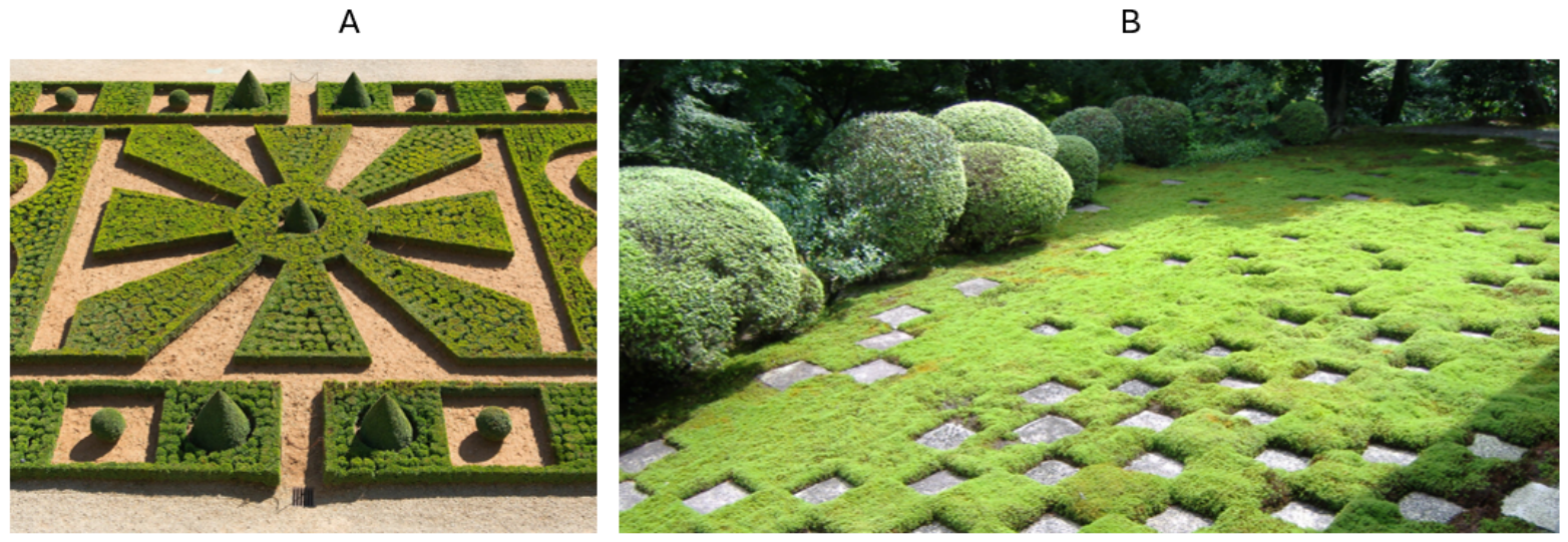
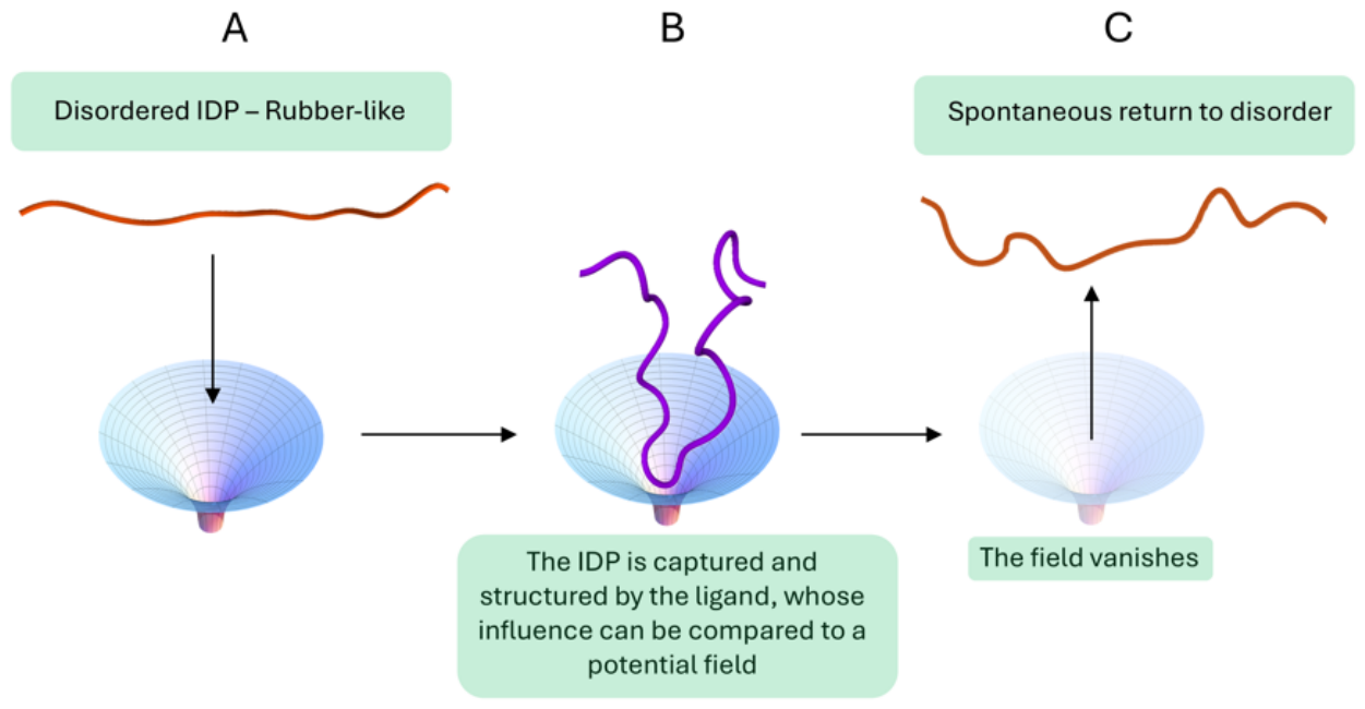
1. The Disordome: Transient Emergent Order in an Electrostatic Landscape
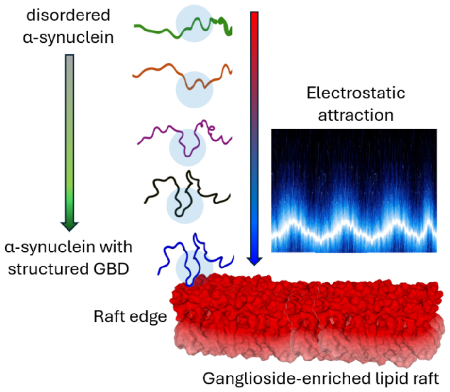
2. A Concrete Illustration: Progressive Structuring of α-Synuclein on a GM3 Raft
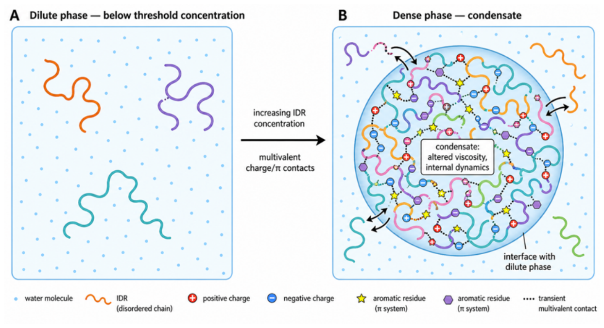
3. A Second Illustration: Emergent Order in an IDR-Driven Condensate

| Protein | Disordered Region | Biological Function | LLPS Mechanism | Cellular Context | Key Reference |
|---|---|---|---|---|---|
| FUS | N-terminal SYGQ-rich low-complexity domain (LCD) | RNA-binding protein, pre-mRNA splicing, transcription regulation | Multivalent tyrosine–arginine contacts drive LLPS; π–π and cation–π interactions stabilize the dense phase | Nuclear speckles, stress granules; aberrant LLPS linked to ALS/FTD | [48] |
| hnRNPA1 | C-terminal low-complexity prion-like domain | mRNA processing, stress granule assembly | Multivalent LCD–LCD interactions; phase separation promotes stress granule formation and can seed pathological fibrillization | Cytoplasmic stress granules; disease-associated mutations increase aggregation propensity | [94] |
| TDP-43 | C-terminal glycine-rich low-complexity domain | RNA/DNA-binding, mRNA splicing and stability | LCD-mediated multivalent interactions drive LLPS; helix–helix contacts within LCD stabilize droplets | Nuclear and cytoplasmic condensates; pathological aggregation in ALS/FTD | [105] |
| Ddx4 (DDX4) | N-terminal RGG/FG-rich disordered region | Germ-cell “nuage” RNA helicase, piRNA pathway | Weakly interacting blocks of charged and aromatic residues; salt- and temperature-responsive LLPS | Perinuclear nuage granules in germ cells | [87] |
| G3BP1 | Intrinsically disordered RGG and acidic regions flanking the NTF2-like domain | RNA-binding, core stress granule scaffold | Multivalent protein–protein and protein–RNA interactions nucleate LLPS above a concentration threshold | Cytoplasmic stress granules under oxidative, ER, or metabolic stress | [110] |
| MED1 (Mediator complex) | Intrinsically disordered activation domain | Transcriptional coactivator, super-enhancer function | Phase separation of coactivators concentrates transcriptional machinery at super-enhancers | Nuclear transcriptional condensates at super-enhancer loci | [111] |
| BRD4 | Intrinsically disordered low-complexity domain | Chromatin reader, transcriptional coactivator | Activation-domain-driven LLPS; disordered domains of transcription factors phase-separate to activate transcription | Nuclear condensates at active gene loci | [111] |
| NUPR1 (p8) | Full-length protein (82 residues, monomeric IDP) | Stress-associated transcriptional regulator; translation control via eIF2α interaction; cell survival | NUPR1-driven LLPS forms stress granules; pharmacological disruption (ZZW-115) blocks droplet formation and induces synthetic lethality | Pancreatic cancer cells (KRAS G12D); endoplasmic reticulum and metabolic stress | [102] |
4. Defining the Disordome: From the Unfoldome to Reversible Order
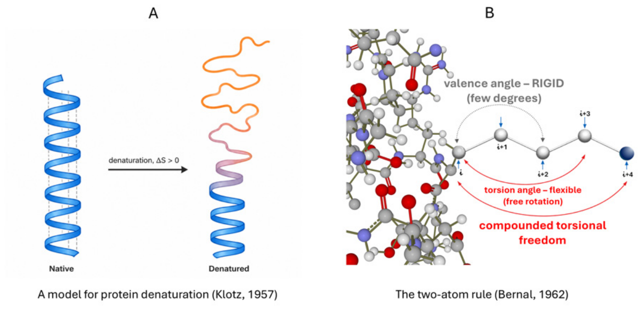
5. Historical Precursors and the Physical Basis of Conformational Freedom: Bernal, Klotz, and the Limits of AlphaFold
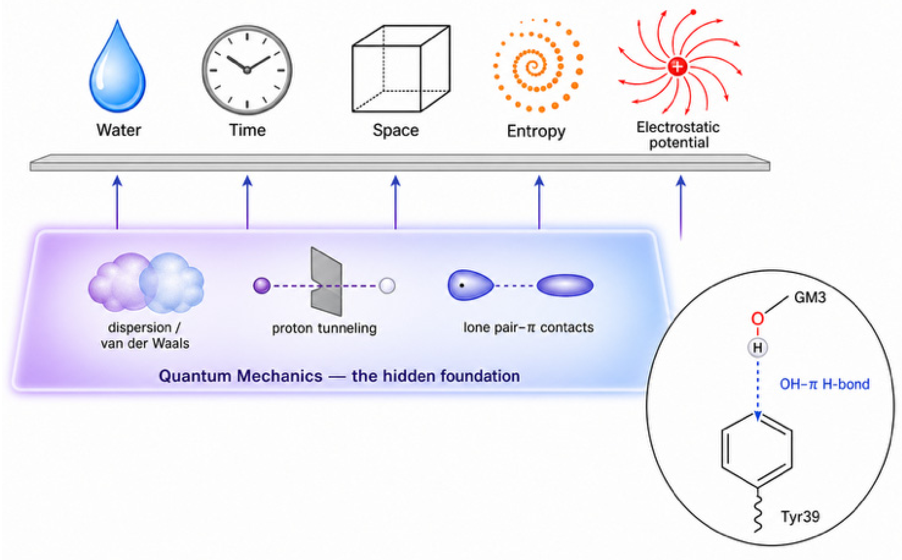
6. Beyond Classical Force Fields: Quantum-Chemical Contributions to the Disordome
7. Conclusions and Perspectives
Author Contributions
Funding
Institutional Review Board Statement
Informed Consent Statement
Data Availability Statement
Acknowledgments
Conflicts of Interest
Abbreviations
| CBD | Cholesterol binding domain |
| GBD | Ganglioside binding domain |
| HDX-MS | Hydrogen/deuterium eXchange mass spectrometry |
| IDPs | Intrinsically disordered proteins |
| IDRs | Intrinsically disordered regions |
| LCD | Low complexity domains |
| LLPS | Liquid–liquid phase separation |
| MS | Mass spectrometry |
| NMR | Nuclear magnetic resonance |
| pLDDT | Predicted local distance difference test |
| RF | RoseTTAFold |
| SGs | Stress granules |
| smFRET | Single-molecule fluorescence resonance energy transfer |
References
- Anfinsen, C.B. Principles that govern the folding of protein chains. Science 1973, 181, 223–230. [Google Scholar] [CrossRef] [Scilit] [PubMed]
- Wright, P.E.; Dyson, H.J. Intrinsically unstructured proteins: Re-assessing the protein structure-function paradigm. J. Mol. Biol. 1999, 293, 321–331. [Google Scholar] [CrossRef] [Scilit] [PubMed]
- Kulkarni, P.; Leite, V.B.P.; Roy, S.; Bhattacharyya, S.; Mohanty, A.; Achuthan, S.; Singh, D.; Appadurai, R.; Rangarajan, G.; Weninger, K.; et al. Intrinsically disordered proteins: Ensembles at the limits of Anfinsen’s dogma. Biophys. Rev. 2022, 3, 011306. [Google Scholar] [CrossRef] [Scilit] [PubMed]
- Dunker, A.K.; Lawson, J.D.; Brown, C.J.; Williams, R.M.; Romero, P.; Oh, J.S.; Oldfield, C.J.; Campen, A.M.; Ratliff, C.M.; Hipps, K.W. Intrinsically disordered protein. J. Mol. Graph. Model. 2001, 19, 26–59. [Google Scholar] [CrossRef] [Scilit] [PubMed]
- Uversky, V.N. A protein-chameleon: Conformational plasticity of alpha-synuclein, a disordered protein involved in neurodegenerative disorders. J. Biomol. Struct. Dyn. 2003, 21, 211–234. [Google Scholar] [CrossRef] [Scilit] [PubMed]
- Uversky, V.N. Intrinsically disordered proteins and their environment: Effects of strong denaturants, temperature, pH, counter ions, membranes, binding partners, osmolytes, and macromolecular crowding. Protein J. 2009, 28, 305–325. [Google Scholar] [CrossRef] [Scilit] [PubMed]
- Uversky, V.N. Introduction to intrinsically disordered proteins (IDPs). Chem. Rev. 2014, 114, 6557–6560. [Google Scholar] [CrossRef] [Scilit] [PubMed]
- Uversky, V.N. Functional roles of transiently and intrinsically disordered regions within proteins. FEBS J. 2015, 282, 1182–1189. [Google Scholar] [CrossRef] [Scilit] [PubMed]
- Uversky, V.N. Dancing Protein Clouds: The Strange Biology and Chaotic Physics of Intrinsically Disordered Proteins. J. Biol. Chem. 2016, 291, 6681–6688. [Google Scholar] [CrossRef] [Scilit] [PubMed]
- Uversky, V.N. Intrinsically disordered proteins and their “mysterious”(meta) physics. Front. Phys. 2019, 7, 10. [Google Scholar] [CrossRef] [Scilit]
- Uversky, V.N. The mysterious unfoldome: Structureless, underappreciated, yet vital part of any given proteome. J. Biomed. Biotechnol. 2010, 2010, 568068. [Google Scholar] [CrossRef] [Scilit] [PubMed]
- Honig, B.; Nicholls, A. Classical electrostatics in biology and chemistry. Science 1995, 268, 1144–1149. [Google Scholar] [CrossRef] [Scilit] [PubMed]
- Zhou, H.-X.; Pang, X. Electrostatic Interactions in Protein Structure, Folding, Binding, and Condensation. Chem. Rev. 2018, 118, 1691–1741. [Google Scholar] [CrossRef] [Scilit] [PubMed]
- Janin, J. The kinetics of protein-protein recognition. Proteins Struct. Funct. Bioinform. 1997, 28, 153–161. [Google Scholar] [CrossRef] [Scilit]
- Ganguly, D.; Zhang, W.; Chen, J. Electrostatically accelerated encounter and folding for facile recognition of intrinsically disordered proteins. PLoS Comput. Biol. 2013, 9, e1003363. [Google Scholar] [CrossRef] [Scilit] [PubMed]
- Klotz, I.M. Some Principles of Energetics in Biochemical Reactions; Academic Press: New York, NY, USA, 1957. [Google Scholar]
- Bernal, J.D. The Structure of Molecules. In Comprehensive Biochemistry, Vol. 1: Atomic and Molecular Structure; Florkin, M., Stotz, E.H., Eds.; Elsevier: Amsterdam, The Netherlands; New York, NY, USA, 1962; pp. 258–276. [Google Scholar]
- Fantini, J. Fundamental Mechanisms in Membrane Receptology: Old Paradigms, New Concepts and Perspectives. Receptors 2024, 3, 107–121. [Google Scholar] [CrossRef] [Scilit]
- Fantini, J.; Azzaz, F.; Di Scala, C.; Aulas, A.; Chahinian, H.; Yahi, N. Conformationally adaptive therapeutic peptides for diseases caused by intrinsically disordered proteins (IDPs). New paradigm for drug discovery: Target the target, not the arrow. Pharmacol. Ther. 2025, 267, 108797. [Google Scholar] [CrossRef] [Scilit] [PubMed]
- Li, P.; Banjade, S.; Cheng, H.-C.; Kim, S.; Chen, B.; Guo, L.; Llaguno, M.; Hollingsworth, J.V.; King, D.S.; Banani, S.F.; et al. Phase transitions in the assembly of multivalent signalling proteins. Nature 2012, 483, 336–340. [Google Scholar] [CrossRef] [Scilit] [PubMed]
- Banjade, S.; Rosen, M.K. Phase transitions of multivalent proteins can promote clustering of membrane receptors. eLife 2014, 3, e04123. [Google Scholar] [CrossRef] [Scilit] [PubMed][Green Version]
- Banani, S.F.; Lee, H.O.; Hyman, A.A.; Rosen, M.K. Biomolecular condensates: Organizers of cellular biochemistry. Nat. Rev. Mol. Cell Biol. 2017, 18, 285–298. [Google Scholar] [CrossRef] [Scilit] [PubMed]
- Schrodinger, E. What is Life? The Physical ASPECT of the Living Cell; University Press: Cambridge, UK, 1951. [Google Scholar]
- Nicolis, G.; Prigogine, I. Self-organization in Nonequilibrium Systems: From Dissipative Structures to Order Through Fluctuations; Wiley: New York, NY, USA, 1977. [Google Scholar]
- Spolar, R.S.; Record, M.T., Jr. Coupling of local folding to site-specific binding of proteins to DNA. Science 1994, 263, 777–784. [Google Scholar] [CrossRef] [Scilit] [PubMed]
- Dyson, H.J.; Wright, P.E. Intrinsically unstructured proteins and their functions. Nat. Rev. Mol. Cell Biol. 2005, 6, 197–208. [Google Scholar] [CrossRef] [Scilit] [PubMed]
- Wright, P.E.; Dyson, H.J. Linking folding and binding. Curr. Opin. Struct. Biol. 2009, 19, 31–38. [Google Scholar] [CrossRef] [Scilit] [PubMed]
- Theisen, F.F.; Staby, L.; Tidemand, F.G.; O’Shea, C.; Prestel, A.; Willemoës, M.; Kragelund, B.B.; Skriver, K. Quantification of Conformational Entropy Unravels Effect of Disordered Flanking Region in Coupled Folding and Binding. J. Am. Chem. Soc. 2021, 143, 14540–14550. [Google Scholar] [CrossRef] [Scilit] [PubMed]
- Ganguly, D.; Otieno, S.; Waddell, B.; Iconaru, L.; Kriwacki, R.W.; Chen, J. Electrostatically accelerated coupled binding and folding of intrinsically disordered proteins. J. Mol. Biol. 2012, 422, 674–684. [Google Scholar] [CrossRef] [Scilit] [PubMed]
- Boehr, D.D.; Nussinov, R.; Wright, P.E. The role of dynamic conformational ensembles in biomolecular recognition. Nat. Chem. Biol. 2009, 5, 789–796, Correction in Nat. Chem. Biol. 2009, 5, 954. https://doi.org/10.1038/nchembio1209-954d. [Google Scholar] [CrossRef] [Scilit] [PubMed]
- Sugase, K.; Dyson, H.J.; Wright, P.E. Mechanism of coupled folding and binding of an intrinsically disordered protein. Nature 2007, 447, 1021–1025. [Google Scholar] [CrossRef] [Scilit] [PubMed]
- Arai, M.; Sugase, K.; Dyson, H.J.; Wright, P.E. Conformational propensities of intrinsically disordered proteins influence the mechanism of binding and folding. Proc. Natl. Acad. Sci. USA 2015, 112, 9614–9619. [Google Scholar] [CrossRef] [Scilit] [PubMed]
- Gallat, F.X.; Laganowsky, A.; Wood, K.; Gabel, F.; van Eijck, L.; Wuttke, J.; Moulin, M.; Härtlein, M.; Eisenberg, D.; Colletier, J.P.; et al. Dynamical coupling of intrinsically disordered proteins and their hydration water: Comparison with folded soluble and membrane proteins. Biophys. J. 2012, 103, 129–136. [Google Scholar] [CrossRef] [Scilit] [PubMed]
- Arya, S.; Singh, A.K.; Bhasne, K.; Dogra, P.; Datta, A.; Das, P.; Mukhopadhyay, S. Femtosecond Hydration Map of Intrinsically Disordered α-Synuclein. Biophys. J. 2018, 114, 2540–2551. [Google Scholar] [CrossRef] [Scilit] [PubMed]
- Schweitzer-Stenner, R. The multifaceted character of water as solvent for proteins: From poor for folded proteins to good for (some) intrinsically disordered proteins and protein segments. Q. Rev. Biophys. 2026, 59, e5. [Google Scholar] [CrossRef] [Scilit] [PubMed]
- Mao, A.H.; Crick, S.L.; Vitalis, A.; Chicoine, C.L.; Pappu, R.V. Net charge per residue modulates conformational ensembles of intrinsically disordered proteins. Proc. Natl. Acad. Sci. USA 2010, 107, 8183–8188. [Google Scholar] [CrossRef] [Scilit] [PubMed]
- Müller-Späth, S.; Soranno, A.; Hirschfeld, V.; Hofmann, H.; Rüegger, S.; Reymond, L.; Nettels, D.; Schuler, B. Charge interactions can dominate the dimensions of intrinsically disordered proteins. Proc. Natl. Acad. Sci. USA 2010, 107, 14609–14614, Correction in Proc. Natl. Acad. Sci. USA 2013, 110, 16693. https://doi.org/10.1073/pnas.1317338110. [Google Scholar] [CrossRef] [Scilit] [PubMed]
- Wuttke, R.; Hofmann, H.; Nettels, D.; Borgia, M.B.; Mittal, J.; Best, R.B.; Schuler, B. Temperature-dependent solvation modulates the dimensions of disordered proteins. Proc. Natl. Acad. Sci. USA 2014, 111, 5213–5218. [Google Scholar] [CrossRef] [Scilit] [PubMed]
- Best, R.B. Emerging consensus on the collapse of unfolded and intrinsically disordered proteins in water. Curr. Opin. Struct. Biol. 2020, 60, 27–38. [Google Scholar] [CrossRef] [Scilit] [PubMed]
- Treloar, L.R.G. The Physics of Rubber Elasticity; Oxford University Press: Oxford, UK, 2005. [Google Scholar]
- Rauscher, S.; Pomès, R. Structural disorder and protein elasticity. In Fuzziness Structural Disorder in Protein Complexes; Springer: New York, NY, USA, 2012; pp. 159–183. [Google Scholar]
- Truong, H.P.; Morgan, I.L.; Koren, G.; Avinery, R.; Beck, R.; Saleh, O.A. Pincus blob elasticity in an intrinsically disordered protein. Eur. Phys. J. E 2023, 46, 100, Correction in Eur. Phys. J. E 2023, 46, 125. https://doi.org/10.1140/epje/s10189-023-00382-8. [Google Scholar] [CrossRef] [Scilit] [PubMed]
- Das, R.K.; Pappu, R.V. Conformations of intrinsically disordered proteins are influenced by linear sequence distributions of oppositely charged residues. Proc. Natl. Acad. Sci. USA 2013, 110, 13392–13397. [Google Scholar] [CrossRef] [Scilit] [PubMed]
- Schreiber, G.; Haran, G.; Zhou, H.X. Fundamental Aspects of Protein−Protein Association Kinetics. Chem. Rev. 2009, 109, 839–860. [Google Scholar] [CrossRef] [Scilit] [PubMed]
- Sheinerman, F.B.; Norel, R.; Honig, B. Electrostatic aspects of protein-protein interactions. Curr. Opin. Struct. Biol. 2000, 10, 153–159. [Google Scholar] [CrossRef] [Scilit] [PubMed]
- Brangwynne, C.P.; Eckmann, C.R.; Courson, D.S.; Rybarska, A.; Hoege, C.; Gharakhani, J.; Jülicher, F.; Hyman, A.A. Germline P granules are liquid droplets that localize by controlled dissolution/condensation. Science 2009, 324, 1729–1732. [Google Scholar] [CrossRef] [Scilit] [PubMed]
- Shin, Y.; Brangwynne, C.P. Liquid phase condensation in cell physiology and disease. Science 2017, 357, eaaf4382. [Google Scholar] [CrossRef] [Scilit] [PubMed]
- Wang, J.; Choi, J.M.; Holehouse, A.S.; Lee, H.O.; Zhang, X.; Jahnel, M.; Maharana, S.; Lemaitre, R.; Pozniakovsky, A.; Drechsel, D.; et al. A Molecular Grammar Governing the Driving Forces for Phase Separation of Prion-like RNA Binding Proteins. Cell 2018, 174, 688–699.e16. [Google Scholar] [CrossRef] [Scilit] [PubMed]
- Vernon, R.M.; Chong, P.A.; Tsang, B.; Kim, T.H.; Bah, A.; Farber, P.; Lin, H.; Forman-Kay, J.D. Pi-Pi contacts are an overlooked protein feature relevant to phase separation. eLife 2018, 7, e31486. [Google Scholar] [CrossRef] [Scilit] [PubMed]
- Elbaum-Garfinkle, S.; Kim, Y.; Szczepaniak, K.; Chen, C.C.; Eckmann, C.R.; Myong, S.; Brangwynne, C.P. The disordered P granule protein LAF-1 drives phase separation into droplets with tunable viscosity and dynamics. Proc. Natl. Acad. Sci. USA 2015, 112, 7189–7194. [Google Scholar] [CrossRef] [Scilit] [PubMed]
- Wang, H.; Shi, Z. The rheology and interfacial properties of biomolecular condensates. Biophys. Rev. 2025, 17, 867–891. [Google Scholar] [CrossRef] [Scilit] [PubMed]
- Hyman, A.A.; Weber, C.A.; Jülicher, F. Liquid-liquid phase separation in biology. Annu. Rev. Cell Dev. Biol. 2014, 30, 39–58. [Google Scholar] [CrossRef] [Scilit] [PubMed]
- Berry, J.; Brangwynne, C.P.; Haataja, M. Physical principles of intracellular organization via active and passive phase transitions. Rep. Prog. Physics. Phys. Soc. 2018, 81, 046601. [Google Scholar] [CrossRef] [Scilit] [PubMed]
- Perunov, N.; Marsland, R.A.; England, J.L. Statistical physics of adaptation. Phys. Rev. X 2016, 6, 021036. [Google Scholar] [CrossRef] [Scilit]
- Qian, H. Phosphorylation energy hypothesis: Open chemical systems and their biological functions. Annu. Rev. Phys. Chem. 2007, 58, 113–142. [Google Scholar] [CrossRef] [Scilit] [PubMed]
- Söding, J.; Zwicker, D.; Sohrabi-Jahromi, S.; Boehning, M.; Kirschbaum, J. Mechanisms for Active Regulation of Biomolecular Condensates. Trends Cell Biol. 2020, 30, 4–14. [Google Scholar] [CrossRef] [Scilit] [PubMed]
- Buchanan, M. The origin of cell division. Nat. Phys. 2017, 13, 526. [Google Scholar] [CrossRef] [Scilit]
- Fantini, J.; Matveeva, M.; Lefebvre, M.; Chahinian, H. What Is life? Rethinking Biology in Light of Fundamental Parameters. Life 2024, 14, 280. [Google Scholar] [CrossRef] [Scilit] [PubMed]
- Masato, A.; Bubacco, L. The αSynuclein half-life conundrum. Neurobiol. Dis. 2024, 196, 106524. [Google Scholar] [CrossRef] [Scilit] [PubMed]
- Stefanis, L.; Emmanouilidou, E.; Pantazopoulou, M.; Kirik, D.; Vekrellis, K.; Tofaris, G.K. How is alpha-synuclein cleared from the cell? J. Neurochem. 2019, 150, 577–590. [Google Scholar] [CrossRef] [Scilit] [PubMed]
- Martinez, Z.; Zhu, M.; Han, S.; Fink, A.L. GM1 specifically interacts with alpha-synuclein and inhibits fibrillation. Biochemistry 2007, 46, 1868–1877. [Google Scholar] [CrossRef] [Scilit] [PubMed]
- Ledeen, R.W.; Wu, G. Gangliosides, α-Synuclein, and Parkinson’s Disease. Prog. Mol. Biol. Transl. Sci. 2018, 156, 435–454. [Google Scholar] [CrossRef] [Scilit] [PubMed]
- Perissinotto, F.; Rondelli, V.; Parisse, P.; Tormena, N.; Zunino, A.; Almásy, L.; Merkel, D.G.; Bottyán, L.; Sajti, S.; Casalis, L. GM1 Ganglioside role in the interaction of Alpha-synuclein with lipid membranes: Morphology and structure. Biophys. Chem. 2019, 255, 106272. [Google Scholar] [CrossRef] [Scilit] [PubMed]
- Fauvet, B.; Mbefo, M.K.; Fares, M.B.; Desobry, C.; Michael, S.; Ardah, M.T.; Tsika, E.; Coune, P.; Prudent, M.; Lion, N.; et al. α-Synuclein in central nervous system and from erythrocytes, mammalian cells, and Escherichia coli exists predominantly as disordered monomer. J. Biol. Chem. 2012, 287, 15345–15364. [Google Scholar] [CrossRef] [Scilit] [PubMed]
- Eliezer, D.; Kutluay, E.; Bussell, R.; Browne, G. Conformational properties of α-synuclein in its free and lipid-associated states. J. Mol. Biol. 2001, 307, 1061–1073. [Google Scholar] [CrossRef] [Scilit] [PubMed]
- Bertoncini, C.W.; Jung, Y.-S.; Fernandez, C.O.; Hoyer, W.; Griesinger, C.; Jovin, T.M.; Zweckstetter, M. Release of long-range tertiary interactions potentiates aggregation of natively unstructured α-synuclein. Proc. Natl. Acad. Sci. USA 2005, 102, 1430–1435. [Google Scholar] [CrossRef] [Scilit] [PubMed]
- Fantini, J.; Yahi, N. Molecular basis for the glycosphingolipid-binding specificity of α-synuclein: Key role of tyrosine 39 in membrane insertion. J. Mol. Biol. 2011, 408, 654–669. [Google Scholar] [CrossRef] [Scilit] [PubMed]
- Fantini, J.; Yahi, N. The driving force of alpha-synuclein insertion and amyloid channel formation in the plasma membrane of neural cells: Key role of ganglioside-and cholesterol-binding domains. In Lipid-mediated Protein Signaling; Springer: Dordrecht, The Netherlands, 2013; pp. 15–26. [Google Scholar] [CrossRef] [Scilit] [PubMed]
- Fantini, J.; Azzaz, F.; Aulas, A.; Chahinian, H.; Yahi, N. Preclinical assessment of a ganglioside-targeted therapy for Parkinson’s disease with the first-in-class adaptive peptide AmyP53. Sci. Rep. 2025, 15, 9144, Correction in Sci. Rep. 2025, 15, 23784. https://doi.org/10.1038/s41598-025-07762-4. [Google Scholar] [CrossRef] [Scilit] [PubMed]
- Fantini, J. Lipid rafts and human diseases: Why we need to target gangliosides. FEBS Open Bio 2023, 13, 1636–1650. [Google Scholar] [CrossRef] [Scilit] [PubMed]
- Bekker, G.J.; Araki, M.; Oshima, K.; Okuno, Y.; Kamiya, N. Mutual induced-fit mechanism drives binding between intrinsically disordered Bim and cryptic binding site of Bcl-xL. Commun. Biol. 2023, 6, 349. [Google Scholar] [CrossRef] [Scilit] [PubMed]
- Yahi, N.; Fantini, J. Deciphering the glycolipid code of Alzheimer’s and Parkinson’s amyloid proteins allowed the creation of a universal ganglioside-binding peptide. PLoS ONE 2014, 9, e104751. [Google Scholar] [CrossRef] [Scilit] [PubMed]
- Fantini, J.; Yahi, N. Targeting gangliosides to treat Alzheimer’s and Parkinson’s diseases: A disruptive approach with the first-in-class peptide AmyP53. Neural. Regen. Res. 2026, 21, 2339–2340. [Google Scholar] [CrossRef] [Scilit] [PubMed]
- Azzaz, F.; Chahinian, H.; Yahi, N.; Fantini, J.; Di Scala, C. AmyP53 Prevents the Formation of Neurotoxic β-Amyloid Oligomers through an Unprecedent Mechanism of Interaction with Gangliosides: Insights for Alzheimer’s Disease Therapy. Int. J. Mol. Sci. 2023, 24, 1760. [Google Scholar] [CrossRef] [Scilit] [PubMed]
- Di Scala, C.; Armstrong, N.; Chahinian, H.; Chabrière, E.; Fantini, J.; Yahi, N. AmyP53, a therapeutic peptide candidate for the treatment of Alzheimer’s and Parkinson’s disease: Safety, stability, pharmacokinetics parameters and nose-to brain delivery. Int. J. Mol. Sci. 2022, 23, 13383. [Google Scholar] [CrossRef] [Scilit] [PubMed]
- Krengel, U.; Bousquet, P.A. Molecular recognition of gangliosides and their potential for cancer immunotherapies. Front. Immunol. 2014, 5, 325. [Google Scholar] [CrossRef] [Scilit] [PubMed]
- Liu, C.; Wu, K.; Choi, H.; Han, H.L.; Zhang, X.; Watson, J.L.; Ahn, G.; Zhang, J.Z.; Shijo, S.; Good, L.L.; et al. Diffusing protein binders to intrinsically disordered proteins. Nature 2025, 644, 809–817. [Google Scholar] [CrossRef] [Scilit] [PubMed]
- Dube, N.; Ramelot, T.A.; Benavides, T.L.; Huang, Y.J.; Moult, J.; Kryshtafovych, A.; Montelione, G.T. Modeling alternative conformational states in CASP16. Proteins Struct. Funct. Bioinform. 2026, 94, 330–347. [Google Scholar] [CrossRef] [Scilit] [PubMed]
- Wu, K.; Jiang, H.; Hicks, D.R.; Liu, C.; Muratspahić, E.; Ramelot, T.A.; Liu, Y.; McNally, K.; Kenny, S.; Mihut, A. Design of intrinsically disordered region binding proteins. Science 2025, 389, eadr8063. [Google Scholar] [CrossRef] [Scilit] [PubMed]
- Jeffery, C.J. An introduction to protein moonlighting. Biochem. Soc. Trans. 2014, 42, 1679–1683. [Google Scholar] [CrossRef] [Scilit] [PubMed]
- Huberts, D.H.; van der Klei, I.J. Moonlighting proteins: An intriguing mode of multitasking. Biochim. Biophys. Acta 2010, 1803, 520–525. [Google Scholar] [CrossRef] [Scilit] [PubMed]
- Copley, S.D. Moonlighting is mainstream: Paradigm adjustment required. Bioessays 2012, 34, 578–588. [Google Scholar] [CrossRef] [Scilit] [PubMed]
- Fantini, J.; Carlus, D.; Yahi, N. The fusogenic tilted peptide (67-78) of α-synuclein is a cholesterol binding domain. Biochim. ET Biophys. Acta 2011, 1808, 2343–2351. [Google Scholar] [CrossRef] [Scilit] [PubMed]
- Di Scala, C.; Yahi, N.; Lelièvre, C.; Garmy, N.; Chahinian, H.; Fantini, J. Biochemical identification of a linear cholesterol-binding domain within Alzheimer’s β amyloid peptide. ACS Chem. Neurosci. 2013, 4, 509–517. [Google Scholar] [CrossRef] [Scilit] [PubMed]
- Di Scala, C.; Yahi, N.; Boutemeur, S.; Flores, A.; Rodriguez, L.; Chahinian, H.; Fantini, J. Common molecular mechanism of amyloid pore formation by Alzheimer’s β-amyloid peptide and α-synuclein. Sci. Rep. 2016, 6, 28781. [Google Scholar] [CrossRef] [Scilit] [PubMed]
- Martin, E.W.; Holehouse, A.S.; Peran, I.; Farag, M.; Incicco, J.J.; Bremer, A.; Grace, C.R.; Soranno, A.; Pappu, R.V.; Mittag, T. Valence and patterning of aromatic residues determine the phase behavior of prion-like domains. Science 2020, 367, 694–699. [Google Scholar] [CrossRef] [Scilit] [PubMed]
- Nott, T.J.; Petsalaki, E.; Farber, P.; Jervis, D.; Fussner, E.; Plochowietz, A.; Craggs, T.D.; Bazett-Jones, D.P.; Pawson, T.; Forman-Kay, J.D.; et al. Phase transition of a disordered nuage protein generates environmentally responsive membraneless organelles. Mol. Cell 2015, 57, 936–947. [Google Scholar] [CrossRef] [Scilit] [PubMed]
- Krainer, G.; Welsh, T.J.; Joseph, J.A.; Espinosa, J.R.; Wittmann, S.; de Csilléry, E.; Sridhar, A.; Toprakcioglu, Z.; Gudiškytė, G.; Czekalska, M.A.; et al. Reentrant liquid condensate phase of proteins is stabilized by hydrophobic and non-ionic interactions. Nat. Commun. 2021, 12, 1085. [Google Scholar] [CrossRef] [Scilit] [PubMed]
- Murthy, A.C.; Dignon, G.L.; Kan, Y.; Zerze, G.H.; Parekh, S.H.; Mittal, J.; Fawzi, N.L. Molecular interactions underlying liquid-liquid phase separation of the FUS low-complexity domain. Nat. Struct. Mol. Biol. 2019, 26, 637–648. [Google Scholar] [CrossRef] [Scilit] [PubMed]
- Wang, H.; Kelley, F.M.; Milovanovic, D.; Schuster, B.S.; Shi, Z. Surface tension and viscosity of protein condensates quantified by micropipette aspiration. Biophys. Rep. 2021, 1. [Google Scholar] [CrossRef] [Scilit] [PubMed]
- Bremer, A.; Farag, M.; Borcherds, W.M.; Peran, I.; Martin, E.W.; Pappu, R.V.; Mittag, T. Deciphering how naturally occurring sequence features impact the phase behaviours of disordered prion-like domains. Nat. Chem. 2022, 14, 196–207. [Google Scholar] [CrossRef] [Scilit] [PubMed]
- Monahan, Z.; Ryan, V.H.; Janke, A.M.; Burke, K.A.; Rhoads, S.N.; Zerze, G.H.; O’Meally, R.; Dignon, G.L.; Conicella, A.E.; Zheng, W.; et al. Phosphorylation of the FUS low-complexity domain disrupts phase separation, aggregation, and toxicity. EMBO J. 2017, 36, 2951–2967. [Google Scholar] [CrossRef] [Scilit] [PubMed]
- Peeples, W.; Rosen, M.K. Mechanistic dissection of increased enzymatic rate in a phase-separated compartment. Nat. Chem. Biol. 2021, 17, 693–702. [Google Scholar] [CrossRef] [Scilit] [PubMed]
- Molliex, A.; Temirov, J.; Lee, J.; Coughlin, M.; Kanagaraj, A.P.; Kim, H.J.; Mittag, T.; Taylor, J.P. Phase separation by low complexity domains promotes stress granule assembly and drives pathological fibrillization. Cell 2015, 163, 123–133. [Google Scholar] [CrossRef] [Scilit] [PubMed]
- Boija, A.; Klein, I.A.; Sabari, B.R.; Dall’Agnese, A.; Coffey, E.L.; Zamudio, A.V.; Li, C.H.; Shrinivas, K.; Manteiga, J.C.; Hannett, N.M.; et al. Transcription Factors Activate Genes through the Phase-Separation Capacity of Their Activation Domains. Cell 2018, 175, 1842–1855.e16. [Google Scholar] [CrossRef] [Scilit] [PubMed]
- Marmor-Kollet, H.; Siany, A.; Kedersha, N.; Knafo, N.; Rivkin, N.; Danino, Y.M.; Moens, T.G.; Olender, T.; Sheban, D.; Cohen, N. Spatiotemporal proteomic analysis of stress granule disassembly using APEX reveals regulation by SUMOylation and links to ALS pathogenesis. Mol. Cell 2020, 80, 876–891.e6. [Google Scholar] [CrossRef] [Scilit] [PubMed]
- Buchan, J.R.; Parker, R. Eukaryotic stress granules: The ins and outs of translation. Mol. Cell 2009, 36, 932–941. [Google Scholar] [CrossRef] [Scilit] [PubMed]
- Yang, P.; Mathieu, C.; Kolaitis, R.-M.; Zhang, P.; Messing, J.; Yurtsever, U.; Yang, Z.; Wu, J.; Li, Y.; Pan, Q. G3BP1 is a tunable switch that triggers phase separation to assemble stress granules. Cell 2020, 181, 325–345.e28. [Google Scholar] [CrossRef] [Scilit] [PubMed]
- Sanders, D.W.; Kedersha, N.; Lee, D.S.; Strom, A.R.; Drake, V.; Riback, J.A.; Bracha, D.; Eeftens, J.M.; Iwanicki, A.; Wang, A. Competing protein-RNA interaction networks control multiphase intracellular organization. Cell 2020, 181, 306–324.e28. [Google Scholar] [CrossRef] [Scilit] [PubMed]
- Wheeler, J.R.; Matheny, T.; Jain, S.; Abrisch, R.; Parker, R. Distinct stages in stress granule assembly and disassembly. eLife 2016, 5, e18413. [Google Scholar] [CrossRef] [Scilit] [PubMed]
- Protter, D.S.W.; Parker, R. Principles and Properties of Stress Granules. Trends Cell Biol. 2016, 26, 668–679. [Google Scholar] [CrossRef] [Scilit] [PubMed]
- Santofimia-Castaño, P.; Xia, Y.; Peng, L.; Velázquez-Campoy, A.; Abián, O.; Lan, W.; Lomberk, G.; Urrutia, R.; Rizzuti, B.; Soubeyran, P.; et al. Targeting the Stress-Induced Protein NUPR1 to Treat Pancreatic Adenocarcinoma. Cells 2019, 8, 1453. [Google Scholar] [CrossRef] [Scilit] [PubMed]
- Murakami, T.; Qamar, S.; Lin, J.Q.; Schierle, G.S.; Rees, E.; Miyashita, A.; Costa, A.R.; Dodd, R.B.; Chan, F.T.; Michel, C.H.; et al. ALS/FTD Mutation-Induced Phase Transition of FUS Liquid Droplets and Reversible Hydrogels into Irreversible Hydrogels Impairs RNP Granule Function. Neuron 2015, 88, 678–690. [Google Scholar] [CrossRef] [Scilit] [PubMed]
- Patel, A.; Lee, H.O.; Jawerth, L.; Maharana, S.; Jahnel, M.; Hein, M.Y.; Stoynov, S.; Mahamid, J.; Saha, S.; Franzmann, T.M.; et al. A Liquid-to-Solid Phase Transition of the ALS Protein FUS Accelerated by Disease Mutation. Cell 2015, 162, 1066–1077. [Google Scholar] [CrossRef] [Scilit] [PubMed]
- Conicella, A.E.; Zerze, G.H.; Mittal, J.; Fawzi, N.L. ALS Mutations Disrupt Phase Separation Mediated by α-Helical Structure in the TDP-43 Low-Complexity C-Terminal Domain. Structure 2016, 24, 1537–1549. [Google Scholar] [CrossRef] [Scilit] [PubMed]
- Wolozin, B.; Ivanov, P. Stress granules and neurodegeneration. Nat. Rev. Neurosci. 2019, 20, 649–666. [Google Scholar] [CrossRef] [Scilit] [PubMed]
- Zhang, P.; Fan, B.; Yang, P.; Temirov, J.; Messing, J.; Kim, H.J.; Taylor, J.P. Chronic optogenetic induction of stress granules is cytotoxic and reveals the evolution of ALS-FTD pathology. eLife 2019, 8, e39578. [Google Scholar] [CrossRef] [Scilit] [PubMed]
- Wegmann, S.; Eftekharzadeh, B.; Tepper, K.; Zoltowska, K.M.; Bennett, R.E.; Dujardin, S.; Laskowski, P.R.; MacKenzie, D.; Kamath, T.; Commins, C.; et al. Tau protein liquid-liquid phase separation can initiate tau aggregation. EMBO J. 2018, 37. [Google Scholar] [CrossRef] [Scilit] [PubMed]
- Ray, S.; Singh, N.; Kumar, R.; Patel, K.; Pandey, S.; Datta, D.; Mahato, J.; Panigrahi, R.; Navalkar, A.; Mehra, S.; et al. α-Synuclein aggregation nucleates through liquid–liquid phase separation. Nat. Chem. 2020, 12, 705–716. [Google Scholar] [CrossRef] [Scilit] [PubMed]
- Guillén-Boixet, J.; Kopach, A.; Holehouse, A.S.; Wittmann, S.; Jahnel, M.; Schlüßler, R.; Kim, K.; Trussina, I.R.; Wang, J.; Mateju, D. RNA-induced conformational switching and clustering of G3BP drive stress granule assembly by condensation. Cell 2020, 181, 346–361.e17. [Google Scholar] [CrossRef] [Scilit] [PubMed]
- Sabari, B.R.; Dall’Agnese, A.; Boija, A.; Klein, I.A.; Coffey, E.L.; Shrinivas, K.; Abraham, B.J.; Hannett, N.M.; Zamudio, A.V.; Manteiga, J.C. Coactivator condensation at super-enhancers links phase separation and gene control. Science 2018, 361, eaar3958. [Google Scholar] [CrossRef] [Scilit] [PubMed]
- Schiavina, M.; Bracaglia, L.; Bolognesi, T.; Rodella, M.A.; Tagliaferro, G.; Tino, A.S.; Pierattelli, R.; Felli, I.C. Intrinsically disordered proteins studied by NMR spectroscopy. J. Magn. Reson. Open 2024, 18, 100143. [Google Scholar] [CrossRef] [Scilit]
- Masson, G.R.; Burke, J.E.; Ahn, N.G.; Anand, G.S.; Borchers, C.; Brier, S.; Bou-Assaf, G.M.; Engen, J.R.; Englander, S.W.; Faber, J.; et al. Recommendations for performing, interpreting and reporting hydrogen deuterium exchange mass spectrometry (HDX-MS) experiments. Nat. Methods 2019, 16, 595–602. [Google Scholar] [CrossRef] [Scilit] [PubMed]
- Beveridge, R.; Migas, L.G.; Das, R.K.; Pappu, R.V.; Kriwacki, R.W.; Barran, P.E. Ion Mobility Mass Spectrometry Uncovers the Impact of the Patterning of Oppositely Charged Residues on the Conformational Distributions of Intrinsically Disordered Proteins. J. Am. Chem. Soc. 2019, 141, 4908–4918. [Google Scholar] [CrossRef] [Scilit] [PubMed]
- Chowdhury, A.; Nettels, D.; Schuler, B. Interaction dynamics of intrinsically disordered proteins from single-molecule spectroscopy. Annu. Rev. Biophys. 2023, 52, 433–462. [Google Scholar] [CrossRef] [Scilit] [PubMed]
- Bianchi, G.; Brocca, S.; Longhi, S.; Uversky, V.N. Liaisons dangereuses: Intrinsic disorder in cellular proteins recruited to viral infection-related biocondensates. Int. J. Mol. Sci. 2023, 24, 2151. [Google Scholar] [CrossRef] [Scilit] [PubMed]
- Sahu, D.; Bastidas, M.; Lawrence, C.W.; Noid, W.G.; Showalter, S.A. Assessing Coupled Protein Folding and Binding Through Temperature-Dependent Isothermal Titration Calorimetry. Methods Enzymol. 2016, 567, 23–45. [Google Scholar] [CrossRef] [Scilit] [PubMed]
- Frederick, K.K.; Marlow, M.S.; Valentine, K.G.; Wand, A.J. Conformational entropy in molecular recognition by proteins. Nature 2007, 448, 325–329. [Google Scholar] [CrossRef] [Scilit] [PubMed]
- Caro, J.A.; Harpole, K.W.; Kasinath, V.; Lim, J.; Granja, J.; Valentine, K.G.; Sharp, K.A.; Wand, A.J. Entropy in molecular recognition by proteins. Proc. Natl. Acad. Sci. USA 2017, 114, 6563–6568. [Google Scholar] [CrossRef] [Scilit] [PubMed]
- Skriver, K.; Theisen, F.F.; Kragelund, B.B. Conformational entropy in molecular recognition of intrinsically disordered proteins. Curr. Opin. Struct. Biol. 2023, 83, 102697. [Google Scholar] [CrossRef] [Scilit] [PubMed]
- Tompa, P.; Fuxreiter, M. Fuzzy complexes: Polymorphism and structural disorder in protein–protein interactions. Trends Biochem. Sci. 2008, 33, 2–8. [Google Scholar] [CrossRef] [Scilit] [PubMed]
- Shoemaker, B.A.; Portman, J.J.; Wolynes, P.G. Speeding molecular recognition by using the folding funnel: The fly-casting mechanism. Proc. Natl. Acad. Sci. USA 2000, 97, 8868–8873. [Google Scholar] [CrossRef] [Scilit] [PubMed]
- Borgia, A.; Borgia, M.B.; Bugge, K.; Kissling, V.M.; Heidarsson, P.O.; Fernandes, C.B.; Sottini, A.; Soranno, A.; Buholzer, K.J.; Nettels, D.; et al. Extreme disorder in an ultrahigh-affinity protein complex. Nature 2018, 555, 61–66. [Google Scholar] [CrossRef] [Scilit] [PubMed]
- Turoverov, K.K.; Kuznetsova, I.M.; Uversky, V.N. The protein kingdom extended: Ordered and intrinsically disordered proteins, their folding, supramolecular complex formation, and aggregation. Prog. Biophys. Mol. Biol. 2010, 102, 73–84. [Google Scholar] [CrossRef] [Scilit] [PubMed]
- Kulkarni, P.; Rajagopalan, K.; Yeater, D.; Getzenberg, R.H. Protein folding and the order/disorder paradox. J. Cell Biochem. 2011, 112, 1949–1952. [Google Scholar] [CrossRef] [Scilit] [PubMed]
- Ramachandran, G.N.; Ramakrishnan, C.; Sasisekharan, V. Stereochemistry of polypeptide chain configurations. J. Mol. Biol. 1963, 7, 95–99. [Google Scholar] [CrossRef] [Scilit] [PubMed]
- Mahadevi, A.S.; Sastry, G.N. Cooperativity in noncovalent interactions. Chem. Rev. 2016, 116, 2775–2825. [Google Scholar] [CrossRef] [Scilit] [PubMed]
- Mati, I.K.; Cockroft, S.L. Molecular balances for quantifying non-covalent interactions. Chem. Soc. Rev. 2010, 39, 4195–4205. [Google Scholar] [CrossRef] [Scilit] [PubMed]
- Pica, A.; Graziano, G. Shedding light on the extra thermal stability of thermophilic proteins. Biopolymers 2016, 105, 856–863. [Google Scholar] [CrossRef] [Scilit] [PubMed]
- Graziano, G. Why small proteins tend to have high denaturation temperatures. Phys. Chem. Chem. Phys. 2020, 22, 16258–16266. [Google Scholar] [CrossRef] [Scilit] [PubMed]
- Merlino, A.; Pontillo, N.; Graziano, G. A driving force for polypeptide and protein collapse. Phys. Chem. Chem. Phys. 2016, 19, 751–756. [Google Scholar] [CrossRef] [Scilit] [PubMed]
- Graziano, G. Is water a good solvent for the denatured state of globular proteins? Chem. Phys. Lett. 2020, 759, 137949. [Google Scholar] [CrossRef] [Scilit]
- Azzaz, F.; Fantini, J. The epigenetic dimension of protein structure. Biomol. Concepts 2022, 13, 55–60. [Google Scholar] [CrossRef] [Scilit] [PubMed]
- Azzaz, F.; Mazzarino, M.; Chahinian, H.; Yahi, N.; Scala, C.D.; Fantini, J. Structure of the Myelin Sheath Proteolipid Plasmolipin (PLLP) in a Ganglioside-Containing Lipid Raft. Front. Biosci. Landmark 2023, 28, 157. [Google Scholar] [CrossRef] [Scilit] [PubMed]
- Azzaz, F.; Yahi, N.; Chahinian, H.; Fantini, J. The Epigenetic Dimension of Protein Structure Is an Intrinsic Weakness of the AlphaFold Program. Biomolecules 2022, 12, 1527. [Google Scholar] [CrossRef] [Scilit] [PubMed]
- Novak, B.; Lotthammer, J.M.; Emenecker, R.J.; Holehouse, A.S. Accurate predictions of disordered protein ensembles with STARLING. Nature 2026, 652, 240–250. [Google Scholar] [CrossRef] [Scilit] [PubMed]
- Jumper, J.; Evans, R.; Pritzel, A.; Green, T.; Figurnov, M.; Ronneberger, O.; Tunyasuvunakool, K.; Bates, R.; Žídek, A.; Potapenko, A.; et al. Highly accurate protein structure prediction with AlphaFold. Nature 2021, 596, 583–589. [Google Scholar] [CrossRef] [Scilit] [PubMed]
- Morcos, F.; Pagnani, A.; Lunt, B.; Bertolino, A.; Marks, D.S.; Sander, C.; Zecchina, R.; Onuchic, J.N.; Hwa, T.; Weigt, M. Direct-coupling analysis of residue coevolution captures native contacts across many protein families. Proc. Natl. Acad. Sci. USA 2011, 108, E1293-1301. [Google Scholar] [CrossRef] [Scilit] [PubMed]
- Brown, C.J.; Takayama, S.; Campen, A.M.; Vise, P.; Marshall, T.W.; Oldfield, C.J.; Williams, C.J.; Dunker, A.K. Evolutionary rate heterogeneity in proteins with long disordered regions. J. Mol. Evol. 2002, 55, 104–110. [Google Scholar] [CrossRef] [Scilit] [PubMed]
- Tunyasuvunakool, K.; Adler, J.; Wu, Z.; Green, T.; Zielinski, M.; Žídek, A.; Bridgland, A.; Cowie, A.; Meyer, C.; Laydon, A.; et al. Highly accurate protein structure prediction for the human proteome. Nature 2021, 596, 590–596. [Google Scholar] [CrossRef] [Scilit] [PubMed]
- Necci, M.; Piovesan, D.; CAID Predictors; DisProt Curators; Tosatto, S.C.E. Critical assessment of protein intrinsic disorder prediction. Nat. Methods 2021, 18, 472–481. [Google Scholar] [CrossRef] [Scilit] [PubMed]
- Wilson, C.J.; Choy, W.Y.; Karttunen, M. AlphaFold2: A Role for Disordered Protein/Region Prediction? Int. J. Mol. Sci. 2022, 23, 4591. [Google Scholar] [CrossRef] [Scilit] [PubMed]
- Ruff, K.M.; Pappu, R.V. AlphaFold and Implications for Intrinsically Disordered Proteins. J. Mol. Biol. 2021, 433, 167208. [Google Scholar] [CrossRef] [Scilit] [PubMed]
- Mehdiabadi, M.; Tosatto, S.C.E.; Piovesan, D. Modeling intrinsically disordered regions from AlphaFold2 to AlphaFold3. Protein Sci. Publ. Protein Soc. 2026, 35, e70426. [Google Scholar] [CrossRef] [Scilit] [PubMed]
- Gopalan, S.; Narayanan, S. Hallucinations in alphafold3 for intrinsically disordered proteins with disorder in biological process residues. arXiv 2025, arXiv:2510.15939. [Google Scholar]
- Abramson, J.; Adler, J.; Dunger, J.; Evans, R.; Green, T.; Pritzel, A.; Ronneberger, O.; Willmore, L.; Ballard, A.J.; Bambrick, J. Accurate structure prediction of biomolecular interactions with AlphaFold 3. Nature 2024, 630, 493–500. [Google Scholar] [CrossRef] [Scilit] [PubMed]
- Yuan, R.; Zhang, J.; Kryshtafovych, A.; Schaeffer, R.D.; Zhou, J.; Cong, Q.; Grishin, N.V. CASP16 protein monomer structure prediction assessment. Proteins Struct. Funct. Bioinform. 2026, 94, 86–105. [Google Scholar] [CrossRef] [Scilit] [PubMed]
- Janson, G.; Valdes-Garcia, G.; Heo, L.; Feig, M. Direct generation of protein conformational ensembles via machine learning. Nat. Commun. 2023, 14, 774. [Google Scholar] [CrossRef] [Scilit] [PubMed]
- Janson, G.; Feig, M. Transferable deep generative modeling of intrinsically disordered protein conformations. PLoS Comput. Biol. 2024, 20, e1012144. [Google Scholar] [CrossRef] [Scilit] [PubMed]
- Uversky, V.N.; Kulkarni, P. Intrinsically disordered proteins: Chronology of a discovery. Biophys. Chem. 2021, 279, 106694. [Google Scholar] [CrossRef] [Scilit] [PubMed]
- Stone, A.J.; Stone, A. The Theory of Intermolecular Forces; Oxford University Press: Oxford, UK, 2013; Volume 2. [Google Scholar]
- Mooibroek, T.J.; Gamez, P.; Reedijk, J. Lone pair–π interactions: A new supramolecular bond? CrystEngComm 2008, 10, 1501–1515. [Google Scholar] [CrossRef] [Scilit]
- Steiner, T.; Koellner, G. Hydrogen bonds with pi-acceptors in proteins: Frequencies and role in stabilizing local 3D structures. J. Mol. Biol. 2001, 305, 535–557. [Google Scholar] [CrossRef] [Scilit] [PubMed]
- Jain, A.; Ramanathan, V.; Sankararamakrishnan, R. Lone pair ··· pi interactions between water oxygens and aromatic residues: Quantum chemical studies based on high-resolution protein structures and model compounds. Protein Sci. Publ. Protein Soc. 2009, 18, 595–605. [Google Scholar] [CrossRef] [Scilit] [PubMed]
- Robustelli, P.; Piana, S.; Shaw, D.E. Developing a molecular dynamics force field for both folded and disordered protein states. Proc. Natl. Acad. Sci. USA 2018, 115, E4758–E4766. [Google Scholar] [CrossRef] [Scilit] [PubMed]
- Piana, S.; Donchev, A.G.; Robustelli, P.; Shaw, D.E. Water dispersion interactions strongly influence simulated structural properties of disordered protein states. J. Phys. Chem. B 2015, 119, 5113–5123. [Google Scholar] [CrossRef] [Scilit] [PubMed]
- Mostajabi Sarhangi, S.; Matyushov, D.V. Electron Tunneling in Biology: When Does it Matter? ACS Omega 2023, 8, 27355–27365. [Google Scholar] [CrossRef] [Scilit] [PubMed]
- Klinman, J.P.; Kohen, A. Hydrogen tunneling links protein dynamics to enzyme catalysis. Annu. Rev. Biochem. 2013, 82, 471–496. [Google Scholar] [CrossRef] [Scilit] [PubMed]
- Hammes-Schiffer, S. Hydrogen tunneling and protein motion in enzyme reactions. Acc. Chem. Res. 2006, 39, 93–100. [Google Scholar] [CrossRef] [Scilit] [PubMed]
- Antoniou, D.; Schwartz, S.D. Internal enzyme motions as a source of catalytic activity: Rate-promoting vibrations and hydrogen tunneling. J. Phys. Chem. B 2001, 105, 5553–5558. [Google Scholar] [CrossRef] [Scilit]
- Ball, P. Water as an active constituent in cell biology. Chem. Rev. 2008, 108, 74–108. [Google Scholar] [CrossRef] [Scilit] [PubMed]


Disclaimer/Publisher’s Note: The statements, opinions and data contained in all publications are solely those of the individual author(s) and contributor(s) and not of MDPI and/or the editor(s). MDPI and/or the editor(s) disclaim responsibility for any injury to people or property resulting from any ideas, methods, instructions or products referred to in the content. |
© 2026 by the authors. Licensee MDPI, Basel, Switzerland. This article is an open access article distributed under the terms and conditions of the Creative Commons Attribution (CC BY) license.
Share and Cite
Fantini, J.; Lefebvre, M.; Aulas, A.; Chahinian, H.; Yahi, N. The Disordome Paradigm: How Intrinsic Disorder Encodes Emergent Biological Function. Molecules 2026, 31, 3662. https://doi.org/10.3390/molecules31203662
Fantini J, Lefebvre M, Aulas A, Chahinian H, Yahi N. The Disordome Paradigm: How Intrinsic Disorder Encodes Emergent Biological Function. Molecules. 2026; 31(20):3662. https://doi.org/10.3390/molecules31203662
Chicago/Turabian StyleFantini, Jacques, Marine Lefebvre, Anaïs Aulas, Henri Chahinian, and Nouara Yahi. 2026. "The Disordome Paradigm: How Intrinsic Disorder Encodes Emergent Biological Function" Molecules 31, no. 20: 3662. https://doi.org/10.3390/molecules31203662
APA StyleFantini, J., Lefebvre, M., Aulas, A., Chahinian, H., & Yahi, N. (2026). The Disordome Paradigm: How Intrinsic Disorder Encodes Emergent Biological Function. Molecules, 31(20), 3662. https://doi.org/10.3390/molecules31203662