Article

Arylidenehydrazinyl 4-Methylthiazole-5-carboxylates: Synthesis, Antileishmanial Activity, and Targeting of Trypanothione Synthetase


1
Laboratório de Síntese Sustentável e Organocalcogênio (LabSO), Instituto de Química (IQ), Universidade Federal de Goiás (UFG), Goiânia 74690-900, GO, Brazil
2
Department of Chemistry, Mirpur University of Science and Technology (MUST), Mirpur 10250, AJK, Pakistan
3
Faculdade de Ciências Farmacêuticas, Alimentos e Nutrição (FACFAN), Universidade Federal do Mato Grosso do Sul (UFMS), Campo Grande 79074-460, MS, Brazil
4
Department of Chemistry, Government Degree College Mirpur, Mirpur 10250, AJK, Pakistan
5
Instituto de Química (INQUI), Universidade Federal do Mato Grosso do Sul (UFMS), Campo Grande 79074-460, MS, Brazil
6
Department of Chemistry & Centre of Advanced Studies in Chemistry, Panjab University, Chandigarh 160014, Punjab, India
7
Instituto de Química, Universidade Federal do Rio Grande do Norte (UFRN), Av. Senador Salgado Filho, s/n, Natal 59078-970, RN, Brazil
*
Authors to whom correspondence should be addressed.
These authors contributed equally to this work.
Molecules 2026, 31(13), 2278; https://doi.org/10.3390/molecules31132278
Submission received: 19 May 2026 / Revised: 17 June 2026 / Accepted: 18 June 2026 / Published: 29 June 2026
(This article belongs to the Special Issue Novel Antiparasitic Molecules for Neglected Tropical Diseases)

Abstract

Background/Objectives: Leishmaniases are a group of neglected tropical diseases that have been overlooked, and new treatments are needed. This is because the parasites that cause it, from the Leishmania genus, have become drug-resistant, and current medications can be toxic. In this regard, arylidenehydrazinyl thiazoles emerge as a potential scaffold for creating new, effective drug candidates to combat this disease. Methods: Here, we report the synthesis of a series of arylidenehydrazinyl thiazole carboxylates (3am, 13 examples, 66–78%) using a simple cost-effective strategy via cyclization of aryl-substituted thiosemicarbazones and ethyl-2-chloro-3-oxobutanoate in an equimolar mixture. These compounds were investigated for their promising bioactive properties. Results: All compounds were fully characterized using spectroscopic techniques (1H-, 13C-NMR, FTIR spectroscopy, and HRMS) to confirm their purity and identity. These arylidenehydrazinyl thiazole carboxylates have been tested for their cytotoxicity against promastigote and intracellular amastigote forms of Leishmania amazonensis (IFLA/BR/1967/PH8) and the NIH/3T3 mouse fibroblast cell lines and their potential interaction with Leishmania trypanothione synthetase was explored through in silico molecular docking studies. Conclusions: The compounds showed little or no cytotoxicity against NIH/3T3 fibroblasts, compounds 3a, 3d and 3m showed low cytotoxicity to promastigote forms, but compounds 3b, 3c, 3h and 3m showed activity against amastigotes (IC50 = 15.92 µM for 3m) at the tested concentrations. In silico molecular docking studies have been deployed to investigate the structural dynamics and the stability of the complex. These findings suggested that the newly developed compounds represent promising preliminary hits for further optimization toward the treatment of diseases associated with Leishmania parasites.

1. Introduction

Neglected tropical diseases (NTDs) remain a profound and persistent challenge to global health equity, disproportionately affecting the world’s most impoverished and marginalized communities [1,2,3]. Among these NTDs is a collection of conditions known as leishmaniases. These are caused by various species of protozoan parasites from the Leishmania genus and are transmitted through the bite of infected sandflies [4,5,6,7].
The burden of leishmaniasis is profoundly concentrated in tropical and subtropical regions, where it disproportionately impacts vulnerable communities with limited access to healthcare and sanitation [8,9,10,11]. The World Health Organization (WHO) estimates that nearly one billion people live in areas endemic for leishmaniasis, with up to one million new cases occurring annually, imposing a catastrophic health, social, and economic burden on approximately two billion people globally [12,13,14,15].
Alarmingly, the epidemiological landscape of leishmaniasis is deteriorating, not improving. Despite ongoing control efforts, the incidence of the disease is rising, with recent studies documenting its spread into regions previously considered non-endemic [16,17,18]. Cutaneous leishmaniasis (CL), the most common clinical form, is at the forefront of this expansion. It is the primary form reported in new cases across vast regions, including the Middle East, North Africa, Asia, and Europe, and remains highly prevalent throughout Latin America [19,20,21,22].
The burden of CL is twofold, encompassing both a deep social and a critical therapeutic crisis. Socially, the disease is highly stigmatizing; its often disfiguring skin lesions can lead to severe psychological distress, social exclusion, and economic hardship, as scars are visibly associated with poverty and illness [23,24,25,26]. This stigma is intensified by the disease’s strong link to social vulnerability, where predisposing factors like malnutrition, poor housing, and limited access to healthcare create a vicious cycle of infection and marginalization [27,28,29,30].
Therapeutically, this crisis is compounded by the severe limitations of existing pharmacologic arsenals [31,32,33,34]. First-line treatments, primarily pentavalent antimonials, are plagued by the necessities of prolonged, parenteral administration and a high frequency of severe adverse effects, including cardiotoxicity and nephrotoxicity, which often outweigh their clinical benefits [35,36,37]. Second-line options, such as amphotericin B, pentamidine, and miltefosine, offer no panacea, being limited by high cost, variable efficacy, emerging parasite resistance, and significant toxicity that precludes their safe use in resource-poor settings [38,39,40].
Heterocyclic scaffold holds an important place in modern pharmaceutical industries, as more than 70% of major commercially available synthetic drugs contain at least one heterocyclic scaffold [41,42,43]. In addition, N-heterocyclic compounds occupy a prominent place in medicinal chemistry since more than half of the FDA-approved small-molecule therapeutics contain at least one of these structures at their core [44,45]. In this regard, thiazole nucleus has gained importance because of their structural diversity.
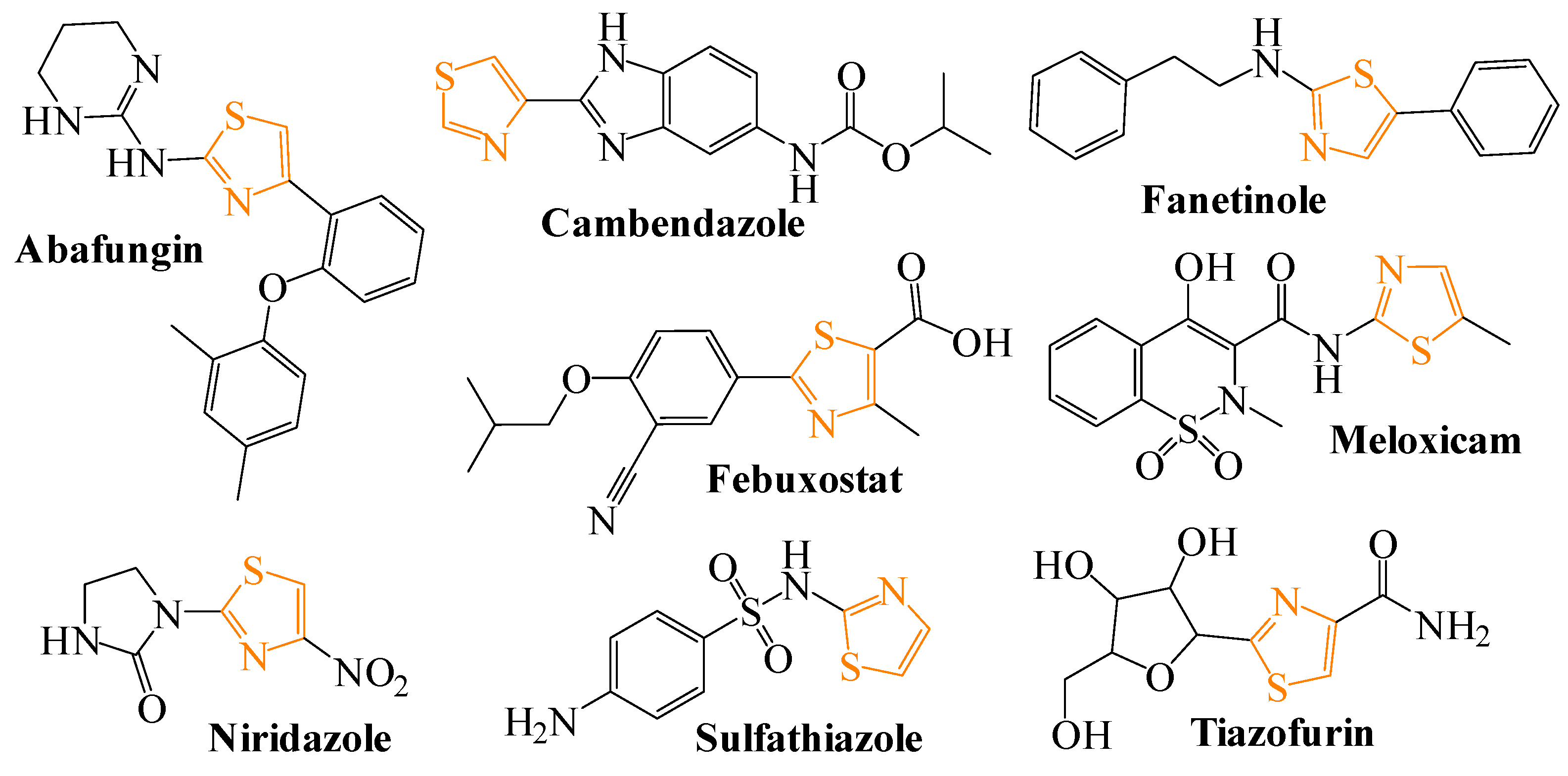
Among thiazoles structures, 1,3-thiazole is a five-membered compound that contains both sulfur and nitrogen in a heterocyclic ring [46,47]. It constitutes a core skeleton of various commercially available drug candidates and the main unit in a diverse range of important scaffolds that are central to several fields of science, biological and medicinal systems [48]. The chemistry of thiazoles was widely developed with respect to anti-microbial, anti-cancer, anti-tubercular, antioxidant, anti-inflammatory, and other therapeutically active agents [49,50,51,52,53,54,55,56,57,58]. Figure 1 shows selected examples of commercially available drugs and bioactive molecules based on thiazole nucleus [46,48,59,60].
A common strategy for designing new compounds with anti-Trypanosomatidae activity is to target enzymes that are essential to the parasite’s metabolism. One such enzyme is trypanothione synthetase (TryS), which plays a key role in the synthesis of antioxidant molecules. Together with trypanothione reductase (TryR), TryS is crucial for maintaining redox balance and, consequently, for the survival of Trypanosomatidae parasites [61,62]. In this regard, compounds containing a thiazole ring with various modifications have been studied in silico and in vitro against Leishmania and Trypanosoma parasites.
As part of our research interest in synthesis and biological evaluation of heterocyclic compounds [63,64,65,66,67,68,69,70] and with the aim of advancing drug discovery of new antileishmanial agents [71,72], here, we describe a new method for the preparation of arylidenehydrazinyl 4-methylthiazole-5-carboxylates derivatives. Moreover, their cytotoxicity against promastigote and intracellular amastigote forms of Leishmania amazonensis and the NIH/3T3 mouse fibroblast cell lines has been described. Furthermore, in silico molecular docking studies were also explored as a preliminary computational approach to investigate the interaction patterns of selected compounds with target proteins of Leishmania trypanothione synthetase.

2. Results and Discussion

2.1. Chemistry

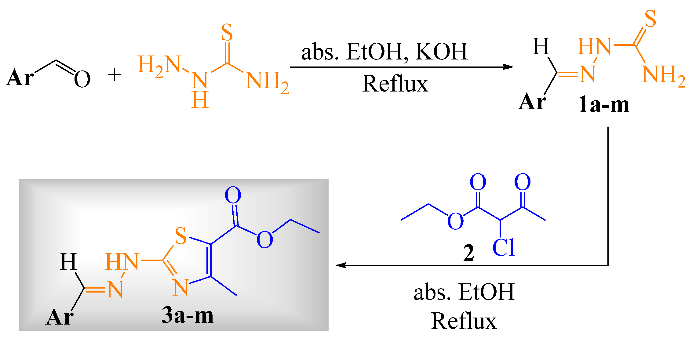
The targeted compounds (3am) were synthesized through the method shown in Scheme 1, with respective structures and yields in Table 1. The procedure involves refluxing equimolar mixture of aryl-substituted thiosemicarbazones (1am) and ethyl 2-chloro-3-oxobutanoate (2) in absolute ethanol. A colored heterocyclic product formed upon pouring in crushed ice.
The formation of cyclized product was first indicated by color changes, melting point and chromatography Rf values (provided in experimental data for each compound). Functional group analysis was carried out through IR spectroscopy. Structural exposition of each individual compound was attained through 1H and 13C NMR spectroscopy. The spectroscopic data is consistent with proposed structure, confirmed by HRMS.
IR spectra indicated the key connections (functional groups) in the synthesized compounds. The presence of ester group, azomethine carbon and methyl group bonded at the fourth position of the thiazole ring is indicated by the absorption band in the range of 1699–1605 cm−1, 1599–1549 cm−1 and 2984–2901 cm−1 respectively. The same linages were supported by 1HNMR spectroscopy. A triplet of three protons in the range of 1.27–1.39 ppm and a downfield quartet of two protons in the range of 4.20–4.33 ppm are an indication of the ethyl group of the ester. A singlet of three protons in the range of 2.22–2.68 ppm is ascribed to the methyl group at C4 of thiazole. A characteristic signal of the azomethine proton was observed in the range of 7.71–8.41 ppm in all cases. The proposed structures were further verified through 13CNMR spectroscopy. Characteristic signals in the 13CNMR spectrum include C2, C4, C5 of thiazole, carbonyl carbon and azomethine carbon. These carbons were observed in the range of 167.4–170.9 ppm, 149.9–159.1 ppm, 110.2–128.9 ppm, 161.9–162.9 ppm and 132.8–153.7 ppm respectively. The calculated masses of the proposed structures are in good agreement with observed masses in the HRMS.

2.2. Biological Assays

The compounds were tested for cytotoxicity against promastigote and amastigote forms of Leishmania (L.) amazonensis (IFLA/BR/1967/PH8) and the NIH/3T3 mouse fibroblast cell lines. All compounds showed little or no cytotoxicity against NIH/3T3 fibroblasts at the concentrations tested (15.625 to 250 µM), as shown in Table 2. Only compounds 3d, 3e and 3m reduced cell viability at higher concentrations, and CC50 values were obtained (Figure S55). These results indicate that, in general, the compounds were not toxic to the cell line tested, a promising factor for in vitro biological activity studies.
As for the promastigote forms of L. amazonensis, some compounds reduced parasite´s viability at the higher concentrations tested (1.56 to 25 µM), Table 2, as noted for compounds 3d and 3m (Figure S56). Considering the small reduction in viability observed, the IC50 values were obtained only for compound 3a (Table 2). Two reference drugs for the leishmaniasis treatment, amphotericin B and pentamidine, were also evaluated as controls in this experiment, and their IC50 are also in Table 2 and Figure S58. It is important to note that compounds displaying little or no in vitro activity against promastigote forms of L. amazonensis may still exhibit significant activity against intracellular amastigotes, which represent the clinically relevant form found in vertebrate hosts [73].
To this end, an experiment was conducted in which murine macrophages infected with L. amazonensis were treated with compounds 3am to evaluate their effect on intracellular amastigotes. This experimental model was designed to mimic in vivo infection environment, in which the compounds must penetrate infected macrophages and reduce the intracellular parasite burden. Four of the compounds exhibited activity against intracellular amastigotes: 3m was the most active, followed by 3c, 3h and 3b (see Table 2 and Figure S57).
A preliminary structure–activity relationship (SAR) analysis was conducted based on the antiamastigote results obtained for the series 3am. The common scaffold—an arylidenehydrazinyl 4-methylthiazole-5-carboxylate—was held constant across all derivatives, and biological differences were thus attributed to the nature and substitution pattern of the arylidene moiety. Among the inactive or poorly active compounds, those bearing electron-donating methyl groups (3f), methoxy groups in symmetric or meta positions (3i, 3j), a hydroxyl/methyl combination (3e), or a bithiazole moiety (3l), showed no meaningful activity against amastigotes, suggesting that these substituents do not favor intracellular parasite inhibition. In contrast, the four active compounds revealed interesting trends: 3b and 3c, bearing 2,6-dichlorophenyl and 3,4-dichlorophenyl substituents, respectively, displayed comparable antiamastigote IC50 values (~32–34 µM), indicating that the presence of two chlorine atoms on the aryl ring is favorable, regardless of their relative position. Compound 3h, featuring a 2,4-dimethoxyphenyl group, also exhibited similar activity (~33.6 µM), suggesting that electron-donating ortho/para methoxy groups may also confer antiamastigote activity, possibly through enhanced membrane permeability. Most notably, compound 3m, bearing a 3-bromothiophen-2-yl heterocyclic substituent instead of a phenyl ring, was the most potent derivative of the series (IC50 = 15.92 µM), highlighting the advantage of replacing the benzene ring with a bromine-substituted thiophene. The combination of the electron-withdrawing bromine atom and the sulfur-containing heteroaromatic system appears to positively modulate both lipophilicity and the capacity to interact with intracellular targets, consistent with its superior docking score against trypanothione synthetase (binding energy: −5.37 kcal/mol, Table 3). Overall, these findings suggest that halogen substitution—particularly on heteroaromatic rings—and ortho/para-directed electron-withdrawing groups are important structural features for antileishmanial activity in this series and should be prioritized in future structural optimization efforts.
Compound 3m showed low activity against promastigotes but was the most active against intracellular amastigotes of L. amazonensis, with the lowest IC50 obtained for this series. It also exhibited good selectivity, as shown in Table 2 by the selectivity index (SI) of 7.6, calculated as the ratio of CC50 to IC50. Although macrophage cytotoxicity would be the most biologically matched comparator for the intracellular amastigote assay, SI values are commonly calculated against different mammalian cell types in antileishmanial screening. We therefore report the NIH/3T3-based SI as a general measure of mammalian selectivity, while acknowledging that a macrophage CC50 would provide a more assay-matched denominator [74]. Compounds 3c, 3h and 3b showed similar antiamastigote activity, whose IC50 values ranged from 32.86 to 34.18 µM. Nevertheless, the results obtained in the present study, while encouraging, remain modest: compound 3m, the most active derivative of the series, exhibited antiamastigote activity with an IC50 of approximately 15 μM, inferior to reference drugs such as amphotericin B and pentamidine, also used as controls here, with IC50 also in Table 2 results are similar to those in the recent literature regarding compounds containing thiazoles moieties. One study evaluated the synthesis and antiparasitic activity of 13 new thiosemicarbazones and 16 naphthyl-thiazole derivatives [75]. Against L. amazonensis, the thiazoles exhibited IC50 values between 16.48 and 247.59 µM for promastigote forms. In assays involving amastigote forms of Leishmania, IC50 values ranged results were recorded from 50.80 to 379.25 µM for thiazoles [5].
Another investigation explored the biological potential of eight 4-(4-chlorophenyl)thiazole compounds containing a naphthyl moiety [76]. For L. amazonensis, these molecules exhibited IC50 values ranging from 19.86 to 200 µM for promastigotes. However, activity against Leishmania amastigotes was characterized as moderate to inactive, with IC50 values between 101 and over 200 µM [76], well above the values obtained for our compounds.
In Leishmania parasites, trypanothione plays a critical role in defending against oxidative stress by neutralizing reactive oxygen species (ROS) produced by both the parasite and the host [77]. As trypanothione and its related enzymes (like trypanothione synthetase and reductase) are absent in humans, they present promising targets for antileishmanial therapies [78]. Disrupting trypanothione synthesis or function weakens the parasite’s antioxidant defenses, leading to its death without affecting human cells [79].
Targeting the trypanothione system has emerged as an effective strategy for the development of selective anti-Leishmania agents with reduced side effects. In the present study, a series of the most active compounds 3b, 3c, 3h, 3i, 3k, 3m and pentamidine was evaluated through molecular docking to preliminarily assess their binding interactions with Leishmania trypanothione synthetase (PDB ID: 2VOB) using AutoDock 4.2 [80]. The protein structure was prepared by removing water molecules and adding polar hydrogens and Kollman charges. The grid box was centered on the active site defined by the co-crystallized ligand to ensure inclusion of key catalytic residues. Docking simulations were carried out using the Lamarckian Genetic Algorithm (LGA) implemented in AutoDock 4.2 with multiple runs to ensure reproducibility and convergence of results.
The in silico docking results demonstrated that the binding energies of the tested compounds ranged from −3.16 to −5.37 kcal/mol compared to the reference compound pentamidine (−2.96 kcal/mol). These results suggest favorable predicted binding affinities and provide computational support for further investigation these compounds as potential anti-Leishmania agent.
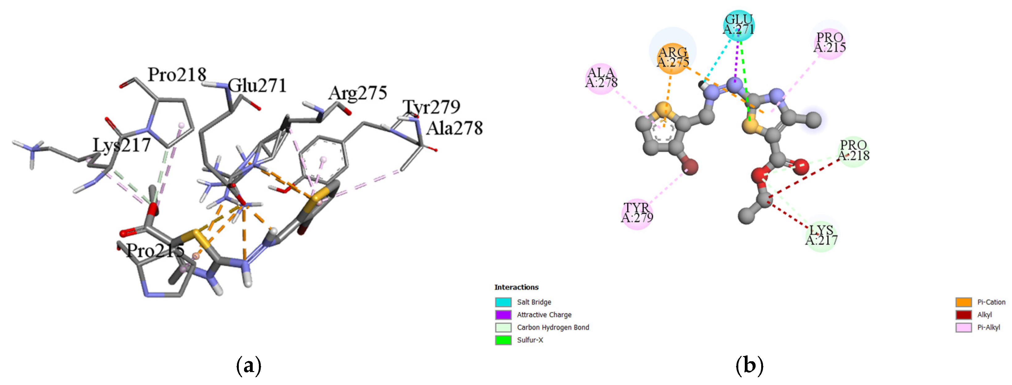
The molecular interactions between compounds 3b, 3c, 3h, 3i, 3k, 3m and the 2VOB protein from Leishmania were examined as shown in Figure 2 (see Figures S53 and S54 in the Supporting Information). These interactions involved hydrogen bonds, π-alkyl, π-sigma, amide-π interactions, and van der Waals forces. These interactions included hydrogen bonding, π-alkyl, π-sigma, amide-π stacking, and van der Waals forces. Table 3 summarizes the binding energies, inhibition constants, and the key amino acid residues involved in hydrogen bond formation. Importantly, the docking results against trypanothione synthetase indicate a possible interaction; however, these findings are considered supportive and hypothesis-generating with respect to the observed antileishmanial activity and do not establish a direct mechanistic link. Direct biochemical evidence of enzyme inhibition, such as enzymatic assays or target engagement studies, would be required to confirm trypanothione synthetase as the molecular target of these compounds.
It is important to emphasize that while molecular docking provides valuable insights into potential binding modes and interactions, these computational predictions require experimental validation. The binding energies and interaction patterns reported herein should be considered as hypothesis-generating data that support further investigation of trypanothione synthetase as a potential target, rather than as definitive evidence of enzyme inhibition.
Interestingly, the compounds with the lowest binding energies for 2VOB protein showed no cytotoxicity for NIH/3T3 fibroblasts or promastigote forms of L. amazonensis. However, two compounds, 3b and 3h, exhibited activity against intracellular amastigote forms of L. amazonensis, as shown in Table 2. While the binding energies listed in Table 3 suggest possible interactions with trypanothione synthetase, this remains a hypothesis that requires experimental validation through enzymatic assays. Furthermore, the molecular properties and aryl substituents of these compounds may facilitate their entry into macrophages and contribute to the observed antileishmanial activity through mechanisms that may or may not involve TryS inhibition.
The distinct metabolic, biochemical, and biological variations between promastigote and amastigote forms throughout Leishmania’s lifecycle [81] may account for differences in enzymatic inhibitory and antileishmanial activities observed across cell types [82]. In this context, the use of intracellular amastigotes provided essential information on the capacity of these compounds to target the clinically relevant stage of the parasite [83], and the partial corroboration of this activity through in vitro interaction with trypanothione synthetase (2VOB) supports a plausible mechanism of action, highlighting this enzyme as a potential target for this chemical class. Taken together, these findings suggest that compound 3m should be regarded as a preliminary hit, and that structural optimization guided by the present pharmacological and enzymatic data may lead to new synthetic molecules with improved potency and selectivity against Leishmania.

3. Materials and Methods

3.1. Synthesis of Ethyl-(E)-2-(2-arylidenehydrazineyl)-4-methylthiazole-5-carboxylates

3.1.1. Materials

All the reagents and solvents used for the synthesis of arylidenehydrazinyl thiazole carboxylates (3am) were of analytical grade and were used without any further purification. The reagents and solvents were purchased from reputed chemical suppliers, i.e., Sigma Aldrich, Riedel-de-Haen, and Analar. Reaction monitoring and retention factor (Rf) determination were performed on pre-coated thin layer chromatography (TLC) sheets (Silica gel 60 F254, Merck, Darmstadt, German), which were visualized either through quenching of ultraviolet or fluorescence light (λmax = 254 and 366 nm, respectively). A digital melting point apparatus was used to determine the uncorrected melting points of all solid compounds. Final products (3am) were characterized using the 1H and 13C NMR spectra on 400 MHz (1H: 400 MHz; 13C: 100 MHz) and 500 MHz (1H: 500 MHz; 13C: 125 MHz) spectrometers, having the residual solvent peaks of CDCl3 (1H: δ 7.26; 13C: δ 77.2), DMSO-d6 (1H: δ 2.50; 13C: δ 39.7), as an indirect reference to tetramethyl silane (TMS). The electron ionization (EI) technique was utilized to set up the time of flight (TOF) equipment used to generate the high-resolution mass spectra (HRMS) operating in the positive ion mode.
All reactions were performed in a well-ventilated fume hood under an inert environment using a Schlenk line. The starting materials, aryl-substituted thiosemicarbazones 1am, were prepared according to our previous report [84].

3.1.2. General Procedure for the Synthesis of Arylidenehydrazinyl Thiazole Carboxylates (3am)

The arylidenehydrazinyl thiazole carboxylate derivatives were prepared by refluxing together aryl-substituted thiosemicarbazones 1am (1 mmol) with equimolar amount of ethyl 2-chloro-3-oxobutanoate 2 in absolute ethanol (15 mL) for a duration of 5 h. Reaction progress was monitored through TLC and upon completion; the reaction was quenched with ice cold water, resulting precipitation. The resulting precipitates were filtered and successively washed with copious deionized water to afford final product 3, which were dried under vacuum. Lastly, purity was rechecked via TLC.

3.1.3. Spectral Data of Synthesized Products (3am)

Ethyl (E)-4-methyl-2-(2-((5-methylfuran-2-yl)methylene)hydrazineyl)thiazole-5-carboxylate (3a). Off-white solid; yield: 72%; melting point: 187–189 °C; Rf: 0.55 (acetone/n-hexane, 1:3); FTIR (ATR) cm−1: 3185 (N-H stretching), 3066 (=C-H stretching), 2922 (C-H aliphatic stretching), 1683 (C=O stretching), 1582 (C=N stretching), 1539, 1522, 1366, 1313 (C=C aromatic ring stretching), 1424 (C-H aliphatic bending), 1088 (C-O stretching), 1012-734 (characteristic of thiazole); 1H-NMR (400 MHz, CDCl3): δ (ppm) 7.71 (s, 1H, H-C=N-), 6.59 (d, 1H, furan-H, J = 4.6 Hz), 6.10 (d, 1H, furan-H, J = 3.4 Hz), 4.30 (q, 2H, -CH2-CH3, J = 7.1 Hz), 2.60 (s, 3H, -CH3), 2.40 (s, 1H, -CH3), 1.37 (t, 3H, CH3-CH2, J = 7.1 Hz); 13C-NMR (100 MHz, CDCl3): δ (ppm) 170.0 (Thiazole C2), 162.6 (C=O), 156.7 (thiazole C4), 155.4 (furan C5), 147.5 (furan C2) 134.6 (H-C=N- azomethine), 114.6 (thiazole C5), 111.0 (furan C3), 108.4 (furan C4), 60.6 (CH2-O-), 17.1 (CH3-), 14.4 (CH3-CH2), 13.9 (-CH3); HRMS: m/z calculated for C13H15N3O3S [M+H]+: calculated 294.0907, found 294.0909, [M + Na]+: calculated 316.0726, found 316.0723, [2M + Na]+: calculated 609.1560, found 609.1551.
Ethyl (E)-2-(2-(2,6-dichlorobenzylidene)hydrazineyl)-4-methylthiazole-5-carboxylate (3b). Off-white solid; yield: 75%; melting point: 201–202 °C; Rf: 0.64 (acetone/n-hexane, 1:3); FTIR (ATR) cm−1: 3136 (N-H stretching), 3988 (=C-H stretching), 2901 (C-H aliphatic stretching), 1699 (C=O stretching), 1580 (C=N stretching), 1549, 1430, 1370, 1313 (C=C aromatic ring stretching), 1409 (C-H aliphatic bending), 1086 (C-O stretching), 975-755 (characteristic of thiazole); 1H-NMR (400 MHz, CDCl3): δ (ppm) 8.27 (s, 1H, H-C=N), 7.39 (d, 2H, Ar-H, J = 8.0 Hz), 7.22 (d, 1H, Ar-H, J = 8.0 Hz), 4.31 (q, 2H, -CH2-CH3, J = 7.2 Hz), 2.67 (s, 3H, -CH3), 1.37 (t, 3H, CH3-CH2, J = 7.2 Hz); 13C-NMR (100 MHz, CDCl3): δ (ppm) 170.9 (thiazole C2), 162.6 (C=O), 156.7 (thiazole C4), 139.3 (C=N, azomethine), 135.0, 130.0, 129.4, 129.2 (Ar-C), 111.8 (thiazole C5), 60.8 (CH2-O), 17.3 (-CH3), 14.4 (CH3-CH2); HRMS: m/z calculated for C14H13Cl2N3O2S [M + H]+: calculated 358.0178, found 358.0168, [M + Na]+: calculated 379.9998, found 379.9994, [2M + Na]+: calculated 737.0103, found 737.0077.
Ethyl (E)-2-(2-(3,4-dichlorobenzylidene)hydrazineyl)-4-methylthiazole-5-carboxylate (3c). Light yellow solid; yield: 73%; melting point: 214–216 °C; Rf: 0.60 (acetone/n-hexane, 1:3); FTIR (ATR) cm−1: 3200 (N-H stretching), 3093 (=C-H stretching), 2934 (C-H aliphatic stretching), 1656 (C=O stretching), 1562 (C=N stretching), 1473, 1368, 1329 (C=C aromatic ring stretching), 1432 (C-H aliphatic bending), 1096 (C-O stretching), 1029-728 (characteristic of thiazole); 1H-NMR (400 MHz, CDCl3): δ (ppm) 7.81 (s, 1H, H-C=N), 7.73 (d, 1H, Ar-H, J = 1.9 Hz), 7.49–7.42 (m, 2H, Ar-H), 4.20 (q, 2H, -CH2-CH3, J = 7.2 Hz), 2.57 (s, 3H, -CH3), 1.34 (t, 3H, CH3-CH2, J = 7.2 Hz); 13C-NMR (100 MHz, CDCl3): δ (ppm) 170.0 (thiazole C2), 162.0 (C=O), 155.9 (thiazole C4), 141.7 (C=N, azomethine), 134.0, 133.6, 130.8, 128.2, 126.1 (Ar-C), 111.9 (thiazole C5), 60.9 (CH2-O), 16.7 (CH3-), 14.4 (CH3-CH2); HRMS: m/z calculated for C14H13Cl2N3O2S [M + H]+: calculated 358.0178, found 358.0178, [M + Na]+: calculated 379.9998, found 379.9995, [2M + Na]+: calculated 737.0103, found 737.0087.
Ethyl (E)-2-(2-(2,4-bis(trifluoromethyl)benzylidene)hydrazineyl)-4-methylthiazole-5-carboxylate (3d). Off-white solid; yield: 71%; melting point: 212–214 °C; Rf: 0.52 (acetone/n-hexane, 1:3); FTIR (ATR) cm−1: 3206 (N-H stretching), 3101 (=C-H stretching), 2984 (C-H aliphatic stretching), 1662 (C=O stretching), 1564 (C=N stretching), 1467, 1393, 1333 (C=C aromatic ring stretching), 1428 (C-H aliphatic bending), 1113 (C-O stretching), 1053-761 (characteristic of thiazole); 1H-NMR (400 MHz, DMSO-d6): δ (ppm) 8.39-8.31 (m, 2H, Ar-H), 8.09 (d, 1H, Ar-H, J = 8.5 Hz), 8.03 (s, 1H, H-C=N), 4.21 (q, 2H, -CH2-CH3, J = 7.1 Hz), 2.48 (s, 3H, -CH3), 1.27 (t, 3H, CH3-CH2, J = 7.1 Hz); 13C-NMR (100 MHz, DMSO-d6): δ (ppm) 179.1 (Ar-C-CF3), 168.1 (thiazole C2), 163.6 (Ar-C-CF3), 162.2 (C=O), 159.1 (thiazole C4), 136.8 (C=N, azomethine), 136.3-122.4 (Ar-C), 119.7 (thiazole C5), 60.8 (CH2-O), 19.0 (-CH3), 14.7 (CH3-CH2-); HRMS: m/z calculated for C16H13F6N3O2S [M + H]+: calculated 426.0705, found 426.0696, [M + Na]+: calculated 448.0525, found 448.0506, [2M + Na]+: calculated 873.1158, found 873.1126.
Ethyl (E)-2-(2-(2-hydroxy-3-methylbenzylidene)hydrazineyl)-4-methylthiazole-5-carboxylate (3e). Light yellow solid; yield: 75%; melting point: 244–246 °C; Rf: 0.54 (acetone/n-hexane, 1:3); FTIR (ATR) cm−1: 3157 (N-H stretching), 3054 (=C-H stretching), 2969 (C-H aliphatic stretching), 1629 (C=O stretching), 1568 (C=N stretching), 1533, 1483, 1374, 1329 (C=C aromatic ring stretching), 1432 (C-H aliphatic bending), 1101 (C-O stretching), 1039-745 (characteristic of thiazole); 1H-NMR (400 MHz, DMSO-d6): δ (ppm) 8.41 (s, 1H, H-C=N), 7.31 (d, 1H, Ar-H, J = 7.8 Hz), 7.19 (d, 1H, Ar-H, J = 6.9 Hz), 6.85 (t, 1H, Ar-H, J = 7.5 Hz), 4.21 (q, 2H, -CH2-CH3, J = 7.1 Hz), 2.45 (s, 3H, -CH3), 2.22 (s, 3H, -CH3), 1.27 (t, 3H, CH3-CH2, J = 7.1 Hz); 13C-NMR (100 MHz, DMSO-d6): δ (ppm) 167.4 (thiazole C2), 161.9 (C=O), 155.9 (thiazole C4), 132.7 (C=N, azomethine), 128.6, 125.4 119.8 (Ar-C), 118.4 (thiazole C5), 60.8 (CH2-O), 16.0 (CH3-), 14.8 (CH3-CH2); HRMS: m/z calculated for C15H17N3O3S [M + H]+: calculated 320.1063, found 320.1066, [M + Na]+: calculated 342.0883, found 342.0882, [2M + Na]+: calculated 661.1873, found 661.1857.
Ethyl (E)-2-(2-(2,5-dimethylbenzylidene)hydrazineyl)-4-methylthiazole-5-carboxylate (3f). Off-white solid; yield: 78%; melting point: 220–222 °C; Rf: 0.69 (acetone/n-hexane, 1:3); FTIR (ATR) cm−1: 3204 (N-H stretching), 3103 (=C-H stretching), 2980 (C-H aliphatic stretching), 1654 (C=O stretching), 1560 (C=N stretching), 1426, 1395, 1364, 1325 (C=C aromatic ring stretching), 1094 (C-O stretching), 1043-724 (characteristic of thiazole); 1H-NMR (400 MHz, CDCl3): δ (ppm) 8.17 (s, 1H, H-C=N), 7.61 (s, 1H, Ar-H), 7.11 (d, 1H, Ar-H, J = 1.8 Hz), 4.30 (q, 2H, -CH2-CH3, J = 7.2 Hz), 2.65 (s, 3H, CH3), 2.45 (s, 3H, -CH3), 2.38 (s, 3H, -CH3) 1.39 (t, 3H, CH3-CH2, J = 7.2 Hz); 13C-NMR (100 MHz, CDCl3): δ (ppm) 170.3 (Thiazole C2), 162.6 (C=O), 156.9 (thiazole C4), 144.0 (C=N, azomethine), 135.8, 134.0, 131.3, 131.1, 130.8, 127.8 (Ar-C), 111.4 (thiazole C5), 60.7 (CH2-O), 21.0 (CH3-), 19.8 (-CH3), 17.3 (-CH3), 14.5 (CH3-CH2); HRMS: m/z calculated for C16H19N3O2S [M + H]+: calculated 318.1271, found 318.1265, [M + Na]+: calculated 340.1090, found 340.1080, [2M + Na] +: calculated 657.2288, found 657.2261.
Ethyl (E)-2-(2-(2,3-dimethoxybenzylidene)hydrazineyl)-4-methylthiazole-5-carboxylate (3g). Gray solid; yield: 75%; melting point: 227–229 °C; Rf: 0.64 (acetone/n-hexane, 1:3); FTIR (ATR) cm−1: 3150 (N-H stretching), 2943 (C-H aliphatic stretching), 1697 (C=O stretching), 1576 (C=N stretching), 1477, 1366, 1304 (C=C aromatic ring stretching), 1417 (C-H aliphatic bending), 1086 (C-O stretching), 1002-736 (characteristic of thiazole); 1H-NMR (400 MHz, CDCl3): δ (ppm) 8.33 (s, 1H, H-C=N), 7.60 (d, 1H, Ar-H, J = 6.5 Hz), 7.11 (t, 1H, Ar-H, J = 8.0 Hz), 6.96 (d, 1H, Ar-H, J = 6.5 Hz), 4.33 (q, 2H, -CH2-CH3, J = 7.2 Hz), 3.91 (s, 3H, -OCH3), 3.88 (s, 3H, -OCH3), 2.68 (s, 3H, -CH3), 1.39 (t, 3H, CH3-CH2, J = 7.2 Hz); 13C-NMR (100 MHz, CDCl3): δ (ppm) 170.5 (Thiazole C2), 162.7 (C=O), 157.6 (thiazole C4), 152.8 (Ar-C-OMe), 148.2 (Ar-C-OMe), 140.1 (C=N, azomethine), 127.4, 124.3, 117.8, 113.7 (Ar-C), 111.6 (thiazole C5), 61.6 (CH2-O), 60.7 (-OCH3), 55.9 (-OCH3), 17.3 (-CH3), 14.5 (CH3-CH2); HRMS: m/z calculated for C16H19N3O4S [M + H]+: calculated 350.1169, found 350.1165, [M + Na]+: calculated 372.0988, found 372.0980, [2M + Na]+: calculated 721.2085, found 721.2062.
Ethyl (E)-2-(2-(2,4-dimethoxybenzylidene)hydrazineyl)-4-methylthiazole-5-carboxylate (3h). Light brown solid; yield: 75%; melting point: 220–222 °C; Rf: 0.60 (acetone/n-hexane, 1:3); FTIR (ATR) cm−1: 3179 (N-H stretching), 3064 (=C-H stretching), 2955 (C-H aliphatic stretching), 1691 (C=O stretching), 1586 (C=N stretching), 1533, 1502, 1370, 1313 (C=C aromatic ring stretching), 1428 (C-H aliphatic bending), 1127 (C-O stretching), 1031-757 (characteristic of thiazole); 1H-NMR (500 MHz, CDCl3): δ (ppm) 8.30 (s, 1H, H-C=N), 7.93 (d, 1H, Ar-H, J = 8.6 Hz), 6.57 (dd, 1H, Ar-H, J = 8.6, 2,4 Hz), 6.45 (d, 1H, Ar-H, J = 2.4 Hz), 4.31 (q, 2H, -CH2-CH3, J = 7.1 Hz), 3.87 (s, 3H, -OCH3), 3.83 (s, 3H, -OCH3), 2.65 (s, 3H, -CH3), 1.38 (t, 3H, CH3-CH2, J = 7.1 Hz); 13C-NMR (125 MHz, CDCl3): δ (ppm) 170.2 (Thiazole C2), 162.8 (C=O), 162.7 (Ar-C-OMe), 159.1 (thiazole C4), 157.6 (Ar-C-OMe), 140.7 (C=N, azomethine), 127.4, 115.2 (Ar-C), 110.9 (thiazole C5), 105.9, 97.9 (Ar-C), 60.6 (CH2-O), 55.5 (2x-OCH3) 17.1 (-CH3), 14.5 (CH3-CH2); HRMS: m/z calculated for C16H19N3O4S [M + H]+: calculated 350.1169, found 350.1164, [M + Na] +: calculated 372.0988, found 372.0982, [2M + Na]+: calculated 721.2085, found 721.2070.
Ethyl (E)-2-(2-(2,5-dimethoxybenzylidene)hydrazineyl)-4-methylthiazole-5-carboxylate (3i). Beige solid; yield: 77%; melting point: 222–224 °C; Rf: 0.66 (acetone/n-hexane, 1:3); FTIR (ATR) cm−1: 3045 (=C-H stretching), 2932 (C-H aliphatic stretching), 1697 (C=O stretching), 1576 (C=N stretching), 1537, 1494, 1368 (C=C aromatic ring stretching), 1422 (C-H aliphatic bending), 1078 (C-O stretching), 1022-794 (characteristic of thiazole); 1H-NMR (500MHz, CDCl3): δ (ppm) 8.35 (s, 1H, H-C=N), 7.52 (d, 1H, Ar-H, J = 3.1 Hz), 6.93 (dd, 1H, Ar-H, J = 8.9, 3.1 Hz), 6.85 (d, 1H, Ar-H, J = 8.9 Hz) 4.30 (q, 2H, -CH2-CH3, J = 7.1 Hz), 3.87 (s, 3H, -OCH3), 3.81 (s, 3H, -OCH3), 2.65 (s, 3H, -CH3), 1.38 (t, 3H, CH3-CH2, J = 7.1 Hz); 13C-NMR (125 MHz, CDCl3): δ (ppm) 170.7 (thiazole C2), 162.8 (C=O), 157.9 (thiazole C4), 153.7 (C=N, azomethine), 152.4, 140.1, 122.8, 117.4, 112.4, 111.2 (Ar-C), 110.2 (thiazole C5), 60.6 (CH2-O), 56.0 (-OCH3), 55.9 (-OCH3), 17.1 (CH3-), 14.5 (CH3-CH2); HRMS: m/z calculated for C16H19N3O4S [M + H]+: calculated 350.1169, found 350.1160, [M + Na]+: calculated 372.0988, found 372.0975, [2M + Na]+: calculated 721.2085, found 721.2064.
Ethyl (E)-2-(2-(3,5-dimethoxybenzylidene)hydrazineyl)-4-methylthiazole-5-carboxylate (3j). Off-white solid; yield: 77%; melting point: 206–208 °C; Rf: 0.66 (acetone/n-hexane, 1:3); FTIR (ATR) cm−1: 3212 (N-H stretching), 3099 (=C-H stretching), 2965 (C-H aliphatic stretching), 1669 (C=O stretching), 1562 (C=N stretching), 1525, 1463, 1364, 1317 (C=C aromatic ring stretching), 1415 (C-H aliphatic bending), 1121 (C-O stretching), 1086-726 (characteristic of thiazole); 1H-NMR (500 MHz, CDCl3): δ (ppm) 7.85 (s, 1H, H-C=N), 6.83 (d, 2H, Ar-H, J = 2.2 Hz), 6.52 (t, 1H, Ar-H, J = 2.2 Hz), 4.32 (q, 2H, -CH2-CH3, J = 7.1 Hz), 3.86 (s, 6H, 2X-OCH3), 2.63 (s, 3H, -CH3), 1.39 (t, 3H, CH3-CH2, J = 7.1 Hz); 13C-NMR (125 MHz, CDCl3): δ (ppm) 169.8 (Thiazole C2), 162.5 (C=O), 161.0 (Ar-C-OMe), 156.6 (thiazole C4), 144.5 (C=N, azomethine), 135.4 (Ar-C), 111.7 (thiazole C5), 105.0, 102.6 (Ar-C), 60.8 (CH2-O), 55.5 (2x-OCH3) 17.1 (-CH3), 14.4 (CH3-CH2); HRMS: m/z calculated for C16H19N3O4S [M + H]+: calculated 350.1169, found 350.1161, [M + Na]+: calculated 372.0988, found 372.0977, [2M + Na]+: calculated 721.2085, found 721.2064.
Ethyl (E)-4-methyl-2-(2-(2,4,5-trimethoxybenzylidene)hydrazineyl)thiazole-5-carboxylate (3k). Light purple solid; yield: 72%; melting point: 234–235 °C; Rf: 0.59 (acetone/n-hexane, 1:3); FTIR (ATR) cm−1: 3140 (N-H stretching), 2936 (C-H aliphatic stretching), 1695 (C=O stretching), 1599 (C=N stretching), 1572, 1516, 1370, 1310 (C=C aromatic ring stretching), 1420 (C-H aliphatic bending), 1072 (C-O stretching), 1024-755 (characteristic of thiazole); 1H-NMR (400 MHz, CDCl3): δ (ppm) 8.31 (s, 1H, H-C=N), 7.48 (s, 1H, Ar-H), 6.48 (s, 1H, Ar-H), 4.30 (q, 2H, -CH2-CH3, J = 7.1 Hz), 3.96 (s, 3H, -OCH3), 3.94 (s, 3H, -OCH3), 3.83 (s, 3H, -OCH3), 2.64 (s, 3H, -CH3), 1.37 (t, 3H, CH3-CH2, J = 7.1 Hz); 13C-NMR (100 MHz, CDCl3): δ (ppm) 170.5 (Thiazole C2), 162.9 (C=O), 158.2 (thiazole C4), 153.3 (Ar-C-OMe), 152.1 (Ar-C-OMe), 143.7 (Ar-C-OMe), 140.3 (C=N, azomethine), 113.9 (Ar-C), 110.9 (thiazole C5), 108.2, 96.6 (Ar-C), 60.5 (CH2-O), 56.5 (-OCH3), 56.3 (-OCH3), 56.1 (-OCH3), 17.3 (-CH3), 14.5 (CH3-CH2); HRMS: m/z calculated for C17H21N3O5S [M + H]+: calculated 380.1275, found 380.1276, [M + Na]+: calculated 402.1094, found 402.1093, [2M + Na]+: calculated 781.2296, found 781.2283.
Ethyl (E)-2-(2-((4,5-dimethylthiazol-2-yl)methylene)hydrazineyl)-4-methylthiazole-5-carboxylate (3l). Yellow solid; yield: 66%; melting point: 219–221 °C; Rf: 0.51 (acetone/n-hexane, 1:3); FTIR (ATR) cm−1: 3152 (N-H stretching), 3039 (=C-H stretching), 2916 (C-H aliphatic stretching), 1605 (C=O, ester stretching), 1564 (C=N stretching), 1531, 1426, 1376, 1356 (C=C aromatic ring stretching), 1111 (C-O stretching), 1024-740 (characteristic of thiazole); 1H-NMR (500 MHz, DMSO-d6): δ (ppm) 8.10 (s, 1H, H-C=N), 4.21 (q, 2H, O-CH2-CH3, J = 7.1 Hz) 2.47 (s, 3H, CH3-), 2.36 (s, 3H, H3C-thiazole-C5), 2.27 (s, 3H, H3C-thiazole-C4), 1.27 (t, 3H, CH3-CH2, J = 7.1 Hz); 13C-NMR (125 MHz, DMSO-d6): δ (ppm) 169.0 (thiazole ring C2), 162.2 (C=O), 159.2 (thiazole C2), 155.2 (thiazole C4), 149.9 (thiazole C4), 147.1 (HC=N- azomethine), 142.4 (thiazole C5), 128.9 (thiazole C5), 60.8 (CH2-O), 14.9 (CH3-) 14.7 (CH3-), 14.6 (CH3-), 11.7 (CH3-CH2-); HRMS: m/z calculated for C13H16N4O2S2, [M + H]+: 325.0787, found 325.0781, [M + Na]+: calculated 347.0607, found 347.0604, [2M + Na]+: calculated 671.1322, found 671.1305.
Ethyl (E)-2-(2-((3-bromothiophen-2-yl)methylene)hydrazineyl)-4-methylthiazole-5-carboxylate (3m). Light yellow solid; yield: 72%; melting point: 210–212 °C; Rf: 0.56 (acetone/n-hexane, 1:3); 1H-NMR (400MHz, DSMO-d6): δ (ppm) 8.23 (s, 1H, H-C=N-), 7.75 (d, 1H, thiophene-H, J = 4.6 Hz), 7.19 (d, 1H, thiophene-H, J = 5.4 Hz), 4.21 (q, 2H, -CH2-CH3, J = 7.2 Hz), 2.47 (s, 3H, -CH3), 1.27 (t, 3H, CH3-CH2, J = 7.2 Hz); 13C-NMR (100 MHz, DMSO-d6): δ (ppm) 168.9 (Thiazole C2), 162.2 (C=O), 133.8 (H-C=N- azomethine) 131.3 (thiophene C5), 131.2 (thiophene C4), 130.2 (thiophene C2), 129.9 (thiophene C3), 113.0 (thiazole C5), 60.7 (CH2-O-), 17.3 (CH3-), 14.8 (CH3-CH2); HRMS: m/z calculated for C12H12BrN3O2S2 [M + H]+: calculated 373.9627, found 373.9627, [M + Na]+: calculated 395.9447, found 395.9441, [2M + Na]+: calculated 768.9001, found 768.8985.

3.2. Biological Evaluation

3.2.1. Cytotoxicity

A suspension containing 104 NIH/3T3 cells (ATCC, catalog number CRL-1658, USA) in RPMI-1240 medium (Sigma-Aldrich, St. Louis, MO, USA) supplemented with 10% (v/v) fetal bovine serum (Cultilab, Campinas, SP, Brazil) and antibiotics (100 units penicillin and 0.01 mg streptomycin/mL, Sigma-Aldrich, USA) was plated in 96-well plates and allowed to adhere, and then it was incubated at 37 °C in a 5% CO2 incubator (ThermoFischer, Waltham, MA, USA) for 18 h. Supernatants were replaced with the supplemented RPMI medium containing the compounds at concentrations ranging from 250 to 15.6 μM in quadruplicates. The plates were then incubated at 37 °C and 5% CO2 for 48 h, after which viability was assessed through the resazurin method: 5 µL of a 0.2 mg/mL (m/v) resazurin solution (Sigma-Aldrich, USA) was added to each well and, after 4 h incubation, absorbance was read at 570 and 600 nm with plate reader spectrophotometer (SpectraMax, Molecular Devices, San Jose, CA, USA). Negative control wells containing only medium and positive controls containing amphotericin B (50 to 0.78 µM, Sigma-Aldrich, USA) and pentamidine (250 to 7.8 µM, Sigma-Aldrich, USA) were also evaluated. The percentage of viability for each compound concentration was calculated compared to the live control with medium alone, considered 100% live cells.

3.2.2. Antipromastigote Activity

Promastigote forms (2 × 10⁵) of L. (L.) amazonensis PH8 strain (IFLA/BR/1967/PH8, code 0575 from CLIOC Leishmania Collection, Oswaldo Cruz Institute, Rio de Janeiro – RJ, Brazil) in Schneider’s Insect medium (Sigma-Aldrich, USA) supplemented with 20% (v/v) fetal bovine serum (Cultilab, Campinas, SP, Brazil) and antibiotics (100 units penicillin and 0.01 mg streptomycin/mL, Sigma-Aldrich, USA) were distributed in 96-well plates containing the compounds at concentrations ranging from 25 to 1.56 μM in quadruplicate. The plates were incubated at 26 °C for 48 h (BOD incubator, Marqlabor, Matão, SP, Brazil), after which viability was assessed spectrophotometrically using the resazurin method (Sigma-Aldrich, USA) as described above. Negative control containing only medium and positive controls containing amphotericin B (5 to 0.078 µM, Sigma-Aldrich, USA) and pentamidine (250 to 3.90 µM, Sigma-Aldrich, USA) were also evaluated, as reference antileishmanial drugs. The percentage of viability for each compound concentration was calculated in relation to the negative control with medium alone, which was considered to represent 100% live cells.

3.2.3. Antiamastigote Activity

The methodology for assessing antileishmanial activity against intracellular amastigotes was adapted [85] using murine peritoneal macrophages. This protocol was approved by the Animal Ethics Committee (CEUA) of Federal University of Mato Grosso do Sul (UFMS) under certificate number 1.041/2019 and Balb/c female mice were provided by Central Animal Facility from Institute of Biosciences, UFMS on 23 April 2019, and ran through 31 July 2024. Peritoneal macrophages were collected after the animals were anesthetized and euthanized. The 96-well microplates were seeded with murine peritoneal macrophages (105) in supplemented RPMI medium and incubated at 37 °C with 5% CO2 for 1 h to allow adhesion. Adherent macrophages were infected with promastigote forms of L. amazonensis in the stationary phase (7-day culture) and then incubated at 35 °C/5% CO2 for 4 h to allow macrophage infection. After this period, the wells were washed with phosphate-buffered solution (PBS) and infected macrophages were then treated for 48 h with the compounds at concentrations ranging from 50 to 0.78 µM. Wells containing infected, untreated macrophages were used as a negative infection control, and wells treated with amphotericin B (1 to 0.0156 µM, Sigma-Aldrich, USA) and pentamidine (10 to 0.156 µM, Sigma-Aldrich, USA) were used as positive control, as reference antileishmanial drugs. After 48 h of incubation, the supernatants were removed, cells were washed with PBS, 150 µL of supplemented Schneider’s medium was added to each well, and the plates were incubated at 26 °C for 5 days to allow viable intracellular amastigotes to transform into promastigotes and multiply. Then, the viability of the promastigote forms in the microplates was assessed using the resazurin method, as described above.

3.2.4. Statistical Analysis

Experiments were conducted in quadruplicate. The one-way ANOVA test and Tukey’s post hoc test were used to compare variances, and the IC50 and CC50 values of the compounds were obtained through nonlinear regression based on the viability results. All graphs and analyses were performed using GraphPad Prism 5.0 software, assuming a 95% confidence interval (GraphPad Software, San Diego, CA, USA).

3.3. Molecular Docking

Molecular docking studies were performed targeting Leishmania trypanothione synthetase using the crystal structure with PDB ID: 2VOB obtained from the RCSB Protein Data Bank. The 3D structures of the antioxidant compounds 3b, 3c, 3h, 3i, 3k and 3m were generated using the Avogadro molecular editor and subsequently optimized by applying the MMFF94 force field. These optimized molecules were then converted into PDBQT format for docking studies. For protein preparation, water molecules were removed, polar hydrogens were added, and Kollman charges were assigned using AutoDock Tools. A grid box measuring 116 × 126 × 110 Å was set to cover the active site and allow ligand flexibility, with the center coordinates fixed at (x: −34.103, y: −8.467, z: 30.552) and a grid spacing of 0.667 Å, based on the protein’s catalytic region. During docking, the ligands were considered flexible, while the protein structure remained rigid. Docking simulations were carried out using the Lamarckian Genetic Algorithm (LGA) embedded in AutoDock 4.2, employing a population size of 150, a maximum of 2,500,000 energy evaluations, and 100 GA runs per ligand. Visualization and analysis of docking results were performed using BIOVIA Discovery Studio Visualizer, Version 21.1.0.20298, Dassault Systèmes BIOVIA, San Diego, CA, USA.

4. Conclusions

In summary, a series of novel arylidenehydrazinyl 4-methylthiazole-5-carboxylate derivatives was synthesized and evaluated as preliminary hits against Leishmania amazonensis. The most active compound of the series, 3m, displayed moderate antiamastigote activity and molecular docking studies provided supporting evidence for interaction with Leishmania trypanothione synthetase as a plausible mechanism of action. Although the biological potency of the present compounds remains modest in comparison with reference drugs, these results represent an encouraging starting point for the rational design of new synthetic molecules. Structural optimization guided by the pharmacological and molecular data reported herein may contribute to the development of more potent and selective antileishmanial candidates targeting trypanothione synthetase.

Supplementary Materials

The following supporting information can be downloaded at: https://www.mdpi.com/article/10.3390/molecules31132278/s1, 1HNMR and 13CNMR—Figures S1–S27; HRMS—Figures S28–S40; IR—Figures S41–S52; Binding model of compounds—Figures S53 and S54; Biological Activity of compounds 3am and for reference drugs amphotericin B and pentamidine—Figures S56–S58.

Author Contributions

Conceptualization: M.H., S.S. and J.R.; methodology: B.d.S.S., H.M., E.M.N. and N.d.S.B.; validation: B.d.S.S., H.M., T.A., E.M.N. and N.d.S.B.; formal analysis: T.A., N.S., V.P.S., A.M.J., G.A.C., A.B. and D.P.d.L.; investigation: B.d.S.S., H.M., E.M.N. and N.d.S.B.; resources: M.H., S.S., T.B.R. and J.R.; data curation: B.d.S.S., H.M. and E.M.N.; writing—original draft: S.S., T.B.R. and J.R.; writing—review and editing: T.B.R. and J.R.; visualization: S.S., T.B.R. and J.R.; supervision: S.S. and J.R.; project administration: M.H., S.S., T.B.R. and J.R.; funding acquisition: S.S. and J.R. All authors have read and agreed to the published version of the manuscript.

Funding

This work was supported by the Conselho Nacional de Desenvolvimento Científico e Tecnológico (CNPq) and the Coordenação de Aperfeiçoamento de Pessoal de Nível Superior (CAPES, Finance Code 001). S.S. and J.R. would like to acknowledge CNPq (308875/2026-5, 401355/2025-0, 316687/2023-5, 309975/2022-0, 404172/2023-7, and 405655/2023-1). S.S. also acknowledges the following FAPEG public call: Chamada Pública FAPEG/SES N° 18/2025 (ARB2025191000003).

Institutional Review Board Statement

All experimental procedures were conducted in compliance with the ethical guidelines approved by the Institutional Animal Care and Use Committee (CEUA/UFMS; protocol number 1.041/2019).

Informed Consent Statement

Not applicable.

Data Availability Statement

The original contributions presented in this study are included in the article and Supplementary Materials. The datasets used and/or analyzed during the current study are available from the corresponding authors upon request.

Acknowledgments

The authors thank the Conselho Nacional de Desenvolvimento Científico e Tecnológico (CNPq), Coordenação de Aperfeiçoamento de Pessoal de Nível Superior (CAPES) and Fundação Universidade Federal de Mato Grosso do Sul (UFMS) and Universidade Federal de Goiás (UFG) for the support offered in this research.

Conflicts of Interest

The authors declare no conflicts of interest.

References

  1. Sun, N.; Amon, J.J. Addressing Inequity: Neglected Tropical Diseases and Human Rights. Health Hum. Rights 2018, 20, 11–25. [Google Scholar] [PubMed]
  2. Ben Beard, C. Forgotten People, Forgotten Diseases: The Neglected Tropical Diseases and Their Impact on Global Health and Development. Emerg. Infect. Dis. 2009, 15, 510–511. [Google Scholar] [CrossRef] [Scilit]
  3. Weld, E.D.; Waitt, C.; Barnes, K.; Garcia Bournissen, F. Twice neglected? Neglected diseases in neglected populations. Br. J. Clin. Pharmacol. 2022, 88, 367–373. [Google Scholar] [PubMed]
  4. Ganasegeran, K.; Abdulrahman, S.A. Epidemiology of Neglected Tropical Diseases. In Neglected Tropical Diseases and Phytochemicals in Drug Discovery; Wiley Online Books: Hoboken, NJ, USA, 2021; pp. 1–36. [Google Scholar]
  5. Akhoundi, M.; Kuhls, K.; Cannet, A.; Votýpka, J.; Marty, P.; Delaunay, P.; Sereno, D. A Historical Overview of the Classification, Evolution, and Dispersion of Leishmania Parasites and Sandflies. PLoS Negl. Trop. Dis. 2016, 10, e0004349. [Google Scholar] [CrossRef] [Scilit] [PubMed]
  6. Sasidharan, S.; Saudagar, P. Leishmaniasis: Where are we and where are we heading? Parasitol. Res. 2021, 120, 1541–1554. [Google Scholar] [CrossRef] [Scilit] [PubMed]
  7. Moriconi, M.; Rugna, G.; Calzolari, M.; Bellini, R.; Albieri, A.; Angelini, P.; Cagarelli, R.; Landini, M.P.; Charrel, R.N.; Varani, S. Phlebotomine sand fly-borne pathogens in the Mediterranean Basin: Human leishmaniasis and phlebovirus infections. PLoS Negl. Trop. Dis. 2017, 11, e0005660. [Google Scholar] [PubMed]
  8. Alvar, J.; Yactayo, S.; Bern, C. Leishmaniasis and poverty. Trends Parasitol. 2006, 22, 552–557. [Google Scholar] [CrossRef] [Scilit] [PubMed]
  9. Sunyoto, T.; Boelaert, M.; Meheus, F. Understanding the economic impact of leishmaniasis on households in endemic countries: A systematic review. Expert Rev. Anti Infect. Ther. 2019, 17, 57–69. [Google Scholar] [PubMed]
  10. Bern, C.; Maguire, J.H.; Alvar, J. Complexities of Assessing the Disease Burden Attributable to Leishmaniasis. PLoS Negl. Trop. Dis. 2008, 2, e313. [Google Scholar] [CrossRef] [Scilit] [PubMed]
  11. Branda, F.; Ali, A.Y.; Ceccarelli, G.; Albanese, M.; Binetti, E.; Giovanetti, M.; Ciccozzi, M.; Scarpa, F. Assessing the Burden of Neglected Tropical Diseases in Low-Income Communities: Challenges and Solutions. Viruses 2024, 17, 29. [Google Scholar] [CrossRef] [Scilit] [PubMed]
  12. Wamai, R.G.; Kahn, J.; McGloin, J.; Ziaggi, G. Visceral leishmaniasis: A global overview. J. Glob. Health Sci. 2020, 2, e3. [Google Scholar] [CrossRef] [Scilit]
  13. Pitt, S.J.; Gunn, A. The One Health concept. Br. J. Biomed. Sci. 2024, 81, 12366. [Google Scholar] [CrossRef] [Scilit] [PubMed]
  14. Costa, C.H.N.; Chang, K.-P.; Costa, D.L.; Cunha, F.V.M. From Infection to Death: An Overview of the Pathogenesis of Visceral Leishmaniasis. Pathogens 2023, 12, 969. [Google Scholar] [CrossRef] [Scilit] [PubMed]
  15. Bamorovat, M.; Sharifi, I.; Khosravi, A.; Reza, M. Global Dilemma and Needs Assessment Toward Achieving Sustainable Development Goals in Controlling Leishmaniasis. J. Epidemiol. Glob. Health 2024, 14, 22–34. [Google Scholar] [CrossRef] [Scilit] [PubMed]
  16. Rocha, R.; Pereira, A.; Maia, C. Non-Endemic Leishmaniases Reported Globally in Humans between 2000 and 2021—A Comprehensive Review. Pathogens 2022, 11, 921. [Google Scholar] [CrossRef] [Scilit] [PubMed]
  17. Malvolti, S.; Malhame, M.; Mantel, C.F.; Le Rutte, E.A.; Kaye, P.M. Human leishmaniasis vaccines: Use cases, target population and potential global demand. PLoS Negl. Trop. Dis. 2021, 15, e0009742. [Google Scholar] [CrossRef] [Scilit] [PubMed]
  18. Lopes, R.; Garcês, A.; Silva, A.; Brilhante-Simões, P.; Martins, Â.; Duarte, E.L.; Coelho, A.C.; Cardoso, L. Distribution of and Relationships between Epidemiological and Clinicopathological Parameters in Canine Leishmaniosis: A Retrospective Study of 15 Years (2009–2023). Pathogens 2024, 13, 635. [Google Scholar] [CrossRef] [Scilit] [PubMed]
  19. Karami, M.; Gorgani-Firouzjaee, T.; Chehrazi, M. Prevalence of cutaneous Leishmaniasis in the Middle East: A systematic review and meta-analysis. Pathog. Glob. Health 2023, 117, 356–365. [Google Scholar] [PubMed]
  20. Abadias-Granado, I.; Diago, A.; Cerro, P.A.; Palma-Ruiz, A.M.; Gilaberte, Y. Cutaneous and Mucocutaneous Leishmaniasis. Actas Dermo-Sifiliogr. (Engl. Ed.) 2021, 6, 491–502. [Google Scholar]
  21. Yadav, P.; Azam, M.; Ramesh, V.; Singh, R. Unusual Observations in Leishmaniasis—An Overview. Pathogens 2023, 12, 297. [Google Scholar] [CrossRef] [Scilit] [PubMed]
  22. Chandler, D.J.; Walker, S.L. African–Asian–European cutaneous leishmaniasis. In Skin Disease in Travelers; Springer: Berlin/Heidelberg, Germany, 2024; pp. 203–214. [Google Scholar]
  23. Nuwangi, H.; Agampodi, T.C.; Price, H.P.; Shepherd, T.; Weerakoon, K.G.; Agampodi, S.B. Stigma associated with cutaneous and mucocutaneous leishmaniasis: A systematic review. PLoS Negl. Trop. Dis. 2023, 17, e0011818. [Google Scholar] [CrossRef] [Scilit] [PubMed]
  24. Kim, J.; Zieneldien, T.; Ma, S.; Cohen, B.A. Cutaneous Leishmaniasis in the Context of Global Travel, Migration, Refugee Populations, and Humanitarian Crises. Clin. Pract. 2025, 15, 77. [Google Scholar] [CrossRef] [Scilit] [PubMed]
  25. Mann, S.; Frasca, K.; Scherrer, S.; Henao-Martínez, A.F.; Newman, S.; Ramanan, P.; Suarez, J.A. A Review of Leishmaniasis: Current Knowledge and Future Directions. Curr. Trop. Med. Rep. 2021, 8, 121–132. [Google Scholar] [CrossRef] [Scilit] [PubMed]
  26. Polidano, K.; Parton, L.; Agampodi, S.B.; Agampodi, T.C.; Haileselassie, B.H.; Lalani, J.M.G.; Mota, C.; Price, H.P.; Rodrigues, S.; Tafere, G.R.; et al. Community Engagement in Cutaneous Leishmaniasis Research in Brazil, Ethiopia, and Sri Lanka: A Decolonial Approach for Global Health. Front. Public Health 2022, 10, 823844. [Google Scholar] [CrossRef] [Scilit] [PubMed]
  27. Grifferty, G.; Shirley, H.; McGloin, J.; Kahn, J.; Orriols, A.; Wamai, R. Vulnerabilities to and the Socioeconomic and Psychosocial Impacts of the Leishmaniases: A Review. Res. Rep. Trop. Med. 2021, 12, 135–151. [Google Scholar] [CrossRef] [Scilit] [PubMed]
  28. McCollum, R.; Barrett, C.; Zawolo, G.; Johnstone, R.; Godwin-Akpan, T.G.; Berrian, H.; Chowdhury, S.; Kollie, J.; Kollie, K.; Rogers, E.; et al. ‘The Lost Peace’: Evidencing the Syndemic Relationship between Neglected Tropical Diseases and Mental Distress in Liberia. Trop. Med. Infect. Dis. 2024, 9, 183. [Google Scholar] [CrossRef] [Scilit] [PubMed]
  29. Naidoo, M.; Shephard, W.; Kambewe, I.; Mtshali, N.; Cope, S.; Rubio, F.A.; Rasella, D. Incorporating social vulnerability in infectious disease mathematical modelling: A scoping review. BMC Med. 2024, 22, 125. [Google Scholar] [CrossRef] [Scilit] [PubMed]
  30. Carvalho, B.M.; Maia, C.; Courtenay, O.; Llabrés-Brustenga, A.; Lotto Batista, M.; Moirano, G.; Van Daalen, K.R.; Semenza, J.C.; Lowe, R. A Climatic Suitability Indicator to Support Leishmania infantum Surveillance in Europe: A Modelling Study. Lancet Reg. Health Eur. 2024, 43, 100971. [Google Scholar] [CrossRef] [Scilit] [PubMed]
  31. Periferakis, A.-T.; Adalis, G.-M.; Periferakis, A.; Troumpata, L.; Periferakis, K.; Dragosloveanu, C.D.M.; Caruntu, A.; Savulescu-Fiedler, I.; Dragosloveanu, S.; Scheau, A.-E.; et al. The Multifaceted Antimicrobial Profile of Piperine in Infectious Disease Management: Current Perspectives and Potential. Pharmaceuticals 2025, 18, 1581. [Google Scholar] [CrossRef] [Scilit] [PubMed]
  32. Tiwari, N.; Gedda, M.R.; Tiwari, V.K.; Singh, S.P.; Singh, R.K. Limitations of current therapeutic options, possible drug targets and scope of natural products in control of leishmaniasis. Mini Rev. Med. Chem. 2018, 18, 26–41. [Google Scholar] [PubMed]
  33. Singh, V.K.; Tiwari, R.; Rajneesh; Kumar, A.; Chuhan, S.B.; Sudarshan, M.; Mehrotra, S.; Gautam, V.; Sundar, S.; Kumar, R. Advancing Treatment for Leishmaniasis: From Overcoming Challenges to Embracing Therapeutic Innovations. ACS Infect. Dis. 2025, 11, 47–68. [Google Scholar] [PubMed]
  34. Brindha, J.; Balamurali, M.M.; Chanda, K. An Overview on the Therapeutics of Neglected Infectious Diseases—Leishmaniasis and Chagas Diseases. Front. Chem. 2021, 9, 37. [Google Scholar] [CrossRef] [Scilit]
  35. Marques, S.A.; Merlotto, M.R.; Ramos, P.M.; Marques, M.E.A. American tegumentary leishmaniasis: Severe side effects of pentavalent antimonial in a patient with chronic renal failure. An. Bras. Dermatol. 2019, 94, 355–357. [Google Scholar] [CrossRef] [Scilit] [PubMed]
  36. Verdan, M.; Taveira, I.; Lima, F.; Abreu, F.; Nico, D. Drugs and nanoformulations for the management of Leishmania infection: A patent and literature review (2015–2022). Expert Opin. Ther. Pat. 2023, 33, 137–150. [Google Scholar] [PubMed]
  37. Frézard, F.; Demicheli, C.; Ribeiro, R.R. Pentavalent antimonials: New perspectives for old drugs. Molecules 2009, 14, 2317–2336. [Google Scholar] [CrossRef] [Scilit] [PubMed]
  38. Wasan, E.; Mandava, T.; Crespo-Moran, P.; Nagy, A.; Wasan, K.M. Review of Novel Oral Amphotericin B Formulations for the Treatment of Parasitic Infections. Pharmaceutics 2022, 14, 2316. [Google Scholar] [CrossRef] [Scilit] [PubMed]
  39. Altamura, F.; Rajesh, R.; Catta-Preta, C.M.C.; Moretti, N.S.; Cestari, I. The current drug discovery landscape for trypanosomiasis and leishmaniasis: Challenges and strategies to identify drug targets. Drug Dev. Res. 2020, 83, 225–252. [Google Scholar] [CrossRef] [Scilit] [PubMed]
  40. Ware, J.M.; O’Connell, E.M.; Brown, T.; Wetzler, L.; Talaat, K.R.; Nutman, T.B.; Nash, T.E. Efficacy and Tolerability of Miltefosine in the Treatment of Cutaneous Leishmaniasis. Clin. Infect. Dis. 2021, 73, e2457–e2562. [Google Scholar] [PubMed]
  41. Lamberth, C.; Dinges, J. (Eds.) The significance of heterocycles for pharmaceuticals and Agrochemicals. In Bioactive Heterocyclic Compound Classes: Agrochemicals; Wiley-VCH Verlag GmbH & Co. KGaA: Weinheim, Germany, 2012; pp. 1–20. [Google Scholar]
  42. Moraes, C.A.O.; Santos, R.B.C.; Cavalcante, M.F.O.; Guilhermi, J.S.; Ali, M.A.; Botteselle, G.V.; Frizon, T.E.A.; Shah, M.I.A.; Liao, L.M.; Beatriz, A.; et al. Urea hydrogen peroxide (UHP) and Ethyl Lactate, an eco-friendly combo system in the direct C(sp2)-H bond selenylation of imidazo[2,1-b]thiazole and related structures. ACS Omega 2023, 8, 39535–39545. [Google Scholar] [PubMed]
  43. Marson, C.M. New and unusual scaffolds in medicinal chemistry. Chem. Soc. Rev. 2011, 40, 5514–5533. [Google Scholar] [CrossRef] [Scilit] [PubMed]
  44. Kumar, A.; Singh, A.K.; Singh, H.; Vijayan, V.; Kumar, D.; Naik, J.; Thareja, S.; Yadav, J.P.; Pathak, P.; Grishina, M.; et al. Nitrogen Containing Heterocycles as Anticancer Agents: A Medicinal Chemistry Perspective. Pharmaceuticals 2023, 16, 299. [Google Scholar] [CrossRef] [Scilit] [PubMed]
  45. Ebenezer, O.; Jordaan, M.A.; Carena, G.; Bono, T.; Shapi, M.; Tuszynski, J.A. An Overview of the Biological Evaluation of Selected Nitrogen-Containing Heterocycle Medicinal Chemistry Compounds. Int. J. Mol. Sci. 2022, 23, 8117. [Google Scholar] [CrossRef] [Scilit] [PubMed]
  46. Haroon, M.; Khalid, M.; Shahzadi, K.; Akhtar, T.; Saba, S.; Rafique, J.; Ali, S.; Irfan, M.; Alam, M.M.; Imran, M. Alkyl 2-(2-(arylidene) alkylhydrazinyl) thiazole-4-carboxylates: Synthesis, acetyl cholinesterase inhibition and docking studies. J. Mol. Struct. 2021, 1245, 131063. [Google Scholar]
  47. Rafique, J.; Saba, S.; Frizon, T.E.A.; Braga, A.L. Fe3O4 Nanoparticles: A Robust and Magnetically Recoverable Catalyst for Direct C–H Bond Selenylation and Sulfenylation of Benzothiazoles. ChemistrySelect 2018, 3, 328–334. [Google Scholar]
  48. Petrou, A.; Fesatidou, M.; Geronikaki, A. Thiazole Ring—A Biologically Active Scaffold. Molecules 2021, 26, 3166. [Google Scholar] [CrossRef] [Scilit] [PubMed]
  49. Nayak, S.; Gaonkar, S.L. A Review on Recent Synthetic Strategies and Pharmacological Importance of 1,3-Thiazole Derivatives. Mini Rev. Med. Chem. 2019, 19, 215–238. [Google Scholar] [CrossRef] [Scilit] [PubMed]
  50. Turan-Zitouni, G.; Altıntop, M.D.; Ozdemir, A.; Kaplancıkli, Z.A.; Alkalin-Çiftçi, G.; Temel, H.E. Synthesis and evaluation of bis-thiazole derivatives as new anticancer agents. Eur. J. Med. Chem. 2016, 107, 288–294. [Google Scholar] [CrossRef] [Scilit] [PubMed]
  51. Al-Said, M.S.; Bashandy, S.I.; Al-Qasoumi, S.I.; Ghorab, M.M. Anti-breast cancer activity of some novel 1,2-dihydropyridine, thiophene and thiazole derivatives. Eur. J. Med. Chem. 2011, 46, 137–141. [Google Scholar] [CrossRef] [Scilit] [PubMed]
  52. Mishchenko, M.; Shtrygol, S.; Kaminskyy, D.; Lesyk, R. Thiazole-Bearing 4-Thiazolidinones as New Anticonvulsant Agents. Sci. Pharm. 2020, 88, 16. [Google Scholar] [CrossRef] [Scilit]
  53. Hossan, A. Synthesis, modelling and molecular docking of new 5-arylazo-2-chloroacetamido thiazole derivatives as antioxidant agent. J. Mol. Struct. 2020, 1206, 127712. [Google Scholar] [CrossRef] [Scilit]
  54. Da Silva, E.B.; Oliveira e Silva, D.A.; Oliveira, A.R.; Mendes, C.H.S.; dos Santos, T.A.R.; da Silva, A.C.; de Castro, M.C.A.; Ferreira, R.S.; Moreira, D.R.M.; Cardoso, M.V.O.; et al. Design and synthesis of potent anti-Trypanosoma cruzi agents’ new thiazoles derivatives which induce apoptotic parasite death. Eur. J. Med. Chem. 2017, 130, 39–50. [Google Scholar] [PubMed]
  55. Braga, S.; Fonseca, N.; Ramos, J.; de Souza-Fagundes, E.; de Oliveira, R. Synthesis and cytotoxicity evaluation of thiosemicarbazones and their thiazole derivatives. Braz. J. Pharm. Sci. 2016, 52, 299–307. [Google Scholar] [CrossRef] [Scilit]
  56. Cordeiro, R.; Kachroo, M. Synthesis and biological evaluation of anti-tubercular activity of Schiff bases of 2-Amino thiazoles. Bioorg. Med. Chem. Lett. 2020, 30, 127655–127662. [Google Scholar] [CrossRef] [Scilit] [PubMed]
  57. Ghoncheh, M.; Pournamdar, Z.; Salehiniya, H. Incidence and Mortality and Epidemiology of Breast Cancer in the World. Asian Pac. J. Cancer Prev. 2016, 17, 43–46. [Google Scholar] [CrossRef] [Scilit] [PubMed]
  58. Portsmouth, S.; van Veenhuyzen, D.; Echols, R.; Machida, M.; Ferreira, J.C.A.; Ariyasu, M.; Tenke, P.; Nagata, T.D. Cefiderocol versus imipenem-cilastatin for the treatment of complicated urinary tract infections caused by Gram-negative uropathogens: A phase 2, randomised, double-blind, non-inferiority trial. Lancet Infect. Dis. 2018, 18, 1319–1328. [Google Scholar] [CrossRef] [Scilit] [PubMed]
  59. Aoki, T.; Yoshizawa, H.; Yamawaki, K.; Yokoo, K.; Sato, J.; Hisakawa, S.; Hasegawa, Y.; Kusano, H.; Sano, M.; Sugimoto, H.; et al. Cefiderocol (S-649266), A new siderophore cephalosporin exhibiting potent activities against Pseudomonas aeruginosa and other gram-negative pathogens including multi-drug resistant bacteria: Structure activity relationship. Eur. J. Med. Chem. 2018, 155, 847–868. [Google Scholar] [PubMed]
  60. Choi, J.J.; McCarthy, M.W. Cefiderocol: A novel siderophore cephalosporin. Expert Opin. Investig. Drugs 2018, 27, 193–197. [Google Scholar] [CrossRef] [Scilit] [PubMed]
  61. Mesías, A.C.; Sasoni, N.; Arias, D.G.; Pérez Brandán, C.; Orban, O.C.F.; Kunick, C.; Robello, C.; Comini, M.A.; Garg, N.J.; Zago, M.P. Trypanothione synthetase confers growth, survival advantage and resistance to anti-protozoal drugs in Trypanosoma cruzi. Free Radic. Biol. Med. 2019, 130, 23–34. [Google Scholar] [CrossRef] [Scilit] [PubMed]
  62. Augusto, A.; Costa, I.; Conceição, J.; Cristiano, M.L.S. Targeting Trypanothione Synthetase and Trypanothione Reductase: Development of Common Inhibitors to Tackle Trypanosomatid Disease. Pharmaceuticals 2025, 18, 1182. [Google Scholar] [CrossRef] [Scilit] [PubMed]
  63. Doerner, C.V.; Scheide, M.R.; Nicoleti, C.R.; Durigon, D.C.; Idiarte, V.D.; Sousa, M.J.A.; Mendes, S.R.; Saba, S.; Neto, J.S.S.; Martins, G.M.; et al. Versatile electrochemical synthesis of selenylbenzo [b] furans derivatives through the cyclization of 2-alkynylphenols. Front Chem. 2022, 10, 880099. [Google Scholar] [CrossRef] [Scilit] [PubMed]
  64. Tornquist, B.L.; de Paula Bueno, G.; Manzano Willig, J.C.; de Oliveira, I.M.; Stefani, H.A.; Rafique, J.; Saba, S.; Iglesias, B.A.; Botteselle, G.V.; Manarin, F. Ytterbium (III) triflate/Sodium Dodecyl Sulfate: A Versatile Recyclable and Water-Tolerant Catalyst for the Synthesis of Bis(indolyl)methanes (BIMs). ChemistrySelect 2018, 3, 6358–6363. [Google Scholar]
  65. Frizon, T.E.A.; Cararo, J.H.; Saba, S.; Dal-Pont, G.C.; Michels, M.; Braga, H.C.; Pimentel, T.; Dal-Pizzol, F.; Valvassori, S.S.; Rafique, J. Synthesis of Novel Selenocyanates and Evaluation of Their Effect in Cultured Mouse Neurons Submitted to Oxidative Stress. Oxid. Med. Cell. Longev. 2020, 2020, 5417024. [Google Scholar] [CrossRef] [Scilit] [PubMed]
  66. Botteselle, G.V.; Elias, W.C.; Bettanin, L.; Canto, R.F.S.; Salin, D.N.O.; Barbosa, F.A.R.; Saba, S.; Gallardo, H.; Ciancaleoni, G.; Domingos, J.B.; et al. Catalytic Antioxidant Activity of Bis-Aniline-Derived Diselenides as GPx Mimics. Molecules 2021, 26, 4446. [Google Scholar] [PubMed]
  67. Dos Santos, D.C.; Rafique, J.; Saba, S.; Almeida, G.M.; Siminski, T.; Pádua, C.; Ourique, F. Apoptosis oxidative damage-mediated and antiproliferative effect of selenylated imidazo[1,2-a]pyridines on hepatocellular carcinoma HepG2 cells and in vivo. J. Biochem. Mol. Toxicol. 2021, 35, e22663. [Google Scholar] [PubMed]
  68. Dos Santos, D.C.; Rafique, J.; Saba, S.; Grinevicius, V.M.A.S.; Filho, D.W.; Zamoner, A.; Braga, A.L.; Pedrosa, R.C.; Ourique, F. IP-Se-06, a Selenylated Imidazo [1,2-a]pyridine, Modulates Intracellular Redox State and Causes Akt/mTOR/HIF-1α and MAPK Signaling Inhibition, Promoting Antiproliferative Effect and Apoptosis in Glioblastoma Cells. Oxid. Med. Cell. Longev. 2022, 2022, 3710449. [Google Scholar] [PubMed]
  69. Rafique, J.; Saba, S.; Rosário, A.R.; Braga, A.L. Regioselective, solvent- and metal-free chalcogenation of imidazo [1,2-a]pyridines by employing I2/DMSO as the catalytic oxidation system. Chem. Eur. J. 2016, 22, 11854–11862. [Google Scholar] [PubMed]
  70. Veloso, I.C.; Delanogare, E.; Machado, A.E.; Braga, S.P.; Rosa, G.K.; De Bem, A.F.; Rafique, J.; Saba, S.; Trindade, R.N.; Galetto, F.Z.; et al. A selanylimidazopyridine (3-SePh-IP) reverses the prodepressant- and anxiogenic-like effects of a high-fat/high-fructose diet in mice. J. Pharm. Pharmacol. 2021, 73, 673–681. [Google Scholar] [PubMed]
  71. Lourenço, E.M.G.; Di Iório, J.F.; da Silva, F.; Fialho, F.L.B.; Monteiro, M.M.; Beatriz, A.; Perdomo, R.T.; Barbosa, E.G.; Oses, J.P.; de Arruda, C.C.P.; et al. Flavonoid Derivatives as New Potent Inhibitors of Cysteine Proteases: An Important Step toward the Design of New Compounds for the Treatment of Leishmaniasis. Microorganisms 2023, 11, 225. [Google Scholar] [CrossRef] [Scilit] [PubMed]
  72. Fermiano, M.H.; Das Neves, A.R.; Da Silva, F.; Barros, M.S.A.; Vieira, C.B.; Stein, A.L.; Frizon, T.E.A.; Braga, A.L.; De Arruda, C.C.P.; Parisotto, E.B.; et al. Selenium-Containing (Hetero)Aryl Hybrids as Potential Antileishmanial Drug Candidates: In Vitro Screening against L. Amazonensis. Biomedicines 2024, 12, 213. [Google Scholar] [PubMed]
  73. De Rycker, M.; Hallyburton, I.; Thomas, J.; Campbell, L.; Wyllie, S.; Joshi, D.; Cameron, S.; Gilbert, I.H.; Wyatt, P.G.; Frearson, J.A.; et al. Comparison of a high-throughput high-content intracellular Leishmania donovani assay with an axenic amastigote assay. Antimicrob. Agents Chemother. 2013, 57, 2913–2922, Erratum in 2014, 58, 7622. [Google Scholar] [PubMed]
  74. Brioschi, M.B.C.; Coser, E.M.; Coelho, A.C.; Gadelha, F.R.; Miguel, D.C. Models for cytotoxicity screening of antileishmanial drugs: What has been done so far? Int. J. Antimicrob. Agents. 2022, 60, 106612. [Google Scholar] [CrossRef] [Scilit] [PubMed]
  75. Santos, N.F.N.; Junior, N.D.S.B.; de Oliveira, J.F.; Duarte, D.M.F.A.; Dos Santos Soares, J.C.; Clara Marques, D.S.; da Silva Santos, A.C.; Nogueira, F.; Alves Pereira, V.R.; Alves de Lima, M.C.; et al. Synthesis, characterization, antioxidant and antiparasitic activities new naphthyl-thiazole derivatives. Exp. Parasitol. 2023, 248, 108498. [Google Scholar] [CrossRef] [Scilit] [PubMed]
  76. Cruz Filho, I.J.D.; Oliveira, J.F.; Santos, A.C.S.; Pereira, V.R.A.; Lima, M.C.A. Synthesis of 4-(4-chlorophenyl)thiazole compounds: In silico and in vitro evaluations as leishmanicidal and trypanocidal agents. An. Acad. Bras. Ciências 2023, 95, e20220538. [Google Scholar] [CrossRef] [Scilit]
  77. Pawłowska, M.; Mila-Kierzenkowska, C.; Szczegielniak, J.; Woźniak, A. Oxidative Stress in Parasitic Diseases—Reactive Oxygen Species as Mediators of Interactions between the Host and the Parasites. Antioxidants 2023, 13, 38. [Google Scholar] [CrossRef] [Scilit] [PubMed]
  78. Dias-Lopes, G.; Cruz, S.M.X.; Pereira, B.A.S.; Zabala-Peñafiel, A.; Alves, C.R. Molecular and biochemical approaches of the trypanothione system in Leishmania spp.: A key player in parasite resistance to antimonial therapy. Biochimie 2025, 234, 40–47. [Google Scholar] [CrossRef] [Scilit] [PubMed]
  79. Saccoliti, F.; Di Santo, R.; Costi, R. Recent Advancement in the Search of Innovative Antiprotozoal Agents Targeting Trypanothione Metabolism. ChemMedChem 2020, 15, 2420–2435. [Google Scholar] [CrossRef] [Scilit] [PubMed]
  80. Phan, T.N.; Park, K.P.; Benítez, D.; Comini, M.A.; Shum, D.; No, J.H. Discovery of novel Leishmania major trypanothione synthetase inhibitors by high-throughput screening. Biochem. Biophys. Res. Commun. 2022, 637, 308–313. [Google Scholar] [CrossRef] [Scilit] [PubMed]
  81. Cohen, A.; Azas, N. Challenges and Tools for In Vitro Leishmania Exploratory Screening in the Drug Development Process: An Updated Review. Pathogens 2021, 10, 1608. [Google Scholar] [CrossRef] [Scilit] [PubMed]
  82. Battista, T.; Federico, S.; Brogi, S.; Pozzetti, L.; Khan, T.; Butini, S.; Ramunno, A.; Fiorentino, E.; Orsini, S.; Di Muccio, T.; et al. Optimization of Potent and Specific Trypanothione Reductase Inhibitors: A Structure-Based Drug Discovery Approach. ACS Infect. Dis. 2022, 8, 1687–1699. [Google Scholar] [CrossRef] [Scilit] [PubMed]
  83. Sereno, D.; Cordeiro da Silva, A.; Mathieu-Daude, F.; Ouaissi, A. Advances and perspectives in Leishmania cell based drug-screening procedures. Parasitol. Int. 2007, 56, 3–7. [Google Scholar] [CrossRef] [Scilit] [PubMed]
  84. Mehmood, H.; Akhtar, T.; Haroon, M.; Shah, M.; Rashid, U.; Woodward, S. Synthesis of hydrazinylthiazole carboxylates: A mechanistic approach for treatment of diabetes and its complications. Future Med. Chem. 2023, 15, 1149–1165. [Google Scholar] [CrossRef] [Scilit] [PubMed]
  85. Sifontes-Rodríguez, S.; Mollineda-Diogo, N.; Monzote-Fidalgo, L.; Escalona-Montaño, A.R.; Escario García-Trevijano, J.A.; Aguirre-García, M.M.; Meneses-Marcel, A. In Vitro and In Vivo Antileishmanial Activity of Thioridazine. Acta parasitol. 2024, 69, 324–331. [Google Scholar] [PubMed]
Figure 1. Commercially available drugs with thiazole nucleus.
Molecules 31 02278 g001
Scheme 1. Synthetic route for the arylidenehydrazinyl 4-methylthiazole-5-carboxylates derivatives.
Molecules 31 02278 sch001
Figure 2. (a) 3D and, (b) 2D binding model of compounds 3m.
Molecules 31 02278 g002
Table 1. Synthesized arylidenehydrazinyl 4-methylthiazole-5-carboxylates derivatives 3am.
Molecules 31 02278 i001
CompoundMolecules 31 02278 i002Yield %
3aMolecules 31 02278 i00372
3bMolecules 31 02278 i00475
3cMolecules 31 02278 i00573
3dMolecules 31 02278 i00671
3eMolecules 31 02278 i00775
3fMolecules 31 02278 i00878
3gMolecules 31 02278 i00975
3hMolecules 31 02278 i01075
3iMolecules 31 02278 i01177
3jMolecules 31 02278 i01277
3kMolecules 31 02278 i01372
3lMolecules 31 02278 i01466
3mMolecules 31 02278 i01572
Table 2. Antiamastigote activity, cytotoxicity and selectivity of compounds 3m, 3c, 3h and 3b.
CompoundCC50 NIH/3T3
(µM ± SD)
IC50 Promastigotes
(µM ± SD)
IC50 Amastigotes
(µM ± SD)
SI
(Promastigotes)
SI
(Amastigotes)
3a>25019.76 ± 1.2>50>12.6nd
3b>250>2534.18 ± 1.2nd>7.3
3c>250>2532.86 ± 1.1nd>7.6
3d56.96 ± 1.6>25>501.8nd
3e102.1>25>50ndnd
3f>250>25>50ndnd
3g>250>25>50ndnd
3h>250>2533.64 ± 1.2nd>7.4
3i>250>25>50ndnd
3j>250>25>50ndnd
3k>250>25>50ndnd
3l>250>25>50ndnd
3m120.7 ± 1.4>2515.92 ± 1.1nd7.6
ANFB10.48 ± 1.20.18 ± 0.070.26 ± 0.0858.240.3
pentamidine23.61 ± 1.82.05 ± 1.30.61 ± 0.0911.538.7
CC50 = cytotoxic concentration against NIH/3T3 cells; IC50 = 50% inhibitory concentration against parasites; SD = standard deviation; SI promastigotes = selectivity index between promastigote forms and NIH/3T3; SI amastigotes = selectivity index between amastigote forms and NIH/3T3. ANFB = amphotericin B; nd = not determined.
Table 3. Binding energies, and number of hydrogen bonding for antioxidants 3b, 3c, 3h, 3i, 3k, 3m [a].
CompoundBinding Energy
(kcal/mol)
Inhibition Constant
(µM)
Amino Acid Residue Involved in H-Bonding, Alkyl/II-Alkyl Interactions
3b−4.1127.12Ser264, Trp363
3c−5.2821.57His543, Lys542, Glu587
3h−5.0331.08Thr641, Gln86, Lys643, Gln103
3i−3.1641.68Gun86, Gln103
3k−5.1429.13Thr641, Leu86, Val101, Gln103
3m−5.3718.47Thr337, Gly338, Lys217, Arg45
Pentamidine−2.9610.21Pro215, Arg275
[a] 2VOB enzyme as target protein was taken. The inhibition constant (Ki) values were estimated automatically by AutoDock 4.2 based on the predicted binding free energies using the standard thermodynamic relationship between binding energy and inhibition constant at 298 K.
Disclaimer/Publisher’s Note: The statements, opinions and data contained in all publications are solely those of the individual author(s) and contributor(s) and not of MDPI and/or the editor(s). MDPI and/or the editor(s) disclaim responsibility for any injury to people or property resulting from any ideas, methods, instructions or products referred to in the content.