Research Progress and Prospects of Flavonoids in the Treatment of Diseases by Regulating Autophagy: A Narrative Review

Abstract
1. Introduction
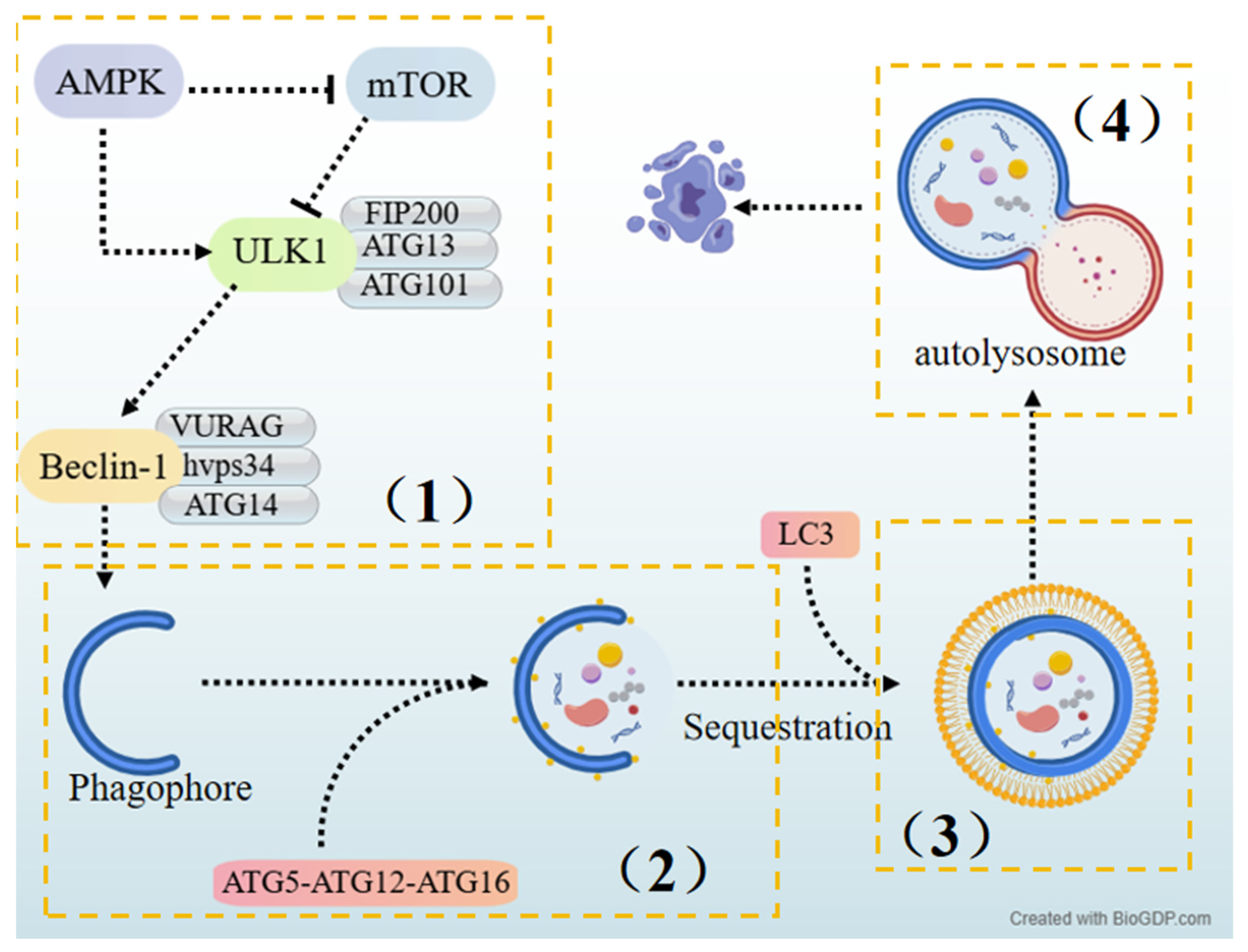
2. Autophagy
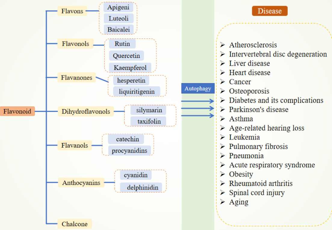
3. Flavonoids
3.1. Flavones
3.2. Flavonols
3.3. Flavanones
3.4. Dihydroflavonols
3.5. Flavanols
3.6. Anthocyanins
3.7. Chalcones

| Category | HCA | Structure | Source | Biological Activity/Application | Experimental Model | Dose Range | Ref. |
|---|---|---|---|---|---|---|---|
| Flavones | Apigenin |  | Dried parsley | Inhibition of apoptosis | B6SJL-Tg transgenic mice | 40, 80 mg/kg | [25] |
| NSC34 cells | 1 μM | ||||||
| Activation of autophagy | Huh7, Huh7.5, HepG2, AML12 cells | 0, 2.5, 5, 10, 20, 40, 80, 160 μM | [26] | ||||
| Anti-inflammation | C57/BL6 mice | 50 mg/kg | [27] | ||||
| Caco-2 cells | 10 μM | ||||||
| Regulation of microglia | CD-1 mice | 25, 50, 75 mg/kg | [28] | ||||
| Anti-atherosclerosis | NLRP3−/−Ldlr−/− mice | 50 mg/kg/day | [29] | ||||
| Luteolin | 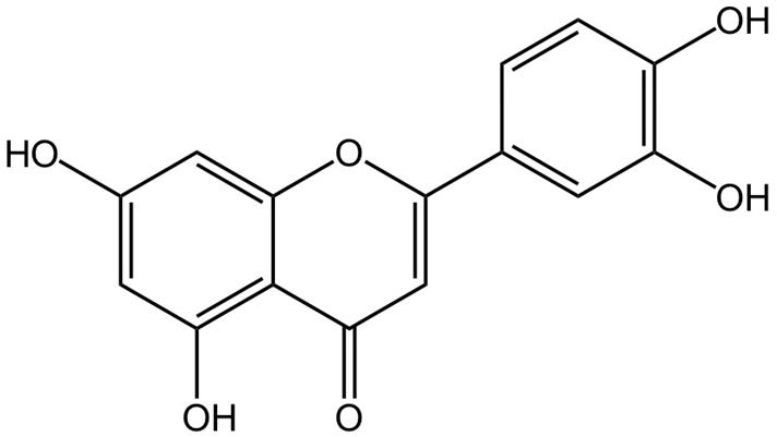 | Honeysuckle flower | Alleviation of liver injury | C57BL/6 mice | 40 mg/kg | [33] | |
| HepG2 cells | 0, 2.5, 5, 10, 20, 30, 40, 80, and 100 μM | ||||||
| Improvement of mitochondrial function | Wistar rats | 150 mg/kg/day | [34] | ||||
| Anticancer | BALB/c nude mice | 20, 40 mg/kg | [35] | ||||
| ESCC cells | 10, 20, 40, 80, and 120 μM | ||||||
| Antioxidant | SD rats | 50, 100 mg/kg/day | [36] | ||||
| Anti-fibrotic | Wistar rats | 10, 50 mg/kg | [37] | ||||
| HSC-T6 cells | 10 μM | ||||||
| Anti-inflammatory | C57BL/6 mice | 10, 20, and 50 mg/kg | [38] | ||||
| H9c2 cells | 10, 20, 50 μM | ||||||
| Baicalein | 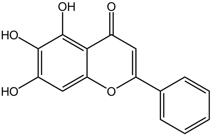 | Scutellaria baicalensis (Huangqin) | Anticancer | C57BL/6 mice | 50, 100, and 200 mg/kg | [42] | |
| H1299, A549 cells | 0, 10, 20, 30, 50, 100, 200, and 500 μM | ||||||
| Regulation of autophagy | SD rats | 100 mg/kg | [43] | ||||
| Improvement of insulin resistance | Glp1r KO mice | 50, 100, and 200 mg/kg | [44] | ||||
| HepG2, C2C12 cells | 40 μM | ||||||
| Antiepileptic | Sprague-Dawley (SD) rats | 50, 100 mg/kg | [45] | ||||
| HT22 cells | 0, 5, 10, 20 μM | ||||||
| Regulation of gut microbiota | C57BL/6J mice | 10 mg/kg and 20 mg/kg | [46] | ||||
| Anti-fibrotic | C57BL/6J mice | 25, 50, or 100 mg/kg | [47] | ||||
| CCC-ESF-1 cells | 5–120 μM | ||||||
| Flavonols | Quercetin |  | Onion | Amelioration of diabetic nephropathy | SD rats | 100 mg | [53] |
| HK-2 cells | 0 μM, 5 μM, 10 μM, 15 μM, 25 μM, and 50 μM | ||||||
| Induction of mitochondrial apoptosis | SK-MEL-28, G-361 cells | 0–400 μM | [54] | ||||
| Anti-inflammatory | A549 cells | 0, 10, 20, and 40 μM | [55] | ||||
| Anticancer | ZR-75-1, MCF-7, T47D, MDA-MB-231, MCF10A, and MDA-kb2 cells | 0, 5, 10, 20 μM | [56] | ||||
| Kaempferol |  | Broccoli | Anti-inflammatory | C57BL/6J mice | 50 mg/kg/day | [61] | |
| Anti-oxidative stress | Sprague-Dawley rats | 3, 10, or 20 mg/kg | [62] | ||||
| Reduction in lipid accumulation | HepG2 cells | 5, 10, and 20 μM | [63] | ||||
| Anti-atherosclerotic | C57BL/6, APOE–/– mice | 100 mg/kg | [64] | ||||
| HAECs cells | 5, 10, or 20 μM | ||||||
| Anti-apoptotic | Albino rats | 250 g/kg | [65] | ||||
| Amelioration of acute myocardial infarction | C57/BL6 mice | 50 mg/kg/day | [66] | ||||
| H9c2 cells | 0, 5, 10, and 20 μM | ||||||
| Anti-fibrotic | C57BL/6 mice | 25, 50, and 100 mg/kg | [67] | ||||
| AML12 cells | 5, 10, and 20 μM | ||||||
| Rutin |  | Sophora japonica (Huaimi) | Anti-inflammatory | HaCaT cells | 0, 1, 5, 10, 15, 30, 50, and 100 μM | [70] | |
| Anticancer | PANC-1, SW1990, MIA PaCa-2 cells | 5–40 μg/mL | [71] | ||||
| Regulation of gut microbiota | C57BL/6J mice | 200 mg/kg | [72] | ||||
| Anti-oxidative stress | BALB/c mice | 35 mg/kg | [73] | ||||
| RK-13, MDBK cells | 10, 20, 40, 80, 160, and 200 μM | ||||||
| Flavanones | Hesperetin | 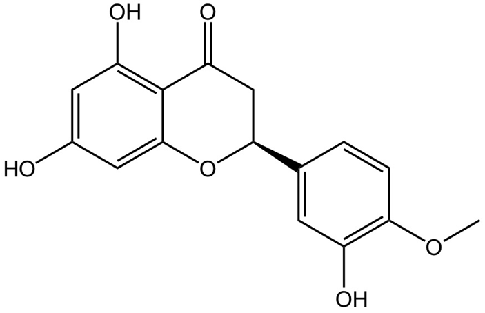 | Orange | Regulation of gut microbiota | C57BL/6 mice | 100 mg/kg/day | [78] |
| RAW264.7 cells | 0.005, 0.01, 0.05, 0.1, 0.2, 0.3, 0.5, 1, 2, and 5 μg/mL | ||||||
| Modulation of autophagy | U937, HL-60 cells | 0, 12.5, 25, 50, and 100 μM | [79] | ||||
| Anti-oxidative stress | MH-S cells | 10, 50 μM | [80] | ||||
| Liquiritigenin | 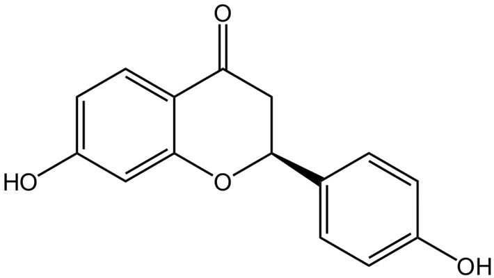 | Glycyrrhiza uralensis (Ural licorice) | Modulation of autophagy | C57BL/6 mice | 30 mg−1 kg−1 day−1 | [83] | |
| MC3T3-E1 cells | 0, 0.5, 1, 5, and 10 μM | ||||||
| Anticancer | MCF-7, BT20 cells | 0, 0.05, 0.1, 0.2, 0.4, and 0.8 mmol/L | [84] | ||||
| Anti-fibrotic | C57BL/6 mice | 25, 50, 100 mg/kg | [85] | ||||
| Pulmonary fibroblasts | 0, 1, 3, 10, 30, and 100 μM | ||||||
| Dihydroflavonols | Silybinin |  | Silybum marianum (Milk thistle) | Anticancer | RPMI 8226, H929 cells | 0–120 μM | [88] |
| HepG2 and Hep3B cells | 20 μmol/L, 40 μmol/L, and 80 μmol/L | [89] | |||||
| Anti-oxidative stress | Swiss Webster mice | 50 mg/kg | [90] | ||||
| Hepatoprotective | Sprague-Dawley rats | 50, 100, 200 mg/kg | [91] | ||||
| Anti-inflammatory | RAW264.7 cells | 0.4, 0.8 μg/mL | [92] | ||||
| Taxifolin (Dihydroquercetin) | 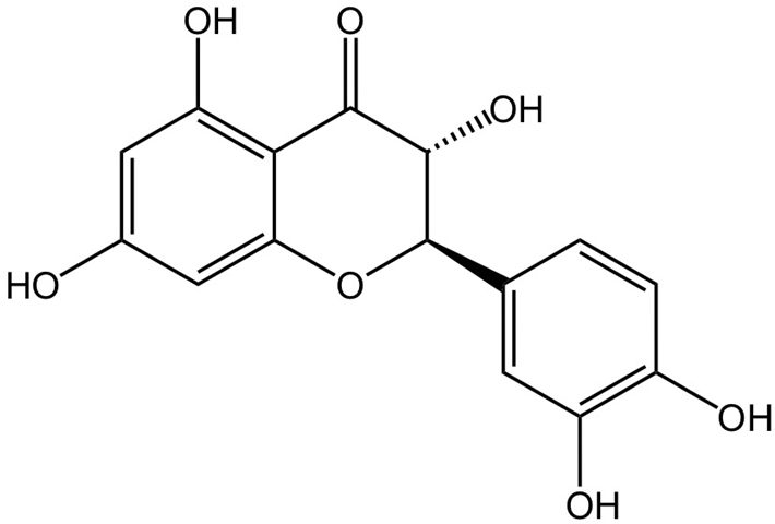 | Siberian larch | Anti-fibrotic | C57BL/6 mice | 10, 50 mg/kg | [94] | |
| HBE, MRC-5 cells | 0, 5, 10, 20, 40, or 80 μM | ||||||
| Regulation of gut microbiota | C57BL/6J mice | 50 mg/kg | [95] | ||||
| Antioxidant | SHR, Wistar rats | 25, 50 mg/kg/d | [96] | ||||
| PC-12 cells | 0.1, 0.5, 1 nM | ||||||
| Anti-inflammatory | BV-2 cells | 200, 400 μM | [97] | ||||
| Flavanols | Catechin | 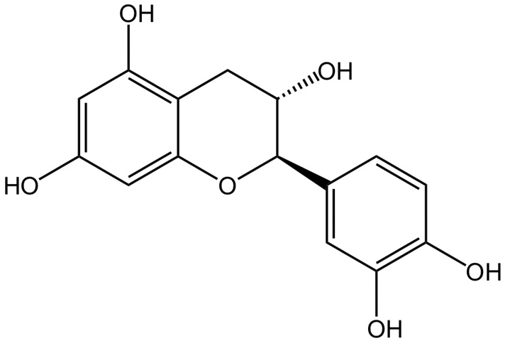 | Tea leaves | Improvement of lipid metabolism | C57BL/6 mice | 1328, 2645, 5289 mg/kg | [101] |
| Anti-inflammatory | SD rats | 0.5, 1, 1.5, and 2 mg/kg | [102] | ||||
| Anti-fibrotic | C57BL/6 mice | 50 μg/kg | [103] | ||||
| PSCs cells | 250 μM | ||||||
| Antihypertensive | SHR rats | 10, 50 mg/kg | [104] | ||||
| Proanthocyanidins | 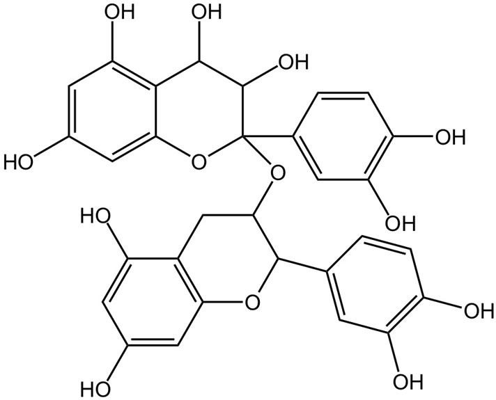 | Grape seeds | Improvement of lipid metabolism | C57BL/6J mice | 1, 2% | [106] | |
| Anti-oxidative stress | Kunming mice | 200 mg/kg/day, 20 mg/kg | [107] | ||||
| Inhibition of apoptosis | SD rats | 10, 20, 40 μM/mL | [108] | ||||
| MG63 cells | 10 mg/kg | ||||||
| Anti-inflammatory | SD rats | 200 mg/kg | [109] | ||||
| Anthocyanins | Cyanidin |  | Blueberry | Anti-fibrotic | C57BL/6 mice | 0, 50, 100, 150, 200 μmol/L | [113] |
| HK-2 cells | 100 mg/kg/day | ||||||
| Regulation of gut microbiota | C57BL/6J mice | 10, 50 mg/kg | [114] | ||||
| Antioxidant | BALB/c male mice | 60 mg/kg | [115] | ||||
| Reduction in pulmonary arterial hypertension | Sprague-Dawley rats | 10 or 20 μM | [116] | ||||
| HPASMCs cells | 40 mg/kg | ||||||
| Anti-inflammatory | Male BALB/c mice | 100 mg/kg | [117] | ||||
| Delphinidin |  | Blueberry | Anticancer | Sprague-Dawley rats | 40 μmol/L | [119] | |
| MDA-MB-231, MCF-7, and MDA-MB-453 cells | 2.6 mg/kg | ||||||
| Antioxidant | C57BL/6J mice | 0–50 μM | [120] | ||||
| Modulation of autophagy | A549 cell line | 10, 20 mg/kg/d | [121] | ||||
| Anti-inflammatory | New Zealand white rabbits | 1.5 μM | [122] | ||||
| Chalcones | —— | 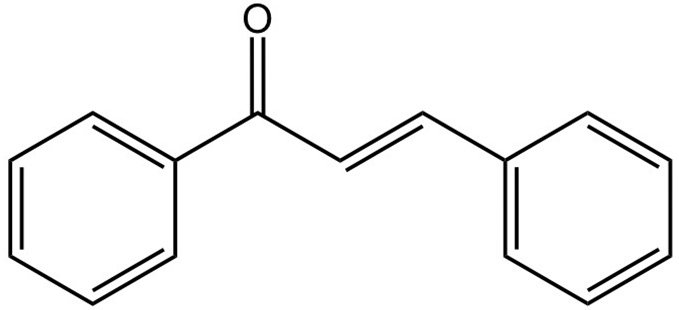 | Asitaba (Angelica keiskei) | Anti-inflammatory | THP-1 cell line | 20 μg/mL | [125] |
| Anticancer | Luc-4T1, MDA-MB-231 cells | 40, 80 mg/kg | [126] |
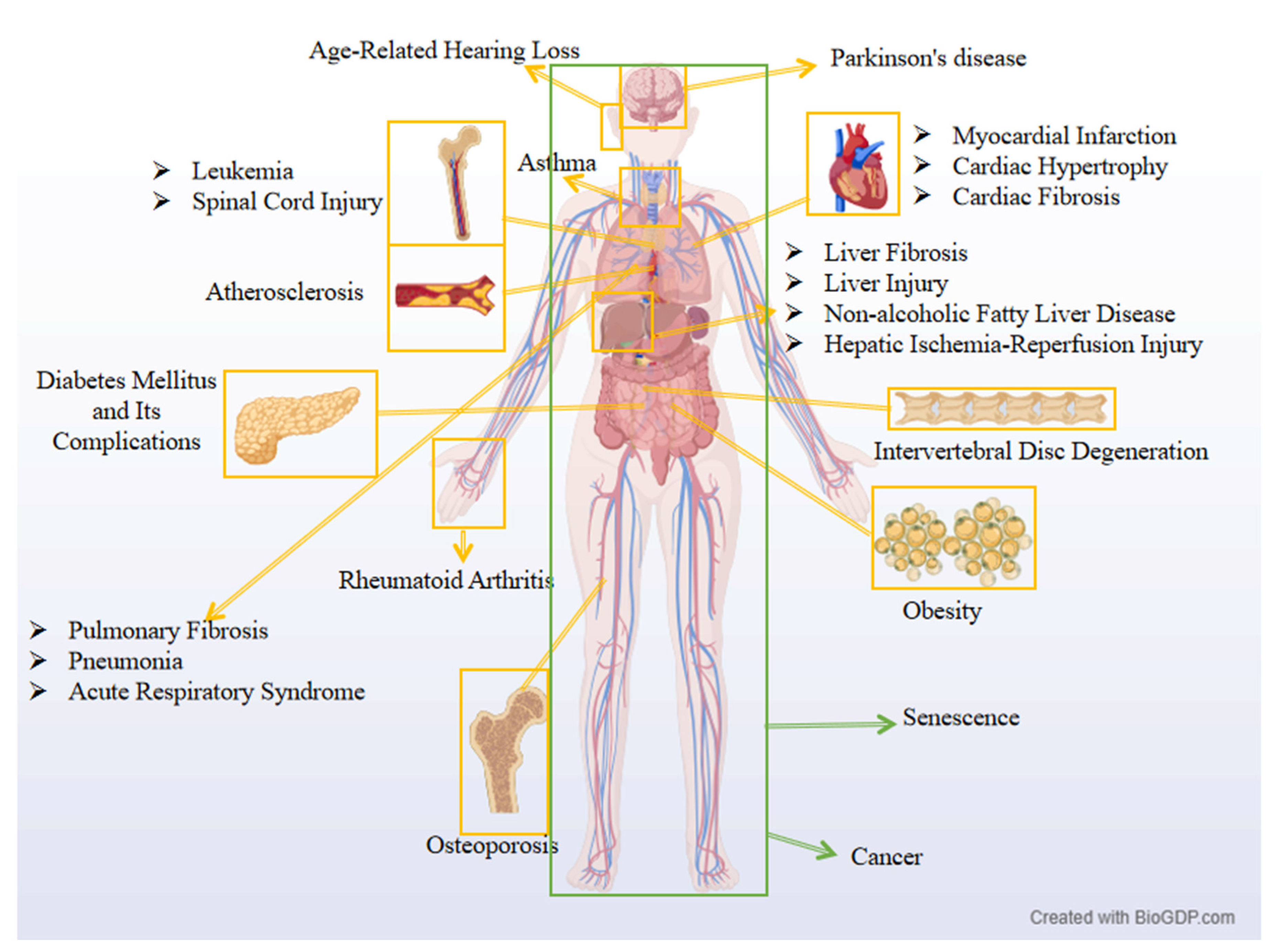
4. Flavonoids Improve Diseases by Regulating Autophagy
4.1. Atherosclerosis
4.2. Intervertebral Disk Degeneration
4.3. Liver Diseases
4.4. Cardiac Diseases
4.5. Cancer
4.6. Osteoporosis
4.7. Diabetes and Its Complications
4.8. Parkinson’s Disease
4.9. Asthma
4.10. Age-Related Hearing Loss
4.11. Leukemia
4.12. Pulmonary Diseases
4.13. Obesity
4.14. Rheumatoid Arthritis
4.15. Spinal Cord Injury
4.16. Cellular Senescence
| HCA | Disease | Promote/ Inhibit | Selective Autophagy/ Autophagy | Mechanisms of Action | Experimental Model | Dose Range | Ref. |
|---|---|---|---|---|---|---|---|
| Apigenin | Atherosclerosis | Activation | Lipophagy | UVRAG, Beclin1, PI3KC3, ATGs, ATG5, ATG3, LC3-II/I ↑ | ApoE−/− mice | 6.25, 12.5 mg/kg/d | [130] |
| Intervertebral disk degeneration | Activation | Autophagy | Lamp2, p62 ↓, LC3-II/I, TFEB ↑ | SD rats | 10 mg/kg | [131] | |
| NP cells | 0–50 μM | ||||||
| Liver fibrosis | Inhibition | Lipophagy | Beclin-1, LC3-II/I ↓, p62 ↑ | C57 mice | 20, 40 mg/kg | [134] | |
| LX2 cell line | 10, 20, 30, 40, 50, 60, 70, or 80 μM | ||||||
| Myocardial ischemia–reperfusion | Activation | Mitophagy | SIRT1, LC3-II/I ↑, p62 ↓ | H9c2 cells | 10 μM | [139] | |
| Luteolin | Triple-negative breast cancer | Activation | Autophagy | Beclin-1, LC3-II/I ↑, P62 ↓ | BALB/c mice | 20, 40 mg/kg | [144] |
| TNBC cell line | 0, 12.5, 25, 50, 100, 200 μM | ||||||
| Osteoporosis | Activation | Autophagy | Beclin-1, ATG5, LC3-II/I ↑, P62 ↓ | SD rats | 50, 100 mg/kg | [152] | |
| BMSC cells | 1, 10, and 100 μM | ||||||
| Diabetes mellitus | Activation | Autophagy | DRAK2, P62 ↓, ULK1, LC3-II/I ↑ | ICR mice | 20 mg/kg | [154] | |
| INS-1E cells | 20 μM | ||||||
| Myocardial injury | Activation | Autophagy | Beclin-1, LC3-II/I ↑, P62 ↓ | Wistar rats | 25, 50, 100 mg/kg | [140] | |
| Baicalein | Cardiac hypertrophy | Activation | Mitophagy | LC3-II/I, FUNDC1 ↑ | C57BL/6 | 25 mg/kg | [141] |
| NRCMs cells | 5–30 μM | ||||||
| Enhancement of antitumor immunity | Activation | Autophagy | CD274, LC3B ↑ | C57 mice | 33 mg/kg | [145] | |
| A549, H1299, 293T cells | 25, 50, 100 μM | ||||||
| Parkinson’s disease | Activation | Mitophagy | p-AMPK/AMPK, LC3-II/I ↑, p-mTOR/mTOR, P62 ↓ | SD rats | 100 mg/kg | [43] | |
| Quercetin | Myocardial fibrosis | Activation | Mitophagy | FOXO3, ATG7, LC3-II/I ↑, p62 ↓ | Wistar rats | 25 mg/kg | [142] |
| 293T, RCF cells | 50 μM | ||||||
| Non-small cell lung cancer | Activation | Mitophagy | P-AMPK/AMPK, Beclin-1, PINK1, Parkin, NOX4, XBP-1, DJ-1 ↑, SHP2 ↓ | SPF male nude mice | 50 mg/kg | [146] | |
| AC16 cells | 80 μM | ||||||
| Intervertebral disk degeneration | Inhibition | Autophagy | Beclin-1, LC3II/I, p38 ↑, p62, p-mTOR/mTOR ↓ | SD rats | 100 mg/kg | [132] | |
| NFC cells | 15, 25 μM | ||||||
| Osteoporosis | Activation | Autophagy | Beclin-1, LC3II/I ↓, p62 ↑ | SD rats | 50 mg/kg | [153] | |
| Non-alcoholic fatty liver disease | Inhibition | Autophagy | Beclin1, LC3II/I ↑, p62 ↓ | C57BL/6J mice | 50 mg/kg | [136] | |
| Asthma | Activation | Autophagy | LC3-II/LC3-I, Beclin-1 ↓, p-PI3K/p-Akt, p-mTOR/mTOR ↑ | BALB/c mice | 10, 20 mg/kg | [160] | |
| Age-related hearing loss | Activation | Mitophagy | PINK1, PARKIN, BNIP3, LC3II/I ↑ | C57BL/6J mice | 50 mg/kg | [161] | |
| Kaempferol | Parkinson’s disease | Promote | Mitophagy | LC3II/I ↑, p62 ↓ | C57BL/6J mice | 50 mg/kg | [159] |
| Diabetic nephropathy | Promote | Autophagy | LC3II/I, Beclin-1, Atg5, Atg7 ↑, p62 ↓ | C57BLKS/J db/db, db/m mice | 50, 100 mg/kg/day | [155] | |
| Hesperetin | Leukemia | Activation | Autophagy | LC3-II/I, Atg5, Beclin-1 ↑, p62 ↓ | U937, HL-60 cells | 0, 12.5, 25, 50, and 100 μM | [79] |
| Pulmonary fibrosis | Activation | Autophagy | BECN1 ↑, BCL2 ↓ | C57BL/6 mice | 50 mg/kg/day | [163] | |
| A549 cells | 10 μM | ||||||
| Diabetic retinopathy | Inhibition | Autophagy | LC3-II/I, Beclin-1 ↑, p62 ↓ | Wistar rats | 50, 100 mg/kg | [156] | |
| Liquiritigenin | Liver injury | Promote | Mitophagy | p-PI3K/PI3K, p-AKT/AKT, p-mTOR/mTOR ↑ | Kunming mice | 20, 40 mg/kg | [135] |
| Obesity | Promote | Lipophagy | LC3-II/I, ATG7 ↑, p62, p-mTOR/mTOR ↓ | 3T3-L1 cells | 10, 20, 25 nM | [167] | |
| Silymarin | Hepatocellular carcinoma | Inhibition | Autophagy | Beclin-1, LC3-I/LC3-II ↑ | H22 cells | 0, 40, and 80 μg/mL | [147] |
| Non-alcoholic fatty liver disease | Inhibition | Mitophagy | Parkin, Bcl-2, LC3-II/I, PINK1, p62 ↓ | Wistar rats | 300 mg/kg | [137] | |
| Lung injury | Promote | Autophagy | LC3-II/I, Beclin-1 ↓ | Wistar rats | 50 mg/kg | [164] | |
| Catechin | Diabetic skin ulcer | Promote | Autophagy | LC3-II/I, Beclin1, ATG5, p-AMPK/AMPK, p-ULK1/ULK1 ↑, p62 ↓ | SD rats | 1 mg/mL | [157] |
| HaCaT, HFF-1 cells | 0, 6.25, 12.5, 25, 50, 100 μM | ||||||
| Diabetic cardiomyopathy | Promote | Autophagy | LC3, Beclin1, p-AMPK/AMPK ↑, p-mTOR/mTOR ↓ | SD rats | 40, 80 mg/kg | [158] | |
| Pneumonia | Promote | Autophagy | LC3, BECN1 ↑, mTOR ↓ | Wistar rats | 15 mg/kg | [165] | |
| Severe acute respiratory syndrome (SARS) | Inhibition | Autophagy | Beclin-1, Atg5, Atg12, LC3-II ↑ | BALB/c mice | 25, 50, 125 mg/kg | [160] | |
| Vero E6 cells | 6.25 mg–195 μg | ||||||
| Proanthocyanidins | Hepatic ischemia–reperfusion injury | Promote | Autophagy | LC3, Beclin-1 ↓, Bcl-2 ↑ | BALB/c mice | 50 mg/kg, 50 mg/kg, 100 mg/kg | [161] |
| Rheumatoid arthritis | Promote | Mitophagy | LC3-II/I ↑, p62 ↓ | RA-FLS cells | 0 to 400 μg/mL | [162] | |
| Alcohol-related liver disease | Promote | Autophagy | p62, LC3-II ↓, Parkin, p-AMPK/AMPK ↑ | C57BL/6J mice | 50 μg/kg | [163] | |
| AML12 cells | 1 μg/mL | ||||||
| Delphinidin | Intervertebral disk degeneration | Promote | Autophagy | LC3-II/LC3-I, Beclin-1, p-AMPK/AMPK, SIRT1 ↑, p62, p-mTOR/mTOR ↓ | hNPCs cells | 0 to 200 μM | [164] |
| Chalcone | Spinal cord injury | Promote | Autophagy | VPS34, Beclin1, LC3-II/LC3-I ↑, CTSD ↓ | C57BL/6J mice | 0, 100, 150, 200, 250 mg/kg/day | [165] |
| Lung cancer | Promote | Autophagy | LC3-II ↑ | BALB/c-nu/nu mice | 20, 40 mg/kg | [148] | |
| A549, H460, H292, MRC-5 cells | 0, 2.5, 5 μM | ||||||
| Breast cancer | Promote | Autophagy | LC3-II, Beclin-1 ↑, p62, p-mTOR ↓ | MDA-MB-231, MCF-7 cells | 5–80 μg/mL | [149] | |
| Cancer | Activation | Autophagy | LC3-II, p53 ↑, p62, mTOR ↓ | TNBC MDA-MB-231, SiHa, MCF-7, PC-3 cells | 1–10 μM | [150] | |
| Osteosarcoma | Activation | Lipophagy | LC3-II ↑, p62 ↓ | MG63, 143BOS cells | 80, 40, 20, 10, 5, 2.5, 1.25, 0.6125 μM | [151] | |
| Aging/Cellular senescence | Inhibition | Autophagy | LC3-II, Beclin-1, p-AMPK/AMPK ↑, p62 ↓ | C57BL/6 mice | 0.5 mg/kg/day | [170] | |
| hBM-MSCs cells | 0–8 μg/mL |
5. Bioavailability and Safety of Flavonoids
6. Summary and Perspectives
Author Contributions
Funding
Data Availability Statement
Conflicts of Interest
Abbreviations
| AKT | Protein Kinase B |
| AMPK | AMP-Activated Protein Kinase |
| ATG | Autophagy-Related Gene |
| Bax | Bcl-2-Associated X Protein |
| Bcl-2 | B-Cell Lymphoma 2 |
| ECM | Extracellular Matrix |
| FOXO3 | Forkhead Box O3 |
| JNK | C-Jun N-Terminal Kinase |
| LC3 | Microtubule-Associated Protein 1 Light Chain 3 |
| mTOR | Mammalian Target of Rapamycin |
| NAFLD | Non-Alcoholic Fatty Liver Disease |
| NF-κB | Nuclear Factor Kappa-Light-Chain-Enhancer of Activated B Cells |
| NLRP3 | NOD-Like Receptor Family Pyrin Domain Containing 3 |
| Nrf2 | Nuclear Factor Erythroid 2-Related Factor 2 |
| OP | Osteoporosis |
| p62 | Sequestosome-1 (SQSTM1) |
| PD | Parkinson’s Disease |
| PI3K | Phosphoinositide 3-Kinase |
| ROS | Reactive Oxygen Species |
| SCI | Spinal Cord Injury |
| SIRT1 | Sirtuin 1 |
| TGF-β | Transforming Growth Factor Beta |
| TNBC | Triple-Negative Breast Cancer |
| ULK1 | Unc-51 Like Autophagy Activating Kinase 1 |
References
- Liu, S.; Yao, S.; Yang, H.; Liu, S.; Wang, Y. Autophagy: Regulator of Cell Death. Cell Death Dis. 2023, 14, 648. [Google Scholar] [CrossRef] [Scilit] [PubMed]
- Lamark, T.; Johansen, T. Mechanisms of Selective Autophagy. Annu. Rev. Cell Dev. Biol. 2021, 37, 143–169. [Google Scholar] [CrossRef] [Scilit]
- Chen, T.; Tu, S.; Ding, L.; Jin, M.; Chen, H.; Zhou, H. The role of autophagy in viral infections. J. Biomed. Sci. 2023, 30, 5. [Google Scholar] [CrossRef] [Scilit]
- Leonte, D.; Ungureanu, D.; Zaharia, V. Flavones and Related Compounds: Synthesis and Biological Activity. Molecules 2023, 28, 6528. [Google Scholar] [CrossRef] [Scilit]
- Wang, Q.; Lu, Y.; Mi, X.; Yang, C.; Ma, W.; Xia, C.; Wang, H. Antidepressant Activity of Flavones from Traditional Chinese Medicine: A Meta-Analysis. Pharm. Biol. 2025, 63, 156–169. [Google Scholar] [CrossRef] [Scilit]
- Álvarez-Mercado, A.I.; Rojano-Alfonso, C.; Micó-Carnero, M.; Caballeria-Casals, A.; Peralta, C.; Casillas-Ramírez, A. New Insights into the Role of Autophagy in Liver Surgery in the Setting of Metabolic Syndrome and Related Diseases. Front. Cell Dev. Biol. 2021, 9, 670273. [Google Scholar] [CrossRef] [Scilit]
- Li, H.; Ye, Z.; Zheng, G.; Su, Z. Polysaccharides Targeting Autophagy to Alleviate Metabolic Syndrome. Int. J. Biol. Macromol. 2024, 283, 137393. [Google Scholar] [CrossRef] [Scilit]
- Li, W.; He, P.; Huang, Y.; Li, Y.F.; Lu, J.; Li, M.; Kurihara, H.; Luo, Z.; Meng, T.; Onishi, M.; et al. Selective Autophagy of Intracellular Organelles: Recent Research Advances. Theranostics 2021, 11, 222–256. [Google Scholar] [CrossRef] [Scilit] [PubMed]
- Jia, J.; Claude-Taupin, A.; Gu, Y.; Choi, S.W.; Peters, R.; Bissa, B.; Mudd, M.H.; Allers, L.; Pallikkuth, S.; Lidke, K.A.; et al. Galectin-3 Coordinates a Cellular System for Lysosomal Repair and Removal. Dev. Cell 2020, 52, 69–87. [Google Scholar] [CrossRef] [Scilit]
- Haidar, M.; Loix, M.; Bogie, J.F.J.; Hendriks, J.J.A. Lipophagy: A New Player in CNS Disorders. Trends Endocrinol. Metab. 2021, 32, 941–951. [Google Scholar] [CrossRef] [Scilit] [PubMed]
- Jiang, S.; Li, H.; Zhang, L.; Mu, W.; Zhang, Y.; Chen, T.; Wu, J.; Tang, H.; Zheng, S.; Liu, Y.; et al. Generic Diagramming Platform (GDP): A Comprehensive Database of High-Quality Biomedical Graphics. Nucleic Acids Res. 2025, 53, D1670–D1676. [Google Scholar] [CrossRef] [Scilit]
- Zhou, Y.; Manghwar, H.; Hu, W.; Liu, F. Degradation Mechanism of Autophagy-Related Proteins and Research Progress. Int. J. Mol. Sci. 2022, 23, 7301. [Google Scholar] [CrossRef] [Scilit] [PubMed]
- Xu, Y.; Qian, C.; Wang, Q.; Song, L.; He, Z.; Liu, W.; Wan, W. Deacetylation of ATG7 Drives the Induction of Macroautophagy and LC3-Associated Microautophagy. Autophagy 2024, 20, 1134–1146. [Google Scholar] [CrossRef] [Scilit]
- Chandra, A.; Rick, J.; Yagnik, G.; Aghi, M.K. Autophagy as a Mechanism for Anti-Angiogenic Therapy Resistance. Semin. Cancer Biol. 2020, 66, 75–88. [Google Scholar] [CrossRef] [Scilit]
- Debnath, J.; Gammoh, N.; Ryan, K.M. Autophagy and Autophagy-Related Pathways in Cancer. Nat. Rev. Mol. Cell Biol. 2023, 24, 560–575. [Google Scholar] [CrossRef] [Scilit] [PubMed]
- Wang, S.Y.; Zhao, J.M.; Zhou, C.L.; Zheng, H.D.; Huang, Y.; Zhao, M.; Zhang, Z.Y.; Wu, L.Y.; Wu, H.G.; Liu, H.R. Herbal Cake-Partitioned Moxibustion Inhibits Colonic Autophagy in Crohn’s Disease via Signaling Involving Distinct Classes of Phosphatidylinositol 3-Kinases. World J. Gastroenterol. 2020, 26, 5997–6014. [Google Scholar] [CrossRef] [Scilit]
- Mahapatra, K.K.; Mishra, S.R.; Behera, B.P.; Patil, S.; Gewirtz, D.A.; Bhutia, S.K. The Lysosome as an Imperative Regulator of Autophagy and Cell Death. Cell. Mol. Life Sci. 2021, 78, 7435–7449. [Google Scholar] [CrossRef] [Scilit]
- Yang, C.; Gong, Q.; Zhu, R.; Fan, Y.; Wang, Q.; Ge, G.; Wang, P. Flavonoids as Promising Drug Leads: Structural Diversity, Synthetic Strategies, and Drug-Likeness Optimization. Eur. J. Med. Chem. 2026, 305, 118549. [Google Scholar] [CrossRef] [Scilit]
- Ma, D.; Guo, Y.; Ali, I.; Lin, J.; Xu, Y.; Yang, M. Accumulation characteristics of plant flavonoids and effects of cultivation measures on their biosynthesis: A review. Plant Physiol. Biochem. 2024, 215, 108960. [Google Scholar] [CrossRef] [Scilit]
- Noguchi, K.; Ueda, C.; Umeda, S.; Kanae, H.; Seki, M.; Hamaguchi, S.; Namekata, I.; Tanaka, H. Chronotropic, Inotropic, and Lusitropic Effects of Flavonoids. Biol. Pharm. Bull. 2026, 49, 12–23. [Google Scholar] [CrossRef] [Scilit]
- Xia, Y.; Liang, C.; Luo, H.; Zhang, Y. Therapeutic Potential of Flavonoids and Flavonoid-Rich Compounds in Irritable Bowel Syndrome. Drug Des. Dev. Ther. 2025, 19, 4895–4910. [Google Scholar] [CrossRef] [Scilit]
- Chen, S.; Wang, X.; Cheng, Y.; Gao, H.; Chen, X. A Review of Classification, Biosynthesis, Biological Activities and Potential Applications of Flavonoids. Molecules 2023, 28, 4982. [Google Scholar] [CrossRef] [Scilit] [PubMed]
- Yoon, J.H.; Kim, M.Y.; Cho, J.Y. Apigenin: A Therapeutic Agent for Treatment of Skin Inflammatory Diseases and Cancer. Int. J. Mol. Sci. 2023, 24, 1498. [Google Scholar] [CrossRef] [Scilit] [PubMed]
- Charrière, K.; Schneider, V.; Perrignon-Sommet, M.; Lizard, G.; Benani, A.; Jacquin-Piques, A.; Vejux, A. Exploring the Role of Apigenin in Neuroinflammation: Insights and Implications. Int. J. Mol. Sci. 2024, 25, 5041. [Google Scholar] [CrossRef] [Scilit]
- Liang, H.; Zhou, X.; Zhang, J.; Xu, W.; Liu, Y.; Wang, X.; Hu, Y.; Xu, R.; Li, X. The Therapeutic Potential of Apigenin in Amyotrophic Lateral Sclerosis through ALDH1A2/Nrf2/ARE Signaling. Mol. Med. 2024, 30, 206. [Google Scholar] [CrossRef] [Scilit]
- Hsu, M.C.; Guo, B.C.; Chen, C.H.; Hu, P.A.; Lee, T.S. Apigenin Ameliorates Hepatic Lipid Accumulation by Activating the Autophagy-Mitochondria Pathway. J. Food Drug Anal. 2021, 29, 240–254. [Google Scholar] [CrossRef] [Scilit]
- Lijun, Z.; Liping, W.; Dandan, Z.; Wenjie, Z.; Renjie, X.; Xueyan, J.; Wenqing, C.; Chen, L.; Lu, Z. Apigenin Mitigates Intestinal Barrier Dysfunction in Sepsis by Modulating the AKT Signaling Pathway. BMC Gastroenterol. 2025, 25, 626. [Google Scholar] [CrossRef] [Scilit]
- Xie, W.; Xie, W.; Jiang, C.; Kang, Z.; Liu, N. Apigenin Alleviates Allodynia and Hyperalgesia in a Mouse Model of Chemotherapy-Induced Peripheral Neuropathy via Regulating Microglia Activation and Polarization. J. Integr. Neurosci. 2023, 22, 64. [Google Scholar] [CrossRef] [Scilit]
- Lu, Z.; Liu, L.; Zhao, S.; Zhao, J.; Li, S.; Li, M. Apigenin Attenuates Atherosclerosis and Non-Alcoholic Fatty Liver Disease through Inhibition of NLRP3 Inflammasome in Mice. Sci. Rep. 2023, 13, 7996. [Google Scholar] [CrossRef] [Scilit] [PubMed]
- Tesio, A.Y.; Robledo, S.N. Analytical Determinations of Luteolin. Biofactors 2021, 47, 141–164. [Google Scholar] [CrossRef] [Scilit]
- Gendrisch, F.; Esser, P.R.; Schempp, C.M.; Wölfle, U. Luteolin as a Modulator of Skin Aging and Inflammation. Biofactors 2021, 47, 170–180. [Google Scholar] [CrossRef] [Scilit]
- Jayawickreme, D.K.; Ekwosi, C.; Anand, A.; Andres-Mach, M.; Wlaź, P.; Socała, K. Luteolin for Neurodegenerative Diseases: A Review. Pharmacol. Rep. 2024, 76, 644–664. [Google Scholar] [CrossRef] [Scilit] [PubMed]
- Han, Z.; Batudeligen; Chen, H.; Narisu; Anda; Xu, Y.; Xue, L. Luteolin Attenuates CCl4-Induced Hepatic Injury by Inhibiting Ferroptosis via SLC7A11. BMC Complement. Med. Ther. 2024, 24, 193. [Google Scholar] [CrossRef] [Scilit]
- Younis, R.L.; El-Gohary, R.M.; Ghalwash, A.A.; Hegab, I.I.; Ghabrial, M.M.; Aboshanady, A.M.; Mostafa, R.A.; El-Azeem, A.H.A.; Farghal, E.E.; Belal, A.A.E.; et al. Luteolin Mitigates D-Galactose-Induced Brain Ageing in Rats: SIRT1-Mediated Neuroprotection. Neurochem. Res. 2024, 49, 2803–2820. [Google Scholar] [CrossRef] [Scilit]
- Yang, Z.; Liu, H.; Song, Y.; Gao, N.; Gao, P.; Hui, Y.; Li, Y.; Fan, T. Luteolin Enhances Drug Chemosensitivity by Downregulating the FAK/PI3K/AKT Pathway in Paclitaxel-Resistant Esophageal Squamous Cell Carcinoma. Int. J. Mol. Med. 2024, 54, 77. [Google Scholar] [CrossRef] [Scilit] [PubMed]
- Taweesap, P.; Potue, P.; Khamseekaew, J.; Iampanichakul, M.; Jan-O, B.; Pakdeechote, P.; Maneesai, P. Luteolin Relieves Metabolic Dysfunction-Associated Fatty Liver Disease Caused by a High-Fat Diet in Rats through Modulating the AdipoR1/AMPK/PPARγ Signaling Pathway. Int. J. Mol. Sci. 2025, 26, 3804. [Google Scholar] [CrossRef] [Scilit]
- Batudeligen; Han, Z.; Chen, H.; Narisu; Xu, Y.; Anda; Han, G. Luteolin Alleviates Liver Fibrosis in Rat Hepatic Stellate Cell HSC-T6: A Proteomic Analysis. Drug Des. Dev. Ther. 2023, 17, 1819–1829. [Google Scholar] [CrossRef] [Scilit] [PubMed]
- Liu, Z.; Gao, S.; Bu, Y.; Zheng, X. Luteolin Protects Cardiomyocytes Cells against Lipopolysaccharide-Induced Apoptosis and Inflammatory Damage by Modulating Nlrp3. Yonsei Med. J. 2022, 63, 220–228. [Google Scholar] [CrossRef] [Scilit]
- Gupta, S.; Buttar, H.S.; Kaur, G.; Tuli, H.S. Baicalein: Promising Therapeutic Applications with Special Reference to Published Patents. Pharm. Pat. Anal. 2022, 11, 23–32. [Google Scholar] [CrossRef] [Scilit]
- Morshed, A.K.M.H.; Paul, S.; Hossain, A.; Basak, T.; Hossain, M.S.; Hasan, M.M.; Hasibuzzaman, M.A.; Rahaman, T.I.; Mia, M.A.R.; Shing, P.; et al. Baicalein as Promising Anticancer Agent: A Comprehensive Analysis on Molecular Mechanisms and Therapeutic Perspectives. Cancers 2023, 15, 2128. [Google Scholar] [CrossRef] [Scilit]
- Chandrashekar, N.; Pandi, A. Baicalein: A Review on Its Anti-Cancer Effects and Mechanisms in Lung Carcinoma. J. Food Biochem. 2022, 46, e14230. [Google Scholar] [CrossRef] [Scilit] [PubMed]
- Li, J.; Zhang, D.; Wang, S.; Yu, P.; Sun, J.; Zhang, Y.; Meng, X.; Li, J.; Xiang, L. Baicalein Induces Apoptosis by Inhibiting the Glutamine-mTOR Metabolic Pathway in Lung Cancer. J. Adv. Res. 2025, 68, 341–357. [Google Scholar] [CrossRef] [Scilit]
- Chen, M.; Peng, L.; Gong, P.; Zheng, X.; Sun, T.; Zhang, X.; Huo, J. Baicalein Induces Mitochondrial Autophagy to Prevent Parkinson’s Disease in Rats via miR-30b and the SIRT1/AMPK/mTOR Pathway. Front. Neurol. 2022, 12, 646817. [Google Scholar] [CrossRef] [Scilit]
- Liu, N.; Cui, X.; Guo, T.; Wei, X.; Sun, Y.; Liu, J.; Zhang, Y.; Ma, W.; Yan, W.; Chen, L. Baicalein Ameliorates Insulin Resistance of HFD/STZ Mice through Activating PI3K/AKT Signal Pathway of Liver and Skeletal Muscle in a GLP-1R-Dependent Manner. Antioxidants 2024, 13, 1246. [Google Scholar] [CrossRef] [Scilit]
- Wu, Z.; Liu, J.; Yin, D.; Huang, J.; Huang, Y.; Wang, P. Baicalein Alleviates Lithium-Pilocarpine-Induced Status Epilepticus by Regulating DNMT1/GABRD Pathway in Rats. Organogenesis 2025, 21, 2519607. [Google Scholar] [CrossRef] [Scilit] [PubMed]
- Lv, W.; Han, S.; Li, K.; Yan, A.; Wang, W.; Lu, W.; Han, J.; Zhang, C. Baicalein Ameliorates DSS-Induced Ulcerative Colitis in Mice by Inhibiting Ferroptosis and Regulating Gut Microbiota. Front. Pharmacol. 2025, 16, 1564783. [Google Scholar] [CrossRef] [Scilit]
- Peng, B.; Hu, Q.; He, R.; Hou, H.; Lian, D.; Chen, Y.; Li, H.; Song, L.; Gao, Y.; Chen, T.; et al. Baicalein Alleviates Fibrosis and Inflammation in Systemic Sclerosis by Regulating B-Cell Abnormalities. BMC Complement. Med. Ther. 2023, 23, 62. [Google Scholar] [CrossRef] [Scilit]
- Daryanavard, H.; Postiglione, A.E.; Mühlemann, J.K.; Muday, G.K. Flavonols Modulate Plant Development, Signaling, and Stress Responses. Curr. Opin. Plant Biol. 2023, 72, 102350. [Google Scholar] [CrossRef] [Scilit]
- Hsieh, Y.H.; Hsu, P.H.; Hu, A.; Cheng, Y.J.; Shih, T.L.; Chen, J.J. Synthesis of Flavonols and Assessment of Their Biological Activity as Anticancer Agents. Molecules 2024, 29, 2041. [Google Scholar] [CrossRef] [Scilit] [PubMed]
- Georgiou, N.; Kakava, M.G.; Routsi, E.A.; Petsas, E.; Stavridis, N.; Freris, C.; Zoupanou, N.; Moschovou, K.; Kiriakidi, S.; Mavromoustakos, T. Quercetin: A Potential Polydynamic Drug. Molecules 2023, 28, 8141. [Google Scholar] [CrossRef] [Scilit]
- Chiang, M.C.; Tsai, T.Y.; Wang, C.J. The Potential Benefits of Quercetin for Brain Health: A Review of Anti-Inflammatory and Neuroprotective Mechanisms. Int. J. Mol. Sci. 2023, 24, 6328. [Google Scholar] [CrossRef] [Scilit]
- Boo, H.J.; Yoon, D.; Choi, Y.; Kim, Y.; Cha, J.S.; Yoo, J. Quercetin: Molecular Insights into Its Biological Roles. Biomolecules 2025, 15, 313. [Google Scholar] [CrossRef] [Scilit]
- Zhang, L.; Wang, X.; Chang, L.; Ren, Y.; Sui, M.; Fu, Y.; Zhang, L.; Hao, L. Quercetin Improves Diabetic Kidney Disease by Inhibiting Ferroptosis and Regulating the Nrf2 in Streptozotocin-Induced Diabetic Rats. Ren. Fail. 2024, 46, 2327495. [Google Scholar] [CrossRef] [Scilit] [PubMed]
- Seo, S.Y.; Ju, W.S.; Kim, K.; Kim, J.; Yu, J.O.; Ryu, J.S.; Kim, J.S.; Lee, H.A.; Koo, D.B.; Choo, Y.K. Quercetin Induces Mitochondrial Apoptosis and Downregulates Ganglioside GD3 Expression in Melanoma Cells. Int. J. Mol. Sci. 2024, 25, 5146. [Google Scholar] [CrossRef] [Scilit]
- Lv, P.; Han, P.; Cui, Y.; Chen, Q.; Cao, W. Quercetin Attenuates Inflammation in LPS-Induced Lung Epithelial Cells via the Nrf2 Signaling Pathway. Immun. Inflamm. Dis. 2024, 12, e1185. [Google Scholar] [CrossRef] [Scilit]
- Tang, H.; Kuang, Y.; Wu, W.; Peng, B.; Fu, Q. Quercetin Inhibits the Metabolism of Arachidonic Acid by Inhibiting the Activity of CYP3A4, Thereby Inhibiting the Progression of Breast Cancer. Mol. Med. 2023, 29, 127. [Google Scholar] [CrossRef] [Scilit]
- Periferakis, A.; Periferakis, K.; Badarau, I.A.; Petran, E.M.; Popa, D.C.; Caruntu, A.; Costache, R.S.; Scheau, C.; Caruntu, C.; Costache, D.O. Kaempferol: Antimicrobial Properties, Sources, Clinical, and Traditional Applications. Int. J. Mol. Sci. 2022, 23, 15054. [Google Scholar] [CrossRef] [Scilit]
- Yang, Y.; Chen, Z.; Zhao, X.; Xie, H.; Du, L.; Gao, H.; Xie, C. Mechanisms of Kaempferol in the Treatment of Diabetes: A Comprehensive and Latest Review. Front. Endocrinol. 2022, 13, 990299. [Google Scholar] [CrossRef] [Scilit]
- Niziński, P.; Krajewska, A.; Oniszczuk, T.; Polak, B.; Oniszczuk, A. Hepatoprotective Effect of Kaempferol-A Review. Molecules 2025, 30, 1913. [Google Scholar] [CrossRef] [Scilit]
- Bangar, S.P.; Chaudhary, V.; Sharma, N.; Bansal, V.; Ozogul, F.; Lorenzo, J.M. Kaempferol: A Flavonoid with Wider Biological Activities and Its Applications. Crit. Rev. Food Sci. Nutr. 2023, 63, 9580–9604. [Google Scholar] [CrossRef] [Scilit]
- Qu, Y.; Li, X.; Xu, F.; Zhao, S.; Wu, X.; Wang, Y.; Xie, J. Kaempferol Alleviates Murine Experimental Colitis by Restoring Gut Microbiota and Inhibiting the LPS-TLR4-NF-κB Axis. Front. Immunol. 2021, 12, 679897. [Google Scholar] [CrossRef] [Scilit]
- Yue, Z.; Zhang, Y.; Zhang, W.; Zheng, N.; Wen, J.; Ren, L.; Rong, X.; Bai, L.; Wang, R.; Zhao, S.; et al. Kaempferol Alleviates Myocardial Ischemia Injury by Reducing Oxidative Stress via the HDAC3-Mediated Nrf2 Signaling Pathway. J. Adv. Res. 2025, 75, 755–764. [Google Scholar] [CrossRef] [Scilit] [PubMed]
- Tie, F.; Ding, J.; Hu, N.; Dong, Q.; Chen, Z.; Wang, H. Kaempferol and Kaempferide Attenuate Oleic Acid-Induced Lipid Accumulation and Oxidative Stress in HepG2 Cells. Int. J. Mol. Sci. 2021, 22, 8847. [Google Scholar] [CrossRef] [Scilit]
- Feng, Z.; Wang, C.; Jin, Y.; Meng, Q.; Wu, J.; Sun, H. Kaempferol-Induced GPER Upregulation Attenuates Atherosclerosis via the PI3K/AKT/Nrf2 Pathway. Pharm. Biol. 2021, 59, 1106–1116. [Google Scholar] [CrossRef] [Scilit]
- BinMowyna, M.N.; AlFaris, N.A. Kaempferol Suppresses Acetaminophen-Induced Liver Damage by Upregulation/Activation of SIRT1. Pharm. Biol. 2021, 59, 146–156. [Google Scholar] [CrossRef] [Scilit]
- Zhou, J.; Zhou, H.; Zhu, J.; Fang, S. Kaempferol Inhibits Cardiomyocyte Pyroptosis via Promoting O-GlcNAcylation of GSDME and Improved Acute Myocardial Infarction. BMC Pharmacol. Toxicol. 2025, 26, 76. [Google Scholar] [CrossRef] [Scilit]
- Zhang, S.; Tang, F.; Zhou, Z.; Li, L.; Tang, Y.; Fu, K.; Tan, Y.; Li, L. Kaempferol Alleviates Carbon Tetrachloride-Induced Liver Fibrosis in Mice by Regulating Intestinal Short-Chain Fatty Acids. Int. J. Mol. Sci. 2025, 26, 6666. [Google Scholar] [CrossRef] [Scilit]
- Forouzanfar, F.; Sahranavard, T.; Tsatsakis, A.; Iranshahi, M.; Rezaee, R. Rutin: A Pain-Relieving Flavonoid. Inflammopharmacology 2025, 33, 1289–1301. [Google Scholar] [CrossRef] [Scilit]
- Liu, H.; Xu, Q.; Wufuer, H.; Li, Z.; Sun, R.; Jiang, Z.; Dou, X.; Fu, Q.; Campisi, J.; Sun, Y. Rutin Is a Potent Senomorphic Agent to Target Senescent Cells and Can Improve Chemotherapeutic Efficacy. Aging Cell 2024, 23, e13921. [Google Scholar] [CrossRef] [Scilit]
- Wu, P.; Liu, Y.; Zhai, H.; Wu, X.; Liu, A. Rutin Alleviates Psoriasis-Related Inflammation in Keratinocytes by Regulating the JAK2/STAT3 Signaling. Ski. Res. Technol. 2024, 30, e70011. [Google Scholar] [CrossRef] [Scilit] [PubMed]
- Huo, M.; Xia, A.; Cheng, W.; Zhou, M.; Wang, J.; Shi, T.; Cai, C.; Jin, W.; Zhou, M.; Liao, Y.; et al. Rutin Promotes Pancreatic Cancer Cell Apoptosis by Upregulating miRNA-877-3p Expression. Molecules 2022, 27, 2293. [Google Scholar] [CrossRef] [Scilit] [PubMed]
- Cai, C.; Cheng, W.; Shi, T.; Liao, Y.; Zhou, M.; Liao, Z. Rutin Alleviates Colon Lesions and Regulates Gut Microbiota in Diabetic Mice. Sci. Rep. 2023, 13, 4897. [Google Scholar] [CrossRef] [Scilit]
- Chen, L.; Li, S.; Li, W.; Yu, Y.; Sun, Q.; Chen, W.; Zhou, H.; Wang, C.; Li, L.; Xu, M.; et al. Rutin Prevents EqHV-8 Induced Infection and Oxidative Stress via Nrf2/HO-1 Signaling Pathway. Front. Cell. Infect. Microbiol. 2024, 14, 1386462. [Google Scholar] [CrossRef] [Scilit]
- Birsa, M.L.; Sarbu, L.G. Novel Dithiocarbamic Flavanones with Antioxidant Properties-A Structure-Activity Relationship Study. Int. J. Mol. Sci. 2024, 25, 13698. [Google Scholar] [CrossRef] [Scilit]
- Cichon, N.; Szelenberger, R.; Stela, M.; Podogrocki, M.; Gorniak, L.; Bijak, M. Flavanones as Modulators of Gut Microbiota and Cognitive Function. Molecules 2025, 30, 2203. [Google Scholar] [CrossRef] [Scilit] [PubMed]
- Taheri, A.; Mobaser, S.E.; Golpour, P.; Nourbakhsh, M.; Tavakoli-Yaraki, M.; Yarahmadi, S.; Nourbakhsh, M. Hesperetin Attenuates the Expression of Markers of Adipose Tissue Fibrosis in Pre-Adipocytes. BMC Complement. Med. Ther. 2023, 23, 315. [Google Scholar] [CrossRef] [Scilit]
- Zhao, S.; Nan, Y.; Yao, R.; Wang, L.; Zeng, X.; Aadil, R.M.; Shabbir, M.A. Antibacterial Activity and Transcriptomic Analysis of Hesperetin against Alicyclobacillus acidoterrestris Vegetative Cells. Foods 2023, 12, 3276. [Google Scholar] [CrossRef] [Scilit]
- Wang, J.; Yao, Y.; Yao, T.; Shi, Q.; Zeng, Y.; Li, L. Hesperetin Alleviated Experimental Colitis via Regulating Ferroptosis and Gut Microbiota. Nutrients 2024, 16, 2343. [Google Scholar] [CrossRef] [Scilit]
- Lin, C.Y.; Chen, Y.H.; Huang, Y.C. Hesperetin Induces Autophagy and Delayed Apoptosis by Modulating the AMPK/Akt/mTOR Pathway in Human Leukemia Cells In Vitro. Curr. Issues Mol. Biol. 2023, 45, 1587–1600. [Google Scholar] [CrossRef] [Scilit]
- Ruan, H.; Yang, D.; Wang, N.; Liu, J.; Huang, L. Hesperetin Alleviates Sepsis-Associated Acute Respiratory Distress Syndrome via the PI3K/Akt/PTEN Axis. Sci. Rep. 2026, 16, 3332. [Google Scholar] [CrossRef] [Scilit] [PubMed]
- Sajeev, A.; Aswani, B.S.; Alqahtani, M.S.; Abbas, M.; Sethi, G.; Kunnumakkara, A.B. Harnessing Liquiritigenin: A Flavonoid-Based Approach for the Prevention and Treatment of Cancer. Cancers 2025, 17, 2328. [Google Scholar] [CrossRef] [Scilit]
- Qiu, Y.; Zhao, Y.; Long, Z.; Song, A.; Huang, P.; Wang, K.; Xu, L.; Molloy, D.P.; He, G. Liquiritigenin Promotes Osteogenic Differentiation and Prevents Bone Loss via Inducing Auto-Lysosomal Degradation and Inhibiting Apoptosis. Genes Dis. 2021, 10, 284–300. [Google Scholar] [CrossRef] [Scilit]
- Xu, S.; Ma, Z.; Xing, L.; Cheng, W. Polygonatum sibiricum Component Liquiritigenin Restrains Breast Cancer Cell Invasion and Migration by Inhibiting HSP90 and Chaperone-Mediated Autophagy. Korean J. Physiol. Pharmacol. 2024, 28, 379–387. [Google Scholar] [CrossRef] [Scilit] [PubMed]
- Hua, Q.; Ren, L. The SIRT1/Nrf2 Signaling Pathway Mediates the Anti-Pulmonary Fibrosis Effect of Liquiritigenin. Chin. Med. 2024, 19, 12. [Google Scholar] [CrossRef] [Scilit] [PubMed]
- Liu, H.; Liu, T.; Zeng, J.; Fang, Q. Silymarin Induces Multiple Myeloma Cell Apoptosis by Inhibiting the JAK2/STAT3 Signaling Pathway. Oncol. Lett. 2025, 30, 426. [Google Scholar] [CrossRef] [Scilit]
- Zhu, Y.; Scholle, F.; Kisthardt, S.C.; Xie, D.Y. Flavonols and Dihydroflavonols Inhibit the Main Protease Activity of SARS-CoV-2 and the Replication of Human Coronavirus 229E. Virology 2022, 571, 21–33. [Google Scholar] [CrossRef] [Scilit]
- Wadhwa, K.; Pahwa, R.; Kumar, M.; Kumar, S.; Sharma, P.C.; Singh, G.; Verma, R.; Mittal, V.; Singh, I.; Kaushik, D.; et al. Mechanistic Insights into the Pharmacological Significance of Silymarin. Molecules 2022, 27, 5327. [Google Scholar] [CrossRef] [Scilit]
- Okiljević, B.; Martić, N.; Govedarica, S.; Andrejić Višnjić, B.; Bosanac, M.; Baljak, J.; Pavlić, B.; Milanović, I.; Rašković, A. Cardioprotective and Hepatoprotective Potential of Silymarin in Paracetamol-Induced Oxidative Stress. Pharmaceutics 2024, 16, 520. [Google Scholar] [CrossRef] [Scilit] [PubMed]
- Yu, L.; Li, T.; Zhang, H.; Ma, Z.; Wu, S. Silymarin Suppresses Proliferation of Human Hepatocellular Carcinoma Cells under Hypoxia through Downregulation of the HIF-1α/VEGF Pathway. Am. J. Transl. Res. 2023, 15, 4521–4532. [Google Scholar]
- Aydemir Celep, N.; Gedikli, S. Protective Effect of Silymarin on Liver in Experimental in the Sepsis Model of Rats. Acta Histochem. Cytochem. 2023, 56, 9–19. [Google Scholar] [CrossRef] [Scilit] [PubMed]
- Mi, X.J.; Le, H.M.; Lee, S.; Park, H.R.; Kim, Y.J. Silymarin-Functionalized Selenium Nanoparticles Prevent LPS-Induced Inflammatory Response in RAW264.7 Cells through Downregulation of the PI3K/Akt/NF-κB Pathway. ACS Omega 2022, 7, 42723–42732. [Google Scholar] [CrossRef] [Scilit] [PubMed]
- Liu, Z.; Qiu, D.; Yang, T.; Su, J.; Liu, C.; Su, X.; Li, A.; Sun, P.; Li, J.; Yan, L.; et al. Research Progress of Dihydroquercetin in the Treatment of Skin Diseases. Molecules 2023, 28, 6989. [Google Scholar] [CrossRef] [Scilit]
- Yuan, L.; Sun, Y.; Zhou, N.; Wu, W.; Zheng, W.; Wang, Y. Dihydroquercetin Attenuates Silica-Induced Pulmonary Fibrosis by Inhibiting Ferroptosis Signaling Pathway. Front. Pharmacol. 2022, 13, 845600. [Google Scholar] [CrossRef] [Scilit]
- Wang, M.; Han, H.; Wan, F.; Zhong, R.; Do, Y.J.; Oh, S.I.; Lu, X.; Liu, L.; Yi, B.; Zhang, H. Dihydroquercetin Supplementation Improved Hepatic Lipid Dysmetabolism Mediated by Gut Microbiota in High-Fat Diet (HFD)-Fed Mice. Nutrients 2022, 14, 5214. [Google Scholar] [CrossRef] [Scilit]
- Fu, Y.; Yuan, P.; Zeng, M.; Zhang, Q.; Hou, Y.; Gao, L.; Wei, Y.; Zheng, Y.; Feng, W.; Zheng, X. Dihydroquercetin Regulates HIF-1α/AKT/NR2B Signalling to Improve Impaired Brain Function in Rats with Metabolic Syndrome. Heliyon 2024, 10, e29807. [Google Scholar] [CrossRef] [Scilit] [PubMed]
- Iwasa, M.; Kato, H.; Iwashita, K.; Yamakage, H.; Kato, S.; Saito, S.; Ihara, M.; Nishimura, H.; Kawamoto, A.; Suganami, T.; et al. Taxifolin Suppresses Inflammatory Responses of High-Glucose-Stimulated Mouse Microglia by Attenuating the TXNIP–NLRP3 Axis. Nutrients 2023, 15, 2738. [Google Scholar] [CrossRef] [Scilit]
- Farhan, M. Green Tea Catechins: Nature’s Way of Preventing and Treating Cancer. Int. J. Mol. Sci. 2022, 23, 10713. [Google Scholar] [CrossRef] [Scilit]
- Diniz, L.R.L.; Elshabrawy, H.A.; Souza, M.T.S.; Duarte, A.B.S.; Datta, S.; de Sousa, D.P. Catechins: Therapeutic Perspectives in COVID-19-Associated Acute Kidney Injury. Molecules 2021, 26, 5951. [Google Scholar] [CrossRef] [Scilit]
- Ferrari, E.; Naponelli, V. Catechins and Human Health: Breakthroughs from Clinical Trials. Molecules 2025, 30, 3128. [Google Scholar] [CrossRef] [Scilit] [PubMed]
- Lee, Y.H.; Huang, C.C.; Lin, H.L.; Chen, Y.X.; Hsu, Y.J. Green Tea Containing Inulin and Catechins Ameliorates High-Fat Diet-Induced Metabolic Dysfunction-Associated Fatty Liver Disease in Mice. Int. J. Med. Sci. 2025, 22, 4325–4334. [Google Scholar] [CrossRef] [Scilit]
- Wang, Z.; Lu, Z.; Chen, Y.; Wang, C.; Gong, P.; Jiang, R.; Liu, Q. Targeting the AKT-P53/CREB Pathway with Epicatechin for Improved Prognosis of Traumatic Brain Injury. CNS Neurosci. Ther. 2024, 30, e14364. [Google Scholar] [CrossRef] [Scilit]
- Kweon, B.; Kim, D.U.; Oh, J.Y.; Park, S.J.; Bae, G.S. Catechin Hydrate Ameliorates Cerulein-Induced Chronic Pancreatitis via the Inactivation of TGF-β/Smad2 Signaling. Mol. Med. Rep. 2023, 28, 208. [Google Scholar] [CrossRef] [Scilit]
- Elbarbry, F.; Jones, G.; Ung, A. Catechin Reduces Blood Pressure in Spontaneously Hypertensive Rats through Modulation of Arachidonic Acid Metabolism. Molecules 2022, 27, 8432. [Google Scholar] [CrossRef] [Scilit]
- Huang, P. Proanthocyanidins May Be Potential Therapeutic Agents for the Treatment of Carotid Atherosclerosis: A Review. J. Int. Med. Res. 2023, 51, 3000605231167314. [Google Scholar] [CrossRef] [Scilit]
- Yi, Y.; Li, T.; Lv, C.; He, W.; Li, W.; Zhou, X.; Qin, S. Proanthocyanidins Isolated from Lotus Seed Skin Mitigate Glycolipid Metabolism Disorder through the p38/Nrf2/NF-κB Signaling Pathway. Acta Biochim. Biophys. Sin. 2024, 56, 1300–1310. [Google Scholar] [CrossRef] [Scilit] [PubMed]
- Huang, Y.; Cui, Y.; Huang, J.; Xinyuan, H.; Zihang, W.; Luo, T.; Li, J. Proanthocyanidins Protects 3-NPA-Induced Ovarian Function Decline by Activating SESTRIN2-NRF2-Mediated Oxidative Stress in Mice. Sci. Rep. 2024, 14, 25643. [Google Scholar] [CrossRef] [Scilit]
- Li, H.; Zhang, Y.; Hao, Y.; Xu, P.; Wang, X.; Zhu, B.; Lu, C.; Xu, K. Proanthocyanidins Inhibit Osteoblast Apoptosis via the PI3K/AKT/Bcl-xL Pathway in the Treatment of Steroid-Induced Osteonecrosis of the Femoral Head in Rats. Nutrients 2023, 15, 1936. [Google Scholar] [CrossRef] [Scilit]
- Li, X.; Wang, M.; Pan, S.; Xian, L.; Zhang, S.; Xian, D.; Zhong, J. Proanthocyanidins Alleviate Henoch-Schönlein Purpura by Mitigating Inflammation and Oxidative Stress through Regulation of the TLR4/MyD88/NF-κB Pathway. Ski. Res. Technol. 2024, 30, e13921. [Google Scholar] [CrossRef] [Scilit] [PubMed]
- Zaa, C.A.; Marcelo, Á.J.; An, Z.; Medina-Franco, J.L.; Velasco-Velázquez, M.A. Anthocyanins: Molecular Aspects on Their Neuroprotective Activity. Biomolecules 2023, 13, 1598. [Google Scholar] [CrossRef] [Scilit]
- Ngamsamer, C.; Sirivarasai, J.; Sutjarit, N. The Benefits of Anthocyanins against Obesity-Induced Inflammation. Biomolecules 2022, 12, 852. [Google Scholar] [CrossRef] [Scilit] [PubMed]
- Deepa, P.; Hong, M.; Sowndhararajan, K.; Kim, S. A Review of the Role of an Anthocyanin, Cyanidin-3-O-β-glucoside in Obesity-Related Complications. Plants 2023, 12, 3889. [Google Scholar] [CrossRef] [Scilit]
- Lu, Q.; Liu, J.; Xiong, Y.; Jian, J.; Wang, J.; Chen, Z.; Wan, S.; Liu, X.; Wang, L. Cyanidin-3-Glucoside Upregulated NDRG2 through the PI3K/AKT Pathway to Alleviate EMT and ECM in Renal Fibrosis. Sci. Rep. 2025, 15, 10695. [Google Scholar] [CrossRef] [Scilit]
- Zhu, L.; Cao, F.; Hu, Z.; Zhou, Y.; Guo, T.; Yan, S.; Xie, Q.; Xia, X.; Yuan, H.; Li, G.; et al. Cyanidin-3-O-Glucoside Alleviates Alcoholic Liver Injury via Modulating Gut Microbiota and Metabolites in Mice. Nutrients 2024, 16, 694. [Google Scholar] [CrossRef] [Scilit]
- Wang, B.; Cui, S.; Mao, B.; Zhang, Q.; Tian, F.; Zhao, J.; Tang, X.; Chen, W. Cyanidin Alleviated CCl4-Induced Acute Liver Injury by Regulating the Nrf2 and NF-κB Signaling Pathways. Antioxidants 2022, 11, 2383. [Google Scholar] [CrossRef] [Scilit]
- Ouyang, S.; Chen, W.; Gaofeng, Z.; Changcheng, L.; Guoping, T.; Minyan, Z.; Yang, L.; Min, Y.; Luo, J. Cyanidin-3-O-β-glucoside Protects against Pulmonary Artery Hypertension Induced by Monocrotaline via the TGF-β1/p38 MAPK/CREB Signaling Pathway. Mol. Med. Rep. 2021, 23, 338. [Google Scholar] [CrossRef] [Scilit]
- Wang, Y.; Chen, J.; Wang, Y.; Zheng, F.; Qu, M.; Huang, Z.; Yan, J.; Bao, F.; Li, X.; Sun, C.; et al. Cyanidin-3-O-Glucoside Extracted from the Chinese Bayberry (Myrica rubra Sieb. et Zucc.) Alleviates Antibiotic-Associated Diarrhea by Regulating Gut Microbiota and Down-Regulating Inflammatory Factors in NF-κB Pathway. Front. Nutr. 2022, 9, 970530. [Google Scholar] [CrossRef] [Scilit]
- Sharma, A.; Choi, H.K.; Kim, Y.K.; Lee, H.J. Delphinidin and Its Glycosides’ War on Cancer: Preclinical Perspectives. Int. J. Mol. Sci. 2021, 22, 11500. [Google Scholar] [CrossRef] [Scilit]
- Han, B.; Peng, X.; Cheng, D.; Zhu, Y.; Du, J.; Li, J.; Yu, X. Delphinidin Suppresses Breast Carcinogenesis through the HOTAIR/microRNA-34a Axis. Cancer Sci. 2019, 110, 3089–3097. [Google Scholar] [CrossRef] [Scilit]
- Bajorat, R.; Grest, S.L.; Bergt, S.; Klawitter, F.; Vollmar, B.; Reuter, D.A.; Bajorat, J. Administration of Delphinidin to Improve Survival and Neurological Outcome in Mice After Cardiac Arrest and Resuscitation. Antioxidants 2024, 13, 1469. [Google Scholar] [CrossRef] [Scilit]
- Kang, S.H.; Bak, D.H.; Chung, B.Y.; Bai, H.W.; Kang, B.S. Delphinidin Enhances Radio-Therapeutic Effects via Autophagy Induction and JNK/MAPK Pathway Activation in Non-Small Cell Lung Cancer. Korean J. Physiol. Pharmacol. 2020, 24, 413–422. [Google Scholar] [CrossRef] [Scilit]
- Sun, B.; Li, F.; Zhang, X.; Wang, W.; Shao, J.; Zheng, Y. Delphinidin-3-O-Glucoside, an Active Compound of Hibiscus sabdariffa Calyces, Inhibits Oxidative Stress and Inflammation in Rabbits with Atherosclerosis. Pharm. Biol. 2022, 60, 247–254. [Google Scholar] [CrossRef] [Scilit]
- Jasim, H.A.; Nahar, L.; Jasim, M.A.; Moore, S.A.; Ritchie, K.J.; Sarker, S.D. Chalcones: Synthetic Chemistry Follows Where Nature Leads. Biomolecules 2021, 11, 1203. [Google Scholar] [CrossRef] [Scilit]
- Hussein, O.J.; Elkhalifa, D.; Hassan, A.F.; Alali, F.; Al Moustafa, A.E.; Khalil, A. The Role of Natural Chalcones and Their Derivatives in Targeting Prostate Cancer: Recent Updates. Int. J. Mol. Sci. 2025, 26, 12082. [Google Scholar] [CrossRef] [Scilit]
- Leu, W.J.; Chu, J.C.; Hsu, J.L.; Du, C.M.; Jiang, Y.H.; Hsu, L.C.; Huang, W.J.; Guh, J.H. Chalcones Display Anti-NLRP3 Inflammasome Activity in Macrophages through Inhibition of Both Priming and Activation Steps-Structure-Activity-Relationship and Mechanism Studies. Molecules 2020, 25, 5960. [Google Scholar] [CrossRef] [Scilit]
- Darwish, M.I.M.; Moustafa, A.M.; Youssef, A.M.; Mansour, M.; Yousef, A.I.; El Omri, A.; Shawki, H.H.; Mohamed, M.F.; Hassaneen, H.M.; Abdelhamid, I.A.; et al. Novel Tetrahydro-[1,2,4]triazolo[3,4-a]isoquinoline Chalcones Suppress Breast Carcinoma through Cell Cycle Arrests and Apoptosis. Molecules 2023, 28, 3338. [Google Scholar] [CrossRef] [Scilit] [PubMed]
- Jebari-Benslaiman, S.; Galicia-García, U.; Larrea-Sebal, A.; Olaetxea, J.R.; Alloza, I.; Vandenbroeck, K.; Benito-Vicente, A.; Martín, C. Pathophysiology of Atherosclerosis. Int. J. Mol. Sci. 2022, 23, 3346. [Google Scholar] [CrossRef] [Scilit]
- Cheng, S.; Gao, H.; Yang, Y.; Zhang, M.; Wang, J.; Li, N.; Ji, Y.; Wang, H.; Zhou, Y. Pharmacological Diversity of Flavonoids and Their Clinical Application Prospects in Neurological Disorders. Phytother. Res. 2025, 39, 5222–5248. [Google Scholar] [CrossRef] [Scilit]
- Yan, C.; Wang, G. Advances in Research on Flavonoids in Tumor Immunotherapy (Review). Mol. Med. Rep. 2025, 31, 150. [Google Scholar] [CrossRef] [Scilit]
- Hu, Z.; Li, Y.; Yao, N.; Zeng, Q.; Huang, X.; Huang, D.; Cai, D.; Chen, Y. Apigenin Attenuates the Atherosclerotic Lesions through Enhancing Selective Autophagy/Lipophagy and Promoting RCT Process. Pharm. Biol. 2025, 63, 387–401. [Google Scholar] [CrossRef] [Scilit] [PubMed]
- Xie, C.; Shi, Y.; Chen, Z.; Zhou, X.; Luo, P.; Hong, C.; Tian, N.; Wu, Y.; Zhou, Y.; Lin, Y.; et al. Apigenin Alleviates Intervertebral Disc Degeneration via Restoring Autophagy Flux in Nucleus Pulposus Cells. Front. Cell Dev. Biol. 2022, 9, 787278. [Google Scholar] [CrossRef] [Scilit]
- Zhang, S.; Liang, W.; Abulizi, Y.; Xu, T.; Cao, R.; Xun, C.; Zhang, J.; Sheng, W. Quercetin Alleviates Intervertebral Disc Degeneration by Modulating p38 MAPK-Mediated Autophagy. Biomed. Res. Int. 2021, 2021, 6631562. [Google Scholar] [CrossRef] [Scilit]
- Bahar, M.E.; Hwang, J.S.; Lai, T.H.; Byun, J.H.; Kim, D.H.; Kim, D.R. The Survival of Human Intervertebral Disc Nucleus Pulposus Cells under Oxidative Stress Relies on the Autophagy Triggered by Delphinidin. Antioxidants 2024, 13, 759. [Google Scholar] [CrossRef] [Scilit]
- Ji, J.; Yu, Q.; Dai, W.; Wu, L.; Feng, J.; Zheng, Y.; Li, Y.; Guo, C. Apigenin Alleviates Liver Fibrosis by Inhibiting Hepatic Stellate Cell Activation and Autophagy via TGF-β1/Smad3 and p38/PPARα Pathways. PPAR Res. 2021, 2021, 6651839. [Google Scholar] [CrossRef] [Scilit]
- Zhang, M.; Xue, Y.; Zheng, B.; Li, L.; Chu, X.; Zhao, Y.; Wu, Y.; Zhang, J.; Han, X.; Wu, Z.; et al. Liquiritigenin Protects against Arsenic Trioxide-Induced Liver Injury by Inhibiting Oxidative Stress and Enhancing mTOR-Mediated Autophagy. Biomed. Pharmacother. 2021, 143, 112167. [Google Scholar] [CrossRef] [Scilit] [PubMed]
- Katsaros, I.; Sotiropoulou, M.; Vailas, M.; Papachristou, F.; Papakyriakopoulou, P.; Grigoriou, M.; Kostomitsopoulos, N.; Giatromanolaki, A.; Valsami, G.; Tsaroucha, A.; et al. The Effect of Quercetin on Non-Alcoholic Fatty Liver Disease (NAFLD) and the Role of Beclin1, P62, and LC3: An Experimental Study. Nutrients 2024, 16, 4282. [Google Scholar] [CrossRef] [Scilit]
- Mokhtari-Andani, F.; Talebi-Garakani, E.; Nasiri, K.; Akbari, A. Exercise Training and Silymarin Consumption Can Ameliorate Mitophagy Signaling Flux in Hepatocytes of Rats with Dexamethasone-Induced Non-Alcoholic Fatty Liver Disease. Sci. Rep. 2025, 15, 36637. [Google Scholar] [CrossRef] [Scilit]
- Yao, Z.; Liu, N.; Lin, H.; Zhou, Y. Proanthocyanidin Alleviates Liver Ischemia/Reperfusion Injury by Suppressing Autophagy and Apoptosis via the PPARα/PGC1α Signaling Pathway. J. Clin. Transl. Hepatol. 2023, 11, 1329–1340. [Google Scholar] [CrossRef] [Scilit] [PubMed]
- He, Q.; Yin, Z.; Chen, Y.; Wu, Y.; Pan, D.; Cui, Y.; Zhang, Z.; Ma, H.; Li, X.; Shen, C.; et al. Cyanidin-3-O-Glucoside Alleviates Ethanol-Induced Liver Injury by Promoting Mitophagy in a Gao-Binge Mouse Model of Alcohol-Associated Liver Disease. Biochim. Biophys. Acta Mol. Basis Dis. 2024, 1870, 167259. [Google Scholar] [CrossRef] [Scilit]
- Tian, C.; Yu, B.; Liu, Y.; Diao, Z.; Wang, Y.; Zhou, J. Apigenin Attenuates Myocardial Ischemia-Reperfusion Injury through miR-448/SIRT1 Axis. Iran. J. Basic Med. Sci. 2025, 28, 602–611. [Google Scholar]
- Dai, X.; Liang, B.; Sun, Y. Luteolin Ameliorates Rat Model of Metabolic Syndrome-Induced Cardiac Injury by Apoptosis Suppression and Autophagy Promotion via NR4A2/p53 Regulation. BMC Complement. Med. Ther. 2025, 25, 14. [Google Scholar] [CrossRef] [Scilit] [PubMed]
- Liu, B.Y.; Li, L.; Liu, G.L.; Ding, W.; Chang, W.G.; Xu, T.; Ji, X.Y.; Zheng, X.X.; Zhang, J.; Wang, J.X. Baicalein Attenuates Cardiac Hypertrophy in Mice via Suppressing Oxidative Stress and Activating Autophagy in Cardiomyocytes. Acta Pharmacol. Sin. 2021, 42, 701–714. [Google Scholar] [CrossRef] [Scilit]
- Hu, J.; Wang, X.; Cui, X.; Kuang, W.; Li, D.; Wang, J. Quercetin Prevents Isoprenaline-Induced Myocardial Fibrosis by Promoting Autophagy via Regulating miR-223-3p/FOXO3. Cell Cycle 2021, 20, 1253–1269. [Google Scholar] [CrossRef] [Scilit]
- de Visser, K.E.; Joyce, J.A. The Evolving Tumor Microenvironment: From Cancer Initiation to Metastatic Outgrowth. Cancer Cell 2023, 41, 374–403. [Google Scholar] [CrossRef] [Scilit] [PubMed]
- Wu, L.; Lin, Y.; Gao, S.; Wang, Y.; Pan, H.; Wang, Z.; Pozzolini, M.; Yang, F.; Zhang, H.; Yang, Y.; et al. Luteolin Inhibits Triple-Negative Breast Cancer by Inducing Apoptosis and Autophagy through SGK1-FOXO3a-BNIP3 Signaling. Front. Pharmacol. 2023, 14, 1200843. [Google Scholar] [CrossRef] [Scilit] [PubMed]
- Hao, B.; Lin, S.; Liu, H.; Xu, J.; Chen, L.; Zheng, T.; Zhang, W.; Dang, Y.; Reiter, R.J.; Li, C.; et al. Baicalein Tethers CD274/PD-L1 for Autophagic Degradation to Boost Antitumor Immunity. Autophagy 2025, 21, 917–933. [Google Scholar] [CrossRef] [Scilit]
- Zhang, J.; Qi, S.; Du, Y.; Dai, H.; Liu, N. Effect of Quercetin on Inhibiting Gefitinib-Activated Non-Small Cell Lung Cancer-Induced Cell Pyroptosis in Cardiomyocytes via Modulating Mitochondrial Autophagy Mediated by the SHP2/ROS/AMPK/XBP-1/DJ-1 Signaling Pathway. Oncol. Rep. 2025, 53, 57. [Google Scholar] [CrossRef] [Scilit] [PubMed]
- Yuan, B.; Wang, R.; Gao, Z.; Mirzeei, H.; Xiang, A.D.; Guo, F. Silymarin Plus Doxorubicin Exerts the Anti-Hepatocellular Carcinoma Effects via Wnt, Apoptosis, Autophagy and Angiogenesis Pathways. Mol. Cell. Probes 2025, 81, 102022. [Google Scholar] [CrossRef] [Scilit]
- Chen, Y.; Liu, Y.; Zhou, J.; Bao, T.; Wang, J.; Ao, M. Adamantyl-Substituted Chalcone CA13 Induces Cytoprotective Autophagy and JNK-Dependent Apoptosis in Lung Cancer Cells. Biomolecules 2025, 16, 54. [Google Scholar] [CrossRef] [Scilit]
- Mendez-Callejas, G.; Piñeros-Avila, M.; Celis, C.A.; Torrenegra, R.; Espinosa-Benitez, A.; Pestana-Nobles, R.; Yosa-Reyes, J. Natural 2′,4-Dihydroxy-4′,6′-Dimethoxy Chalcone Isolated from Chromolaena tacotana Inhibits Breast Cancer Cell Growth through Autophagy and Mitochondrial Apoptosis. Plants 2024, 13, 570. [Google Scholar] [CrossRef] [Scilit]
- Mendez-Callejas, G.; Piñeros-Avila, M.; Yosa-Reyes, J.; Pestana-Nobles, R.; Torrenegra, R.; Camargo-Ubate, M.F.; Bello-Castro, A.E.; Celis, C.A. A Novel Tri-Hydroxy-Methylated Chalcone Isolated from Chromolaena tacotana with Anti-Cancer Potential Targeting Pro-Survival Proteins. Int. J. Mol. Sci. 2023, 24, 15185. [Google Scholar] [CrossRef] [Scilit]
- Rossi, M.; Pellegrino, C.; Rydzyk, M.M.; Farruggia, G.; de Biase, D.; Cetrullo, S.; D’Adamo, S.; Bisi, A.; Blasi, P.; Malucelli, E.; et al. Chalcones Induce Apoptosis, Autophagy and Reduce Spreading in Osteosarcoma 3D Models. Biomed. Pharmacother. 2024, 179, 117284. [Google Scholar] [CrossRef] [Scilit]
- Tang, L.; Fan, X.; Xu, Y.; Zhang, Y.; Li, G. Luteolin Inhibits Dexamethasone-Induced Osteoporosis by Autophagy Activation Through miR-125b-5p/SIRT3/AMPK/mTOR Axis, an In Vitro and In Vivo Study. Food Sci. Nutr. 2025, 13, e70071. [Google Scholar] [CrossRef] [Scilit] [PubMed]
- Xiong, Y.; Huang, C.W.; Shi, C.; Peng, L.; Cheng, Y.T.; Hong, W.; Liao, J. Quercetin Suppresses Ovariectomy-Induced Osteoporosis in Rat Mandibles by Regulating Autophagy and the NLRP3 Pathway. Exp. Biol. Med. 2023, 248, 2363–2380. [Google Scholar] [CrossRef] [Scilit]
- Han, M.; Lu, Y.; Tao, Y.; Zhang, X.; Dai, C.; Zhang, B.; Xu, H.; Li, J. Luteolin Protects Pancreatic β Cells against Apoptosis through Regulation of Autophagy and ROS Clearance. Pharmaceuticals 2023, 16, 975. [Google Scholar] [CrossRef] [Scilit]
- Sheng, H.; Zhang, D.; Zhang, J.; Zhang, Y.; Lu, Z.; Mao, W.; Liu, X.; Zhang, L. Kaempferol Attenuated Diabetic Nephropathy by Reducing Apoptosis and Promoting Autophagy through AMPK/mTOR Pathways. Front. Med. 2022, 9, 986825. [Google Scholar] [CrossRef] [Scilit]
- Alshaman, R.; Kolieb, E.; El-Sayed, R.M.; Gouda, S.G.; Alattar, A.; Zaitone, S.A.; Abdelmaogood, A.K.K.; Elabbasy, L.M.; Eltrawy, A.H.; Sayd, F.Y.; et al. Computational and Experimental Approaches Exploring the Role of Hesperetin in Improving Autophagy in Rat Diabetic Retinopathy. Biomedicines 2024, 12, 552. [Google Scholar] [CrossRef] [Scilit] [PubMed]
- Tian, C.; Feng, Y.; Chen, T.; Zhang, Z.; He, X.; Jiang, L.; Liu, M. EGCG Restores Keratinocyte Autophagy to Promote Diabetic Wound Healing through the AMPK/ULK1 Pathway. Front. Biosci. 2023, 28, 324. [Google Scholar] [CrossRef] [Scilit]
- Jia, Q.; Yang, R.; Mehmood, S.; Li, Y. Epigallocatechin-3-Gallate Attenuates Myocardial Fibrosis in Diabetic Rats by Activating Autophagy. Exp. Biol. Med. 2022, 247, 1591–1600. [Google Scholar] [CrossRef] [Scilit]
- Han, X.; Zhao, S.; Song, H.; Xu, T.; Fang, Q.; Hu, G.; Sun, L. Kaempferol Alleviates LD-Mitochondrial Damage by Promoting Autophagy: Implications in Parkinson’s Disease. Redox Biol. 2021, 41, 101911. [Google Scholar] [CrossRef] [Scilit]
- Lang, L.; Liu, S.; Zhang, W.; Zhang, J.; Liu, H. Quercetin Regulates Autophagy and Attenuates Airway Inflammation in a Murine Model of Asthma: Association with PI3K/Akt/mTOR Signaling Pathway Modulation. Front. Pharmacol. 2026, 17, 1755094. [Google Scholar] [CrossRef] [Scilit]
- Feng, M.; Zhou, X.; Yang, T.; Chen, Z.; Yang, G.; Jiang, X.; Yuan, W. Quercetin Prevents Age-Related Hearing Loss in C57BL/6J Mice by Activating Mitophagy and Inhibiting the NLRP3 Inflammasome. PLoS ONE 2026, 21, e0342423. [Google Scholar] [CrossRef] [Scilit]
- Lin, Q.; Fan, W.; Chen, Z.; Chen, B.; Zhang, C. Hesperetin Alleviates Bleomycin-Induced Pulmonary Fibrosis by Modulating Cellular Senescence and Promoting Impaired Autophagy in a CISD2-Dependent Manner. FASEB J. 2025, 39, e71083. [Google Scholar] [CrossRef] [Scilit]
- Genc, A.; Sahin, E.; Cankaya, E. Ameliorative Role of Silymarin in Methotrexate-Induced Pulmonary Damage: A Multi-Pathway Molecular Approach. J. Biochem. Mol. Toxicol. 2026, 40, e70829. [Google Scholar] [CrossRef] [Scilit] [PubMed]
- Shen, M.; You, Y.; Xu, C.; Chen, Z. Epigallocatechin-3-Gallate Attenuates Lipopolysacharide-Induced Pneumonia via Modification of Inflammation, Oxidative Stress, Apoptosis, and Autophagy. BMC Complement. Med. Ther. 2024, 24, 147. [Google Scholar] [CrossRef] [Scilit]
- Yang, C.C.; Wu, C.J.; Chien, C.Y.; Chien, C.T. Green Tea Polyphenol Catechins Inhibit Coronavirus Replication and Potentiate the Adaptive Immunity and Autophagy-Dependent Protective Mechanism to Improve Acute Lung Injury in Mice. Antioxidants 2021, 10, 928. [Google Scholar] [CrossRef] [Scilit]
- Qin, H.; Song, Z.; Zhao, C.; Yang, J.; Xia, F.; Wang, L.; Ali, A.; Zheng, W. Liquiritigenin Inhibits Lipid Accumulation in 3T3-L1 Cells via mTOR-Mediated Regulation of the Autophagy Mechanism. Nutrients 2022, 14, 1287. [Google Scholar] [CrossRef] [Scilit]
- Heo, Y.R.; Son, C.N.; Baek, W.K.; Kim, S.H. Grape Seed Proanthocyanidin Extract Induces Apoptotic and Autophagic Cell Death in Rheumatoid Arthritis Fibroblast-Like Synoviocytes. Arch. Rheumatol. 2022, 37, 393–403. [Google Scholar] [CrossRef] [Scilit]
- Zhang, H.; Ni, W.; Yu, G.; Geng, Y.; Lou, J.; Qi, J.; Chen, Y.; Li, F.; Ye, H.; Ma, H.; et al. 3,4-Dimethoxychalcone, a Caloric Restriction Mimetic, Enhances TFEB-Mediated Autophagy and Alleviates Pyroptosis and Necroptosis after Spinal Cord Injury. Theranostics 2023, 13, 810–832. [Google Scholar] [CrossRef] [Scilit] [PubMed]
- Maharajan, N.; Ganesan, C.D.; Moon, C.; Jang, C.H.; Oh, W.K.; Cho, G.W. Licochalcone D Ameliorates Oxidative Stress-Induced Senescence via AMPK Activation. Int. J. Mol. Sci. 2021, 22, 7324. [Google Scholar] [CrossRef] [Scilit] [PubMed]
- Rahmani, A.H.; Alsahli, M.A.; Almatroudi, A.; Almogbel, M.A.; Khan, A.A.; Anwar, S.; Almatroodi, S.A. The Potential Role of Apigenin in Cancer Prevention and Treatment. Molecules 2022, 27, 6051. [Google Scholar] [CrossRef] [Scilit]
- Zhu, M.; Sun, Y.; Su, Y.; Guan, W.; Wang, Y.; Han, J.; Wang, S.; Yang, B.; Wang, Q.; Kuang, H. Luteolin: A Promising Multifunctional Natural Flavonoid for Human Diseases. Phytother. Res. 2024, 38, 3417–3443. [Google Scholar] [CrossRef] [Scilit]
- Chmiel, M.; Stompor-Gorący, M. Promising Role of the Scutellaria baicalensis Root Hydroxyflavone-Baicalein in the Prevention and Treatment of Human Diseases. Int. J. Mol. Sci. 2023, 24, 4732. [Google Scholar] [CrossRef] [Scilit]
- Liu, L.; Barber, E.; Kellow, N.J.; Williamson, G. Improving Quercetin Bioavailability: A Systematic Review and Meta-Analysis of Human Intervention Studies. Food Chem. 2025, 477, 143630. [Google Scholar] [CrossRef] [Scilit]
- Fallah, M.; Davoodvandi, A.; Nikmanzar, S.; Aghili, S.; Mirazimi, S.M.A.; Aschner, M.; Rashidian, A.; Hamblin, M.R.; Chamanara, M.; Naghsh, N.; et al. Silymarin (Milk Thistle Extract) as a Therapeutic Agent in Gastrointestinal Cancer. Biomed. Pharmacother. 2021, 142, 112024. [Google Scholar] [CrossRef] [Scilit]


Disclaimer/Publisher’s Note: The statements, opinions and data contained in all publications are solely those of the individual author(s) and contributor(s) and not of MDPI and/or the editor(s). MDPI and/or the editor(s) disclaim responsibility for any injury to people or property resulting from any ideas, methods, instructions or products referred to in the content. |
© 2026 by the authors. Licensee MDPI, Basel, Switzerland. This article is an open access article distributed under the terms and conditions of the Creative Commons Attribution (CC BY) license.
Share and Cite
Xue, S.; Wang, Q.; Guo, X.; Chen, X.; Zhou, Y.; Yang, J.; Zhang, Y.; Niu, W. Research Progress and Prospects of Flavonoids in the Treatment of Diseases by Regulating Autophagy: A Narrative Review. Molecules 2026, 31, 2055. https://doi.org/10.3390/molecules31122055
Xue S, Wang Q, Guo X, Chen X, Zhou Y, Yang J, Zhang Y, Niu W. Research Progress and Prospects of Flavonoids in the Treatment of Diseases by Regulating Autophagy: A Narrative Review. Molecules. 2026; 31(12):2055. https://doi.org/10.3390/molecules31122055
Chicago/Turabian StyleXue, Shuang, Qiao Wang, Xuan Guo, Xingtong Chen, Yunyue Zhou, Jinbiao Yang, Yukun Zhang, and Wenying Niu. 2026. "Research Progress and Prospects of Flavonoids in the Treatment of Diseases by Regulating Autophagy: A Narrative Review" Molecules 31, no. 12: 2055. https://doi.org/10.3390/molecules31122055
APA StyleXue, S., Wang, Q., Guo, X., Chen, X., Zhou, Y., Yang, J., Zhang, Y., & Niu, W. (2026). Research Progress and Prospects of Flavonoids in the Treatment of Diseases by Regulating Autophagy: A Narrative Review. Molecules, 31(12), 2055. https://doi.org/10.3390/molecules31122055