Article

Effects of Exogenous Selenium on Accumulations of Selenium, GABA and Antioxidant Activity of Chestnut During Germination

1
Research Center of Rural Vitalization, Hebei Normal University of Science and Technology, Qinhuangdao 066004, China
2
Chestnut Research Center, Hebei Normal University of Science and Technology, Qinhuangdao 066004, China
3
College of Food Science and Technology, Hebei Normal University of Science and Technology, Qinhuangdao 066004, China
*
Author to whom correspondence should be addressed.
Molecules 2026, 31(11), 1847; https://doi.org/10.3390/molecules31111847

Submission received: 27 April 2026 / Revised: 23 May 2026 / Accepted: 26 May 2026 / Published: 27 May 2026

Abstract

The objective of this study was to investigate the effect of exogenous selenium on selenium enrichment and antioxidant activity of germinated chestnuts. We treated ‘Zaofeng’ chestnuts with Na2SeO3 at concentration of 0, 20, 40, 60 and 80 mg/L, and analyzed, during germination, the level of total Se, SeCys2, MeSecys, SeIV, SeMet, SeVI, γ-aminobutyric acid (GABA), antioxidant enzyme (phenylalanine ammonialyase (PAL), glutathione peroxidase (GPX), superoxide dismutase (SOD) and catalase (CAT)) activity, non-enzymatic antioxidant substances (total polyphenols and flavonoids) content and antioxidant capacity (DPPH, ABTS). The results indicated that low concentrations of selenium (20–40 mg/L) significantly promoted the organic transformation of selenium, with a Se-enrichment rate over 74%. Antioxidant enzyme (PAL, SOD, CAT) activities and total phenol content were enhanced by 1.1 to 1.9-fold compared with the control, leading to a 12.2–29.2% improvement in antioxidant capacity (DPPH and ABTS). In contrast, the high concentration of selenium (80 mg/L) induced oxidative stress, inhibiting enzyme (PAL, SOD, CAT) activities (reduced by 14.1–20.5%) and decreasing antioxidant capacity (DPPH) by approximately 19.0%. During chestnut germination, selenite was absorbed by the embryo and subsequently transformed into organic Se in vivo, ultimately being stored mainly as SeCys2. The selenium enrichment rate decreased significantly with increasing Na2SeO3 treatment concentration: from 86.4% at 20 mg/L to 62.2% at 80 mg/L. Furthermore, treatment with 40 mg/L Na2SeO3 led to a significant increase in GABA content of germinated chestnuts, reaching 1.3 times that of the control group. Overall, germination with 20–40 mg/L Na2SeO3 is an effective condition for producing Se-enriched chestnut sprouts with enhanced GABA and antioxidant capacity, offering a potential functional food ingredient.

1. Introduction

Selenium (Se) is an essential trace element for human health, playing a vital role in various biological functions such as anti-cancer, antioxidation, anti-aging, heart protection, vision protection, immunity improvement, and reproductive capacity enhancement [1,2]. Prolonged Se deficiency can result in sub-health and predispose individuals to a range of associated ailments [3,4]. However, due to the variation in soil Se levels across different regions, the Se content in food sources, particularly organic Se, is generally low in many areas of the world [5]. One viable approach for Se supplementation is to enhance the Se concentration in agricultural products through exogenous Se biofortification [6]. Among various biofortification approaches, seed germination offers a short-term, low-cost strategy to increase Se bioavailability [7]. Assessing the conversion efficiency of organic Se in agricultural products and ensuring balanced nutrition are crucial for the safe cultivation of Se-enriched foods.
Common inorganic forms include SeO2−4 and SeO2−3, while major organic forms include SeCys2, MeSeCys, and SeMet [8,9]. While inorganic Se is abundant and cost-effective, organic Se is primarily derived from the conversion of inorganic forms within organisms [10]. Currently, the biological enrichment of exogenous Se using plant seeds as raw materials is recognized as the convenient, efficient, and safe technology for Se-enriched agricultural production [11,12,13]. Seed sprouts are known to accumulate Se from various inorganic sources [14,15] and incorporate Se into newly synthesized proteins during sprout growth [16]. Studies have shown that bean sprouts germinated under inorganic Se treatment for 5 to 7 days exhibited significantly higher Se content compared to untreated bean sprouts [10]. Inorganic Se treatment has also been found to enhance Se absorption by garden cress sprouts [17]. During germination, Se accumulates in brown rice, primarily in Se-containing proteins [18]. Studies on various Se-enriched foods, including mushroom [19], ovalbumin [20], and Zea mays [21], have confirmed the potent effects of Se on specific intracellular selenoproteins and crucial antioxidant activities. Furthermore, the germination of seeds has been shown to increase the content and activity of bioactive compounds including polyphenols, flavonoids, vitamins, and free amino acids, particularly γ-aminobutyric acid (GABA) [22]. GABA, an essential inhibitory neurotransmitter in the central nervous system, plays a key role in enhancing sleep quality and reducing blood pressure [23]. With favorable water solubility, thermal stability, and safety for consumption, GABA finds wide application in food production [24].
In recent years, researchers have focused on optimizing the technological parameters for Se and GABA enrichment during crop germination [25]. Although Se is known to modulate stress-induced GABA accumulation in some plants, no information is available on whether exogenous Se affects GABA metabolism in germinated chestnut. In our previous study, the GABA content and antioxidant capacity of chestnuts were significantly increased by the germination process [26]. Several studies have demonstrated Se biofortification in legume sprouts, but similar work in tree nuts such as chestnuts is lacking, and the effects of Se-biofortification on the secondary metabolites of germinated chestnuts remain unclear.
In this study, Se-enriched chestnuts were cultivated under varying concentrations of Na2SeO3 solution to evaluate the accumulation of selenium in different parts of germinated chestnuts, and the changes of the antioxidant enzyme activity system. Meanwhile, the GABA, total polyphenol, flavonoid content and antioxidant capacity were assessed. This study aimed to (1) determine the optimal Se concentration for chestnut germination, (2) quantify the accumulation of Se and GABA, and (3) evaluate the changes in antioxidant activity under Se treatment. This present work focuses on nutritional biofortification rather than stress-tolerance studies, and this knowledge gap is practically relevant for developing Se-enriched sprouted nut products.

2. Results and Discussion

2.1. Effects of Exogenous Na2SeO3 on Total Se Content in Germinated Chestnuts

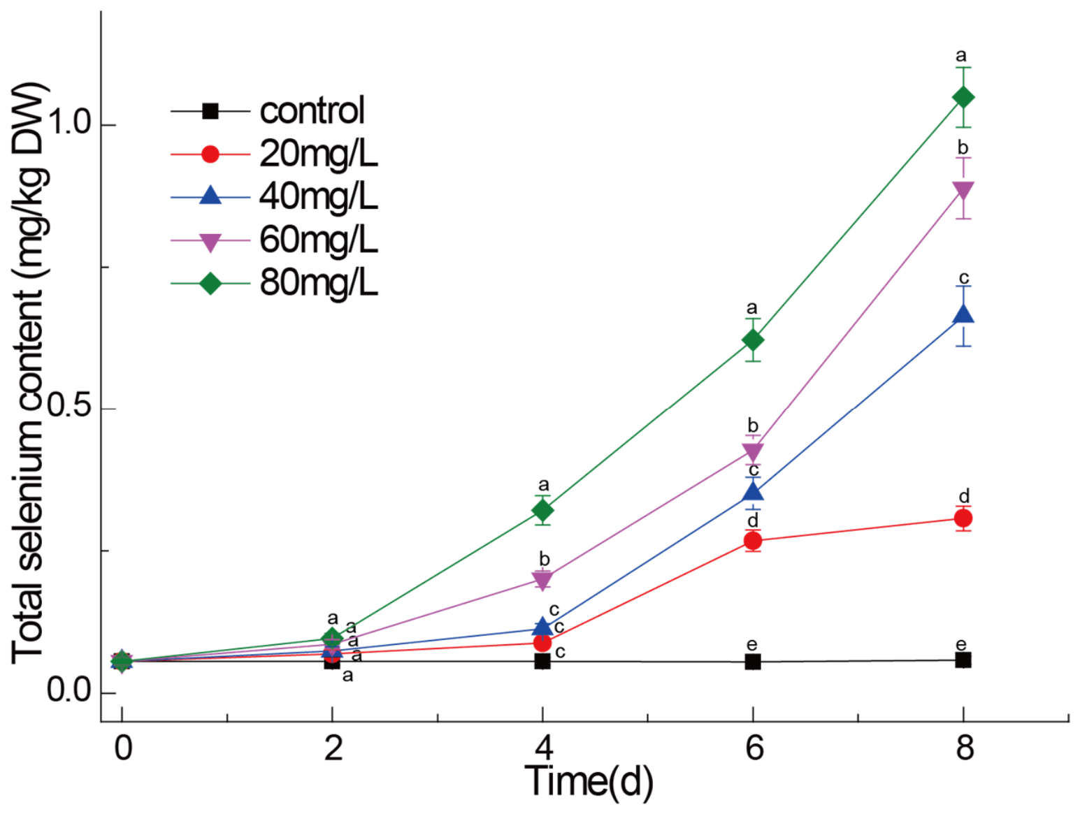
With the increase in exogenous Na2SeO3 concentration and the prolongation of germination time, the total selenium content in germinated chestnuts increased significantly (p < 0.05) (Figure 1). In the control, the total Se content in chestnuts remained within the range of 0.056 to 0.058 mg/kg throughout the germination process, with no significant differences observed (p > 0.05). The selenium of chestnuts under four concentrations of exogenous Na2SeO3 did not increase significantly in the first 2 days (p > 0.05). This may be because the sodium selenite on the surface of the chestnuts was not absorbed into chestnut tissue 2 d after sowing, and was washed away during germination to prepare samples. Under 80 and 60 mg/L Na2SeO3 treatment, the total Se content in chestnuts significantly increased (p < 0.05) from the 4th day, reached 1.049 and 0.889 mg/kg by the 8th day post-sowing respectively. Under 40 and 20 mg/L Na2SeO3 treatment, the total Se content in chestnuts significantly increased (p < 0.05) from the 6th day. The applied 20, 40, 60 and 80 mg/L Na2SeO3 correspond to actual selenium concentrations of approximately 9.1, 18.2, 27.4 and 36.5 mg Se/L, respectively. These results indicated that germination in the presence of a Na2SeO3 solution is an efficient method for Se biofortification of germinated chestnuts.
In this study, the control chestnuts sourced from the primary chestnut-producing region in Qianxi county, Tangshan city, Hebei province, China, exhibited a total Se content ranging from 0.056 to 0.058 mg/kg, significantly below the recommended standard for Se-enriched food by China Nutrition Society (0.1 to 1.0 mg/kg) [27]. Different plant species exhibit varying capacities for Se uptake and accumulation. For instance, rye grains treated with selenite accumulated Se up to 55 µg/g during germination [28], while alfalfa germination enriched Se concentrations up to 150 µg/g DW [29]. The Se content accumulated by garden cress germinated with 8 mg/L selenite solution (39 μg/g) surpasses that of lupine bean sprouts grown under the same conditions (11 μg/g dm) [17]. Therefore, the appropriate Se treatment and germination duration are pivotal factors in producing high-quality Se-enriched sprouts. Germinated chestnuts treated with 20 and 40 mg/L for 6–8 days, 60 mg/L for 4–8 days, 80 mg/L Na2SeO3 for 4–6 days all could serve as viable sources for Se-enriched foods, meeting the regulatory standards for Se enrichment in foods and agricultural products [6]. Se accumulation during different germination stages involves distinct processes. In the initial stage of germination (2–4 d after sowing), seeds absorb selenite and water from the environment, leading to swelling and restoration of metabolic activity [30]. During this period, selenite enters the seeds and cells through the aleurone layer with the absorbed water [18]. The total Se content in the germ experiences a rapid increase during the middle–late stage (4–8 d after sowing) of germination, a pattern similar to observations in mung beans germinated via Na2SeO3 treatment [10].

2.2. Effects of Exogenous Na2SeO3 on Se Speciation and Content in Germinated Chestnut

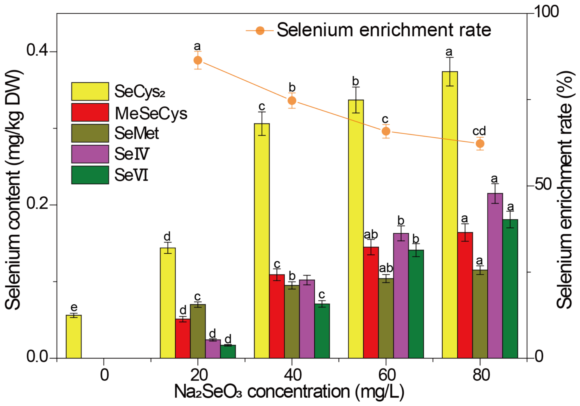
Due to the low bioavailability and high toxicity of inorganic Se [31], efforts should be directed towards enhancing the organic speciation of Se [32]. Considering nutritional value and safety aspects, the organic Se content holds greater significance, underscoring the importance of understanding Se form and transformation during plant germination. The Na2SeO3 treatment resulted in a significant (p < 0.05) increase in SeCys2, MeSecys, SeMet, SeIV and SeVI content of germinated chestnuts, while Se-enrichment rate decreased gradually (Figure 2). On the 8th day after sowing, the SeCys2 content ranged from 0.056 to 0.374 mg/kg, the MeSeCys content ranged from 0 to 0.164 mg/kg, the SeMet content ranged from 0 to 0.115 mg/kg, and the Se-enrichment rate reduced from 86.42% to 62.24%. After treatment with sodium selenite, selenium in germinated chestnuts mainly exists in organic form, which indicates that germinated chestnuts can effectively transform and accumulate organic selenium. Similar results also appeared in the experiment of mung bean germination treated by Na2SeO3 soaking [33]. Plants primarily convert inorganic Se into selenomethionine and incorporate it into protein [34]. Se-Met is the main form of organic selenium in cereals and leguminous plants, while the main selenium compound in selenium-enriched plants such as garlic, onion and wild leek is SeMeCys [35]. The main forms of organic selenium in different parts of some selenium-enriched plants are also significantly different. For example, in selenium-enriched broccoli, the major form of selenium in roots is Se-Met, while the main form of selenium in fruits is SeMeCys [36]. In this study, SeCys2 was the most abundant and stable form of Se detected in chestnuts under the experimental conditions used. After being absorbed by plants, inorganic selenium will be mixed with Cys and Met to form Se-Cys and Se-Met respectively, and then further integrated into protein to form selenoprotein [17]. When cruciferous plants are cultivated in Se-rich soil, they convert less than 40% of the total Se content into selenoamino acids [37]. In contrast, when grown in Se-poor conditions, primary species can incorporate over 95% of the total Se content as selenoamino acids [38,39]. In this study, the Se-enrichment rate decreased gradually with the increase in Na2SeO3 concentration in this study. This may be because when the concentration of Na2SeO3 reaches a certain threshold, the ability of Se assimilation and transformation into organic selenium decreases. This is basically consistent with the research results of selenium-enriched soybean sprouts [40].

2.3. Effects of Exogenous Na2SeO3 on Phenylalanine Ammonialyase (PAL), Glutathione Peroxidase (GPX), Superoxide Dismutase (SOD) and Catalase (CAT) Activity of Germinated Chestnuts

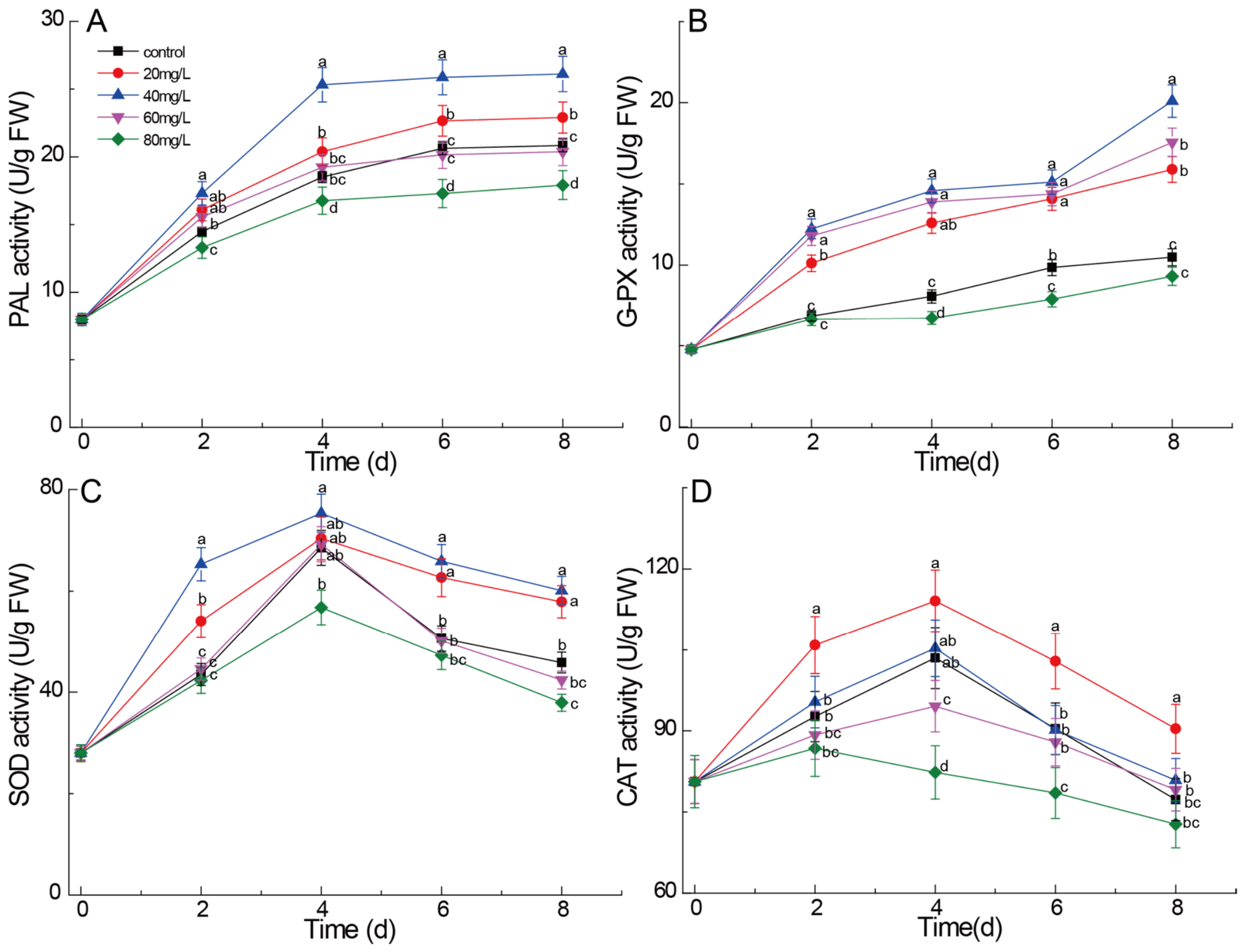
PAL is a key enzyme in the phenylpropane metabolic pathway [41]. As shown in Figure 3A, PAL activity first increased (0–4 d after sowing) and then stabilized (4–8 d after sowing) during germination. Low concentrations of Na2SeO3 (20 and 40 mg/L) significantly enhanced PAL activity during the whole germination period (p < 0.05). It is worth noting that the PAL activity reached the highest at 40 mg/L, reaching 26.11 U/g FW on the 8th day after sowing, which was 25.2% higher than that of the control group. This finding is consistent with previous research reports, indicating that sodium selenite can enhance PAL activity in many plant systems. For example, the PAL activity of chicory was increased by 36.4% by the combined treatment of NO and nano-Se, while the PAL activity of bitter gourd seedlings was increased by 39% by nano-Se [42]. In contrast, high concentration of Na2SeO3 (80 mg/L) inhibited PAL activity. On the 8th day after sowing, it decreased to 17.92 U/g FW, which was 14.1% lower than that of the control group. This inhibition may be attributed to the excessive accumulation of reactive oxygen species exceeding the antioxidant capacity of cells, leading to oxidative damage of enzyme proteins and destruction of redox homeostasis of cells [43]. There was no significant difference in PAL activity between 60 mg/L treatment and control group (p > 0.05).
In all selenium treatment groups, GPX activity increased continuously during the whole germination period, and reached the maximum on the 8th day (Figure 3B). GPX activity in the control group increased moderately from 4.77 U/g FW on day 0 to 10.48 U/g FW on day 8. GPX, as a selenase containing selenium in the form of selenocysteine in its active center, is the most sensitive to selenium treatment among all the detected enzymes. The activity of GPX increased significantly under the treatment of 40 mg/L selenium (1.92 times that of the control group), which highlighted the efficiency of selenium bioaugmentation in germinated chestnuts. The 20 and 60 mg/L treatments also significantly increased GPX activity, reaching 15.90 and 17.56 U/g FW respectively. This result confirmed earlier studies, and showed that trace selenium could enhance GPX activity in germinated seeds of wheat, rice and various vegetables [44]. However, the 80 mg/L treatment reduced GPX activity to 9.31 U/g FW on the 8th day, which was only slightly lower than that of the control group (p > 0.05), indicating that it was inhibited under excessive selenium concentration. This shows that excessive selenium interferes with the synthesis or stability of selenase [45]. This inhibition may be due to selenium-induced oxidative stress interfering with protein biosynthesis [46].
SOD serves as the first line of defense against superoxide anions, catalyzing the dismutation of O2· into H2O2 and O2 [47]. Unlike PAL and GPX, SOD activity rapidly increased from day 0 to day 4 across all groups, peaked on day 4, and then gradually declined by day 8 (Figure 3C), consistent with the typical unimodal curve of SOD during seed germination. At the peak (day 4), SOD activity in the control group was 68.51 U/g FW. Treatment with 40 mg/L Se significantly increased SOD activity to 75.32 U/g FW, which was 9.9% higher than the control. The 40 mg/L treatment maintained the highest SOD activity throughout germination. In contrast, the 80 mg/L treatment resulted in SOD activity of 56.76 U/g FW on day 4, 17.1% lower than the control. The promoting effect of low Se concentration (20–40 mg/L) on SOD activity is consistent with previous findings in alfalfa seeds [29]. A possible explanation for this activation is that Se at low concentrations may induce mild oxidative stress, which could in turn upregulate the expression of antioxidant enzyme-encoding genes (including SOD) via ROS-mediated signaling pathways involving transcription factors such as WRKY and DREB, as suggested by previous studies [48]. However, direct evidence for this mechanism in germinating chestnuts is still lacking.
With the prolongation of germination time, CAT activity in all treatment groups showed a trend of first increasing and then decreasing, peaking on day 4 (Figure 3D). The effect of different selenium concentrations on CAT activity was concentration-dependent: low-concentration (20 mg/L) treatment significantly increased CAT activity at all time points (p < 0.05), reaching a maximum of 114.09 U/g FW on day 4, which was 10.2% higher than the control. The 40 mg/L Na2SeO3 treatment resulted in slightly higher CAT activity than CK, but the difference was not significant (p > 0.05). The 60 and 80 mg/L Na2SeO3 treatments significantly inhibited CAT activity, with the 80 mg/L Na2SeO3 showing a 20.5% reduction compared to the control on day 4, and remaining at the lowest level throughout germination. Low-concentration Se significantly increased CAT activity during chestnut germination, consistent with the antioxidant effects of selenium in other plants [49,50]. When Se concentration reached 60 mg/L or higher, CAT activity was markedly inhibited. High Se concentration causes excessive selenium accumulation in plants, inducing bursts of reactive oxygen species (ROS) such as superoxide anion (O2) and hydrogen peroxide (H2O2). Additionally, selenium binds to sulfhydryl (-SH) groups, disrupting the structure of enzyme proteins and inactivating the CAT active center [51]. Of note, although CAT activity was inhibited under high Se treatment, a small increase followed by a rapid decline was still observed in the early germination stage (0–2 d after sowing), indicating that seeds can still initiate a limited antioxidant response at the beginning of germination, but this response is quickly suppressed as selenium toxicity intensifies.
The decrease in SOD and CAT activities after day 4 of germination (Figure 3C,D) can be attributed to several factors. First, the early stage of germination (days 0–4) involves intense metabolic reactivation and ROS production, which induces antioxidant enzymes. After day 4, the stored reserves are partially depleted, and the overall metabolic rate may slow down, leading to reduced enzyme synthesis. Second, in the presence of exogenous selenium, a hormetic effect may occur: low Se transiently stimulates antioxidant enzymes, but prolonged exposure or higher Se levels could cause moderate oxidative damage, suppressing enzyme activity. Third, it is known that in many seeds, the peak of antioxidant enzyme activity coincides with radicle emergence, after which activity naturally declines as the seed shifts from germination to early growth. Future studies measuring enzyme protein levels and gene expression could further clarify the regulation.
In a word, Na2SeO3 synergistically regulates the antioxidant enzyme network in germinating chestnuts. At 20–40 mg/L, selenium enhances the activities of PAL, SOD, GPX, and CAT, thereby strengthening H2O2 scavenging and phenolic biosynthesis pathways, whereas a high concentration (80 mg/L) causes irreversible oxidative damage.

2.4. Effects of Exogenous Na2SeO3 on GABA Content in Germinated Chestnuts

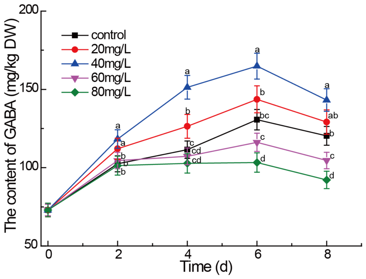
The changes in GABA content during chestnut germination under different Na2SeO3 treatments are shown in Figure 4. In all treatment groups, GABA content gradually increased from day 0 to day 6, reflecting the natural activation of GABA biosynthesis during germination, peaked at day 6, and then decreased from day 6 to day 8. The 40 mg/L Na2SeO3 treatment consistently maintained the highest GABA content throughout germination, reaching 164.87 mg/kg DW on day 6, which was 26.2% higher than the control (p < 0.05). In contrast, the 80 mg/L treatment resulted in GABA levels 20.9% lower than the control (p < 0.05), indicating that the inhibitory effect of high Se concentration becomes more pronounced during the late germination stage when GAD activity is naturally elevated. Low Se concentrations (20 and 40 mg/L) significantly promoted GABA accumulation throughout the germination period, whereas the high Se concentration (80 mg/L) inhibited GABA accumulation.
The stimulatory effect at low Se concentrations (20–40 mg/L) can be attributed to the induction of mild oxidative stress. Sub-toxic levels of selenium generate moderate amounts of reactive oxygen species (ROS), which act as signaling molecules to activate stress-responsive transcription factors and upregulate the expression of GAD and other genes involved in GABA metabolism [52]. This interpretation is consistent with the recognition of GABA as a novel protective agent capable of mitigating oxidative stress [53]. In a study of cut lily flowers, exogenous GABA treatment elevated antioxidant enzyme activities, prevented hydrogen peroxide accumulation, and maintained higher levels of total phenols and soluble proteins, thereby delaying senescence [54]. Conversely, the increase in endogenous GABA accumulation under mild selenium-induced stress may similarly contribute to the antioxidant defense system of germinating chestnuts. At high Se concentration (80 mg/L), the capacity of the cellular antioxidant system is overwhelmed, leading to excessive ROS accumulation, which causes oxidative damage to the GAD enzyme protein, disruption of cellular redox homeostasis, and inhibition of GABA biosynthesis [55]. The reduction in GABA content under the 80 mg/L Se treatment (20.9% lower than the control) is largely consistent with results observed in highland barley seeds [56].
The optimal Se concentration for GABA enhancement identified in this study (40 mg/L Na2SeO3) differs from the concentrations reported for other crop species. In foxtail millet, the appropriate concentrations were 60 mg/L for soaking and 2 mg/L for spraying [25]. These differences may reflect species-specific variations in Se sensitivity, as well as differences in experimental conditions including germination duration, temperature, seed size, and Se application method. The chestnut, as a large-seeded species with substantial nutrient reserves, may exhibit distinct patterns of GABA accumulation and Se responsiveness compared to small-seeded cereals or legumes. The observation that GABA is the major amino compound during chestnut germination and early seedling growth highlights the particular importance of this metabolite in the chestnut’s nitrogen metabolism and stress adaptation.

2.5. Effects of Exogenous Na2SeO3 on Total Polyphenols and Flavonoids Content in Germinated Chestnuts

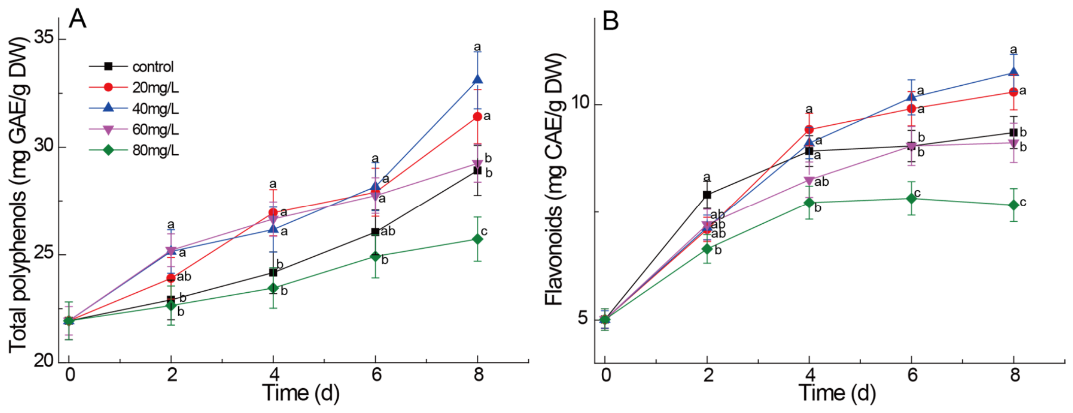
Under all treatments, the total polyphenols content (TPC) in germinating chestnuts gradually increased throughout the 8-day germination period (Figure 5A), reflecting the natural activation of phenylpropanoid metabolism during seed germination. On day 8, the TPC in the control group (0 mg/L) was 28.92 mg GAE/g DW. Low selenium concentrations (20 and 40 mg/L) significantly increased TPC, with the highest value (33.11 mg GAE/g DW) observed at 40 mg/L, which was 14.5% higher than the control. The TPC at 20 mg/L (31.42 mg GAE/g DW) was also slightly higher than the control, but the difference was not significant (p > 0.05). These findings are generally consistent with observations in other germinated seeds [57]. In contrast, the 80 mg/L Na2SeO3 treatment reduced TPC to 25.72 mg GAE/g DW, which was 11.1% lower than the control, indicating that phenolic biosynthesis is inhibited under excessive selenium stress.
Across all treatments, total flavonoid content (TFC) in germinating chestnuts accumulated rapidly during the first 4 days, and the accumulation rate slowed down during the later period (4–8 days) (Figure 5B). Low selenium concentrations (20 and 40 mg/L) showed no significant difference in TFC from the control during the first 4 days (p > 0.05), but TFC was significantly higher than the control from day 6 to day 8 (p < 0.05). These findings are generally consistent with increased flavonoid accumulation observed in other selenium-biofortified germinated seeds [58,59]. In contrast, the 80 mg/L treatment reduced TFC to 7.66 mg CAE/g DW, which was 18.1% lower than the control.
Low concentrations of Na2SeO3 (20–40 mg/L) significantly promoted the accumulation of total polyphenols and flavonoids in germinated chestnuts, indicating that appropriate selenium biofortification during seed germination can effectively enhance the accumulation of phenolic compounds in various crops. For example, in black soybeans [57], selenium biofortification significantly increased total phenolic and total flavonoid contents in germinated seeds. Similarly, selenium-enriched germination significantly increased total phenolic and total flavonol contents in hulless barley seeds [56] and soybeans [40]. It is speculated that the underlying mechanism is the activation of the phenylpropanoid pathway in germinating chestnuts, which is generally consistent with the changes in PAL activity described above (Figure 3A).

2.6. Effects of Exogenous Na2SeO3 on Antioxidant Capacity of Germinated Chestnuts

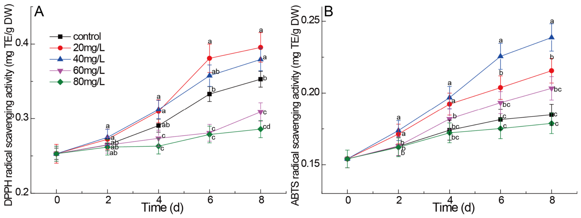
DPPH radical scavenging activity, a widely used indicator of antioxidant capacity based on hydrogen atom transfer, showed a gradual increase throughout the germination period across all treatments (Figure 6A). This reflected the natural accumulation of phenolic compounds and other antioxidants during seed germination, consistent with the enhanced PAL activity and increased phenolic content observed in this study. On day 8, the DPPH scavenging activity in the control (0 mg/L) was 0.353 mg TE/g DW. Low selenium concentrations (20 mg/L) significantly increased DPPH scavenging activity, which was 12.2% higher than the control. These results are generally consistent with findings in foxtail millet sprouts [25]. In contrast, the effect of high selenium concentration on DPPH scavenging activity gradually weakened. The DPPH scavenging activity at 80 mg/L on day 8 was 0.286 mg TE/g DW, which was 19.0% lower than the control, indicating that hydrogen atom transfer-based antioxidant capacity is inhibited under excessive selenium stress.
ABTS radical scavenging activity reflects antioxidant capacity based on electron transfer. Similar to DPPH scavenging activity, ABTS radical scavenging activity increased steadily throughout the germination period, reaching a maximum on day 8 across all treatments (Figure 6B). The ABTS scavenging activity in the control group on day 8 was 0.185 mg TE/g DW. Low selenium concentrations significantly increased ABTS scavenging activity, with the highest value (0.239 mg TE/g DW) observed at 40 mg/L, which was 29.2% higher than the control. The 20 mg/L treatment also significantly increased ABTS scavenging activity to 0.216 mg TE/g DW (16.5% higher than the control). These findings are consistent with increased ABTS radical scavenging activity observed in germinated seeds of mung bean [60]. The ABTS scavenging activity of germinated chestnuts under 80 mg/L treatment was 0.179 mg TE/g DW (slightly lower than the control), but the difference was not significant (p > 0.05).
Notably, ABTS scavenging activity was consistently higher than DPPH scavenging activity across all treatments. This observation is not uncommon, as ABTS and DPPH radicals differ in their redox potential, solubility, and reaction kinetics, and the two assays often yield different absolute values when applied to the same sample matrix. This study demonstrates that low concentrations of Na2SeO3 (20–40 mg/L) significantly enhance both DPPH and ABTS radical scavenging activities in germinating chestnuts, indicating that selenium biofortification during seed germination can effectively improve the total antioxidant capacity of various crops. In foxtail millet sprouts, soaking with 60 mg/L Na2SeO3 combined with spraying 2 mg/L Na2SeO3 significantly increased free and bound phenolic contents, thereby enhancing in vitro antioxidant activity [25]. Similarly, mung bean seeds and sprouts treated with 30 mg/L Na2SeO3 exhibited significantly elevated ABTS and DPPH radical scavenging activities, accompanied by specific enrichment of benzoic acid, rutin, and luteolin glycoside [61]. Wheat sprouts also showed increased ABTS, DPPH, and SOD activities under moderate selenium biofortification (0.25–0.50 mg/L) [62]. Peanut seeds soaked with 7.5 μmol/L Na2SeO3 exhibited significantly improved antioxidant capacity and sprout quality [63]. Furthermore, chickpea sprouts treated with 2 mg/100 g seeds Na2SeO3 showed a significant 33% increase in antioxidant capacity [64].
Overall, the enhancement of antioxidant properties by Se treatment can be explained by the ‘stress-response’ hypothesis [44]. Selenium at low (20 mg/L) to moderate (40 mg/L) concentrations generates reactive oxygen species (ROS) at sub-toxic levels, which in turn upregulates the expression and activity of antioxidant enzymes as a defense mechanism [51]. This is consistent with the well-known hormetic effect of Se in plants. In this study, the highest Se concentration (80 mg/L) caused a decrease in antioxidant enzyme activities, indicating that the stress exceeded the adaptive capacity of the chestnut tissue. Therefore, the observed increase in antioxidant activity is not due to a direct radical-scavenging role of Se itself, but rather to Se-induced mild oxidative stress that triggers an endogenous antioxidant response.

2.7. Practical Implications and Limitations

Based on our laboratory-scale results, germination with 20 mg/L sodium selenite is effective for producing Se-enriched chestnut sprouts with enhanced GABA and antioxidant capacity. However, direct extrapolation to open-field or commercial farming requires caution. In a controlled germination facility (e.g., sprout production units), adding Se to the soaking water is technically simple and low-cost. For open-field chestnut production, applying Se to soil or as a foliar spray is more common, but our study does not directly test those methods. The optimal concentration for field application would likely differ due to soil adsorption, rainfall, and microbial transformation. This study provides proof-of-concept for Se biofortification of germinated chestnuts under controlled conditions, but field application should be preceded by dose-optimization and safety trials.

3. Materials and Methods

3.1. Chemicals and Reagents

Sodium selenite was purchased from MedChem Express (Taian Havay Chemicals Co., Ltd., Taian, China). SeIV and SeVI standards were obtained from the National Institute of Metrology (Beijing, China). SeCys2, SeMet, and MeSeCys (methylselenocysteine) standards were purchased from Sigma-Aldrich (St. Louis, MO, USA). Protease XIV, lipase, Tris-HCl, L-phenylalanine, sodium borate, methionine, NBT, EDTA-Na2, riboflavin, potassium phosphate monobasic/dibasic, GSH, sodium azide, hydrogen peroxide (H2O2), Folin–Ciocalteu reagent, gallic acid, catechin, DPPH (2,2-diphenyl-1-picrylhydrazyl), ABTS (2,2′-azino-bis(3-ethylbenzothiazoline-6- sulfonic acid)), and Trolox were purchased from Sigma-Aldrich (St. Louis, MO, USA), while 2,3-Diaminonaphthalene was purchased from Macklin Biochemical Co., Ltd. (Shanghai, China). Potassium persulfate was purchased from Aladdin Biochemical Technology Co., Ltd. (Shanghai, China). Phosphate-buffered saline (PBS, 50 mmol/L, pH 7.8), EDTA, PVP-K30, Triton X-100, BCA protein assay kit, PAL activity assay kit, GPX activity assay kit, SOD activity assay kit, and CAT activity assay kit were purchased from Solarbio Science & Technology Co., Ltd. (Beijing, China). AccQ·Tag Ultraborate buffer and AccQ·Tag reagent were purchased from Waters Corporation (Milford, MA, USA).

3.2. Plant Materials and Treatments

The seeds of chestnut (Castanea mollissima) variety “Zaofeng” were harvested from healthy trees in the primary chestnut production region of Qinglong County, Qinhuangdao City, Hebei Province, China. The seeds were stored at −3 °C for preservation. Following the selection of uniform seeds, they were washed with sterile water, soaked for 12 h, and then evenly distributed on trays (50 cm × 30 cm × 15 cm) filled with sterilized sand mixed with sterile water (control) or varying concentrations (20, 40, 60, and 80 mg/L) of Na2SeO3. The trays were maintained at 22 °C and 85% relative humidity for 8 days [26]. Radicle emergence was observed on day 3, and full germination (radicle > 2 mm) was reached by day 4. Ten chestnut seeds per replicate were collected at 0, 2, 4, 6 and 8 days post-sowing, and the whole chestnuts were gently rinsed three times with deionized water to remove surface-adsorbed Se. The outer hard shell and the brown seed coat were then removed, and only the edible kernel was ground into powder after seed coat removal and stored at −40 °C for subsequent physiological and biochemical analysis. All results are expressed on a dry weight basis (dw) after drying a subsample at 105 °C to constant weight. Each treatment was replicated three times, with each replicate consisting of 30 seeds.

3.3. Determination of Se Content

3.3.1. Total Se Content

The total selenium content was determined using fluorescence spectrophotometry (Cary Eclipse; Agilent Instruments, Santa Clara, CA, USA) [13]. The procedure was as follows: 0.5 g of dried chestnut powder was digested overnight by heating with 10 mL of a mixed solution of HNO3 and HClO4 (9:1, v/v). Subsequently, 5 mL of HCl (6 mol/L) was added to the digest. The heat-digested sample was then reacted with 2,3-diaminonaphthalene, and after extraction with cyclohexane, the fluorescence intensity was measured. A calibration curve was prepared using a selenium standard solution, and the total selenium content was calculated and expressed as mg/kg DW.

3.3.2. Five Se Speciation Content

Accurately weigh 0.3000 g of dried germinated chestnut sample and place in a digestion vessel. Add 5 mL of HNO3 and 1 mL of H2O2, and perform microwave-assisted digestion. After digestion, cool the solution and transfer the digest to a 10 mL volumetric flask. Rinse the digestion vessel three times with ultrapure water, combine the washings, and dilute to the mark with ultrapure water. Subsequently, transfer 0.5 mL of the digest to a 20 mL centrifuge tube, and sequentially add 60 mg of protease XIV, 30 mg of lipase, and 10 mL of Tris-HCl buffer. Mix thoroughly, then ultrasonicate for 30 min. Place the tube in a constant temperature water bath shaker and shake horizontally at 37 °C and 150 r/min. After incubation in the dark for 20 h, centrifuge the sample at 10,000 r/min for 30 min. Filter the supernatant through a 0.22 μm aqueous filter membrane and store at −20 °C. Add 5 mL of hydrolysis solution containing 100 mg of catabolic enzyme to the remaining residue, and repeat the above steps. Combine the two supernatants and filter through a 0.22 μm aqueous filter membrane. Perform Se speciation analysis using HPLC-ICP-MS [65]. Prepare a mixed standard solution using Secys2 (>99% purity), MeSeCys (>99% purity), SeMet (>99% purity), SeIV and SeVI standards (>99% purity). Using the external standard method, construct a calibration curve by plotting Se concentration on the X-axis against chromatographic peak area on the Y-axis with a series of mixed standard solutions. The speciation content of Se in the sample was determined using the provided formula (1):
X = ( C     C 0 ) × V m × 1000
where: X—speciation content of Se in the sample (mg/kg); C—determination of concentration of sample extract (μg/L); C0—concentration of sample blank determination (μg/L); V—total volume of sample extract at constant volume (L); m—weight of sample (g).

3.3.3. Calculation of Selenium Enrichment Rate

The enrichment rate of selenium was calculated by Equations (2) and (3), respectively [66].
Ose = CSeCys2 + CMeSeCys + CSeMet
Rse (%) = Ose/Tse × 100
where CSeCys2 represents the SeCys2 content of germinated chestnuts, CMeSeCys represents the MeSeCys content of germinated chestnuts, CSeMet represents the SeMet content of germinated chestnuts, Tse represents the total selenium content of germinated chestnuts, Ose represents the organic selenium content, and Rse represents the selenium enrichment rate.

3.4. Determination of PAL, GPX, SOD and CAT Activity

The extraction and activity assays of PAL, GPX, SOD, and CAT were performed following Du et al. (2024) [67]. Fresh germinated chestnut samples (3 g) were homogenized with 6 mL of ice-cold PBS (50 mmol/L, pH 7.8) containing 1 mmol/L EDTA, 2% (m/v) PVP-K30, and 0.3% Triton X-100. After grinding for 60 s, the mixture was centrifuged at 10,000× g for 5 min at 4 °C. The resulting supernatant served as the crude enzyme extract for all assays, with activity determination based on Zhang et al. (2025) [68].
PAL activity assay: A total of 0.1 mL of crude enzyme extract was mixed with 2.0 mL of 0.1 mol/L borate buffer (pH 8.8) and 1.0 mL of 0.02 mol/L L-phenylalanine solution. After thorough mixing, the mixture was incubated in a constant temperature water bath at 37 °C for 30 min. The reaction was terminated by adding 0.5 mL of 35% trichloroacetic acid (TCA). The absorbance was measured at 290 nm. One unit (U) of PAL activity was defined as the amount of enzyme causing an increase of 0.01 in absorbance at 290 nm per 30 min under the assay conditions.
SOD activity assay: A total of 0.1 mL of crude enzyme extract was sequentially mixed with 1.7 mL of PBS (50 mmol/L, pH 7.8), 0.3 mL of methionine (130 mmol/L), 0.3 mL of NBT (750 μmol/L), 0.3 mL of EDTA-Na2 (100 μmol/L), and 0.3 mL of riboflavin (20 μmol/L). A control tube was prepared by replacing the crude enzyme extract with an equal volume of PBS. The reaction mixture was illuminated under a fluorescent lamp (4000 lx) for 15 min, and then the absorbance was measured at 560 nm. One unit of SOD activity was defined as the amount of enzyme required to inhibit 50% of NBT photoreduction.
GPX activity assay: A total of 0.1 mL of crude enzyme extract was mixed with 0.6 mL of 50 mmol/L potassium phosphate buffer (pH 7.0), 0.1 mL of 1 mmol/L GSH, and 0.1 mL of 1 mmol/L sodium azide. After pre-incubation at 37 °C for 5 min, the reaction was initiated by adding 0.1 mL of H2O2 solution (1.5 mmol/L). The mixture was immediately vortexed and allowed to react for exactly 10 min. The reaction was terminated by adding 0.5 mL of 10% (w/v) TCA, followed by centrifugation at 10,000× g for 5 min at 4 °C. Then, 0.4 mL of the supernatant was mixed with 2.6 mL of 50 mmol/L potassium phosphate buffer (pH 7.0) and 0.5 mL of DTNB chromogenic solution (10 mmol/L). After incubation at room temperature for 5 min, the absorbance was measured at 412 nm. The control tube was prepared by replacing the crude enzyme extract with an equal volume of potassium phosphate buffer. One unit of GPX activity was defined as a change of 0.005 in absorbance at 412 nm per minute.
CAT activity assay: A total of 0.1 mL of crude enzyme extract was mixed with 2.9 mL of PBS (50 mmol/L, pH 7.0) containing 20 mmol/L H2O2. The activity was determined by monitoring the decrease in absorbance at 240 nm. The control solution was PBS with H2O2 but without enzyme. One unit of CAT activity was defined as a decrease of 0.01 in absorbance at 240 nm per minute.
All absorbance values were measured by a UV-Vis spectrophotometer (UT-1901; METASH, Shanghai, China), and all enzyme activities are expressed as U/g.

3.5. Determination of GABA Content

The procedures were based on Jannoey, P., et al. [69] with slight modifications. Briefly, 0.2 g of dried germinated chestnut sample power was extracted using 1 mL of 0.1 mol/L HCl at 70 °C for 1 h, and centrifuged at 12,000 rpm for 10 min. Then, 10 µL of the supernatant was mixed with 70 µL of AccQ·Tag Ultra Borate Buffer and 20 µL of accqtag reagent in a derivatization tube. After vortexing, the mixture was heated at 55 °C for 10 min and then cooled. HPLC analysis was performed at a flow rate of 0.5 mL/min, column temperature of 55 °C, and the injection volume was 1 μL. GABA content was quantified using a standard curve and expressed as mg/kg DW.

3.6. Determination of Total Polyphenol and Flavonoid Content

The extraction process for total polyphenol content (TPC) and total flavonoid content (TFC) was similar to that of the GABA. TPC was determined using the Folin–Ciocalteu method [66]. Briefly, 500 µL of sample extract was mixed with 250 µL of Folin–Ciocalteu reagent and 3.00 mL of distilled water. After 5 min, 750 µL of 7% Na2CO3 was added, and the mixture was incubated at 20 °C for 1 h. Absorbance was measured at 765 nm. TPC was quantified against a gallic acid calibration curve and expressed as mg gallic acid equivalents (GAE)/g DW. TFC was measured according to Ren et al. (2024) [70]. Briefly, 250 µL of sample extract was mixed with 1250 µL of distilled water and 75 µL of 5% NaNO2 solution. After 5 min, 150 µL of 10% AlCl3·6H2O was added; then, 500 µL of 1 mol/L NaOH and 275 µL of distilled water. TFC was calculated using a calibration using a catechin (CAE) standard curve and expressed as mg CAE/g DW. The absorbance of the mixture was measured at 510 nm. All absorbance values were measured by a UV-Vis spectrophotometer (UT-1901; METASH, Shanghai, China).

3.7. Determination of DPPH Radical Scavenging Activity

DPPH radical scavenging activity was measured using a modified method of Farhadi et al. (2016) [71]. In brief, 0.2 g of dried germinated chestnut sample power was extracted using 2 mL of methanol. After ultrasonic extraction at 100–200 W for 20 min, the mixture was centrifuged at 10,000× g for 5 min. Then, 100 µL of the supernatant was mixed with 3.9 mL of 0.1 mmol/L DPPH in methanol and kept in the dark for 30 min. Absorbance was then read at 517 nm using a UV-Vis spectrophotometer (UT-1901; METASH, Shanghai, China). The scavenging activity was quantified against a Trolox standard curve and expressed as mg Trolox equivalents per gram of dry weight (mg TE/g DW).

3.8. Determination of ABTS Radical Scavenging Activity

ABTS radical cation scavenging activity was determined according to the method of Spiegel et al. (2022) [72] with minor modifications. Briefly, 0.2 g of dried germinated chestnut sample power was extracted using 2 mL of methanol. After ultrasonic extraction at 100–200 W for 20 min, the mixture was centrifuged at 10,000× g for 5 min. Then, 100 µL of the supernatant was mixed with 1.9 mL of 0.325 mol/L phosphate buffer and 2.0 mL of ABTS working solution and incubated in the dark at room temperature for 10 min. The absorbance was measured at 734 nm using a UV-Vis spectrophotometer (UT-1901; METASH, Shanghai, China). A control was prepared by replacing the sample extract with the corresponding solvent. The scavenging activity was calculated as the percentage of inhibition and expressed as mg Trolox equivalents per gram of dry weight (mg TE/g DW) using a Trolox standard curve.

3.9. Statistical Analysis

All experiments were performed with three biological replicates, and the results are reported as mean values with standard errors. All statistical analyses were carried out using SPSS 22.0 (SPSS Inc. Chicago, IL, USA), while graphical analysis was conducted using Origin 2023 (Microcal Software, Northampton, MA, USA). One-way ANOVA followed by Tukey’s HSD post-hoc test (p < 0.05) was used to compare treatments.

4. Conclusions

In summary, exogenous Na2SeO3 treatment exerts dose-dependent effects on selenium biofortification, phenolic metabolism, antioxidant enzyme activities, and total antioxidant capacity in germinating chestnuts. Low selenium concentrations (20–40 mg/L) significantly promoted total selenium accumulation and the conversion of organic selenium (SeMet, SeCys2 and MeSecys), with the highest proportion of organic selenium (81.3%) observed at 20 mg/L. Concurrently, low selenium treatment upregulated the activities of PAL, GPX, SOD, and CAT, promoted the accumulation of γ-aminobutyric acid (GABA), total polyphenols and flavonoids, and consequently enhanced DPPH and ABTS radical scavenging activities. The 40 mg/L Na2SeO3 treatment was identified as the optimal concentration, yielding the greatest improvements across all measured parameters. In contrast, high selenium concentration (80 mg/L) gradually inhibited these beneficial effects, leading to a decreased proportion of organic selenium, reduced enzyme activities, diminished accumulation of phenolics and GABA, and weakened antioxidant capacity, indicating oxidative stress and metabolic disorder. These findings reveal that the functional activity of germinating chestnuts exhibits a “low-dose promotion, high-dose inhibition” response to selenium treatment, and establish 40 mg/L Na2SeO3 as the optimal concentration for producing selenium-enriched germinated chestnuts with improved nutritional quality and antioxidant potential. This study provides a theoretical basis and technical parameters for the development of functional foods from selenium-enriched germinated chestnuts.

Author Contributions

Writing—original draft, H.W.; review and editing, W.L.; formal analysis, F.P.; methodology, Z.Z.; data curation, J.C. and J.S.; investigation, L.H.; validation, Y.J.; conceptualization, M.W.; project administration, J.Y. All authors have read and agreed to the published version of the manuscript.

Funding

This work was financially supported by the Scientific Research Fund of Hebei Normal University of Science and Technology (2024YB038), the Hebei Transformation Project of Agricultural Scientific and Technological Achievements (2026JNZ-S28), and the Hebei Scientific Research Projects of Colleges and Universities (BJ2026130).

Institutional Review Board Statement

Not applicable.

Informed Consent Statement

Not applicable.

Data Availability Statement

The original contributions presented in this study are included in the article; further inquiries can be directed to the corresponding author.

Conflicts of Interest

The authors declare no conflicts of interest.

References

  1. Duntas, L.H.; Benvenga, S. Selenium: An element for life. Endocrine 2015, 48, 756–775. [Google Scholar] [CrossRef] [PubMed]
  2. Yang, W.; Huang, G.; Chen, F.; Huang, H. Extraction/synthesis and biological activities of selenopolysaccharide. Trends Food Sci. Tech. 2021, 109, 211–218. [Google Scholar] [CrossRef]
  3. Lin, Y.; Jiang, L.Q. Research progress on the immunomodulatory effect of trace element selenium and its effect on immune-related diseases. Food Ther. Health Care. 2020, 2, 86–98. [Google Scholar] [CrossRef]
  4. Ho, Y.S.; So, K.F.; Chang, R.C.C. Anti-aging herbal medicine-how and why can they be used in aging-associated neurodegenerative diseases? Ageing Res. Rev. 2010, 9, 354–362. [Google Scholar] [CrossRef]
  5. Wang, H.; Shi, D.; Chen, Y.; Wang, Z.; Yuan, Y.; Yue, T. Dietary supplementation with novel selenium-enriched Pichia kudriavzevii regulates gut microbiota and host metabolism in mice. Food Funct. 2024, 15, 10896–10912. [Google Scholar] [CrossRef]
  6. Chen, Z.; Lu, Y.; Dun, X.; Wang, X.; Wang, H. Research progress of selenium-enriched foods. Nutrients 2023, 15, 4189. [Google Scholar] [CrossRef]
  7. Miyahira, R.F.; Lopes, J.O.; Antunes, A.E.C. The use of sprouts to improve the nutritional value of food products: A brief review. Plant Food Hum. Nutr. 2021, 76, 143–152. [Google Scholar] [CrossRef]
  8. Burk, R.F.; Hill, K.E. Regulation of selenium metabolism and transport. Annu. Rev. Nutr. 2015, 35, 109–134. [Google Scholar] [CrossRef]
  9. Kubachka, K.M.; Hanley, T.; Mantha, M.; Wilson, R.A.; Falconer, T.M.; Kassa, Z.; Oliveira, A.; Landero, J.; Caruso, J. Evaluation of selenium in dietary supplements using elemental speciation. Food Chem. 2017, 218, 313–320. [Google Scholar] [CrossRef]
  10. Cheng, H.; Li, L.; Dong, J.; Wang, S.; Wu, S.; Rao, S.; Li, L.; Cheng, S.; Li, L. Transcriptome and physiological determination reveal the effects of selenite on the growth and selenium metabolism in mung bean sprouts. Food Res. Int. 2023, 169, 112880. [Google Scholar] [CrossRef]
  11. Zhang, M.; Wang, L.; Wang, J.; Zhao, Q.; Sun, Y. Feasibility of cultivating Se-enriched crops using localized natural Se enrichment resources. Food Chem. 2025, 467, 142333. [Google Scholar] [CrossRef] [PubMed]
  12. Li, M.; Zhou, S.; Zhou, C.; Peng, Y.; Wen, X.; Cong, W.; Zhang, F.; Ni, Y. Unlocking the potential of selenium (Se) bio-fortification and optimal food processing of barely grains on the alleviation of Se-deficiency in highland diets. J. Food Compos. Anal. 2025, 142, 107436. [Google Scholar] [CrossRef]
  13. Diowksz, A.; Kordialik-Bogacka, E.; Ambroziak, W. Se-enriched sprouted seeds as functional additives in sourdough fermentation. LWT-Food Sci. Technol. 2014, 56, 524–528. [Google Scholar] [CrossRef]
  14. Ávila, F.W.; Yang, Y.; Faquin, V.; Ramos, S.J.; Guilherme, L.R.G.; Thannhauser, T.W.; Li, L. Impact of selenium supply on Se-methylselenocysteine and glucosinolate accumulation in selenium-biofortified Brassica sprouts. Food Chem. 2014, 165, 578–586. [Google Scholar] [CrossRef]
  15. Ortega-Hernández, E.; Antunes-Ricardo, M.; Cisneros-Zevallos, L.; Jacobo-Velázquez, D.A. Selenium, sulfur, and methyl jasmonate treatments improve the accumulation of lutein and glucosinolates in kale sprouts. Plants 2022, 11, 1271. [Google Scholar] [CrossRef]
  16. Lintschinger, J.; Fuchs, N.; Moser, J.; Kuehnelt, D.; Goessler, W. Selenium- enriched sprouts: A raw material for fortified cereal-based diets. J. Agri. Food Chem. 2000, 48, 5362–5368. [Google Scholar] [CrossRef]
  17. Frias, J.; Gulewicz, P.; Martinez-Villaluenga, C.; Peñas, E.; Piskula, M.K.; Kozlowska, H.; Ciska, E.; Gulewicz, K.; Vidal-Valverde, C. Changes in nutritional value and cytotoxicity of garden cress germinated with different selenium solutions. J. Agri. Food Chem. 2010, 58, 2331–2336. [Google Scholar] [CrossRef]
  18. Liu, K.; Chen, F.; Zhao, Y.; Gu, Z.; Yang, H. Selenium accumulation in protein fractions during germination of Se-enriched brown rice and molecular weights distribution of Se-containing proteins. Food Chem. 2011, 127, 1526–1531. [Google Scholar] [CrossRef]
  19. Kora, A.J. Nutritional and antioxidant significance of selenium-enriched mushrooms. Bull. Nat. Res. Centre. 2020, 44, 34. [Google Scholar] [CrossRef]
  20. Li, C.P.; He, Z.; Wang, X.; Yang, L.; Yin, C.; Zhang, N.; Lin, J.; Zhao, H. Selenization of ovalbumin by dry-heating in the presence of selenite: Effect on protein structure and antioxidant activity. Food Chem. 2014, 148, 209–217. [Google Scholar] [CrossRef] [PubMed]
  21. Pinzon-Nuñez, D.A.; Wiche, O.; Bao, Z.; Xie, S.; Fan, B.; Zhang, W.; Tang, M.; Tian, H. Selenium species and fractions in the rock–soil–plant interface of maize (Zea mays L.) grown in a natural ultra-rich Se environment. Int. J. Env. Res. Pub. He. 2023, 20, 4032. [Google Scholar] [CrossRef]
  22. Idowu, A.T.; Olatunde, O.O.; Adekoya, A.E.; Idowu, S. Germination: An alternative source to promote phytonutrients in edible seeds. Food Qual. Safety. 2020, 4, 129–133. [Google Scholar] [CrossRef]
  23. Nikmaram, N.; Dar, B.N.; Roohinejad, S.; Koubaa, M.; Barba, F.J.; Greiner, R.; Johnson, S.K. Recent advances in γ-aminobutyric acid (GABA) properties in pulses: An overview. J. Sci. Food Agr. 2017, 97, 2681–2689. [Google Scholar] [CrossRef]
  24. Tiansawang, K.; Luangpituksa, P.; Varanyanond, W.; Hansawasdi, C. GABA (γ-aminobutyric acid) production, antioxidant activity in some germinated dietary seeds and the effect of cooking on their GABA content. Food Sci. Technol. 2016, 36, 313–321. [Google Scholar] [CrossRef]
  25. Sun, S.; Zhang, J.; Li, Y.; Xu, Y.; Yang, R.; Luo, L.; Xiang, J. Effects of sodium selenite on accumulations of selenium and GABA, phenolic profiles, and antioxidant activity of Foxtail Millet during germination. Foods 2024, 13, 3916. [Google Scholar] [CrossRef] [PubMed]
  26. Yuan, J.; Wang, H.; Jiang, Y.; Jiang, Y.; Tang, Y.; Li, X.; Zhao, Y. Utilization of germinated seeds as functional food ingredients: Optimization of nutrient composition and antioxidant activity evolution based on the germination characteristics of Chinese chestnut (Castanea mollissima). Foods 2024, 13, 2605. [Google Scholar] [CrossRef] [PubMed]
  27. Ren, X.; Wang, Y.; Sun, J.; Liang, K.; Zhu, H.; Li, Y.; Gao, J.; Zhang, Y.; Huang, S.; Zhu, D. Legal standards for selenium enriched foods and agricultural products: Domestic and international perspectives. Nutrients 2024, 16, 3659. [Google Scholar] [CrossRef]
  28. Frias, J.; Gulewicz, P.; Martínez-Villaluenga, C.; Pilarski, R.; Blazquez, E.; Jiménez, B.; Gulewicz, K.; Vidal-Valverde, C. Influence of germination with different selenium solutions on nutritional value and cytotoxicity of lupin seeds. J. Agri. Food Chem. 2009, 57, 1319–1325. [Google Scholar] [CrossRef] [PubMed]
  29. Kovács, Z.; Soós, Á.; Kovács, B.; Kaszás, L.; Elhawat, N.; Bákonyi, N.; Razem, M.; Kári, M.; Prokisch, J.; Domokos-Szabolcsy, É.; et al. Uptake dynamics of ionic and elemental selenium forms and their metabolism in multiple-harvested alfalfa (Medicago sativa L.). Plants 2021, 10, 1277. [Google Scholar] [CrossRef]
  30. Chauhan, R.; Awasthi, S.; Srivastava, S.; Dwivedi, S.; Pilon-Smits, E.A.; Dhankher, O.P.; Tripathi, R.D. Understanding selenium metabolism in plants and its role as a beneficial element. Crit. Rev. Env. Sci. Tec. 2019, 49, 1937–1958. [Google Scholar] [CrossRef]
  31. Wang, D.; Dinh, Q.T.; Thu, T.T.A.; Zhou, F.; Yang, W.; Wang, M.; Song, W.W.; Liang, D. Effect of selenium-enriched organic material amendment on selenium fraction transformation and bioavailability in soil. Chemosphere 2018, 199, 417–426. [Google Scholar] [CrossRef]
  32. Tolu, J.; Thiry, Y.; Bueno, M.; Jolivet, C.; Potin-Gautier, M.; Le Hécho, I. Distribution and speciation of ambient selenium in contrasted soils, from mineral to organic rich. Sci. Total Environ. 2014, 479, 93–101. [Google Scholar] [CrossRef]
  33. Wang, K.; Yuan, Y.; Luo, X.; Shen, Z.; Huang, Y.; Zhou, H.; Gao, X. Effects of exogenous selenium application on nutritional quality and metabolomic characteristics of mung bean (Vigna radiata L.). Front. Plant Sci. 2022, 13, 961447. [Google Scholar] [CrossRef]
  34. Zhang, X.; He, H.; Xiang, J.; Yin, H.; Hou, T. Selenium-containing proteins/peptides from plants: A review on the structures and functions. J. Agri. Food Chem. 2020, 68, 15061–15073. [Google Scholar] [CrossRef] [PubMed]
  35. Rizwan, M.; Ali, S.; Rehman, M.Z.U.; Rinklebe, J.; Tang, D.C.; Tack, F.M.; Abbasi, G.H.; Hussain, A.; Igalavithana, A.D.; Lee, B.C.; et al. Effects of selenium on the uptake of toxic trace elements by crop plants: A review. Crit. Rev. Env. Sci. Tec. 2021, 51, 2531–2566. [Google Scholar] [CrossRef]
  36. Pedrero, Z.; Madrid, Y.; Hartikainen, H.; Cámara, C. Protective effect of selenium in broccoli (Brassica oleracea) plants subjected to cadmium exposure. J. Agri. Food Chem. 2008, 56, 266–271. [Google Scholar] [CrossRef] [PubMed]
  37. Deng, N.; Zuo, X.; Stack, A.G.; Lee, S.S.; Zhou, Z.; Weber, J.; Hu, Y. Selenite and selenate sequestration during coprecipitation with barite: Insights from mineralization processes of adsorption, nucleation, and growth. Environ. Sci. Technol. 2022, 56, 15518–15527. [Google Scholar] [CrossRef]
  38. Xu, X.; Wang, J.; Wu, H.; Cui, X.; Hou, D.; Cui, J.; Lu, R.; Lin, A. Synthesis and characterization of chitosan oligosaccharide selenium nanoparticles and selenium enrichment evaluation and mechanism on soybean sprouts. ACS Agr. Sci. Technol. 2022, 3, 67–81. [Google Scholar] [CrossRef]
  39. Chen, B.; He, M.; Zhong, C.; Hu, B. Chiral speciation of selenoamino acids in biological samples. J. Chromatogr. A 2014, 1363, 62–70. [Google Scholar] [CrossRef]
  40. Huang, Y.; Lei, N.; Xiong, Y.; Liu, Y.; Tong, L.; Wang, F.; Fan, B.; Maesen, P.; Blecker, C. Influence of selenium biofortification of soybeans on speciation and transformation during seed germination and sprouts quality. Foods 2022, 11, 1200. [Google Scholar] [CrossRef]
  41. Kong, J.Q. Phenylalanine ammonia-lyase, a key component used for phenylpropanoids production by metabolic engineering. RSC Adv. 2015, 5, 62587–62603. [Google Scholar] [CrossRef]
  42. Kang, L.; Wu, Y.; Jia, Y.; Chen, Z.; Kang, D.; Zhang, L.; Pan, C. Nano-selenium enhances melon resistance to Podosphaera xanthii by enhancing the antioxidant capacity and promoting alterations in the polyamine, phenylpropanoid and hormone signaling pathways. J. Nanobiotechnol. 2023, 21, 377. [Google Scholar] [CrossRef]
  43. He, L.; He, T.; Farrar, S.; Ji, L.; Liu, T.; Ma, X. Antioxidants maintain cellular redox homeostasis by elimination of reactive oxygen species. Cell. Physiol. Biochem. 2017, 44, 532–553. [Google Scholar] [CrossRef]
  44. Tan, Z.; Xuan, Z.; Wu, C.; Cheng, Y.; Xu, C.; Ma, X.; Wang, D. Effects of selenium on the AsA-GSH system and photosynthesis of pakchoi (Brassica chinensis L.) under lead stress. J. Soil Sci. Plant Nut. 2022, 22, 5111–5122. [Google Scholar] [CrossRef]
  45. Ferguson, L.R.; Karunasinghe, N.; Zhu, S.; Wang, A.H. Selenium and its’ role in the maintenance of genomic stability. Mutat. Res.-Fundam. Mol. Mech. Mutagen. 2012, 733, 100–110. [Google Scholar] [CrossRef]
  46. Zhang, T.; Qi, M.; Wu, Q.; Xiang, P.; Tang, D.; Li, Q. Recent research progress on the synthesis and biological effects of selenium nanoparticles. Front. Nutr. 2023, 10, 1183487. [Google Scholar] [CrossRef]
  47. Ighodaro, O.M.; Akinloye, O.A. First line defence antioxidants-superoxide dismutase (SOD), catalase (CAT) and glutathione peroxidase (GPX): Their fundamental role in the entire antioxidant defence grid. Alex. J. Med. 2018, 54, 287–293. [Google Scholar] [CrossRef]
  48. Singh, A.; Kumar, A.; Yadav, S.; Singh, I.K. Reactive oxygen species-mediated signaling during abiotic stress. Plant Gene 2019, 18, 100173. [Google Scholar] [CrossRef]
  49. He, L.X.; Zheng, A.X.; Du, B.; Luo, H.W.; Lu, R.H.; Du, P.; Chen, Y.L.; Zhang, T.T.; Lai, R.F.; Tang, X.R. Low-concentration sodium selenite applications improve oxidation resistance of filling-stage rice. Appl. Ecol. Env. Res. 2019, 17, 989–998. [Google Scholar] [CrossRef]
  50. Liu, Y.; Ding, X.; Lv, Y.; Cheng, Y.; Li, C.; Yan, L.; Tian, S.; Zou, X. Exogenous serotonin improves salt tolerance in rapeseed (Brassica napus L.) seedlings. Agronomy 2021, 11, 400. [Google Scholar] [CrossRef]
  51. Battin, E.E.; Brumaghim, J.L. Antioxidant activity of sulfur and selenium: A review of reactive oxygen species scavenging, glutathione peroxidase, and metal-binding antioxidant mechanisms. Cell Biochem. Biophys. 2009, 55, 1–23. [Google Scholar] [CrossRef]
  52. Scuto, M.; Majzúnová, M.; Torcitto, G.; Antonuzzo, S.; Rampulla, F.; Di Fatta, E.; Trovato Salinaro, A. Functional food nutrients, redox resilience signaling and neurosteroids for brain health. Int. J. Mol. Sci. 2024, 25, 12155. [Google Scholar] [CrossRef]
  53. Ansari, M.I.; Jalil, S.U.; Ansari, S.A.; Hasanuzzaman, M. GABA shunt: A key-player in mitigation of ROS during stress. Plant Growth Regul. 2021, 94, 131–149. [Google Scholar] [CrossRef]
  54. Aghdam, M.S.; Naderi, R.; Jannatizadeh, A.; Babalar, M.; Sarcheshmeh, M.A.A.; Faradonbe, M.Z. Impact of exogenous GABA treatments on endogenous GABA metabolism in anthurium cut flowers in response to postharvest chilling temperature. Plant Physiol. Bioch. 2016, 106, 11–15. [Google Scholar] [CrossRef]
  55. Wu, X.; Jia, Q.; Ji, S.; Gong, B.; Li, J.; Lü, G.; Gao, H. Gamma-aminobutyric acid (GABA) alleviates salt damage in tomato by modulating Na+ uptake, the GAD gene, amino acid synthesis and reactive oxygen species metabolism. BMC Plant Biol. 2020, 20, 465. [Google Scholar] [CrossRef]
  56. Yin, X.; Wang, S.; Wang, Z.; Wen, H.; Bai, T.; Zhang, Y. Effects of Pretreatment Methods on Gamma-Aminobutyric Acid Enrichment and Quality Improvement in Highland Barley Beverages. Foods 2024, 13, 4053. [Google Scholar] [CrossRef]
  57. Lee, A.L.; Yu, Y.P.; Hsieh, J.F.; Kuo, M.I.; Ma, Y.S.; Lu, C.P. Effect of germination on composition profiling and antioxidant activity of the polysaccharide-protein conjugate in black soybean [Glycine max (L.) Merr.]. Int. J. Biol. Macromol. 2018, 113, 601–606. [Google Scholar] [CrossRef]
  58. Huang, J.; Qian, J.; Wang, S.; Li, Y.; Zhai, X.; Olajide, T.M.; Shen, G.X.; Liao, X. Effect of selenium biofortification on bioactive compounds and antioxidant activity in germinated black soybean. J. Food Sci. 2022, 87, 1009–1019. [Google Scholar] [CrossRef]
  59. Mitharwal, S.; Chauhan, K. Impact of germination on the proximate composition, functional properties, and structural characteristics of black soybean (Glycine max L. Merr). J. Food Process. Pres. 2022, 46, e17202. [Google Scholar] [CrossRef]
  60. Kapravelou, G.; Martínez, R.; Perazzoli, G.; Sanchez Gonzalez, C.; Llopis, J.; Cantarero, S.; Goua, M.; Bermano, G.; Prados, J.; Melguizo, C.; et al. Germination improves the polyphenolic profile and functional value of mung bean (Vigna radiata L.). Antioxidants 2020, 9, 746. [Google Scholar] [CrossRef]
  61. Kathuria, D.; Hamid; Chavan, P.; Jaiswal, A.K.; Thakur, A.; Dhiman, A.K. A comprehensive review on sprouted seeds bioactives, the impact of novel processing techniques and health benefits. Food Rev. Int. 2024, 40, 370–398. [Google Scholar] [CrossRef]
  62. Islam, M.Z.; Park, B.J.; Kang, H.M.; Lee, Y.T. Influence of selenium biofortification on the bioactive compounds and antioxidant activity of wheat microgreen extract. Food Chem. 2020, 309, 125763. [Google Scholar] [CrossRef]
  63. Lei, S.; Wu, Q.; Liu, Y.; Hao, M.; Liu, R.; Yu, F.; Zhang, L. Effects of soaking seeds with selenite on the physiological characteristics and quality of peanut sprouts. Plant Soil Environ. 2025, 71, 387–397. [Google Scholar] [CrossRef]
  64. Longkumer, I.; Akbar, U.; Singh, J.; Rasane, P.; Nanda, V.; Pandey, V.K.; Kaur, S.; Abdi, G. Elicitors enhance bioactive compound production in sprouts and improve health benefits. Discov. Sustain. 2025, 6, 919. [Google Scholar] [CrossRef]
  65. Zhan, J.P.; Qi, M.; Wang, C.; Wang, X.F.; Wang, H.Z.; Dun, X.L. Precise determination of selenium forms and contents in selenium-enriched rapeseed seedlings and flowering stalks by HPLC-ICP-MS. J. Agri. Food Chem. 2024, 72, 9477–9486. [Google Scholar] [CrossRef]
  66. Xiang, J.; Yuan, Y.; Du, L.; Zhang, Y.; Li, C.; Beta, T. Modification on phenolic profiles and enhancement of antioxidant activity of proso millets during germination. Food Chem. X 2023, 18, 100628. [Google Scholar] [CrossRef]
  67. Du, Y.; Mi, S.; Wang, H.; Yuan, S.; Yang, F.; Yu, H.; Xie, Y.; Guo, Y.; Cheng, Y.; Yao, W. Intervention mechanisms of cold plasma pretreatment on the quality, antioxidants and reactive oxygen metabolism of fresh wolfberries during storage. Food Chem. 2024, 473, 137106. [Google Scholar] [CrossRef]
  68. Zhang, J.; Liu, J.; Han, Z.; He, X.; Herrera-Balandrano, D.D.; Xiang, J. Comprehensive evaluation on phenolic derivatives and antioxidant activities of diverse yellow maize varieties. Food Chem. 2025, 464, 141602. [Google Scholar] [CrossRef]
  69. Jannoey, P.; Niamsup, H.; Lumyong, S.; Tajima, S.; Nomura, M.; Chairote, G. γ-aminobutyric acid (GABA) accumulations in rice during germination. Chiang Mai J. Sci. 2010, 37, 124–133. [Google Scholar]
  70. Ren, Y.; Zhang, Q.; Li, X.; Zhang, T.; Tian, D.; Liu, L.; Dong, X.; Wang, Z.Y.; Chai, M. Effects of selenium content on growth, antioxidant activity, and key selenium-enriched gene expression in alfalfa sprouts. Foods 2024, 13, 2261. [Google Scholar] [CrossRef]
  71. Farhadi, K.; Esmaeilzadeh, F.; Hatami, M.; Forough, M.; Molaie, R. Determination of phenolic compounds content and antioxidant activity in skin, pulp, seed, cane and leaf of five native grape cultivars in West Azerbaijan province, Iran. Food Chem. 2016, 199, 847–855. [Google Scholar] [CrossRef]
  72. Spiegel, M. Current trends in computational quantum chemistry studies on antioxidant radical scavenging activity. J. Chem. Inf. Model. 2022, 62, 2639–2658. [Google Scholar] [CrossRef]
Figure 1. Dynamic changes of total selenium content in germinated chestnuts under different concentrations of Na2SeO3 treatments. Note: Values in the figure were shown as the means ± standard error (n = 3). Vertical bars represent the standard errors of the means. Different letters represent significant differences under different Na2SeO3 treatments, p-value < 0.05.
Molecules 31 01847 g001
Figure 2. The effects of different concentrations of Na2SeO3 treatments on SeCys2, MeSeCys, SeMet, SeIV, SeVI and the selenium enrichment rate in germinated chestnuts. Note: Values in the figure were shown as the means ± standard error (n = 3). Vertical bars represent the standard errors of the means. Different letters represent significant differences under different Na2SeO3 treatments, p-value < 0.05.
Molecules 31 01847 g002
Figure 3. Dynamic changes of PAL (A), GPX (B), SOD (C) and CAT (D) of germinated chestnuts under different concentrations of Na2SeO3 treatments. Note: Values in the figures were shown as the means ± standard error (n = 3). Vertical bars represent the standard errors of the means. Different letters represent significant differences under different Na2SeO3 treatments, p-value < 0.05.
Molecules 31 01847 g003
Figure 4. Comparison of γ-aminobutyric acid (GABA) content in chestnut under different concentrations of Na2SeO3 treatments. Note: Values in the figure were shown as the means ± standard error (n = 3). Vertical bars represent the standard errors of the means. Different letters represent significant differences under different Na2SeO3 treatments, p-value < 0.05.
Molecules 31 01847 g004
Figure 5. Dynamic changes of total polyphenol (A) and flavonoid (B) content of germinated chestnut under different concentrations of Na2SeO3 treatments. Note: Values in the figures were shown as the means ± standard error (n = 3). Vertical bars represent the standard errors of the means. Different letters represent significant differences under different Na2SeO3 treatments, p-value < 0.05.
Molecules 31 01847 g005
Figure 6. Dynamic changes of DPPH (A) and ABTS (B) radical scavenging activity of germinated chestnuts under different concentrations of Na2SeO3 treatments. Note: Values in the figures were shown as the means ± standard error (n = 3). Vertical bars represent the standard errors of the means. Different letters represent significant differences under different Na2SeO3 treatments, p-value < 0.05.
Molecules 31 01847 g006
Disclaimer/Publisher’s Note: The statements, opinions and data contained in all publications are solely those of the individual author(s) and contributor(s) and not of MDPI and/or the editor(s). MDPI and/or the editor(s) disclaim responsibility for any injury to people or property resulting from any ideas, methods, instructions or products referred to in the content.