
Silver Nanoparticles (AgNPs) as Potential Antiviral Agents: Synthesis, Biophysical Properties, Safety, Challenges and Future Directions─Update Review

 ,
,  , , ,
, , ,
Abstract
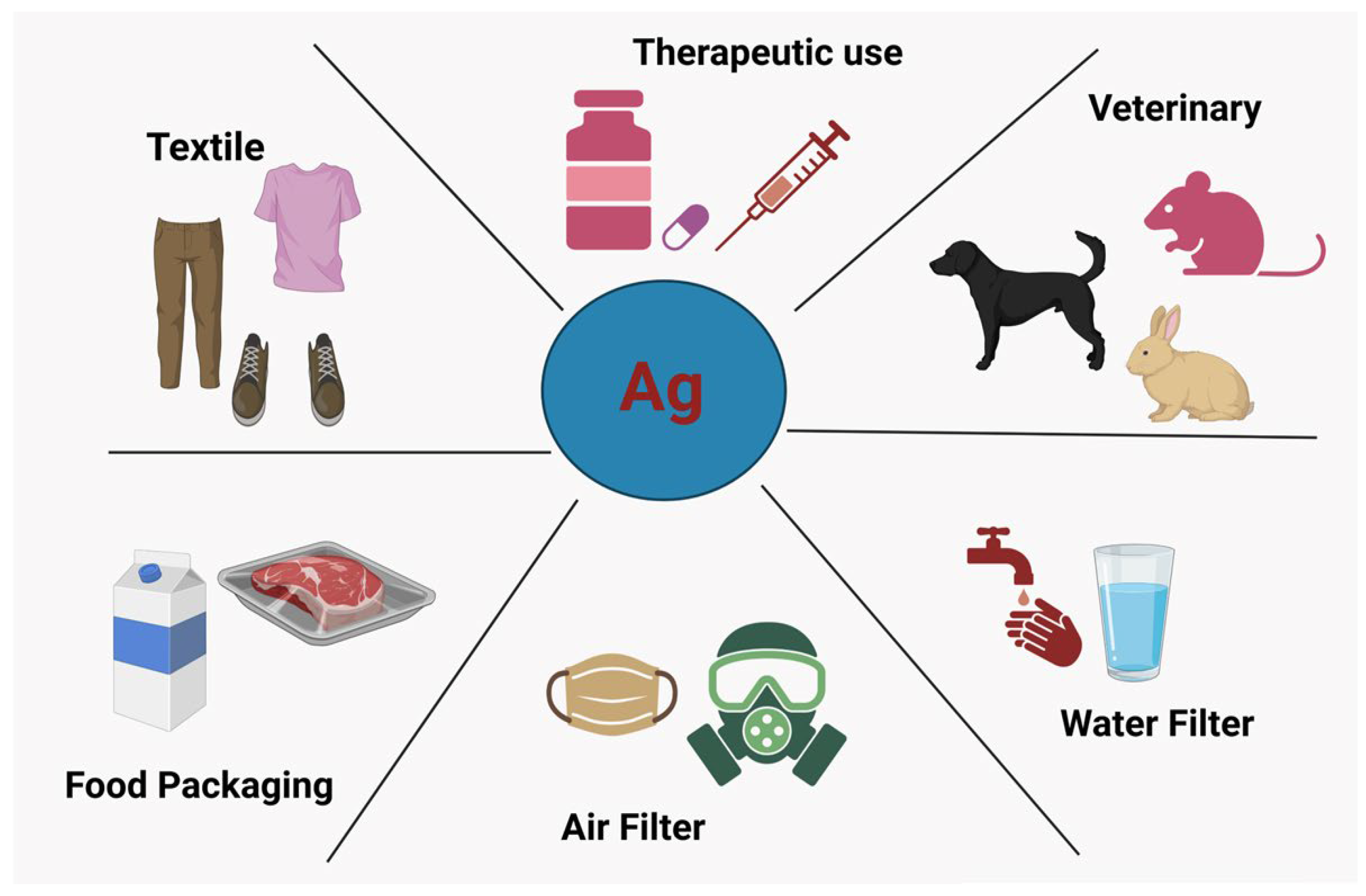
1. Introduction
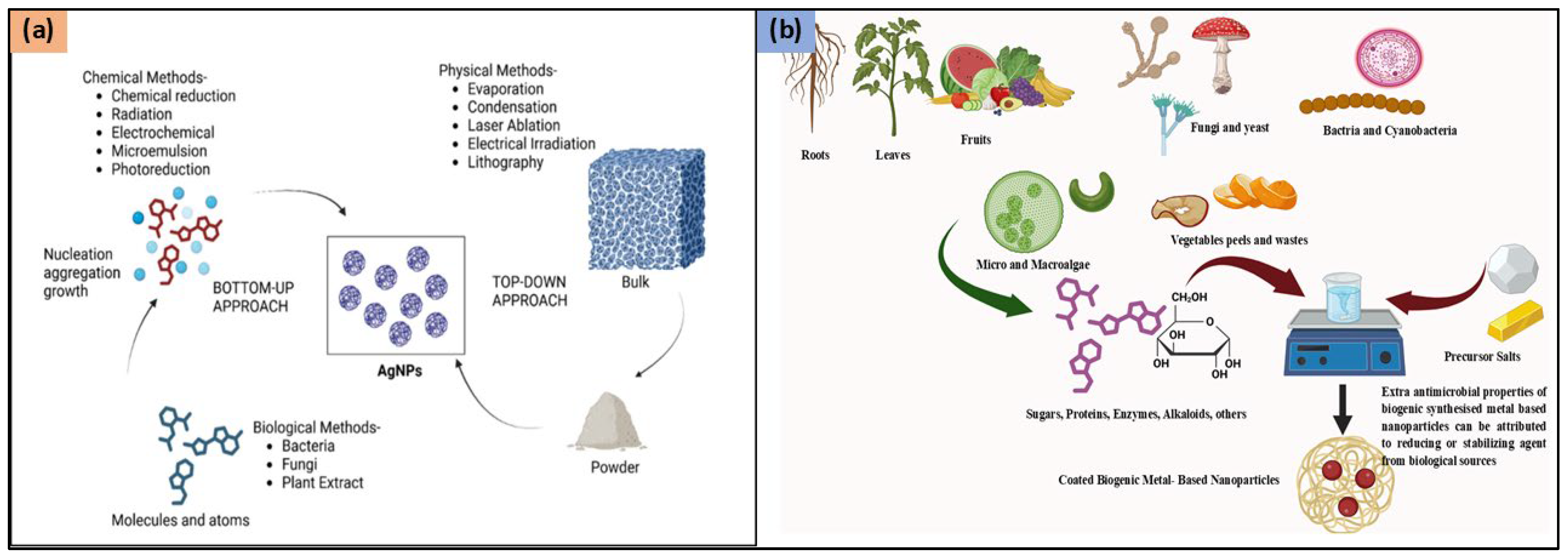
2. Synthesis
2.1. Physical Methods
2.2. Chemical Methods
2.3. Green Synthesis
3. Biophysical Properties
3.1. Shape and Crystallinity
3.2. Melting Temperature
3.3. Optical Properties
3.4. Electrical Properties
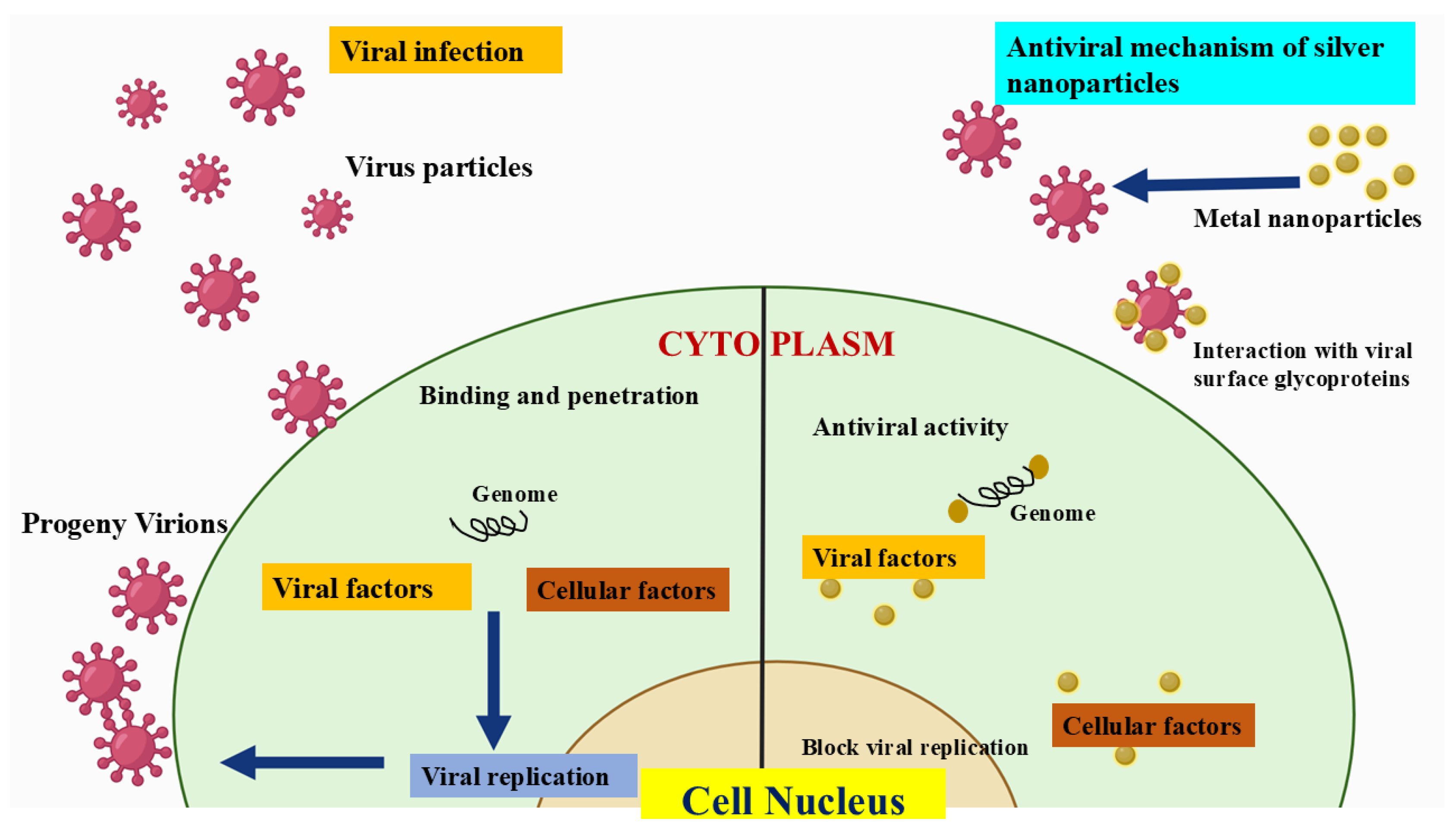
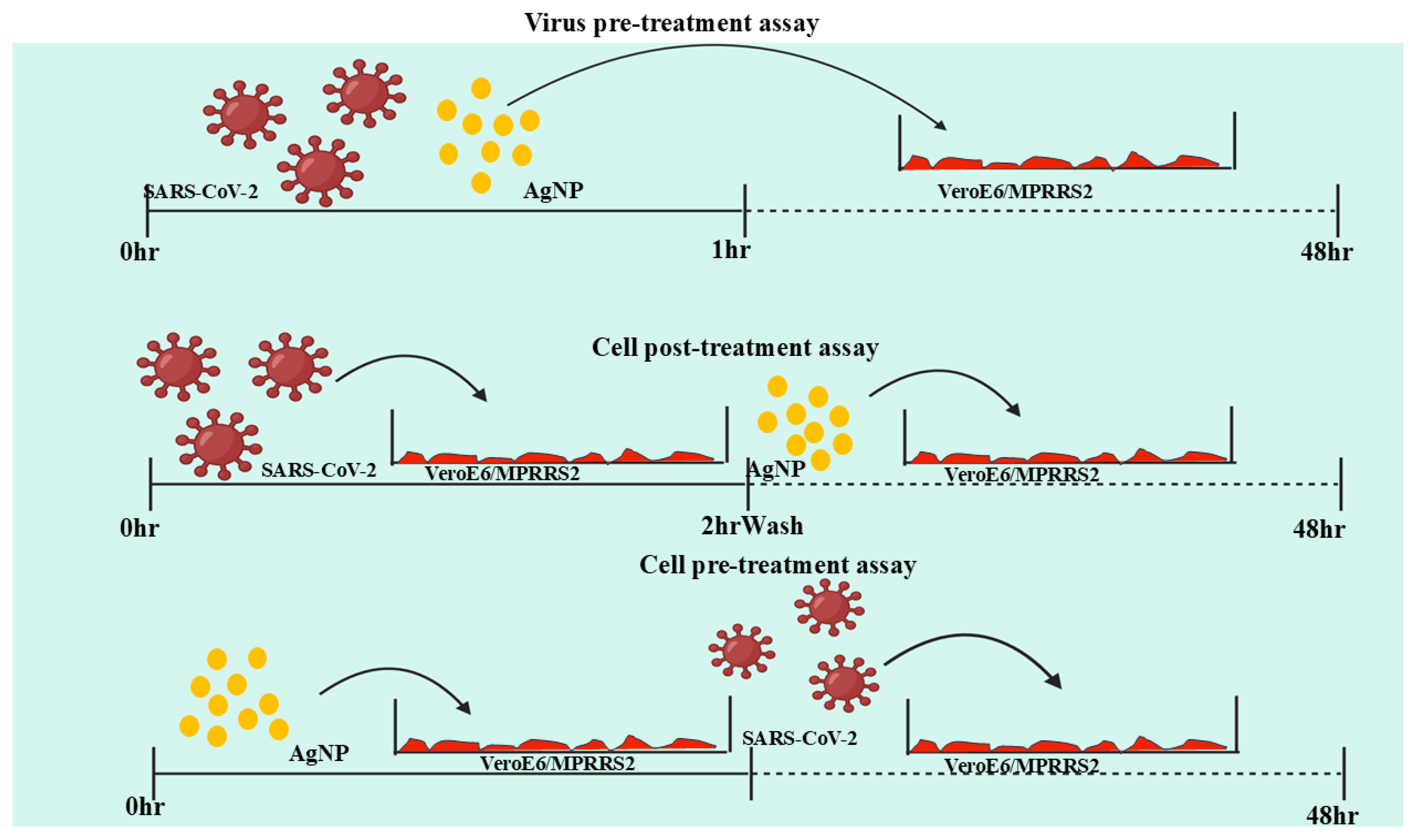
4. An Update on Antiviral AgNPs
5. Safety of AgNPs
Limitations of AgNPs
6. Challenges and Future Directions
Author Contributions
Funding
Institutional Review Board Statement
Informed Consent Statement
Data Availability Statement
Acknowledgments
Conflicts of Interest
References
- Tomaszewska, E.; Bednarczyk, K.; Janicka, M.; Chodkowski, M.; Krzyzowska, M.; Celichowski, G.; Grobelny, J.; Ranoszek-Soliwoda, K. The Influence of the AgNPs Ligand on the Antiviral Activity Against HSV-2. Int. J. Nanomed. 2025, 20, 2659–2671. [Google Scholar] [CrossRef]
- Idres, Y.M.; Idris, A.; Gao, W. Preclinical Testing of Antiviral SiRNA Therapeutics Delivered in Lipid Nanoparticles in Animal Models–a Comprehensive Review. Drug Deliv. Transl. Res. 2025, 1–18. [Google Scholar] [CrossRef]
- Yadav, S.; Mali, S.N.; Pandey, A. Biogenic Nanoparticles as Safer Alternatives for Gastric Ulcers: An Update on Green Synthesis Methods, Toxicity, and Their Efficacy in Controlling Inflammation. Biol. Trace Elem. Res. 2024, 1–20. [Google Scholar]
- Morens, D.M.; Fauci, A.S. Emerging Pandemic Diseases: How We Got to COVID-19. Cell 2020, 182, 1077–1092. [Google Scholar] [CrossRef]
- Morens, D.M.; Folkers, G.K.; Fauci, A.S. The Challenge of Emerging and Re-Emerging Infectious Diseases. Nature 2004, 430, 242–249. [Google Scholar] [CrossRef]
- Gonçalves, B.C.; Lopes Barbosa, M.G.; Silva Olak, A.P.; Belebecha Terezo, N.; Nishi, L.; Watanabe, M.A.; Marinello, P.; Zendrini Rechenchoski, D.; Dejato Rocha, S.P.; Faccin-Galhardi, L.C. Antiviral Therapies: Advances and Perspectives. Fundam. Clin. Pharmacol. 2021, 35, 305–320. [Google Scholar] [CrossRef]
- Ranade, T.; Sati, A.; Pratap, A.; Mali, S.N. Curcumin-Integrated Biopolymer Films for Active Packaging: Current Trends and Future Directions. Chem. Pap. 2025, 79, 1303–1334. [Google Scholar] [CrossRef]
- Yaqoob, A.A.; Umar, K.; Ibrahim, M.N.M. Silver Nanoparticles: Various Methods of Synthesis, Size Affecting Factors and Their Potential Applications–a Review. Appl. Nanosci. 2020, 10, 1369–1378. [Google Scholar] [CrossRef]
- Vu, K.B.; Phung, T.K.; Tran, T.T.T.; Mugemana, C.; Giang, H.N.; Nhi, T.L.P. Polystyrene Nanoparticles Prepared by Nanoprecipitation: A Recyclable Template for Fabricating Hollow Silica. J. Ind. Eng. Chem. 2021, 97, 307–315. [Google Scholar] [CrossRef]
- Rajapaksha, P.; Orrell-Trigg, R.; Shah, D.; Cheeseman, S.; Vu, K.B.; Ngo, S.T.; Murdoch, B.J.; Choudhury, N.R.; Yin, H.; Cozzolino, D.; et al. Broad Spectrum Antibacterial Zinc Oxide-Reduced Graphene Oxide Nanocomposite for Water Depollution. Mater. Today Chem. 2023, 27, 101242. [Google Scholar] [CrossRef]
- Doan, L.; Nguyen, L.T.; Nguyen, N.T.N. Modifying Superparamagnetic Iron Oxides Nanoparticles for Doxorubicin Delivery Carriers: A Review. J. Nanoparticle Res. 2023, 25, 73. [Google Scholar] [CrossRef]
- Natsuki, J.; Natsuki, T.; Hashimoto, Y. A Review of Silver Nanoparticles: Synthesis Methods, Properties and Applications. Int. J. Mater. Sci. Appl. 2015, 4, 325–332. [Google Scholar] [CrossRef]
- Bamal, D.; Singh, A.; Chaudhary, G.; Kumar, M.; Singh, M.; Rani, N.; Mundlia, P.; Sehrawat, A.R. Silver Nanoparticles Biosynthesis, Characterization, Antimicrobial Activities, Applications, Cytotoxicity and Safety Issues: An Updated Review. Nanomaterials 2021, 11, 2086. [Google Scholar] [CrossRef]
- Xu, L.; Wang, Y.Y.; Huang, J.; Chen, C.Y.; Wang, Z.X.; Xie, H. Silver Nanoparticles: Synthesis, Medical Applications and Biosafety. Theranostics 2020, 10, 8996–9031. [Google Scholar] [PubMed]
- Bouafia, A.; Laouini, S.E.; Ahmed, A.S.A.; Soldatov, A.V.; Algarni, H.; Chong, K.F.; Ali, G.A.M. The Recent Progress on Silver Nanoparticles: Synthesis and Electronic Applications. Nanomaterials 2021, 11, 2318. [Google Scholar] [CrossRef]
- Khayati, G.R.; Janghorban, K. The Nanostructure Evolution of Ag Powder Synthesized by High Energy Ball Milling. Adv. Powder Technol. 2012, 23, 393–397. [Google Scholar] [CrossRef]
- Tien, D.-C.; Liao, C.-Y.; Huang, J.-C.; Tseng, K.-H.; Lung, J.-K.; Tsung, T.-T.; Kao, W.-S.; Tsai, T.-H.; Cheng, T.-W.; Yu, B.-S.; et al. Novel Technique for Preparing A Nano-Silver Water Suspension by The Arc-Discharge Method. Rev. Adv. Mater. Sci. 2008, 18, 752–758. [Google Scholar]
- Tien, D.C.; Tseng, K.H.; Liao, C.Y.; Huang, J.C.; Tsung, T.T. Discovery of Ionic Silver in Silver Nanoparticle Suspension Fabricated by Arc Discharge Method. J. Alloys Compd. 2008, 463, 408–411. [Google Scholar] [CrossRef]
- Elwakil, B.H.; Eldrieny, A.M.; Almotairy, A.R.Z.; El-Khatib, M. Potent Biological Activity of Newly Fabricated Silver Nanoparticles Coated by a Carbon Shell Synthesized by Electrical Arc. Sci. Rep. 2024, 14, 5324. [Google Scholar] [CrossRef]
- Gharieb, M.A.; Khalil, A.M.; Menshawy, S.; El-Aty, A.; El-Khatib, A.M. The Impact of Different Temperatures on NanoSilver Carbon Manufacturing by Arc Discharge Method. Alfarama J. Basic. Appl. Sci. 2024, 5, 409–416. [Google Scholar] [CrossRef]
- Amendola, V.; Meneghetti, M. Laser Ablation Synthesis in Solution and Size Manipulation of Noble Metal Nanoparticles. Phys. Chem. Chem. Phys. 2009, 11, 3805–3821. [Google Scholar] [CrossRef]
- Sadrolhosseini, A.R.; Mahdi, M.A.; Alizadeh, F.; Rashid, S.A. Laser Ablation Technique for Synthesis of Metal Nanoparticle in Liquid. In Laser Technology and Its Applications; IntechOpen: Rijeka, Croatia, 2018. [Google Scholar]
- Rahmah, M.I.; Ahmed, A.M.; Rashid, T.M.; Qasim, A.J. Preparation of Silver Nanoparticles Using Laser Ablation for In Vitro Treatment of MCF-7 Cancer Cells with Antibacterial Activity. Plasmonics 2024, 19, 2097–2105. [Google Scholar] [CrossRef]
- Rafique, M.; Rafique, M.S.; Kalsoom, U.; Afzal, A.; Butt, S.H.; Usman, A. Laser Ablation Synthesis of Silver Nanoparticles in Water and Dependence on Laser Nature. Opt. Quantum Electron. 2019, 51, 179. [Google Scholar] [CrossRef]
- Kenmotsu, S.; Hirasawa, M.; Tamadate, T.; Matsumoto, C.; Osone, S.; Inomata, Y.; Seto, T. Surface-Enhanced Raman Scattering on Size-Classified Silver Nanoparticles Generated by Laser Ablation. ACS Omega 2024, 9, 37716–37723. [Google Scholar] [CrossRef]
- Mohammed, A.A.; Jawad, K.H.; Çevik, S.; Sulaiman, G.M.; Albukhaty, S.; Sasikumar, P. Investigating the Antimicrobial, Antioxidant, and Anticancer Effects of Elettaria Cardamomum Seed Extract Conjugated to Green Synthesized Silver Nanoparticles by Laser Ablation. Plasmonics 2024, 19, 1187–1200. [Google Scholar] [CrossRef]
- Niaz, U.; Hemat, S.; Jamil, A.; Aziz, M.S. Exploring the Relationship between Confinement Geometry and the Formation of High-Quality Silver Nanoparticles by Laser Ablation in Liquid Media. Indian. J. Phys. 2024, 98, 4989–4995. [Google Scholar] [CrossRef]
- Alharbi, A.M.; Ahmed, N.M.; Abdul Rahman, A.; Zahirah Noor Azman, N.; Algburi, S.; Wadi, I.A.; Binzowaimil, A.M.; Aldaghri, O.; Ibnaouf, K.H. Development of ZnO and Si Semiconductor-Based Ultraviolet Photodetectors Enhanced by Laser-Ablated Silver Nanoparticles. Photonics Nanostruct 2024, 58, 101228. [Google Scholar] [CrossRef]
- Raffi, M.; Rumaiz, A.K.; Hasan, M.M.; Shah, S.I. Studies of the Growth Parameters for Silver Nanoparticle Synthesis by Inert Gas Condensation. J. Mater. Res. 2007, 22, 3378–3384. [Google Scholar] [CrossRef]
- Jeevika, A.; Shankaran, D.R. Functionalized Silver Nanoparticles Probe for Visual Colorimetric Sensing of Mercury. Mater. Res. Bull. 2016, 83, 48–55. [Google Scholar] [CrossRef]
- Al-Mubaddel, F.S.; Haider, S.; Al-Masry, W.A.; Al-Zeghayer, Y.; Imran, M.; Haider, A.; Ullah, Z. Engineered Nanostructures: A Review of Their Synthesis, Characterization and Toxic Hazard Considerations. Arab. J. Chem. 2017, 10, S376–S388. [Google Scholar] [CrossRef]
- Veerasamy, R.; Xin, T.Z.; Gunasagaran, S.; Xiang, T.F.W.; Yang, E.F.C.; Jeyakumar, N.; Dhanaraj, S.A. Biosynthesis of Silver Nanoparticles Using Mangosteen Leaf Extract and Evaluation of Their Antimicrobial Activities. J. Saudi Chem. Soc. 2011, 15, 113–120. [Google Scholar] [CrossRef]
- Agnihotri, S.; Mukherji, S.; Mukherji, S. Size-Controlled Silver Nanoparticles Synthesized over the Range 5-100 Nm Using the Same Protocol and Their Antibacterial Efficacy. RSC Adv. 2014, 4, 3974–3983. [Google Scholar] [CrossRef]
- Khan, S.U.; Saleh, T.A.; Wahab, A.; Khan, M.H.U.; Khan, D.; Khan, W.U.; Rahim, A.; Kamal, S.; Khan, F.U.; Fahad, S. Nanosilver: New Ageless and Versatile Biomedical Therapeutic Scaffold. Int. J. Nanomed. 2018, 13, 733–762. [Google Scholar]
- Naganthran, A.; Verasoundarapandian, G.; Khalid, F.E.; Masarudin, M.J.; Zulkharnain, A.; Nawawi, N.M.; Karim, M.; Abdullah, C.A.C.; Ahmad, S.A. Synthesis, Characterization and Biomedical Application of Silver Nanoparticles. Materials 2022, 15, 427. [Google Scholar] [CrossRef]
- Malik, M.A.; Wani, M.Y.; Hashim, M.A. Microemulsion Method: A Novel Route to Synthesize Organic and Inorganic Nanomaterials. 1st Nano Update. Arab. J. Chem. 2012, 5, 397–417. [Google Scholar]
- Hak, J.; Jahan, I.; Sharma, K.; Farooqui, N.A. Article in Community Practitioner: The Journal of the Community Practitioners’ & Health Visitors’ Association. Community Pract. 2024, 83, 9–26. [Google Scholar] [CrossRef]
- dos Santos, M.A.; Paterno, L.G.; Moreira, S.G.C.; Sales, M.J.A. Original Photochemical Synthesis of Ag Nanoparticles Mediated by Potato Starch. SN Appl. Sci. 2019, 1, 554. [Google Scholar] [CrossRef]
- Raveendran, P.; Fu, J.; Wallen, S.L. Completely “Green” Synthesis and Stabilization of Metal Nanoparticles. J. Am. Chem. Soc. 2003, 125, 13940–13941. [Google Scholar] [CrossRef]
- Pinzaru, I.; Coricovac, D.; Dehelean, C.; Moacă, E.-A.; Mioc, M.; Baderca, F.; Sizemore, I.; Brittle, S.; Marti, D.; Calina, C.D.; et al. Stable PEG-Coated Silver Nanoparticles–A Comprehensive Toxicological Profile. Food Chem. Toxicol. 2018, 111, 546–556. [Google Scholar] [CrossRef]
- Pencheva, D.; Bryaskova, R.; Kantardjiev, T. Polyvinyl Alcohol/Silver Nanoparticles (PVA/AgNps) as a Model for Testing the Biological Activity of Hybrid Materials with Included Silver Nanoparticles. Mater. Sci. Eng. C 2012, 32, 2048–2051. [Google Scholar] [CrossRef]
- Abdel-Halim, E.S.; El-Rafie, M.H.; Al-Deyab, S.S. Polyacrylamide/Guar Gum Graft Copolymer for Preparation of Silver Nanoparticles. Carbohydr. Polym. 2011, 85, 692–697. [Google Scholar] [CrossRef]
- Singh, B.; Dhiman, A.; Kumar, S. Designing Silver Nanoparticles Impregnated Acacia and Tragacanth Gum Based Copolymeric Hydrogels for Drug Delivery Applications. Results Surf. Interfaces 2024, 16, 100256. [Google Scholar] [CrossRef]
- Srikar, S.K.; Giri, D.D.; Pal, D.B.; Mishra, P.K.; Upadhyay, S.N. Green Synthesis of Silver Nanoparticles: A Review. Green. Sustain. Chem. 2016, 06, 34–56. [Google Scholar] [CrossRef]
- Huang, J.; Li, Q.; Sun, D.; Lu, Y.; Su, Y.; Yang, X.; Wang, H.; Wang, Y.; Shao, W.; He, N.; et al. Biosynthesis of Silver and Gold Nanoparticles by Novel Sundried Cinnamomum Camphora Leaf. Nanotechnology 2007, 18, 105104. [Google Scholar] [CrossRef]
- Ahmed, S.; Saifullah; Ahmad, M.; Swami, B.L.; Ikram, S. Green Synthesis of Silver Nanoparticles Using Azadirachta Indica Aqueous Leaf Extract. J. Radiat. Res. Appl. Sci. 2016, 9, 1–7. [Google Scholar] [CrossRef]
- Ashraf, J.M.; Ansari, M.A.; Khan, H.M.; Alzohairy, M.A.; Choi, I. Green Synthesis of Silver Nanoparticles and Characterization of Their Inhibitory Effects on AGEs Formation Using Biophysical Techniques. Sci. Rep. 2016, 6, 20414. [Google Scholar] [CrossRef]
- Das, R.; Kumar, P.; Singh, A.K.; Agrawal, S.; Albukhaty, S.; Bhattacharya, I.; Tiwari, K.N.; Mishra, S.K.; Tripathi, A.K.; AlMalki, F.A.; et al. Green Synthesis of Silver Nanoparticles Using Trema Orientalis (L.) Extract and Evaluation of Their Antibacterial Activity. Green. Chem. Lett. Rev. 2025, 18, 2444679. [Google Scholar] [CrossRef]
- Singla, S.; Jana, A.; Thakur, R.; Kumari, C.; Goyal, S.; Pradhan, J. Green Synthesis of Silver Nanoparticles Using Oxalis Griffithii Extract and Assessing Their Antimicrobial Activity. OpenNano 2022, 7, 100047. [Google Scholar] [CrossRef]
- Widatalla, H.A.; Yassin, L.F.; Alrasheid, A.A.; Rahman Ahmed, S.A.; Widdatallah, M.O.; Eltilib, S.H.; Mohamed, A.A. Green Synthesis of Silver Nanoparticles Using Green Tea Leaf Extract, Characterization and Evaluation of Antimicrobial Activity. Nanoscale Adv. 2022, 4, 911–915. [Google Scholar] [CrossRef]
- Sharifi-Rad, M.; Elshafie, H.S.; Pohl, P. Green Synthesis of Silver Nanoparticles (AgNPs) by Lallemantia Royleana Leaf Extract: Their Bio-Pharmaceutical and Catalytic Properties. J. Photochem. Photobiol. A Chem. 2024, 448, 115318. [Google Scholar] [CrossRef]
- Mejía-Méndez, J.L.; Sánchez-Ante, G.; Cerro-López, M.; Minutti-Calva, Y.; Navarro-López, D.E.; Lozada-Ramírez, J.D.; Bach, H.; López-Mena, E.R.; Sánchez-Arreola, E. Green Synthesis of Silver Nanoparticles with Extracts from Kalanchoe Fedtschenkoi: Characterization and Bioactivities. Biomolecules 2024, 14, 782. [Google Scholar] [CrossRef] [PubMed]
- Ghasemi, S.; Dabirian, S.; Kariminejad, F.; Koohi, D.E.; Nemattalab, M.; Majidimoghadam, S.; Zamani, E.; Yousefbeyk, F. Process Optimization for Green Synthesis of Silver Nanoparticles Using Rubus Discolor Leaves Extract and Its Biological Activities against Multi-Drug Resistant Bacteria and Cancer Cells. Sci. Rep. 2024, 14, 4130. [Google Scholar] [CrossRef]
- Taleb Safa, M.A.; Koohestani, H. Green Synthesis of Silver Nanoparticles with Green Tea Extract from Silver Recycling of Radiographic Films. Results Eng. 2024, 21, 101808. [Google Scholar] [CrossRef]
- Losetty, V.; Devanesan, S.; AlSalhi, M.S.; Velu, P.P.; Muthupillai, D.; Kumar, K.A.; Lakkaboyana, S.K. Green Synthesis of Silver Nanoparticles Using Malachra Alceifolia (Wild Okra) for Wastewater Treatment and Biomedical Applications with Molecular Docking Approach. Environ. Sci. Pollut. Res. 2024, 31, 55562–55576. [Google Scholar] [CrossRef]
- Gangal, A.; Bachhar, V.; Joshi, V.; Akhtar, N.; Duseja, M.; Sethiya, N.K.; Shukla, R.K. Green Synthesis of Silver Nanoparticles from the Essential Oil of Curcuma Amada and Their Antihyperglycemic Effect in STZ Induced Diabetic Rats. Inorg. Chem. Commun. 2024, 168, 112873. [Google Scholar] [CrossRef]
- Lima, A.K.O.; Vieira, Í.R.S.; Souza, L.M.d.S.; Florêncio, I.; Silva, I.G.M.d.; Tavares Junior, A.G.; Machado, Y.A.A.; Santos, L.C.d.; Taube, P.S.; Nakazato, G.; et al. Green Synthesis of Silver Nanoparticles Using Paullinia Cupana Kunth Leaf Extract Collected in Different Seasons: Biological Studies and Catalytic Properties. Pharmaceutics 2025, 17, 356. [Google Scholar] [CrossRef]
- Ratan, Z.A.; Mashrur, F.R.; Chhoan, A.P.; Shahriar, S.M.; Haidere, M.F.; Runa, N.J.; Kim, S.; Kweon, D.H.; Hosseinzadeh, H.; Cho, J.Y. Silver Nanoparticles as Potential Antiviral Agents. Pharmaceutics 2021, 13, 2034. [Google Scholar] [CrossRef]
- Maaz, K. Silver Nanoparticles: Fabrication, Characterization and Applications; IntechOpen: Rijeka, Croatia, 2018; ISBN 9781789234787. [Google Scholar]
- Mukherji, S.; Bharti, S.; Shukla, G.; Mukherji, S. Synthesis and Characterization of Size- and Shape-Controlled Silver Nanoparticles. Phys. Sci. Rev. 2019, 4, 20170082. [Google Scholar] [CrossRef]
- Haes, A.J.; Haynes, C.L.; McFarland, A.D.; Schatz, G.C.; Van Duyne, R.P.; Zou, S. Plasmonic Materials for Surface-Enhanced Sensing and Spectroscopy. MRS Bull. 2005, 30, 368–375. [Google Scholar] [CrossRef]
- Kelly, K.L.; Coronado, E.; Zhao, L.L.; Schatz, G.C. The Optical Properties of Metal Nanoparticles: The Influence of Size, Shape, and Dielectric Environment. J. Phys. Chem. B 2003, 107, 668–677. [Google Scholar] [CrossRef]
- Bastys, V.; Pastoriza-Santos, I.; Rodríguez-González, B.; Vaisnoras, R.; Liz-Marzán, L.M. Formation of Silver Nanoprisms with Surface Plasmons at Communication Wavelengths. Adv. Funct. Mater. 2006, 16, 766–773. [Google Scholar] [CrossRef]
- Métraux, G.S.; Mirkin, C.A. Rapid Thermal Synthesis of Silver Nanoprisms with Chemically Tailorable Thickness. Adv. Mater. 2005, 17, 412–415. [Google Scholar] [CrossRef]
- Sau, T.K.; Murphy, C.J. Room Temperature, High-Yield Synthesis of Multiple Shapes of Gold Nanoparticles in Aqueous Solution. J. Am. Chem. Soc. 2004, 126, 8648–8649. [Google Scholar] [CrossRef] [PubMed]
- He, R.; Qian, X.; Yin, J.; Zhu, Z. Preparation of Polychrome Silver Nanoparticles in Different Solvents. J. Mater. Chem. 2002, 12, 3783–3786. [Google Scholar] [CrossRef]
- Tsuji, M.; Miyamae, N.; Lim, S.; Kimura, K.; Zhang, X.; Hikino, S.; Nishio, M. Crystal Structures and Growth Mechanisms of Au@Ag Core−Shell Nanoparticles Prepared by the Microwave−Polyol Method. Cryst. Growth Des. 2006, 6, 1801–1807. [Google Scholar] [CrossRef]
- Xiao, J.P.; Xie, Y.; Tang, R.; Chen, M.; Tian, X.B. Novel Ultrasonically Assisted Templated Synthesis of Palladium and Silver Dendritic Nanostructures. Adv. Mater. 2001, 13, 1887–1891. [Google Scholar] [CrossRef]
- Tsuji, M.; Hashimoto, M.; Nishizawa, Y.; Kubokawa, M.; Tsuji, T. Microwave-Assisted Synthesis of Metallic Nanostructures in Solution. Chem.–A Eur. J. 2005, 11, 440–452. [Google Scholar] [CrossRef]
- Wiley, B.J.; Im, S.H.; Li, Z.-Y.; McLellan, J.; Siekkinen, A.; Xia, Y. Maneuvering the Surface Plasmon Resonance of Silver Nanostructures through Shape-Controlled Synthesis. J. Phys. Chem. B 2006, 110, 15666–15675. [Google Scholar] [CrossRef]
- Ahmed, M.J.; Murtaza, G.; Mehmood, A.; Bhatti, T.M. Green Synthesis of Silver Nanoparticles Using Leaves Extract of Skimmia Laureola: Characterization and Antibacterial Activity. Mater. Lett. 2015, 153, 10–13. [Google Scholar] [CrossRef]
- Raza, M.A.; Kanwal, Z.; Rauf, A.; Sabri, A.N.; Riaz, S.; Naseem, S. Size- and Shape-Dependent Antibacterial Studies of Silver Nanoparticles Synthesized by Wet Chemical Routes. Nanomaterials 2016, 6, 74. [Google Scholar] [CrossRef]
- Dhaka, A.; Chand Mali, S.; Sharma, S.; Trivedi, R. A Review on Biological Synthesis of Silver Nanoparticles and Their Potential Applications. Results Chem. 2023, 6, 101108. [Google Scholar]
- Dhayalan, M.; Riyaz, S.M.; Karikalan, P.; Srinivasan, N. Biomedical Applications of Silver Nanoparticles. In Silver Micro-Nanoparticles—Properties, Synthesis, Characterization, and Applications; Kumar, S., Kumar, P., Pathak, C.S., Eds.; IntechOpen: Rijeka, Croatia, 2021; ISBN 978-1-83968-660-3. [Google Scholar]
- Syafiuddin, A.; Salmiati; Salim, M.R.; Beng Hong Kueh, A.; Hadibarata, T.; Nur, H. A Review of Silver Nanoparticles: Research Trends, Global Consumption, Synthesis, Properties, and Future Challenges. J. Chin. Chem. Soc. 2017, 64, 732–756. [Google Scholar] [CrossRef]
- Roduner, E. Nanoscopic Materials: Size-Dependent Phenomena and Growth Principles; RSC: London, UK, 2015; ISBN 9781782624943. [Google Scholar]
- Attarian Shandiz, M. Effective Coordination Number Model for the Size Dependency of Physical Properties of. J. Phys. Condens. Matter 2008, 20, 325237. [Google Scholar] [CrossRef]
- Allen, G.L.; Bayles, R.A.; Gile, W.W.; Jesser, W.A. Small Particle Melting of Pure Metals. Thin Solid. Film. 1986, 144, 297–308. [Google Scholar] [CrossRef]
- Ide, E.; Angata, S.; Hirose, A.; Kobayashi, K.F. Metal–Metal Bonding Process Using Ag Metallo-Organic Nanoparticles. Acta Mater. 2005, 53, 2385–2393. [Google Scholar] [CrossRef]
- Evanoff, D.D.; Chumanov, G. Size-Controlled Synthesis of Nanoparticles. 2. Measurement of Extinction, Scattering, and Absorption Cross Sections. J. Phys. Chem. B 2004, 108, 13957–13962. [Google Scholar] [CrossRef]
- González, A.L.; Noguez, C. Optical Properties of Silver Nanoparticles. Phys. Status Solidi C 2007, 4, 4118–4126. [Google Scholar] [CrossRef]
- Alshehri, A.H.; Jakubowska, M.; Młożniak, A.; Horaczek, M.; Rudka, D.; Free, C.; Carey, J.D. Enhanced Electrical Conductivity of Silver Nanoparticles for High Frequency Electronic Applications. ACS Appl. Mater. Interfaces 2012, 4, 7007–7010. [Google Scholar] [CrossRef]
- Bhagat, M.; Rajput, S.; Arya, S.; Khan, S.; Lehana, P. Biological and Electrical Properties of Biosynthesized Silver Nanoparticles. Bull. Mater. Sci. 2015, 38, 1253–1258. [Google Scholar]
- Shivananda, C.S.; Lakshmeesha Rao, B. Sangappa Structural, Thermal and Electrical Properties of Silk Fibroin–Silver Nanoparticles Composite Films. J. Mater. Sci. Mater. Electron. 2020, 31, 41–51. [Google Scholar] [CrossRef]
- Yamari, I.; Abchir, O.; Mali, S.N.; Errougui, A.; Talbi, M.; Kouali, M.E.; Chtita, S. The Anti-SARS-CoV-2 Activity of Novel 9, 10-Dihydrophenanthrene Derivatives: An Insight into Molecular Docking, ADMET Analysis, and Molecular Dynamics Simulation. Sci. Afr. 2023, 21, e01754. [Google Scholar] [CrossRef]
- Jeevanandam, J.; Krishnan, S.; Hii, Y.S.; Pan, S.; Chan, Y.S.; Acquah, C.; Danquah, M.K.; Rodrigues, J. Synthesis Approach-Dependent Antiviral Properties of Silver Nanoparticles and Nanocomposites. J. Nanostructure Chem. 2022, 12, 809–831. [Google Scholar]
- Luceri, A.; Francese, R.; Lembo, D.; Ferraris, M.; Balagna, C. Silver Nanoparticles: Review of Antiviral Properties, Mechanism of Action and Applications. Microorganisms 2023, 11, 629. [Google Scholar] [CrossRef]
- Demchenko, V.; Mamunya, Y.; Sytnyk, I.; Iurzhenko, M.; Krivtsun, I.; Rybalchenko, N.; Naumenko, K.; Artiukh, L.; Kowalczuk, M.; Demchenko, O.; et al. Fabrication of Polylactide Composites with Silver Nanoparticles by Sputtering Deposition and Their Antimicrobial and Antiviral Applications. Polym. Int. 2025, 74, 207–216. [Google Scholar] [CrossRef]
- de Souza, T.B.; Rosa, A.S.; Constantino-Teles, P.; Ferreira, V.N.S.; Archanjo, B.S.; Soares, C.A.G.; Picciani, P.H.S.; Allão Cassaro, R.A.; Miranda, M.D.; Poneti, G. Silver Nanoparticles-Functionalized Textile against SARS-CoV-2: Antiviral Activity of the Capping Oleylamine Molecule. ACS Appl. Mater. Interfaces 2025, 17, 4. [Google Scholar] [CrossRef]
- Martín-Faivre, L.; Prince, L.; Cornu, C.; Villeret, B.; Sanchez-Guzman, D.; Rouzet, F.; Sallenave, J.M.; Garcia-Verdugo, I. Pulmonary Delivery of Silver Nanoparticles Prevents Influenza Infection by Recruiting and Activating Lymphoid Cells. Biomaterials 2025, 312, 122721. [Google Scholar] [CrossRef]
- EL Bagoury, G.F.; Mahmoud, A.H.; Kassem, S.; Elhabashy, R. Green Synthesis of Silver Nanoparticles Using Green Tea Extract and Evaluation of Their Antiviral Potential against Foot-and-Mouth Disease Virus Serotype O: An In-Vitro Study. Egypt. J. Vet. Sci. 2025, 1–11. [Google Scholar] [CrossRef]
- Gattucci, F.; Lallukka, M.; Grifasi, N.; Piumetti, M.; Miola, M. Tannic Acid-Assisted Green Functionalization of Clinoptilolite: A Step-by-Step Characterization of Silver Nanoparticles in Situ Reduction. Ceram. Int. 2025, 51, 13051–13057. [Google Scholar] [CrossRef]
- Amaral, M.V.M.V.; Carraro, C.B.; Antoniêto, A.C.C.; Costa, M.N.; Fraga-Silva, T.F.C.; Cipriano, U.G.; Abuná, R.P.F.; Rodrigues, T.S.; Martins, R.B.; Luzenti, A.M.; et al. Biogenic Silver Nanoparticles Produced by Trichoderma Reesei Inhibit SARS-CoV-2 Infection, Reduce Lung Viral Load and Ameliorate Acute Pulmonary Inflammation. Curr. Res. Biotechnol. 2025, 9, 100277. [Google Scholar] [CrossRef]
- Sahu, S.K.; Sahoo, P.R.; Dash, S.; Mishra, S.R.; Behera, P.C. Antimicrobial Activity of Silver Nanoparticles Against Common Bovine Mastitis Pathogens: A Comparative Analysis. Curr. Microbiol. 2025, 82, 121. [Google Scholar] [CrossRef]
- Obasi, D.E.; Nebolisa, N.M.; Akinwunmi, A.R.; Abimbolu, A.K.; Ezeorah, M.C.; Areola, O.M.; Donatus, U.D.; Oladipupo, V.T.; Ohiani, J.J.; Ayanleke, T.A.; et al. Eco-Friendly and Facile Production Method, Natural Products Chemistry, and Pharmacological Properties of Silver Nanoparticles Using Telfaria Occidentalis Leaf and Stem Extracts. Eur. J. Sustain. Dev. Res. 2025, 9, em0280. [Google Scholar] [CrossRef]
- Fereydani, M.; Jalalian, A.; Saber, N. Green Synthesis of Silver Nanoparticles from Cuscuta Epithymum Extract, Evaluation of Antibacterial, Antioxidant Activity, Cytotoxic Effect on MCF-7 Cell Line; Elsevier: Amsterdam, The Netherlands, 2019. [Google Scholar]
- Długosz, O.; Żebracka, A.; Sochocka, M.; Franz, D.; Ochnik, M.; Chmielowiec-Korzeniowska, A.; Banach, M. Selective and Complementary Antimicrobial and Antiviral Activity of Silver, Copper, and Selenium Nanoparticle Suspensions in Deep Eutectic Solvent. Env. Environ. Res. 2025, 264, 120351. [Google Scholar] [CrossRef]
- Barabadi, H.; Vahidi, H.; Karami, K.; Kamali, M.; Jounaki, K.; Jahani, R.; Hosseini, O.; Amidi, S.; Ashouri, F. Cephalosporium Aphidicola-Derived Silver Nanoparticles: In Vitro Physicochemical, Antibacterial, Antifungal, Biofilm Inhibition, Biofilm Degradation, Antioxidant, Alpha-Amylase, and Urease Inhibitory Properties. Bionanoscience 2025, 15, 48. [Google Scholar] [CrossRef]
- Srikhao, N.; Ounkaew, A.; Srichiangsa, N.; Phanthanawiboon, S.; Boonmars, T.; Artchayasawat, A.; Theerakulpisut, S.; Okhawilai, M.; Kasemsiri, P. Green-Synthesized Silver Nanoparticle Coating on Paper for Antibacterial and Antiviral Applications. Polym. Bull. 2023, 80, 9651–9668. [Google Scholar] [CrossRef]
- Naumenko, K.; Zahorodnia, S.; Pop, C.V.; Rizun, N. Antiviral Activity of Silver Nanoparticles against the Influenza A Virus. J. Virus Erad. 2023, 9, 100330. [Google Scholar] [CrossRef]
- Elnosary, M.E.; Aboelmagd, H.A.; Sofy, M.R.; Sofy, A.R.; Elshazly, E.H. Antiviral and Antibacterial Properties of Synthesis Silver Nanoparticles with Nigella Arvensis Aqueous Extract. Egypt. J. Chem. 2023, 66, 209–223. [Google Scholar] [CrossRef]
- El-Ganainy, S.M.; Soliman, A.M.; Ismail, A.M.; Sattar, M.N.; Farroh, K.Y.; Shafie, R.M. Antiviral Activity of Chitosan Nanoparticles and Chitosan Silver Nanocomposites against Alfalfa Mosaic Virus. Polymers 2023, 15, 2961. [Google Scholar] [CrossRef]
- Doszpoly, A.; Shaalan, M.; El-Matbouli, M. Silver Nanoparticles Proved to Be Efficient Antivirals In Vitro against Three Highly Pathogenic Fish Viruses. Viruses 2023, 15, 1689. [Google Scholar] [CrossRef]
- Al-Askar, A.A.; Aseel, D.G.; El-Gendi, H.; Sobhy, S.; Samy, M.A.; Hamdy, E.; El-Messeiry, S.; Behiry, S.I.; Elbeaino, T.; Abdelkhalek, A. Antiviral Activity of Biosynthesized Silver Nanoparticles from Pomegranate (Punica Granatum L.) Peel Extract against Tobacco Mosaic Virus. Plants 2023, 12, 2103. [Google Scholar] [CrossRef] [PubMed]
- Gaikwad, S.; Ingle, A.; Gade, A.; Rai, M.; Falanga, A.; Incoronato, N.; Russo, L.; Galdiero, S.; Galdiero, M. Antiviral Activity of Mycosynthesized Silver Nanoparticles against Herpes Simplex Virus and Human Parainfluenza Virus Type 3. Int. J. Nanomed. 2013, 8, 4303–4314. [Google Scholar] [CrossRef]
- Makhlof, M.E.M.; Diab, H.A.; Mabrouk, M.E.; Abd El Kareem, M.S. Antiviral and Antioxidant Activity, Green Synthesis, and Optimization of Silver Nanoparticles Derived from Ulva Lactuca. Egypt. J. Phycol. 2024, 25, 1–42. [Google Scholar] [CrossRef]
- Butler, M.R.; Hrncirova, J.; Jacot, T.A.; Dutta, S.; Clark, M.R.; Doncel, G.F.; Cooper, J.B. Detection and Quantification of Antiviral Drug Tenofovir Using Silver Nanoparticles and Surface Enhanced Raman Spectroscopy (SERS) with Spatially Resolved Hotspot Selection. Front. Nanotechnol. 2023, 5, 1270474. [Google Scholar] [CrossRef]
- Abdulsattar Ali, A.; Tahir Maher, F.; Ahmed Al-Bajari, S. Green biosynthesis of silver nanoparticles from taraxacum officinale roots plant and studying its antiviral properties to coronavirus (SARS-CoV-2) infected lung cells. J. Hyg. Eng. Des. 2023, 42, 361–369. [Google Scholar]
- Khan, R.; Naureen, H.; Javed, A.; Khalid, M.; Khan, H. Alocasia Odora–Mediated Synthesis of Silver Nanoparticles, Their Cytotoxicity, and Virucidal Potential. Appl. Microbiol. Biotechnol. 2023, 107, 111–123. [Google Scholar] [CrossRef]
- Pilaquinga, F.; Bosch, R.; Morey, J.; Bastidas-Caldes, C.; Torres, M.; Toscano, F.; Debut, A.; Pazmiño-Viteri, K.; de las Nieves Piña, M. High in Vitro Activity of Gold and Silver Nanoparticles from Solanum Mammosum L. against SARS-CoV-2 Surrogate Phi6 and Viral Model PhiX174. Nanotechnology 2023, 34, 175705. [Google Scholar] [CrossRef]
- 111. Rybalchenko, N.P.; Hnatiuk; Artiukh, L.O.; Naumenko, S.; Zaremba, P.Y.; Demchenko, V.L.; Kokhtych, L.M.; Iurzhenko, M.V.; Rybalchenko, T.V.; Ovsyankina, V.; et al. Antimicrobial and Antiviral Activity of Nanocomposites Based on Polyelectrolyte Complexes with Silver Nanoparticles. Mikrobiolohichnyi Zhurnal 2024, 86, 36–50. [Google Scholar] [CrossRef]
- Sinclair, T.R.; Van Den Hengel, S.K.; Raza, B.G.; Rutjes, S.A.; De Roda Husman, A.M.; Peijnenburg, W.J.G.M.; Roesink, H.D.W.; De Vos, W.M. Surface Chemistry-Dependent Antiviral Activity of Silver Nanoparticles. Nanotechnology 2021, 32, 365101. [Google Scholar] [CrossRef]
- Bharti, S.; Mukherji, S.; Mukherji, S. Antiviral Application of Colloidal and Immobilized Silver Nanoparticles. Nanotechnology 2021, 32, 205102. [Google Scholar] [CrossRef]
- Emam, M.H.; Elezaby, R.S.; Swidan, S.A.; Loutfy, S.A.; Hathout, R.M. Enhancing Polyacrylonitrile Nanofibers Antiviral Activity Using Greenly Synthesized Silver Nanoparticles. Arch. Pharm. 2025, 358, e202400943. [Google Scholar] [CrossRef]
- Chen, L.; Liang, J. An Overview of Functional Nanoparticles as Novel Emerging Antiviral Therapeutic Agents. Mater. Sci. Eng. C 2020, 112, 110924. [Google Scholar] [CrossRef]
- Park, S.J.; Park, H.H.; Kim, S.Y.; Kim, S.J.; Woo, K.; Ko, G.P. Antiviral Properties of Silver Nanoparticles on a Magnetic Hybrid Colloid. Appl. Env. Environ. Microbiol. 2014, 80, 2343–2350. [Google Scholar] [CrossRef]
- Rakowska, P.D.; Tiddia, M.; Faruqui, N.; Bankier, C.; Pei, Y.; Pollard, A.J.; Zhang, J.; Gilmore, I.S. Antiviral Surfaces and Coatings and Their Mechanisms of Action. Commun. Mater. 2021, 2, 53. [Google Scholar]
- Sadiq, S.; Khan, I.; Shen, Z.; Wang, M.; Xu, T.; Khan, S.; Zhou, X.; Bahadur, A.; Rafiq, M.; Sohail, S.; et al. Recent Updates on Multifunctional Nanomaterials as Antipathogens in Humans and Livestock: Classification, Application, Mode of Action, and Challenges. Molecules 2023, 28, 7674. [Google Scholar] [CrossRef] [PubMed]
- Gurunathan, S.; Qasim, M.; Choi, Y.; Do, J.T.; Park, C.; Hong, K.; Kim, J.H.; Song, H. Antiviral Potential of Nanoparticles—Can Nanoparticles Fight against Coronaviruses? Nanomaterials 2020, 10, 1645. [Google Scholar] [CrossRef]
- Hadinejad, F.; Morad, H.; Jahanshahi, M.; Zarrabi, A.; Pazoki-Toroudi, H.; Mostafavi, E. A Novel Vision of Reinforcing Nanofibrous Masks with Metal Nanoparticles: Antiviral Mechanisms Investigation. Adv. Fiber Mater. 2023, 5, 1273–1317. [Google Scholar]
- Galdiero, S.; Falanga, A.; Vitiello, M.; Cantisani, M.; Marra, V.; Galdiero, M. Silver Nanoparticles as Potential Antiviral Agents. Molecules 2011, 16, 8894–8918. [Google Scholar] [CrossRef]
- Manisekaran, R.; Chettiar, A.-D.R.; Marasamy, L.; Ibarra, V.C.; Lopez-Ayuso, C.A.; Chavez-Granados, P.A.; Kandasamy, G.; Acosta-Torres, L.S.; Arthikala, M.-K. Silver-Nanoparticles-Based Composites for Antimicrobial Applications: An Update. ChemistrySelect 2024, 9, e202403772. [Google Scholar] [CrossRef]
- Sati, A.; Ranade, T.N.; Mali, S.N.; Ahmad Yasin, H.K.; Pratap, A. Silver Nanoparticles (AgNPs): Comprehensive Insights into Bio/Synthesis, Key Influencing Factors, Multifaceted Applications, and Toxicity─A 2024 Update. ACS Omega 2025, 10, 7549–7582. [Google Scholar] [CrossRef]
- Szymańska, E.; Orłowski, P.; Winnicka, K.; Tomaszewska, E.; Bąska, P.; Celichowski, G.; Grobełny, J.; Basa, A.; Krzyżowska, M. Multifunctional Tannic Acid/Silver Nanoparticle-Based Mucoadhesive Hydrogel for Improved Local Treatment of HSV Infection: In Vitro and in Vivo Studies. Int. J. Mol. Sci. 2018, 19, 387. [Google Scholar] [CrossRef]
- Frippiat, T.; Art, T.; Delguste, C. Silver Nanoparticles as Antimicrobial Agents in Veterinary Medicine: Current Applications and Future Perspectives. Nanomaterials 2025, 15, 202. [Google Scholar] [CrossRef]
- Wang, D.; Yin, C.; Bai, Y.; Zhou, M.; Wang, N.; Tong, C.; Yang, Y.; Liu, B. Chitosan-Modified AgNPs Efficiently Inhibit Swine Coronavirus-Induced Host Cell Infections via Targeting the Spike Protein. Biomolecules 2024, 14, 1152. [Google Scholar] [CrossRef] [PubMed]
- He, Q.; Lu, J.; Liu, N.; Lu, W.; Li, Y.; Shang, C.; Li, X.; Hu, L.; Jiang, G. Antiviral Properties of Silver Nanoparticles against SARS-CoV-2: Effects of Surface Coating and Particle Size. Nanomaterials 2022, 12, 990. [Google Scholar] [CrossRef] [PubMed]
- Mali, S.N.; Pandey, A. Multiple QSAR and Molecular Modelling for Identification of Potent Human Adenovirus Inhibitors. J. Indian. Chem. Soc. 2021, 98, 100082. [Google Scholar] [CrossRef]
- Lara, H.H.; Ixtepan-Turrent, L.; Garza-Treviño, E.N.; Rodriguez-Padilla, C. PVP-Coated Silver Nanoparticles Block the Transmission of Cell-Free and Cell-Associated HIV-1 in Human Cervical Culture. J. Nanobiotechnology 2010, 8, 15. [Google Scholar] [CrossRef]
- Lara, H.H.; Ayala-Nuñez, N.V.; Ixtepan-Turrent, L.; Rodriguez-Padilla, C. Mode of Antiviral Action of Silver Nanoparticles against HIV-1. J. Nanobiotechnology 2010, 8, 1. [Google Scholar]
- Baram-Pinto, D.; Shukla, S.; Gedanken, A.; Sarid, R. Inhibition of HSV-1 Attachment, Entry, and Cell-to-Cell Spread by Functionalized Multivalent Gold Nanoparticles. Small 2010, 6, 1044–1050. [Google Scholar] [CrossRef]
- Sun, L.; Singh, A.; Vig, K.; Pillai, S.; Singh, S. Silver Nanoparticles Inhibit Replication of Respiratory Syncytial Virus. J. Biomed. Nanotechnol. 2008, 4, 149–158. [Google Scholar]
- Rogers, J.V.; Parkinson, C.V.; Choi, Y.W.; Speshock, J.L.; Hussain, S.M. A Preliminary Assessment of Silver Nanoparticle Inhibition of Monkeypox Virus Plaque Formation. Nanoscale Res. Lett. 2008, 3, 129. [Google Scholar] [CrossRef]
- Papp, I.; Sieben, C.; Ludwig, K.; Roskamp, M.; Böttcher, C.; Schlecht, S.; Herrmann, A.; Haag, R. Inhibition of Influenza Virus Infection by Multivalent Sialic-Acid- Functionalized Gold Nanoparticles. Small 2010, 6, 2900–2906. [Google Scholar] [CrossRef]
- Mali, S.N.; Pratap, A.P.; Thorat, B.R. The Rise of New Coronavirus Infection (COVID-19): A Recent Update and Potential Therapeutic Candidates. Eurasian J. Med. Oncol. 2020, 4, 35–41. [Google Scholar]
- Habibi, S.; Ghajarieh, A. Application of Nanofibers in Virus and Bacteria Filtration. Russ. J. Appl. Chem. 2022, 95, 486–498. [Google Scholar]
- Elbeshehy, E.K.F.; Elazzazy, A.M.; Aggelis, G. Silver Nanoparticles Synthesis Mediated by New Isolates of Bacillus Spp., Nanoparticle Characterization and Their Activity against Bean Yellow Mosaic Virus and Human Pathogens. Front. Microbiol. 2015, 6, 453. [Google Scholar] [CrossRef]
- Elechiguerra, J.L.; Burt, J.L.; Morones, J.R.; Camacho-Bragado, A.; Gao, X.; Lara, H.H.; Yacaman, M.J. Interaction of Silver Nanoparticles with HIV-1. J. Nanobiotechnology 2005, 3, 6. [Google Scholar] [CrossRef]
- Chen, N.; Zheng, Y.; Yin, J.; Li, X.; Zheng, C. Inhibitory Effects of Silver Nanoparticles against Adenovirus Type 3 in Vitro. J. Virol. Methods 2013, 193, 470–477. [Google Scholar] [CrossRef]
- Kamarudin, D.; Hashim, N.A.; Ong, B.H.; Che Hassan, C.R.; Abdul Manaf, N. Synthesis of Silver Nanoparticles Stabilised by PVP for Polymeric Membrane Application: A Comparative Study. Mater. Technol. 2022, 37, 289–301. [Google Scholar] [CrossRef]
- Nie, P.; Zhao, Y.; Xu, H. Synthesis, Applications, Toxicity and Toxicity Mechanisms of Silver Nanoparticles: A Review. Ecotoxicol. Env. Environ. Saf. 2023, 253, 114636. [Google Scholar]
- Jaswal, T.; Gupta, J. A Review on the Toxicity of Silver Nanoparticles on Human Health. Mater. Today Proc. 2021, 81, 859–863. [Google Scholar]
- Noga, M.; Milan, J.; Frydrych, A.; Jurowski, K. Toxicological Aspects, Safety Assessment, and Green Toxicology of Silver Nanoparticles (AgNPs)—Critical Review: State of the Art. Int. J. Mol. Sci. 2023, 24, 5133. [Google Scholar] [CrossRef] [PubMed]
- Alwan, S.; Al-Saeed, M.; Abid, H. Safety Assessment and Biochemical Evaluation of the Effect of Biogenic Silver Nanoparticles (Using Bark Extract of C. Zeylanicum) on Rattus Norvegicus Rats. Baghdad J. Biochem. Appl. Biol. Sci. 2021, 2, 133–145. [Google Scholar] [CrossRef]
- Vuković, B.; Milić, M.; Dobrošević, B.; Milić, M.; Ilić, K.; Pavičić, I.; Šerić, V.; Vrček, I.V. Surface Stabilization Affects Toxicity of Silver Nanoparticles in Human Peripheral Blood Mononuclear Cells. Nanomaterials 2020, 10, 1390. [Google Scholar] [CrossRef]
- Tortella, G.R.; Rubilar, O.; Durán, N.; Diez, M.C.; Martínez, M.; Parada, J.; Seabra, A.B. Silver Nanoparticles: Toxicity in Model Organisms as an Overview of Its Hazard for Human Health and the Environment. J. Hazard. Mater. 2020, 390, 121974. [Google Scholar]
- Elyousfi, S.; Dellali, M.; Mezni, A.; Ben Ali, M.; Hedfi, A.; Almalki, M.; Mezni, A.; Rohal-Lupher, M.; Dervishi, A.; Boufahja, F. Toxicity of Silver Nanoparticles on the Clam Ruditapes Decussatus Assessed through Biomarkers and Clearance Rate. Mater. Res. Express 2021, 8, 105005. [Google Scholar] [CrossRef]
- Pinheiro, S.K.d.P.; Lima, A.K.M.; Miguel, T.B.A.R.; Filho, A.G.S.; Ferreira, O.P.; Pontes, M.d.S.; Grillo, R.; Miguel, E.d.C. Assessing Toxicity Mechanism of Silver Nanoparticles by Using Brine Shrimp (Artemia Salina) as Model. Chemosphere 2024, 347, 140673. [Google Scholar] [CrossRef]
- Al Baloushi, K.S.Y.; Senthilkumar, A.; Kandhan, K.; Subramanian, R.; Kizhakkayil, J.; Ramachandran, T.; Shehab, S.; Kurup, S.S.; Alyafei, M.A.M.; Al Dhaheri, A.S.; et al. Green Synthesis and Characterization of Silver Nanoparticles Using Moringa Peregrina and Their Toxicity on MCF-7 and Caco-2 Human Cancer Cells. Int. J. Nanomed. 2024, 19, 3891–3905. [Google Scholar] [CrossRef]
- Taha, N.M.; Youssef, F.S.; Auda, H.M.; El-Bahy, M.M.; Ramadan, R.M. Efficacy of Silver Nanoparticles against Trichinella Spiralis in Mice and the Role of Multivitamin in Alleviating Its Toxicity. Sci. Rep. 2024, 14, 5843. [Google Scholar] [CrossRef]
- Dinç, B. Comprehensive Toxicity Assessment of Silver Nanoparticles on Bacteria, Human Vein Endothelial Cells, and Caenorhabditis Elegans. Results Chem. 2025, 14, 102092. [Google Scholar] [CrossRef]
- Kakakhel, M.A.; Wu, F.; Sajjad, W.; Zhang, Q.; Khan, I.; Ullah, K.; Wang, W. Long-Term Exposure to High-Concentration Silver Nanoparticles Induced Toxicity, Fatality, Bioaccumulation, and Histological Alteration in Fish (Cyprinus Carpio). Env. Environ. Sci. Eur. 2021, 33, 14. [Google Scholar] [CrossRef]
- Thwala, M.; Klaine, S.; Musee, N. Exposure Media and Nanoparticle Size Influence on the Fate, Bioaccumulation, and Toxicity of Silver Nanoparticles to Higher Plant Salvinia Minima. Molecules 2021, 26, 2305. [Google Scholar] [CrossRef] [PubMed]
- Yan, Z.; Zhou, Y.; Zhu, P.; Bao, X.; Su, P. Polystyrene Nanoplastics Mediated the Toxicity of Silver Nanoparticles in Zebrafish Embryos. Front. Mar. Sci. 2023, 10, 1195125. [Google Scholar] [CrossRef]
- Sredojević, D.; Lazić, V.; Pirković, A.; Periša, J.; Murafa, N.; Spremo-Potparević, B.; Živković, L.; Topalović, D.; Zarubica, A.; Jovanović Krivokuća, M.; et al. Toxicity of Silver Nanoparticles Supported by Surface-Modified Zirconium Dioxide with Dihydroquercetin. Nanomaterials 2022, 12, 3195. [Google Scholar] [CrossRef]
- Sambale, F.; Wagner, S.; Stahl, F.; Khaydarov, R.R.; Scheper, T.; Bahnemann, D. Investigations of the Toxic Effect of Silver Nanoparticles on Mammalian Cell Lines. J. Nanomater. 2015, 2015, 136765. [Google Scholar] [CrossRef]
- Santos, T.S.; Silva, T.M.; Cardoso, J.C.; de Albuquerque-Júnior, R.L.C.; Zielinska, A.; Souto, E.B.; Severino, P.; Mendonça, M.D.C. Biosynthesis of Silver Nanoparticles Mediated by Entomopathogenic Fungi: Antimicrobial Resistance, Nanopesticides, and Toxicity. Antibiotics 2021, 10, 852. [Google Scholar]
- Greulich, C.; Braun, D.; Peetsch, A.; Diendorf, J.; Siebers, B.; Epple, M.; Köller, M. The Toxic Effect of Silver Ions and Silver Nanoparticles towards Bacteria and Human Cells Occurs in the Same Concentration Range. RSC Adv. 2012, 2, 6981–6987. [Google Scholar] [CrossRef]
- Cho, Y.M.; Mizuta, Y.; Akagi, J.I.; Toyoda, T.; Sone, M.; Ogawa, K. Size-Dependent Acute Toxicity of Silver Nanoparticles in Mice. J. Toxicol. Pathol. 2018, 31, 73–80. [Google Scholar] [CrossRef]
- Jian, Y.; Chen, X.; Ahmed, T.; Shang, Q.; Zhang, S.; Ma, Z.; Yin, Y. Toxicity and Action Mechanisms of Silver Nanoparticles against the Mycotoxin-Producing Fungus Fusarium Graminearum. J. Adv. Res. 2022, 38, 1–12. [Google Scholar] [CrossRef]
- Zhao, Z.; Xu, L.; Wang, Y.; Li, B.; Zhang, W.; Li, X. Toxicity Mechanism of Silver Nanoparticles to Chlamydomonas Reinhardtii: Photosynthesis, Oxidative Stress, Membrane Permeability, and Ultrastructure Analysis. Environ. Sci. Pollut. Res. 2020, 28, 15032–15042. [Google Scholar] [CrossRef]
- Souza, L.R.R.; Corrêa, T.Z.; Thaís Bruni, A.; Da Veiga, M.A.M.S. The Effects of Solubility of Silver Nanoparticles, Accumulation, and Toxicity to the Aquatic Plant Lemna Minor. Environ. Sci. Pollut. Res. 2021, 28, 16720–16733. [Google Scholar] [CrossRef]
- Ke, M.; Li, Y.; Qu, Q.; Ye, Y.; Peijnenburg, W.J.G.M.; Zhang, Z.; Xu, N.; Lu, T.; Sun, L.; Qian, H. Offspring Toxicity of Silver Nanoparticles to Arabidopsis Thaliana Flowering and Floral Development. J. Hazard. Mater. 2020, 386, 121975. [Google Scholar] [CrossRef]
- Marinho, C.S.; Matias, M.V.F.; Toledo, E.K.M.; Smaniotto, S.; Ximenes-da-Silva, A.; Tonholo, J.; Santos, E.L.; Machado, S.S.; Zanta, C.L.P.S. Toxicity of Silver Nanoparticles on Different Tissues in Adult Danio Rerio. Fish. Physiol. Biochem. 2021, 47, 239–249. [Google Scholar] [CrossRef]
- Maziero, J.S.; Thipe, V.C.; Rogero, S.O.; Cavalcante, A.K.; Damasceno, K.C.; Ormenio, M.B.; Martini, G.A.; Batista, J.G.S.; Viveiros, W.; Katti, K.K.; et al. Species-Specific in Vitro and in Vivo Evaluation of Toxicity of Silver Nanoparticles Stabilized with Gum Arabic Protein. Int. J. Nanomed. 2020, 15, 7359–7376. [Google Scholar] [CrossRef]
- Abdelkhaliq, A.; Van Der Zande, M.; Peters, R.J.B.; Bouwmeester, H. Combination of the BeWo B30 Placental Transport Model and the Embryonic Stem Cell Test to Assess the Potential Developmental Toxicity of Silver Nanoparticles. Part. Fibre Toxicol. 2020, 17, 11. [Google Scholar] [CrossRef]
- Chen, R.J.; Huang, C.C.; Pranata, R.; Lee, Y.H.; Chen, Y.Y.; Wu, Y.H.; Wang, Y.J. Modulation of Innate Immune Toxicity by Silver Nanoparticle Exposure and the Preventive Effects of Pterostilbene. Int. J. Mol. Sci. 2021, 22, 2536. [Google Scholar] [CrossRef] [PubMed]
- Michalec, S.; Nieckarz, W.; Klimek, W.; Lange, A.; Matuszewski, A.; Piotrowska, K.; Hotowy, A.; Kunowska-Slósarz, M.; Sosnowska, M. Green Synthesis of Silver Nanoparticles from Chlorella vulgaris Aqueous Extract and Their Effect on Salmonella enterica and Chicken Embryo Growth. Molecules 2025, 30, 1521. [Google Scholar] [CrossRef]
- Emma, N.; Judith, S.; Peter, M.; Naomi, M. Sub-Acute and Chronic Toxicity of Silver Nanoparticles Synthesized by Azadirachta Indica Extract. Afr. J. Biotechnol. 2020, 19, 320–331. [Google Scholar] [CrossRef]
- Khoshnamvand, M.; Hao, Z.; Fadare, O.O.; Hanachi, P.; Chen, Y.; Liu, J. Toxicity of Biosynthesized Silver Nanoparticles to Aquatic Organisms of Different Trophic Levels. Chemosphere 2020, 258, 127346. [Google Scholar] [CrossRef]
- Somda, D.; Bargul, J.L.; Wesonga, J.M.; Wachira, S.W. Green Synthesis of Brassica Carinata Microgreen Silver Nanoparticles, Characterization, Safety Assessment, and Antimicrobial Activities. Sci. Rep. 2024, 14, 29273. [Google Scholar] [CrossRef]
- Sati, A.; Nandiwdekar, O.; Ratnaparkhi, A.; Doke, R.B.; Pinjari, D.V.; Mali, S.N.; Pratap, A.P. Bio-Based Alkyd–Polyesteramide–Polyurethane Coatings from Castor, Neem, and Karanja Oils with Inherent Antimicrobial Properties for Enhanced Hygiene. Coatings 2025, 15, 370. [Google Scholar] [CrossRef]
- Haghighat, F.; Kim, Y.; Sourinejad, I.; Yu, I.J.; Johari, S.A. Titanium Dioxide Nanoparticles Affect the Toxicity of Silver Nanoparticles in Common Carp (Cyprinus Carpio). Chemosphere 2021, 262, 127805. [Google Scholar] [CrossRef]
- Chen, F.; Aqeel, M.; Maqsood, M.F.; Khalid, N.; Irshad, M.K.; Ibrahim, M.; Akhter, N.; Afzaal, M.; Ma, J.; Hashem, M.; et al. Mitigation of Lead Toxicity in Vigna Radiata Genotypes by Silver Nanoparticles. Environ. Pollut. 2022, 308, 119606. [Google Scholar] [CrossRef]
- Chi, Z.; Lin, H.; Li, W.; Zhang, X.; Zhang, Q. In vitro assessment of the toxicity of small silver nanoparticles and silver ions to the red blood cells. Environ. Sci. Pollut. Res. 2018, 25, 32373–32380. [Google Scholar]
- Prucek, R.; Tuček, J.; Kilianová, M.; Panáček, A.; Kvítek, L.; Filip, J.; Kolář, M.; Tománková, K.; Zbořil, R. The Targeted Antibacterial and Antifungal Properties of Magnetic Nanocomposite of Iron Oxide and Silver Nanoparticles. Biomaterials 2011, 32, 4704–4713. [Google Scholar] [CrossRef] [PubMed]
- Torres-Mendieta, R.; Nguyen, N.H.A.; Guadagnini, A.; Semerad, J.; Łukowiec, D.; Parma, P.; Yang, J.; Agnoli, S.; Sevcu, A.; Cajthaml, T.; et al. Growth Suppression of Bacteria by Biofilm Deterioration Using Silver Nanoparticles with Magnetic Doping. Nanoscale 2022, 14, 18143–18156. [Google Scholar] [CrossRef] [PubMed]
- Zhang, X.; Niu, H.; Yan, J.; Cai, Y. Immobilizing Silver Nanoparticles onto the Surface of Magnetic Silica Composite to Prepare Magnetic Disinfectant with Enhanced Stability and Antibacterial Activity. Colloids Surf. A Physicochem. Eng. Asp. 2011, 375, 186–192. [Google Scholar] [CrossRef]
- Ratti, M.; Naddeo, J.J.; Tan, Y.; Griepenburg, J.C.; Tomko, J.; Trout, C.; O’Malley, S.M.; Bubb, D.M.; Klein, E.A. Irradiation with Visible Light Enhances the Antibacterial Toxicity of Silver Nanoparticles Produced by Laser Ablation. Appl. Phys. A Mater. Sci. Process 2016, 122, 346. [Google Scholar] [CrossRef]


| Plant Used | Characterization Methods | Size (nm) | Properties | Method | Application and Significance | Reference |
|---|---|---|---|---|---|---|
| Lallemantia royleana | TEM | 34.47 ± 1.6 | Spherical shape, antioxidant, antimicrobial, anti-inflammatory, cytotoxic | Green synthesis using leaf extract | Biocatalytic degradation of methylene blue and biopharmaceutical applications, with potential environmental and medical benefits from green synthesis using leaf extract. | [51] |
| Kalanchoe fedtschenkoi | UV-Vis, FTIR, SEM, Zeta Potential | 39.9, 111, 42 | Antibacterial (Escherichia coli, Staphylococcus aureus, Pseudomonas aeruginosa), antioxidant | Green synthesis using plant extracts | Strong antibacterial and antioxidant properties, with biomedical and nanotechnology applications, achieved through green synthesis using plant extracts. | [52] |
| Rubus discolor | UV-Vis (λmax 456.01 nm), TEM, XRD, EDX, Zeta Potential (−44.2 mV) | 37 | Crystalline structure, high stability, antibacterial (MDR Escherichia coli, Pseudomonas aeruginosa), cytotoxic (A431, MCF7, HepG2) | Green synthesis using leaf extract, Response Surface Methodology | Effective in medical applications and antimicrobial coatings, with phenolics, tannins, and flavonoids contributing to bioactivity, synthesized through green methods and Response Surface Methodology. | [53] |
| Green tea | XRD, FESEM, DLS | 50 | Quasi-spherical shape, antibacterial (Staphylococcus aureus, E. coli) | Green synthesis from recycled silver (radiographic films) | Sustainable nanoparticle production and antimicrobial coatings, utilizing recycled silver from radiographic films for eco-friendly synthesis. | [54] |
| Malachra alceifolia | UV-Vis, XRD, SEM | 10–55 (avg. 28) | FCC crystalline structure, antibacterial Pseudomonas aeruginosa, Staphylococcus aureus, antioxidant (DPPH scavenging, IC50: 0.87 mg/mL) | Green synthesis using leaf extract | Biomedical, antimicrobial, and antioxidant applications, with strong activity attributed to green synthesis using leaf extract. | [55] |
| Curcuma amada | Not specified | Not specified | Various potential applications | Surfactant-free, eco-friendly synthesis using rhizome essential oil | Eco-friendly synthesis using rhizome essential oil, focusing on biomedical and nanotechnology applications with a sustainable approach. | [56] |
| Paullinia cupana Kunth (Guarana) | UV-Vis, DLS, Zeta Potential, MET, NTA, EDX | 39.33–126.2 | Spherical morphology, high colloidal stability, antibacterial, antioxidant, cytotoxic against cancer cells, effective against Leishmania | Green synthesis using aqueous leaf extract | Sustainable AgNP synthesis with diverse applications in biomedical, environmental, and industrial fields. Seasonal extract variation enhances properties. | [57] |
| Sr.No | Method of Synthesis of AgNPs | Type of Virus/Pathogen | Characterization of AgNPs | Shape of AgNPs | Size of AgNPs | Viral/Bacterial/Disease Model | Assays/Evaluation Parameters | Mechanism of Action (MOA) | Doses | References |
|---|---|---|---|---|---|---|---|---|---|---|
| 1 | Sputtering deposition on polylactide (PLA) films | Herpes simplex virus type 1, Influenza A virus | WAXS, TEM, TGA, DSC | Not specified | 5.9 nm | In vitro: MDCK, BHK, Hep-2 cells | Antimicrobial, antiviral, and cytotoxicity assays | Weak virucidal effect, not cytotoxic | Not specified | [88] |
| 2 | Oleylamine-capped AgNPs deposited on nonwoven textile | SARS-CoV-2 | FTIR, DLS, TEM, ICP-OES | Spherical | 8 ± 2 nm | In vitro: Virus inactivation on textile surface | Virus inactivation assay (99.6% in 2 min) | Direct virucidal activity | Microgram amounts | [89] |
| 3 | Direct pulmonary administration of AgNPs | Influenza virus, Murine pneumonia virus | Not specified | Not specified | Not specified | In vivo: Mice model | Viral load reduction, cytokine analysis, immune response evaluation | Enhances NK cell migration and IFN-γ production | Not specified | [90] |
| 4 | Green tea extract-mediated synthesis | Foot-and-mouth disease virus (FMDV) | TEM, FTIR, DLS | Spherical | 15.1–16.9 nm (TEM), 28.86 nm (DLS) | In vitro: BHK-21 cells | TCID50, cytopathic effect inhibition assay, IC50, SI | Suppresses viral replication in early stages | IC50: 2.05–2.45 µg/mL | [91] |
| 5 | In situ functionalization of clinoptilolite with AgNPs using tannic acid | Bacterial pathogens (Gram-positive and Gram-negative) | FTIR, TEM, Compositional analysis | Spherical | 2–4 nm | Not virus-specific (Bacterial model) | Zone of inhibition test | Antibacterial activity via surface adsorption | Not applicable | [92] |
| 6 | Green synthesis using Trichoderma reesei fungus | SARS-CoV-2 | UV-Vis, TEM, SEM, DLS, Zeta Potential | Spherical | 7–50 nm (TEM), 86.74 nm (DLS major) | In vitro: Vero E6, Calu-3 cells; In vivo: Syrian hamsters | Cytotoxicity (MTT), RT-qPCR, Immunohistochemistry, Inflammasome Activation | Binds spike protein, reduces replication, modulates immune response | Not specified | [93] |
| 7 | Green and chemical synthesis using Citrus limon extract | Bovine mastitis pathogens (Staphylococcus aureus, E. coli) | UV-Vis, Electron Microscopy, Zeta Sizing, FTIR, GC-MS | Not specified | 10–20 nm | In vitro: Antimicrobial testing | MIC50 (46.10 and 49.93 µg/mL for green AgNPs), MIC50 (77.39 and 86.50 µg/mL for chemical AgNPs) | Disrupts bacterial cell viability | Not specified | [94] |
| 8 | Green synthesis using Telfairia occidentalis leaves and stems | Not virus-specific (Anti-inflammatory, Anti-diabetic, Antioxidant applications) | UV-Vis, FTIR, SEM | Spherical | 10–80 nm (SEM), 43.66 nm mean | In vitro: Antioxidant, anti-diabetic, and anti-inflammatory assays | α-glucosidase inhibition, protein denaturation inhibition, radical scavenging assays | Phytochemicals stabilize and reduce AgNPs, inhibit enzymes, and neutralize free radicals | Not specified | [95] |
| 9 | Green synthesis using Cuscuta epithymum extract | Not virus-specific (Antioxidant, Antibacterial, Antitumor properties) | UV-Vis, FESEM, TEM, XRD, FTIR | Spherical | 15–60 nm | In vitro: Antioxidant, antibacterial, and cytotoxicity assays (MCF-7 cells) | DPPH assay (IC50 = 45.55 mg/L), Disk Diffusion for antibacterial activity, MTT cytotoxicity assay (IC50 = 42.53 mg/L, 36.78 mg/L, 26.86 mg/L at 12, 24, 48 h) | Reduction of Ag+ ions to AgNPs, oxidative stress modulation, antibacterial and antitumor activity | Not specified | [96] |
| 10 | Deep eutectic solvent (DES) method using betaine, glucose, and ethylene glycol | Influenza A/H1N1, Human Coronavirus (HCoV-OC43), Vesicular Stomatitis Virus (VSV) | STEM, XPS, DLS, UV-VIS | Not specified | 50–100 | Human Influenza A/H1N1, HCoV-OC43 (Betacoronavirus 1), VSV (Rhabdoviridae) | ROS generation assays, API assays, MIC/MBC determination, Cell decomposition rate assays | ROS generation leading to enzyme inactivation and inhibition of metabolic processes | Virus titer reduction of 93.7−99.96% | [97] |
| 11 | Fungal-mediated synthesis using Cephalosporium aphidicola (eco-friendly approach) | Not specified | UV-Vis, FT-IR, EDX, FE-SEM, DLS | Spherical | 59.52 | Not specified | Antibacterial and biofilm degradation assays, DPPH radical scavenging, Alpha-amylase inhibition, Urease inhibition | Antimicrobial, biofilm degradation, enzyme inhibition, antioxidant activity | 1 mg/mL (72.81% DPPH scavenging, 86.06% alpha-amylase inhibition, 80.84% urease inhibition) | [98] |
| 12 | Green synthesis using Trema orientalis (L.) leaf extract | Not specified | UV-Vis, FTIR, XRD, TEM, AFM | Spherical, Crystalline | 14.04–34.38 (avg. 26.81) | Not specified | Antibacterial assay (Agar well diffusion), MIC | Flavonoids mediate Ag+ reduction and stabilization, leading to antibacterial activity | MIC50 = 55.31 μg/mL; Zone of inhibition: 9, 10, 13, 14 mm at 25, 50, 75, and 100 µg/mL | [48] |
| 13 | Green synthesis using Garcinia mangostana (GM) peel extract and citric acid for active packaging | Not specified | UV-Vis, Elemental analysis, Silver mapping | Spherical | 2.36–294.73 | Not specified | Virus inactivation assay, Antibacterial (E. coli, Staphylococcus aureus), Water resistance, Tensile strength analysis | Surface roughness increased hydrophobicity, synergistic effect of AgNPs, citric acid, and GM extract | AgNPs-150 coated paper showed complete virus inactivation within 1 min | [99] |
| 14 | Silver nanoparticles (AgNPs) (20 mg/mL) coated with natural resins from Noble Elements LLC | Influenza A (H1N1), strain A/FM/1/47 | Size: 10 ± 1.5 nm, Stable dispersion, Natural resin coating | Likely spherical (inferred from uniform dispersion) | 10 ± 1.5 | MDCK cells, H1N1 virus (1 × 10⁷ TCID50/mL) | MTT and Neutral Red (CC50 = 80 μg/mL), Virucidal activity, Pre- and Post-exposure assays, Infective titer assay | Prevents viral attachment, disrupts envelope, inhibits replication. Selective Index: Pre-exposure = 88, Post-exposure = 667 (higher than oseltamivir) | 0.0002 to 100 μg/mL tested. Effective concentrations: Pre-exposure = 4.5 μg/mL, Post-exposure = 0.6 μg/mL | [100] |
| 15 | Green synthesis using Nigella arvensis aqueous extract | HSV-1, HAV, Adenovirus | UV-Vis, XRD, TEM | Spherical | 2–9 (avg. 2.5) | In vitro cell culture | Antiviral efficacy assay, MIC/MBC determination, Color change (yellow to brown) | Inhibits viral replication by 53.6% (HSV-1), 86% (HAV), 17.3% (Adenovirus) | MNTC: 10.56 µg/mL; MIC: 5.7–10.2 μg/mL; MBC: 22.3–36.8 μg/mL | [101] |
| 16 | Chitosan nanoparticles (CS-NPs) and chitosan silver nanocomposites (CS-Ag NC) | Alfalfa mosaic virus (AMV) in pepper plants | Electron microscopy | Spherical | Uniform | AMV-infected pepper plants | ELISA, Symptomatology, RT-PCR, Agronomic metrics (plant height, fresh and dry pod weight, number of pods) | Induces phenol, proline, and capsaicin production; inhibits AMV replication | 400 ppm CS-NPs (90% inhibition), 200 ppm CS-Ag NC (91% inhibition) | [102] |
| 17 | Polyvinylpyrrolidone (PVP)-stabilized AgNPs | Spring viraemia of carp virus (SVCV), European catfish virus (ECV), Ictalurid herpesvirus 2 (IcHV-2) | TEM, DLS | Spherical, Electron-dense | 10.2 ± 1.6 (TEM), 22.4 ± 5.3 (DLS) | Fish viruses in EPC cells | Virus pretreatment, Cell pretreatment, Cell post-treatment, Delayed post-treatment (24 h after infection) | Inhibits viral replication, disrupts viral envelope, prevents host cell binding | 25 ng/mL (safe concentration), Reduction in viral load: 70–330× (ECV), 10–54× (SVCV), 5–17× (IcHV-2) | [103] |
| 18 | Green synthesis using Punica granatum biowaste peel extract | Tobacco mosaic virus (TMV) | SEM, TEM, UV-Vis, XRD, DLS, EDX, FTIR, Zeta potential | Spherical, Condensed | 61–97 (SEM), 33.37 ± 12.7 (TEM) | TMV-infected tomato plants | Greenhouse study (TB, TA, TD treatments), PR gene expression, Oxidative stress markers, Antioxidant enzyme assays | Reduces viral accumulation, delays viral replication, enhances PR gene expression, restores flavonoid biosynthesis | TD strategy (dual treatment) most effective | [104] |
| 19 | Biological synthesis using fungi | Herpes Simplex Virus and Human Parainfluenza Virus Type 3 | TEM, UV-Vis, zeta potential | Spherical | 46 nm and 40 nm | VERO cells | MTT assay, cotreatment assay, cell pretreatment assay, cell post-treatment assay, Virus pretreatment assay | Inhibits viral replication | ID50-10 mg/mL, | [105] |
| 21 | Green synthesis using Ulva lactuca extract; AgNO3 (4 mM) + algal extract (5:5) at 60 °C under light for 84 h | Adenovirus Type 2 | UV-Vis, TEM, XRD, SEM | Spherical and distinct AgNPs | 4.08–27.57 nm (avg. 10.29 nm) | In vitro: Vero cell line (African green monkey kidney cells) | MTT (cytotoxicity), Plaque Reduction, TCID50 (viral infectivity) | Weak antiviral activity via possible inhibition of viral entry or replication. Lower activity than Amantadine | 2–3000 µg/mL tested. CC50 = 20.34 µg/mL, 9.83% inhibition at 2 µg/mL | [106] |
| 22 | Hydroxylamine-reduced Ag colloidal nanoparticles (AgNO3 + NH2OHHCl, 350 rpm, 45 min) | Not applicable (focus on antiviral drug detection) | UV-Vis, DLS (size and distribution) | Spherical | 56.42 nm (average) | Not applicable (focus on Tenofovir (TFV) detection) | SERS, PLS Regression, CHAOS Theory-based Spectral Ranking | AgNPs serve as a SERS substrate, enhancing Raman signals for ultra-sensitive TFV detection, aiding in HIV drug adherence monitoring | TFV detection down to 25 ng/mL, using double-deposition for enhanced sensitivity | [107] |
| 23 | Green synthesis using Taraxacum officinale (dandelion) root extract; AgNO3 (0.315 g in 100 mL) reduced under alkaline conditions (pH 10) with NaOH, followed by microwave heating | SARS-CoV-2 | XRD, FTIR, FESEM | Spherical | 15–60 nm (FESEM), 11–22 nm (XRD crystallite size) | In vitro: WI-38 human lung fibroblast cells infected with SARS-CoV-2 | XTT assay (cell viability), Plaque Reduction, Microscopic analysis for viral inhibition | Blocks viral entry, disrupts viral proteins, inhibits replication; Alcoholic extract showed stronger antiviral activity due to smaller particle size | 50, 25, and 10 mg/L tested. IC50: 32.50 mg/L (alcoholic), 29.03 mg/L (aqueous) | [108] |
| 24 | Green synthesis using Alocasia odora rhizome (RE) and stem extract (SE); AgNPs synthesized from aqueous extracts | Dengue virus type 2 (DENV-2) | UV-Vis, SEM, EDX, FTIR | Spherical | RNP: 60.83–64.66 nm, SNP: 54.64–149.06 nm | In vitro: Huh-7 cell line infected with DENV-2 | MTT assay (cytotoxicity), Plaque Reduction, Microscopic analysis for cytopathic effects (CPE) | SNP and RNP significantly reduce viral infectivity titer; SNP shows stronger cytopathic effects against DENV-2 | 12.5 µg/mL: SNP reduces virus-infected cells by 73% ± 2.64, RNP by 70% ± 5 | [109] |
| 25 | Green synthesis using Solanum mammosum (Sm) leaf extract; AgNPs and AuNPs synthesized using NaBH4 as a reducing agent | PhiX174 (non-enveloped) and Phi6 (enveloped) bacteriophages (surrogate models for SARS-CoV-2) | UV-Vis, TEM, FTIR, HPLC-DAD | Spherical | AuNPs-Sm: 5.34 ± 2.25 nm, AgNPs-Sm: 15.92 ± 8.03 nm | In vitro: Phi6 with Pseudomonas syringae host, PhiX174 with Escherichia coli host | Antiviral assay (viral inactivation), Cytotoxicity assays (A549, HFF cell lines) | AgNPs-Sm and AuNPs-Sm inactivate Phi6, likely by interacting with viral envelope proteins; AgNPs are more effective than AuNPs | AgNPs-Sm: 99.94% viral inactivation at 0.01 mg/mL, AuNPs-Sm: 99.30% at 1 mg/mL | [110] |
| S. No | Method of Preparation | Shape and Size | In Vitro/In Vivo | Toxicity and Effects | References |
|---|---|---|---|---|---|
| 1 | Co-precipitation using silver nitrate and trisodium citrate | Spherical, ~25 nm, <40 nm thick | In vitro: Tested on HepG2 and lung cells (IC50 measured) In vivo: Infected mice treated orally; organ biomarkers (ALT, AST, urea, creatinine) assessed | Mild liver and kidney toxicity; reversible with multivitamins. Strong antiparasitic effect and reduced oxidative stress when combined with supplements. | [150] |
| 2 | Commercial AgNP colloid (Nanocid®) TiO2: Powder suspended and sonicated | Spherical, AgNPs: Avg 7.29 nm TiO2: ~32.3 nm | In vivo only: Common carp exposed to AgNPs, TiO2NPs, or their mixture. Histology, bioaccumulation, enzyme activity, and growth performance assessed | Co-exposure increased toxicity. Caused gill tissue damage, reduced antioxidant enzymes, increased silver bioaccumulation in liver/intestine, and reduced weight gain. | [173] |
| 3 | Green synthesis using microgreen extract (BCME) + AgNO3 under heat and stirring | Spherical, avg. 34.68 nm Crystalline structure confirmed | In vitro only: Cytotoxicity tested on Vero cells using MTT assay | Low cytotoxicity on Vero cells. Safe for biological use and exhibited strong antimicrobial activity. | [171] |
| 4 | Green synthesis using “Katti Peptide” + gum arabic protein | Core: 20 ± 5 nm (TEM); Hydrodynamic: 70–80 nm | In vitro: Daphnia similis, zebrafish embryos In vivo: Sprague Dawley rats | EC50 (Daphnia): 4.4 μg/L LC50 (Zebrafish): 177 μg/L No adverse effects in rats up to 10 mg/kg | [165] |
| 5 | Green synthesis (method not detailed) | Not specified | Plant-based (In vivo) | AgNPs improved growth under Pb stress Reduced oxidative damage; enhanced chlorophyll, antioxidants | [174] |
| 6 | Aqueous leaf extract of Moringa peregrina | Spherical; 18–27 nm (HR-TEM) | In vitro: MCF-7 and Caco-2 cell lines | IC50: 26.93 μg/mL (MCF-7) IC50: 41.59 μg/mL (Caco-2) Good antioxidant and anticancer activity | [149] |
| 7 | Green synthesis using methanolic bark extract of Azadirachta indica | Spherical; ~45 nm (SEM) | In vivo: Swiss albino rats | Sub-acute (28 days) and chronic (180 days): No major toxicity up to 10 mg/kg Liver damage at 30 mg/kg | [169] |
| 8 | Methods not detailed in the document | Spherical; ~2 nm | In vitro (fungal cultures) | Effective against resistant strains Induced ROS, DON production Cellular damage observed | [160] |
| 9 | Pre-synthesized spherical AgNPs | Spherical, 18–30 nm | In vivo: Female NMRI mice | 4 mg/kg dose led to increased IL-6, IL-1β, reduced pinopods Nanoparticle accumulation in endometrium | [175] |
| 10 | Commercial AgNPs with coatings (lipoic acid, citrate, BPEI), Ag2S | Spherical; 45–51 nm (TEM) | In vitro: BeWo b30 cell layer + Mouse embryonic stem cell test | Pristine AgNPs slightly cross placenta; embryotoxicity observed only at cytotoxic levels; Ag2S least toxic | [166] |
| 11 | Chemical reduction (NaBH4 and AgNO3) | Spherical; 55 ± 7 nm | In vitro: HUVECs, bacteria In vivo: C. elegans | Toxic to all systems in a dose-dependent manner; IC50 (HUVECs): 38 μg/mL; reduced motility and reproduction in C. elegans | [151] |
| 12 | Green synthesis using entomopathogenic fungi | Not specified | In vitro (MTT, Comet assays, pest control bioassays) | Toxic to pests; good antimicrobial activity; low cytotoxicity; potential for eco-friendly biopesticide | [157] |
Disclaimer/Publisher’s Note: The statements, opinions and data contained in all publications are solely those of the individual author(s) and contributor(s) and not of MDPI and/or the editor(s). MDPI and/or the editor(s) disclaim responsibility for any injury to people or property resulting from any ideas, methods, instructions or products referred to in the content. |
© 2025 by the authors. Licensee MDPI, Basel, Switzerland. This article is an open access article distributed under the terms and conditions of the Creative Commons Attribution (CC BY) license (https://creativecommons.org/licenses/by/4.0/).
Share and Cite
Sati, A.; Ranade, T.N.; Mali, S.N.; Yasin, H.K.A.; Samdani, N.; Satpute, N.N.; Yadav, S.; Pratap, A.P. Silver Nanoparticles (AgNPs) as Potential Antiviral Agents: Synthesis, Biophysical Properties, Safety, Challenges and Future Directions─Update Review. Molecules 2025, 30, 2004. https://doi.org/10.3390/molecules30092004
Sati A, Ranade TN, Mali SN, Yasin HKA, Samdani N, Satpute NN, Yadav S, Pratap AP. Silver Nanoparticles (AgNPs) as Potential Antiviral Agents: Synthesis, Biophysical Properties, Safety, Challenges and Future Directions─Update Review. Molecules. 2025; 30(9):2004. https://doi.org/10.3390/molecules30092004
Chicago/Turabian StyleSati, Abhinav, Tanvi N. Ranade, Suraj N. Mali, Haya Khader Ahmad Yasin, Nehal Samdani, Nikil Navnath Satpute, Susmita Yadav, and Amit P. Pratap. 2025. "Silver Nanoparticles (AgNPs) as Potential Antiviral Agents: Synthesis, Biophysical Properties, Safety, Challenges and Future Directions─Update Review" Molecules 30, no. 9: 2004. https://doi.org/10.3390/molecules30092004
APA StyleSati, A., Ranade, T. N., Mali, S. N., Yasin, H. K. A., Samdani, N., Satpute, N. N., Yadav, S., & Pratap, A. P. (2025). Silver Nanoparticles (AgNPs) as Potential Antiviral Agents: Synthesis, Biophysical Properties, Safety, Challenges and Future Directions─Update Review. Molecules, 30(9), 2004. https://doi.org/10.3390/molecules30092004