Article

Phytochemical Characterization and Anti-Biofilm Activity of Primula veris L. Roots

by
Antoaneta Trendafilova
1,*,
Desislava Raykova
1,
Viktoria Ivanova
1,
Miroslav Novakovic
2,
Paraskev Nedialkov
3,
Tsvetelina Paunova-Krasteva
4,
Ralitsa Veleva
5 and
Tanya Topouzova-Hristova
5
1
Institute of Organic Chemistry with Centre of Phytochemistry, Bulgarian Academy of Sciences, 1113 Sofia, Bulgaria
2
Institute of Chemistry, Technology and Metallurgy, National Institute of the Republic of Serbia, University of Belgrade, 11000 Belgrade, Serbia
3
Pharmacognosy Department, Faculty of Pharmacy, Medical University of Sofia, 1000 Sofia, Bulgaria
4
Stephan Angeloff Institute of Microbiology, Bulgarian Academy of Sciences, 1113 Sofia, Bulgaria
5
Department of Cellular and Developmental Biology, Faculty of Biology, Sofia University St Kliment Ohridski, 1164 Sofia, Bulgaria
*
Author to whom correspondence should be addressed.
Molecules 2025, 30(8), 1702; https://doi.org/10.3390/molecules30081702
Submission received: 26 February 2025 / Revised: 1 April 2025 / Accepted: 8 April 2025 / Published: 10 April 2025
(This article belongs to the Special Issue Discovery, Isolation, and Mechanisms of Bioactive Natural Products)

Abstract

:
In this study, three new undescribed triterpene saponins named primulasaponin III–V (1921) were isolated from the roots of Primula veris L. of Bulgarian origin together with the known primulasaponin I. Their structures were elucidated via 1D and 2D NMR spectroscopy and HR-ESI-MS. In addition, 17 known phenolic compounds (six flavones, three acetophenones, four bisbibenzyls, and four phenolic glycosides) were identified in the chloroform and methanol extracts. Among them, flavone, 2′-methoxyflavone, 3′-methoxyflavone, 3′-hydroxy-4′,5′-dimethoxyflavone, 2′,5′-dimethoxyflavone, 3′-methoxy-4′,5′-methylendioxyflavone, paeonol, 2-primeverosyl-5-methoxy-acetophenone, and paeonolide were detected for the first time in the roots of P. veris. The minimum inhibitory and minimum bactericidal concentrations of the chloroform and methanol extracts of P. veris roots and the saponin-enriched fraction were determined, with MIC values ranging between 0.5 and 1 mg/mL. Additionally, the tested samples were evaluated for their ability to inhibit biofilm formation in the presence of sub-MICs. All tested samples showed better biofilm inhibition of Gram-negative strains compared to Gram-positive strains. The strongest effect was observed for the chloroform extract against the biofilm formation of Pseudomonas aeruginosa, while the saponin-enriched fraction showed the highest percentage of biofilm inhibition of Escherichia coli, Staphylococcus aureus, and Staphylococcus mutans. At the same time, chloroform extract showed lower cytotoxicity against human keranocyte cell line HaCaT, as compared with methanol extract and the saponin-enriched fraction.

Graphical Abstract

1. Introduction

Genus Primula is the largest one in the Primulaceae family and is distributed in the temperate and cold regions of the Northern Hemisphere [1]. Primula veris L. (syn. P. officinalis (L.) Hill), commonly known as cowslip, is an herbaceous perennial plant native to almost all of Europe, with its area of inhabitance extending from the northern parts of the Mediterranean to Scandinavia, as well as large parts of Asia [2,3]. In folk medicine, the leaf and root extracts (infusion, decoction, or tincture) of P. veris and other Primula species are used to treat various respiratory diseases, such as coughs, bronchitis and colds, skin problems, wound healing, ulcers; for toothaches; and against snake poison [1,4,5,6,7]. Primulae radix and Primulae flos are listed in the European pharmacopeia, and their extracts are incorporated in various expectorant and diuretic drugs [8]. Additionally, primrose is a valuable horticultural plant often used as an ornamental species [9].
Primula species are known to produce various polyphenolic compounds such as flavonoids; mono-, di-, and tri-glycosides; phenolic acids; phenolic glycosides (primulaverin and primeverin); and epicuticular flavonoids [1,5,10]. The latter are accumulated on the external surface of the plant and are mainly aglycones substituted with hydroxy, methoxy, methylenedioxy, and acetyl groups. These compounds exhibit anti-asthmatic, anti-inflammatory, antiviral, antioxidant, and antigenotoxic activities [11,12,13,14]. Epicuticular flavonoids are considered to be chemotaxonomic markers for the genus [15,16,17,18,19]. Aside from polyphenols, cowslip is also known for its species-specific saponins. So far, primula acids (primulasaponin) I and II, priverosaponin B, priverosaponin B-22-acetate, and primacrosaponin have been isolated from P. veris [9,10,20,21,22,23]. The secretolytic and expectorant activity of P. veris is attributed to the presence of saponins [24]. Other compounds found in the Primula genus are bisbibenzyls, which were initially considered as chemical markers of liverworts but were identified for the first time in higher plants, specifically in P. veris subsp. macrocalyx from Mt. Altay in Russia [25]. Recently, riccardin C and other bisbibenzyls were found in Russian, Armenian, and Iranian P. veris subsp. macrocalyx [26] and Serbian P. veris subsp. columnae [27]. Bisbibenzyl derivatives exhibited diverse biological activities such as antitumor and anti-leukemia [28] and antibacterial and antifungal activities [29].
The increasing prevalence of biofilm-associated infections, which often lead to chronic conditions, has drawn significant attention from the scientific community in the search for suitable inhibitors of bacterial biofilms. According to data from the U.S. National Institutes of Health, approximately 65% of all bacterial infections and 80% of chronic infections are associated with biofilm formation in patients [30]. The main reason for this is that antimicrobial agents can be up to 1000 times less effective against biofilms compared to free-living bacterial cells, making biofilm eradication particularly challenging [31,32]. Furthermore, biofilm tolerance is attributed to both the composition of the exopolysaccharide matrix and the phenotypic heterogeneity within the biofilm, which consists of metabolically active and inactive cells [33,34]. Despite the diverse bioactivities of Primula species and their metabolites, there is a lack of information regarding their antibiofilm potential. To the best of our knowledge, there is only one report on the antibiofilm activity of P. vulgaris root extract against Candida albicans [35]. Literature data on the antibacterial properties of Primula species are also scarce and focused mainly on extracts obtained from the aerial parts of P. veris [36,37,38], P. officinalis [39], P. vulgaris [35,40,41,42], and some non-European Primula species [43,44,45]. There are only two reports on the antibacterial activity of P. vulgaris and P. vulgaris subsp. rubra roots [35,42].
In Bulgarian flora, the genus Primula is represented by eight species, one of which is Primula veris L. [46,47]. A literature survey revealed only three reports on Bulgarian populations of P. veris: a study on the reproductive potential (embryological features and pollen and seed viability) and genetic diversity of P. veris [48], the establishment of ex situ collection of P. veris [49], and the presence of exudate flavonoids in the leaves of P. veris and their inhibitory activity on L. perrene seed germination [50].
The insufficient data for the chemical content of the Bulgarian Primula populations prompted us to conduct in-depth phytochemical investigations, starting with the roots of Primula veris L. The second goal of this work was to expand the knowledge of the biological activity of cowslip by studying its antibiofilm properties.

2. Results and Discussion

2.1. Phytochemical Characterization of Primula veris

The air-dried and ground roots of P. veris were successively extracted with chloroform and methanol to obtain the corresponding extracts. Further purification of the chloroform extract led to the isolation of 13 compounds belonging to three different classes, namely flavones (16), acetophenones (79), and bisbibenzyls (1013) (Figure 1). A comparison of their 1H NMR data (Supplementary Part I) led to the identification of flavones (1) [51], 2′-methoxyflavone (2) [51], 3′-methoxyflavone (3) [51], 2′,5′-dimethoxyflavone (4) [51], 3′-hydroxy-4′,5′-dimethoxyflavone (5) [51], 3′-methoxy-4′,5′-methylendioxyflavone (6) [51], 4-hydroxyacetophenone (7) [27], 4-hydroxy-3-methoxyacetophenone (8) [27], 2-hydroxy-4-methoxyacetophenone (paeonol) (9) [52], riccardin C (10) [27], 8′-oxoriccardin C (11) [27], 8′-hydroxyisomarchantin C (12) [27], and 8′-hydroxydihydroptyhantol A (13) [27]. To the best of our knowledge, flavones 16 are detected for the first time in the roots of P. veris, although they have been previously reported as constituents of the leaves of P. veris and other Primula species [1,50,51]. Paeonol (9) is also described for the first time in the roots of P. veris. It has been recently detected as one of the major volatile components in P. vulgaris Huds. subsp. vulgaris [53]. Acetophenones 7 and 8 and bisbibenzyls 1013 have been recently isolated from Serbian P. veris subsp. columnae [27].
The methanol extract was subjected to polyamide column chromatography to give three main fractions, A–C. The subsequent separation of fraction B led to the isolation of two pairs of closely related compounds. A comparison of their 1H NMR data (Supplementary Part I.) with those published in the literature allowed for the identification of the phenolic glycosides primulaverin (14) [10], primeverin (15) [10], 2-primeverosyl-5-methoxy-acetophenone (16) [54], and paeonolide (paeonol 2-O-β-xylopyranosyl (1→6)-β-glucopyranoside, 17) [54,55]. Compounds 14/15 and 16/17 were detected in ratios of 1:0.07 and 1:0.2, respectively. Primulaverin (14) and primeverin (15) are characteristic compounds of P. veris roots, and the content of primulaverin is usually higher than that of primeverin [8,9,10,56]. These compounds degrade over storage time through the activity of the enzyme primverase and produce a typical odor which may serve as an indicator of the age of the plant material [56]. It is worth mentioning that 2-primeverosyl-5-methoxy-acetophenone (16) and paeonolide (17) are reported here for the first time in P. veris. These compounds have been previously reported as constituents of P. maximowiczii of Chinese origin [54].
Column chromatography of fraction C and further purification resulted in the isolation of the known primulasaponin I (primulic acid I) (18) [20] and three undescribed previously compounds, named primulasaponins III–V (1921). Their structures were determined using NMR techniques (1H, 13C, COSY, HSQC, HMBC, and ROESY) and mass spectrometry (HR-ESI-MS) (Supplementary Part II). The NMR data of compound 19 (Table 1) showed the presence of seven signals corresponding to the tertiary methyls [δH/C 0.87/16.8 (H/C-24), 0.90/16.8 (H/C-25), 0.92/25.0 (H/C-30), 0.93/33.9 (H/C-29), 1.06/28.3 (H/C-23), 1.18/18.4 (H/C-26), and 1.22/19.9 (H/C-27)], ten signals corresponding to the methylene groups [δH 0.99 (m) and 1.74 (m)/δC 40.3 (C-1), δH 1.75 (m) and 2.02 (m)/δC 27.1 (C-2), δH 1.44 (m) and 1.50 (m)/δC 18.7 (C-6), δH 1.20 (m) and 1.56 (m)/δC 35.2 (C-7), δH 1.45 (m) and 1.66 (m)/δC 19.9 (C-11), δH 1.28 (m) and 2.03 (m)/δC 33.6 (C-12), δH 1.20 (m) and 1.99 (m)/δC 37.0 (C-15), δH 1.19 (m) and 2.37 (dd, 12.1 and 14.5 Hz)/δC 39.8 (C-19), δH 1.15 (m) and 2.06 (m)/δC 37.5 (C-21), δH 1.42 (m) and 1.94 (m)/δC 27.2 (C-22)], three methine signals [δH 0.72 (dd, 1.8 and 11.5 Hz/δC 56.8 (C-5), δH 1.22 (m)/δC 51.4 (C-9) and δH 1.67 (m)/δC 47.6 (C-18)], and three oxygenated methine signals [δH 3.20 (dd, 4.0 and 11.3 Hz)/δC 92.1 (C-3), δH 3.78 (m)/δC 77.3 (C-16) and δH 4.60 (s)/δC 99.7 (C-28)]. In addition, the positions of seven quaternary carbons were determined from the observed HMBC correlations (Figure 2) as follows: H-5, H-23, and H-24 with C-4 (δC 40.7); H-5 and H-25 with C-10 (δC 37.8); H-26 with C-8 (δC 43.34); H-26 and H-27 with C-14 (δC 43.9); H-27 and H-28 with C-13 (δC 88.9); H-16 and H-28 with C-17 (δC 48.7); and H-29 and H-20 with C-20 (δC 32.4). Furthermore, four glycosyl units were determined as glucuronyl [δH 4.45 (d, 7.7 Hz)/δC 105.8 (C-1′)], galactosyl [δH 5.20 (d, 7.8 Hz)/δC 100.8 (C-1″)], rhamnosyl [δH 5.28 (d, 1.2 Hz)/δC 102.0 (C-1‴)], and glucosyl [δH 4.88/δC 102.5 (C-1⁗)]. The coupling constants (Table 1) revealed β-configuration for glucuronic acid, galactose, and glucose and α-configuration for rhamnose. In addition, the HMBC correlation between H-3 and an anomeric carbon at δC 105.75 (C-1′) indicated that the glycosyl part is attached at C-3 (Figure 2), while HMBC correlations - H-1″/C-3′ (δC 81.2), H-1‴/C-2″ (δC 76.2), and H-1⁗/C-2′ (δC 79.3) determined the linkage of the glycosyl units. The comparison of these data with those for primulasaponin I (18) showed the same tetrasaccharide moiety at C-3 and almost the same triterpene framework. The only difference was the presence of an additional hydroxy group at C-28, which was confirmed by the HMBC correlations H-28/C-13, H-28/C-16, and H-28/C-17. Additionally, the HR-ESI-MS of 19 displayed a deprotonated molecular ion peak [M−H] at m/z 1119.55784 (calcd. 1119.55928), which corresponds to a compound with a molecular formula of C54H88O24, and differed from that of 18 with 16 Da ([M−H] at m/z 1103.56257, Figure SII-9). The relative stereochemistry at C-3, C-16, and C-28 was determined using ROESY experiment (Figure 2A). Thus, the observed correlations H-3/H-5 and H-5/H-9 revealed their α-axial orientation (i.e., β-orientation of the oxygen function at C-3), while the correlations H-25/H-26 and H-26/H-15 (δH 1.99) indicated that they were β-orientated. In addition, the correlations H-15 (δH 1.99)/H-28 and H-16/H-28 confirmed the α-position of the hydroxyl groups at C-16 and C-28. Therefore, compound 19 was identified as 3-O-β-(β-D-glucopyranosyl-(1→2)-[α-L-rhamnopyranosyl-(1→2)-β-D-galactopyranosyl]-(1→3)-β-D-glucuronopyranosyl)-16α,28α-dihydroxy-13β,28-epoxy-oleanane, named primulasaponin III.
Compound 20 had a molecular formula of C55H90O24, determined based on its deprotonated molecular ion peak [M−H] (m/z 1133.57382, calcd. 1133.57493). Extensive 1D and 2D NMR analysis showed that compound 20 differed from 19 only in the aglycone part. Thus, the signal for H-28 in 19 was shifted upfield at δH 4.17 (s) and that for C-28 was shifteddownfield at δC 105.80 (Table 1). Furthermore, an additional signal for the methoxy group appeared at δH/C 3.31 (s)/55.36. The location of the latter at C-28 was deduced from the HMBC correlation between the C-28 and the methyl of a methoxy group at δH 3.31 as well as between H-28 and the carbon of the methoxy group at δC 55.4 (Figure 2B). Furthermore, the observed correlations H-25/H-26 and H-26/H-15 (δH 1.99) in the ROESY experiment indicated their β-orientation, while the correlations H-15 (δH 1.99)/H-28 and H-16/H-28 confirmed the α-position of the C-16 hydroxy and C-28 methoxy groups. Therefore, compound 20 was identified as 3-O-β-(β-D-glucopyranosyl-(1→2)-[α-L-rhamnopyranosyl-(1→2)-β-D-galactopyranosyl]-(1→3)-β-D-glucuronopyranosyl)-16α-hydroxy-28-methoxy-13β,28-epoxy-oleanane, named primulasaponin IV.
The HR-ESI-MS of compound 21 displayed a deprotonated molecular ion peak [M−H] at m/z 1117.54210 (calcd. 1117.54363), which corresponds to a compound with a molecular formula of C54H86O24. The NMR data (Table 1) of 21 were very similar to those of compound 19 and showed a difference only in the aglycone part, namely at C-28. Thus, an additional signal at δC 182.02 appeared instead of the signals for an oxymethine group at δH 4.60 (s)/δC 99.7 (C-28). The presence of a carbonyl group was also confirmed by the downfield shift of C-13 (δC 94.9). Thus, the structure of compound 21 was elucidated as 3-O-β-(β-D-glucopyranosyl-(1→2)-[α-L-rhamnopyranosyl-(1→2)-β-D-galactopyranosyl]-(1→3)-β-D-glucuronopyranosyl)-16α-hydroxy-oleanan-28,13β-olide, named primulasaponin V.
Finally, the monosaccharides of compounds 1821 obtained by acidic hydrolysis of a portion of the saponin-containing fraction C were identified as D-glucose, D-galactose, D-glucuronic acid, and L-rhamnose via TLC comparison with authentic standards.
The new saponins 1921 could be described as oxygenated derivatives of primulasaponin I (18), differing in the nature of the substituent at C-28. It seems that oleanane triterpenoids with the 16α-hydroxy and 13β, 28-epoxy ring are characteristic for Primula species [57], while C-28 carbonyl derivatives (28→13 lactones) like compound 21 have not been found in Primulaceae species so far. The studied population of P. veris showed a different chemical profile from those reported previously for P. veris, P. veris subsp. macrocalyx, P. veris subsp. veris, and commercially available root extracts of Primula sp. [9,10,20,21,22,23]. Primulasaponin I (18) was found in the roots of all studied plants excluding P. veris subsp. macrocalyx [22]. It would be speculative to believe that intraspecific differences lead to different compositions, especially since some of the studies so far lack precise data on the taxonomic identity of the species. Moreover, environmental and ecological factors could also contribute to the differences in the main chemical components of plants.

2.2. Minimum Inhibitory Concentration (MIC) and Minimum Bactericidal Concentration (MBC) of Primula veris Extracts and the Saponin-Enriched Fraction

In our study, we used four bacterial strains responsible for infections in the respiratory tract, the urogenital tract, skin, and oral cavity, as well as wound infections and implant-associated infections [34,58]. To determine the MIC of the methanol (ME) and chloroform (CE) extracts from Primula veris roots, as well as the saponin-enriched fraction (SF), the oxidation–reduction indicator resazurin (7-hydroxy-3H-phenoxazin-3-one 10-oxide) was used. This is a blue dye that transforms into resorufin upon changes in the metabolic activity of bacterial cells. It is known that this indicator dye is suitable for antibacterial screening of plant extracts and phytochemicals [59,60], essential oils [61], biosurfactants, and others. Data for the determination of the MIC and MBC are summarized in Table 2. The microdilution test showed that for both Gram-negative strains, MIC varied from 1 to 0.5 mg/mL for all three tested extracts. The lowest antimicrobial activity was recorded against P. aeruginosa after treatment with the methanol extract and the saponin fraction. For E. coli, all three tested extracts showed a MIC of 0.5 mg/mL. Among the Gram-positive strains, the lowest MIC of 0.5 mg/mL was observed for the saponin-enriched fraction. In comparison with the literature data, some differences in MIC values were observed in extracts from other Primula species.
Studies on the methanol extract from P. auriculata leaves reported antimicrobial activity at concentrations of 1 mg/mL against S. aureus and lower values of 0.5 mg/mL against E. coli [43]. After a broader study on five Primula species, namely P. denticulata, P. elliptica, P. macrophylla, P. rosea, and P. stuarti, it was found that MIC values of methanol extracts with a range from 0.5 mg/mL to 0.125 mg/mL depending on the species inhibited S. aureus [62]. A study on plant extracts from the roots of P. vulgaris subspecies rubra reported identical MIC values of 1.25 mg/mL for all three tested strains—S. aureus, E. coli, and P. aeruginosa. A difference was observed only in P. spina-christi for root extracts, where MIC values for S. aureus were determined at 0.625 mg/mL, while for the two Gram-negative strains, they remained within the same range as P. vulgaris at 1.25 mg/mL [35].

2.3. Inhibitory Effects of Sub-MICs of Primula veris Extracts and the Saponin-Enriched Fraction on Biofilm Formation

The inhibitory effects of the sub-MICs of ME, CE, and SF on biofilm formation were compared. The crystal violet staining method, which is widely used for biofilm analysis, was applied. The concentration-dependent antibiofilm effects were examined over a 24 h treatment period during biofilm formation and compared against an untreated control sample.
The results, illustrating the percentage of biofilm inhibition by the applied samples, are shown in Figure 3. The data revealed a clear trend of higher inhibition rates in Gram-negative strains compared to Gram-positive strains, where the highest inhibition by the saponin-enriched fraction barely reached 22%. As can be seen from Figure 3, all three tested 1/2 sub-MICs showed a significant inhibition percentage of P. aeruginosa‘s biofilm, with the highest effect recorded for the chloroform extract (62.8%), followed by the methanol extract (50.6%) and the saponin-enriched fraction (48.1%). In contrast, the SF (49.6%) and ME (42.4%) demonstrated better antibiofilm effects on E. coli than that of CE (37.1%). The observed effect for the two Gram-positive strains confirmed a general trend, with the highest inhibition percentage found in the saponin-enriched fraction and the lowest in the methanol extract. Nonetheless, an inhibition of over 10% was recorded in four of the tested 1/2 MICs for both Gram-positive strains.
Data on the effects of sub-minimal inhibitory concentrations of plant extracts on biofilm formation have also been reported by other authors [63,64,65]. But in comparison with our findings, we identified only one study reporting the antibiofilm activity of P. vulgaris root extract against C. albicans, an opportunistic pathogen responsible for vaginal candidiasis, with an MIC of 156 µg/mL [35]. The observed differences in the activity of the different extracts are not surprising as the extracts contained different types of compounds, which in turn influences their ability to inhibit biofilm formation. Similar results have been already reported in the literature for Myrsine umbellata (Primulaceae) [66], Cyclamen hederiflolium (Primulaceae) [67], Etlingera elatior [68], Inula sp. [69], etc. It is known that some phytochemicals, besides acting as potential biofilm inhibitors, can also suppress quorum-sensing signaling pathways, bioluminescence, and pigment production [69,70]. In certain cases, biofilm formation may have a protective role [71]. Regarding the flavonoid group, there is evidence of the group’s antibiofilm potential against pathogens such as S. aureus, a causative agent of infections in implantable devices, where targeted inhibition of Bap-mediated biofilm formation leads to a reduction in catheter colonization [72]. The antibiofilm and anti-amyloid activity of various flavonoids has also been demonstrated in S. mutans and E. coli [73,74]. Saponin-rich extracts and pure saponins are also reported to possess good antibiofilm activity against P. aeruginosa, E. coli, and S. aureus [67,75,76,77,78]. There are several hypotheses regarding the inhibitory properties of plant phytochemicals on biofilms. First, they penetrate the biofilm matrix by forming micropores and channels, allowing interaction with bacterial cell wall proteins and leading to its disruption, increased permeability, and the accumulation of the treatment compound in the cytoplasm. In some cases, cell lysis may occur, resulting in the leakage of intracellular components and subsequent cell death. Simultaneously, the adhesion potential of the cell is also disrupted. Phytochemicals can damage DNA and RNA structures and inhibit various enzymes involved in replication, transcription, and translation processes. As a result, gene expression, intracellular metabolism, and cell proliferation are inhibited. Their impact on translation leads to impaired synthesis of EPS, QS molecules, virulence factors, and motility structures. Moreover, efflux pumps are affected, and proton gradients are disrupted. Along with the formation of micropores, this facilitates the penetration of various antibiofilm agents [79,80]. Considering the above, we assume that the higher antibiofilm effectiveness of the plant extracts tested against Gram-negative bacteria may be due to two main reasons. On the one hand, their impact on biofilm cells is influenced by the composition of the bacterial cell wall. In Gram-positive bacteria, the cell wall consists mainly of a thick peptidoglycan layer, which may hinder the action of plant molecules and their access to the cell. In contrast, in Gram-negative bacteria, we assume that the penetration of treatment compounds into the cell is facilitated by the thinner peptidoglycan layer. Additionally, it is possible that a mechanism is activated to specifically inhibit certain pathways involved in exopolysaccharide matrix production, leading to its suppression and, consequently, the inability to form a biofilm, a phenomenon also confirmed by other authors [58,81]. In summary, the strategy of biofilm suppression using plant extracts appears promising. It could reduce reliance on conventional antibiotics and consequently limit antibiotic resistance.

2.4. Cytotoxicity Against Human Keratinocyte Cell Line HaCaT

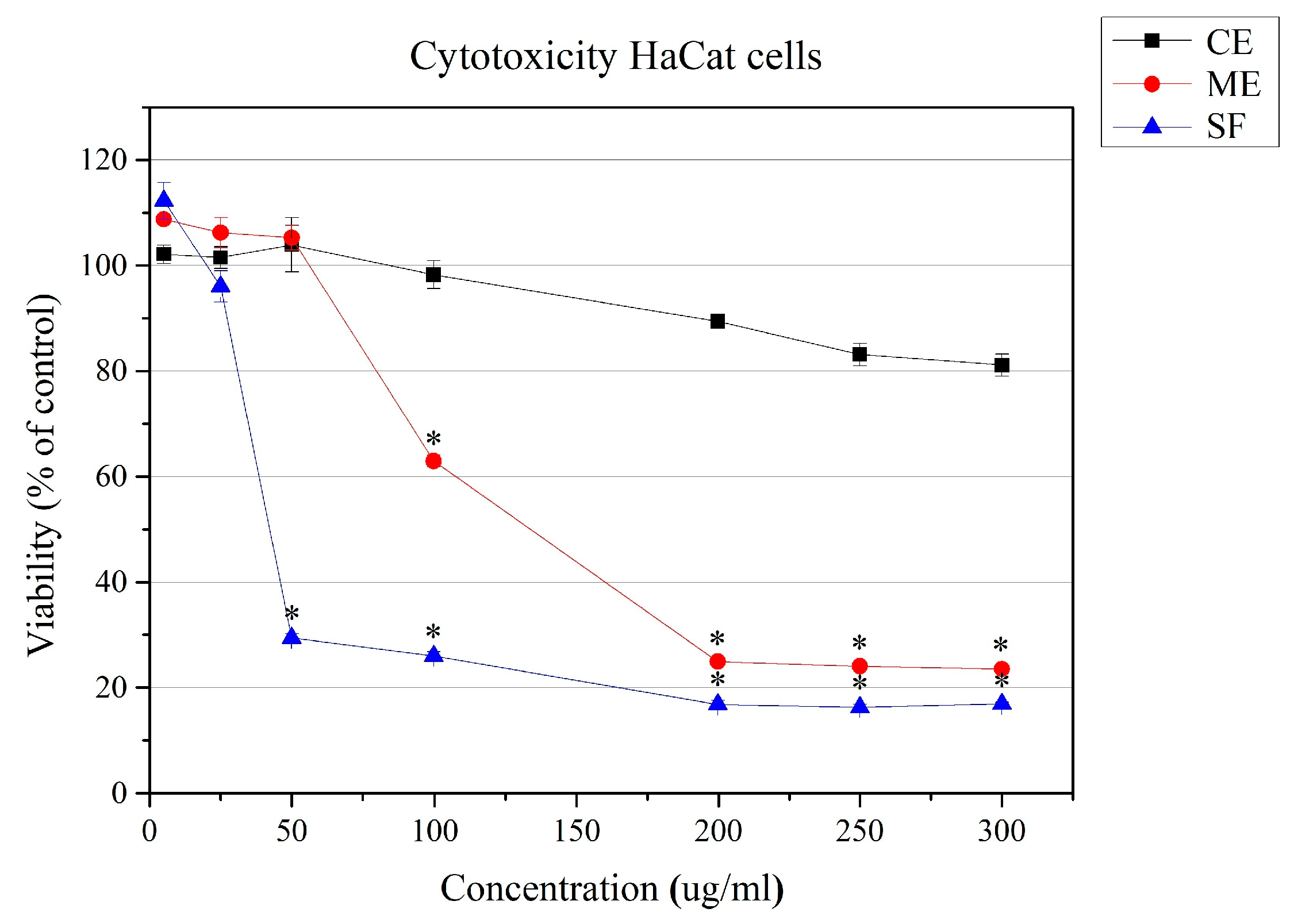
The cytotoxicity assessment of the methanol (ME) and chloroform (CE) extracts of P. veris roots and the saponin-enriched fraction (SF) on a human keratinocyte cell line was performed using the CV test (crystal violet staining) to avoid the influence of enzyme-based cytotoxicity assays due to a reaction with polyphenols [82]. Our results showed the expected high cytotoxicity of the saponin-enriched fraction (SF), which is caused by the interaction of saponins with cell membranes [83]. The methanol extract also showed relatively high activity, while the chloroform extract was not toxic in the tested concentration range up to 300 μg/mL, and cell survival was above 80% (Figure 4). Combined with the high antibiofilm activity, this makes the chloroform extract of P. veris roots a promising subject for future research.
The effect of saponins in ethnomedicine is well known, and their action at the organism level has been studied to some extent [84]. Little is known about their action in vitro at the cellular level. There is evidence that saponins interact with cell membranes, causing temporary or permanent micropores in tumor and actively dividing cells, thus causing cell cycle arrest, disturbances in ionic balance and/or apoptosis in actively dividing cells, and they are potential candidates for application in antitumor therapies. There is evidence of specific effects on specific stress-associated signaling pathways and the activation of apoptosis in myeloid leukemia and other tumor cells by triterpenoid saponins and saikosaponin [85]. When applied externally, their contact with highly differentiated and non-dividing keratinocytes will not have a negative effect, while the development of melanomas or other skin diseases associated with increased proliferation and inflammation, such as psoriasis, for example, as well as wound healing, would be well influenced [4].

3. Materials and Methods

3.1. General Experimental Procedures

Specific optical rotation values were measured on a Jasco P-2000 polarimeter (Jasco, Tokyo, Japan) at the D line of a sodium lamp at 20 °C by using a 0.5 dm quartz cell. The [α] 20 D is given in deg·cm3·g−1·dm−1 and concentration (c) in g·cm−3. The 1D and 2D NMR (1H and 13C NMR, DEPT, COSY, HSQC, HMBC, and ROESY) spectra were recorded on a Bruker Avance NEO 600 spectrometer (Biospin GmbH, Rheinstetten, Germany) with the operating frequencies at 600 MHz (1H) and 150 MHz (13C) using the residual solvent’s signals (δH 7.26 in 1H and δC 77.00 ppm in 13C for CDCl3 and δH 3.31 in 1H and δC 49.3 ppm in 13C for CD3OD) as a reference. The chemical shifts (δ) are expressed in ppm and coupling constants (J) in Hz. HRESIMS spectra were acquired in negative mode on the Q Exactive Plus (Thermo Fisher Scientific, Inc., Bremen, Germany) mass spectrometer equipped with a heated HESI-II source. IR spectra were recorded on a Shimadzu IR Spirit FT-IR spectrometer using QATR-S as a single-reflection ATR measurement attachment.
Polyamide (Carl Roth GmbH + Co. KG, Karlsruhe, Germany), Silica gel 60 (70-230 mesh ASTM) (Merck KGaA, Darmstadt, Germany), and LiChroprep® RP-18 (40–63 µm) (Merck KGaA, Darmstadt, Germany) were used as adsorbents for column chromatography (CC). MPLC was performed on LiChroprep® RP-8 (Merck, Darmstadt, Germany). Thin-layer chromatography (TLC) on Silica gel 60 F254 (Merck, Darmstadt, Germany) and Silica gel RP-18 (Merck, Darmstadt, Germany) plates was used to monitor the separation of the extracts and for preparative TLC. The spots were visualized by spraying with concentrated H2SO4or with the NP reagent (1% diphenylboronic acid 2-aminoethyl ester in ethyl acetate), followed by heating at 105 °C. All solvents used were of HPLC grade.

3.2. Plant Material

The roots of Primula veris L. were collected in April 2023 in the vicinity of Assenovgrad town, Bulgaria (GPS 42.014051; 24.855882), at an altitude of 276 m. A voucher specimen (SOM 179 373) was deposited in the herbarium of the Institute of Biodiversity and Ecosystem research, Bulgarian Academy of Sciences, Sofia, Bulgaria. The plant was identified by Assoc. Prof. Dr Vladimir Vladimirov from the Institute of Biodiversity and Ecosystem Research, Bulgarian Academy of Sciences, Sofia, Bulgaria.

3.3. Extraction and Isolation of Individual Compounds

The air-dried and finely powdered roots of P. veris (52 g) were sequentially extracted with CHCl3 and MeOH (3 × 500 mL) at room temperature for 24 h each. The extracts were filtered and concentrated under vacuum using a rotary evaporator to obtain the corresponding CHCl3 (0.328 g) and MeOH (3.98 g) extracts.
A portion of the CHCl3 extract (0.200 g) was subjected to CC on the silica gel using the n-hexane/ethyl acetate mixture with increasing polarity (from 10:1 to 0:1). Fourteen fractions (F1–F14) were collected via TLC monitoring (Silica gel, n-hexane/ethyl acetate, 3:1 and RP-18 MeOH/H2O 8:2). Prep. TLC (Silica gel, n-hexane/diethyl ether, 2:1, twice development) of fr. F3 (12 mg) afforded compound 9 (1.2 mg). Prep. TLC (Silica gel, n-hexane/diethyl ether, 2:1) of fr. F5 (8.0 mg) and F7 (5.0 mg) yielded compounds 1 (5.3 mg) and 3 (3.0 mg). Prep. TLC (RP-18, H2O/MeOH, 2:8) of fr. F9 (6 mg) gave compound 2 (1.3 mg) and a mixture of compounds 7 and 8 (0.9 mg and a ratio of 0.3:1, as deduced from 1H NMR). Prep. TLC (RP-18, H2O/MeOH, 2:8) of fr. F11 (13 mg) afforded 4.6 mg of compound 10, 1.4 mg of compound 12, and a mixture of compounds 4 and 6 (0.7 mg and a ratio of 1:0.5, as deduced from 1H NMR). Prep. TLC (RP-18, H2O/MeOH, 2:8) of fr. F13 (10 mg) led to the isolation of compounds 11 (3.8 mg), 5 (1.1 mg), and 13 (0.9 mg).
A portion of the methanolic extract (3.0 g) was separated by CC on Polyamide using H2O/MeOH mixtures (from 0:1 to 1:0) to give 3 fractions A-C. MPLC on LiChroprep RP-8 of a portion of fr. B (60 mg) with H2O/MeOH (0:1 to 0:1) afforded a mixture of compounds 14 and 15 (23.6 mg in a ratio of 1:0.07, as deduced from 1H NMR) and afforded a mixture of compounds 16 and 17 (12.6 mg in a ratio of 1:0.2, as deduced from 1H NMR). Fr. C (360 mg) was subjected to MPLC on LiChroprep RP-8 (H2O/MeOH, 1:1 to 0:1) to give 7 subfractions C1–C7. MPLC on LiChroprep RP-8 (H2O/MeOH, 4:6 to 0:1) of fr. C2 afforded 8.12 mg of 19. Prep. TLC (RP-18, H2O/MeOH, 4:6) of fr. C3 afforded 5.2 mg of 21. MPLC on LiChroprep RP-8 (H2O/MeOH, 4:6 to 0:1) of fr. C5 yielded 25.2 mg of 18. MPLC on LiChroprep RP-8 (H2O/MeOH, 4:6 to 0:1) of fr. C6 gave 5.2 mg of 18 and 7.6 mg of 20.
Primulasaponin III (19): Amorphous white powder, [α] 20 D −0.09 (c 0.6, MeOH); FT-IR (ATR): νmax 3352, 2947, 1604, and 1037 cm−1; 1H (600 MHz, CD3OD) and 13C (150 MHz, CD3OD): Table 1; HR-ESI-MS m/z: 1119.55784 [M−H] (calcd. for C54H87O24 1119.55928).
Primulasaponin IV (20): Amorphous white powder, [α] 20 D −0.01 (c 0.154, MeOH); FT-IR (ATR): νmax 3330, 2921, 1609, and 1043 cm−1; 1H (600 MHz, CD3OD) and 13C (150 MHz, CD3OD): Table 1; HR-ESI-MS m/z: 1133.57382 [M−H] (calcd. for C55H89O24 1133.57493).
Primulasaponin V (21): Amorphous white powder, [α] 20 D −0.07 (c 0.375, MeOH); FT-IR (ATR): νmax 3350, 2924, 1744, 1604, and 1040 cm−1; 1H (600 MHz, CD3OD) and 13C (150 MHz, CD3OD): Table 1; HR-ESI-MS m/z: 1117.54210 [M−H] (calcd. for C54H85O24 1117.54363).

3.4. Hydrolysis of Saponin-Enriched Fraction

A portion of fraction C (SF) (10 mg) that was dissolved in 1 mL of MeOH was mixed with 5% HCl (5 mL) and refluxed for 4 h. The reaction mixture was concentrated under reduced pressure to remove MeOH. After extraction with CH2Cl2, the aqueous solution was concentrated under vacuum to obtain the sugar residue and compared with authentic standards (D-glucose, D-galactose, D-glucuronic acid, and L-rhamnose) by TLC (Silica gel, CH3CN/H2O, 85:15).

3.5. Bacterial Strains, Growth Medium, and Cultural Conditions

This study included two Gram-negative and two Gram-positive bacterial strains. Pseudomonas aeruginosa 15692 (ATCC) is a strain from the International Reference Panel [86] isolated from wounds, Escherichia coli 25922 (ATCC), Staphylococcus aureus 29213 (ATCC), and Streptococcus mutans 35668 (ATCC). For long-term storage, the strains were frozen at −80 °C with the addition of the cryoprotectant 8% DMSO. Before experimental work, each strain was inoculated in a growth medium according to its specific requirements: P. aeruginosa and S. aureus in tryptic soy broth (TSA, Sigma, Burlington, MA, USA), E. coli in nutrient broth (HiMedia, Bedford, PA, USA), and S. mutans in brain heart infusion (HiMedia, Bedford, PA, USA).

3.6. Evaluation of the Minimum Inhibitory Concentration (MIC) and the Minimum Bactericidal Concentration (MBC)

For the evaluation of MIC and MBC, standard culture media made of Mueller–Hinton broth (MHB) and Mueller–Hinton agar (MHA) (HiMedia, Bedford, PA, USA) were used. MIC was tested using the widely applied microdilution method, which relies on the oxidation–reduction indicator resazurin to assess changes in the metabolic activity of bacterial cells [59]. For the experiment, the test substances were diluted over a wide concentration range, from 2 mg/mL to 0.0156 mg/mL, with the addition of DMSO at a final concentration of 2% to avoid toxicity to bacterial cells. The initial concentration of bacterial strains was applied after densitometric measurement following the McFarland standard to a final cell concentration of 1 × 10⁶ CFU/mL. Aliquots of 100 µL of the tested extracts (chloroform, methanol, and saponin fraction) at concentrations of 2 mg/mL were added to the first column of the plate, followed by serial two-fold dilutions in the MHB medium to the final test concentration. To each well, 10 µL of a 0.01% sterile aqueous resazurin solution (Sigma, New York, NY, USA) and 10 µL of bacterial suspension were added. The plates were incubated for 24 h at 37 °C under static conditions. As a positive control, 10 µL of ciprofloxacin (Sigma, New York, NY, USA) was added, while a sterile nutrient medium served as the negative control. After the incubation period, the lowest concentration of the test substance where a visual color change from blue to pink was observed was recorded as the minimum inhibitory concentration (MIC). To determine the MBC, samples were taken from the wells of the plate using a sterile swab and plated onto an MHA medium. The lowest concentration of the substances where no bacterial growth was observed was considered the MBC.

3.7. Biofilm Formation Experiments

To determine the antibiofilm effect of the applied plant extracts, the bacterial strains were cultivated in minimal salt medium M63 with the following composition: KH2PO4 (0.02 M), K2HPO4 (0.02 M), (NH4)2SO4 (0.02 M), MgSO4 (0.1 mM), and glucose (0.04 M), with a pH of 7.5. As a source of bacterial inoculum, the strains were incubated in the media described above for 18 h at 37 °C. The cultures were then diluted at a ratio of 1:100 in M63, and the plant extracts were applied in the presence of sub-MICs (1/2 MIC). The prepared suspensions were pipetted into 96-well U-bottomed polystyrene microtiter plates (Corning, Corning, NY, USA) with a final volume of 150 μL per well in six replicates per sample. The control sample contained only bacterial inoculum diluted in M63 with the addition of 2% DMSO (untreated probe). The prepared plates were incubated for 24 h at 37 °C under static conditions. After incubation, the non-adherent planktonic cells were removed by washing the wells with phosphate-buffered saline (PBS, pH 7.2), and the attached cells were stained with a 0.1% aqueous solution of crystal violet for 15 min. The final step involved extracting the dye with 70% ethanol for Gram-negative bacteria and a 95% ethanol–acetone (4:1) dilution for Gram-positive bacteria. Absorbance was measured at a wavelength of 595 nm using an ELISA plate reader (LTEK INNO, Gyeonggi-do, Republic of Korea). The quantitative data were analyzed as the means ± standard deviation (SD) using OriginPro 6.1 software.

3.8. Cytotoxicity Assessment

The cytotoxicity assessment of methanol (ME) and chloroform (CE) extracts from P. veris roots and the saponin-enriched fraction (SF) on a human keratinocyte cell line was performed via the CV assay (crystal violet staining) as previously described [87]. All experiments were performed in triplicate. The optical density of the samples was measured at 570 nm using an Epoch Microplate Spectrophotometer (BioTek® Instruments Inc., Winooski, VT, USA) with Gen5™ Data Analysis software, version 1.11.5. The results are presented as a percentage of cell viability compared to the control of untreated cells. The data were analyzed with OriginPro 9.0 and presented as a mean value ± SE. Statistical significance is according to one-way ANOVA at the 0.05 level (* p < 0.05).

4. Conclusions

In summary, the current study showed the presence of various phenolic compounds (flavones, acetophenones, phenolic glycosides, and bisbibenzyls) and saponins closely related to primulasaponin I in the roots of Primula veris of Bulgarian origin. Among them, three triterpene saponins were found to be new compounds undescribed so far. This investigation contributes to the phytochemical characterization of cowslip in Europe, but detailed phytochemical analyses of more populations of P. veris are needed to understand the intraspecific and interspecific variability of this well-known medicinal plant. Additionally, the antibiofilm properties of the chloroform and methanol extracts and the saponin-enriched fraction obtained from the roots of P. veris were studied for the first time. The obtained results revealed better biofilm inhibition of the tested samples of Gram-negative bacterial strains (P. aeruginosa and E. coli) than Gram-positive ones (S. aureus and S. mutans). Expanding knowledge on biofilm inhibition mechanisms, particularly the modulation of bacterial virulence traits by plant extracts, provides advantages for discovering other agents with similar effects and potential applicability in medical practice for treating biofilm-related infections.

Supplementary Materials

The following supporting information can be downloaded at https://www.mdpi.com/article/10.3390/molecules30081702/s1, Supplementary Part I. Figures SI-1–SI-13: 1H NMR spectra of the known compounds 117; Supplementary Part II. Figures SII-1–SII-36: Spectral data (1H and 13C NMR, DEPT, COSY, HSQC, HMBC, ROESY, HR-ESI-MS, and IR) of primulasaponin I (18) and new saponins 1921.

Author Contributions

Conceptualization, A.T. and M.N.; methodology and investigation, A.T., V.I., D.R., P.N., T.P.-K., R.V. and T.T.-H.; data curation, A.T., M.N., P.N., T.P.-K. and T.T.-H.; writing—original draft preparation, A.T. and T.P.-K.; writing—review and editing, A.T., M.N., P.N., T.P.-K. and T.T.-H.; visualization, A.T., R.V. and T.P.-K.; supervision and project administration, A.T. and M.N. All authors have read and agreed to the published version of the manuscript.

Funding

This research received no external funding.

Institutional Review Board Statement

Not applicable.

Informed Consent Statement

Not applicable.

Data Availability Statement

Data are contained within the article and Supplementary Materials.

Acknowledgments

The authors are thankful to the bilateral project of the Bulgarian Academy of Sciences and the Serbian Academy of Sciences and Arts “Phytochemical and metabolomics investigation of biologically active compounds from Primula species and medicinal mushrooms” and to Vladimir Vladimirov for the identification of the plant material.

Conflicts of Interest

The authors declare no conflicts of interest.

References

  1. Colombo, P.S.; Flamini, G.; Rodondi, G.; Giuliani, C.; Santagostini, L.; Fico, G. Phytochemistry of European Primula species. Phytochemistry 2017, 143, 132–144. [Google Scholar] [CrossRef] [PubMed]
  2. The Plant List. Primula veris L. Available online: http://www.theplantlist.org/tpl1.1/record/kew-2563733 (accessed on 21 February 2025).
  3. Plants of the World Online Kew Science. Primula veris L. Available online: https://powo.science.kew.org/taxon/urn:lsid:ipni.org:names:702751-1 (accessed on 21 February 2025).
  4. Kahraman, C.; Sari, S.; Küpeli Akkol, E.; Tatli Cankaya, I. Bioactive saponins of Primula vulgaris Huds. promote wound healing through inhibition of collagenase and elastase enzymes: In vivo, in vitro and in silico evaluations. Chem. Biodivers. 2022, 19, e202200582. [Google Scholar] [CrossRef] [PubMed]
  5. Alam, F.; Din, K.M.; Sarfraz, M.; Qudoos, A.; Malik, S. Genus Primula and its role in phytomedicine; a systematic review. Phytomedicine Plus 2024, 4, 100510. [Google Scholar] [CrossRef]
  6. Kemmerich, B. Evaluation of efficacy and tolerability of a fixed combination of dry extracts of Thyme herb and Primrose root in adults suffering from acute bronchitis with productive cough: A prospective, double-blind, placebo-controlled multicentre clinical trial. Arzneim. Forsch. Drug Res. 2007, 57, 607–615. [Google Scholar] [CrossRef]
  7. Yasar, B.; Kutlu, G.; Tornuk, F. Edible Flowers as Sources of Bioactive Compounds: Determination of phenolic extraction conditions. Int. J. Gastron. Food Sci. 2022, 30, 100618. [Google Scholar] [CrossRef]
  8. Sarropoulou, V.; Sarrou, E.; Angeli, A.; Martens, S.; Maloupa, E.; Grigoriadou, K. Species-specific secondary metabolites from Primula veris subsp. veris obtained in vitro adventitious root cultures: An alternative for sustainable production. Sustainability 2023, 15, 2452. [Google Scholar] [CrossRef]
  9. Stefanis, I.; Chatzopoulou, P.; Krigas, N.; Karioti, A. Exploring the chemical content of Primula veris L. subsp. veris wild-growing populations along a climate gradient: An HPLC-PDA-MS quality assessment of flowers, leaves and roots for sustainable exploitation. Horticulturae 2023, 9, 1120. [Google Scholar] [CrossRef]
  10. Müller, A.; Ganzera, M.; Stuppner, H. Analysis of phenolic glycosides and saponins in Primula elatior and Primula veris (Primula Root) by liquid chromatography, evaporative light scattering detection and mass spectrometry. J. Chromatogr. A 2006, 1112, 218–223. [Google Scholar] [CrossRef]
  11. Ozkan, M.T.; Aliyazicioglu, R.; Demir, S.; Misir, S.; Turan, I.; Yildirmis, S.; Aliyazicioglu, Y. Phenolic characterisation and antioxidant activity of Primula vulgaris and its antigenotoxic effect on fibroblast cells. Jundishapur. J. Nat. Pharm. Prod. 2017, 12, e40073. [Google Scholar] [CrossRef]
  12. Latypova, G.M.; Bychenkova, M.A.; Katayev, V.A.; Perfilova, V.N.; Tyurenkov, I.N.; Mokrousov, I.S.; Prokofiev, I.I.; Salikhov, S.M.; Iksanova, G.R. Composition and cardioprotective effects of Primula veris L. solid herbal extract in experimental chronic heart failure. Phytomedicine 2019, 54, 17–26. [Google Scholar] [CrossRef]
  13. Tünde, J.; Eleonora, M.; Laura, V.; Neagu, O.; Annamaria, P. Bioactive compounds and antioxidant capacity of Primula veris L. flower extracts. Analele Univ. Din Oradea Fasc. Ecotoxicologie Zooteh. De Ind. 2015, XIV B, 235–241. [Google Scholar]
  14. Tokalov, S.V.; Kind, B.; Wollenweber, E.; Gutzeit, H.O. Biological effects of epicuticular flavonoids from Primula denticulata on human leukemia cells. J. Agric. Food. Chem. 2003, 52, 239–245. [Google Scholar] [CrossRef] [PubMed]
  15. Bhutia, T.D.; Valant-Vetschera, K.M. Diversification of exudate flavonoid profiles in further Primula spp. Nat. Prod. Commun. 2012, 7, 587–589. [Google Scholar] [CrossRef] [PubMed]
  16. Valant-Vetschera, K.M.; Bhutia, T.D.; Wollenweber, E. Chemodiversity of exudate flavonoids in Dionysia (Primulaceae): A comparative study. Phytochemistry 2010, 71, 937–947. [Google Scholar] [CrossRef]
  17. Valant-Vetschera, K.M.; Bhutia, T.D.; Wollenweber, E. Exudate flavonoids of Primula spp: Structural and biogenetic chemodiversity. Nat. Prod. Commun. 2009, 4, 365–370. [Google Scholar] [CrossRef] [PubMed]
  18. Bhutia, T.D.; Valant-Vetschera, K.M.; Brecker, L. Orphan flavonoids and dihydrochalcones from Primula exudates. Nat. Prod. Commun. 2013, 8, 1081–1084. [Google Scholar] [CrossRef] [PubMed]
  19. Bhutia, T.D.; Valant-Vetschera, K.M.; Adlassnig, W.; Brecker, L. Flavonoids in selected Primula spp.: Bridging micromorphology with chemodiversity. Nat. Prod. Commun. 2012, 7, 1469–1473. [Google Scholar] [CrossRef]
  20. Siems, K.; Jaensch, M.; Jakupovic, J. Structures of the two saponins isolated from commercially available root extract of Primula sp. Planta Med. 1998, 64, 272–274. [Google Scholar] [CrossRef]
  21. Apel, L.; Kammerer, D.R.; Stintzing, F.C.; Spring, O. Comparative metabolite profiling of triterpenoid saponins and flavonoids in flower color mutations of Primula veris L. Int. J. Mol. Sci. 2017, 18, 153. [Google Scholar] [CrossRef]
  22. Çaliş, İ.; Yürüker, A.; Rüegger, H.; Wright, A.D.; Sticker, O. Triterpene saponins from Primula veris subsp. macrocalyx and Primula elatior subsp. meyeri. J. Nat. Prod. 1992, 55, 1299–1306. [Google Scholar] [CrossRef]
  23. Włodarczyk, M.; Pasikowski, P.; Osiewała, K.; Frankiewicz, A.; Dryś, A.; Gleńsk, M. In search of high-yielding and single-compound-yielding plants: New sources of pharmaceutically important saponins from the Primulaceae family. Biomolecules 2020, 10, 376. [Google Scholar] [CrossRef] [PubMed]
  24. Herbal Medicinal Product European Medicines Agency (EMA). Primulae Radix. Available online: https://www.ema.europa.eu/en/medicines/herbal/primulae-radix (accessed on 14 February 2025).
  25. Kosenkova, Y.S.; Polovinka, M.P.; Komarova, N.I.; Korchagina, D.V.; Kurochkina, N.Y.; Cheremushkina, V.A.; Salakhutdinov, N.F. Riccardin C, a bisbibenzyl compound from Primula Macrocalyx. Chem. Nat. Compd. 2007, 43, 712–713. [Google Scholar] [CrossRef]
  26. Bukvicki, D.; Kovtonyuk, N.K.; Legin, A.A.; Keppler, B.K.; Brecker, L.; Asakawa, Y.; Valant-Vetschera, K. Hunting for bis-bibenzyls in Primula veris subsp. macrocalyx (Bunge) Lüdi: Organ-specific accumulation and cytotoxic activity. Phytochem. Lett. 2021, 44, 90–97. [Google Scholar] [CrossRef]
  27. Novakovic, M.; Ilic-Tomic, T.; Djordjevic, I.; Andjelkovic, B.; Tesevic, V.; Milosavljevic, S.; Asakawa, Y. Bisbibenzyls from Serbian Primula veris subsp. columnae (Ten.) Lȕdi and P. acaulis (L.) L. Phytochemistry 2023, 212, 113719. [Google Scholar] [CrossRef]
  28. Novakovic, M.; Bukvicki, D.; Andjelkovic, B.; Ilic-Tomic, T.; Veljic, M.; Tesevic, V.; Asakawa, Y. Cytotoxic activity of riccardin and perrottetin derivatives from the liverwort Lunularia cruciata. J. Nat. Prod. 2019, 82, 694–701. [Google Scholar] [CrossRef] [PubMed]
  29. Xie, C.F.; Qu, J.B.; Wu, X.Z.; Liu, N.; Ji, M.; Lou, H.X. Antifungal macrocyclic bis(bibenzyls) from the Chinese liverwort Ptagiochasm intermedlum L. Nat. Prod. Res. 2010, 24, 515–520. [Google Scholar] [CrossRef] [PubMed]
  30. Zafer, M.M.; Mohamed, G.A.; Ibrahim, S.R.M.; Ghosh, S.; Bornman, C.; Elfaky, M.A. Biofilm-mediated infections by multidrug-resistant microbes: A comprehensive exploration and forward perspectives. Arch. Microbiol. 2024, 206, 101. [Google Scholar] [CrossRef]
  31. Olsen, I. Biofilm-specific antibiotic tolerance and resistance. Eur. J. Clin. Microbiol. Infect. Dis. 2015, 34, 877–886. [Google Scholar] [CrossRef]
  32. Abebe, G.M. The role of bacterial biofilm in antibiotic resistance and food contamination. Int. J. Microbiol. 2020, 2020, 1705814. [Google Scholar] [CrossRef]
  33. Ciofu, O.; Moser, C.; Jensen, P.Ø.; Høiby, N. Tolerance and resistance of microbial biofilms. Nat. Rev. Microbiol. 2022, 20, 621–635. [Google Scholar] [CrossRef]
  34. Damyanova, T.; Paunova-Krasteva, T. What we still don’t know about biofilms—current overview and key research information. Microbiol. Res. 2025, 16, 46. [Google Scholar] [CrossRef]
  35. Oyardi, O.; Hacioglu, M.; Özdemir, E.; Erbay, M.Ş.; Kültür, Ş.; Bozkurt Güzel, C. Screening of antimicrobial, antibiofilm and cytotoxic activities of some medicinal plants from Balıkesir Province, Türkiye: Pointing to the potential effects of Allium paniculatum flower. Turk. J. Pharm. Sci. 2023, 21, 252–258. [Google Scholar] [CrossRef]
  36. Başbülbül, G.; Özmen, A.; Biyik, H.H.; Şen, Ö. Antimitotic and antibacterial effects of the Primula veris L. flower extracts. Caryologia 2008, 61, 88–91. [Google Scholar] [CrossRef]
  37. Karapınar, Ç.; Öz, M. Chemical content of volatile oil of Primula veris subsp. columnae, obtaining the methanol extracts and their biological activities. Bioresources 2023, 18, 4475–4491. [Google Scholar] [CrossRef]
  38. Ivanišová, E.; Horňák, M.; Čech, M.; Harangozo, Ľ.; Kačániová, M.; Grygorieva, O.; Kowalczewski, P.Ł. Polyphenol content, mineral compounds composition, antimicrobial and antioxidant activities of selected medicinal herbs from Slovak Republic. Appl. Sci. 2023, 13, 1918. [Google Scholar] [CrossRef]
  39. Chilku, E.; Ivic Kolevska, S.; Kadifkova Panovska, T. Antioxidant and antibacterial properties of some commercial plants from Macedonia. World J. Pharm. Pharm. Sci. 2017, 6, 1767–1778. [Google Scholar] [CrossRef]
  40. Tosun, F.; Kizilay, Ç.A.; Şener, B.; Vural, M. The evaluation of plants from Turkey for in vitro antimycobacterial activity. Pharm. Biol. 2005, 43, 58–63. [Google Scholar] [CrossRef]
  41. Yayli, N.; Tosun, G.; Yayli, B.; Gündoǧan, Z.; Coşkunçelebi, K.; Karaoǧlu, Ş.A. Altitude variation in the composition of essential oils, fatty acid methyl esters, and antimicrobial activities of two subspecies of Primula vulgaris grown in Turkey. Nat. Prod. Commun. 2016, 11, 1505–1510. [Google Scholar] [CrossRef]
  42. Majid, A.; Hassan, S.; Hussain, W.; Khan, A.; Hassan, A.; Khan, A.; Khan, T.; Ahmad, T.; Rehman, M.U. In vitro approaches of Primula vulgaris leaves and roots extraction against human pathogenic bacterial strains. World Appl. Sci. J. 2014, 30, 575–580. Available online: https://idosi.org/wasj/wasj30(5)14/6.pdf (accessed on 14 February 2025).
  43. Jaberian, H.; Piri, K.; Nazari, J. Phytochemical composition and in vitro antimicrobial and antioxidant activities of some medicinal plants. Food Chem. 2013, 136, 237–244. [Google Scholar] [CrossRef]
  44. Khan, S.; Shaheen, H.; Aziz, S.; Nasar, S. Diversity and distribution of genus Primula in Kashmir Region: An indicator genus of the western Himalayan Mountain wetlands and glacial forelands. Biodivers. Conserv. 2021, 30, 1673–1688. [Google Scholar] [CrossRef]
  45. Najmus-Saqib, Q.; Alam, F.; Ahmad, M. Antimicrobial and cytotoxicity activities of the medicinal plant Primula macrophylla. J. Enzyme. Inhib. Med. Chem. 2009, 24, 697–701. [Google Scholar] [CrossRef] [PubMed]
  46. Peev, D. Flora of the Republic of Bulgaria; Velchev, V., Ed.; Bulgarian Academy of Sciences: Sofia, Bulgaria, 1982; Volume 8, pp. 324–336. [Google Scholar]
  47. Assyov, B.; Petrova, A.; Dimitrov, D.; Vassilev, P. Conspectus of the Bulgarian Vascular Flora. Distribution Maps and Floristic Elements, 4th ed.; Assyov, B., Petrova, A., Eds.; Bulgarian Biodiversity Foundation: Sofia, Bulgaria, 2012. [Google Scholar]
  48. Yankova-Tsvetkova, E.; Yurukova-Grancharova, P.; Aneva, I.; Zhelev, P. On the reproductive potential in Primula veris L. (Primulaceae): Embryological features, pollen and seed viability, genetic diversity. Plants 2021, 10, 2296. [Google Scholar] [CrossRef]
  49. Yankova-Tsvetkova, E.; Petrova, M.; Grigorova, I.; Traykova, B.; Stanilova, M. The establishment of an ex situ collection of Primula veris in Bulgaria. Plants 2022, 11, 3018. [Google Scholar] [CrossRef] [PubMed]
  50. Nikolova, M.; Yankova-Tsvetkova, E.; Stefanova, T.; Berkov, S. Exudate flavonoids of Primula veris leaves and their inhibitory activity on Lolium perrene seed germination. Proc. Bulg. Acad. Sci. 2023, 76, 388–393. [Google Scholar] [CrossRef]
  51. Budzianowski, J.; Morozowska, M.; Wesołowska, M. Lipophilic Flavones of Primula veris L. from field cultivation and in vitro cultures. Phytochemistry 2005, 66, 1033–1039. [Google Scholar] [CrossRef]
  52. Matsumoto, M. 2′-Hydroxy-4′-Methoxyacetophenone (Paeonol) in Exacum affine Cv. Biosci. Biotechnol. Biochem. 1994, 58, 1892–1893. [Google Scholar] [CrossRef]
  53. Has, M.; Kucuk, S.; Kurkcuoglu, M. Anatomical and Volatile Components Investigations on Primula vulgaris Huds. subsp. vulgaris (Primulaceae). Ann. Phytomedicine Int. J. 2021, 10, 63–66. [Google Scholar] [CrossRef]
  54. Qu, G.L.; Zhang, H.M.; Deng, Z.W.; Kong, D.Y.; Geng, Z.F.; Du, S.S. Study on chemical constituents of Primula maximowiczii Regel: Part III. Chin. Pharm. J. 2011, 46, 93–97. [Google Scholar]
  55. Xu, S.J.; Yang, L.; Zeng, X.; Zhang, M.; Wang, Z.T. Characterization of compounds in the Chinese herbal drug mu-dan-pi by liquid chromatography coupled to electrospray ionization mass spectrometry. Rapid Commun. Mass Spectrom. 2006, 20, 3275–3288. [Google Scholar] [CrossRef]
  56. Baczek, K.; Przybył, J.L.; Mirgos, M.; Kosakowska, O.; Szymborska-Sandhu, I.; Wȩglarz, Z. Phenolics in Primula veris L. and P. elatior (L.) Hill raw materials. Int. J. Anal. Chem. 2017, 2017, 2871579. [Google Scholar] [CrossRef] [PubMed]
  57. Foubert, K.; Theunis, M.; Apers, S.; Vlietinck, A.J.; Pieters, L. Chemistry, distribution and biological activities of 13,28-epoxy-oleanane saponins from the plant families Myrsinaceae and Primulaceae. Curr. Org. Chem. 2008, 12, 629–642. [Google Scholar] [CrossRef]
  58. Damyanova, T.; Dimitrova, P.D.; Borisova, D.; Topouzova-Hristova, T.; Haladjova, E.; Paunova-Krasteva, T. An overview of biofilm-associated infections and the role of phytochemicals and nanomaterials in their control and prevention. Pharmaceutics 2024, 16, 162. [Google Scholar] [CrossRef]
  59. Sarker, S.D.; Nahar, L.; Kumarasamy, Y. Microtitre plate-based antibacterial assay incorporating resazurin as an indicator of cell growth, and its application in the in vitro antibacterial screening of phytochemicals. Methods 2007, 42, 321–324. [Google Scholar] [CrossRef] [PubMed]
  60. Święciło, A.; Rybczyńska-Tkaczyk, K. Resazurin method for evaluation of bioactive compounds from cranberry extracts using the metabolic activity of a ΔSOD1 mutant of saccharomyces cerevisiae yeast under severe osmotic stress. J. AOAC Int. 2020, 103, 422–427. [Google Scholar] [CrossRef] [PubMed]
  61. Hussain, A.I.; Anwar, F.; Nigam, P.S.; Sarker, S.D.; Moore, J.E.; Rao, J.R.; Mazumdar, A. Antibacterial activity of some Lamiaceae essential oils using resazurin as an indicator of cell growth. LWT Food Sci. Technol. 2011, 44, 1199–1206. [Google Scholar] [CrossRef]
  62. Khan, S.; Shaheen, H.; Mehmood, A.; Nasar, S.; Khan, T. Ethnobotanical and antibacterial study of Primula plants traditionally used in the indigenous communities of Western Himalaya, Pakistan. Saudi J. Biol. Sci. 2022, 29, 3244–3254. [Google Scholar] [CrossRef] [PubMed]
  63. El-Sayed, N.R.; Samir, R.; Abdel-Hafez, L.J.M.; Ramadan, M.A. Olive leaf extract modulates quorum sensing genes and biofilm formation in multi-drug resistant Pseudomonas aeruginosa. Antibiotics 2020, 9, 526. [Google Scholar] [CrossRef]
  64. Paunova-Krasteva, T.; Hemdan, B.A.; Dimitrova, P.D.; Damyanova, T.; El-Feky, A.M.; Elbatanony, M.M.; Stoitsova, S.; El-Liethy, M.A.; El-Taweel, G.E.; El Nahrawy, A.M. Hybrid Chitosan/CaO-based nanocomposites doped with plant extracts from Azadirachta indica and Melia azedarach: Evaluation of antibacterial and antibiofilm activities. Bionanoscience 2023, 13, 88–102. [Google Scholar] [CrossRef]
  65. Ansari, F.A.; Husain, F.M.; Pichtel, J.; Meena, R.P.; Khan, M.H.; Khan, A.S.; Alam, N. Withania somnifera (L.) Dunal: A medicinally important plant inhibits pathogenic biofilms. Microbe 2025, 6, 100227. [Google Scholar] [CrossRef]
  66. Laskoski, L.V.; Bandeira, D.M.; Batista, J.M.; da Costa, W.F.; Baeza, L.C.; Kuo, L.H.; Pinto, F.G.d.S. Phytochemical prospection and evaluation of antimicrobial, antioxidant and antibiofilm activities of extracts and essential oil from leaves of Myrsine umbellata Mart. (Primulaceae). Braz. J. Biol. 2022, 82, e263865. [Google Scholar] [CrossRef]
  67. Ozay, C.; Temel, A.; Turel, S.; Akgul, I. Investigation of anti-inflammatory, antibiofilm, antioxidant and cytotoxic activities of Cyclamen hederifolium (Primulaceae). Farmacia 2023, 71, 1208–1216. [Google Scholar] [CrossRef]
  68. Hamdan, H.F.; Ross, E.E.R.; Jalil, M.T.M.; Hashim, M.A.; Yahya, M.F.Z.R. Antibiofilm efficacy and mode of action of Etlingera elatior extracts against Staphylococcus aureus. Malays. Appl. Biol. 2024, 53, 27–34. [Google Scholar] [CrossRef]
  69. Dimitrova, P.D.; Ivanova, V.; Trendafilova, A.; Paunova-Krasteva, T. Anti-biofilm and anti-quorum-sensing activity of Inula extracts: A strategy for modulating Chromobacterium violaceum virulence factors. Pharmaceuticals 2024, 17, 573. [Google Scholar] [CrossRef] [PubMed]
  70. Dimitrova, P.D.; Damyanova, T.; Paunova-Krasteva, T. Chromobacterium violaceum: A model for evaluating the anti-quorum sensing activities of plant substances. Sci. Pharm. 2023, 91, 33. [Google Scholar] [CrossRef]
  71. Yu, Y.; Kang, X.; Liu, T.; Wang, Y.; Tang, J.; Peng, W.; Martin, F.M.; Tan, H. Inoculation of the Morchella importuna mycosphere with Pseudomonas chlororaphis alleviated a soil-borne disease caused by Paecilomyces penicillatus. Biol. Fertil. Soils. 2024, 61, 141–161. [Google Scholar] [CrossRef]
  72. Matilla-Cuenca, L.; Gil, C.; Cuesta, S.; Rapún-Araiz, B.; Žiemytė, M.; Mira, A.; Lasa, I.; Valle, J. Antibiofilm activity of flavonoids on Staphylococcal biofilms through targeting BAP amyloids. Sci. Rep. 2020, 10, 18968. [Google Scholar] [CrossRef]
  73. Zeng, Y.; Nikitkova, A.; Abdelsalam, H.; Li, J.; Xiao, J. Activity of quercetin and kaemferol against Streptococcus mutans biofilm. Arch. Oral. Biol. 2019, 98, 9–16. [Google Scholar] [CrossRef]
  74. Pruteanu, M.; Hernández Lobato, J.I.; Stach, T.; Hengge, R. Common plant flavonoids prevent the assembly of amyloid curli fibres and can interfere with bacterial biofilm formation. Environ. Microbiol. 2020, 22, 5280–5299. [Google Scholar] [CrossRef]
  75. Parai, D.; Islam, E.; Mitra, J.; Mukherjee, S.K. Effect of Bacoside a on growth and biofilm formation by Staphylococcus aureus and Pseudomonas aeruginosa. Can. J. Microbiol. 2016, 63, 169–178. [Google Scholar] [CrossRef]
  76. Stanković, J.; Godevac, D.; Tešević, V.; Dajić-Stevanović, Z.; Ćirić, A.; Soković, M.; Novaković, M. Antibacterial and antibiofilm activity of flavonoid and saponin derivatives from Atriplex tatarica against Pseudomonas aeruginosa. J. Nat. Prod. 2019, 82, 1487–1495. [Google Scholar] [CrossRef]
  77. Chen, Y.; Gao, Y.; Li, Y.; Yin, J. Anti-biofilm activity of assamsaponin A, theasaponin E1, and theasaponin E2 against Candida albicans. Int. J. Mol. Sci. 2024, 25, 3599. [Google Scholar] [CrossRef]
  78. Araújo, N.J.S.; Silva, A.R.P.; Costa, M.S.; Freitas, T.S.; Filho, J.M.B.; Matos, Y.M.L.S.; Morais-Braga, M.F.B.; Pereira Junior, F.N.; Silva, C.A.P.; Souza, E.O.; et al. Chemical characterization UPLC-ESI-QToF-MSE, antibacterial and antibiofilm potential of Sarcomphalus joazeiro (MART.) Hauenschild. Food Biosci. 2022, 50, 102066. [Google Scholar] [CrossRef]
  79. Choudhary, M.; Verma, V.; Saran, R.; Bhagyawant, S.S.; Srivastava, N. Natural biosurfactant as antimicrobial agent: Strategy to action against fungal and bacterial activities. Cell. Biochem. Biophys. 2022, 80, 245–259. [Google Scholar] [CrossRef] [PubMed]
  80. Yadav, H.; Mahalvar, A.; Pradhan, M.; Yadav, K.; Kumar Sahu, K.; Yadav, R. Exploring the potential of phytochemicals and nanomaterial: A boon to antimicrobial treatment. Med. Drug Discov. 2023, 17, 100151. [Google Scholar] [CrossRef]
  81. Silva, E.; Teixeira, J.A.; Pereira, M.O.; Rocha, C.M.R.; Sousa, A.M. Evolving biofilm inhibition and eradication in clinical settings through plant-based antibiofilm agents. Phytomedicine 2023, 119, 154973. [Google Scholar] [CrossRef] [PubMed]
  82. Wang, P.; Henning, S.M.; Heber, D. Limitations of MTT and MTS-based assays for measurement of antiproliferative activity of green tea polyphenols. PLoS ONE 2010, 5, e10202. [Google Scholar] [CrossRef]
  83. Koczurkiewicz, P.; Czyz, J.; Podolak, I.; Wójcik, K.; Galanty, A.; Janeczko, Z.; Michalik, M. Multidirectional effects of triterpene saponins on cancer cells—mini-review of in vitro studies. Acta Biochim. Pol 2015, 62, 383–393. [Google Scholar] [CrossRef]
  84. Francis, G.; Kerem, Z.; Makkar, H.P.S.; Becker, K. The biological action of saponins in animal systems: A review. Br. J. Nutr. 2002, 88, 587–605. [Google Scholar] [CrossRef]
  85. Sun, X.H.; Chai, Y.H.; Bai, X.T.; Li, H.X.; Yang, P.P.; Xi, Y.M. Saikosaponin A mediates the anti-acute myeloid leukemia effect via the P-JNK signaling pathway induced by endoplasmic reticulum stress. Drug Des. Devel. Ther. 2025, 19, 1983–2001. [Google Scholar] [CrossRef]
  86. De Soyza, A.; Hall, A.J.; Mahenthiralingam, E.; Drevinek, P.; Kaca, W.; Drulis-Kawa, Z.; Stoitsova, S.R.; Toth, V.; Coenye, T.; Zlosnik, J.E.A.; et al. Developing an international Pseudomonas aeruginosa reference panel. Microbiologyopen 2013, 2, 1010–1023. [Google Scholar] [CrossRef] [PubMed]
  87. Trendafilova, A.; Staleva, P.; Petkova, Z.; Ivanova, V.; Evstatieva, Y.; Nikolova, D.; Rasheva, I.; Atanasov, N.; Topouzova-Hristova, T.; Veleva, R.; et al. Phytochemical profile, antioxidant potential, antimicrobial activity, and cytotoxicity of dry extract from Rosa damascena Mill. Molecules 2023, 28, 7666. [Google Scholar] [CrossRef] [PubMed]
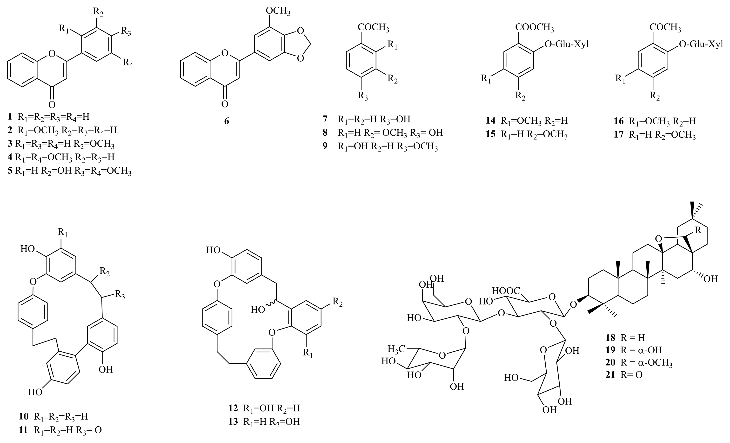
Figure 1. Structures of isolated compounds from P. veris roots.
Molecules 30 01702 g001
Figure 2. Important HMBC and ROESY correlations for compounds 19 (A) and 20 (B).
Molecules 30 01702 g002
Figure 3. Inhibition of biofilm formation under the influence of methanol (ME) and chloroform (CE) extracts of P. veris roots and the saponin-enriched fraction (SF). The antibiofilm effects were examined after a 24 h treatment period and calculated as a percentage of the biofilm in the untreated control sample. (A) P. aeruginosa; (B) E. coli; (C) S. aureus; (D) S. mutans.
Molecules 30 01702 g003
Figure 4. Inhibition of cellular viability under the influence of the methanol (ME) and chloroform (CE) extracts of P. veris roots and the saponin-enriched fraction (SF). Cytotoxicity was examined at after a 24 h treatment period and calculated as a percentage of the untreated control cells (* p < 0.05).
Molecules 30 01702 g004
Table 1. 1H (600 MHz) and 13C (150 MHz) NMR data of compounds 1921 in CD3OD (δ in ppm, multiplicity, and J in Hz).
Position192021
δCδHδCδHδCδH
140.30.99 m 140.30.98 m40.21.02 m
1.74 m1.75 m1.75 m
227.11.75 m27.11.75 m27.01.75 m
2.02 m1.95 m2.01 m
392.13.20 dd (4.0, 11.3)92.33.19 dd (4.0, 11.3)91.93.21 dd (4.0, 11.3)
440.7-40.7-40.7-
556.80.72 dd (1.8, 11.5)56.80.73 dd (1.8, 11.5)56.80.76 dd (1.8, 11.5)
618.71.44 m18.71.43 m18.71.43 m
1.50 m1.50 m1.55 m
735.21.20 m35.21.22 m34.91.24 m
1.56 m1.55 m1.59 m
843.3-43.4-43.0-
951.41.22 m51.41.23 m50.81.34 m
1037.8-37.8-37.8-
1119.91.45 m19.91.45 m19.41.55 m
1.66 m1.65 m1.65 m
1233.61.28 m33.71.28 m32.21.56 m
2.03 m2.00 m2.15 m
1388.9-89.0-94.9-
1443.9-44.0-43.0-
1537.01.20 m37.41.20 m37.41.20 m
1.99 m1.99 m1.99 m
1677.33.78 m77.23.82 m76.23.79 m
1748.7-48.9 49.1-
1847.61.67 m48.01.68 m52.61.92 m
1939.81.19 m39.81.17 m39.21.30 m
2.37 dd (12.1, 14.5)2.28 dd (12.1, 14.5)2.49 dd (12.1, 14.5)
2032.4-32.4-32.2-
2137.51.15 m37.11.15 m36.71.21 m
2.06 m2.01 m2.09 m
2227.21.42 m26.51.42 m28.71.63 m
1.94 m1.61 m1.83 m
2328.31.06 s28.21.06 s28.21.07 s
2416.80.87 s16.80.87 s16.70.88 s
2516.80.90 s16.80.91 s16.80.91 s
2618.81.18 s18.91.18 s18.31.08 s
2719.91.22 s19.81.21 s19.71.38 s
2899.74.60 brs106.94.17 brs182.0-
2933.90.93 s33.90.92 s33.50.96
3025.00.92 s24.90.88 s24.90.92
OCH3 55.43.31 s
1′105.84.45 d (7.7)105.84.48 d (7.7)105.84.46 d (7.7
2′79.33.92 dd (7.7, 8.8)79.13.93 dd (7.7, 8.8)79.33.92 dd (7.7, 8.8)
3′81.24.06 t (8.8)81.04.05 t (8.8)81.24.07 t (8.8)
4′72.23.59 m72.23.61 m72.33.59 m
5′75.93.79 m75.93.78 m75.93.79 m
6′176.6- -
1″100.85.20 d (7.8)100.95.20 d (7.8)100.85.20 d (7.8)
2″76.23.79 m76.13.79 m76.23.79 m
3″75.93.72 m76.03.74 m76.03.74 m
4″71.93.72 m71.83.72 m71.83.72 m
5″76.93.53 m76.93.53 m76.93.53 m
6″62.73.65 dd (4.1, 11.5)62.83.65 dd (4.1, 11.5)62.73.67 dd (4.1, 11.5)
3.81 m3.81 m3.81 m
1‴102.05.28 d (1.2)102.15.28 d (1.2)102.05.29 d (1.2)
2‴72.63.97 dd (1.2, 3.5)72.63.96 dd (1.2, 3.5)72.63.96 dd (1.2, 3.5)
3‴72.33.71 dd (3.5, 9.5)72.33.71 dd (3.5, 9.5)72.23.71 dd (3.5, 9.5)
4‴73.73.44 t (9.5)73.83.45 t (9.5)73.73.43 t (9.5)
5‴70.34.10 dq (9.5, 6.5)70.24.11 dq (9.5, 6.5)70.24.11 dq (9.5, 6.5)
6‴17.91.27 d (6.5)17.91.27 d (6.5)17.91.27 d (6.5)
1⁗102.54.88102.64.88102.64.88
2⁗76.23.22 dd (7.2, 9.5)76.13.22 dd (7.2, 9.5)76.13.22 dd (7.2, 9.5)
Glu-377.93.35 m77.93.35 m77.93.35 m
4⁗72.43.05 t (9.5)72.63.05 t (9.5)72.63.05 t (9.5)
5⁗78.13.40 m78.13.40 m78.13.40 m
6⁗63.53.54 dd (8.0, 12.5)63.53.54 dd (8.0, 12.5)63.53.55 dd (8.0, 12.5)
3.87 dd (2.5, 12.5)3.87 dd (2.5, 12.5)3.88 dd (2.5, 12.5)
1 Multiplicity: m—multiplet; d—doublet; dd—doublet of doublets; t—triplet; s—singlet; brs—broad singlets.
Table 2. Determination of the MIC and MBC (mg/mL) of P. veris extracts and the saponin-enriched fraction.
Sample 1P. aeruginosaE. coliS. aureusS. mutans
MICMBCMICMBCMICMBCMICMBC
CE1.02.00.50.51.02.01.02.0
ME0.51.00.51.01.02.01.02.0
SF0.51.00.51.00.52.00.52.0
1 Chloroform extract (CE), methanol extract (ME), and saponin-enriched fraction (SF).
Disclaimer/Publisher’s Note: The statements, opinions and data contained in all publications are solely those of the individual author(s) and contributor(s) and not of MDPI and/or the editor(s). MDPI and/or the editor(s) disclaim responsibility for any injury to people or property resulting from any ideas, methods, instructions or products referred to in the content.