Article

Phytochemical Profile, Antioxidant Capacity and Anticancer Potential of Water Extracts from In Vitro Cultivated Salvia aethiopis

1
Institute of Plant Physiology and Genetics, Bulgarian Academy of Sciences, Acad. G. Bonchev Str., Bl. 21, 1113 Sofia, Bulgaria
2
Department of Pathology, Institute of Experimental Morphology, Pathology and Anthropology with Museum, Bulgarian Academy of Sciences, 1113 Sofia, Bulgaria
3
Department of Molecular Biology of Cell Cycle, Institute of Molecular Biology “Acad. R. Tsanev”, Bulgarian Academy of Sciences, Acad. G. Bonchev Str., Bl. 21, 1113 Sofia, Bulgaria
4
Department of Medical Chemistry and Biochemistry, Medical University–Sofia, 1431 Sofia, Bulgaria
5
Laboratory of Biologically Active Substances, Institute of Organic Chemistry with Centre of Phytochemistry, Bulgarian Academy of Sciences, 4000 Plovdiv, Bulgaria
6
Department of Synaptic Signaling and Communication, Institute of Neurobiology, Bulgarian Academy of Sciences, 1113 Sofia, Bulgaria
*
Author to whom correspondence should be addressed.
Molecules 2025, 30(7), 1427; https://doi.org/10.3390/molecules30071427
Submission received: 28 February 2025 / Revised: 20 March 2025 / Accepted: 22 March 2025 / Published: 23 March 2025

Abstract

:
Salvia aethiopis L. (Mediterranean sage) is a medicinal plant known for its rich phenolic content and different therapeutic properties. This study evaluated the phytochemical composition, antioxidant capacity and anticancer potential of water extracts from in vitro cultivated S. aethiopis. The extract exhibited a high total polyphenol (110.03 ± 0.7 mg GAE/g) and flavonoid (7.88 ± 0.25 mg QE/g) content, along with a strong oxygen radical absorbance capacity (an ORAC value of 3677.9 ± 24.8 µmol TE/g). LC-HRMS analysis identified 21 bioactive compounds, including salvianic acid C, rosmarinic acid, salvianolic acid K and various organic acids. A cytotoxicity evaluation using the Neutral Red Uptake assay showed that the extract had a low toxicity to non-cancerous BALB/3T3 cells. An antiproliferative activity assessment via the MTT assay revealed selective cytotoxicity against Hep G2 hepatocellular carcinoma cells (IC50 = 353.8 ± 21.8 µg/mL) and lung (A549) and prostate (PC-3) carcinoma cell lines. Migration assays and cytopathological evaluations confirmed the significant inhibition of cancer cell proliferation, the suppression of migration and G2/M cell cycle arrest. Flow cytometry revealed considerable increases in apoptotic and necrotic cell populations following treatment with S. aethiopis extract. These findings showed the potential of S. aethiopis as a promising source of bioactive compounds with antioxidant and anticancer properties, supporting its further exploration for therapeutic applications.

Graphical Abstract

1. Introduction

Aromatic and medicinal plants release a number of secondary metabolites like flavonoids and other polyphenols with a wide range of beneficial effects. Their potent antioxidant properties through scavenging reactive oxygen species (ROS), the activation of antioxidant enzymes, the inhibition of oxidases and the reduction of α-tocopheryl radicals [1,2] are complemented by their anti-inflammatory, antimicrobial, anti-aging, anticancer, cardioprotective, neuroprotective and UV-protective activities [3]. The multifunctional nature of these compounds underpins their importance not only in traditional medicine but also in the modern development of pharmaceutical products with preventive and therapeutic applications [4].
Plants of the genus Salvia (Lamiaceae family) have long been recognized for their medicinal properties and widely used in traditional medicine [5]. Among them, Salvia aethiopis L. (Mediterranean or African sage) synthesizes several types of valuable secondary metabolites, amongst which are abietane diterpenoids like aethiopinone salvipisone and sesterpenes [6,7,8]. It also produces phenolic acids like rosmarinic and caffeic acid [5,9,10], which contribute to its broad spectrum of bioactivities—antimicrobial, antifungal, antimycobacterial, antioxidant, anti-inflammatory and anti-cholinesterase effects [3,11,12,13,14,15,16,17]. Notably, S. aethiopis also exhibits cytotoxic properties against different cancer cell lines, with ethiopinone, for example, shown to induce apoptosis in human melanoma cells [18]. However, available data on the anticancer activity of S. aethiopis L. remain scarce and limited to extracts obtained using organic solvents [10,14,19]. To our knowledge, the anticancer potential of in vitro obtained and cultivated S. aethiopis plants has not yet been investigated.
Cancer, often referred to as the disease of modern life, remains one of the leading causes of premature death worldwide [20]. Due to global population growth and aging, the cancer burden is projected to rise to 21.4 million new cases and 13.2 million deaths annually by 2030 [21,22]. Despite extensive research, the precise etiology of many neoplastic diseases remains unclear. However, oxidative stress, oxidative DNA damage and chronic inflammation have been identified as specific risk factors [23,24]. Currently, cancer is not curable, and conventional treatment methods such as surgery, radiation therapy and chemotherapy often cause damage to healthy cells and tissues. Given their rich composition of bioactive secondary metabolites, medicinal plants with a demonstrated cytotoxic activity against cancer cells represent a promising avenue for the development of safer, more affordable and effective anticancer therapies.
This research aimed to explore the chemical composition, antioxidant capacity and anticancer effects of water extracts from cultivated S. aethiopis, as neither in vitro obtained and cultivated plants nor the water extracts of those have been studied so far. The study examined the antiproliferative activity of the S. aethiopis extract on A540 lung, PC-3 prostate and Hep G2 hepatocellular carcinoma cell lines (used as in vitro models for some of the most common cancer types in humans), for which no data on the effects of S. aethiopis extracts is available in the literature. The effects of the extract on cell migration, cell cycle progression and apoptosis were also examined to determine whether the anticipated anticancer activity of S. aethiopis was supported by antimetastatic properties, cancer cell cycle arrest and pathological alterations in the cancer cells.

2. Results

2.1. Phytochemical Analyses

2.1.1. Total Polyphenol and Flavonoid Content and Antioxidant Activity

The total polyphenol and flavonoid content, along with the antioxidant activity of freeze-dried extracts from in vitro cultivated S. aethiopis plants, are presented in Table 1. The extracts contained 110.03 ± 0.7 µg GAE/g of total polyphenols and 7.88 ± 0.25 µg QE/g of total flavonoids. The antioxidant activity, assessed by the ORAC assay, was 3677.9 ± 24.8 µmol TE/g, and by the HORAC assay it was 889.6 ± 14.3 µmol GAE/g, indicating a strong capacity for scavenging free radicals.

2.1.2. LC-HRMS Analysis

An LC-HRMS analysis in negative ionization mode was performed to identify the phytochemical compounds of the investigated S. aethiopis L. extract. Compounds were identified based on MS and MS2 data analysis, comparing their retention times and fragmentation patterns with standard compounds and previously reported data. The results are summarized in Table 2.
The dominant constituent (compound 12 in Table 2) (Figures S1 and S2 in the Supplementary Material), exhibited a molecular ion [M-H] at m/z 359.0722, corresponding to the molecular formula C18H16O8. The MS2 spectrum revealed high-intensity fragment ions at m/z 161, 197 and 179, which are characteristic of rosmarinic acid fragmentation, rosmarinic acid being a major polyphenol compound in the Lamiaceae family. Another dominant compound identified was salvianic acid C (compound 9) with an [M-H] ion at m/z 377.0832, corresponding to the molecular formula C18H18O9. Analysis of the MS/MS spectrum of compound 10 (an [M-H] ion at m/z 569.1068 and molecular formula C24H26O16) suggested that the compound was an ester of salvianic acid C and 2,3,4,5-tetrahydroxyhexanedioic acid, supported by the observed in the MS2 spectrum fragment ions at m/z 389 ([M-H-180]), 371 ([M-H-180-H2O]), 327 ([M-H-180-H2O-CO2]), 197, 153 and 129 ([209-2H2O-CO2]) being some of the highest peaks. An ion [M-H] at m/z 341.1038 (C12H22O11), exhibited fragment peaks at m/z 161 ([M-H-C6H12O6]), corresponding to a neutral loss of hexose, and at m/z 179, corresponding to a neutral loss of a hexose moiety, suggesting the compound to be a disaccharide composed of two hexoses (compound 2). Compound 3 with the molecular formula C4H6O5, was identified as malic acid [25]. The MS/MS spectrum of compound 4, with an [M-H] ion at m/z 191.0172 (C6H8O7), exhibited a significant peak at m/z 111 [M-H-CO2-2H2O]. Thus, compound 4 was deduced as citric acid [25].
The analysis of compound 13 (m/z 475.0817, molecular formula C22H20O12), indicating the presence of different fragment ions at m/z 299 ([M-H-176]) after the loss of a glucuronic acid moiety, 284 [M-H-176-CH3], 175 a [glucuronic acid moiety] and 113, identified it as a methoxylated flavonoid-O-glucuronide. In the MS/MS spectra of four other compounds (11, 16, 18 and 19) an ion at m/z 113 was observed. In the MS/MS spectrum of compound 11 (an [M-H] at m/z 651.1111), the ion fragment at m/z 351, corresponding to an O-glucuronyl-glucuronic acid moiety, was the most significant, identifying compound 11 as an O-(O-glucuronyl-O-glucuronide)methoxylated flavonoid. Its MS/MS spectrum showed fragment ions also at m/z 299 [a methoxylated flavonoid] and 193 [glucuronic acid]-, confirming compound 11 as an O-(O-glucuronyl-O-glucuronide)methoxylated flavonoid. In the MS/MS spectrum of compound 16 (an [M-H] at m/z 813.1404 and molecular formula C37H34O21), fragment ions at m/z 633 ([M-H-C9H8O4]) after a neutral loss of caffeic acid, 513 ([M-H-C16H12O6]) after a neutral loss of a methoxylated flavonoid, 351 corresponding to an [O-glucuronyl-glucuronic acid moiety] and 337 corresponding to an [O-caffeoyl-glucuronic acid moiety], were identified. This compound we therefore considered to be an O-(O-caffeoyl-O-glucuronyl-O-glucuronide)methoxylated flavonoid. Compounds 18 (an [M-H] ion at m/z 857.1658) and 19 (an [M-H] ion at m/z 827.1561) showed fragmentation patterns similar to that of compound 16. These compounds were considered to be an O-(O-sinapoyl-O-glucuronyl-O-glucuronide)methoxylated flavonoid and an O-(O-feruloyl-O-glucuronyl-O-glucuronide)methoxylated flavonoid, respectively. Such types of compounds, according to the information from the Lotus natural products database, are isolated from Medicago sativa L. [31].
The [M-H] ion at m/z 475.1754 of compound 8 corresponded to the molecular formula C21H32O12. The neutral loss of 146 Da (a rhamnosyl moiety) leading to the formation of an ion at m/z 329, the ion at m/z 161 corresponding to a [glucose moiety], the ion at m/z 149 corresponding to a [5-ethenyl-2-methoxyphenol] and the ion with highest relative abundance observed in the MS/MS spectrum at m/z 113 ([161-CH2O-H2O]) identified compound 8 as a 2-(3-hydroxy-4-methoxyphenyl)ethyl-O-(rhamnosyl)glucupyranoside. In its MS spectrum, compound 21 showed an [M-H] ion at m/z 313.0675 (C17H14O6). The fragment ion at m/z 161 as a base peak was associated with a [caffeic acid moiety] and the fragment ion at m/z 151 corresponded to a [(3,4-dihydroxyphenyl)acetaldehyde] suggesting that compound 21 was a 2-(3,4-dihydroxyphenyl)ethenyl 3-(3,4-dihydroxyphenyl)prop-2-enoate. Compound 17, with an [M-H] ion at m/z 555.1065 based on data in the literature as shown in Table 2, was found to be salvianolic acid K. Two other compounds with [M-H] ions at m/z 207.0634 and 629.2346 were found in the extract but have not been identified.
According to the data in the literature, compounds 3, 5, 13 and 20 were found in extracts of Salvia aethiopis [10], and compounds 2 [25,27], 17 [27], 6, 7 and 13 [25] and compound 21 [32] were discovered in extracts of other representatives of Salvia species. It should be noted that according to the already published scientific data, the twelve compounds (1, 4, 810, 11, 1416, 18 and 19), have not been identified so far in S. aethiopis extracts. The MS/MS spectra of all the compounds in Table 2 are presented in Figures S3–S23. The molecular structures of some of the dominant compounds of the extract of S. aethiopis are presented in Figure S24.
In summary, the presented results reveal the rich phytochemical composition of S. aethiopis aqueous extracts, with the major constituents being mostly rosmarinic acid and salvianic acid C but also danshensu, malic acid, isopropylmalic acid, gluconic acid and citric acid. Of note, also, is the presence of salvianolic acid K as a representative of a major class of phenolic acids–salvianolic acids, with important bioactive properties.

2.2. Assessment of Cytotoxicity and Antiproliferative Activity of S. aetiopis Extract

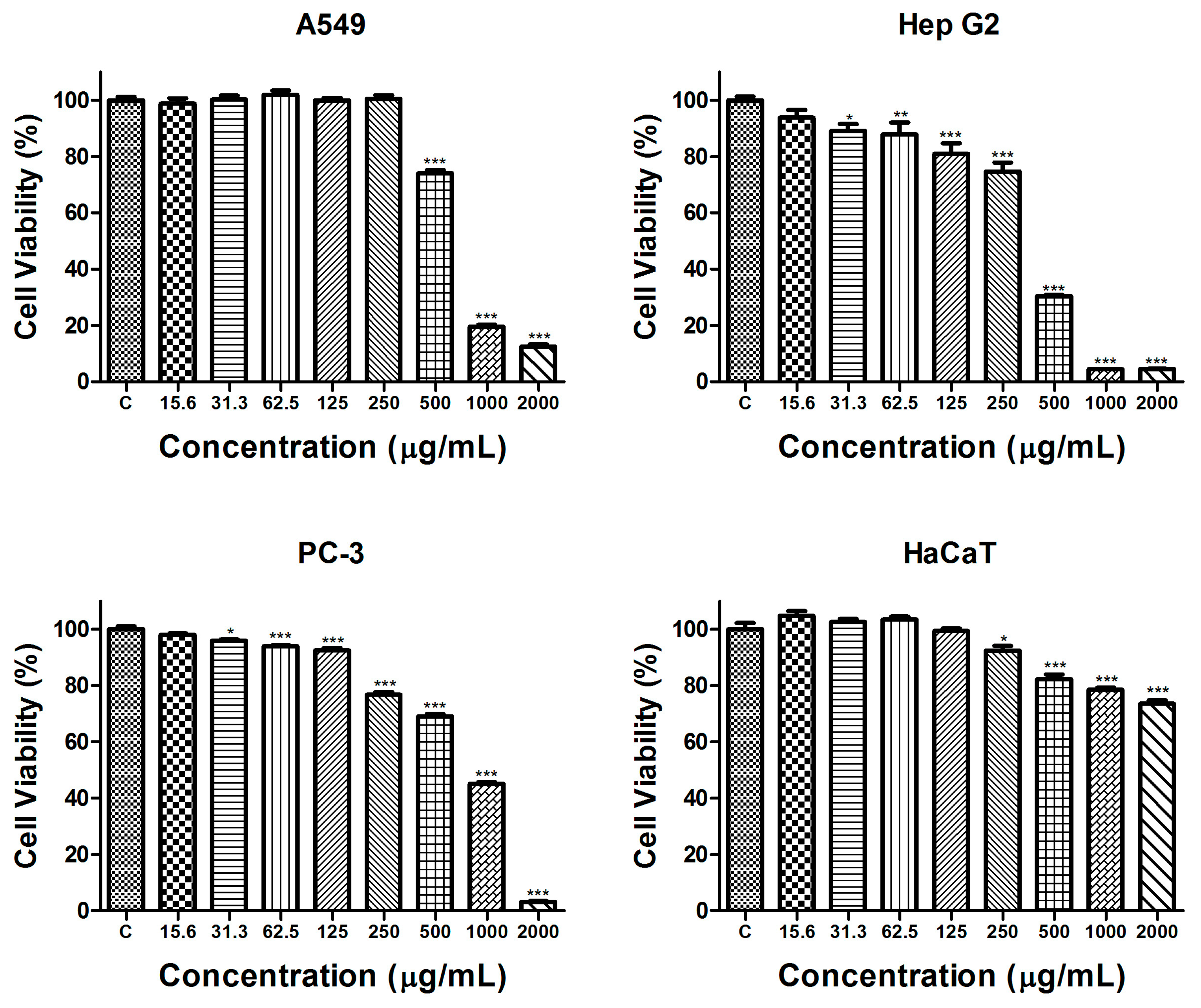
2.2.1. Cytotoxicity Evaluation

The in vitro safety assessment of the S. aethiopis extract was conducted using the standard BALB/3T3 Neutral Red Uptake (NRU) cytotoxicity assay. The test evaluated the cell viability and potential cytotoxic effects of the extract. The results of the cytotoxicity evaluation are presented in Figure 1.
As evident from the presented data, the tested extract showed a very low cytotoxicity. At concentrations of 250 µg/mL, 500 µg/mL and 1000 µg/mL, cell viability was reduced by 10.30 ± 1.89%, 12.81 ± 1.597% and 22.16 ± 2.160%, respectively. Even at the highest tested concentration of 2000 µg/mL, cytotoxicity remained below 50%, demonstrating a relatively low toxic effect on BALB/3T3 cells.

2.2.2. Antiproliferative Activity

The potential of S. aethiopis extract to inhibit the proliferation of cancer cells was evaluated by the MTT (3-(4,5-dimethylthiazol-2-yl)-2,5-diphenyltetrazolium bromide) assay following 72 h of exposure. To assess the selectivity of its antiproliferative effect, the effect of the extract on cancer cells was compared to its effect on the human non-cancerous HaCaT cell line. The results of the assay are presented in Figure 2.
A concentration-dependent antiproliferative effect of the tested extract with varying degrees of sensitivity in different cell lines was detected. In lung carcinoma cells (A549), a statistically significant decrease in cell proliferation was found only at the highest concentrations of 500 µg/mL, 1000 µg/mL and 2000 µg/mL. In contrast, hepatocellular and prostate carcinoma cells showed greater sensitivity, with significant inhibition occurring at all concentrations above 30 µg/mL. In the non-cancerous HaCaT cell line, the effects of the extract on cell proliferation were significantly weaker. Even at the highest tested concentration, cell viability remained at 73.6 ± 2.87% compared to untreated control cells. To further evaluate the antiproliferative potential of the S. aethiopis extract in different cancer cell lines, the half-maximal inhibitory concentration (IC50) and selectivity index (SI), relative to the non-cancerous control cell line, were determined (Table 3).
The results indicate that the extract showed stronger effects and the highest selectivity in the hepatocellular carcinoma (Hep G2) cell line. Based on the MTT assay findings, Hep G2 was identified as the most suitable model for further studies on the mechanisms underlying the observed antiproliferative effects.

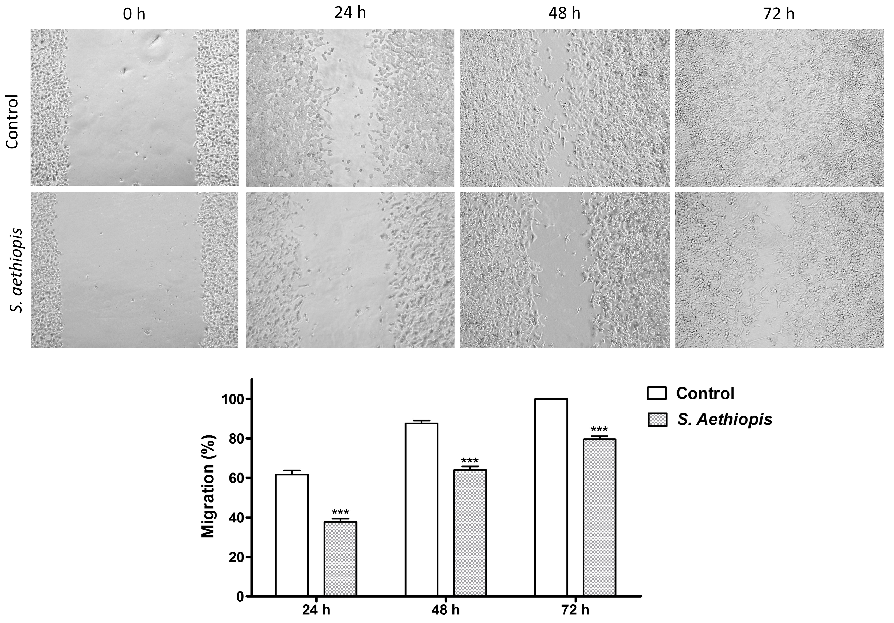
2.2.3. Migration Assay

The effect of S. aethiopis extract on cancer cell migration was evaluated using the wound-healing scratch assay (Figure 3). Cell migration was quantified by measuring the mean wound area at different time points and comparing it to the initial (0 h) scratch area in the corresponding cell culture.
In the control cell cultures, 61.72%, 87.54% and 100.00% of the wounded area was healed at 24 h, 48 h and 72 h, respectively. Treatment with S. aethiopis extract significantly inhibited the migration of Hep G2 carcinoma cells, reducing wound closure to 37.70%, 63.94% and 79.55% at the corresponding time points.

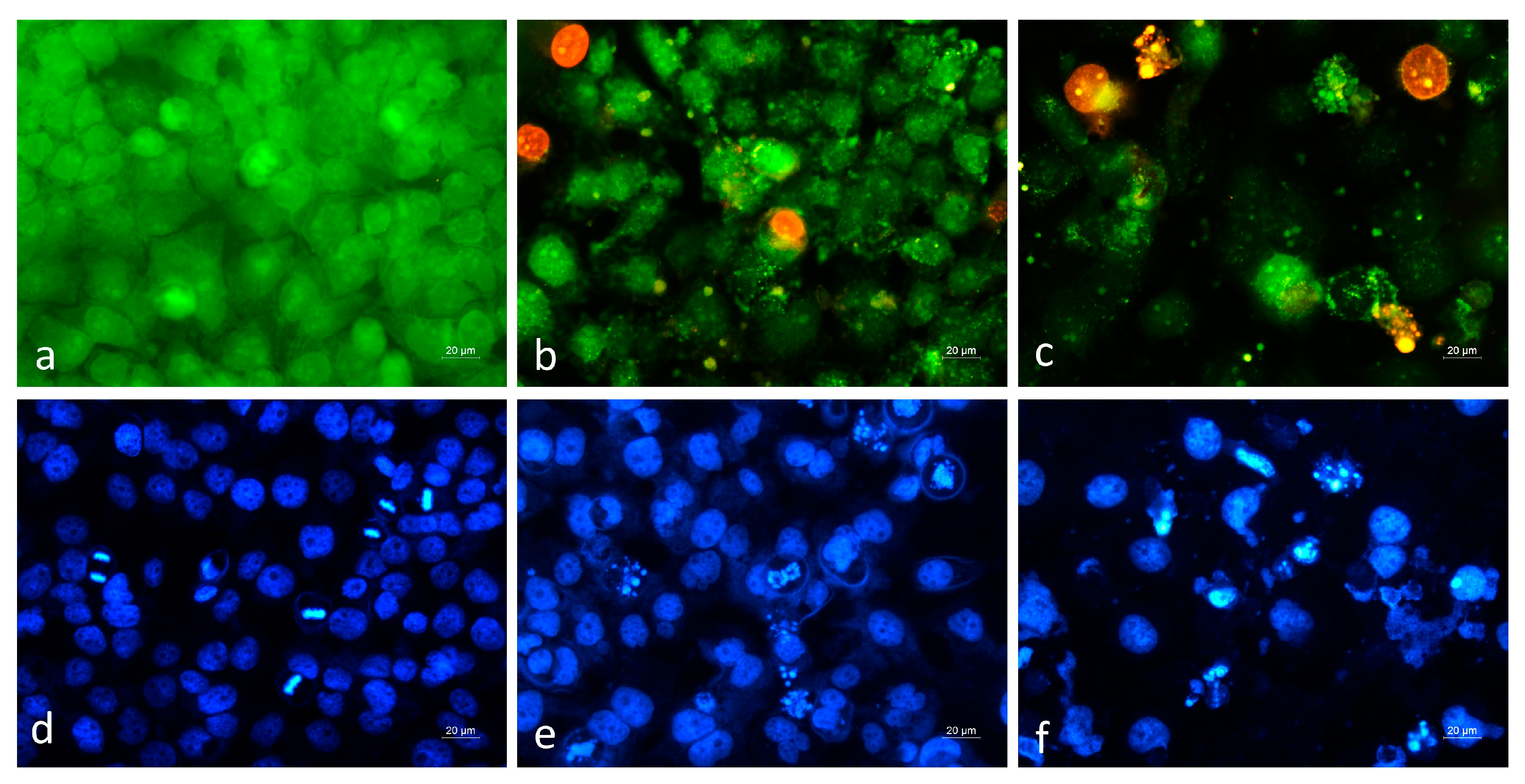
2.2.4. Cytopathological Analysis

Cytopathological changes in Hep G2 cancer cells following treatment with S. aethiopis extract were compared to those in cell cultures treated with the standard cytostatic agent, doxorubicin (Figure 4).
Fluorescence microscopy revealed that in the control untreated cell cultures stained with AO/EB, a monolayer of green-stained viable cells was observed, with numerous cells actively undergoing mitosis (Figure 4a). Treatment with S. aethiopis extract led to pronounced morphological alterations, including a reduction in monolayer confluency and the presence of early apoptotic, late apoptotic and necrotic cells (Figure 4b). Cells treated with the positive control, doxorubicin, showed similar morphological changes; however, a higher number of cells displayed typical apoptotic features, such as nuclear condensation and fragmentation (Figure 4c). Further confirmation of the ability of S. aethiopis extract to induce cancer cell death via apoptotic and necrotic pathways was provided by DAPI nuclear staining. Cells treated with S. aethiopis extract showed characteristic apoptotic nuclear alterations, though these changes were more frequent and pronounced in doxorubicin-treated cultures (Figure 4e,f).

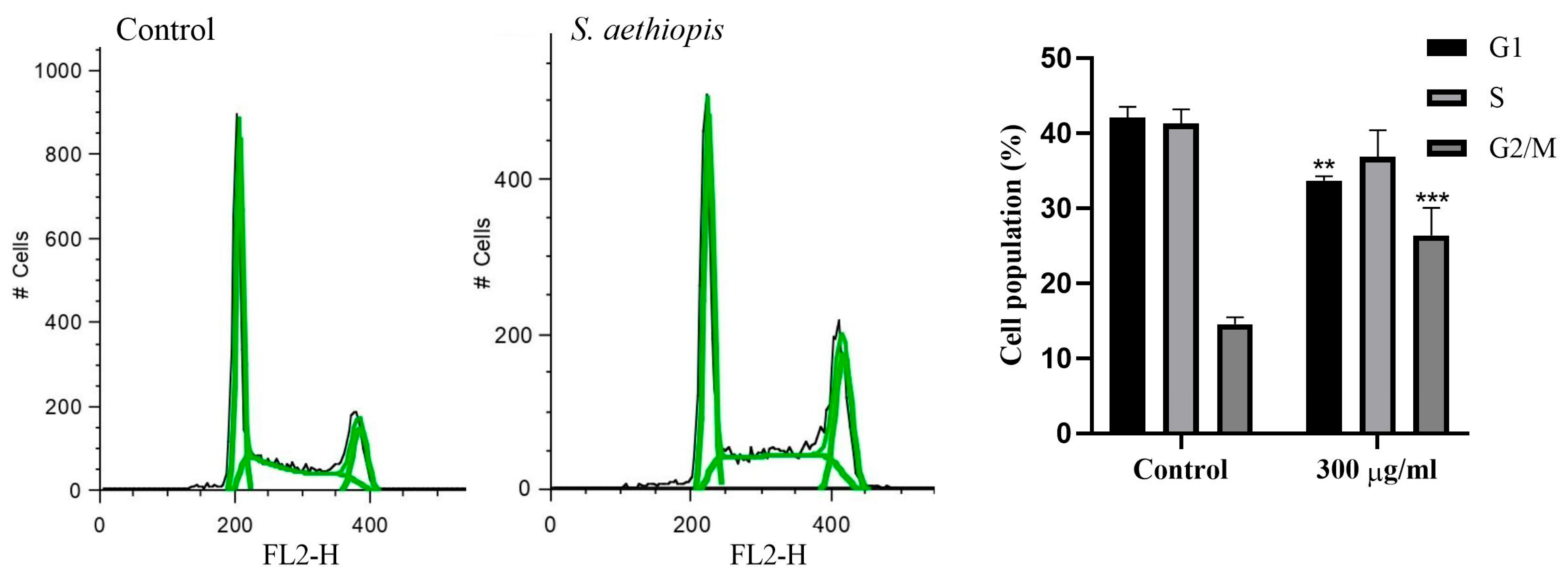
2.2.5. Cell Cycle Analysis

The effect of S. aethiopis extract on cell cycle progression was assessed by a fluorescence-activated cell sorting (FACS) analysis. Propidium iodide (PI)-stained control and extract-treated Hep G2 cells were examined for changes in cell cycle distribution (Figure 5). The cells were analyzed for distribution in the individual phases of the cell cycle by assessing FL2 peak heights (FL2-H, Fluorescence Channel 2—Height).
As evident from the data presented in Figure 5, exposure to S. aethiopis extract led to significant changes in cell cycle phase distribution. Compared to untreated control cells, the extract caused a decrease in G1- and S-phase cell populations, while the G2/M-phase population showed a statistically significant increase.

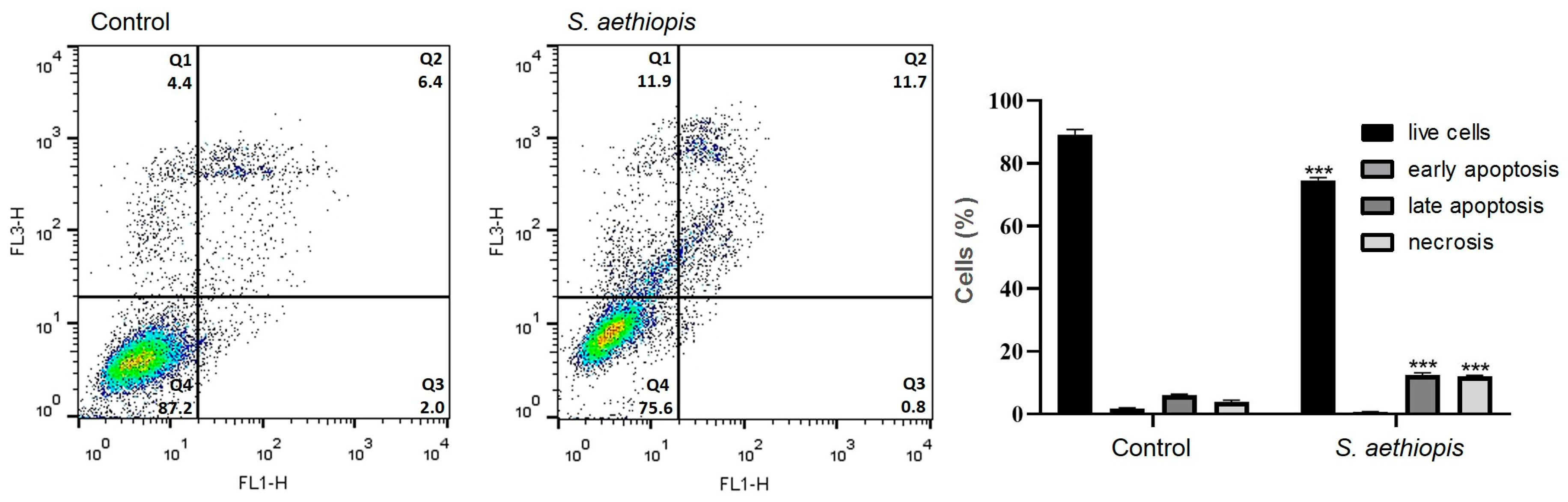
2.2.6. Apoptosis Assay

A flow cytometry analysis of Annexin V-FITC/PI-stained Hep G2 cells was performed to quantify the live, apoptotic and necrotic cell populations in untreated and extract-treated cell cultures (Figure 6).
The results of the Annexin V-FITC/PI FACS analysis were consistent with the data obtained from fluorescent microscopy and showed a statistically significant increase in both late apoptotic and necrotic cell populations following treatment with S. aethiopis extract.

3. Discussion

In the present research, the anticancer potential of water extracts from cultivated S. aethiopis plants on lung carcinoma A549, prostate adenocarcinoma PC-3 and hepatocellular carcinoma Hep G2 cell lines, used as in vitro models for some of the most common cancer types in humans, was investigated. The chemical composition and antioxidant properties of the S. aethiopis extract were also analyzed.
In a first step, the safety of the S. aethiopis extract was evaluated using the standard BALB/3T3 Neutral Red Uptake (NRU) cytotoxicity test. This test is a validated in vitro method for the assessment of basal cytotoxicity, with high predictive value for human acute toxicity. The assay measures cell viability by assessing the retention of a neutral red dye in lysosomes, distinguishing viable from non-viable cells. Neutral red (3-amino-7-dimethylamino-2-methylphenazine hydrochloride) is a weak cationic dye that readily diffuses through the cell membrane and accumulates in the lysosomes of viable cells, whereas it is excluded from dead cells. The NRU test results indicated the very low cytotoxicity of the extract, supporting its potential for therapeutic uses.
Next, the antiproliferative potential of the S. aethiopis extract was assessed on human A549 (lung carcinoma), PC-3 (prostate adenocarcinoma) and Hep G2 (hepatocellular carcinoma) cancer cells using the MTT assay. To evaluate the impact of the S. aethiopis extract on cell proliferation, a low seeding density (1000 cells/well) and long exposure time (72 h) were used. The non-cancer human cell line HaCaT, treated under the same conditions, served as a control to assess selectivity. The results showed that the tested extract inhibited the proliferation of all cell lines in a concentration-dependent manner. Among the cancer cell lines, Hep G2 exhibited the highest sensitivity to the extract, followed by A549 and PC-3, whereas HaCaT cells were the least affected, showing once again the safety of the extract on normal cells, a significant advantage when developing new anticancer treatments. Selectivity was quantified using the selectivity index (SI), calculated as the IC50 ratio of cancer versus normal cells. The SI is an important parameter for evaluating the anticancer effect of a compound, as it indicates the compound’s ability to preferentially target cancer cells while sparing normal, healthy cells. The obtained data indicated that the S. aethiopis extract demonstrated high selectivity towards the Hep G2 cells and moderate selectivity against the A549 and PC-3 cells.
Existing literature on the anticancer activity of S. aethiopis L. is scarce and limited to extracts obtained with organic solvents [10,19]. Additionally, no studies have investigated the anticancer potential of in vitro cultivated S. aethiopis, nor has its activity been evaluated against liver, lung and prostate cancer. Thus, Firuzi et al. [19] studied the cytotoxicity of methanol and dichloromethane extracts of eleven Salvia species, including S. aethiopis, against HL60 (human acute promyelocytic leukemia cells), K562 (human chronic myelogenous leukemia cells) and MCF-7 (human breast adenocarcinoma) and reported the significant cytotoxic activity of S. aethiopis against all the tested cell lines. Luca et al. [10] also conducted a study on the potential activity of S. aethiopis against breast cancer. They investigated the cytotoxic effects of ethanol extracts from S. aethiopis on human breast carcinoma MCF-7 and MDA-MB-231 (triple-negative breast cancer), but their findings indicated no significant reduction in the cell viability over the concentration range tested (25–100 μg/mL).
To our knowledge, the present study is the first to investigate the anticancer effects of water extracts from in vitro cultivated S. aethiopis on liver, lung and prostate cancer, demonstrating significant cytotoxic activity against all three cancer types, with the strongest impact observed on Hep G2 liver cancer cells.
To elucidate the mechanisms underlying the antiproliferative activity of S. aethiopis extracts, cell migration, cytomorphological and flow cytometric analyses were performed using Hep G2 cells, selected for their high responsiveness. The wound-healing (scratch) assay demonstrated the ability of the tested extract to inhibit cancer cell migration, consistent with previous studies reporting that different extracts from Salvia spp. and their bioactive compounds suppress cancer cell migration and metastasis in in vitro and in vivo cancer models [33,34,35]. In addition to its inhibitory effect on cancer cell migration, the S. aethiopis water extract was also found to suppress cancer cell proliferation by inducing apoptosis, necrosis and cell cycle arrest. The extract’s ability to disrupt cell cycle progression, evidenced by G2/M-phase arrest, suggests interference with key regulatory pathways involved in cancer cell proliferation. Similar findings, characterized by a reduction in the number of G0/G1-phase cells and the accumulation of cells in G2/M, have been reported for SW480 and HT-29 colon cancer cells treated with Salvia rosmarinus extracts [26,36], suggesting common bioactive compounds and anticancer mechanisms in the two Salvia species. The fluorescent microscopy of AO/EB- and DAPI-stained cancer cells revealed distinct apoptotic and necrotic morphological changes following S. aethiopis extract treatment. These effects were further confirmed and quantified by the fluorescence-activated cell sorting analysis of Annexin V-FITC/PI-stained Hep G2 cells, which demonstrated a significant increase in both late apoptotic and necrotic cell populations. Taken together with previous reports, the present findings indicate that Salvia spp. extracts trigger cancer cell death through different mechanisms, including both necrosis and apoptosis [34,35,36,37,38].
The strong antiproliferative and antimetastatic activity of S. aethiopis may be attributed to its rich phenolic content and convincing antioxidant properties. Polyphenols, particularly flavonoids, are well known for their antioxidant effects and potential role in cancer prevention and treatment [39,40].
The current analysis revealed a total polyphenol content of 110.03 ± 0.7 mg/g in the freeze-dried extract, comparable to values reported by other studies, including Tosun et al. [12] (82.1 mg GAE/g); Luca et al. [10] (81.43 mg/g); Hanganu et al. [41] (22.25 ± 0.24 mg/g); Dogan [42] (41.50 mg GAE/g); Caylak [43] (90.75 ± 0.35 mg GAE/g), and Firuzi et al. [19] (14.13 ± 0.90 mg GAE/g dried plant). Additionally, Poyraz et al. [3] reported polyphenol levels of 94.36 mg GAE/g in methanol extracts and 290.62 mg GAE/g in ethyl acetate extracts of S. aethiopis. However, it is important to note that, to our knowledge, only two studies examine the polyphenol and flavonoid content in extracts from cultivated S. aethiopis plants [10,12], and no reports exist for in vitro obtained and further cultivated plants. Regarding the antioxidant activity, the current study employed ORAC and HORAC assays, whereas most available literature reports are based on DPPH, FRAP and ABTS assays, making direct comparisons challenging. Reported values include DPPH: 158.76 ± 0.82 IC50 (μg/mL) and FRAP: 1399 ± 5.01 (mmol Trolox/mg d.w.) [41]; DPPH: 42.00 ± 0.10 μg TE/mL, ABTS: 17.00 ± 0.10 EC50 (μg/mL), FRAP: 36.94 ± 0.18 μmol TE/100 mL [10]; DPPH: 26.54 ± 3.8%, FRAP: 28.78 ± 5.2 mg-Trolox/g [43].
Looking deeper into the chemical composition of the water extracts of S. aethiopis, attempting to more precisely define its antitumor components, rosmarinic acid appears to be the major dominant compound. Other prevailing compounds are salvianic acid C, gluconic acid, malic acid, citric acid and isopropylmalic acid, as well as salvianic acid A ((R)-3-(3, 4-Dihydroxyphenyl)-2-hydroxypropanoic acid, danshensu) and a compound identified as salvianolic acid K. Salvianolic acids and salvianic acid A are primary active constituents of the traditional Chinese medicinal herb, Salvia miltiorrhiza [44,45]—in Chinese, dānshēn, where the alternative name of salvianic acid A, danshensu, comes from.
Consistent with the findings presented in the current work, other studies have also identified rosmarinic acid, salvianolic acids and malic acid in S. aethiopis extracts [9,10,46].
Rosmarinic acid (α-o-caffeoyl-3,4-dihydroxy phenyl lactic acid), originally isolated from the herb rosemary (R. officinalis L., also known as S. rosmarinus) in 1958, and nowadays reported to be present in 39 plant taxa, including S. aethiopis [9,10,46], has been extensively studied and validated for its anticancer properties, demonstrating the ability to selectively induce cancer cell apoptosis, suppress tumor growth and inhibit cancer cell proliferation and migration [9,47,48,49]. Breast cancer, ovarian cancer, colorectal cancer and skin cancer, including melanoma and leukemia, are just a few of the cancer types affected [47,49]. Rosmarinic acid has been shown to hold promise for the treatment of lung and hepatocellular cancer as well. Thus, phenolic acid has been demonstrated to have a 50% inhibitory effect on A549 cell proliferation, targeting cyclooxygenase-2, a well-known mediator in tumorigenesis [50]. Furthermore, it was shown that rosmarinic acid decreased the viability of human Hep G2 liver cancer cells in a dose-dependent manner while inducing cancer cell apoptosis, as demonstrated by AO/EB and DAPI staining of the cells and observed changes in the expression of the apoptosis-related proteins Bax and Bcl-2, complemented by the activation of caspase-3 and caspase-9, which prevented the migration and invasion of the Hep G2 cancer cells [51]. Another study further confirmed that rosmarinic acid decreased cell proliferation, inhibited migration and invasion and induced the apoptosis of Hep G2 cells [52]. The above findings are in line with the results presented in the current work, showing extracts of S. aethiopis with a high content of rosmarinic acid having detrimental effects on A549 and Hep G2 cancer cells, mediated in Hep G2 cells by cell apoptosis shown by AO/EB and DAPI staining experiments and Annexin-V/propidium iodide-based flow cytometry, accompanied by the decreased migration capacity of the cancer cells as demonstrated by the wound-healing (scratch) assay. Moreover, recent studies have revealed that, beyond its direct anticancer effects, rosmarinic acid is capable of reversing cancer resistance to first-line chemotherapeutics and has a protective effect against the toxicity caused by radiotherapy and chemotherapy, mostly due to its free radical-scavenging ability [53].
There are more than 10 different types of salvianolic acids, known as salvianolic acid A, salvianolic acid B, salvianolic acid C, etc., the most abundant of which are salvianolic acid A and salvianolic acid B. Danshensu and caffeic acid in different combinations are the main building blocks of the various salvianolic acids. Thus, salvianolic acid A is formed by one molecule of danshensu and two molecules of caffeic acid, while salvianolic acid B is formed by three molecules of danshensu and one molecule caffeic acid [54]. Similar to salvianolic acid A, salvianolic acid K also contains one moiety of danshensu and a dimer of caffeic acid, but the difference with salvianolic acid A lies in the specific configuration and the way the moieties are bonded within the salvianolic acid K molecule. [55]. While data on the anticancer activity of salvianolic acid K are missing, Silva et al. [56] report on Thymus zygis subsp. zygis, an endemic Portuguese plant, with the major phenolic compounds identified being rosmarinic acid and high amounts of salvianolic acid K and I, whose extracts exhibit significant scavenging activity for ABTS+, hydroxyl (•OH) and nitric oxide (NO) radicals and significant antiproliferative effects against the human colon adenocarcinoma cell line Caco-2, and the human hepatocellular carcinoma cell line Hep G2 used in the present research as well [56]. Data in the scientific literature on the strong antioxidant activity and anticancer effects of the similar-in-structure salvianolic acid A, as well as of salvianolic acid B, are abundant. Regarding the antioxidant activity of salvianolic acid A, it has been shown to not only sequester free radicals directly but to exert cytoprotective effects against oxidative stress by the activation of the Nrf2/HO-1 axis mediated by Akt/mTORC1 activation [57]. Regarding the anticancer activity of salvianolic acid A and salvianolic acid B, it has been shown against numerous types of cancer (amongst which are breast, ovarian and colorectal cancer, squamous cell carcinoma, melanoma and not least liver and lung cancer) and exerted by inducing apoptosis of cancer cells, influencing their cell cycle and inhibiting the proliferative, metastatic and invasive ability of cancer cells by targeting multiple signaling pathways [37,46], only a few of which being the enhancing of caspase-3 activity, downregulating Bcl-2 expression, upregulating Bax expression and disrupting the mitochondrial membrane potential in breast cancer cells [58], inhibiting the AKT/mTOR signaling in hepatocellular carcinoma cells salvianolic acid B [59]) and influencing the PTEN/PI3K/AKT signaling pathway in human lung cancer A549 cells [44]. Recently Tang et al. [60] have shown the anticancer activity of salvianolic acid A on a most aggressive, triple-negative subtype of breast cancer by influencing the polarization of tumor-associated macrophages identified as significant contributors to the growth and metastasis of the cancer. The cytotoxic effects of salvianolic acids on hepatocellular carcinoma and A549 lung cancer cells [44,59] align closely with the current research. Further advantages of salvianolic acids A and B, enhancing their anticancer capacity, are their potency in sensitizing cancer cells to chemotherapeutic agents [61,62] and their ability to inhibit epithelial–mesenchymal transition [44], a process when epithelial cells switch to the mesenchymal cell phenotype and gain migration and invasion capabilities, facilitating tumor progression [63].
Danshensu has also shown anticancer effects, including the suppression of epithelial–mesenchymal transition, the inhibition of tumor angiogenesis and invasion, and the ability to overcome or reduce chemoresistance, while enhancing radiosensitivity in cancer therapy [57,64,65]. Moreover, danshensu exhibits direct antiproliferative activity against A549 lung cancer cells [50] and exerts hepatoprotective effects by inhibiting Hep G2/2.2.15 cell growth, alleviating chronic alcoholic liver disease, counteracting hepatic fibrosis and reducing colon cancer-related liver metastasis [64,66,67,68].
Thus, we believe that the strong cytotoxic effect of the S. aethiopis extract against A549 and Hep G2 cancer cell lines was mostly due to rosmarinic acid, being the predominant compound in the extract, and to salvianolic acids and danshensu; all three constituents having demonstrated convincing cytotoxic effects against lung (A549) and hepatocellular cancer (Hep G2) cells, as well as protective effects on the liver tissue (danshensu). Of course, the combination with malic acid, citric acid and gluconic acid, with reported anticarcinogenic properties and activities [69,70,71], likely contributes to the cytotoxic efficiency of the S. aethiopis extract.
The results of the present study, limited to in vitro experiments, demonstrating selective anticancer effects on a panel of cell lines through pathological changes in the cancer cells and the inhibition of cancer cell migration, provide a strong foundation for future in vivo experiments using laboratory animals. These in vivo experiments would additionally allow to determine the optimal extract dose, to assess the extract’s bioavailability and pharmacokinetics, to further evaluate the extract’s safety including acute and chronic toxicity, to follow the long-term effects of the extract on tumor progression and recurrence and to explore the combination of the S. aethiopis extract with other natural or synthetic compounds to possibly enhance its anticancer activity, all in complex living systems, providing a more comprehensive understanding of the therapeutic potential of S. aethiopis and paving the way for its development as an effective anticancer agent.

4. Materials and Methods

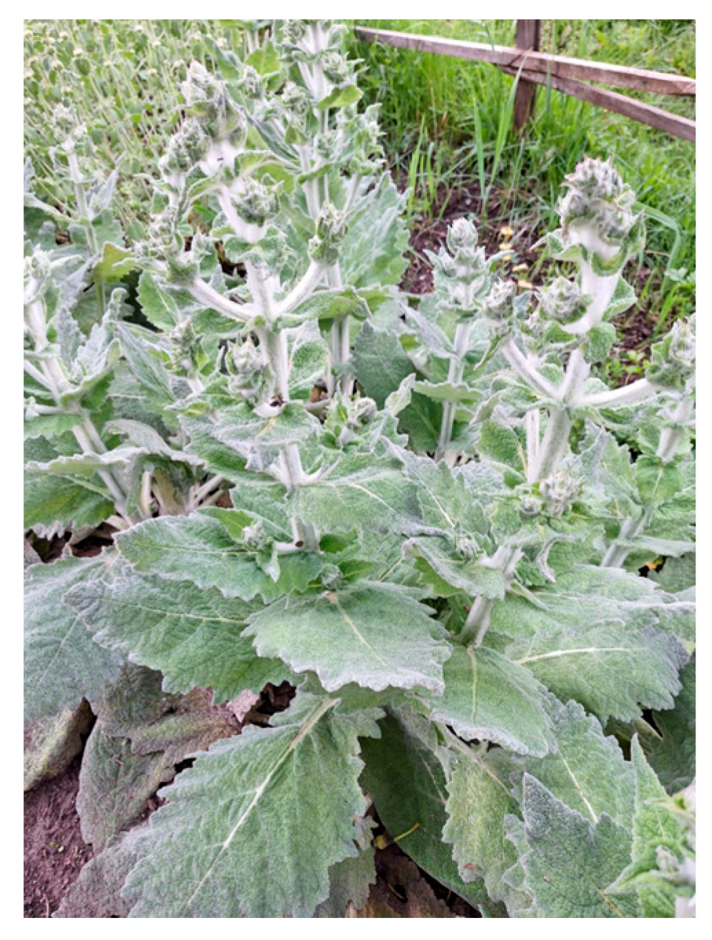
4.1. Plant Materials

Salvia aethiopis plants were obtained by an in vitro micropropagation technique and cultivated in an open field in Elin Pelin, Sofia region, at an altitude of 618 m above sea level (Figure 7). The aerial parts of the plants (flowers, leaves and stems) were harvested at the end of the vegetation period (June–July, 2023). The collected plant material was dried at room temperature and subsequently used for extract preparation and phytochemical and antitumor analysis.

4.2. Phytochemical Analysis

4.2.1. Extraction and Freeze-Dried Procedure

Plant material was first dried and ground to a fine powder using a laboratory mill. For each extraction, 5 g of the powder was steeped in 200 mL of hot water (90 °C) for 15 min. The mixture was then centrifuged at 6000× g with an MPW-260R centrifuge (MPW Med. Instruments, Warszawa, Poland), and the supernatants were carefully collected and freeze-dried for 96 h in an Alpha 1–4 LD plus laboratory freeze-drier (manufactured by Martin Christ Gefriertrocknungsanlagen GmbH, Osterode am Harz, Germany). The resulting freeze-dried extracts were subsequently used to evaluate potential antitumor activity.

4.2.2. Quantification of Total Polyphenols and Flavonoid Content

For total polyphenol quantification, the method developed by Singleton and Rossi [72] was employed. Gallic acid was used to establish a standard calibration and the results presented as mg gallic acid equivalents (GAEs) per g dry weight (DW) ± SD.
The total flavonoid content was measured using aluminum chloride (AlCl3) as a reagent, following the protocol by Chang et al. [73]. Quercetin dehydrate served as the calibration standard, and the results were expressed as mg quercetin equivalents (QEs) per g DW ± SD.

4.2.3. Evaluation of Antioxidant Activity

The antioxidant potential was assessed by the measurement of oxygen radical absorbance capacity (ORAC) using a microplate reader (FLUOstar OPTIMA; BMG Labtech, Ortenberg, Germany), following the method described by Ou et al. [74], with modifications by Denev et al. [75]. The results are presented as micromole Trolox equivalents (μmol TEs) per gram DW ± SD.
The hydroxyl radical averting capacity (HORAC) of the freeze-dried extract was evaluated based on the protocol of Ou et al. [76]. The results are expressed as micromole gallic acid equivalents (μmol GAEs) per gram DW ± SD.

4.2.4. Liquid Chromatography–High Resolution Mass Spectrometry Analysis (LC-HRMS)

Sample Preparation

Each sample (10.0 mg accurately weighted) was mixed with 1.0 mL of 1% formic acid. The solution was extracted with 200 µL of dichloroethane, vortexing it for 5 min, followed by centrifugation at 13,000 rpm for 5 min. Then, 10 µL of the upper phase was used for analysis.

LC-HRMS Analysis

Analyses were performed using an Orbitrap IQ-X® hybrid mass spectrometer (ThermoScientific Co., Waltham, MA, USA) coupled with a heated electrospray ionization (HESI) module and Vanquish® Ultra High-Performance Liquid Chromatography (UHPLC) system (ThermoScientific Co., Waltham, MA, USA).
Chromatographic conditions. Separations was carried out on a Nucleodur C18 Isis analytical column (100 × 2.1 mm, 3 µm; Macherey-Nagel, Düren, Germany) under gradient elution at a flow rate of 300 µL/min. The mobile phases were as follows: A, 0.1% formic acid in water and B 0.1% formic acid in acetonitrile and with the gradient program 0% B for 2 min, 0–40% B for 19 min, 40–95% B for 2 min, 95% B for 2 min, 95–0% B for 2 min and 0% B for 3 min.
Mass spectrometry conditions. Full-scan mass spectra were recorded in negative ion mode across the m/z range of 120–1800 at a resolution of 120,000. Data-dependent reaction monitoring (ddMS2) was performed at a resolution of 17,500 with a 2 amu precursor isolation window. The negative ionization parameters were as follows: spray voltage—3.5 kV; capillary temperature,320 °C; probe heater temperature, 300 °C; sheath gas flow, 20 units; auxiliary gas, 12 units; sweep gas, 3 units (arbitrary values set by IQ-X Tune Software version 3.3 SP3); and S-Lens RF level, 50.00. Nitrogen served as the nebulization and collision gas in the HCD cell. The normalized collision energy was set between 20–35%. Metabolites were quantified using 5 ppm mass tolerance filters against the theoretical m/z values. Data were acquired and processed using XCalibur® software (Software version 4.1, ThermoScientific Co., Waltham, MA, USA).

4.3. Antitumor Activity

4.3.1. Cell Lines

The following permanent cell lines were obtained from the American Type Culture Collection (ATCC, Rockville, MD, USA): murine embryonic fibroblasts (BALB/3T3, CCL-163), human lung carcinoma (A549, CRM-CCL-185), human prostate adenocarcinoma (PC-3, CRL-1435) and human hepatocellular carcinoma (Hep G2, HB-8065). The human keratinocyte cell line HaCaT (CVCL_0038) was acquired from the CLS Cell Lines Service (Cytion, Eppelheim, Germany) and employed as a non-tumorigenic control to assess the selectivity of the anticancer effects. All cell lines were cultured in Dulbecco Modified Eagle Medium (DMEM), supplemented with 10% fetal bovine serum, 100 U/mL penicillin and 100 μg/mL streptomycin. Cultures were kept at 37 °C in a humidified atmosphere containing 5% CO2 (HEPA Class 100, Thermo Scientific, Waltham, MA, USA). Upon reaching confluence, cell monolayers were detached using a 0.05% trypsin–0.02% ethylendiaminotetraacetic acid (EDTA) solution, and cells in the exponential growth phase were used for subsequent experiments.

4.3.2. BALB/3T3 Neutral Red Uptake Assay

The cytotoxicity of the S. aethiopis extract was evaluated by the BALB/3T3 Neutral Red Uptake assay. BALB/3T3 cells were seeded in 96-well flat-bottomed plates at a density of approximately 1 × 10⁵ cells/mL in DMEM supplemented with 10% FBS (100 µL/well) and incubated overnight at 37 °C with 5% CO2. The cells were then exposed to different concentrations of the extract (ranging from 15.6 to 2000 μg/mL) and incubated for 24 h, 48 h and 72 h. Following treatment, the wells were washed twice with PBS, and 100 µL of DMEM containing neutral red dye (50 μg/mL) was added to each well. After 2 h of incubation under standard culture conditions, the neutral red medium was removed, the wells were washed with PBS, and a desorbing solution of ethanol/water/acetic acid (50:49:1) was added. The plates were incubated for 10 min, and absorbance was measured by a TECAN microplate reader (SunriseTM, Grödig/Salzburg, Austria) at 540 nm.

4.3.3. Assessment of Antiproliferative Activity

The antiproliferative activity of the S. aethiopis extract on cancer cell lines was evaluated using the standard MTT colorimetric assay. Non-tumorigenic HaCaT cells were used as a reference to determine the selectivity of the extract toward cancer cells. Cells were detached with 0.25% Trypsin–EDTA, counted in a hemocytometer and diluted to a concentration of 1 × 10⁴ cells/mL before being plated in 96-well plates. The cells were incubated overnight at 37 °C in a humidified atmosphere containing 5% CO2. Following overnight incubation at 37 °C in a humidified 5% CO2 atmosphere, the cells were treated with S. aethiopis extract at concentrations ranging from 15.6 to 1000 μg/mL, with untreated cells as controls. Each concentration was tested in quintuplicate. After 72 h exposure, the cells were rinsed with PBS, followed by the addition of 100 μL of MTT solution (5 mg/mL in DMEM) to each well. After a further 3 h incubation at 37 °C, the supernatants were removed and replaced with 100 μL of DMSO/ethanol solution (1:1 ratio) to solubilize the purple formazan crystals. Absorbance was measured at 570 nm using a TECAN SunriseTM ELISA plate reader (Grodig/Salzburg, Austria). Cell viability was expressed as a percentage relative to the untreated control.

4.3.4. Cytopathological Studies—Live/Dead Staining with Acridine Orange and Ethidium Bromide

To evaluate cytopathological changes in Hep G2 hepatocellular carcinoma cells treatment with S. aethiopis extract, dual fluorescent live/dead staining with acridine orange (AO) and ethidium bromide (EtBr) was performed. Cells were plated on sterile glass coverslips in 24-well plates at a density of 1 × 10⁵ cells per well and allowed to adhere overnight. The next day, cells were exposed to the extract at its IC50 concentration for 72 h. The control groups included untreated HepG2 cells (negative control) and doxorubicin-treated cells (a positive control) cultured under the same conditions. Post-treatment, the coverslips were removed, gently rinsed with phosphate-buffered saline (PBS) and stained with a mixture of AO (10 μg/mL in PBS) and EtBr (10 μg/mL each in PBS). The stained cells were then mounted on glass slides and immediately visualized under a Leica DM fluorescence microscope (5000B, Wetzlar, Germany) before fluorescence fading could occur.

4.3.5. Nuclear Morphology Analysis by DAPI Staining

The nuclear morphology of Hep G2 cells treated with S. aethiopis extract was investigated by staining them with the DNA-binding fluorescent dye 4′,6-diamidino-2-phenylindole (DAPI). Hep G2 cells were cultivated and treated as described in the previous section. Following incubation, the glass coverslips were washed twice with phosphate-buffered saline (PBS) to remove detached cells. The cells were then fixed with ice-cold methanol, and DAPI staining was performed according to the manufacturer’s protocol. Samples of treated and untreated (control) cells were mounted with Mowiol on microscope slides and stored in the dark until examination with a Leica fluorescence microscope (DM 5000B, Wetzlar, Germany).

4.3.6. Flow Cytometric Analysis

The cell cycle progression and apoptosis of Hep G2 hepatocellular carcinoma cells following treatment with S. aethiopis extract were evaluated by flow cytometry. Cells were seeded in 6-well plates, incubated for 24 h and then treated with S. aethiopis extract at its IC50 concentration for 72 h. Untreated Hep G2 cells served as controls. Following the treatment period, cells were harvested using Trypsin–EDTA solution (Sigma-Aldrich, Burlington, MA, USA) and washed twice with cold PBS by centrifugation (1000 rpm, 10 min), and the resulting cell pellets were collected for the s cell cycle and apoptosis analyses, as detailed in subsequent sections.

4.3.7. Cell Cycle Assay

Hep G2 cells, untreated controls and those treated with S. aethiopis extract, were washed with PBS and fixed with 70% ice-cold ethanol, added dropwise under continuous vortexing. The cells were then stored overnight at −20 °C. The next day, the fixed cells were washed with PBS and incubated with RNase A (20 µg/mL; Roche Diagnostics GmbH, Mannheim, Germany) for 30 min. Subsequently, the cells were stained with PI (20 µg/mL) and analyzed by flow cytometry (Becton Dickinson, BD Biosciences, San Jose, CA, USA) to assess the distribution of cell cycle phases. The proportions of cells in G1, S and G2-M were determined with FlowJo™ v10.8 software (BD Biosciences, San Jose, CA, USA). Data are expressed as the mean ± standard error of the mean from three independent replicates.

4.3.8. Apoptosis Detection Assay

Apoptosis was assessed using the Annexin V-FITC Apoptosis Detection Kit (sc-4252 AK, Santa Cruz Biotechnology, Inc., Dallas, TX, USA) following the manufacturer’s protocol. Control and treated cells were stained and incubated for 15 min at room temperature, then analyzed by a flow cytometer (BD FACSCalibur™). The proportions of viable, early apoptotic, late apoptotic and necrotic cells were quantified with FlowJo software (BD Biosciences, San Jose, CA, USA).

4.4. Statistical Analysis

Data are expressed as the mean ± standard error of the mean (SEM). Statistical analyses were conducted using GraphPad Prism 9.0 software. A one-way analysis (ANOVA) was applied, with the Shapiro–Wilks test to assess data normality, followed by Tukey’s post hoc test for multiple comparisons. A p-value of less than 0.05 (p < 0.05) was considered statistically significant.

5. Conclusions

This study demonstrates that aqueous extracts from cultivated Salvia aethiopis possess potent anticancer properties against hepatocellular, liver and prostate carcinoma, with the most pronounced effects against liver cancer. The extract selectively inhibits hepatocellular carcinoma cell proliferation by induced apoptosis, necrosis and altered cell cycle progression, while maintaining a low toxicity to non-cancerous cells. The anticancer activity is underlined by a high content of phenolics in the S. aethiopis extracts, including rosmarinic acid and salvianolic acids, as well as salvianolic acid K and danshensu, known for their strong anticancer activity. These findings underscore the potential of S. aethiopis as a valuable source of bioactive compounds for therapeutic applications, warranting further investigation into its mechanism of action and in vivo efficacy.

Supplementary Materials

The following supporting information can be downloaded at: https://www.mdpi.com/article/10.3390/molecules30071427/s1, Figure S1. UPLC-DAD chromatogram at 280 nm of extract of Salvia aethiopis; Figure S2. The total ion chromatogram (TIC) of the extract of Salvia aethiopis; Figure S3. MS/MS spectrum of compound 1 (gluconic acid) using ESI in negative ionization mode; Figure S4. MS/MS spectrum of compound 2 (disaccharide) using ESI in negative ionization mode; Figure S5. MS/MS spectrum of compound 3 (malic acid) using ESI in negative ionization mode; Figure S6. MS/MS spectrum of compound 4 (citric acid) using ESI in negative ionization mode; Figure S7. MS/MS spectrum of compound 5 (danshensu) using ESI in negative ionization mode; Figure S8. MS/MS spectrum of compound 6 (protocatechuic acid hexoside) using ESI in negative ionization mode; Figure S9. MS/MS spectrum of compound 7 (isopropylmalic acid) using ESI in negative ionization mode; Figure S10. MS/MS spectrum of compound 8 (2-(3-hydroxy-4-methoxyphenyl)ethyl-O-(rhamnosyl)glucopyranoside) using ESI in negative ionization mode; Figure S11. MS/MS spectrum of compound 9 (salvianic acid C) using ESI in negative ionization mode; Figure S12. MS/MS spectrum of compound 11 (ester of salvianic acid C and 2,3,4,5-tetrahydroxyhexanedioic acid) using ESI in negative ionization mode; Figure S13. MS/MS spectrum of compound 11 (O-(O-glucuronyl-O-glucuronide)methoxylated flavonoid) using ESI in negative ionization mode; Figure S14. MS/MS spectrum of compound 12 (rosmarinic acid) using ESI in negative ionization mode; Figure S15. MS/MS spectrum of compound 13 (methoxylated flavonoid-O-glucuronide) using ESI in negative ionization mode; Figure S16. MS/MS spectrum of compound 14 (unknown) using ESI in negative ionization mode; Figure S17. MS/MS spectrum of compound 15 (unknown) using ESI in negative ionization mode; Figure S18. MS/MS spectrum of compound 16 (O-(O-caffeoyl-O-glucuronyl-O-glucuronide)methoxylated flavonoid) using ESI in negative ionization mode; Figure S19. MS/MS spectrum of compound 17 (salvianolic acid K) using ESI in negative ionization mode; Figure S20. MS/MS spectrum of compound 18 (O-(O-sinapoyl-O-glucuronyl-O-glucuronide)methoxylated flavonoid) using ESI in negative ionization mode; Figure S21. MS/MS spectrum of compound 19 (O-(O-feruloyl-O-glucuronyl-O-glucuronide)methoxylated flavonoid) using ESI in negative ionization mode; Figure S22. MS/MS spectrum of compound 20 (dihydroxy-oxooctadecenoic acid) using ESI in negative ionization mode; Figure S23. MS/MS spectrum of compound 21 (2-(3,4-dihydroxyphenyl)ethenyl 3-(3,4-dihydroxyphenyl)prop-2-enoate) using ESI in negative ionization mode; Figure S24. Molecular structure of several compounds of the extract of Salvia aethiopis.

Author Contributions

Conceptualization, A.G. and K.T.; methodology, K.T.; software, A.G. and I.S.; validation, A.G., I.S. and M.L.; formal analysis, I.S., P.D., V.D., A.V., I.I. and V.L.; investigation, A.G., I.S., V.D., K.T., M.L. and A.V., data curation, A.G., P.P.-K. and K.T.; writing—original draft preparation, P.P.-K., A.G. and K.T.; writing—review and editing, P.P.-K., V.V. and P.D.; visualization, A.G.; supervision, V.V. and K.T.; project administration, K.T. and V.V.; funding acquisition, K.T. All authors have read and agreed to the published version of the manuscript.

Funding

This research was funded by the National Science Fund—Bulgaria, grant number KP-06-N56/16.

Institutional Review Board Statement

Not applicable.

Informed Consent Statement

Not applicable.

Data Availability Statement

All data are contained in the manuscript.

Acknowledgments

The authors are grateful to Maria Petrova, Lyudmila Dimitrova and Margarita Dimitrova (Institute of Plant Physiology and Genetics, Bulgarian Academy of Sciences) for their invaluable assistance in obtaining the in vitro micropropagated plants and in their cultivation.

Conflicts of Interest

The authors declare no conflicts of interest.

References

  1. Mierziak, J.; Kostyn, K.; Kulma, A. Flavonoids as important molecules of plant interactions with the environment. Molecules 2014, 19, 16240–16265. [Google Scholar] [CrossRef] [PubMed]
  2. Shen, N.; Wang, T.; Gan, Q.; Liu, S.; Wang, L.; Jin, B. Plant flavonoids: Classification, distribution, biosynthesis, and antioxidant activity. Food Chem. 2022, 383, 132531. [Google Scholar]
  3. Poyraz, İ.E.; Çiftçi, G.A.; Öztürk, N. Phenolic contents, in vitro antioxidant and cytotoxicity activities of Salvia aethiopis L. and S. ceratophylla L. (Lamiaceae). Rec. Nat. Prod. 2017, 11, 345–355. [Google Scholar]
  4. Roy, A.; Khan, A.; Ahmad, I.; Alghamdi, S.; Rajab, B.S.; Babalghith, A.O.; Alshahrani, M.Y.; Islam, S.; Islam, M.R. Flavonoids a bioactive compound from medicinal plants and its therapeutic applications. Biomed. Res. Int. 2022, 2022, 5445291. [Google Scholar] [PubMed]
  5. Moshari Nasirkandi, A.; Iaccarino, N.; Romano, F.; Graziani, G.; Alirezalu, A.; Alipour, H.; Amato, J. Chemometrics based analysis of the phytochemical profile and antioxidant activity of Salvia species from Iran. Sci. Rep. 2024, 14, 17317. [Google Scholar]
  6. Boya, M.T.; Valverde, S. An orthoquinone isolated from Salvia aethiopis. Phytochemistry 1981, 20, 1367–1368. [Google Scholar]
  7. Rodríguez, B.; Fernandez-Gadea, F.; Savona, G. A rearranged abietane diterpenoid from the root of Salvia aethiopis. Phytochemistry 1984, 23, 1805–1806. [Google Scholar]
  8. Zhumaliyeva, G.; Zhussupova, A.; Zhusupova, G.E.; Błońska-Sikora, E.; Cerreto, A.; Omirbekova, N.; Zhunusbayeva, Z.; Gemejiyeva, N.; Ramazanova, M.; Wrzosek, M.; et al. Natural compounds of Salvia L. genus and molecular mechanism of their biological activity. Biomedicines 2023, 11, 3151. [Google Scholar] [CrossRef]
  9. Kostić, M.; Miladinović, B.; Milutinović, M.; Branković, S.; Živanović, S.; Zlatković, B.; Kitić, D. Rosmarinic and caffeic acid content and antioxidant potential of the Salvia aethiopis L. extracts. Acta Medica Median. 2017, 56, 121–128. [Google Scholar]
  10. Luca, S.V.; Skalicka-Woźniak, K.; Mihai, C.-T.; Gradinaru, A.C.; Mandici, A.; Ciocarlan, N.; Miron, A.; Aprotosoaie, A.C. Chemical profile and bioactivity evaluation of Salvia species from Eastern Europe. Antioxidants 2023, 12, 1514. [Google Scholar] [CrossRef]
  11. Veličković, D.T.; Ranđelović, N.V.; Ristić, M.S.; Šmelcerović, A.A.; Veličković, A.S. Chemical composition and antimicrobial action of the ethanol extracts of Salvia pratensis L. Salvia glutinosa L. and Salvia aethiopis L. J. Serb. Chem. Soc. 2002, 67, 639–646. [Google Scholar]
  12. Tosun, F.; Kizilay, C.A.; Sener, B.; Vural, M.; Palittapongarnpim, P. Antimycobacterial screening of some Turkish plants. J. Ethnopharmacol. 2004, 95, 273–275. [Google Scholar] [CrossRef] [PubMed]
  13. Hernández-Pérez, M.; Rabanal, R.M.; de la Torre, M.C.; Rodríguez, B. Analgesic, anti-inflammatory, antipyretic and haematological effects of aethiopinone, an o-naphthoquinone diterpenoid from Salvia aethiopis roots and two hemisynthetic derivatives. Planta Med. 1995, 61, 505–509. [Google Scholar] [CrossRef]
  14. Hernandez-Perez, M.; Rabanal, R.M.; Arias, A.; De La Torre, M.C.; Rodriguez, B. Aethiopinone, an antibacterial and cytotoxic agent from Salvia aethiopis roots. Pharm. Biol. 1999, 37, 17–21. [Google Scholar] [CrossRef]
  15. Naghibi, F.; Mosaddegh, M.; Mohammadi Motamed, M.; Ghorbani, A. Labiatae family in folk medicine in Iran: From ethnobotany to pharmacology. Iran. J. Pharm. Res. 2010, 4, 63–79. [Google Scholar]
  16. Şenol, F.S.; Orhan, I.; Celep, F.; Kahraman, A.; Doğan, M.; Yilmaz, G.; Şener, B. Survey of 55 Turkish Salvia taxa for their acetylcholinesterase inhibitory and antioxidant activities. Food Chem. 2010, 120, 34–43. [Google Scholar] [CrossRef]
  17. Wake, G.; Court, J.; Pickering, A.; Lewis, R.; Wilkins, R.; Perry, E. CNS acetylcholine receptor activity in European medicinal plants traditionally used to improve failing memory. J. Ethnopharmacol. 2000, 69, 105–114. [Google Scholar] [CrossRef] [PubMed]
  18. Vaccaro, M.C.; Alfieri, M.; Malafronte, N.; De Tommasi, N.; Leone, A. Increasing the synthesis of bioactive abietane diterpenes in Salvia sclarea hairy roots by elicited transcriptional reprogramming. Plant Cell Rep. 2017, 36, 375–386. [Google Scholar] [CrossRef]
  19. Firuzi, O.; Miri, R.; Asadollahi, M.; Eslami, S.; Jassbi, A.R. Cytotoxic, antioxidant and antimicrobial activities and phenolic contents of eleven Salvia species from Iran. Iran. J. Pharm. Res. 2013, 12, 801–810. [Google Scholar]
  20. Nwosu, N. Cancer: A Disease of Modern Times? Cureus 2024, 16, e74666. [Google Scholar] [CrossRef]
  21. Njoya, E.M. Chapter 31—Medicinal plants, antioxidant potential, and cancer. In Cancer, Oxidative Stress and Dietary Antioxidants, 2nd ed.; Preedy, V.R., Patel, V.B., Eds.; Elsevier Inc.: Amsterdam, The Netherlands, 2021; pp. 349–357. [Google Scholar]
  22. Sung, H.; Ferlay, J.; Siegel, R.L.; Laversanne, M.; Soerjomataram, I.; Jemal, A.; Bray, F. Global cancer statistics 2020: GLOBOCAN estimates of incidence and mortality worldwide for 36 cancers in 185 countries. CA Cancer J. Clin. 2021, 71, 209–249. [Google Scholar] [CrossRef] [PubMed]
  23. Reuter, S.; Gupta, S.C.; Chaturvedi, M.M.; Aggarwal, B.B. Oxidative stress, inflammation, and cancer: How are they linked? Free Radic. Biol. Med. 2010, 49, 1603–1616. [Google Scholar] [CrossRef]
  24. Sorriento, D. Oxidative stress and inflammation in cancer. Antioxidants 2024, 13, 1403. [Google Scholar] [CrossRef]
  25. Llorent-Martínez, E.J.; Nilofar; Sinan, K.I.; Darendelioglu, E.; Bahsi, M.; Polat, R.; Cakilcioglu, U.; Orlando, G.; Ferrante, C.; Zengin, G. Incorporating the HPLC-ESI-Q-TOF-MS profiles with the biochemical properties of eight Salvia species. eFood 2025, 6, e70021. [Google Scholar] [CrossRef]
  26. MassBank of North America (MoNA037822). Available online: https://mona.fiehnlab.ucdavis.edu/spectra/display/MoNA037822 (accessed on 14 February 2025).
  27. Zengin, G.; Llorent-Martínez, E.J.; Fernández-de Córdova, M.L.; Bahadori, M.B.; Mocan, A.; Locatelli, M.; Aktumsek, A. Chemical composition and biological activities of extracts from three Salvia species: S. blepharochlaena, S. euphratica var. leiocalycina, and S. verticillata subsp. amasiaca. Ind. Crops. Prod. 2018, 111, 11–21. [Google Scholar] [CrossRef]
  28. Wang, K.; Sun, Z.; Zhu, F.; Xu, Y.; Zhou, F. Development of a high-resolution mass-spectrometry-based method and software for human leukocyte antigen typing. Front. Immunol. 2023, 14, 1188381. [Google Scholar] [CrossRef]
  29. El-Gazar, A.A.; Emad, A.M.; Ragab, G.M.; Rasheed, D.M. Mentha pulegium L.(Pennyroyal, Lamiaceae) extracts impose abortion or fetal-mediated toxicity in pregnant rats; evidenced by the modulation of pregnancy hormones, MiR-520, MiR-146a, TIMP-1 and MMP-9 protein expressions, inflammatory state, certain related signaling pathways, and metabolite profiling via UPLC-ESI-TOF-MS. Toxins 2022, 14, 347. [Google Scholar] [CrossRef]
  30. MassBank of North America (MoNA035378). Available online: https://mona.fiehnlab.ucdavis.edu/spectra/display/MoNA035378 (accessed on 14 February 2025).
  31. Stochmal, A.; Simonet, A.M.; Macias, F.A.; Oleszek, W. Alfalfa (Medicago sativa L.) flavonoids. 2. Tricin and chrysoeriol glycosides from aerial parts. J. Agric. Food Chem. 2001, 49, 5310–5314. [Google Scholar] [CrossRef] [PubMed]
  32. Shirota, O.; Nagamatsu, K.; Sekita, S. Neo-clerodane diterpenes from the hallucinogenic sage Salvia divinorum. J. Nat. Prod. 2006, 69, 1782–1786. [Google Scholar] [CrossRef]
  33. Ezema, C.A.; Ezeorba, T.P.C.; Aguchem, R.N.; Okagu, I.U. Therapeutic benefits of Salvia species: A focus on cancer and viral infection. Heliyon 2022, 8, e08763. [Google Scholar] [CrossRef]
  34. Pérez-Sánchez, A.; Barrajón-Catalán, E.; Ruiz-Torres, V.; Agulló-Chazarra, L.; Herranz-López, M.; Valdés, A.; Cifuentes, A.; Micol, V. Rosemary (Rosmarinus officinalis) extract causes ROS-induced necrotic cell death and inhibits tumor growth in vivo. Sci. Rep. 2019, 9, 808. [Google Scholar] [CrossRef] [PubMed]
  35. Jaglanian, A.; Termini, D.; Tsiani, E. Rosemary (Rosmarinus officinalis L.) extract inhibits prostate cancer cell proliferation and survival by targeting Akt and mTOR. Biomed. Pharmacother. 2020, 131, 110717. [Google Scholar] [CrossRef] [PubMed]
  36. Shakeri, A.; Delavari, S.; Ebrahimi, S.N.; Asili, J.; Emami, S.A.; Tayarani-Najaran, Z. A new tricyclic abietane diterpenoid from Salvia chloroleuca and evaluation of cytotoxic and apoptotic activities. Rev. Bras. Farmacogn. 2019, 29, 30–35. [Google Scholar] [CrossRef]
  37. Shakeri, A.; Farahmand, S.S.; Tayarani-Najaran, Z.; Emami, S.A.; Kúsz, N.; Hohmann, J.; Boozari, M.; Tavallaie, F.Z.; Asili, J. 4,5-Seco-5,10-friedo-abietane-type diterpenoids with anticancer activity from Salvia atropatana Bunge. Naunyn Schmiedebergs Arch. Pharmacol. 2021, 394, 241–248. [Google Scholar] [CrossRef]
  38. Telang, N. Antiproliferative and pro apoptotic effects of rosemary and constituent terpenoids in a model for the HER 2 enriched molecular subtype of clinical breast cancer. Oncol. Lett. 2018, 16, 5489–5497. [Google Scholar]
  39. Roszkowski, S. Application of polyphenols and flavonoids in oncological therapy. Molecules 2023, 28, 4080. [Google Scholar] [CrossRef] [PubMed]
  40. de Luna, F.C.F.; Ferreira, W.A.S.; Casseb, S.M.M.; de Oliveira, E.H.C. Anticancer potential of flavonoids: An overview with an emphasis on tangeretin. Pharmaceuticals 2023, 16, 1229. [Google Scholar] [CrossRef]
  41. Hanganu, D.; Olah, N.K.; Pop, C.E.; Vlase, L.; Oniga, I.; Ciocarlan, N.; Matei, A.; Pu¸sca¸s, C.M.; Silaghi-Dumitrescu, R.; Benedec, D. Evaluation of polyphenolic profile and antioxidant activity for some Salvia species. Farmacia 2019, 67, 801–805. [Google Scholar] [CrossRef]
  42. Dogan, H. Minerals and bioactive content of some Salvia species in cultivated condition. C. R. Acad. Bulg. Sci. 2020, 73, 1398–1408. [Google Scholar]
  43. Caylak, E. Determination of antioxidant and anti-inflammatory properties of Salvia aethiopis/sclarea and synthesized silver nanoparticles. Ann. Med. Res. 2024, 31, 169–176. [Google Scholar] [CrossRef]
  44. Yang, Y.; Qiu, S.; Qian, L.; Tian, Y.; Chen, Y.; Bi, L.; Chen, W. OCF can repress tumor metastasis by inhibiting epithelial-mesenchymal transition involved in PTEN/PI3K/AKT pathway in lung cancer cells. PLoS ONE 2017, 12, e0174021. [Google Scholar] [PubMed]
  45. Qin, T.; Rasul, A.; Sarfraz, A.; Sarfraz, I.; Hussain, G.; Anwar, H.; Riaz, A.; Liu, S.; Wei, W.; Li, J.; et al. Salvianolic acid A & B: Potential cytotoxic polyphenols in battle against cancer via targeting multiple signaling pathways. Int. J. Biol. Sci. 2019, 15, 2256–2264. [Google Scholar]
  46. Tohma, H.; Köksal, E.; Kılıç, Ö.; Alan, Y.; Yılmaz, M.A.; Gülçin, İ.; Bursal, E.; Alwasel, S.H. RP-HPLC/MS/MS Analysis of the phenolic compounds, antioxidant and antimicrobial activities of Salvia L. species. Antioxidants 2016, 5, 38. [Google Scholar] [CrossRef]
  47. Hossan, S.; Rahman, S.; Jahan, R.; Al-Nahain, A.; Rahmatullah, M. Rosmarinic acid: A review of its anticancer action. WJPPS 2014, 3, 57–70. [Google Scholar]
  48. El Kantar, S.; Yassin, A.; Nehmeh, B.; Labaki, L.; Mitri, S.; Naser Aldine, F.; Hirko, A.; Caballero, S.; Monck, E.; Garcia-Maruniak, A.; et al. Deciphering the therapeutical potentials of rosmarinic acid. Sci. Rep. 2022, 12, 15489. [Google Scholar] [CrossRef] [PubMed]
  49. Ijaz, S.; Iqbal, J.; Abbasi, B.A.; Ullah, Z.; Yaseen, T.; Kanwal, S.; Mahmood, T.; Sydykbayeva, S.; Ydyrys, A.; Almarhoon, Z.M.; et al. Rosmarinic acid and its derivatives: Current insights on anticancer potential and other biomedical applications. Biomed. Pharmacother. 2023, 162, 114687. [Google Scholar] [PubMed]
  50. Tao, L.; Wang, S.; Zhao, Y.; Sheng, X.; Wang, A.; Zheng, S.; Lu, Y. Phenolcarboxylic acids from medicinal herbs exert anticancer effects through disruption of COX-2 activity. Phytomedicine 2014, 21, 1473–1482. [Google Scholar]
  51. Jin, B.; Liu, J.; Gao, D.; Xu, Y.; He, L.; Zang, Y.; Li, N.; Lin, D. Detailed studies on the anticancer action of rosmarinic acid in human Hep-G2 liver carcinoma cells: Evaluating its effects on cellular apoptosis, caspase activation and suppression of cell migration and invasion. J. BUON 2020, 25, 1383–1389. [Google Scholar]
  52. An, Y.; Zhao, J.; Zhang, Y.; Wu, W.; Hu, J.; Hao, H.; Qiao, Y.; Tao, Y.; An, L. Rosmarinic acid induces proliferation suppression of hepatoma cells associated with NF-κB signaling pathway. Asian Pac. J. Cancer Prev. 2021, 22, 1623–1632. [Google Scholar]
  53. Villegas, C.; Cortez, N.; Ogundele, A.V.; Burgos, V.; Pardi, P.C.; Cabrera-Pardo, J.R.; Paz, C. Therapeutic applications of rosmarinic acid in cancer-chemotherapy-associated Resistance and Toxicity. Biomolecules 2024, 14, 867. [Google Scholar] [CrossRef]
  54. Ma, L.; Tang, L.; Yi, Q. Salvianolic acids: Potential source of natural drugs for the treatment of fibrosis disease and cancer. Front. Pharmacol. 2019, 10, 97. [Google Scholar]
  55. Available online: https://pubchem.ncbi.nlm.nih.gov/compound/Salvianolic-acid-K (accessed on 18 February 2025).
  56. Silva, A.M.; Martins-Gomes, C.; Souto, E.B.; Schäfer, J.; Santos, J.A.; Bunzel, M.; Nunes, F.M. Thymus zygis subsp. zygis an endemic Portuguese plant: Phytochemical profiling, antioxidant, anti-proliferative and anti-inflammatory activities. Antioxidants 2020, 9, 482. [Google Scholar] [CrossRef] [PubMed]
  57. Zhang, H.; Liu, Y.-Y.; Jiang, Q.; Li, K.-R.; Zhao, Y.-X.; Cao, C.; Yao, J. Salvianolic acid A protects RPE cells against oxidative stress through activation of Nrf2/HO-1 signaling. Free Radic. Biol. Med. 2014, 69, 219–228. [Google Scholar] [PubMed]
  58. Wang, X.; Wang, C.; Zhang, L.; Li, Y.; Wang, S.; Wang, J.; Yuan, C.; Niu, J.; Wang, C.; Lu, G. Salvianolic acid A shows selective cytotoxicity against multidrug-resistant MCF-7 breast cancer cells. Anticancer Drugs 2015, 26, 210–223. [Google Scholar] [PubMed]
  59. Gong, L.; Di, C.; Xia, X.; Wang, J.; Chen, G.; Shi, J.; Chen, P.; Xu, H.; Zhang, W. AKT/mTOR signaling pathway is involved in salvianolic acid B-induced autophagy and apoptosis in hepatocellular carcinoma cells. Int. J. Oncol. 2016, 49, 2538–2548. [Google Scholar]
  60. Tang, C.; Jiang, S.T.; Li, C.X.; Jia, X.F.; Yang, W.L. The effect of salvianolic acid A on tumor-associated macrophage polarization and its mechanisms in the tumor microenvironment of triple-negative breast cancer. Molecules 2024, 29, 1469. [Google Scholar] [CrossRef]
  61. Chen, F.Y.; Bi, L.; Qian, L.; Gao, J.; Jiang, Y.C.; Chen, W.P. Identification of multidrug resistance gene MDR1 associated microRNA of salvianolic acid A reversal in lung cancer. China J. Chin. Mater. Med. 2016, 41, 3279–3284. [Google Scholar]
  62. Cai, J.; Chen, S.; Zhang, W.; Zheng, X.; Hu, S.; Pang, C.; Lu, J.; Xing, J.; Dong, Y. Salvianolic acid A reverses paclitaxel resistance in human breast cancer MCF-7 cells via targeting the expression of transgelin 2 and attenuating PI3 K/Akt pathway. Phytomedicine 2014, 21, 1725–1732. [Google Scholar]
  63. Casas, E.; Kim, J.; Bendesky, A.; Ohno-Machado, L.; Wolfe, C.J.; Yang, J. Snail2 is an essential mediator of Twist1-induced epithelial mesenchymal transition and metastasis. Cancer 2011, 71, 245–254. [Google Scholar]
  64. Cao, Y.; Lu, K.; Xia, Y.; Wang, Y.; Wang, A.; Zhao, Y. Danshensu attenuated epithelial-mesenchymal transformation and chemoresistance of colon cancer cells induced by platelets. Front. Biosci. 2022, 27, 160. [Google Scholar] [CrossRef]
  65. Cao, H.Y.; Ding, R.L.; Li, M.; Yang, M.N.; Yang, L.L.; Wu, J.B.; Yang, B.; Wang, J.; Luo, C.L.; Wen, Q.L. Danshensu, a major water-soluble component of Salvia miltiorrhiza, enhances the radioresponse for Lewis Lung Carcinoma xenografts in mice. Oncol. Lett. 2017, 13, 605–612. [Google Scholar] [PubMed]
  66. Duan, S.P.; Zhu, L.H.; Li, P.; Song, X.W.; Wang, H.W.; Shen, B.S. Effect and mechanism of danshensu on hepatitis B virus reverse transcriptase and antigen expression. Zhongguo Zhong Yao Za Zhi 2016, 41, 1297–1301. (In Chinese) [Google Scholar]
  67. Lan, Y.; Yan, R.; Shan, W.; Chu, J.; Sun, R.; Wang, R.; Zhao, Y.; Wang, Z.; Zhang, N.; Yao, J. Salvianic acid A alleviates chronic alcoholic liver disease by inhibiting HMGB1 translocation via down-regulating BRD4. J. Cell Mol. Med. 2020, 24, 8518–8531. [Google Scholar]
  68. Cao, G.; Zhu, R.; Jiang, T.; Tang, D.; Kwan, H.Y.; Su, T. Danshensu, a novel indoleamine 2,3-dioxygenase1 inhibitor, exerts anti-hepatic fibrosis effects via inhibition of JAK2-STAT3 signaling. Phytomedicine 2019, 63, 153055. [Google Scholar] [PubMed]
  69. Arslan, M.E. Anticarcinogenic properties of malic acid on glioblastoma cell line through necrotic cell death mechanism. MANAS J. Eng. 2021, 9, 22–29. [Google Scholar]
  70. Chen, X.; Lv, Q.; Liu, Y.; Deng, W. Effect of Food Additive Citric Acid on The Growth of Human Esophageal Carcinoma Cell Line EC109. Cell J. 2017, 18, 493–502. [Google Scholar]
  71. Mycielska, M.E.; Mohr, M.T.J.; Schmidt, K.; Drexler, K.; Rümmele, P.; Haferkamp, S.; Schlitt, H.J.; Gaumann, A.; Adamski, J.; Geissler, E.K. Potential use of gluconate in cancer therapy. Front. Oncol. 2019, 9, 522. [Google Scholar]
  72. Singleton, V.; Rossi, J. Colorimetry of total phenolic with phosphomolibdiphosphotungstic acid reagents. Am. J. Enol. Vitic. 1965, 16, 144–158. [Google Scholar]
  73. Chang, C.C.; Yang, M.H.; Wen, H.M.; Chern, J.C. Estimation of total flavonoid content in propolis by two complementary colometric methods. J. Food Drug Anal. 2002, 10, 3. [Google Scholar]
  74. Ou, B.; Hampsch-Woodill, M.; Prior, R.L. Development and validation of an improved oxygen radical absorbance capacity assay using fluorescein as the fluorescent probe. J. Agric. Food Chem. 2001, 49, 4619–4626. [Google Scholar]
  75. Denev, P.; Ciz, M.; Ambrozova, G.; Lojek, A.; Yanakieva, I.; Kratchanova, M. Solid-phase extraction of berries’ anthocyanins and evaluation of their antioxidative properties. Food Chem. 2010, 123, 1055–1061. [Google Scholar]
  76. Ou, B.; Hampsch-Woodill, M.; Flanagan, J.; Deemer, E.K.; Prior, R.L.; Huang, D. Novel Fluorometric Assay for Hydroxyl Radical Prevention Capacity Using Fluorescein as the Probe. J. Agric. Food Chem. 2002, 50, 2772–2777. [Google Scholar] [PubMed]
Figure 1. Cytotoxicity of S. aethiopis extract on BALB/3T3 cells assessed by the Neutral Red Uptake (NRU) test. Data are expressed as mean ± SD. Statistical significance: *** p < 0.001.
Molecules 30 01427 g001
Figure 2. Antiproliferative effect of S. aethiopis extract on the cell viability of human cell lines assessed by the MTT test after 72 h of exposure. Data are presented as means ± SD. Statistical significance: * p < 0.05, ** p < 0.01 and *** p < 0.001.
Molecules 30 01427 g002
Figure 3. Effect of S. aethiopis extract on the migration capacity of hepatocellular carcinoma (Hep G2) cells. Statistical significance: *** p < 0.001.
Molecules 30 01427 g003
Figure 4. Cytopathological alterations induced by S. aethiopis extract in hepatocellular carcinoma (Hep G2) cells. (a,d) Negative control (untreated cells); (b,e) cells treated with S. aethiopis extract (300 µg/mL); (c,f) positive control (cells treated with 6 µg/mL doxorubicin). Upper panel: AO/EB staining. Lower panel: DAPI staining. Fluorescence microscopy, 40× objective.
Molecules 30 01427 g004
Figure 5. Effect of S. aethiopis extract on the cell cycle progression of Hep G2 hepatocellular carcinoma cells. Cells were analyzed for distribution in the individual phases of the cell cycle by examining FL2 peak heights (FL2-H, Fluorescence Channel 2—Height) (left and middle panel). Statistical significance: ** p < 0.01 and *** p < 0.001.
Molecules 30 01427 g005
Figure 6. Apoptotic response of Hep G2 hepatocellular carcinoma cells following treatment with S. aethiopis extract as assessed by flow cytometry. Statistical significance: *** p < 0.001.
Molecules 30 01427 g006
Figure 7. Cultivated S. aethiopis in the experimental field.
Molecules 30 01427 g007
Table 1. Total polyphenol and flavonoid content and antioxidant activity of freeze-dried extract from in vitro cultivated Salvia aethiopis.
SampleTotal Polyphenols
(mg GAE/g DW)
Total Flavonoids
(mg QE/g DW)
ORAC
(µmol TE/g DW)
HORAC
(µmol GAE/g DW)
S. aethiopis (cultivated) 110.03 ± 0.77.88 ± 0.253677.9 ± 24.8889.6 ± 14.3
Table 2. Identification of phytochemical compounds of Salvia aethiopis extract by LC-HRMS in negative mode. The numbers in parentheses indicate the corresponding compound identified either in a scientific publication or by database matching.
PeakRT
(min)
Experimental m/z
Error (ppm)
Molecular
Formula
MS2 Fragments
m/z, (R.I., %)
Proposed Compound
10.48195.0483
(−13.7)
C6H12O771.0125 (8), 75.0073 (51), 85.0280 (7), 87.0071 (19), 99.0070 (14), 105.0176 (21), 129.0173 (32),
147.0275 (7), 159.0273 (6), 177.0378 (11)
Gluconic acid
[25,26]
20.54341.1038
(−14.9)
C12H22O1159.0127 (76), 71.0125 (62), 89.0229 (100), 95.0122 (9), 101.0227 (70),113.0226 (59), 119.0331 (39),
131.0329 (9), 143.0328 (17), 161.0430 (15),
179.0534 (44)
Disaccharide
[25,27]
30.56133.0122
(−14.9)
C4H6O571.0124 (38), 72.9916 (15), 89.0227 (7), 115.0017 (100) Malic acid
[10,25]
41.20191.0172
(−13.8)
C6H8O785.0279 (29), 99.0070 (6), 111.0069 (100),
117.0173 (21), 129.0172 (14), 154.9962 (16),
173.0065 (27)
Citric acid
[25]
54.55197.0429
(−13.5)
C9H10O572.9917 (55), 123.0433 (78), 134.0353 (13),
135.0432 (100), 179.0326 (58)
Danshensu
(3-(3,4-Dihydroxyphenyl)-2-hydroxypropanoic acid) [28]
65.94315.0679
(−13.5)
C13H16O9153.0170 (34), 152.0092 (98), 109.0278 (36),
108.0200 (90)
Protocatechuic acid hexoside [25,29]
76.91175.0586
(−14.9)
C7H12O585.0643 (32), 113.0589 (36), 115.0381 (87),
131.0693 (8), 157.0482 (8), 175.0585 (90)
Isopropylmalic acid
[25,30]
88.29475.1754
(−14.0)
C21H32O1271.0122 (12), 85.0277 (12), 101.0225 (9), 103.0380 (8) 113.0223 (72), 149.0583 (6), 161.0427 (8), 329.1194 (8)2-(3-Hydroxy-4-methoxyphenyl)ethyl-O-(rhamnosyl)glucopyranoside
99.43377.0832
(−13.4)
C18H18O9133.0274 (5), 135.0432 (17), 137.02220 (7), 161.0220 (100), 179.0323 (17), 197.0427 (31), 359.0721 (22)Salvianic acid C
109.92569.1068
(−14.4)
C24H26O16111.0069 (23), 121.0275 (9), 129.0173 (100.00), 138.0303 (5), 147.0276 (28), 153.0532 (28),
173.0063 (26), 182.0193(5), 191.0169 (16),
197.0424 (96), 265.0676 (23), 327.0671 (33),
371.0561 (52), 389.0666 (21)
Ester of salvianic acid C and 2,3,4,5-tetrahydroxyhexanedioic acid
1112.02651.1111
(−13.9)
C28H28O18113.0226 (35), 175.0221 (9), 193.0323 (23),
284.0286 (6), 285.0367 (6), 289.0525 (4),
299.0520 (15), 351.0520 (100)
O-(O-Glucuronyl-O-glucuronide)methoxylated flavonoid
1213.09359.0722
(−14.5)
C18H16O8123.0434 (5), 132.0202 (7), 135.0432 (13),
161.0220 (100), 179.0324 (18), 197.0427 (30)
Rosmarinic acid [10]
1313.76475.0817
(−17.3)
C22H20O12113.0225 (26), 175.0220 (7), 284.0284 (32),
299.0516 (100)
Methoxylated flavonoid-O-glucoronide [25]
1413.96207.0634
(−14.2)
C11H12O4135.0429 (100), 136.0510 (5), 162.0299 (6),
163.0375 (11), 164.0456 (8), 174.0295 (9), 177.0529, 192.0399 (15)
Unknown
1514.39629.2346
(−16.2)
C29H42O15153.0533 (5), 165.1258 (5), 196.0346 (5), 197.0423 (47), 207.1357 (11), 209.1150 (30), 211.0580 (15),
224.0293 (6), 225.1462 (8), 239.0523 (81), 251.1250 (8), 269.1352 (100), 285.0577 (6), 299.0731 (27),
359.0930 (5)
Unknown
1614.87813.1404
(−14.0)
C37H34O21113.0225 (35), 135.0430 (45), 161.0218 (39),
175.0219 (15), 179.0322 (80), 193.0322 (29),
203.0318 (6), 245.0420 (18), 284.0287 (14),
285.0366 (17), 289.0526 (5), 299.0521 (45),
333.0417 (21), 337.0517 (14), 351.0517 (55),
355.0616 (5), 513.0811 (100), 633.0996 (6)
O-(O-Caffeoyl-O-glucuronyl-O-glucuronide)methoxylated flavonoid
1715.04555.1065
(−14.2)
C27H24O13133.0274 (5), 135.0431 (80), 161.0220 (100),
179.0322 (25), 197.0426 (33), 295.0573 (10),
359.0718 (97), 401.0821 (7), 493.1060 (19)
Salvianolic acid K
[27]
1815.65857.1658
(−13.8)
C39H38O22113.0225 (26), 149.0220 (16), 164.0452 (31),
175.0218 (10), 179.0685 (5), 223.0579 (79),
284.0287 (16), 285.0362 (6), 299.0521 (41),
333.0417 (25), 351.0516 (7), 399.0880 (5),
557.1068 (100), 633.1000 (6)
O-(O-Sinapoyl-O-glucuronyl-O-glucuronide)methoxylated flavonoid
1916.03827.1561
(−13.2)
C38H36O21113.0225 (27), 134.0351 (37), 149.0584 (7),
175.0217 (10), 193.0473 (87), 284.0284 (16),
285.0365 (7), 299.0518 (44), 333.0413 (27),
351.0527 (7), 369.0772 (5), 527.0952 (100), 633.0990 (6)
O-(O-Feruloyl-O-glucuronyl-O-glucuronide)methoxylated flavonoid
2017.49327.2132
(−12.0)
C18H32O5137.0952 (13), 171.1001 (100), 201.1100 (8), 211.1310 (18), 221.1160 (5), 229.1415 (11)Dihydroxy-oxo-octadecenoic acid [10,25]
2117.58313.0675
(−11.7)
C17H14O6133.0273 (14), 151.0376 (7), 161.0219 (100)2-(3,4-dihydroxyphenyl)ethenyl 3-(3,4-dihydroxyphenyl)prop-2-enoate
Table 3. Half-maximal inhibitory concentration (IC50) of S. aethiopis extract in human cancer cell lines and selectivity index (SI) relative to non-cancerous cells, as determined by the MTT assay. IC50 values are expressed as mean ± standard deviation (SD).
Cell LinesIC50 (μg/mL)Selectivity Index (SI)
HaCaT (non-tumorigenic)>2000-
A549 (lung carcinoma)683.4 ± 7.5>2.9
Hep G2 (hepatocellular carcinoma)353.8 ± 21.8>5.7
PC-3 (prostate carcinoma)720.1 ± 15.2>2.8
Disclaimer/Publisher’s Note: The statements, opinions and data contained in all publications are solely those of the individual author(s) and contributor(s) and not of MDPI and/or the editor(s). MDPI and/or the editor(s) disclaim responsibility for any injury to people or property resulting from any ideas, methods, instructions or products referred to in the content.

