Review

Exosomal MicroRNA: Diagnostic Potential and Role in Breast Cancer Dissemination

by
Svetlana Tamkovich
1,*,
Alexandra Borisova
1,
Andrey Shevela
1,
Alexander Chernyavskiy
2 and
Alyona Chernyshovа
1
1
Institute of Chemical Biology and Fundamental Medicine, Siberian Branch of Russian Academy of Sciences, 630090 Novosibirsk, Russia
2
E.N. Meshalkin National Medical Research Center, Ministry of Health of the Russian Federation, 630055 Novosibirsk, Russia
*
Author to whom correspondence should be addressed.
Molecules 2025, 30(19), 3858; https://doi.org/10.3390/molecules30193858
Submission received: 22 August 2025 / Revised: 17 September 2025 / Accepted: 22 September 2025 / Published: 23 September 2025
(This article belongs to the Topic Liquid Biopsy: A Modern Method Transforming Biomedicine)

Abstract

Liquid biopsy, which analyzes tumor secretomes in biological fluids, allows us to not only diagnose cancer, but also evaluate the effectiveness of antitumor therapy, predict the prognosis of the disease, and select targeted therapy. One of the promising sources for identifying tumor markers using liquid biopsy is exosomes—small extracellular vesicles (sEVs) (30–150 nm in size) that are secreted by all types of cells, including tumor cells, to exchange information. It is known that during the maturation process, mainly biologically active proteins and non-coding RNA are packaged into exosomes, and tumor cells secrete significantly more exosomes than normal cells. Taking into account the involvement of microRNAs in the mechanisms of carcinogenesis, their high stability in EVs, and ease of detection, exosomal microRNAs are the most promising tumor markers for creating panels that can serve as a guide both for clarifying diagnostics and for making therapeutic decisions on effective cancer treatment, including breast cancer (BC). The purpose of this review is to summarize information on the shortcomings of modern methods for diagnosing early BC, the involvement of exosomal microRNAs in BC dissemination (impact on the immune system, epithelial–mesenchymal transition, proliferation, invasion, migration, angiogenesis, and metastasis), and the high diagnostic potential of exosomal microRNAs for detecting early BC.

1. Introduction

Breast cancer (BC) is the most common type of cancer in women. According to the WHO, in 2022, 2,296,840 new cases of BC were registered worldwide, of which 666,103 were fatal [1]. In Russia, BC also ranks first in terms of incidence and mortality among malignant tumors in women aged 40–85 years, with an incidence increase of 13.03% over the past 10 years (from 47.05 in 2013 to 57.28 per 100,000 population in 2023) [2]. The difficulties in BC diagnosis are often due to the asymptomatic course of the disease, as well as the lack of reliable biomarkers that can detect malignant processes in the early stages. It is known that early diagnosis of neoplasms is associated with increased effectiveness of antitumor therapy and a significant reduction in mortality [3]. At the same time, BC diagnosis at late stages correlates with a decrease in five-year survival and an increase in the cost of treatment [4]. Nevertheless, in the Russian Federation in 2022, more than a quarter of new cases of BC were diagnosed at late (III-IV) stages of the disease [5].
The BC diagnosis is made based on physical examination data in combination with imaging methods and confirmed by pathological examination. Imaging methods include bilateral mammography and ultrasound (US) examination of the mammary glands and regional lymph nodes [6]. Magnetic resonance imaging (MRI) is recommended only in cases of familial breast cancer with the presence of a BRCA mutation and suspicion of multifocality.
Mammography, which is the standard diagnostic method, can detect neoplasms measuring 10 mm or more, but its use in dynamic observation is limited by the radiation dose. It has also been shown that mammography can be used to predict the patient’s HER2 status, which can be used for diagnostic and therapeutic purposes [7]. The disadvantages of mammography include age restrictions (increased breast density in patients younger than 40 years), a relatively high frequency of false-negative results in the examination of palpable tumors (up to 15% [6]), and a significant frequency of false-positive results (up to 61% in women aged 40–50 years [8]). To reduce the frequency of false-positive results, mammography is used in conjunction with other imaging methods [9].
US imaging is widely used in the diagnosis of malignant breast tumors. Due to the absence of ionizing radiation, US is safe for examining pregnant and breastfeeding women. Other advantages of this method include its accessibility and higher sensitivity in relation to examinations of denser breast tissue. The limitations of ultrasound include the inability to detect microcalcifications [10] and a significant decrease in sensitivity with a high content of fatty tissue in the mammary gland [11] and the dependence of the results of data interpretation on the qualifications of the physician.
MRI and positron emission tomography (PET) are not subject to the above limitations and provide an accurate assessment of the size and location of lesions with a diameter of 10 mm or more, as well as visualization of microcalcifications and residual malignant foci in patients who have undergone surgery [12]. However, these methods are not used for population screening [11]. PET is an effective method for the instrumental diagnosis of BC. However, in addition to the limitations inherent in MRI, PET is characterized by its inability to detect lesions smaller than 10 mm in diameter [11].
Another modern and relatively accessible diagnostic method for studying breast tumors in nuclear medicine is mammoscintigraphy, which has a number of advantages over the above methods for detecting BC. In particular, the degree of accumulation of the radiopharmaceutical (99mTc-MIBI, 99mTc-Technetril, etc.) in the tumor focus does not depend on the density of the glandular tissue, and allows for detecting tumors from 6 mm in size, including those with multicentric growth [12,13]. The sensitivity of the analysis depends on the biological subtype of BC and is generally 82% for palpable (76% for luminal A and 100% for triple-negative and HER2-positive subtypes) [12] and 52% for non-palpable neoplasms [14].
The main method for diagnosing BC is biopsy. The accuracy of biopsy diagnosis depends on the volume of biomaterial, its purity, and the accuracy of tumor sample collection [15]. As a result, the sensitivity and specificity of this method vary between 52% and 93% [16].
The problem of detecting BC in situ and at stage I of the disease is associated with both the asymptomatic course of the disease and the limitations of instrumental methods of analysis and their availability, as well as the lack of reliable tumor markers for diagnosing the disease. Since the probability of successful treatment when detecting malignant neoplasms at stage I reaches 92–98% [17], the search for new markers in the biological fluids of patients for the timely BC detection is one of the urgent tasks of clinical oncology in general, and molecular oncology in particular. When diagnosing BC using traditional methods, it is necessary to take into account the heterogeneity of the disease both within the nosological form and within a specific case of the disease, since the tumor node may contain foci with different levels of expression of estrogen receptors, progesterone, and HER2. In this regard, biopsy of the central part of the neoplasm, traditionally used for diagnostic purposes, does not guarantee the collection of representative material in the case of a heterogeneous tumor [18]. In addition, during the treatment and progression of malignant neoplasms, their biological characteristics may change. In particular, the sensitivity of the tumor to therapy changes due to the loss of receptors in metastatic and recurrent foci, as well as the acquisition of new mutations, while a repeat biopsy is often difficult, and sometimes technically impossible [19].
Currently, the detection of circulating tumor cells (CTCs) [19] and extracellular vesicles (EVs), in particular exosomes [20], as well as biopolymers of these cells (nucleic acids [21,22,23,24], proteins [25], and their complexes (NPCs, i.e., nucleoprotein complexes [26,27]) and metabolites [28]) in biological fluids such as blood, urine, tears, saliva, and others, is called liquid biopsy (Figure 1). Currently, an active search is underway for cancer markers that can be used for early and accurate detection and prognosis of tumors using liquid biopsy. For example, it has been shown that the activity of serum proMMP-2 and proMMP-9 (matrix metalloproteinases) in BC patients positively correlates with the size of the tumor, as well as with the presence of metastases in the lymph nodes [29]. At the same time, these markers can also potentially help determine the subtype of BC, since it has been shown that tumor proMMP-2 activity and adjacent tissue aMMP-2 activity are elevated in patients with basal-like carcinoma compared to luminal type A BC [30]. Another marker that may indicate the presence of cancer is lactate dehydrogenase (LDH). It has previously been shown that elevated blood LDH levels can be informative for diagnosing and prognosing the course of the disease, as well as assessing the effectiveness of therapy along with other laboratory tests. Serum LDH is known to be elevated in melanoma, lung cancer, and BC [31], and its activity increases in late stages of lymphoma [32]. It has also been shown that the activity of LDH release from cells in patients with BC negatively correlates with the activity of NK cells [33].
The advantages of using this developed method compared to tissue biopsy include the possibility of conducting an analysis without obtaining tumor tissue, which allows identifying both primary neoplasms and secondary metastatic foci. Another advantage of liquid biopsy is the reflection of the heterogeneity of the neoplasm, since the genetic and phenotypic heterogeneity of the tumor cell population is a serious problem both in establishing the subtype of BC using histological analysis of biopsy material and in treating patients, increasing the likelihood of therapeutic, primarily drug, resistance [34].
Given the higher concentration of exosomes in biological fluids (at least 108 exosomes/mL compared to 10 circulating tumor cells/mL), they appear to be a more promising source of biomarkers for use in liquid biopsy [35]. Furthermore, the molecular cargo of EVs has a number of advantages over NPCs. Firstly, the contents of exosomes are the result of sorting, unlike circulating nucleic acids, which are mainly formed during necrosis and apoptosis [36], which ensures higher specificity of exosomes as biomarkers. Second, the composition of exosomes depends on the composition of the parent cell [37,38], with vesicles containing receptors and adhesion molecules characteristic of the parent cell on their membrane. These surface markers direct exosomes to target cells, which allows us to not only identify the origin of the exosome, but also predict the location of metastases [39]. Thirdly, the lipid membrane of exosomes protects their molecular cargo from hydrolases, ensuring the stability of RNA and proteins over a long period of time, which facilitates the detection of tumor markers compared to tumor DNA, which circulates in the blood in small quantities [40,41]. Fourth, exosomes contain a higher concentration of biomarkers compared to individually circulating molecules in the NPCs [42]. Numerous studies have shown that tumor-derived exosomes promote proliferation, migration, angiogenesis, epithelial–mesenchymal transition (EMT), and resistance to hormone and chemotherapy [43,44]. Thus, liquid biopsy based on the analysis of the composition of exosomes provides more reliable results compared to approaches based on the analysis of nucleic acids in the composition of NPCs [45].

2. MicroRNA Biogenesis and Packaging into Exosomes

MicroRNAs are a class of small non-coding RNAs approximately 22 nucleotides in length that participate in post-transcriptional regulation of gene expression by binding to mRNA in the 3′-untranslated region. This interaction can lead to mRNA degradation or inhibition of protein synthesis. Thus, microRNAs play an important role in the regulation of various biological processes: cell proliferation, differentiation, apoptosis, embryonic development, metabolism, etc., since ~60% of human genes are regulated by microRNAs [46].
MicroRNA genes are transcribed into pri-miRNAs, which mature with the help of the Drosha protein complex, forming microRNA precursors (pre-miRNAs). These pre-miRNAs are transported into the cytoplasm through nuclear pores, where they undergo further hydrolysis by the ribonuclease Dicer to form mature microRNAs. The microRNAs are then either packaged into exosomes [47,48] or secreted from the cell into the bloodstream as part of NPCs (e.g., with Ago-2, NPM1) [49] or high-density lipoproteins, which makes them resistant to degradation [50]. Once inside recipient cells as part of exosomes, microRNAs can modulate processes related to proliferation, invasion, metastasis, angiogenesis, and drug resistance [51,52].
The composition and concentration of microRNAs in exosomes differ from those present in the parent cell [53]; moreover, there is a set of microRNAs that are predominantly found in exosomes [54,55]. Although the main mechanism of RNA sorting into exosomes has not yet been fully studied, it is assumed that microRNAs contain specific sequences that are recognized by proteins involved in the sorting of nucleic acids into EVs [55]. To date, several potential pathways for sorting microRNAs into exosomes have been proposed [52,55]:
(1) the neural sphingomyelinase 2 (nSMase2)-dependent pathway;
(2) RNA-binding proteins mediated pathways (AGO2, YBX-1, SYNCRIP, MEX3C, MVP, La protein);
(3) 3′-miRNA sequence-dependent pathway.
Neural sphingomyelinase is a hydrolase involved in the cleavage of sphingolipids into phosphatidylcholine and ceramide, the latter playing a key role in exosome biogenesis [56]. Indirect evidence that nSMase2 is involved in the sorting of microRNAs into exosomes is that inhibition of this enzyme reduces microRNA secretion, while stimulation increases the level of exosomal microRNAs [57,58].
RNA-binding proteins participate in the sorting of specific microRNAs into exosomes. One such protein is sumoylated heterogeneous nuclear ribonucleoprotein (hnRNPA2B1), which specifically recognizes sequences at the 3′-ends of exosomal microRNAs. The sumoylation process, in turn, regulates its binding to microRNAs [59]. Specifically, the GGAG motif (EXO motif) can be recognized by sumoylated hnRNPA2B1 [60], while the GGCU motif (hEXO motif) is recognized by the SYNCRIP protein [61]. This interaction facilitates the selective packaging of certain microRNAs into exosomes [49], indicating that certain sequence elements are crucial for sorting. It is also suggested that the RISC complex may be another regulator of miRNA sorting into exosomes. Phosphorylation of the AGO2 protein, which is part of this complex, is controlled by the KRAS-MEK-ERK signaling pathway and affects the content of miRNAs in exosomes. When AGO2 is knocked out, the content of exosomal microRNAs, such as miR-142-3p, miR-150, and miR-451, decreases [62].
It is assumed that different subpopulations of exosomes [63] may use different mechanisms for sorting microRNAs. For example, some studies have shown selective sorting of RNA in sEVs with high density compared to non-selective sorting in larger vesicles, highlighting the complexity and specificity of microRNA packaging [64]. The packaging of microRNAs into exosomes is also influenced by modification of the 3′-end (uridylation and adenylation), which distinguishes exosomal microRNAs from cellular ones [65].

3. Molecular Cargo and Features of Exosome Circulation in the Blood

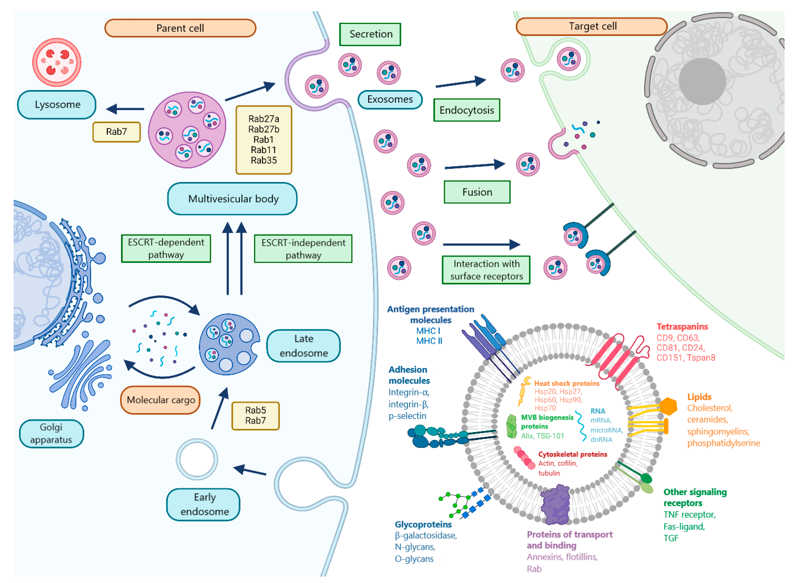
The formation of exosomes, unlike other types of vesicles, is a multi-step process. In the first stage, invagination of the plasma membrane occurs, resulting in the formation of early endosomes. Next, invagination of the plasma membrane leads to the formation of intraluminal vesicles (ILVs). The subsequent accumulation of multiple ILVs leads to the formation of multivesicular bodies (MVBs), which can be degraded by fusion with lysosomes or autophagosomes, or transported to the plasma membrane and, by fusion with it, release ILVs into the intercellular space in the form of exosomes. Transformation into MVB can occur both with and without the participation of the ESCRT protein complex (Figure 2) [66].
The mechanisms of exosome secretion are dependent on numerous factors, including the type of parent cell and the molecular cargo of the vesicles. It is evident that Rab GTPases, in conjunction with SNAP and SNARE proteins, assume a pivotal function in this process. Furthermore, Rab27a and Rab27b have been shown to promote the fusion of MVB with the plasma membrane, and Rab-1, Rab11, and Rab35 have been identified as regulators of the process of exosome secretion [67]. Exosomes have been demonstrated to interact with target cells through three principal mechanisms: interaction with cell receptors, fusion with the cell membrane, and endocytosis [49].
Regardless of their origin, exosomes contain a universal set of components, such as proteins involved in the formation, secretion, transport, and binding of exosomes to recipient cells, as well as lipids, metabolites, and other components that partially reflect the composition of the parent cells (Figure 2) [68]. Examples of universal exosomal proteins include tetraspanins (CD9, CD63 and CD81), adhesion receptors, annexins, integrins and flotillins. These proteins play a role in the processes of recognition and binding to the recipient cell [68,69,70]. MMPs, heat shock matrix proteins (Hsp70 and Hsp90), and proteins involved in biogenesis (Alix and TSG 101) are also considered to be universal exosomal proteins.
It has been established that enzymes comprise approximately one-third of exosome proteins. MMPs have been shown to belong to tetraspanin-associated proteases [71]. The process of ADAM-mediated hydrolysis of the extracellular domains of transmembrane proteins has been shown to regulate a number of cellular processes, including cell adhesion, migration, and intercellular interactions [72]. MMPs have been shown to hydrolyze type IV collagen, elastin, fibronectin and laminin, as well as cell surface proteins, including E-cadherin, fibrin and IL-1 [72]. MMP-2 and MMP-9 are the most abundant MMPs in exosomes [73,74]. As demonstrated in the extant literature, there is evidence to suggest that ADAM-10 levels increase in blood exosomes in BC [75,76]. However, the levels of other tetraspanin-associated proteases in exosomes in this disease remain to be elucidated.
Tetraspanin non-associated proteases include PAPP-A and 20S proteasomes. The former is a metalloproteinase [77], while the β-subunits of the latter exhibit caspase-like, trypsin-like, and chymotrypsin-like activity [78]. As demonstrated in the extant literature, there is an elevated level of 20S proteasome observed in patients diagnosed with BC, both in tumor tissue and in the circulating exosome form found in blood [79].
Various types of RNA have been identified in exosomes, including microRNA, messenger RNA (mRNA), ribosomal RNA (rRNA), long non-coding RNA (lncRNA), circular RNA, and others [80].
lncRNAs are a class of non-coding RNA molecules longer than 200 nucleotides that participate in the regulation of gene expression. These RNAs are capable of modulating translation processes, ensuring protein stability, and controlling mRNA translocation [81]. It has been established that lncRNAs play a key role in the development of various types of malignant neoplasms, including BC. For example, the HOTAIR miRNA is involved in invasion, metastasis, and tumor growth in various types of cancer, including BC; a positive correlation has also been found between this miRNA in exosomes and the expression level of the ERBB2 (HER-2/Neu) gene [82]. Some lncRNAs have oncogenic activity, while others exhibit tumor-suppressive properties. Differential lncRNA expression significantly influences the development of primary and acquired resistance to radiation, endocrine, immune and targeted therapies [83]. High levels of MIR100HG, T376626, and XIST microRNAs are associated with an unfavorable prognosis in triple-negative BC (TNBC) [84,85], while suppression of MIR100HG expression significantly reduces the proliferation of MDA-MB-231 cells [86]. It has also been shown that SNHG1 miRNA participates in the modulation of tumor-associated M2 macrophage polarization, thereby stimulating BC progression [85,86].
Currently, the role of circular RNAs in the BC dissemination is also being actively researched. Despite an incomplete understanding of the functions of these RNAs, it is assumed that they are involved in tumor formation, progression and metastasis through interaction with microRNAs and proteins [87]. Circular RNAs are considered as possible tumor markers, partly due to their stability, which is acquired through their non-linear shape. It has also been shown that there are exosomal sorting mechanisms for these RNAs, which are likely related to the level of microRNA targets in the parent cell [88]. In vitro and in vivo studies have shown that the levels of some circular RNAs differ between healthy donors and BC patients [89] and also depend on the degree of tumor malignancy, with these observations being characteristic of both blood samples and exosomes [90].
Exosomal tumor-derived mRNA has been demonstrated to exert influence on a variety of processes in recipient cells [91]. It has been demonstrated that MCF-7 exosomes contain hTERT mRNA, which can be transcribed into active telomerase, thus promoting oncogenic processes [92], while exosomal WNT5a mRNA from the MCF-7 BC cell line promotes invasion by activating the β-catenin-independent WNT-signaling pathway in macrophages [93].

4. The Role of Exosomal MicroRNA in BC Dissemination

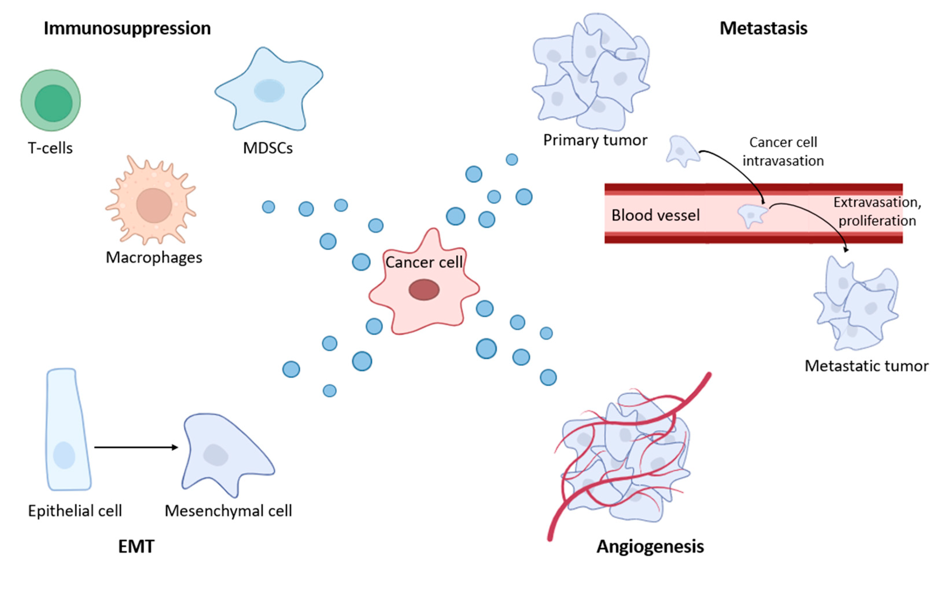
Exosomes play a key role in regulating various biological processes, both normal and pathological, including cancer (Figure 3). Their influence spans embryogenesis, immune response, tissue regeneration, hemostasis, angiogenesis, etc. [94].
Despite the rapid growth in the number of scientific publications devoted to the study of exosomes over the past ten years, many questions remain in understanding the regulation of intercellular communication through exosomes. This concerns, in particular, the dynamics and regulation of exosome secretion by cells, the mechanisms of intracellular sorting of molecules in exosomes, and the targeted transport of their contents between cells.

4.1. The Influence of BC Exosomes on the Functional Activity of the Immune System

BC-derived exosomes have been demonstrated to play a pivotal role in the formation of a microenvironment with a less active immune system within the tumor, thereby exerting an immunosuppressive effect. One way in which tumor-derived exosomes influence the immune system is by stimulating the polarization of tumor-associated M2 macrophages (TAMs), which regulate all stages of tumor progression, including angiogenesis, immunosuppression, drug resistance and metastasis in BC [95,96]. In particular, the role of tumor-derived exosomes in macrophage polarization has been demonstrated in cell lines mimicking TNBC [97]. It has been found that exosomal miR-222 also participates in the stimulation of TAM polarization in adriamycin resistance [98]. By influencing the phenotype of macrophages, microRNAs indirectly affect other elements of the immune system. It has been shown that under conditions of endoplasmic reticulum stress, the content of miR-27a-3p is increased in the exosomes of BC cells, which indirectly stimulates macrophage polarization and contributes to an increase in the frequency of T cell apoptosis [99].
The molecular cargo of exosomes can affect not only macrophages but also other cells of the immune system. For example, in BC, miR-9 and miR-181a in tumor-derived exosomes stimulated the development of myeloid-derived suppressor cells, which suppress the immune response [100].
Furthermore, exosomal microRNAs have been demonstrated to elicit an immunomodulatory effect, which can be exploited for therapeutic purposes. For instance, the administration of exosomes containing Let-7i, miR-142 and miR-155 to mice with BC had an effect on T-cell activation and dendritic cell maturation [101]. In addition, let-7 likely prevents the establishment of immunosuppressive pre-metastatic niches in BC, as the occurrence of an immunosuppressive effect is contingent upon the low content of this microRNA in exosomes, concomitant with the transition of neutrophils to a pro-tumor phenotype [102].
Consequently, the immunosuppressive microenvironment formed is pivotal in enabling the tumor to evade immune surveillance. The comprehension of these mechanisms has the potential to facilitate the development of novel immunotherapeutic approaches that are aimed at restoring the antitumor immune response and overcoming resistance to therapy.

4.2. BC-Derived Exosomes Stimulate EMT, Proliferation, Invasion and Migration

It is currently known that the molecular cargo of exosomes, including microRNAs, is involved in all stages of the EMT, up to the formation of metastases [103,104].
In vitro and in vivo studies show that exosomal miR-181d-5p promotes EMT by reducing the levels of the transcription factors CDX2 and HOXA5 [105]. A similar study found that miR-191–5p levels are elevated in both BC cell lines and blood exosomes from BC patients, and that it stimulates EMT by negatively regulating KLF6 [106]. In the MCF-7 cell line, it has been shown that cancer stem cells can mediate EMT and chemoresistance, most likely through exosomal transport of miR-155 [107]. Another in vitro study revealed the role of miR-197 cancer stem cell EVs in EMT [108].
EMT is also stimulated by exosomes from cells in the tumor microenvironment, such as cancer-associated fibroblasts (CAFs). In BC cell lines, it has been shown that miR-21, -378e, and -143 in CAF exosomes promote EMT, tumor cell growth, and the development of an aggressive tumor phenotype [109]. At the same time, it has been shown that the activation of normal fibroblasts is induced by miR-370-3p in the exosomes of BC cells, contributing to increased migration, invasion, and EMT of cancer cells [110].
MicroRNAs in the exosomes of BC cell lines are also involved in stimulating the invasion and migration of tumor cells. For example, MCF-7 and MDA-MB231 cells show increased levels of exosomal miR-301a, miR-106b, and miR-328, which are involved in the processes of increased cell migration and invasion [111]. Incubation of exosomes containing miR-222 with various BC cell lines promoted cell migration and invasion by suppressing PDLIM2 expression [112]. Furthermore, studies have demonstrated that exosomal miR-10b functions as a tumor suppressor by reducing the protein levels of its target genes, including HOXD 10 and KLF4, and by enhancing the invasive capacity of BC cells [113]. Additionally, exosomal miR-105 has been shown to regulate cell migration by targeting ZO-1 [114]. In addition to stimulating migration and invasion, microRNAs in exosomes can also suppress these processes. For example, a study of the role of histone demethylase LSD1 in BC found that knockdown of this gene leads to a decrease in the level of miR-1290 secreted in exosomes, which stimulates EMT, migration, and invasion of BC cells [115]. Furthermore, studies have demonstrated that miR-5100 exerts a regulatory effect on invasion, migration, and EMT in both pancreatic cancer and BC [116].
Exosomal non-coding RNAs not only stimulate cell invasion and migration, but also influence cell proliferation. For example, it has been shown that exosomal miR-130a and miR-425 increase the viability of MCF-7 cells, whose proliferation is associated with signaling pathways such as TOR, ErbB, MAPK and TGF-β [117]. Incubation of exosomes from the culture medium of the HCC1806 line, which mimics TNBC, with the non-carcinogenic MCF10A line caused an increase in cell proliferation, which was probably due to changes in 138 genes and the expression of 70 microRNAs affecting PI3K/AKT, MAPK, and HIF1A [118]. It has been shown that in BC, the level of exosomal miR-222 is significantly increased in the plasma of patients in the late stages of the disease and correlates with the high aggressiveness of BC cell lines [119].

4.3. BC-Derived Exosomes Stimulate Angiogenesis and Metastasis

Exosomes of tumor origin play an important role in regulating angiogenesis accompanying tumor development. In particular, MMPs participate in creating conditions for capillary growth by degrading the basal membrane, as well as in the release and induction of transcription of angiogenic factors [116].
In addition, it is known that exosomal microRNAs also play a role in angiogenesis in BC. Members of the miR-17-92 cluster, for example, have a proangiogenic effect by targeting thrombospondin 1, VEGFA, and TIMP. In particular, miR-20a promotes the expansion of the vascular network in BC, probably mediating this effect through VEGFA [120]. When studying the effect of intracellular calcium ion concentration on angiogenesis, an increase in miR-145 levels was found in exosomes obtained from BC cells with reduced calcium concentration. Treatment of HUVECs with exosomes from these cells reduced their ability to undergo angiogenesis. This effect is likely due to the influence of miR-145 on IRS1 [121]. It is also suggested that increased levels of miR-92a in both plasma exosomes and total blood exosomes of BC patients lead to a proangiogenic effect [43], which has also been demonstrated in other neoplasms [122,123].
It is evident that tumor cells are pivotal in the process of metastasis, as evidenced by their ability to form metastatic niches and stimulate the formation of primary metastatic nodes. For instance, exosomal miR-122 has been demonstrated to promote the formation of metastatic niches by altering glucose consumption in recipient cells [124]. In addition, it has been demonstrated that exosomes with elevated levels of miR-200 have the capacity to enhance the metastatic potential of BC cells in both in vivo and in vitro contexts [125].
Tumor cells are characterized by metastasis to specific organs with the formation of organotropic metastases. At the initial stage, the interaction between tumor cells and target organs leads to the formation of pre-metastatic niches. This process is then reinforced by other intercellular interactions, including tumor cell exosomes. In particular, the study showed that exosome secretion by T47D-CXCR4 cells is enriched in mRNA associated with stem cells and metastasis, confirming the predisposition of breast cancer cells to metastasis and the role of exosomes in stimulating tumor progression and metastasis [126].
TNBC is characterized by an increased frequency of metastasis to the lungs and brain, which distinguishes it from other molecular subtypes, for which metastatic involvement of bones and soft tissues is more typical. Numerous studies have demonstrated the upregulation of microRNAs, such as miR-9, miR-20a-5p, and miR-155, in highly metastatic TNBC cells and tumor-derived exosomes. Transfer of miR-20a-5p by exosomes to bone marrow macrophages was shown to stimulate osteoclastogenesis [127,128]. Studies show that elevated levels of exosomal miR-105 are characteristic of BC with high metastatic potential [129]. A number of other studies have identified a significant correlation between elevated levels of exosomal miR-210 and the presence of metastases in the lymph nodes in patients diagnosed with BC. The secretion of this microRNA is regulated by nSMase2, which promotes the metastasis of cancer cells by inducing angiogenesis in the tumor microenvironment, enhancing endothelial cell migration and capillary formation [56]. In addition, it has been demonstrated that cancer exosomes play a role in the disruption of the blood–brain barrier, which may lead to the formation of brain metastases. One of the putative mediators of this process is miR-181c, which alters actin localization [130].
The role of exosomal miR-210 as one of the key factors of tumor angiogenesis and brain metastasis was also noted, with high expression of miR-210 correlating with poor survival in BC patients with brain metastases [131]. Studies conducted on cell lines mimicking HER2+ BC have shown that miR-146a-5p not only promotes resistance to trastuzumab but also stimulates angiogenesis, proliferation, and migration [132]. Some microRNAs, such as miR-218, are known to act as suppressors of angiogenesis, migration, and metastasis [133]. Another example of a tumor suppressor is miR-124-3p, which targets the MTDH oncogene [134]. In vitro, downregulation of miR-145 has been shown to induce angiogenesis. Presumably, this effect is observed due to the fact that IRS1 is no longer regulated by miR-145 [122].
Therefore, the role of exosomes as important messengers in intercellular communication is significant both within the primary breast tumor microenvironment and in metastatic niches.

5. Exosomal MicroRNAs as Markers for BC Detection

Since exosomes mainly contain RNA less than 200 nucleotides in size [135], microRNA attracts the most attention from researchers. In particular, on 11 July 2025, a search of the PubMed database for the combination ‘microRNA and exosome and cancer’ yielded 4978 articles, while a search for ‘exosomes and microRNA and breast cancer’ yielded 608 articles.
To date, according to the ExoCarta database, 2838 microRNAs have been identified in the molecular cargo of exosomes. Although it is still unclear which microRNAs are involved in carcinogenesis, the level of tissue-specific microRNAs in exosomes can be used to develop diagnostics for malignant neoplasms, including BC.
In 2016, Exosome Diagnostics launched the first ExoDX test on the US market, which allows assessing the advantages of prostate cancer liquid biopsy using ERG, PCA3, and SPDEF mRNA in urine exosomes [136]. This test currently has Breakthrough Device Designation status from the FDA, but has not yet been approved for widespread use in clinical practice. It is imperative to recognize that 20–25% of women diagnosed with localized BC receive neoadjuvant therapy as their initial treatment line. Consequently, the identification of effective biomarkers in liquid biopsy for the diagnosis and prognosis of treatment response is a pressing task in the field of molecular oncology. A comparative analysis of CTCs and exosomal microRNAs in blood samples before and after neoadjuvant therapy showed that elevated levels of exosomal miRNA-21, miRNA-222 and miRNA-155 significantly correlate with the presence of CTCs, indicating the potential role of both exosomal microRNAs and CTCs in improving diagnosis and prognosis for patients undergoing neoadjuvant therapy [137].
A promising biomarker for the diagnosis and prognosis of BC may be the level of exosomal miR-148a in the blood, which is significantly reduced in patients with BC and correlates with an unfavorable prognosis [138]. Furthermore, the level of exosomal miR-223-3p has been shown to have potential as an additional tool in the diagnostic toolkit for BC, with a statistically significant increase in its concentration in plasma exosome samples observed in patients with ductal carcinoma of the breast, even at the initial stage of the disease [139]. In addition, exosomes associated with the surface of blood cells may serve as a more accurate source of tumor markers, in particular microRNAs [140].
The combination of exosomal microRNAs with other clinical biomarkers significantly improves the analytical characteristics of diagnostic systems based on liquid biopsy. For example, a panel of four exosomal microRNAs (miR-424, miR-423, miR-660, and let7-i) can differentiate between healthy donors and BC patients with 98.6% sensitivity and 100% specificity [141]. It is noteworthy that in this study, urine exosomes served as a probable source of tumor markers.
Using NGS (Next Generation sequencing) of serum exosomal microRNAs and subsequent verification by real-time PCR (polymerase chain reaction), a highly effective panel of markers has been developed for the differential diagnosis of breast neoplasms: the combination of miR-142-5p and miR-320a allows the luminal A subtype to be distinguished from the TNBC with 100% sensitivity and 93.8% specificity [142]. In addition, a diagnostic panel based on the analysis of miR-195, miR-210, miR-21, and miR-16 has a sensitivity of 71.4% and a specificity of 100% for BC diagnosis [143]. The potential of erythrocyte exosomes as sources of tumor markers, including for BC, is also being studied [144].
Despite the promising potential of miRNAs, the implementation of EV-based liquid biopsy into clinical practice is associated with several challenges. First, there is currently no EV isolation method available and scalable for use in a healthcare setting, guaranteeing high purity and the ability to simultaneously analyze large numbers of samples. Methods used for EV isolation, such as ultracentrifugation, density gradient centrifugation, and polymer precipitation, are time-consuming and may not be suitable for routine analysis. Methods based on immunospecific binding of EVs to a carrier are expensive, which is also a serious limitation for implementation into clinical practice [145]. Second, there are currently no standardized approaches to quantification and normalization of vesicular miRNAs. To date, researchers have used various strategies for miRNA level normalization, including the use of endogenous controls, exogenous controls, mean normalization, and paired miRNA normalization, which often do not allow comparison of data obtained by different groups [146,147,148,149].

6. Exosomal MicroRNAs Associated with BC Relapse

To identify key molecular mechanisms underlying BC recurrence, a bioinformatics analysis was performed. Based on the analysis of literature data, sixteen exosomal microRNAs were selected, the level of which changes depending on the presence or absence of relapse and pathological complete response (pCR) to therapy, the latter of which is associated with a more favorable prognosis for BC patients (Table 1).
The target genes of the selected microRNAs were initially identified using the DIANA-miRPath v4.0 database (http://62.217.122.229:3838/app/miRPathv4). DIANA-miRPath enables the identification of KEGG (Kyoto Encyclopedia of Genes and Genomes) and GO (Gene Ontology) biological pathways that are enriched with target genes for specific microRNAs. This allows us to determine the potential influence of these microRNAs on cellular processes associated with BC recurrence. The results show that these microRNAs are involved in BC pathways and other cancer pathways, as well as in cell cycle and apoptosis-related processes and in mTOR, FoxO and p53 signaling pathways. All studied miRNAs, except miR-548ab, are involved in BC-related pathways and participate in the regulation of the expression of 89 target genes.
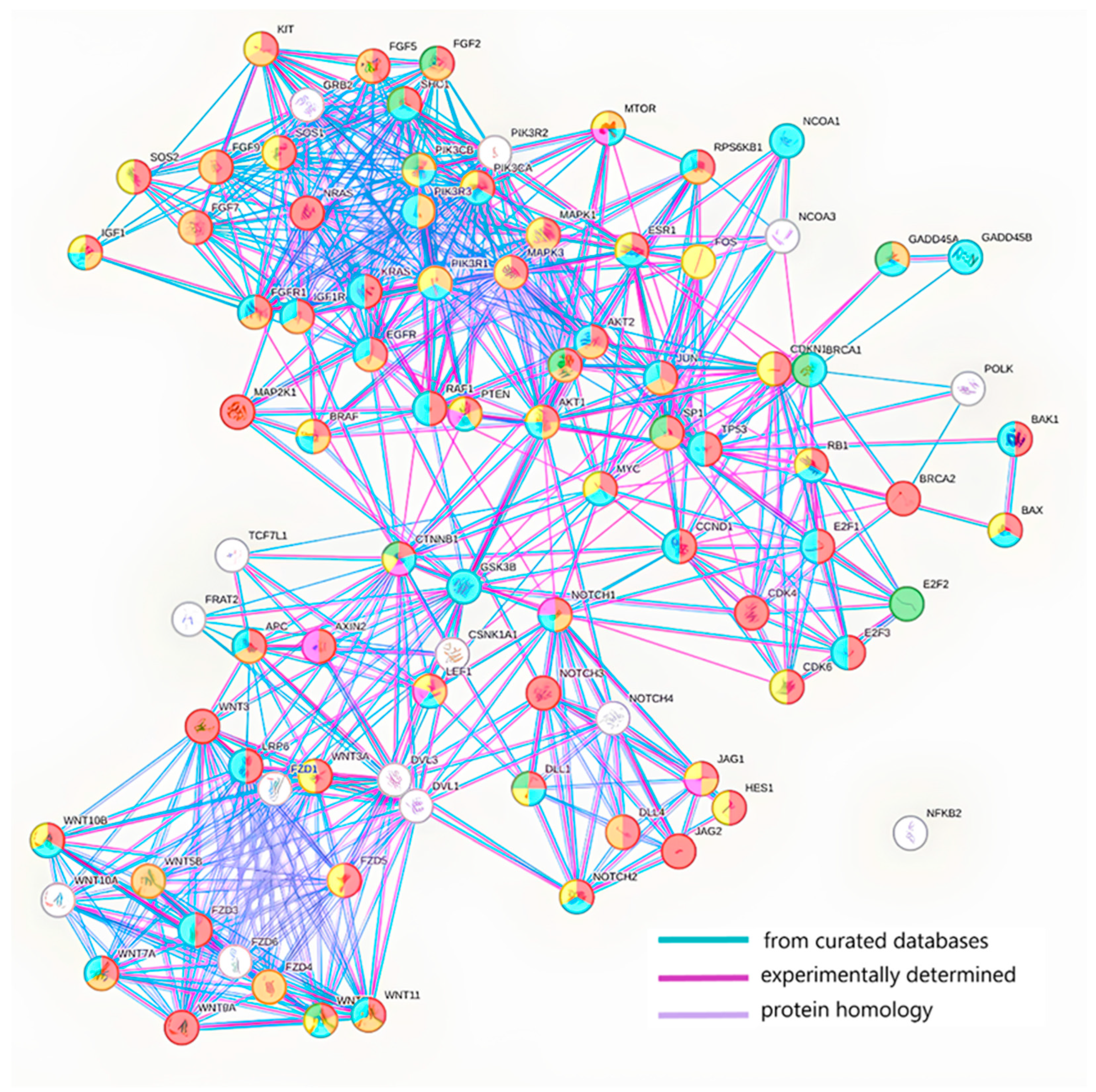
The STRING v12.0 database (string-db.org) was used to analyze interactions between the protein products of target genes. STRING provides information on known and predicted protein interactions, including physical interactions, protein homology and participation in common biological pathways. Using target genes identified in DIANA-miRPath, an interaction network was constructed to identify key proteins that may play a central role in regulating the processes leading to BC recurrence. The analysis revealed that these proteins are involved in regulating proliferation, apoptosis, cell migration, EMT, immune processes and angiogenesis (Figure 4).
The obtained protein interaction network was imported into the Cytoscape software, version 3.10.3 (cytoscape.org) to identify hub proteins. Hub proteins were identified based on degree centrality analysis, which was calculated using Cytoscape’s built-in NetworkAnalyzer tool. Degree Centrality determines the number of direct interactions a protein has with other proteins in the network, so proteins with the highest number of interactions were identified as hub proteins. Network topology analysis, particularly Degree Centrality, enabled us to identify key proteins that may play a central role in regulating processes that lead to breast cancer recurrence. The degree value ranged from 26 to 0, with 17 proteins having the highest values (21–26): PIK3R1, CTNNB1, KRAS, MAPK1, PIK3R2, PIK3CA, MAPK3, EGFR, AKT1, TP53, PIK3CB, NRAS, GSK3B, SHC1, DVL1, GRB2 and DVL3. These results highlight the significant role of these proteins in dissemination and, in particular, BC recurrence.
Thus, the bioinformatics analysis revealed several miRNAs, associated genes, biological pathways and protein interactions that are potentially involved in BC recurrence. The identified protein hubs and biological pathways are promising targets for further research into developing new strategies for preventing and treating BC recurrence. Further experimental confirmation of the role of these microRNAs and their targets will allow the creation of panels for assessing the prognosis of the course and selection of targeted therapy for BC.

7. Conclusions

Modern instrumental methods of BC diagnostics have a number of limitations related to early detection of the disease and differentiation of benign and malignant tumors. Rapid development of molecular oncology opens up prospects for the emergence of new tumor markers in the next five years, including those based on exosomal microRNAs for the detection of early BC. In combination with instrumental diagnostic methods, these markers will allow more effective detection of BC and assessment of the disease prognosis. Elucidation of detailed mechanisms of tumor spread involving exosomal microRNAs will allow the development of gene-directed therapeutic approaches to increase the effectiveness of BC treatment. This, in turn, will lead to a decrease in disability and improvement in the quality of life of women suffering from this disease.

Author Contributions

Conceptualization, S.T. and A.C. (Alyona Chernyshova); formal analysis, A.B.; writing—original draft preparation, S.T. and A.B.; writing—review and editing, S.T., A.S., A.C. (Alexander Chernyavskiy) and A.C. (Alyona Chernyshova). All authors have read and agreed to the published version of the manuscript.

Funding

This research was funded by the Russian Science Foundation, grant 25-15-20023, https://rscf.ru/project/25-15-20023/ (accessed on 21 September 2025), and the Government of the Novosibirsk region of the Russian Federation, grant 30-2025-001040.

Acknowledgments

We thank Yu. Gotalskaya for creating Figure 3.

Conflicts of Interest

The authors declare no conflicts of interest.

Abbreviations

The following abbreviations are used in this manuscript:
BCBreast cancer
CAFCancer-associated fibroblasts
CTCCirculating tumor cells
EMTEpithelial–mesenchymal transition
GOGene Ontology
ILVIntraluminal vesicles
KEGGKyoto Encyclopedia of Genes and Genomes
LDHLactate dehydrogenase
MMPsMatrix metalloproteinases
MRIMagnetic resonance imaging
MVBMultivesicular body
NGSNext Generation sequencing
NPCNucleoprotein complex
pCRPathological complete response
PCRPolymerase chain reaction
PETPositron emission tomography
RNARibonucleic acid
sEVsSmall extracellular vesicles
TAMTumor-associated macrophages
TNBCTriple-negative breast cancer
USUltrasound examination
WHOWorld Health Organization

References

  1. Sung, H.; Ferlay, J.; Siegel, R.L.; Laversanne, M.; Soerjomataram, I.; Jemal, A.; Bray, F. Global Cancer Statistics 2022: GLOBOCAN Estimates of Incidence and Mortality Worldwide for 36 Cancers in 185 Countries. CA Cancer J. Clin. 2021, 71, 209–249. [Google Scholar] [CrossRef]
  2. Starinskij, V.V.; Shahzadova, A.O.; Kaprin, A.D. Malignant Neoplasms in Russia in 2023 (Incidence and Mortality); MNIOI im. P.A. Gercena−filial FGBU «NMIC radiologii» Minzdrava Rossii: Moscow, Russia, 2023; p. 252. (In Russian) [Google Scholar]
  3. Winters, S.; Martin, C.; Murphy, D.; Shokar, N.K. Breast Cancer Epidemiology, Prevention, and Screening. Prog. Mol. Biol. Transl. Sci. 2017, 151, 1–32. [Google Scholar] [CrossRef]
  4. Connal, S.; Cameron, J.M.; Sala, A.; Brennan, P.M.; Palmer, D.S.; Palmer, J.D.; Perlow, H.; Baker, M.J. Liquid biopsies: The future of cancer early detection. J. Transl. Med. 2023, 21, 118. [Google Scholar] [CrossRef]
  5. Shakhzadova, A.O.; Starinsky, V.V.; Lisichnikova, I.V. Cancer care to the population of Russia in 2022. Sib. J. Oncol. 2023, 22, 5–13. [Google Scholar] [CrossRef]
  6. Semiglazov, V.F.; Semiglazov, V.V.; Krivorotko, P.V.; Paltuev, R.M.; Dashyan, G.A.; Semiglazova, T.Y.; Bessonov, A.A.; Nikolaev, K.S. Guidelines for the Treatment of Early Breast Cancer; PJSC “T8 Publishing Technologies”: Sankt-Peretburg, Russia, 2016; p. 154. (In Russian) [Google Scholar]
  7. Radenkovic, S.; Konjevic, G.; Isakovic, A.; Stevanovic, P.; Gopcevic, K.; Jurisic, V. HER2-positive breast cancer patients: Correlation between mammographic and pathological findings. Radiat. Prot. Dosim. 2014, 162, 125–128. [Google Scholar] [CrossRef]
  8. Chan, C.H.F.; Coopey, S.B.; Freer, P.E.; Hughes, K.S. False-negative rate of combined mammography and ultrasound for women with palpable breast masses. Breast Cancer Res. Treat. 2015, 153, 699–702. [Google Scholar] [CrossRef]
  9. McDonald, E.S.; Clark, A.S.; Tchou, J.; Zhang, P.; Freedman, G.M. Clinical Diagnosis and Management of Breast Cancer. J. Nucl. Med. 2016, 57, 9S–16S. [Google Scholar] [CrossRef]
  10. Lehman, C.D. Clinical indications: What is the evidence? Eur. J. Radiol. 2012, 81, S82–S84. [Google Scholar] [CrossRef]
  11. Geisel, J.; Raghu, M.; Hooley, R. The Role of Ultrasound in Breast Cancer Screening: The Case for and Against Ultrasound. Semin. Ultrasound. CT MRI 2018, 39, 25–34. [Google Scholar] [CrossRef]
  12. Tamkovich, S.N.; Voytsitskiy, V.E.; Laktionov, P.P. Modern Methods in Breast Cancer Diagnostics. Biochem. Suppl. Ser. B Biomed. Chem. 2014, 8, 302–313. [Google Scholar] [CrossRef]
  13. Jafari, S.H.; Saadatpour, Z.; Salmaninejad, A.; Momeni, F.; Mokhtari, M.; Nahand, J.S.; Rahmati, M.; Mirzaei, H.; Kianmehr, M. Breast cancer diagnosis: Imaging techniques and biochemical markers. J. Cell Physiol. 2018, 233, 5200–5213. [Google Scholar] [CrossRef]
  14. Popova, N.S.; Novikov, S.N.; Krzhivitsky, P.I.; Zhukova, L.A.; Krivorotko, P.V.; Artemyeva, A.S.; Valitova, A.A.; Khoroshavina, A.A.; Chernaya, A.V.; Bryanceva, Z.V.; et al. Diagnostic capabilities of scintimammography in detecting multicentric and minimal breast cancer of various molecular subtypes. Vopr. Onkologii. 2023, 69, 708–714. (In Russian) [Google Scholar] [CrossRef]
  15. Basova, T.S.; Fedorov, N.M.; Fadeeva, A.I.; Shmalts, E.A.; Kovalik, A.V.; Sepiashvili, G.G. Mammoscintigraphy and OFECT with 99MTS-MIBI in diagnostics of breast neoplasms. Acad. J. West Sib. 2019, 15, 55–57. [Google Scholar]
  16. Pesapane, F.; Suter, M.B.; Rotili, A.; Penco, S.; Nigro, O.; Cremonesi, M.; Bellomi, M.; Jereczek-Fossa, B.A.; Pinotti, G.; Cassano, E. Will traditional biopsy be substituted by radiomics and liquid biopsy for breast cancer diagnosis and characterisation? Med. Oncol. 2020, 37, 29. [Google Scholar] [CrossRef]
  17. Wesoła, M.; Jeleń, M. The diagnostic efficiency of fine needle aspiration biopsy in breast cancers—Review. Adv. Clin. Exp. Med. 2013, 22, 887–892. [Google Scholar]
  18. Kunihiko, H.; Kornelia, P. Intratumoral Heterogeneity: More Than Just Mutations. Trends Cell Biol. 2019, 29, 569–579. [Google Scholar] [CrossRef]
  19. Shimomura, A.; Shiino, S.; Kawauchi, J.; Takizawa, S.; Sakamoto, H.; Matsuzaki, J.; Ono, M.; Takeshita, F.; Niida, S.; Shimizu, C.; et al. Novel combination of serum microRNA for detecting breast cancer in the early stage. Cancer Sci. 2016, 107, 326–334. [Google Scholar] [CrossRef]
  20. Satomi-Tsushita, N.; Shimomura, A.; Matsuzaki, J.; Yamamoto, Y.; Kawauchi, J.; Takizawa, S.; Aoki, Y.; Sakamoto, H.; Kato, K.; Shimizu, C.; et al. Serum microRNA-based prediction of responsiveness to eribulin in metastatic breast cancer. PLoS ONE 2019, 14, e0222024. [Google Scholar] [CrossRef]
  21. Shiino, S.; Matsuzaki, J.; Shimomura, A.; Kawauchi, J.; Takizawa, S.; Sakamoto, H.; Aoki, Y.; Yoshida, M.; Tamura, K.; Kato, K.; et al. Serum miRNA-based Prediction of Axillary Lymph Node Metastasis in Breast Cancer. Clin. Cancer Res. 2019, 25, 1817–1827. [Google Scholar] [CrossRef]
  22. Sato, J.; Shimomura, A.; Kawauchi, J.; Matsuzaki, J.; Yamamoto, Y.; Takizawa, S.; Sakamoto, H.; Ohno, M.; Narita, Y.; Ochiya, T.; et al. Brain metastasis-related microRNAs in patients with advanced breast cancer. PLoS ONE 2019, 14, e0221538. [Google Scholar] [CrossRef]
  23. Ye, Q.; Ling, S.; Zheng, S.; Xu, X. Liquid biopsy in hepatocellular carcinoma: Circulating tumor cells and circulating tumor DNA. Mol. Cancer 2019, 18, 114. [Google Scholar] [CrossRef]
  24. Tay, T.K.Y.; Tan, P.H. Liquid Biopsy in Breast Cancer: A Focused Review. Arch. Pathol. Lab. Med. 2020, 145, 678–686. [Google Scholar] [CrossRef]
  25. Tamkovich, S.; Tupikin, A.; Kozyakov, A.; Laktionov, P. Size and Methylation Index of Cell-Free and Cell-Surface-Bound DNA in Blood of Breast Cancer Patients in the Contest of Liquid Biopsy. Int. J. Mol. Sci. 2022, 23, 8919. [Google Scholar] [CrossRef]
  26. Shefer, A.; Tutanov, O.; Belenikin, M.; Tsentalovich, Y.P.; Tamkovich, S. Blood Plasma Circulating DNA-Protein Complexes: Involvement in Carcinogenesis and Prospects for Liquid Biopsy of Breast Cancer. J. Pers. Med. 2023, 13, 1691. [Google Scholar] [CrossRef]
  27. Tutanov, O.; Shefer, A.; Shefer, E.; Ruzankin, P.; Tsentalovich, Y.; Tamkovich, S. DNA-Binding Proteins and Passenger Proteins in Plasma DNA-Protein Complexes: Imprint of Parental Cells or Key Mediators of Carcinogenesis Processes? Int. J. Mol. Sci. 2024, 25, 5165. [Google Scholar] [CrossRef]
  28. Wang, X.; Li, X.; Dong, T.; Yu, W.; Jia, Z.; Hou, Y.; Yang, J.; Liu, Y. Global biomarker trends in triple-negative breast cancer research: A bibliometric analysis. Int. J. Surg. 2024, 110, 7962–7983. [Google Scholar] [CrossRef]
  29. Stankovic, S.; Konjevic, G.; Gopcevic, K.; Jovic, V.; Inic, M.; Jurisic, V. Activity of MMP-2 and MMP-9 in sera of breast cancer patients. Pathol. Res. Pract. 2010, 206, 241–247. [Google Scholar] [CrossRef]
  30. Radenkovic, S.; Konjevic, G.; Jurisic, V.; Karadzic, K.; Nikitovic, M.; Gopcevic, K. Values of MMP-2 and MMP-9 in tumor tissue of basal-like breast cancer patients. Cell Biochem. Biophys. 2014, 68, 143–152. [Google Scholar] [CrossRef]
  31. Jurisic, V.; Radenkovic, S.; Konjevic, G. The Actual Role of LDH as Tumor Marker, Biochemical and Clinical Aspects. Adv. Exp. Med. Biol. 2015, 867, 115–124. [Google Scholar] [CrossRef]
  32. Konjević, G.; Jurisić, V.; Spuzić, I. Association of NK cell dysfunction with changes in LDH characteristics of peripheral blood lymphocytes (PBL) in breast cancer patients. Breast Cancer Res. Treat. 2001, 66, 255–263. [Google Scholar] [CrossRef]
  33. Jurisić, V.; Konjević, G.; Jancić-Nedeljkov, R.; Sretenović, M.; Banicević, B.; Colović, M.; Spuzić, I. The comparison of spontaneous LDH release activity from cultured PBMC with sera LDH activity in non-Hodgkin’s lymphoma patients. Med. Oncol. 2004, 21, 179–185. [Google Scholar] [CrossRef]
  34. Shchegolev, Y.Y.; Sorokin, D.V.; Scherbakov, A.M.; Andreeva, O.E.; Salnikova, D.I.; Mikhaevich, E.I.; Gudkova, M.V.; Krasil’nikov, M.A. Exosomes are involved in the intercellular transfer of rapamycin resistance in the breast cancer cells. Bioimpacts 2023, 13, 313–321. [Google Scholar] [CrossRef]
  35. Kok, V.C.; Yu, C.-C. Cancer-Derived Exosomes: Their Role in Cancer Biology and Biomarker Development. Int. J. Nanomed. 2020, 15, 8019–8036. [Google Scholar] [CrossRef]
  36. Cai, X.; Janku, F.; Zhan, Q.; Fan, J.-B. Accessing Genetic Information with Liquid Biopsies. Trends Genet. 2015, 31, 564–575. [Google Scholar] [CrossRef]
  37. Ludwig, N.; Whiteside, T.L.; Reichert, T.E. Challenges in Exosome Isolation and Analysis in Health and Disease. Int. J. Mol. Sci. 2019, 20, 4684. [Google Scholar] [CrossRef]
  38. Tutanov, O.; Orlova, E.; Proskura, K.; Grigor’eva, A.; Yunusova, N.; Tsentalovich, Y.; Alexandrova, A.; Tamkovich, S. Proteomic Analysis of Blood Exosomes from Healthy Females and Breast Cancer Patients Reveals an Association between Different Exosomal Bioactivity on Non-tumorigenic Epithelial Cell and Breast Cancer Cell Migration in Vitro. Biomolecules 2020, 10, 495. [Google Scholar] [CrossRef]
  39. Hoshino, A.; Costa-Silva, B.; Shen, T.-L.; Rodrigues, G.; Hashimoto, A.; Tesic Mark, M.; Molina, H.; Kohsaka, S.; Di Giannatale, A.; Ceder, S.; et al. Tumour exosome integrins determine organotropic metastasis. Nature 2015, 527, 329–335. [Google Scholar] [CrossRef]
  40. Ge, Q.; Zhou, Y.; Lu, J.; Bai, Y.; Xie, X.; Lu, Z. miRNA in plasma exosome is stable under different storage conditions. Molecules 2014, 19, 1568–1575. [Google Scholar] [CrossRef]
  41. Yu, W.; Hurley, J.; Roberts, D.; Chakrabortty, S.K.; Enderle, D.; Noerholm, M.; Breakefield, X.O.; Skog, J.K. Exosome-based liquid biopsies in cancer: Opportunities and challenges. Ann. Oncol. 2021, 32, 466–477. [Google Scholar] [CrossRef]
  42. Cheng, L.; Sharples, R.A.; Scicluna, B.J.; Hill, A.F. Exosomes provide a protective and enriched source of miRNA for biomarker profiling compared to intracellular and cell-free blood. J. Extracell. Vesicles 2014, 3. [Google Scholar] [CrossRef]
  43. Konoshenko, M.; Sagaradze, G.; Orlova, E.; Shtam, T.; Proskura, K.; Kamyshinsky, R.; Yunusova, N.; Alexandrova, A.; Efimenko, A.; Tamkovich, S. Total Blood Exosomes in Breast Cancer: Potential Role in Crucial Steps of Tumorigenesis. Int. J. Mol. Sci. 2020, 21, 7341. [Google Scholar] [CrossRef]
  44. Dong, X.; Bai, X.; Ni, J.; Zhang, H.; Duan, W.; Graham, P.; Li, Y. Exosomes and breast cancer drug resistance. Cell Death Dis. 2020, 11, 987. [Google Scholar] [CrossRef]
  45. Min, L.; Zhu, S.; Chen, L.; Liu, X.; Wei, R.; Zhao, L.; Yang, Y.; Zhang, Z.; Kong, G.; Li, P.; et al. Evaluation of circulating small extracellular vesicles derived miRNAs as biomarkers of early colon cancer: A comparison with plasma total miRNAs. J. Extracell. Vesicles 2019, 8, 1643670. [Google Scholar] [CrossRef]
  46. Datta, N.; Johnson, C.; Kao, D.; Gurnani, P.; Alexander, C.; Polytarchou, C.; Monaghan, T.M. MicroRNA-based therapeutics for inflammatory disorders of the microbiota-gut-brain axis. Pharmacol. Res. 2023, 194, 106870. [Google Scholar] [CrossRef]
  47. Huang, X.; Zhu, X.; Yu, Y.; Zhu, W.; Jin, L.; Zhang, X.; Li, S.; Zou, P.; Xie, C.; Cui, R. Dissecting miRNA signature in colorectal cancer progression and metastasis. Cancer Lett. 2021, 501, 66–82. [Google Scholar] [CrossRef]
  48. Abou Madawi, N.A.; Darwish, Z.E.; Omar, E.M. Targeted gene therapy for cancer: The impact of microRNA multipotentiality. Med. Oncol. 2024, 41, 214. [Google Scholar] [CrossRef]
  49. Zhang, J.; Li, S.; Li, L.; Li, M.; Guo, C.; Yao, J.; Mi, S. Exosome and exosomal microRNA: Trafficking, sorting, and function. Genom. Proteom. Bioinform. 2015, 13, 17–24. [Google Scholar] [CrossRef]
  50. Ho, P.T.B.; Clark, I.M.; Le, L.T.T. MicroRNA-Based Diagnosis and Therapy. Int. J. Mol. Sci. 2022, 23, 7167. [Google Scholar] [CrossRef]
  51. Li, B.; Cao, Y.; Sun, M.; Feng, H. Expression, regulation, and function of exosome-derived miRNAs in cancer progression and therapy. FASEB J. 2021, 35, e21916. [Google Scholar] [CrossRef]
  52. Groot, M.; Lee, H. Sorting Mechanisms for MicroRNAs into Extracellular Vesicles and Their Associated Diseases. Cells 2020, 9, 1044. [Google Scholar] [CrossRef]
  53. Jenjaroenpun, P.; Kremenska, Y.; Nair, V.M.; Kremenskoy, M.; Joseph, B.; Kurochkin, I.V. Characterization of RNA in exosomes secreted by human breast cancer cell lines using next-generation sequencing. PeerJ 2013, 1, e201. [Google Scholar] [CrossRef]
  54. Gurung, S.; Perocheau, D.; Touramanidou, L.; Baruteau, J. The exosome journey: From biogenesis to uptake and intracellular signalling. Cell Commun. Signal 2021, 19, 47. [Google Scholar] [CrossRef]
  55. Kossinova, O.A.; Gopanenko, A.V.; Tamkovich, S.N.; Krasheninina, O.A.; Tupikin, A.E.; Kiseleva, E.; Yanshina, D.D.; Malygin, A.A.; Ven’yaminova, A.G.; Kabilov, M.R.; et al. Cytosolic YB-1 and NSUN2 are the only proteins recognizing specific motifs present in mRNAs enriched in exosomes. Biochim. Biophys. Acta Proteins Proteom. 2017, 1865, 664–673. [Google Scholar] [CrossRef]
  56. Trajkovic, K.; Hsu, C.; Chiantia, S.; Rajendran, L.; Wenzel, D.; Wieland, F.; Schwille, P.; Brügger, B.; Simons, M. Ceramide triggers budding of exosome vesicles into multivesicular endosomes. Science 2008, 319, 1244–1247. [Google Scholar] [CrossRef]
  57. Kosaka, N.; Iguchi, H.; Yoshioka, Y.; Takeshita, F.; Matsuki, Y.; Ochiya, T. Secretory mechanisms and intercellular transfer of microRNAs in living cells. J. Biol. Chem. 2010, 285, 17442–17452. [Google Scholar] [CrossRef]
  58. Kosaka, N.; Iguchi, H.; Hagiwara, K.; Yoshioka, Y.; Takeshita, F.; Ochiya, T. Neutral Sphingomyelinase 2 (nSMase2)-dependent Exosomal Transfer of Angiogenic MicroRNAs Regulate Cancer Cell Metastasis. J. Biol. Chem. 2013, 288, 10849–10859. [Google Scholar] [CrossRef]
  59. Villarroya-Beltri, C.; Gutiérrez-Vázquez, C.; Sánchez-Cabo, F.; Pérez-Hernández, D.; Vázquez, J.; Martin-Cofreces, N.; Martinez-Herrera, D.J.; Pascual-Montano, A.; Mittelbrunn, M.; Sánchez-Madrid, F. Sumoylated hnRNPA2B1 controls the sorting of miRNAs into exosomes through binding to specific motifs. Nat. Commun. 2013, 4, 2980. [Google Scholar] [CrossRef]
  60. Zhou, X.; Wang, L.; Zou, W.; Chen, X.; Roizman, B.; Zhou, G.G. hnRNPA2B1 Associated with Recruitment of RNA into Exosomes Plays a Key Role in Herpes Simplex Virus 1 Release from Infected Cells. J. Virol. 2020, 94, e00367-20. [Google Scholar] [CrossRef]
  61. Santangelo, L.; Giurato, G.; Cicchini, C.; Montaldo, C.; Mancone, C.; Tarallo, R.; Battistelli, C.; Alonzi, T.; Weisz, A.; Tripodi, M. The RNA-Binding Protein SYNCRIP Is a Component of the Hepatocyte Exosomal Machinery Controlling MicroRNA Sorting. Cell Rep. 2016, 17, 799–808. [Google Scholar] [CrossRef]
  62. McKenzie, A.J.; Hoshino, D.; Hong, N.H.; Cha, D.J.; Franklin, J.L.; Coffey, R.J.; Patton, J.G.; Weaver, A.M. KRAS-MEK Signaling Controls Ago2 Sorting into Exosomes. Cell Rep. 2016, 15, 978–987. [Google Scholar] [CrossRef]
  63. Kowal, J.; Arras, G.; Colombo, M.; Jouve, M.; Morath, J.P.; Primdal-Bengtson, B.; Dingli, F.; Loew, D.; Tkach, M.; Théry, C. Proteomic comparison defines novel markers to characterize heterogeneous populations of extracellular vesicle subtypes. Proc. Natl. Acad. Sci. USA 2016, 113, 968–977. [Google Scholar] [CrossRef]
  64. Temoche-Diaz, M.M.; Shurtleff, M.J.; Nottingham, R.M.; Yao, J.; Fadadu, R.P.; Lambowitz, A.M.; Schekman, R. Distinct mechanisms of microRNA sorting into cancer cell-derived extracellular vesicle subtypes. eLife 2019, 8, e47544. [Google Scholar] [CrossRef]
  65. Koppers-Lalic, D.; Hackenberg, M.; Bijnsdorp, I.V.; van Eijndhoven, M.A.J.; Sadek, P.; Sie, D.; Zini, N.; Middeldorp, J.M.; Ylstra, B.; de Menezes, R.X.; et al. Nontemplated Nucleotide Additions Distinguish the Small RNA Composition in Cells from Exosomes. Cell Rep. 2014, 8, 1649–1658. [Google Scholar] [CrossRef]
  66. Yue, B.; Yang, H.; Wang, J.; Ru, W.; Wu, J.; Huang, Y.; Lan, X.; Lei, C.; Chen, H. Exosome biogenesis, secretion and function of exosomal miRNAs in skeletal muscle myogenesis. Cell Prolif. 2020, 53, e12857. [Google Scholar] [CrossRef]
  67. Wei, H.; Chen, Q.; Lin, L.; Sha, C.; Li, T.; Liu, Y.; Yin, X.; Xu, Y.; Chen, L.; Gao, W.; et al. Regulation of exosome production and cargo sorting. Int. J. Biol. Sci. 2021, 17, 163–177. [Google Scholar] [CrossRef]
  68. Kalluri, R.; LeBleu, V.S. The biology, function, and biomedical applications of exosomes. Science 2020, 367, 977. [Google Scholar] [CrossRef]
  69. Ekström, K.; Crescitelli, R.; Pétursson, H.I.; Johansson, J.; Lässer, C.; Olofsson Bagge, R. Characterization of surface markers on extracellular vesicles isolated from lymphatic exudate from patients with breast cancer. BMC Cancer 2022, 22, 50. [Google Scholar] [CrossRef]
  70. Yunusova, N.V.; Tugutova, E.A.; Tamkovich, S.N.; Kondakova, I.V. Role of tetraspanins and exosomal proteases in tumor progression. Biomed. Chem. 2018, 64, 123–133. [Google Scholar] [CrossRef]
  71. Matthews, A.L.; Noy, P.J.; Reyat, J.S.; Tomlinson, M.G. Regulation of A disintegrin and metalloproteinase (ADAM) family sheddases ADAM10 and ADAM17: The emerging role of tetraspanins and rhomboids. Platelets 2017, 28, 333–341. [Google Scholar] [CrossRef]
  72. Shimoda, M.; Khokha, R. Metalloproteinases in extracellular vesicles. Biochim. Biophys. Acta 2017, 1864, 1989–2000. [Google Scholar] [CrossRef]
  73. Yunusova, N.V.; Patysheva, M.R.; Molchanov, S.V.; Zambalova, E.A.; Grigor’eva, A.E.; Kolomiets, L.A.; Ochirov, M.O.; Tamkovich, S.N.; Kondakova, I.V. Metalloproteinases at the surface of small extrcellular vesicles in advanced ovarian cancer: Relationships with ascites volume and peritoneal canceromatosis index. Clin. Chim. Acta 2019, 494, 116–122. [Google Scholar] [CrossRef]
  74. Deep, G.; Jain, A.; Kumar, A.; Agarwal, C.; Kim, S.; Leevy, W.M.; Agarwal, R. Exosomes secreted by prostate cancer cells under hypoxia promote matrix metalloproteinases activity at pre-metastatic niches. Mol. Cancerogen. 2020, 59, 323–332. [Google Scholar] [CrossRef]
  75. Tamkovich, S.N.; Yunusova, N.V.; Tugutova, E.; Somov, A.K.; Proskura, K.V.; Kolomiets, L.A.; Stakheeva, M.N.; Grigor’eva, A.E.; Laktionov, P.P.; Kondakova, I.V. Protease Cargo in Circulating Exosomes of Breast Cancer and Ovarian Cancer Patients. Asian. Pac. J. Cancer Prev. 2019, 20, 255–262. [Google Scholar] [CrossRef]
  76. Somov, A.K.; Yunusova, N.V.; Shtam, T.A.; Proskura, K.V.; Zambalova, E.A.; Laktionov, P.P.; Tamkovich, S.N. ADAM-10 on the surface of exosomes from breast cancer patients blood: Newly mechanisms tumor dissemination. Vopr. Onkol. 2019, 65, 678–683. [Google Scholar] [CrossRef]
  77. Laursen, L.S.; Overgaard, M.T.; Søe, R.; Boldt, H.B.; Sottrup-Jensen, L.; Giudice, L.C.; Conover, C.A.; Oxvig, C. Pregnancy-associated plasma protein-A (PAPP-A) cleaves insulin-like growth factor binding protein (IGFBP)-5 independent of IGF: Implications for the mechanism of IGFBP-4 proteolysis by PAPP-A. FEBS Lett. 2001, 504, 36–40. [Google Scholar] [CrossRef]
  78. Kisselev, A.F.; Callard, A.; Goldberg, A.L. Importance of the different proteolytic sites of the proteasome and the efficacy of inhibitors varies with the protein substrate. J. Biol. Chem. 2006, 281, 8582–8590. [Google Scholar] [CrossRef]
  79. Yunusova, N.; Kolegova, E.; Sereda, E.; Kolomiets, L.; Villert, A.; Patysheva, M.; Rekeda, I.; Grigor’eva, A.; Tarabanovskaya, N.; Kondakova, I.; et al. Plasma Exosomes of Patients with Breast and Ovarian Tumors Contain an Inactive 20S Proteasome. Molecules 2021, 26, 6965. [Google Scholar] [CrossRef]
  80. Hashimoto, K.; Ochiya, T.; Shimomura, A. Liquid biopsy using non-coding RNAs and extracellular vesicles for breast cancer management. Breast Cancer 2025, 32, 16–25. [Google Scholar] [CrossRef]
  81. Xu, Z.; Chen, Y.; Ma, L.; Chen, Y.; Liu, J.; Guo, Y.; Yu, T.; Zhang, L.; Zhu, L.; Shu, Y. Role of exosomal non-coding RNAs from tumor cells and tumor-associated macrophages in the tumor microenvironment. Mol. Ther. 2022, 30, 3133–3154. [Google Scholar] [CrossRef]
  82. Wang, Y.L.; Liu, L.C.; Hung, Y.; Chen, C.J.; Lin, Y.Z.; Wu, W.R.; Wang, S.C. Long non-coding RNA HOTAIR in circulatory exosomes is correlated with ErbB2/HER2 positivity in breast cancer. Breast 2019, 46, 64–69. [Google Scholar] [CrossRef]
  83. Kashyap, D.; Sharma, R.; Goel, N.; Buttar, H.S.; Garg, V.K.; Pal, D.; Rajab, K.; Shaikh, A. Coding roles of long non-coding RNAs in breast cancer: Emerging molecular diagnostic biomarkers and potential therapeutic targets with special reference to chemotherapy resistance. Front. Genet. 2023, 13, 993687. [Google Scholar] [CrossRef]
  84. Yang, Q.; Fu, Y.; Wang, J.; Yang, H.; Zhang, X. Roles of lncRNA in the diagnosis and prognosis of triple-negative breast cancer. J. Zhejiang Univ. Sci. B 2023, 24, 1123–1140. [Google Scholar] [CrossRef]
  85. Wang, S.; Ke, H.; Zhang, H.; Ma, Y.; Ao, L.; Zou, L.; Yang, Q.; Zhu, H.; Nie, J.; Wu, C.; et al. LncRNA MIR100HG promotes cell proliferation in triple-negative breast cancer through triplex formation with p27 loci. Cell Death Dis. 2018, 9, 805. [Google Scholar] [CrossRef]
  86. Zong, S.; Dai, W.; Guo, X.; Wang, K. LncRNA-SNHG1 promotes macrophage M2-like polarization and contributes to breast cancer growth and metastasis. Aging 2021, 13, 23169–23181. [Google Scholar] [CrossRef]
  87. Conn, V.M.; Chinnaiyan, A.M.; Conn, S.J. Circular RNA in cancer. Nat. Rev. Cancer 2024, 24, 597–613. [Google Scholar] [CrossRef]
  88. Zhou, W.Y.; Cai, Z.R.; Liu, J.; Wang, D.S.; Ju, H.Q.; Xu, R.H. Circular RNA: Metabolism, functions and interactions with proteins. Mol. Cancer 2020, 19, 172. [Google Scholar] [CrossRef]
  89. Li, X.; Ma, F.; Wu, L.; Zhang, X.; Tian, J.; Li, J.; Cao, J.; Ma, Y.; Zhang, L.; Wang, L. Identification of Hsa_circ_0104824 as a Potential Biomarkers for Breast Cancer. Technol. Cancer Res. Treat. 2020, 19, 1533033820960745. [Google Scholar] [CrossRef]
  90. Wang, J.; Zhang, Q.; Zhou, S.; Xu, H.; Wang, D.; Feng, J.; Zhao, J.; Zhong, S. Circular RNA expression in exosomes derived from breast cancer cells and patients. Epigenomics 2019, 11, 411–421. [Google Scholar] [CrossRef]
  91. Matsuno, Y.; Kanke, T.; Maruyama, N.; Fujii, W.; Naito, K.; Sugiura, K. Characterization of mRNA profiles of the exosome-like vesicles in porcine follicular fluid. PLoS ONE 2019, 14, e0217760. [Google Scholar] [CrossRef]
  92. Gutkin, A.; Uziel, O.; Beery, E.; Nordenberg, J.; Pinchasi, M.; Goldvaser, H.; Henick, S.; Goldberg, M.; Lahav, M. Tumor cells derived exosomes contain hTERT mRNA and transform nonmalignant fibroblasts into telomerase positive cells. Oncotarget 2016, 7, 59173–59188. [Google Scholar] [CrossRef]
  93. Menck, K.; Klemm, F.; Gross, J.C.; Pukrop, T.; Wenzel, D.; Binder, C. Induction and transport of Wnt 5a during macrophage-induced malignant invasion is mediated by two types of extracellular vesicles. Oncotarget 2013, 4, 2057–2066. [Google Scholar] [CrossRef]
  94. Vyas, K.S.; Kaufman, J.; Munavalli, G.S.; Robertson, K.; Behfar, A.; Wyles, S.P. Exosomes: The latest in regenerative aesthetics. Regen. Med. 2023, 18, 181–194. [Google Scholar] [CrossRef]
  95. Munir, M.T.; Kay, M.K.; Kang, M.H.; Rahman, M.M.; Al-Harrasi, A.; Choudhury, M.; Moustaid-Moussa, N.; Hussain, F.; Rahman, S.M. Tumor-Associated Macrophages as Multifaceted Regulators of Breast Tumor Growth. Int. J. Mol. Sci. 2021, 22, 6526. [Google Scholar] [CrossRef]
  96. Zhao, X.; Qu, J.; Sun, Y.; Wang, J.; Liu, X.; Wang, F.; Zhang, H.; Wang, W.; Ma, X.; Gao, X.; et al. Prognostic significance of tumor-associated macrophages in breast cancer: A meta-analysis of the literature. Oncotarget 2017, 8, 30576–30586. [Google Scholar] [CrossRef]
  97. Piao, Y.J.; Kim, H.S.; Hwang, E.H.; Woo, J.; Zhang, M.; Moon, W.K. Breast cancer cell-derived exosomes and macrophage polarization are associated with lymph node metastasis. Oncotarget 2017, 9, 7398–7410. [Google Scholar] [CrossRef]
  98. Chen, W.; Wang, D.; Zhu, B.; Zhu, Y.; Zheng, L.; Feng, Z.; Qin, X. Exosomal miR-222 from adriamycin-resistant MCF-7 breast cancer cells promote macrophages M2 polarization via PTEN/Akt to induce tumor progression. Aging 2021, 13, 10415–10430. [Google Scholar] [CrossRef]
  99. Yao, X.; Tu, Y.; Xu, Y.; Guo, Y.; Yao, F.; Zhang, X. Endoplasmic reticulum stress-induced exosomal mir-27a-3p promotes immune escape in breast cancer via regulating pd-L1 expression in macrophages. J. Cell Mol. Med. 2020, 24, 9560–9573. [Google Scholar] [CrossRef]
  100. Jiang, M.; Zhang, W.; Zhang, R.; Liu, P.; Ye, Y.; Yu, W.; Guo, X.; Yu, J. Cancer exosome-derived miR-9 and miR-181a promote the development of early-stage MDSCs via interfering with SOCS3 and PIAS3 respectively in breast cancer. Oncogene 2020, 39, 4681–4694. [Google Scholar] [CrossRef]
  101. Khani, A.T.; Sharifzad, F.; Mardpour, S.; Hassan, Z.M.; Ebrahimi, M. Tumor extracellular vesicles loaded with exogenous Let-7i and miR-142 can modulate both immune response and tumor microenvironment to initiate a powerful anti-tumor response. Cancer Lett. 2021, 501, 200–209. [Google Scholar] [CrossRef]
  102. Qi, M.; Xia, Y.; Wu, Y.; Zhang, Z.; Wang, X.; Lu, L.; Dai, C.; Song, Y.; Xu, K.; Ji, W.; et al. Lin28B-high breast cancer cells promote immune suppression in the lung pre-metastatic niche via exosomes and support cancer progression. Nat. Commun. 2022, 13, 897. [Google Scholar] [CrossRef]
  103. Otmani, K.; Rouas, R.; Lagneaux, L.; Krayem, M.; Duvillier, H.; Berehab, M.; Lewalle, P. Acute myeloid leukemia-derived exosomes deliver miR-24-3p to hinder the T-cell immune response through DENN/MADD targeting in the NF-κB signaling pathways. Cell Commun. Signal 2023, 21, 253. [Google Scholar] [CrossRef]
  104. Crow, J.; Atay, S.; Banskota, S.; Artale, B.; Schmitt, S.; Godwin, A.K. Exosomes as mediators of platinum resistance in ovarian cancer. Oncotarget 2017, 8, 11917–11936. [Google Scholar] [CrossRef]
  105. Wang, H.; Wei, H.; Wang, J.; Li, L.; Chen, A.; Li, Z. MicroRNA-181d-5p-Containing Exosomes Derived from CAFs Promote EMT by Regulating CDX2/HOXA5 in Breast Cancer. Mol. Ther. Nucleic. Acids 2020, 19, 654–667. [Google Scholar] [CrossRef]
  106. Pan, L.; Liu, W.; Zhao, H.; Chen, B.; Yue, X. MiR-191-5p inhibits KLF6 to promote epithelial-mesenchymal transition in breast cancer. Technol. Health Care 2023, 31, 2251–2265. [Google Scholar] [CrossRef]
  107. Santos, J.C.; Lima, N.D.S.; Sarian, L.O.; Matheu, A.; Ribeiro, M.L.; Derchain, S.F.M. Exosome-mediated breast cancer chemoresistance via miR-155 transfer. Sci. Rep. 2018, 8, 829. [Google Scholar] [CrossRef]
  108. Li, L.; Xiong, Y.; Wang, N.; Zhu, M.; Gu, Y. Breast Cancer Stem Cells-derived Extracellular Vesicles Affect PPARG Expression by Delivering MicroRNA-197 in Breast Cancer Cells. Clin. Breast Cancer 2022, 22, 478–490. [Google Scholar] [CrossRef]
  109. Donnarumma, E.; Fiore, D.; Nappa, M.; Roscigno, G.; Adamo, A.; Iaboni, M.; Russo, V.; Affinitom, A.; Puoti, I.; Quintavalle, C.; et al. Cancer-associated fibroblasts release exosomal microRNAs that dictate an aggressive phenotype in breast cancer. Oncotarget 2017, 8, 19592–19608. [Google Scholar] [CrossRef]
  110. Ren, Z.; Lv, M.; Yu, Q.; Bao, J.; Lou, K.; Li, X. MicroRNA-370-3p shuttled by breast cancer cell-derived extracellular vesicles induces fibroblast activation through the CYLD/Nf-kappaB axis to promote breast cancer progression. FASEB J. 2021, 35, e21383. [Google Scholar] [CrossRef]
  111. Kruger, S.; Abd Elmageed, Z.Y.; Hawke, D.H.; Wörner, P.M.; Jansen, D.A.; Abdel-Mageed, A.B.; Alt, E.U.; Izadpanah, R. Molecular characterization of exosome-like vesicles from breast cancer cells. BMC Cancer 2014, 14, 44. [Google Scholar] [CrossRef]
  112. Spirina, L.V.; Kondakova, I.V.; Klisho, E.V.; Kakurina, G.V.; Shishkin, D.A. Metaloproteinases as neoangiogenesis regulators in cancer. Sib. J. Oncol. 2007, 1, 67–71. [Google Scholar]
  113. Singh, R.; Pochampally, R.; Watabe, K.; Lu, Z.; Mo, Y.-Y. Exosome-mediated transfer of miR-10b promotes cell invasion in breast cancer. Mol. Cancer 2014, 13, 256. [Google Scholar] [CrossRef]
  114. Zhou, W.; Fong, M.Y.; Min, Y.; Somlo, G.; Liu, L.; Palomares, M.R.; Yu, Y.; Chow, A.; O’Connor, S.T.F.; Chin, A.R.; et al. Cancer-secreted miR-105 destroys vascular endothelial barriers to promote metastasis. Cancer Cell 2014, 25, 501–515. [Google Scholar] [CrossRef]
  115. Zhang, N.; Chen, Z.; Xin, B.; Shi, Y.; Yao, Y.; Yang, J.; Wang, X.; Hu, X. LSD1 inhibits the invasion and migration of breast cancer through exosomes. Sci. Rep. 2024, 14, 20817. [Google Scholar] [CrossRef]
  116. Yue, S.; Ye, X.; Zhou, T.; Gan, D.; Qian, H.; Fang, W.; Yao, M.; Zhang, D.; Shi, H.; Chen, T. PGRN-/- TAMs-derived exosomes inhibit breast cancer cell invasion and migration and its mechanism exploration. Life Sci. 2021, 264, 118687. [Google Scholar] [CrossRef]
  117. Chen, W.-X.; Cheng, L.; Pan, M.; Qian, Q.; Zhu, Y.-L.; Xu, L.-Y.; Ding, Q. D Rhamnose β-Hederin against human breast cancer by reducing tumor-derived exosomes. Oncol. Lett. 2018, 16, 5172–5178. [Google Scholar] [CrossRef]
  118. Ozawa, P.M.M.; Alkhilaiwi, F.; Cavalli, I.J.; Malheiros, D.; de Souza Fonseca Ribeiro, E.M.; Cavalli, L.R. Extracellular vesicles from triple-negative breast cancer cells promote proliferation and drug resistance in non-tumorigenic breast cells. Breast Cancer Res. Treat. 2018, 172, 713–723. [Google Scholar] [CrossRef]
  119. Ding, J.; Xu, Z.; Zhang, Y.; Tan, C.; Hu, W.; Wang, M.; Xu, Y.; Tang, J. Exosome-mediated miR-222 transferring: An insight into NF-κB-mediated breast cancer metastasis. Exp. Cell Res. 2018, 369, 129–138. [Google Scholar] [CrossRef]
  120. Luengo-Gil, G.; Gonzalez-Billalabeitia, E.; Perez-Henarejos, S.A.; Navarro Manzano, E.; Chaves-Benito, A.; Garcia-Martinez, E.; Garcia-Garre, E.; Vicente, V.; Ayala de la Peña, F. Angiogenic role of miR-20a in breast cancer. PLoS ONE 2018, 13, e0194638. [Google Scholar] [CrossRef]
  121. Pan, S.; Zhao, X.; Shao, C.; Fu, B.; Huang, Y.; Zhang, N.; Dou, X.; Zhang, Z.; Qiu, Y.; Wang, R.; et al. STIM1 promotes angiogenesis by reducing exosomal miR-145 in breast cancer MDA-MB-231 cells. Cell Death Dis. 2021, 12, 38. [Google Scholar] [CrossRef]
  122. Zhao, W.; Wu, Y.; Wang, Y.; Li, T.; Liu, Q.; Hou, Z. Exosomal miR-92a-3p modulates M2 macrophage polarization in colorectal cancer: Implications for tumor migration and angiogenesis. Med. Oncol. 2025, 42, 96. [Google Scholar] [CrossRef]
  123. Chen, S.; Chen, X.; Luo, Q.; Liu, X.; Wang, X.; Cui, Z.; He, A.; He, S.; Jiang, Z.; Wu, N.; et al. Retinoblastoma cell-derived exosomes promote angiogenesis of human vesicle endothelial cells through microRNA-92a-3p. Cell Death Dis. 2021, 12, 695. [Google Scholar] [CrossRef]
  124. Fong, M.Y.; Zhou, W.; Liu, L.; Alontaga, A.Y.; Chandra, M.; Ashby, J.; Chow, A.; O’Connor, S.T.F.; Li, S.; Chin, A.R.; et al. Breast-cancer-secreted miR-122 reprograms glucose metabolism in premetastatic niche to promote metastasis. Nat. Cell Biol. 2015, 17, 183–194. [Google Scholar] [CrossRef]
  125. Le, M.T.N.; Hamar, P.; Guo, C.; Basar, E.; Perdigão-Henriques, R.; Balaj, L.; Lieberman, J. miR-200-containing extracellular vesicles promote breast cancer cell metastasis. J. Clin. Investig. 2014, 124, 5109–5128. [Google Scholar] [CrossRef]
  126. Rodríguez, M.; Silva, J.; Herrera, A.; Herrera, M.; Peña, C.; Martín, P.; Gil-Calderón, B.; Larriba, M.J.; Coronado, M.J.; Soldevilla, B.; et al. Exosomes enriched in stemness/metastatic-related mRNAS promote oncogenic potential in breast cancer. Oncotarget 2015, 6, 40575–40587. [Google Scholar] [CrossRef]
  127. Kia, V.; Paryan, M.; Mortazavi, Y.; Biglari, A.; Mohammadi-Yeganeh, S. Evaluation of exosomal miR-9 and miR-155 targeting PTEN and DUSP14 in highly metastatic breast cancer and their effect on low metastatic cells. J. Cell Biochem. 2019, 120, 5666–5676. [Google Scholar] [CrossRef]
  128. Guo, L.; Zhu, Y.; Li, L.; Zhou, S.; Yin, G.; Yu, G.; Cui, H. Breast cancer cell-derived exosomal miR-20a-5p promotes the proliferation and differentiation of osteoclasts by targeting SRCIN1. Cancer Med. 2019, 8, 5687–5701. [Google Scholar] [CrossRef]
  129. Li, C.; Zhou, T.; Chen, J.; Li, R.; Chen, H.; Luo, S.; Chen, D.; Cai, C.; Li, W. The role of Exosomal miRNAs in cancer. J. Transl. Med. 2022, 20, 6. [Google Scholar] [CrossRef]
  130. Tominaga, N.; Kosaka, N.; Ono, M.; Katsuda, T.; Yoshioka, Y.; Tamura, K.; Lötvall, J.; Nakagama, H.; Ochiya, T. Brain metastatic cancer cells release microRNA-181c-containing extracellular vesicles capable of destructing blood-brain barrier. Nat. Commun. 2015, 6, 6716. [Google Scholar] [CrossRef]
  131. Hong, L.; Yang, J.; Han, Y.; Lu, Q.; Cao, J.; Syed, L. High expression of miR-210 predicts poor survival in patients with breast cancer: A meta-analysis. Gene 2012, 507, 135–138. [Google Scholar] [CrossRef]
  132. Cabello, P.; Torres-Ruiz, S.; Adam-Artigues, A.; Forés-Martos, J.; Martínez, M.T.; Hernando, C.; Zazo, S.; Madoz-Gúrpide, J.; Rovira, A.; Burgués, O.; et al. miR-146a-5p Promotes Angiogenesis and Confers Trastuzumab Resistance in HER2+ Breast Cancer. Cancers 2023, 15, 2138. [Google Scholar] [CrossRef]
  133. Shojaei, S.; Moradi-Chaleshtori, M.; Paryan, M.; Koochaki, A.; Sharifi, K.; Mohammadi-Yeganeh, S. Mesenchymal stem cell-derived exosomes enriched with miR-218 reduce the epithelial-mesenchymal transition and angiogenesis in triple-negative breast cancer cells. Eur. J. Med. Res. 2023, 28, 516. [Google Scholar] [CrossRef]
  134. Shi, P.; Liu, Y.; Yang, H.; Hu, B. Breast cancer derived exosomes promoted angiogenesis of endothelial cells in microenvironment via circHIPK3/miR-124-3p/MTDH axis. Cell Signal 2022, 95, 110338. [Google Scholar] [CrossRef]
  135. Yunusova, N.; Dzhugashvili, E.; Yalovaya, A.; Kolomiets, L.; Shefer, A.; Grigor’eva, A.; Tupikin, A.; Kondakova, I.; Tamkovich, S. Comparative Analysis of Tumor-Associated microRNAs and Tetraspanines from Exosomes of Plasma and Ascitic Fluids of Ovarian Cancer Patients. Int. J. Mol. Sci. 2023, 24, 464. [Google Scholar] [CrossRef]
  136. McKiernan, J.; Donovan, M.J.; O’Neill, V.; Bentink, S.; Noerholm, M.; Belzer, S.; Skog, J.; Kattan, M.W.; Partin, A.; Andriole, G.; et al. A Novel Urine Exosome Gene Expression Assay to Predict High-grade Prostate Cancer at Initial Biopsy. JAMA Oncol. 2016, 2, 882–889. [Google Scholar] [CrossRef]
  137. Rodríguez-Martínez, A.; de Miguel-Pérez, D.; Ortega, F.G.; García-Puche, J.L.; Robles-Fernández, I.; Exposito, J.; Martorell-Marugan, J.; Carmona-Sáez, P.; Garrido-Navas, M.D.C.; Rolfo, C.; et al. Exosomal miRNA profile as complementary tool in the diagnostic and prediction of treatment response in localized breast cancer under neoadjuvant chemotherapy. Breast Cancer Res. 2019, 21, 21. [Google Scholar] [CrossRef]
  138. Li, D.; Wang, J.; Ma, L.-J.; Yang, H.-B.; Jing, J.-F.; Jia, M.-M.; Zhang, X.-J.; Guo, F.; Gao, J.-N. Identification of serum exosomal miR-148a as a novel prognostic biomarker for breast cancer. Eur. Rev. Med. Pharmacol. Sci. 2020, 24, 7303–7309. [Google Scholar] [CrossRef]
  139. Yoshikawa, M.; Iinuma, H.; Umemoto, Y.; Yanagisawa, T.; Matsumoto, A.; Jinno, H. Exosome-encapsulated microRNA-223-3p as a minimally invasive biomarker for the early detection of invasive breast cancer. Oncol. Lett. 2018, 15, 9584–9592. [Google Scholar] [CrossRef]
  140. Tamkovich, S.; Tutanov, O.; Efimenko, A.; Grigor’eva, A.; Ryabchikova, E.; Kirushina, N.; Vlassov, V.; Tkachuk, V.; Laktionov, P. Blood circulating exosomes contain distinguishable fractions of free and cell-surface-associated vesicles. Curr. Mol. Med. 2019, 19, 273–285. [Google Scholar] [CrossRef]
  141. Hirschfeld, M.; Rücker, G.; Weiß, D.; Berner, K.; Ritter, A.; Jäger, M.; Erbes, T. Urinary Exosomal MicroRNAs as Potential Non-invasive Biomarkers in Breast Cancer Detection. Mol. Diagn. Ther. 2020, 24, 215–232. [Google Scholar] [CrossRef]
  142. Ozawa, P.M.M.; Vieira, E.; Lemos, D.S.; Souza, I.L.M.; Zanata, S.M.; Pankievicz, V.C.; Tuleski, T.R.; Souza, E.M.; Wowk, P.F.; Urban, C.; et al. Identification of miRNAs Enriched in Extracellular Vesicles Derived from Serum Samples of Breast Cancer Patients. Biomolecules 2020, 10, 150. [Google Scholar] [CrossRef]
  143. Miranda, F.S.; Slaibi-Filho, J.; Calasans dos Santos, G.; Carmo, N.T.; Kaneto, C.M.; Borin, T.F.; Luiz, W.B.; Gastalho Campos, L.C. MicroRNA as a promising molecular biomarker in the diagnosis of breast cancer. Front. Mol. Biosci. 2024, 11, 1337706. [Google Scholar] [CrossRef]
  144. Yang, L.; Huang, S.; Zhang, Z.; Liu, Z.; Zhang, L. Roles and Applications of Red Blood Cell-Derived Extracellular Vesicles in Health and Diseases. Int. J. Mol. Sci. 2022, 23, 5927. [Google Scholar] [CrossRef]
  145. Bhadra, M.; Sachan, M. An overview of challenges associated with exosomal miRNA isolation toward liquid biopsy-based ovarian cancer detection. Heliyon 2024, 10, e30328. [Google Scholar] [CrossRef]
  146. Zou, R.; Loke, S.Y.; Tang, Y.C.; Too, H.-P.; Zhou, L.; Lee, A.S.G.; Hartman, M. Development and validation of a circulating microRNA panel for the early detection of breast cancer. Br. J. Cancer 2022, 126, 472–481. [Google Scholar] [CrossRef]
  147. Chekka, L.M.S.; Langaee, T.; Johnson, J.A. Comparison of Data Normalization Strategies for Array-Based MicroRNA Profiling Experiments and Identification and Validation of Circulating MicroRNAs as Endogenous Controls in Hypertension. Front. Genet. 2022, 13, 836636. [Google Scholar] [CrossRef]
  148. Schwarzenbach, H.; da Silva, A.M.; Calin, G.; Pantel, K. Data Normalization Strategies for MicroRNA Quantification. Clin. Chem. 2015, 61, 1333–1342. [Google Scholar] [CrossRef]
  149. Skryabin, G.O.; Vinokurova, S.V.; Elkina, N.V.; Denisova, D.A.; Beliaeva, A.A.; Zhordania, K.I.; Bagrov, D.V.; Enikeev, A.D.; Galetsky, S.A.; Komelkov, A.V.; et al. Comparison of methods for microRNA isolation from extracellular vesicles obtained from ascitic fluids. Biochemistry 2022, 87, 1354–1366. [Google Scholar] [CrossRef]
  150. Sueta, A.; Yamamoto, Y.; Tomiguchi, M.; Takeshita, T.; Yamamoto-Ibusuki, M.; Iwase, H. Differential expression of exosomal miRNAs between breast cancer patients with and without recurrence. Oncotarget 2017, 8, 69934–69944. [Google Scholar] [CrossRef]
  151. Sueta, A.; Fujiki, Y.; Goto-Yamaguchi, L.; Tomiguchi, M.; Yamamoto-Ibusuki, M.; Iwase, H.; Yamamoto, Y. Exosomal miRNA profiles of triple-negative breast cancer in neoadjuvant treatment. Oncol. Lett. 2021, 22, 819. [Google Scholar] [CrossRef]
  152. Stevic, I.; Müller, V.; Weber, K.; Fasching, P.A.; Karn, T.; Marmé, F.; Schem, C.; Stickeler, E.; Denkert, C.; van Mackelenbergh, M.; et al. Specific microRNA signatures in exosomes of triple-negative and HER2-positive breast cancer patients undergoing neoadjuvant therapy within the GeparSixto trial. BMC Med. 2018, 16, 179. [Google Scholar] [CrossRef]
  153. Ni, Q.; Stevic, I.; Pan, C.; Müller, V.; Oliveira-Ferrer, L.; Pantel, K.; Schwarzenbach, H. Different signatures of miR-16, miR-30b and miR-93 in exosomes from breast cancer and DCIS patients. Sci. Rep. 2018, 8, 12974. [Google Scholar] [CrossRef] [PubMed]
Figure 1. The principle of liquid biopsy and promising tumor markers.
Molecules 30 03858 g001
Figure 2. Biogenesis and composition of exosomes.
Molecules 30 03858 g002
Figure 3. The role of exosomes in BC dissemination.
Molecules 30 03858 g003
Figure 4. Network of interactions between target genes involved in BC. Regulation of cell population proliferation—red color; regulation of apoptotic processes—blue; regulation of EMT—pink color; regulation of cell migration—orange color; regulation of angiogenesis—green color; regulation of immune system processes—yellow color.
Molecules 30 03858 g004
Table 1. MicroRNAs associated with BC relapse and pCR.
#MicroRNAsLevel Change
1miR-338-3p in serum exosomes of patients with relapse compared to patients without relapse [150]
2miR-340-5p
3miR-124-3p
4miR-29b-3p in serum exosomes of patients with relapse compared to patients without relapse [150]
5miR-20b-5p
6miR-17-5p
7miR-130a-3p
8miR-18a-5p
9miR-195-5p in serum exosomes of patients with relapse compared to patients without relapse [150]
in serum exosomes of non-pCR patients with relapse compared to those without it [151]
10miR-486-5p in serum exosomes of patients with relapse compared to patients without relapse [150]
11miR-93-5p
12miR-548ab in serum exosomes of non-pCR patients with relapse compared to those without it [151]
13miR-155 plasma exosomes after therapy, associated with pCR [152]
14miR-301
15miR-30b in plasma exosomes of patients with relapse compared with primary BC patients [153]
16miR-16 in plasma exosomes of patients with relapse compared with healthy donors [153]
Disclaimer/Publisher’s Note: The statements, opinions and data contained in all publications are solely those of the individual author(s) and contributor(s) and not of MDPI and/or the editor(s). MDPI and/or the editor(s) disclaim responsibility for any injury to people or property resulting from any ideas, methods, instructions or products referred to in the content.