Review

Natural Products with Potent Antimycobacterial Activity (2000–2024): A Review

Department of Pharmaceutical Chemistry and Pharmacognosy, School of Pharmacy, College of Health Sciences, Addis Ababa University, Addis Ababa P.O. Box. 1176, Ethiopia
*
Author to whom correspondence should be addressed.
Molecules 2025, 30(18), 3708; https://doi.org/10.3390/molecules30183708
Submission received: 31 July 2025 / Revised: 22 August 2025 / Accepted: 26 August 2025 / Published: 12 September 2025

Abstract

Tuberculosis (TB), an infection caused by Mycobacterium tuberculosis, affects nearly one-third of the world’s population. It is estimated that TB infects around ten million people worldwide, with no less than two million fatalities annually. It is one of the treatable infections due to improved diagnostic tools and therapeutic agents. However, the disease remains a threat to humankind due to the emergence of multidrug- and extensively drug-resistant strains of M. tuberculosis. This has driven many researchers to look for new antitubercular medications with better efficacy, safety, and affordability. As has always been the case, natural products have provided huge potential as a source of remedies for various infectious and non-infectious diseases. This review aims to report discoveries and updates of antitubercular natural products with minimum inhibitory concentration (MIC) values of less than or 10 µg/mL or 50 µM and selectivity indices of greater than 10. The review discusses 36 naturally occurring compounds from various classes, isolated from both terrestrial and aquatic organisms, including higher plants and microorganisms. Perusal of the literature reveals that most of these promising compounds are alkaloids, terpenoids, steroids, and peptides. Rufomycin I, a cyclic heptapeptide from Streptomyces sp., showed potent activity against drug-sensitive and isoniazid-resistant M. tuberculosis H37Rv (MIC < 0.004 µM), surpassing isoniazid (MIC = 0.23 µM), likely by inhibiting ClpC1 transcription. Hapalindole A also displayed strong activity (MIC < 0.6 µM). Current TB drugs have become less effective; therefore, natural products such as hapalindole A and rufomycin I, owing to their potent activity, selectivity, and novelty, are increasingly recognized as potential lead compounds against TB.

1. Introduction

Tuberculosis (TB), one of the leading causes of death, is caused by M. tuberculosis [1,2]. M. tuberculosis (e.g., H37Rv and its attenuated counterpart H37Ra) is a classic slow-growing pathogen, with its doubling time in the laboratory being approximately 24 h, varying with the environment and with the time of visible colony development [3,4,5]. M. avium, being part of the M. avium complex (MAC), is also slow-growing and causes opportunistic pulmonary and disseminated infections, mainly among immunocompromised individuals [6]. In contrast, M. smegmatis is non-pathogenic and rapidly growing, possessing a 3-day colony growth rate and serving as a model in biosafety level 1 laboratories for the study of mycobacterial physiology and genetics [7]. Remarkably, using invasion assays of HEp-2 epithelial cells, M. smegmatis was not invasive or capable of multiplication unless it had been transformed with an invasive locus from M. avium, where it possesses the ability to invade and grow intracellularly, highlighting its universality as a substitute for the dissection of pathogenic traits [6].
M. tuberculosis H37Ra replicates the drug sensitivity profile of its virulent counterpart to most of the first- and second-line antitubercular agents. This makes M. tuberculosis H37Ra an excellent, safer substitute for in vitro drug screening and pharmacodynamic assays [8,9]. M. smegmatis can be used as a safe strain in a high-throughput model for screening anti-TB compounds with comparable MIC values to M. tuberculosis H37Rv for most TB drugs [7]. Other rapidly growing non-tuberculous species such as M. aurum have been observed to maintain drug susceptibility profiles similar to that of M. tuberculosis and be used as surrogate models for intracellular drug screening [9].
Around 2 billion people are latently infected with tuberculosis, yet only 10% of them develop active disease during their lifetime [10]. TB is treatable due to improved diagnostic tools and therapeutic agents in the 20th century [11,12]. Nevertheless, the emergence of HIV infection and the spread of multidrug-resistant TB (MDR-TB) in the 1980s and 1990s brought challenges in treatment [11]. Since 2000, global TB incidence has reduced by 1.7% per year, and age-standardized mortality rates fell from approximately 1.8 million deaths in 1990 to nearly 1.2 million in 2021. Incidents fell 11% between 2015 and 2020, but nevertheless, 10 million new cases continued each year. The COVID-19 pandemic reversed TB death gains, which increased to levels of 2017, as the rate fell by only 9% in 2020, considerably short of the 35% target. Inequities persist between regions, with Europe and parts of Africa recording steeper declines, while South/Southeast Asia and most low- and middle-income countries continue to bear heavy burdens. In 2023, TB once more became the leading infectious cause of death with 10.8 million cases and 1.25 million deaths [2].
The current pharmacotherapy of TB is challenged by the emergence of drug-resistant bacterial strains, side effects, and long duration of treatment. Hence, there is a pressing need to develop alternative treatments with novel modes of action, good oral bioavailability, shorter treatment duration, efficacy against MDR- and XDR-TB, broad availability and affordability, reduced pill burden, low dosing frequency, and minimal drug–drug interactions [13]. This could be achieved through looking into natural products or synthetic molecules. Natural products provide a large pool of molecules that could have an antimycobacterial effect or scaffold in anti-TB drug discovery [14].
Natural products could be screened for their antitubercular activity based on phenotypic (drug to target) or the target to drug screening [15,16]. Most anti-TB medications advanced to clinics through whole-cell screening, followed by analysis of the mechanism of action and identification of potential targets [17]. But even for early screening, antibacterial new drug discovery will be efficient and effective if it begins with a target that has been identified [15]. In this review, we shall discuss natural products with potent antimycobacterial activity reported in the English language from 2000 to 2024 with MICs less than 10 µg/mL or 50 µM and a selectivity index greater than 10. Data were collected from PubMed, Google Scholar, and relevant Internet sources by using keywords such as tuberculosis, mechanisms of action, minimum inhibitory concentration, selectivity index, and natural products. Mechanism of action and structure–activity relationships of the compounds will be discussed wherever reported.

2. Targets of Mycobacteria for Antitubercular Drug Discovery

2.1. Cell Wall of M. tuberculosis

The cell wall of M. tuberculosis is extremely intricately designed and contains elements that are involved in bacterial pathogenicity, host cell communication, and treatment resistance [18]. So, several medications have been produced that target the pathogen’s cell wall. The mycolic acid production pathway is one of the potential targets in the M. tuberculosis cell wall [19]. The production of mycolic acid is carried out by two enzymes, enoyl acyl carrier protein reductase (InhA) and -ketoacyl synthase, both of which are members of the fatty acid synthase (FAS) family [20]. Lysis of the bacterial cell results from the inhibition of these enzymes, which inhibits the bacteria’s ability to synthesize cell walls [11].
Since InhA is only present in bacteria, it makes it a good target for antibacterial treatments [21,22]. It is one of the enzymes responsible for producing mycolic acid, a component of the cell wall of M. tuberculosis [19]. There are three known binding sites on this NADH-dependent enzyme. Site I of NAD contains the tyrosine and ribose residues [23], Site II contains hydrophobic pockets that allow the binding of alkyl groups [24], and Site III has not been thoroughly explored but is thought to provide hydrophilic binding in which the phosphate group of NAD binds [25]. Isoniazid (INH) and ethionamide (ETH), two currently marketed medications, target this enzyme. Isoniazid is a prodrug that must be activated by catalase–peroxidase to form an unstable free radical, which binds to the NADH of InhA and forms a covalent bond, thereby inactivating the enzyme [22] and ultimately leading to mycobacterial cell death. To prevent the activation of INH, where the bacteria would otherwise develop drug resistance, the bacteria undergo a KatG mutation. Therefore, research on compounds that will prevent InhA from activating through KatG as antimycobacterial drugs will be ideal [26].
In addition to InhA, β-hydroxyacyl-acyl carrier protein (ACP) dehydratase complex (HadABC), β-ketoacyl ACP reductase (MabA), and -ketoacyl-acyl carrier protein (ACP) synthases (KasA and KasB, mtFabH, and mtFab) are enzymes involved in the production of mycolic acid (Figure 1) [27,28]. As a result, these enzymes could serve as a target for antimycobacterial drugs.

2.2. Nucleic Acids

Nucleic acids play an important role in the survival of mycobacteria. Notwithstanding this, there are few drugs approved for the treatment of TB on these targets [21].

2.2.1. Purine and Pyrimidine Ribonucleotide Synthesis

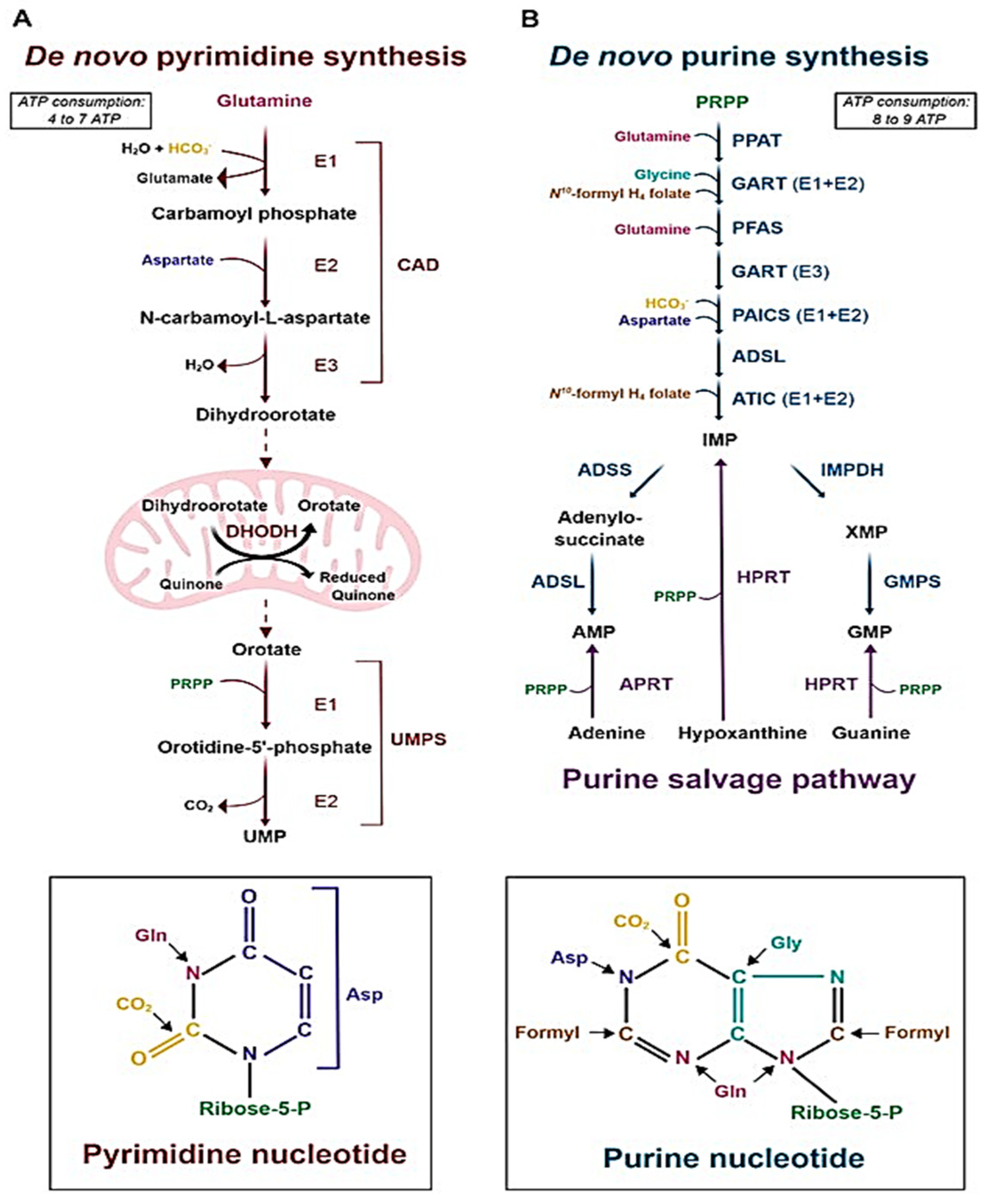
In purine biosynthesis, an aminoimidazole moiety is produced and connected to a ribose by a sequence of reactions that are metabolized by the enzyme phosphoribosylpyrophosphate (PRPP) synthetase, which is dependent on inorganic phosphate, an essential metabolite for bacterial survival [22,23]. The C4 and C5 of imidazole then undergo cyclization, resulting in the production of inosine-5′-monophosphate (IMP), which is then transformed into guanosine 5′-monophosphate (GMP) by IMP dehydrogenase (IMPDH, GuaB) [29,30]. GuaB, one of the three IMPDH homologues that are currently known, is thought to be a viable therapeutic target because of its critical involvement in the metabolism of guanine nucleotides and as the cascade’s rate-limiting enzyme [21,25].
Uridine monophosphate, a precursor for pyrimidine nucleotides, is produced during the multi-enzymatic cascade reaction of pyrimidine biosynthesis [24]. Promising therapeutic targets have been investigated, including the cascade that orotate phosphoribosyltransferase (OPRT) catalyzes to transform orotate into orotidine 5′-monophosphate (OMP) [31,32]. Additionally, PRPP synthase, an enzyme that synthesizes PRPP, in turn, is necessary to produce both pyrimidine and purine nucleotides. Therefore, blocking this enzyme will also likely be a target for drugs (Figure 2) [29,30].

2.2.2. Deoxyribonucleic Acid (DNA) Replication

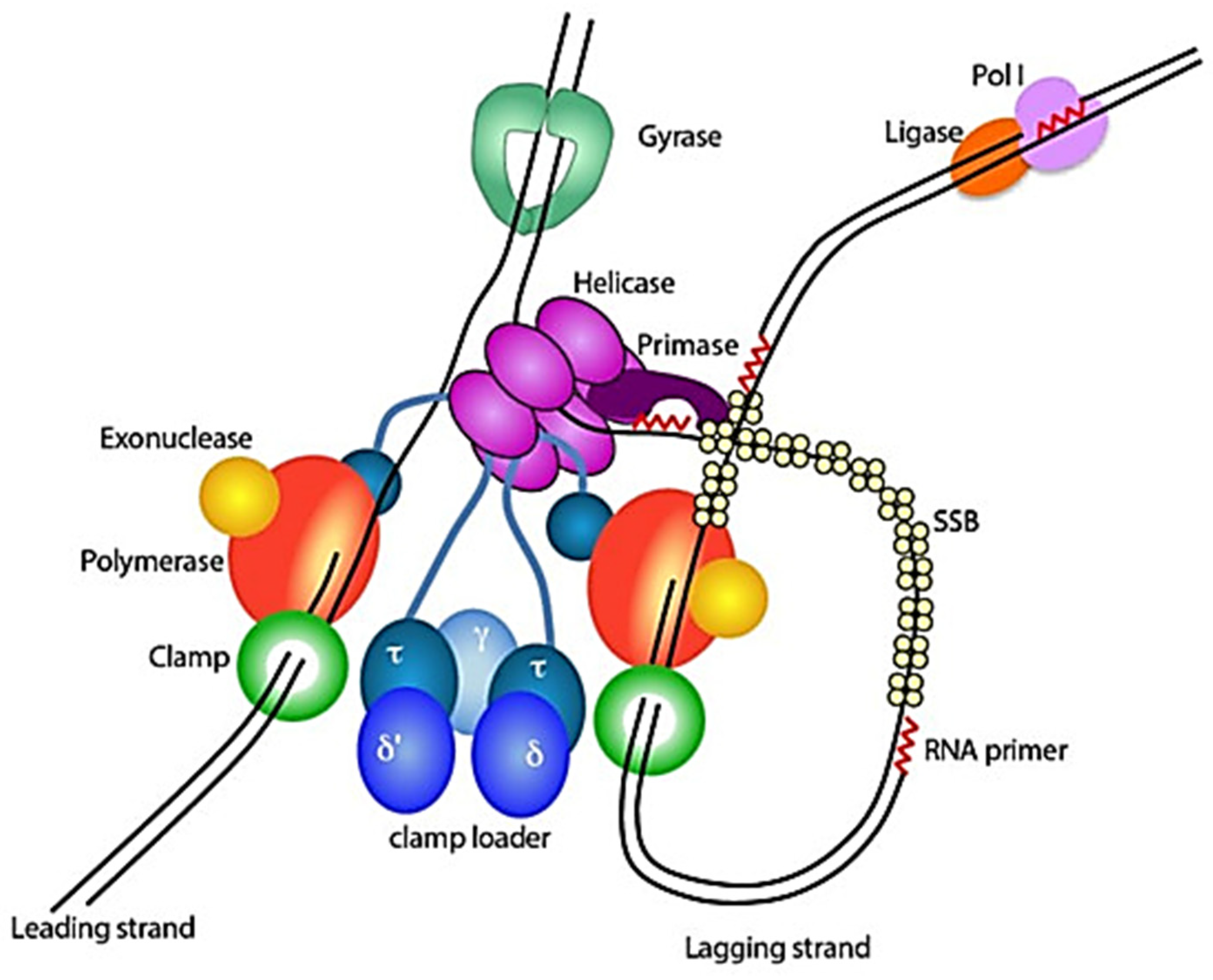
A multistep, multiprotein replisome in bacteria performs DNA replication in a highly controlled and coordinated manner [34]. These replisome proteins catalyze the production of RNA primers, clamp loading, DNA polymerization, and DNA unwinding [29]. Nearly 3950 genes make up the genome of M. tuberculosis, and 10% of them are necessary for the mycobacteria to survive [35,36]. The DnaA replication initiator, the primosomal (P) helicase, the DnaB helicase, the DnaG primase, the single-stranded DNA binding proteins (SSB), the clamp loader subunits, the DNA polymerases I and III, the DnaN-clamp, the DNA ligase I, and type I and II topoisomerases are among the 15 genes that are necessary for DNA replication (Figure 3) [34,37]. The single topoisomerase II class member, DNA gyrase (mostly DNA gyrA), is the only target for fluoroquinolones in the treatment of MDR-TB [38]. Therefore, there is a great potential to search for targets as DNA replication inhibitors and anti-mycobacterial drugs.

2.2.3. DNA Repair

Due to the release of reactive oxygen species (ROS) and reactive nitrogen species (RNS), M. tuberculosis continuously sustains DNA damage in its host cell [39]. One of the RNS, nitrogen monoxide, interacts with oxygen to form nitrous anhydride, which then nitrosates amines and amides to produce potent DNA alkylating agents that endanger the survival of the bacteria because of genome instability [29]. Multi-enzymatic systems, including Nucleotide Excision Repair (NER) and Base Excision Repair (BER), recombination repair systems, and single proteins that directly reverse DNA damage, are all part of M. tuberculosis DNA repair mechanisms [40]. As a result, antimycobacterial agents that target this bacterial pathway will have potential. However, they possess a challenge of selectivity [29].

2.3. Protein Synthesis (RNA Translation)

The ribosome of M. tuberculosis is the key component in the translation of RNA into proteins [19]. The smaller subunit of ribosomes, known as 30S, contains 21 ribosomal proteins (RPs) and 16S ribosomal RNA (rRNA), which are used to decode messenger RNA (mRNA) sequences, and the larger subunit, known as 50S, contains 37 RPs and 20S and 5S rRNA and is used to form peptide bonds via the peptidyl transferase center (PTC) [19,41]. The 70S functional ribosome is made up of the two subunits [42]. The two subunits form the functional 70S ribosome [41]. Since these subunits’ interface is where the incoming transfer RNA (tRNA) passes via the aminoacyl (A-site), peptidyl (+P-site), and exit (E-site) sites, it is a crucial translational step [12]. The decoding center in the 30S and PTC and polypeptide exit site at the 50S subunit on the ribosome are prospective targets on the basis of the already available antibiotics such as aminoglycosides [43,44].

2.4. Energy Metabolism

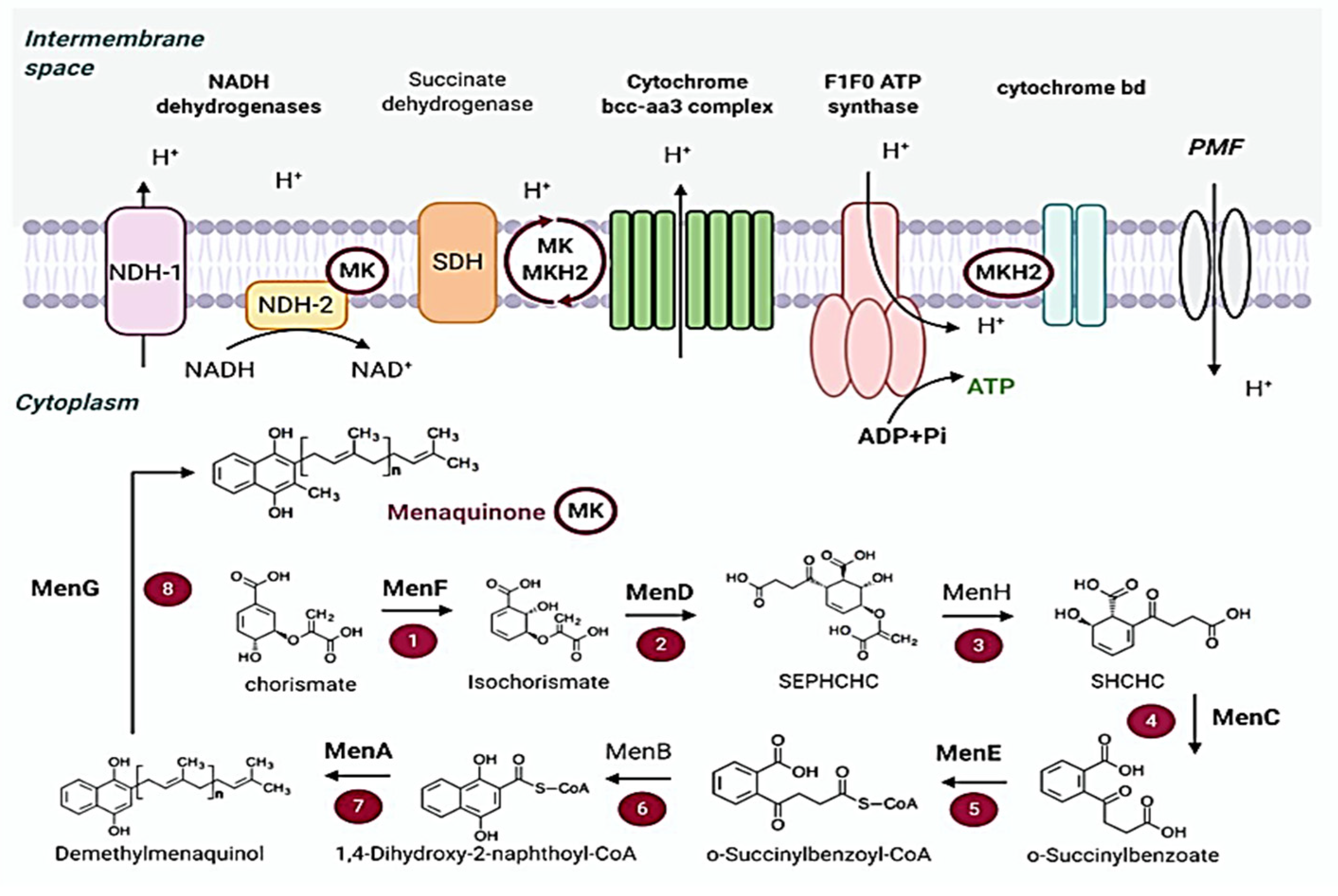
M. tuberculosis produces ATP through oxidative phosphorylation, which is essential for growth and survival [45]. M. tuberculosis can produce ATP via substrate-level phosphorylation; however, it is insufficient [46]. Therefore, the primary source of energy for the bacteria is oxidative phosphorylation. NADH dehydrogenases and succinate dehydrogenases aid in the entry of the electron transport chain that transfers electrons to menaquinone, generating a protein complex and proton motive force (PMF), in the process of oxidative phosphorylation [46,47]. Adenosine triphosphatase (ATP synthase) uses the energy of this enzyme to produce ATP [35]. Since oxidative phosphorylation differs significantly between prokaryotes and eukaryotes, there are questions about the selectivity of antitubercular medications that target this route (Figure 4) [46,48]. Despite this, there are already available clinical and experimental medications that more specifically block the routes used by mycobacteria to synthesize ATP, revealing alternate targets for treating MDR and XDR infections.

3. Natural Products with Potent Antimycobacterial Activities

Numerous natural compounds derived from both aquatic and terrestrial sources, such as microbes and higher plants, have demonstrated potent anti-mycobacterial properties against various strains of mycobacteria. In vitro assays are used to determine the inhibitory concentration (IC) or minimum inhibitory concentration (MIC) of natural compounds [49,50]. For further study as potential anti-TB drug candidates, natural compounds with in vitro MIC ≤ 10 µg/mL or 50 µM are considered to have a significant antimycobacterial action. Therefore, these types of compounds are considered for further investigation, provided that their selectivity indices (SI) are greater than 10 [51,52]. This review focuses on natural products discovered between 2000 and 2024, with particular emphasis on compounds identified from 2020 to 2024 that exhibit potent in vitro activity against M. avium, M. bovis, M. smegmatis, and M. tuberculosis (Table 1). These products should demonstrate minimum inhibitory concentration (MIC) values of ≤10 µg/mL or 50 µM, along with a selectivity index (SI) greater than 10. The bioactive compounds meeting these criteria mainly fall within the classes of alkaloids, terpenes, peptides, and steroids.

3.1. Alkaloids

Alkaloids, which are a diverse group of nitrogen-containing natural products, have demonstrated promising potential against TB. Many plants, cyanobacteria, and marine-derived alkaloids have demonstrated potent antimycobacterial activity, with high selectivity indices (Table 1).
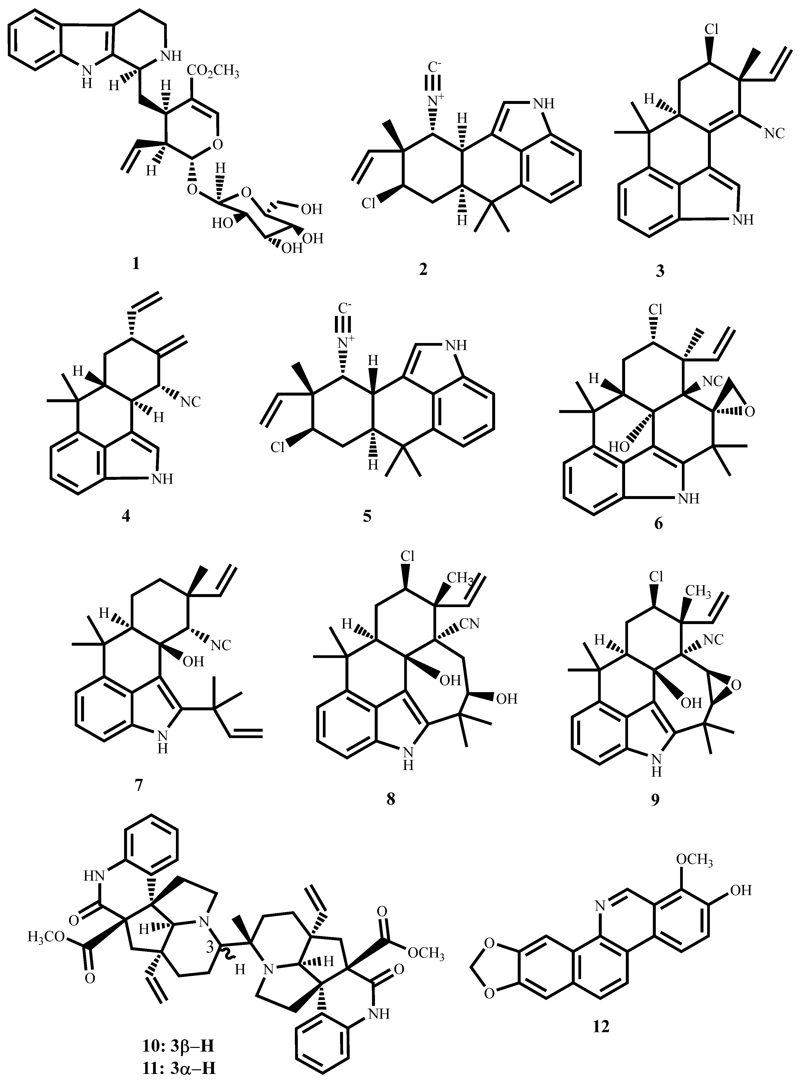
Strictosidine (1, Figure 5), a monoterpene indole obtained from the methanol extract of the leaves and twigs of Psychotria nuda (Cham. & Schltdl.) Wawra (Rubiaceae), showed an MIC value of 13.7 µM against M. tuberculosis H37Rv with a cytotoxic effect of 170.93 µM against RAW264.7 cells in the MTT assay [53].
The hapalindole-type alkaloids (25, Figure 5) and, fischambiguine B (6) and ambiguine isonitriles (79) from Fischerella ambigua and Westiellopsis species exhibit potent antimycobacterial activity (Table 1). Their antimycobacterial activity revealed key structure–activity relationships where epoxidation at C25–26, as in fischambiguine B (6), radically enhances potency and selectivity against M. tuberculosis H37Rv (MIC ~2 µM; SI > 60). Similarly, ambiguine E isonitrile (9) also contains an epoxy ring, which is fused with the seven-membered E ring. However, its activity dropped by 10-fold. This implies that the spiro-epoxy containing six six-membered rings is essential for activity. Moreover, chlorination at C13 is non-influential on activity. Hapalindole G, which lacks the epoxide but remains chlorinated, is moderately active and less selective (SI > 18). For ambiguine isonitriles, compounds C and M are moderately active against M. tuberculosis (MIC = 7 µM; SI ≈ 11), but ambiguine E is exceptionally selective for M. smegmatis (MIC = 1.4 µM; SI = 30.42), while being less active against M. tuberculosis. Taken together, these findings point to the significant role played by the epoxide moiety in optimizing antimycobacterial activity and reaffirm the isonitrile core as being requisite for lower activity [54,55,56,57].
Suadamin A (10) and Suadamin B (11) are dimeric monoterpenoid quinoline alkaloids, which were obtained from Melodinus suaveolens (Hance) Champ, ex Benth. Both compounds exhibited antimycobacterial activity against M. tuberculosis H37Rv, with Suadamin A having a significantly lower MIC (6.76 μM) (Table 1). The SAR analysis revealed that their stereochemistry at the C-3 position is the main cause of the difference in potency [58]. Stereochemistry often dictates drug–target interactions since substituent spatial arrangements determine how the compound fits into the binding site of its mycobacterial target. For Suadamins, the C-3 stereogenic center controls the orientation of the most critical functional groups in the dimeric framework that influence hydrogen bonding, hydrophobic contact, and steric complementarity with the bacterial enzyme or membrane constituent responsible for the antimycobacterial effect.
For Suadamin A, the C-3 stereochemistry facilitates the quinoline and monoterpenoid functionalities to assume a conformation that better complements the mycobacterial target’s binding pocket, increasing activity. Conversely, the other stereochemistry of Suadamin B likely causes steric clash or pharmacophore misalignment, consequently reducing binding affinity and antimycobacterial activity. This conforms to broad general medicinal chemistry principles where stereochemical differences, occasionally even a sole chiral center, can result in large variations in bioactivity, as in thalidomide enantiomers or quinine versus quinidine [74,75]. Thus, the higher selectivity index and potency of Suadamin A emphasize the importance of stereochemistry in natural product drug discovery. The findings suggest that precise stereochemical control at important centers such as C-3 should be considered in any possible synthetic or semi-synthetic modification of suadamins and related alkaloids for drug development against tuberculosis.
Another isoquinoline-based alkaloid, decarine (12), isolated from the roots of Zanthoxylum capense (Thunb.) Harv., has been shown to exhibit substantial activity against M. tuberculosis H37Ra and H37Rv, with MIC values of 9.71 μM and 5.01 μM, respectively (SI = 41.2) [59].

3.2. Simple Amide and Peptides

Cyclic peptides and usual amides have become potential antimycobacterial drugs, showing potent activity against both drug-sensitive and drug-resistant M. tuberculosis strains. The cyclic peptides and amides contained in this review are mostly derived from Streptomyces and Zanthoxylum capense (Thunb.) Harv (Table 1). They have been shown to exhibit bactericidal activity at low MIC values with low cytotoxicity. The mechanisms of action of cyclic peptides and amides, particularly their inhibition of ClpC1 ATPase, provide a new direction for the discovery of therapeutics against tuberculosis.
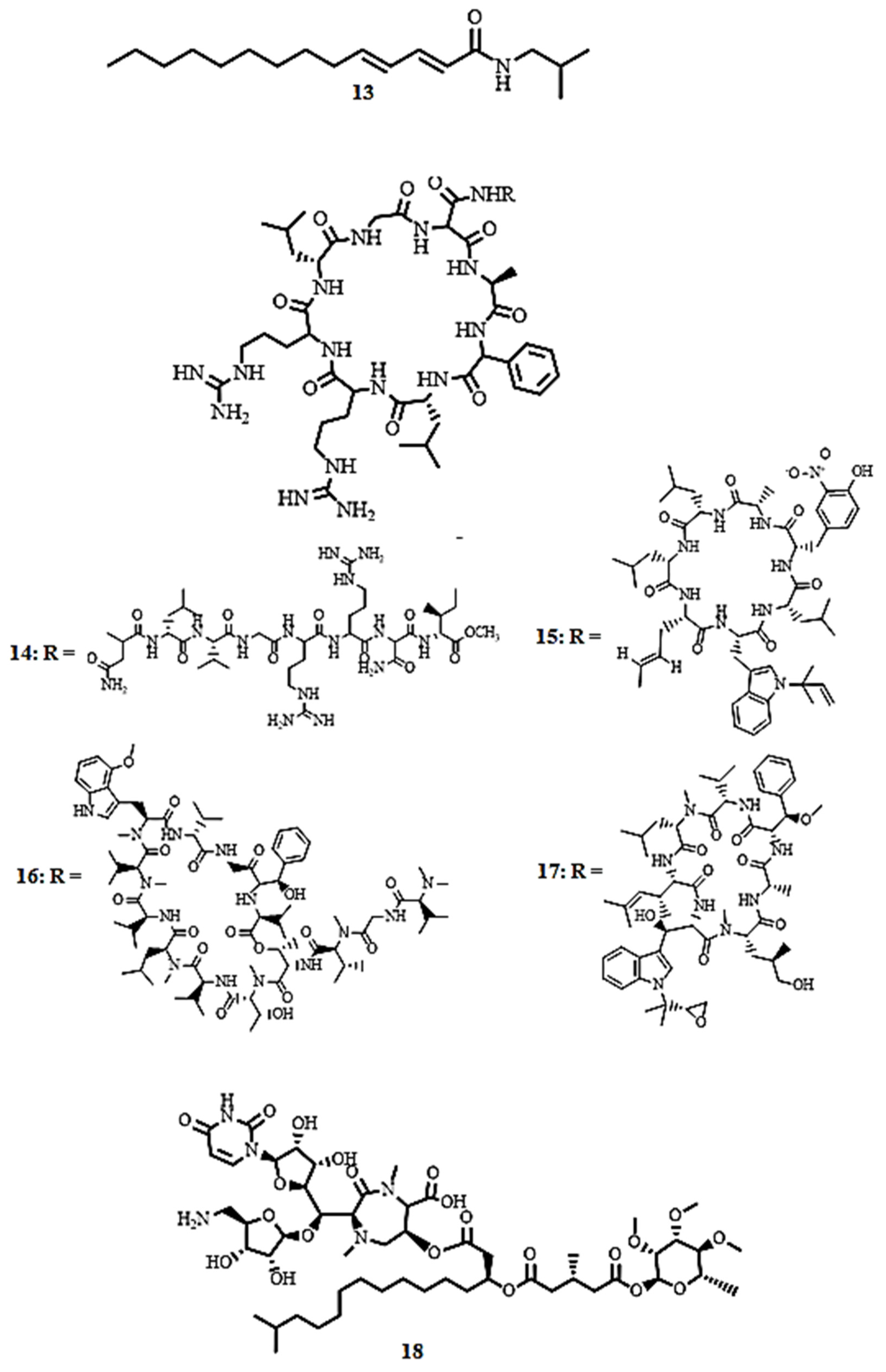
(2E,4E)-N-Isobutyl-2,4-tetradecadienamide (13, Figure 6), a simple amide isolated from the roots of Zanthoxylum capense (Thunb.) Harv demonstrated antimycobacterial activity against M. tuberculosis H37Rv with MIC values of 5.73 µM (SI = 38.2), in the broth micro-dilution method [59].
Lassomycin (14, Figure 6), a cyclic peptide isolated from Lentzea kentuckyensis, exhibits potent bactericidal activity against M. smegmatis, M. tuberculosis H37Rv, and M. avium subsp. paratuberculosis, including drug-resistant (MDR) and extensively drug-resistant (XDR) strains, with MICs ranging from < 0.004 to 1.66 µM and low cytotoxicity against human NIH 3T3 and HepG2 cells (IC50 = 187.34 µM) [16]. Molecular docking studies reveal that lassomycin is sequestered in the acidic ATPase pocket of the ClpC1 caseinolytic protein, inhibiting its proteolytic activity [9]. Cyclic peptides rufomycin I (15, MIC < 0.004 µM, SI > 250), ecumicin (16, MIC = 0.16 µM, SI > 192), and cyclomarin A (17, MIC = 0.094 µM, SI = 26.7), derived from Streptomyces sp., were active against drug-sensitive and drug-resistant M. tuberculosis H37Rv, M. smegmatis, and M. avium, with rufomycin I being more potent than isoniazid (MIC = 0.23 µM) [60,61]. These cyclic peptides targeting ClpC1 represent a novel and promising antibacterial approach. Instead of classical enzyme inhibition, they disable the essential proteolytic machinery of M. tuberculosis by decoupling or deregulating critical functional cycles. This innovative approach, whether through uncoupling ATP hydrolysis from proteolysis, selectively inhibiting substrate turnover, or misdirecting the protease, leads to a lethal collapse of proteostasis [60,61]. Consequently, these compounds emerge as lead candidates for the development of new anti-TB drugs, offering a unique strategy to treat both drug-sensitive and drug-resistant mycobacterial infections.
Caprazamycin B (18, Figure 6), a novel peptide from one of the Streptomyces isolates, showed excellent inhibitory activity against M. tuberculosis H37Rv Kurono and M. bovis Ravenel, with the MIC value of 2.73 µM. The compound also showed a very good safety profile with no in vivo cytotoxicity observed in mice at 4368.4 µM, which corresponds to a selectivity index (SI) value above 1500 [62]. These results bring into focus caprazamycin B as a promising molecule for further development as a selective antitubercular agent.

3.3. Terpenoids

Strong activity against M. tuberculosis H37Rv and M. tuberculosis M299 was demonstrated by the squalene-type triterpenoid eurylene (19, Figure 7), isolated from the roots of Homalolepis suffruticosa (Engl.) Devecchi & Pirani, with MICs of 2.35 µM and 3.36 µM, respectively. These compounds had better selectivity (SI > 20) when tested with the MTT method [63].
12-Deacetoxyscalarin 19-acetate (20), a scalarane type sesterterpenoid, was isolated from marine sponges. It is an extremely potent antimycobacterial compound against M. tuberculosis H37Ra with an MIC of 4 µM (Table 1) [64]. Its action mode is the inhibition of farnesyltransferase, an enzyme catalyzing post-translational modification of proteins by attaching a farnesyl group [76]. This inhibition also prevents the functioning of membrane-associated proteins and leads to compromised bacterial membrane integrity and subsequent cell death. This positions 12-deacetoxyscalarin 19-acetate as a lead compound in the design of novel antimycobacterial agents targeting membrane-associated processes.
Nieves et al. [65] reported that (−)-8,15-diisocyano-11(20)-amphilectene (21) exhibited strong antimycobacterial activity against M. tuberculosis H37Ra (MIC = 9.8 µM) with moderate selectivity toward mammalian cells (SI = 10.2).

3.4. Steroids

Steroidal compounds have displayed potent inhibitory activity against M. tuberculosis, including the drug-sensitive strains, particularly the triterpene and phytosterol derivatives. The steroids, including saringosterol and stigmastene, were made from plant and marine algae, and had relatively low MIC values, some equaling the potency of standard drugs such as rifamycin. The selectivity indices and structure–activity relationships were promising, particularly in terms of the role of unsaturated ketone moieties, indicating the steroids could have utility as scaffolds for anti-TB drug development (Table 1).
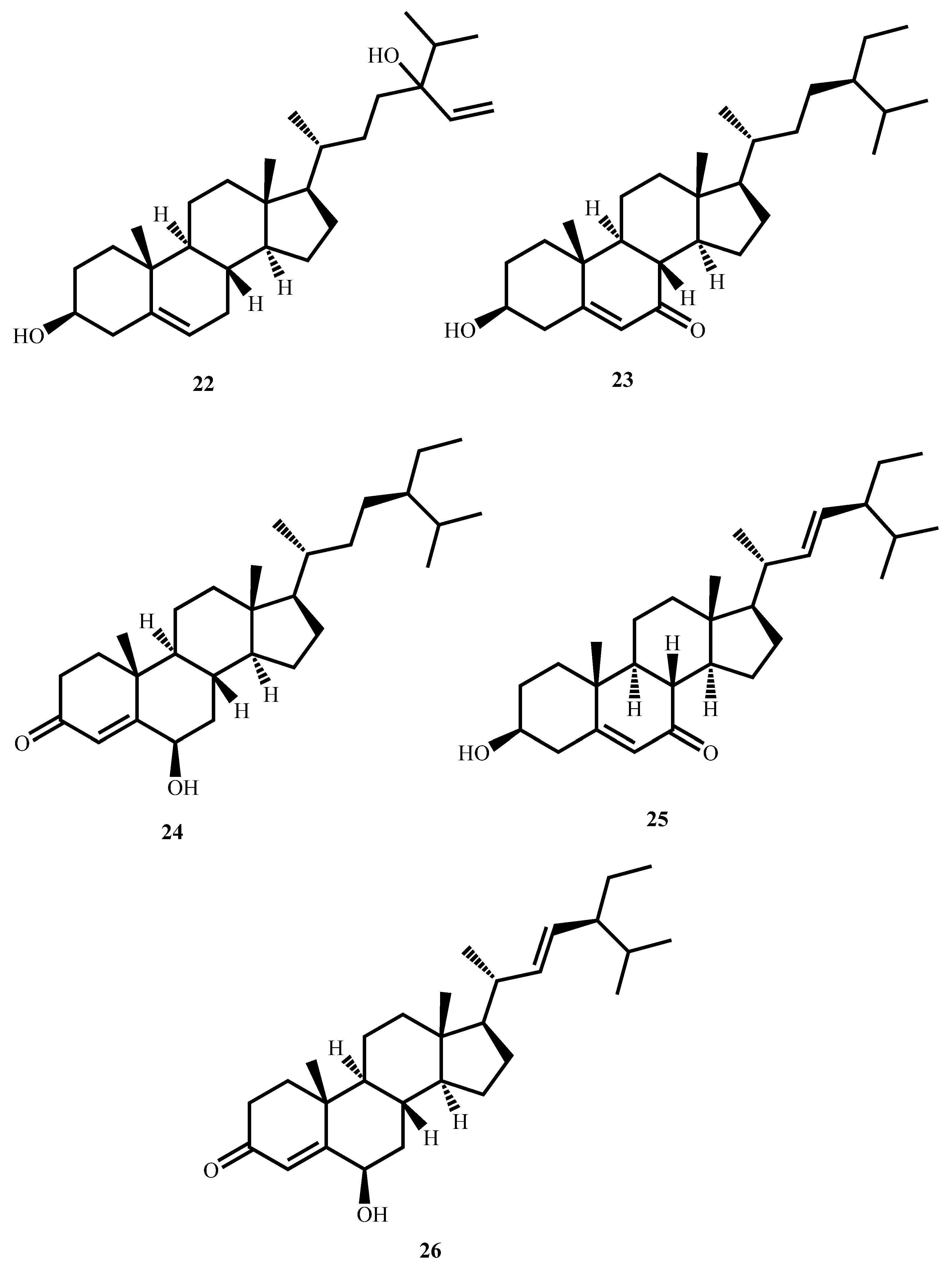
Saringosterol (22, Figure 8), a triterpene steroid from the brown alga Lessonia nigrescens, possesses potent antimycobacterial activity against M. tuberculosis H37Rv (MIC = 0.58 µM). The single 24S and 24R epimers of saringosterol were also analyzed in the study, and they presented MIC values of approximately 2.33 µM and 0.29 µM, respectively, indicating that the 24R epimer is more active. The findings indicate the promise of saringosterol and its epimers as potential leads for the development of new antimycobacterial agents [66].
Bioassay-guided fractionation of the aerial parts of Thalia multiflora Horkel ex Koernicke (Marantaceae) using dichloromethane–methanol (1:1) gave stigmast-5-ene-3β-ol-7-one (23, MIC = 4.62 µM), stigmast-4-ene-6β-ol-3-one (24, MIC = 10.07 µM), stigmast-5,22-diene-3β-ol-7-one (25, MIC = 2.34 µM), and stigmast-4,22-diene-6β-ol-3-one (26, MIC = 2.35 µM) against M. tuberculosis H37Rv [67]. Structure–activity relationship studies revealed that the presence of an α,β–unsaturated ketone on ring A or ring B significantly enhances antitubercular activity. Of interest, all of the compounds were found to be highly selective against the pathogen with selectivity indices greater than 10 against Vero cells, denoting low cytotoxicity against mammalian cells. The findings suggest that these stigmastane analogs are good prospects for further development as selective antitubercular drugs.

3.5. Miscellaneous Compounds

There exists a wide variety of structurally diverse natural products with antimycobacterial activities, including fatty acid derivatives, polyketides, lactones, organic acids, and others. Certain compounds (e.g., falcarindiol and micromolide) exhibited low values of MIC with selectivity indices and good mechanisms of action (e.g., inhibition of the biosynthesis of para-aminobenzoate (PABA) and succinate dehydrogenase). The potent activity of these compounds against drug-sensitive and resistant strains of M. tuberculosis supports and may indicate their potential for use in future explorations for anti-TB drugs (Table 1).
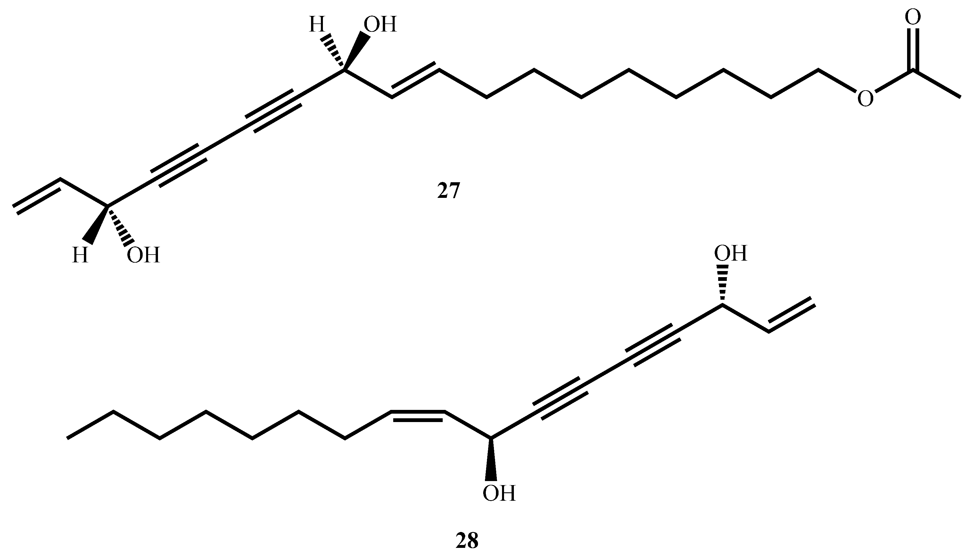
11(S),16(R)-Dihydroxy-octadeca-9Z,17-dien-12,14-diyn-1-yl acetate (27, Figure 9) and falcarindiol (28), isolated from the methanol extract of Angelica sinensis (Oliv.) Diels roots, were robustly antimycobacterially active against M. tuberculosis Erdman, with MICs of 4.21 µM and 23.04 µM, respectively [68]. Both compounds showed high selectivity against the pathogen, with selectivity indices of >117 and >19, respectively, indicating minimal toxicity toward mammalian cells. Structure–activity relationship (SAR) investigations revealed that antimycobacterial activity is maintained with the hydroxyl group at C16 and with an unsaturation at C9–C10 [68]. The findings suggest that the compounds are good candidates for further development as selective antimycobacterial drugs and provide valuable information for designing derivatives with improved efficacy and safety profiles [40].
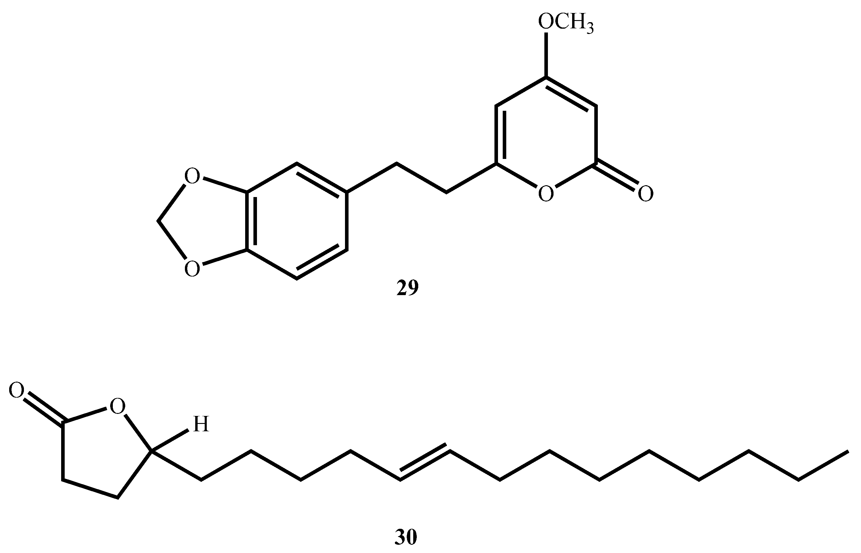
Two lactones with potential antimycobacterial activity were isolated. Kavalactone 5,6-dehydro-7,8-dihydromethysticin (29, Figure 10) from the leaves and stems of Piper sanctum (Miq.) Schltdl. ex C. DC. inhibited M. tuberculosis H37Rv with an MIC of 12.04 µM [69]. Micromolide (30), a γ-lactone oleic acid derivative from Micromelum hirsutum Oliv., was even more active (MIC = 5.35 µM) with a high SI value (63) against Vero cells. Micromolide was further demonstrated to exhibit robust efficacy under more physiological conditions with an EC90 of 19.97 µM against the virulent Erdman strain in J774 mouse macrophages, and thus it presents as a promising lead for future in vivo studies [70].
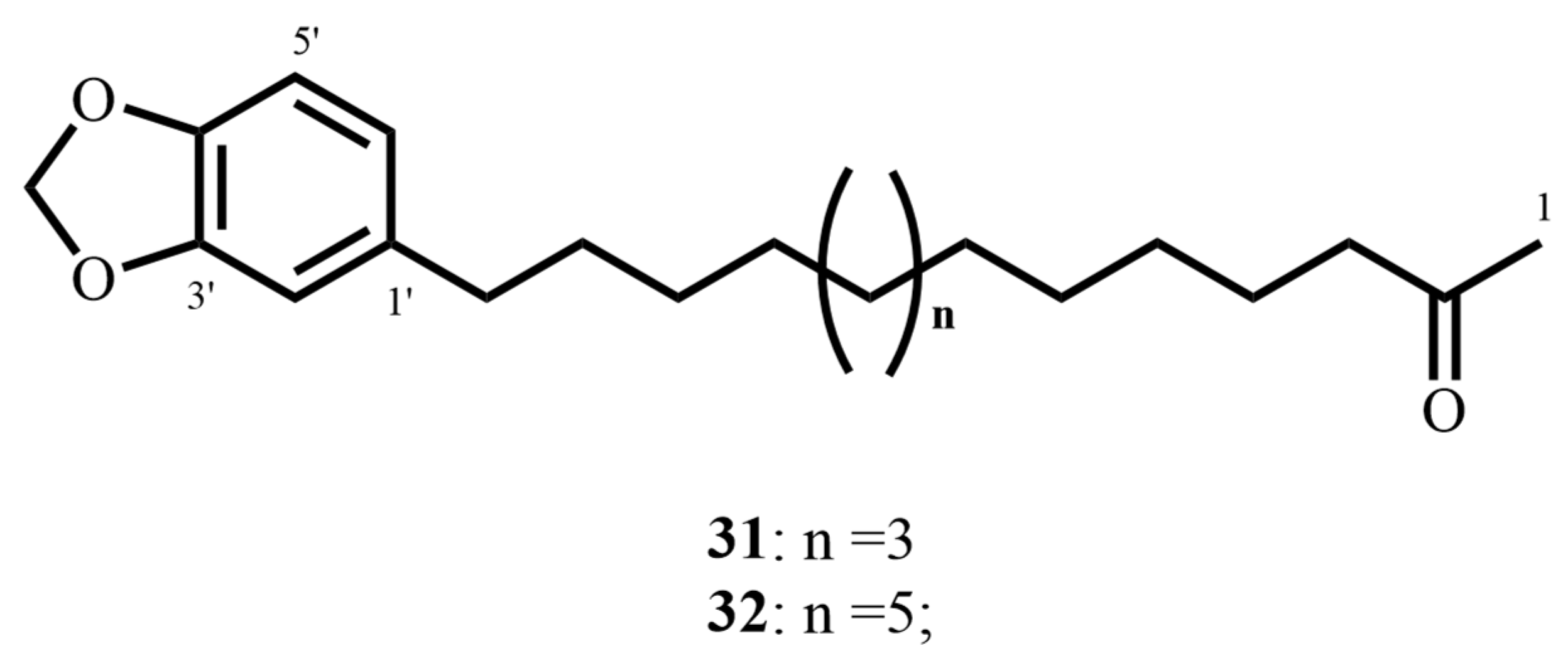
Besides these lactones, two aryl-alkyl ketones, 2-oxo-14-(3′,4′-methylenedioxyphenyl)-tetradecane (31, Figure 11) and 2-oxo-16-(3′,4′-methylenedioxyphenyl)-hexadecane (32), were identified in P. sanctum. Both of these compounds inhibited M. tuberculosis H37Rv with MIC ranges of 12.04–18.81 µM and excellent selectivity indices greater than 10 against Vero cells. This combination of potent antimycobacterial activity with minimal cytotoxicity positions the aryl-alkyl ketones, as well as the lactones, very favorably as scaffolds for selective antitubercular drug development [69].
Linoleic acid (33, Figure 12), isolated from the CH2Cl2 extract of the stem bark of Warburgia ugandensis, showed powerful activity against M. aurum and M. phlei with MIC values of 14.27 µM. In addition, it showed activity against M. fortuitum (MIC = 28.53 µM) [71]. M. tuberculosis has evolved to utilize long-chain fatty acids (LCFAs) as a preferred carbon source. However, LCFAs have long been known to exhibit bactericidal effects against M. tuberculosis in vitro by disrupting the bacterial membrane, leading to hyperpolarization and subsequent cell death. A deeper understanding of how M. tuberculosis regulates the uptake and metabolism of LCFAs, and how these pathways can be therapeutically targeted, is essential for the development of novel anti-TB agents.
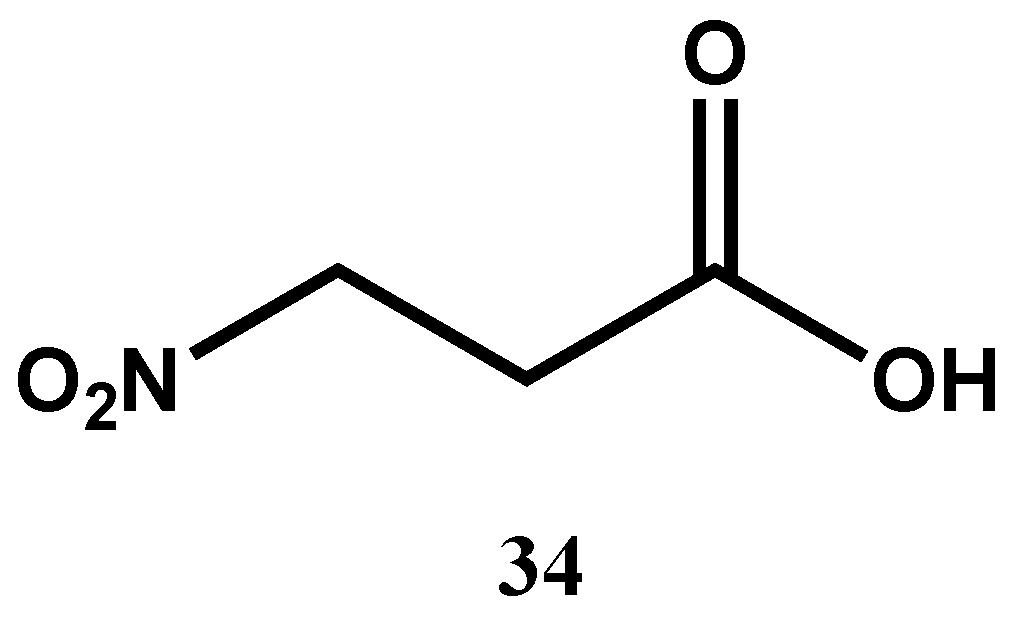
3-Nitropropionic acid (34, Figure 13) isolated from the endophytic fungal strain Phomopsis sp. strain usia5 was found to be a potent antimycobacterial with an MIC of 3.3 μM against M. tuberculosis H37Ra, without any detectable cytotoxicity against Vero cells [72]. Its mode of action was predicted to inhibit succinate dehydrogenase, a critical enzyme of the tricarboxylic acid (TCA) cycle and the electron transport chain [77]. By inhibiting succinate dehydrogenase, 3-nitropropionic acid decouples both energy generation and redox homeostasis in mycobacteria, which are essential for their survival and persistence. Its strong antimycobacterial activity, low host toxicity, and distinct mechanism of action compared to conventional antitubercular drugs make it a promising lead compound for the development of novel therapeutics and validate energy metabolism as a target for antibacterial drug discovery.
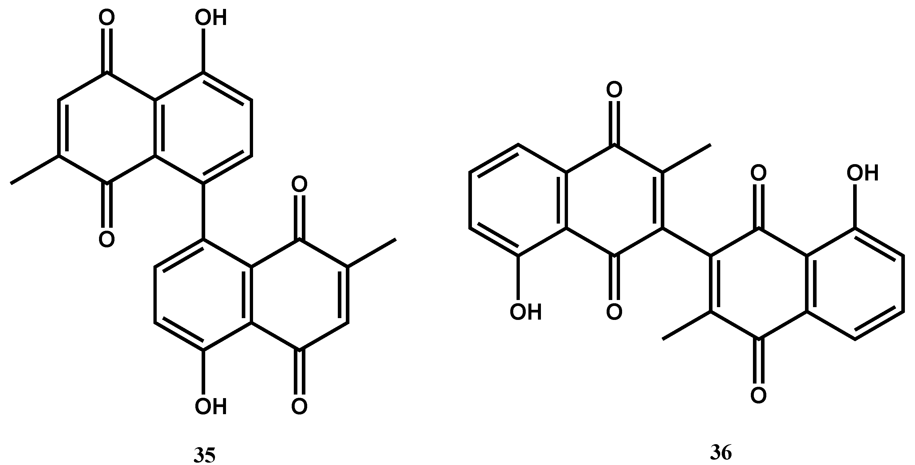
Maritinone (35, Figure 14) and 3,3′-biplumbagin (36), naphthoquinones isolated from the n-hexane extract of the stem bark of Diospyros anisandra S.F.Blake, demonstrated strong activity against drug-sensitive M. tuberculosis H37Rv with an MIC value of 8.34 µM each (SI > 10; against Vero cells) [73]. The above anthraquinones have been found to inhibit flavin-dependent thymidylate synthase ThyX, which is responsible for catalyzing the essential methylation of dUMP to dTMP for DNA synthesis in M. tuberculosis [78]. Targeting ThyX is an excellent strategy for antitubercular lead optimization: the enzyme is essential for mycobacterial replication and viability in latent and intracellular infections, yet absent in humans, thereby offering highly selective inhibition with low host toxicity. Structural and mechanistic differences between ThyX and the human thymidylate synthase (ThyA), including flavin cofactor dependency and distinct active-site topology, also allow for the potential development of selective inhibitors, providing a promising avenue toward novel tuberculosis treatments [79].

4. Conclusions

Existing therapies have had little impact on the management of TB on a worldwide scale, and new regimens are urgently required. The emergence of MDR and XDR forms of M. tuberculosis has rendered many of the existing therapeutic agents ineffective, yet again emphasizing the need for drugs with novel modes of action and targets. Natural products from a vast array of sources have been incredibly promising as antimycobacterial compounds with potential clinical use. Agents such as hapalindole A (MIC < 0.6 µM; SI > 53), lassomycin (MIC = 0.78–1.56 µg/mL; SI > 224), rufomycin I (MIC < 0.004 µM; SI > 250), ecumicin (MIC = 0.26 µg/mL; SI > 192), and cyclomarin A (MIC = 0.094 µM; SI = 26.7) have shown excellent in vitro activity with high selectivity indices, making them candidates of interest for drug discovery. These findings suggest that natural products remain one of the richest sources of leads for new drugs and will undoubtedly remain involved in antimycobacterial drug discovery. But optimization of such discoveries into drugs of clinical utility is highly constrained. Most anti-TB natural product leads are found through phenotypic screens versus rational target-based technologies, which can be inhibitive to hit-to-lead optimization. Other difficulties include natural product structural complexity, sustainable large-scale production challenges, inadequate solubility and biocompatibility, potential host toxicity, and yield variability depending on environmental or seasonal parameters, with the further barrier of prolonged preclinical screening mechanisms.
These difficulties can be addressed with multidisciplinary approaches involving natural product chemistry, microbiology, pharmacology, and computational sciences. Semi-synthetic derivatization of lead-like scaffolds can enhance pharmacokinetic properties, and metabolomics, genomics, synthetic biology, and artificial intelligence have the potential to accelerate lead discovery and optimization. Representative M. tuberculosis-specific screening platforms and in silico ADMET and molecular docking studies should be incorporated early in the drug development pipeline to enhance efficiency. In addition to this, sustainable sourcing schemes such as microbial fermentation, endophytic culture, and plant tissue culture need to be guaranteed to provide compound availability consistency. Finally, natural products provide unparalleled chemical diversity and new mechanisms of action, but their complete potential as sources for TB drug discovery depends on high-tech, cooperative, and technologically synergistic approaches. With sustained investment and global cooperation, the projects could finally offer safe, affordable, and cost-effective anti-TB drugs to counteract the increasing menace of drug-resistant TB.

Author Contributions

Conceptualization, B.P., K.A., D.B. and M.Y.Y.; software and data curation, B.P., K.A. and M.Y.Y.; writing—original draft preparation, B.P., K.A. and D.B.; writing—review and editing, B.P., M.Y.Y., K.A. and D.B., who have all likewise contributed to this work. All authors have read and agreed to the published version of the manuscript.

Funding

This study was partially funded by the International Science Program (ISP), Uppsala University, through the ETH:02 project.

Institutional Review Board Statement

Not applicable.

Informed Consent Statement

Not applicable.

Data Availability Statement

There is no additional data associated with this article.

Acknowledgments

One of the authors (B.P.) would like to express his gratitude for the School of Pharmacy, Addis Ababa University.

Conflicts of Interest

The authors declare no conflicts of interest.

References

  1. Abrahams, K.A.; Besra, G.S. Mycobacterial drug discovery. RSC Med. Chem. 2020, 11, 1354–1365. [Google Scholar] [CrossRef]
  2. World Health Organization. The WHO Global Tuberculosis Report; World Health Organization: Geneva, Switzerland, 2023.
  3. Beste, D.J.V.; Espasa, M.; Bonde, B.; Kierzek, A.M.; Stewart, G.R.; McFadden, J. The genetic requirements for fast and slow growth in Mycobacteria. PLoS ONE 2009, 4, e5349. [Google Scholar]
  4. Gill, W.P.; Harik, N.S.; Whiddon, M.R.; Liao, R.P.; Mittler, J.E.; Sherman, D.R. A replication clock for Mycobacterium tuberculosis. Nat Med. 2009, 15, 211–214. [Google Scholar]
  5. Li, A.H.; Lam, W.L.; Stokes, R.W. Characterization of genes differentially expressed within macrophages by virulent and attenuated Mycobacterium tuberculosis identifies candidate genes involved in intracellular growth. Microbiology 2008, 154 Pt 8, 2291–2303. [Google Scholar] [CrossRef]
  6. Bermudez, L.E.; Shelton, K.; Young, L.S. Comparison of the ability of Mycobacterium avium, M. smegmatis and M. tuberculosis to invade and replicate within HEp-2 epithelial cells. Tuber. Lung Dis. 1995, 76, 240–247. [Google Scholar] [CrossRef]
  7. Chaturvedi, V.; Dwivedi, N.; Tripathi, R.P.; Sinha, S. Evaluation of Mycobacterium smegmatis as a possible surrogate screen for selecting molecules active against multidrug-resistant Mycobacterium tuberculosis. J. Gen. Appl. Microbiol. 2007, 53, 333–337. [Google Scholar] [CrossRef]
  8. Zheng, H.; Lu, L.; Wang, B.; Pu, S.; Zhang, X.; Zhu, G.; Zhu, G.; Shi, W.; Zhang, L.; Wang, H.; et al. Genetic basis of virulence attenuation revealed by comparative genomic analysis of Mycobacterium tuberculosis strain H37Ra versus H37Rv. PLoS ONE 2008, 3, e2375. [Google Scholar] [CrossRef]
  9. Heinrichs, M.T.; May, R.J.; Heider, F.; Reimers, T.; B Sy, S.K.; Peloquin, C.A.; Derendorf, H. Mycobacterium tuberculosis strains H37ra and H37rv have equivalent minimum inhibitory concentrations to most antituberculosis drugs. Int. J. Mycobacteriol. 2018, 7, 156–161. [Google Scholar] [CrossRef]
  10. Bueno-Sánchez, J.G.; Martínez-Morales, J.R.; Stashenko, E.E.; Ribón, W. Anti-tubercular activity of eleven aromatic and medicinal plants occurring in Colombia. Biomed. Rev. Inst. Nac. Salud 2009, 29, 51–60. [Google Scholar]
  11. Zhang, Y.; Amzel, L.M. Tuberculosis drug targets. Curr. Drug Targets 2002, 3, 131–154. [Google Scholar] [CrossRef]
  12. Lawn, S.D.; Zumla, A.I. Tuberculosis. Lancet 2011, 378, 57–72. [Google Scholar] [CrossRef]
  13. Shehzad, A.; Rehman, G.; Ul-Islam, M.; Khattak, W.A.; Lee, Y.S. Challenges in the development of drugs for the treatment of tuberculosis. Braz. J. Infect. Dis. 2013, 17, 74–81. [Google Scholar] [CrossRef]
  14. Verpoorte, R. Exploration of nature’s chemodiversity: The role of secondary metabolites as leads in drug development. Drug Discov. Today 1998, 3, 232–238. [Google Scholar] [CrossRef]
  15. Kana, B.D.; Karakousis, P.C.; Parish, T.; Dick, T. Future target-based drug discovery for tuberculosis? Tuberculosis 2014, 94, 551–556. [Google Scholar] [CrossRef]
  16. Gavrish, E.; Sit, C.S.; Cao, S.; Kandror, O.; Spoering, A.; Peoples, A.; Ling, L.; Fetterman, A.; Hughes, D.; Bissell, A.; et al. Lassomycin, a ribosomally synthesized cyclic peptide, kills Mycobacterium tuberculosis by targeting the ATP-dependent protease ClpC1P1P2. Chem. Biol. 2014, 21, 509–518. [Google Scholar] [CrossRef]
  17. Oh, S.; Trifonov, L.; Yadav, V.D.; Barry, C.E.; Boshoff, H.I. Tuberculosis drug discovery: A decade of hit assessment for defined targets. Front. Cell. Infect. Microbiol. 2021, 11, 611304. [Google Scholar] [CrossRef]
  18. Barry, C.E. Interpreting cell wall “virulence factors” of Mycobacterium tuberculosis. Trends Microbiol. 2001, 9, 237–241. [Google Scholar] [CrossRef]
  19. Shetye, G.S.; Franzblau, S.G.; Cho, S. New tuberculosis drug targets, their inhibitors, and potential therapeutic impact. Transl. Res. 2020, 220, 68–97. [Google Scholar] [CrossRef]
  20. Martínez-Hoyos, M.; Perez-Herran, E.; Gulten, G.; Encinas, L.; Álvarez-Gómez, D.; Alvarez, E.; Ferrer-Bazaga, S.; García-Pérez, A.; Ortega, F.; Angulo-Barturen, I.; et al. Antitubercular drugs for an old target: GSK693 as a promising InhA direct inhibitor. EBioMedicine 2016, 8, 291–301. [Google Scholar] [CrossRef]
  21. Campaniço, A.; Moreira, R.; Lopes, F. Drug discovery in tuberculosis. New drug targets and antimycobacterial agents. Eur. J. Med. Chem. 2018, 150, 525–545. [Google Scholar] [CrossRef]
  22. Purohit, H.J.; Cheema, S.; Lal, S.; Raut, C.P.; Kalia, V.C. In search of drug targets for Mycobacterium tuberculosis. Infect. Disord. Drug Targets 2007, 7, 245–250. [Google Scholar] [CrossRef]
  23. Kuo, M.R.; Morbidoni, H.R.; Alland, D.; Sneddon, S.F.; Gourlie, B.B.; Staveski, M.M.; Leonard, M.; Gregory, J.S.; Janjigian, A.D.; Yee, C.; et al. Targeting tuberculosis and malaria through inhibition of enoyl reductase: Compound activity and structural data. J. Biol. Chem. 2003, 278, 20851–20859. [Google Scholar] [CrossRef]
  24. Rozwarski, D.A.; Vilchèze, C.; Sugantino, M.; Bittman, R.; Sacchettini, J.C. Crystal structure of the Mycobacterium tuberculosis enoyl-ACP reductase, InhA, in complex with NAD+ and a C16 fatty acyl substrate. J. Biol. Chem. 1999, 274, 15582–15589. [Google Scholar] [CrossRef]
  25. Shirude, P.S.; Madhavapeddi, P.; Naik, M.; Murugan, K.; Shinde, V.; Nandishaiah, R.; Bhat, J.; Kumar, A.; Hameed, S.; Holdgate, G.; et al. Methyl-thiazoles: A novel mode of inhibition with the potential to develop novel inhibitors targeting InhA in Mycobacterium tuberculosis. J. Med. Chem. 2013, 56, 8533–8542. [Google Scholar] [CrossRef]
  26. Almeida Da Silva, P.E.A.; Palomino, J.C. Molecular basis and mechanisms of drug resistance in Mycobacterium tuberculosis: Classical and new drugs. J. Antimicrob. Chemother. 2011, 66, 1417–1430. [Google Scholar] [CrossRef]
  27. Duan, X.; Xiang, X.; Xie, J. Crucial components of Mycobacterium type II fatty acid biosynthesis (Fas-II) and their inhibitors. FEMS Microbiol. Lett. 2014, 360, 87–99. [Google Scholar] [CrossRef]
  28. Sun, M.; Ge, S.; Li, Z. The role of phosphorylation and acylation in the regulation of drug resistance in Mycobacterium tuberculosis. Biomedicines 2022, 10, 2592. [Google Scholar] [CrossRef]
  29. Miggiano, R.; Morrone, C.; Rossi, F.; Rizzi, M. Targeting genome integrity in Mycobacterium tuberculosis: From nucleotide synthesis to DNA replication and repair. Molecules 2020, 25, 1205. [Google Scholar] [CrossRef]
  30. Warner, D.F.; Evans, J.C.; Mizrahi, V. Nucleotide metabolism and DNA replication. Microbiol. Spectr. 2014, 2, 633–656. [Google Scholar] [CrossRef]
  31. Donini, S.; Garavaglia, S.; Ferraris, D.M.; Miggiano, R.; Mori, S.; Shibayama, K.; Rizzi, M. Biochemical and structural investigations on phosphoribosylpyrophosphate synthetase from Mycobacterium smegmatis. PLoS ONE 2017, 12, e0175815. Available online: https://journals.plos.org/plosone/article?id=10.1371/journal.pone.0175815 (accessed on 11 December 2022).
  32. Breda, A.; Martinelli, L.K.B.; Bizarro, C.V.; Rosado, L.A.; Borges, C.B.; Santos, D.S.; Basso, L.A. Wild-type phosphoribosylpyrophosphate synthase (PRS) from Mycobacterium tuberculosis: A bacterial Class II PRS? PLoS ONE 2012, 7, e39245. [Google Scholar] [CrossRef]
  33. Villa, E.; Ali, E.S.; Sahu, U.; Ben-Sahra, I. Cancer cells tune the signaling pathways to empower de novo synthesis of nucleotides. Cancers 2019, 11, 688. [Google Scholar] [CrossRef]
  34. Hedstrom, L. IMP Dehydrogenase: Structure, mechanism, and inhibition. Chem. Rev. 2009, 109, 2903–2928. [Google Scholar] [CrossRef] [PubMed]
  35. Ditse, Z.; Lamers, M.H.; Warner, D.F. DNA replication in Mycobacterium tuberculosis. Microbiol. Spectr. 2017, 5, 1128. [Google Scholar] [CrossRef]
  36. Wang, T.-C.; Chen, F.-C. The evolutionary landscape of the Mycobacterium tuberculosis genome. Gene 2013, 518, 187–193. [Google Scholar] [CrossRef]
  37. DeJesus, M.A.; Gerrick, E.R.; Xu, W.; Park, S.W.; Long, J.E.; Boutte, C.C.; Rubin, E.J.; Schnappinger, D.; Ehrt, S.; Fortune, S.M.; et al. Comprehensive essentiality analysis of the Mycobacterium tuberculosis genome via saturating transposon mutagenesis. mBio 2017, 8, e02133–e02216. [Google Scholar] [CrossRef] [PubMed]
  38. Santos, J.A.; Lamers, M.H. Novel antibiotics targeting bacterial replicative DNA polymerases. Antibiotics 2020, 9, 776. [Google Scholar] [CrossRef]
  39. Reiche, M.A.; Warner, D.F.; Mizrahi, V. Targeting DNA replication and repair for the development of novel therapeutics against tuberculosis. Front. Mol. Biosci. 2017, 4, 75. [Google Scholar] [CrossRef]
  40. Durbach, S.I.; Springer, B.; Machowski, E.E.; North, R.J.; Papavinasasundaram, K.G.; Colston, M.J.; Böttger, E.C.; Mizrahi, V. DNA alkylation damage as a sensor of nitrosative stress in Mycobacterium tuberculosis. Infect. Immun. 2003, 71, 997–1000. [Google Scholar] [CrossRef] [PubMed]
  41. Mizrahi, V.; Andersen, S.J. DNA repair in Mycobacterium tuberculosis. What have we learnt from the genome sequence? Mol. Microbiol. 1998, 29, 1331–1339. [Google Scholar] [CrossRef]
  42. Kumar, N.; Sharma, S.; Kaushal, P.S. Protein synthesis in Mycobacterium tuberculosis as a potential target for therapeutic interventions. Mol. Aspects Med. 2021, 81, 101002. [Google Scholar] [CrossRef]
  43. Yang, K.; Chang, J.; Cui, Z.; Li, X.; Meng, R.; Duan, L.; Thongchol, J.; Jakana, J.; Huwe, C.M.; Sacchettini, J.C.; et al. Structural insights into species-specific features of the ribosome from the human pathogen Mycobacterium tuberculosis. Nucleic Acids Res. 2017, 45, 10884–10894. [Google Scholar] [CrossRef]
  44. Wilson, D.N. Ribosome-targeting antibiotics and mechanisms of bacterial resistance. Nat. Rev. Microbiol. 2014, 12, 35–48. [Google Scholar] [CrossRef] [PubMed]
  45. Poehlsgaard, J.; Douthwaite, S. The bacterial ribosome as a target for antibiotics. Nat. Rev. Microbiol. 2005, 3, 870–881. [Google Scholar] [CrossRef] [PubMed]
  46. Bald, D.; Koul, A. Respiratory ATP synthesis: The new generation of mycobacterial drug targets? FEMS Microbiol. Lett. 2010, 308, 1–7. [Google Scholar] [CrossRef]
  47. Bald, D.; Villellas, C.; Lu, P.; Koul, A. Targeting energy metabolism in Mycobacterium tuberculosis, a new paradigm in antimycobacterial drug discovery. mBio 2017, 8, e00272–e00317. [Google Scholar] [CrossRef]
  48. Wellington, S.; Hung, D.T. The expanding diversity of Mycobacterium tuberculosis drug targets. ACS Infect. Dis. 2018, 4, 696–714. [Google Scholar] [CrossRef]
  49. Foo, C.S.-Y.; Pethe, K.; Lupien, A. Oxidative phosphorylation—An update on a new, essential target space for drug discovery in Mycobacterium tuberculosis. Appl. Sci. 2020, 10, 2339. [Google Scholar] [CrossRef]
  50. Bueno, J.; Kouznetsov, V. Antimycobacterial susceptibility testing methods for natural products research. Braz. J. Microbiol. Publ. Braz. Soc. Microbiol. 2010, 41, 270–277. [Google Scholar] [CrossRef]
  51. Jorgensen, J.H.; Ferraro, M.J. Antimicrobial susceptibility testing: A review of general principles and contemporary practices. Clin. Infect. Dis. 2009, 49, 1749–1755. [Google Scholar] [CrossRef]
  52. Orme, I.; Program, T.D.S. Search for new drugs for treatment of tuberculosis. Antimicrob. Agents Chemother. 2001, 45, 1943–1946. [Google Scholar] [CrossRef] [PubMed]
  53. de Carvalho Junior, A.R.; Oliveira Ferreira, R.; de Souza Passos, M.; da Silva Boeno, S.I.; Glória das Virgens, L.d.L.; Ventura, T.L.B.; Calixto, S.D.; Lassounskaia, E.; de Carvalho, M.G.; Braz-Filho, R.; et al. Antimycobacterial and nitric oxide production inhibitory activities of triterpenes and alkaloids from Psychotria nuda (Cham. & Schltdl.) Wawra. Molecules 2019, 24, 1026. [Google Scholar]
  54. Kim, H.; Lantvit, D.; Hwang, C.H.; Kroll, D.J.; Swanson, S.M.; Franzblau, S.G.; Orjala, J. Indole alkaloids from two cultured cyanobacteria, Westiellopsis sp. and Fischerella muscicola. Bioorg. Med. Chem. 2012, 20, 5290–5295. [Google Scholar] [CrossRef]
  55. Mo, S.; Krunic, A.; Chlipala, G.; Orjala, J. Antimicrobial ambiguine isonitriles from the cyanobacterium Fischerella ambigua. J. Nat. Prod. 2009, 72, 894–899. [Google Scholar] [CrossRef]
  56. Mo, S.; Krunic, A.; Santarsiero, B.D.; Franzblau, S.G.; Orjala, J. Hapalindole-related alkaloids from the cultured cyanobacterium Fischerella ambigua. Phytochemistry 2010, 71, 2116–2123. [Google Scholar] [CrossRef] [PubMed]
  57. Raveh, A.; Carmeli, S. Antimicrobial ambiguines from the cyanobacterium Fischerella sp. collected in Israel. J. Nat. Prod. 2007, 70, 196–201. [Google Scholar] [CrossRef]
  58. Gao, X.-H.; Fan, Y.-Y.; Liu, Q.-F.; Cho, S.-H.; Pauli, G.F.; Chen, S.-N.; Yue, J.-M. Suadimins A-C, Unprecedented dimeric quinoline alkaloids with antimycobacterial activity from Melodinus suaveolens. Org. Lett. 2019, 21, 7065–7068. [Google Scholar] [CrossRef]
  59. Luo, X.; Pires, D.; Aínsa, J.A.; Gracia, B.; Duarte, N.; Mulhovo, S.; Anes, E.; Ferreira, M.-J.U. Zanthoxylum capense constituents with antimycobacterial activity against Mycobacterium tuberculosis in vitro and ex vivo within human macrophages. J. Ethnopharmacol. 2013, 146, 417–422. [Google Scholar] [CrossRef]
  60. Choules, M.P.; Wolf, N.M.; Lee, H.; Anderson, J.R.; Grzelak, E.M.; Wang, Y.; Ma, R.; Gao, W.; McAlpine, J.B.; Jin, Y.-Y.; et al. Rufomycin targets ClpC1 proteolysis in Mycobacterium tuberculosis and M. abscessus. Antimicrob. Agents Chemother. 2019, 63, e02204-18. [Google Scholar] [PubMed]
  61. Gao, W.; Kim, J.-Y.; Chen, S.-N.; Cho, S.-H.; Choi, J.; Jaki, B.U.; Jin, Y.-Y.; Lankin, D.C.; Lee, J.-E.; Lee, S.-Y.; et al. Discovery and characterization of the tuberculosis drug lead ecumicin. Org. Lett. 2014, 16, 6044–6047. [Google Scholar] [CrossRef]
  62. Igarashi, M.; Nakagawa, N.; Doi, N.; Hattori, S.; Naganawa, H.; Hamada, M. Caprazamycin B, a novel anti-tuberculosis antibiotic, from Streptomyces sp. J. Antibiot. 2003, 56, 580–583. [Google Scholar]
  63. Boeno, S.I.S.; Passos, M.d.S.; Félix, M.; Calixto, S.D.; Júnior, A.R.C.; Barbosa Siqueira, L.F.; Muzitano, M.F.; Braz-Filho, R.; Vieira, I.J.C. Antimycobacterial activity of milemaronol, a new squalene-type triterpene, and other isolate? Nat. Prod. Commun. 2020, 15, 1934578X20925589. [Google Scholar] [CrossRef]
  64. Wonganuchitmeta, S.; Yuenyongsawad, S.; Keawpradub, N.; Plubrukarn, A. Antitubercular sesterterpenes from the Thai sponge Brachiaster Sp. J. Nat. Prod. 2004, 67, 1767–1770. [Google Scholar]
  65. Nieves, K.; Prudhomme, J.; Le Roch, K.G.; Franzblau, S.G.; Rodríguez, A.D. Natural product-based synthesis of novel anti-infective isothiocyanate- and isoselenocyanate-functionalized amphilectane diterpenes. Bioorg. Med. Chem. Lett. 2016, 26, 854–857. [Google Scholar] [CrossRef]
  66. Wächter, G.A.; Franzblau, S.G.; Montenegro, G.; Hoffmann, J.J.; Maiese, W.M.; Timmermann, B.N. Inhibition of Mycobacterium tuberculosis growth by saringosterol from Lessonia nigrescens. J. Nat. Prod. 2001, 64, 1463–1464. [Google Scholar] [CrossRef]
  67. Gutierrez-Lugo, M.-T.; Wang, Y.; Franzblau, S.G.; Suarez, E.; Timmermann, B.N. Antitubercular sterols from Thalia multiflora Horkel Ex Koernicke. Phytother. Res. PTR 2005, 19, 876–880. [Google Scholar] [CrossRef]
  68. Deng, S.; Wang, Y.; Inui, T.; Chen, S.-N.; Farnsworth, N.R.; Cho, S.; Franzblau, S.G.; Pauli, G.F. Anti-TB polyynes from the roots of Angelica sinensis. Phytother. Res. PTR 2008, 22, 878–882. [Google Scholar] [CrossRef]
  69. Mata, R.; Morales, I.; Pérez, O.; Rivero-Cruz, I.; Acevedo, L.; Enriquez-Mendoza, I.; Bye, R.; Franzblau, S.; Timmermann, B. Antimycobacterial compounds from Piper sanctum. J. Nat. Prod. 2004, 67, 1961–1968. [Google Scholar] [CrossRef] [PubMed]
  70. Ma, C.; Case, R.J.; Wang, Y.; Zhang, H.-J.; Tan, G.T.; Van Hung, N.; Cuong, N.M.; Franzblau, S.G.; Soejarto, D.D.; Fong, H.H.; et al. Anti-tuberculosis constituents from the stem bark of Micromelum hirsutum. Planta Med. 2005, 71, 261–267. [Google Scholar]
  71. Wube, A.A.; Bucar, F.; Gibbons, S.; Asres, K. Sesquiterpenes from Warburgia ugandensis and their antimycobacterial activity. Phytochemistry 2005, 66, 2309–2315. [Google Scholar] [CrossRef] [PubMed]
  72. Chomcheon, P.; Wiyakrutta, S.; Sriubolmas, N.; Ngamrojanavanich, N.; Isarangkul, D.; Kittakoop, P. 3-Nitropropionic acid (3-NPA), a potent antimycobacterial agent from endophytic fungi:  Is 3-NPA in some plants produced by endophytes? J. Nat. Prod. 2005, 68, 1103–1105. [Google Scholar] [CrossRef]
  73. Uc-Cachón, A.H.; Borges-Argáez, R.; Said-Fernández, S.; Vargas-Villarreal, J.; González-Salazar, F.; Méndez-González, M.; Cáceres-Farfán, M.; Molina-Salinas, G.M. Naphthoquinones isolated from Diospyros anisandra exhibit potent activity against pan-resistant first-line drugs Mycobacterium tuberculosis strains. Pulm. Pharmacol. Ther. 2014, 27, 114–120. [Google Scholar] [CrossRef] [PubMed]
  74. Clayden, J.; Greeves, N.; Warren, S.; Wothers, P. Stereochemistry and Drug Interactions. In Organic Chemistry; Oxford University Press: Oxford, UK, 2012. [Google Scholar]
  75. Nguyen, L.A.; He, H.; Pham-Huy, C. Chiral drugs: An overview. Int. J. Biomed. Sci. 2006, 2, 85–100. [Google Scholar] [CrossRef] [PubMed]
  76. Ledroit, V.; Debitus, C.; Ausseil, F.; Raux, R.; Menou, J.-L.; Hill, B. Heteronemin as a protein farnesyl transferase inhibitor. Pharm. Biol. 2004, 42, 454–456. [Google Scholar] [CrossRef]
  77. Scallet, A.C.; Haley, R.L.; Scallet, D.M.; Duhart, H.M.; Binienda, Z.K. 3-Nitropropionic acid inhibition of succinate dehydrogenase (complex II) activity in cultured Chinese hamster ovary cells: Antagonism by L-carnitine. Ann. N. Y. Acad. Sci. 2003, 993, 305–312; discussion 345–309. [Google Scholar] [CrossRef]
  78. Sarkar, A.; Ghosh, S.; Shaw, R.; Patra, M.M.; Calcuttawala, F.; Mukherjee, N.; Das Gupta, S.K. Mycobacterium tuberculosis thymidylate synthase (ThyX) is a target for plumbagin, a natural product with antimycobacterial activity. PLoS ONE 2020, 15, e0228657. [Google Scholar] [CrossRef]
  79. Basta, T.; Boum, Y.; Briffotaux, J.; Becker, H.F.; Lamarre-Jouenne, I.; Lambry, J.C.; Skouloubris, S.; Liebl, U.; Graille, M.; van Tilbeurgh, H.; et al. Mechanistic and structural basis for inhibition of thymidylate synthase ThyX. Open Biol. 2012, 2, 120120. [Google Scholar] [CrossRef]
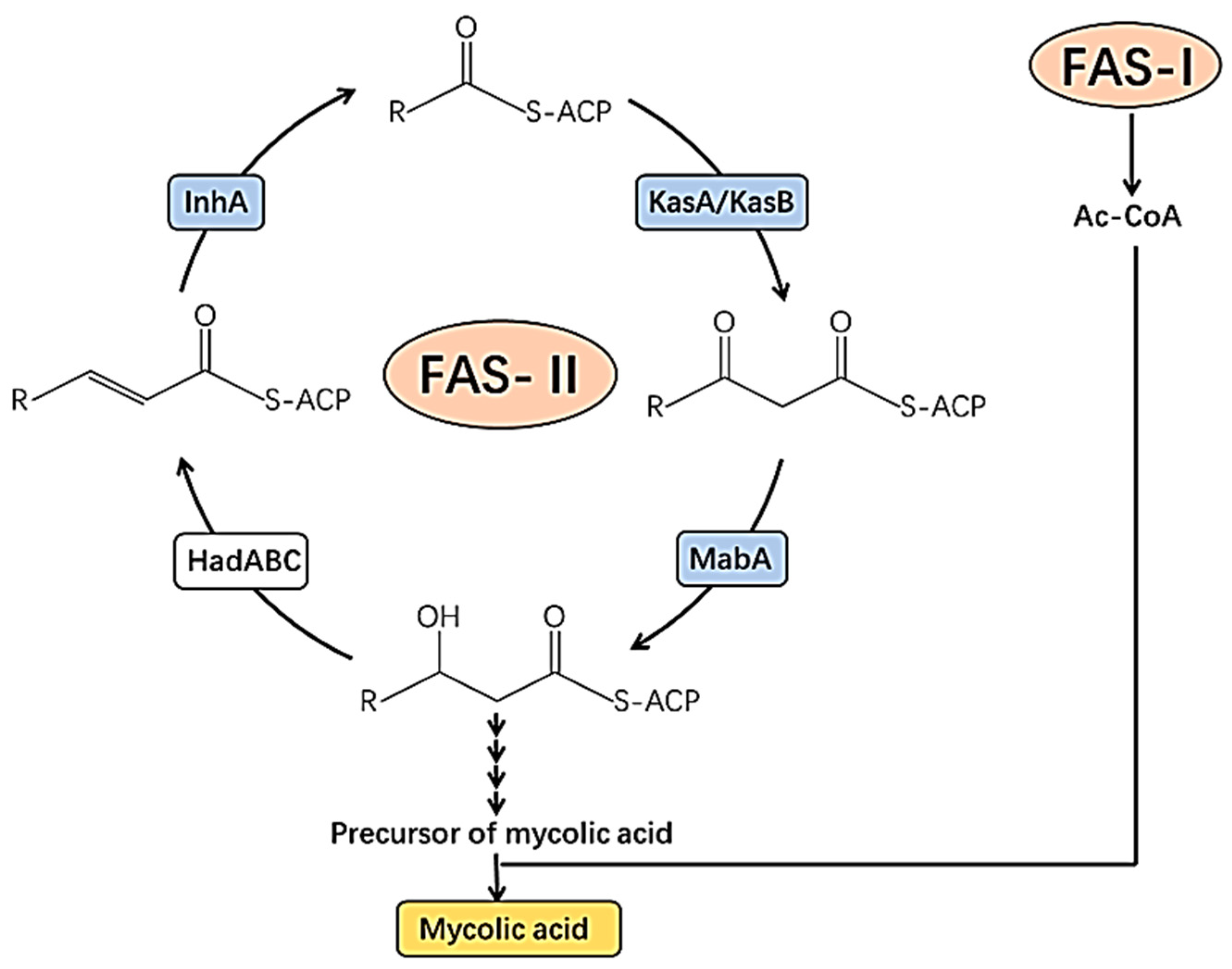
Figure 1. Potential target of the mycobacterial cell wall for drug targets. Ac-CoA, acetyl-coenzyme A; FAS-I, fatty acid synthase type I; HadABC, beta-hydroxyacyl-ACP dehydratase complex; InhA, 2-transenoyl-acyl carrier protein reductase; KasA/KasB, beta-ketoacyl-acyl carrier protein synthases A/B; MabA, beta-ketoacyl-acyl carrier protein reductase [28].
Molecules 30 03708 g001
Figure 2. The de novo pyrimidine and purine biosynthesis pathways. (A) Diagram of the de novo pyrimidine biosynthesis pathway. Pyrimidine biosynthesis enzymes: CAD: Carbamoyl-Phosphate Synthetase 2, Aspartate Transcarbamylase, And Dihydroorotase; DHODH: Dihydroorotate Dehydrogenase; UMPS: Uridine Monophosphate Synthetase. (B) Diagram of the de novo and purine salvage pathways. Purine biosynthesis enzymes: PPAT: phosphoribosyl pyrophosphate amidotransferase; GART: Glycinamide Ribonucleotide Transformylase; PFAS: Phosphoribosylformylglycinamidine Synthase; PAICS: Phosphoribosylaminoimidazole Carboxylase and Phosphoribosylaminoimidazolesuccinocarboxamide Synthase; ADSL: Adenylosuccinate Lyase; ATIC: 5-Aminoimidazole-4-Carboxamide Ribonucleotide Formyltransferase; IMPDH: Inosine Monophosphate Dehydrogenase; GMPS: Guanine Monophosphate Synthase; ADSS: Adenylosuccinate Synthase; HPRT: hypoxanthine phosphoribosyltransferase; APRT: adenine phosphoribosyltransferase [30,33].
Molecules 30 03708 g002
Figure 3. Schematic diagram of bacterial replisome: DNA gyrase unwinds supercoiling ahead of the fork, and DNA helicase unwinds DNA and invites primase to lay down RNA primers; leading and lagging strands are synthesized by DNA polymerases bound to clamps, which become loaded through a τ-containing clamp loader complex that bridges polymerases with helicase. SSB protects single-stranded DNA, and Polymerase I, and subsequently DNA ligase, removes primers and nicks to finish Okazaki fragments [37].
Molecules 30 03708 g003
Figure 4. Schematic representation of ATP synthesis in M. tuberculosis: Menaquinone is reduced by NADH dehydrogenases (primarily NDH-2) and succinate dehydrogenases and donates electrons through terminal oxidases (Cyt-bcc-aa3 or Cyt-bd), generating a proton motive force that is used by F1F0-ATP synthase for the production of ATP [48].
Molecules 30 03708 g004
Figure 5. Structures of alkaloids (112) with potent antimycobacterial activity.
Molecules 30 03708 g005
Figure 6. Structures of an amide (13) and peptides (1418) with potent antimycobacterial activity.
Molecules 30 03708 g006
Figure 7. Structures of terpenoids (1921) with potent antimycobacterial activity.
Molecules 30 03708 g007
Figure 8. Structures of steroids (2226) with potent antimycobacterial activity.
Molecules 30 03708 g008
Figure 9. Structures of polyacetylenes (27, 28) with potent antimycobacterial activity.
Molecules 30 03708 g009
Figure 10. Structures of lactones (29, 30) with potent antimycobacterial activity.
Molecules 30 03708 g010
Figure 11. Structures of aryl-alkyl ketones (31, 32) with potent antimycobacterial activity.
Molecules 30 03708 g011
Figure 12. Structure of a fatty acid (33) with potent antimycobacterial activity.
Molecules 30 03708 g012
Figure 13. Structure of a nitroacid (34) with potent antimycobacterial activity.
Molecules 30 03708 g013
Figure 14. Structures of naphthoquinones (35, 36) with potent antimycobacterial activity.
Molecules 30 03708 g014
Table 1. Summary of natural products with potent antimycobacterial activity and selectivity indices greater than 10 from 2000 to 2024.
Compound’s Name
(Class of Compounds)
Molecular
Formula
Molecular Weight (g/mol)SourceMIC in µM (Mycobacterial Strain)Anti-Mycobacterial Assay Methods Cytotoxicity in µM (Cell Line)Selectivity IndexRef
Strictosidine (1)
(Alkaloid)
C27H34N2O9530.6Psychotria nuda (Cham. & Schltdl.) Wawra13.7 (M. tuberculosis H37Rv)MTT170.93 (RAW264.7 cells)12.47[53]
Hapalindole A (2)
(Alkaloid)
C21H23ClN2338.9Westiellopsis sp. and Fischerella muscicola<0.6 (M. tuberculosis H37Rv)MABA31.9 (Vero cells)>53[54,55,56,57]
Hapalindole I (3)
(Alkaloid)
C21H21ClN2336.92 (M. tuberculosis H37Rv)>100 (Vero cells)>50
Hapalindole X (4)
(Alkaloid)
C21H22N2302.422.5 (M. tuberculosis H37Rv)35.2 (Vero cells)14.08
Hapalindole G (5)
(Alkaloid)
C21H23ClN2338.96.8 (M. tuberculosis H37Rv)>128 (Vero cells)>18.8
Fischambiguine B (6)
(Alkaloid)
C26H29ClN2O2436.182 (M. tuberculosis H37Rv)>128 (Vero cells)>64
Ambiguine C isonitrile (7)
(Alkaloid)
C26H32N2O388.57.0 (M. tuberculosis H37Rv)78.3 (Vero cells)11.25
Ambiguine M isonitrile (8)
(Alkaloid)
C26H31ClN2O2439.0Fischerella ambigua7.5 (M. tuberculosis H37Rv)79.8 (Vero cells)10.64
Ambiguine E isonitrile (9)
(Alkaloid)
C26H29ClN2O2437.01.4 (M. smegmatis)42.6 (Vero cells)30.43
Suadamin A (10)
(Alkaloid)
C43H48N4O6716.88Melodinus suaveolens (Hance) Champ, ex Benth6.76 (M. tuberculosis H37Rv)MABA143.3 (VERO cells)21.2[58]
Suadamin B (11)
(Alkaloid)
C43H48N4O6716.8833.47 (M. tuberculosis H37Rv)
Decarine (12)
(Alkaloid)
C19H13NO4319.3Zanthoxylum capense (Thunb.) Harv5.01 (M. tuberculosis H37Rv)Broth microdilution method206.7 (THP-1)41.2[59]
9.71 (M. tuberculosis H37Ra)21.3
(2E,4E)-N-isobutyl-2,4-tetradecadienamide (13)
(Amide)
C18H33NO279.46Z. capense (Thunb.) Harv5.73 (M. tuberculosis H37Rv)Broth microdilution method219 (THP-1)38.2[59]
Lassomycin (14)
(Peptide)
C82H142N30O201868.23Lentzea kentuckyensis0.41–0.83 (M. tuberculosis H37Rv)MABA187.34 (Human NIH 3T3 and HepG2 cells)225.72–456.93[60]
1.65 (M. tuberculosis H37Rv resistant to INH, RIF, STR, EMB, PZA, FQ)113.5
0.07–0.13 M. avium subsp. Paratuberculosis1441.1–2676
0.41–1.06 M. smegmatis176.4–456.92
Rufomycin I (15)
(Peptide)
C54H77N9O101012.56Streptomyces sp.0.02 (M. tuberculosis H37Rv)MABA>50 (Vero cell)>2500[61]
<0.004 (M. tuberculosis H37Rv resistant to INH
0.073 M. smegmatis
<0.02 (M. bovis)
Ecumicin (16)
(Peptide)
C83H134N14O171600.0Streptomyces sp.0.16 (M. tuberculosis H37Rv)MABA>63 (Vero cell line)>3150[61]
<0.12 (M. tuberculosis H37Rv resistant to INH)
1.7 (M. smegmatis)
<0.2 M. bovis
Cyclomarin A (17)
(Peptide)
C56H82N8O111043.3Streptomyces sp.0.094 (M. tuberculosis H37Rv)MABA>50 (Vero cell line)>531[61]
1.6 (M. smegmatis)
<0.02 (M. bovis)
Caprazamycin B (18)
(Peptide)
C53H86N5O221144.6Streptomyces sp.2.73 (M. tuberculosis H37Rv) >4368.33 (Mice)>1600[62]
Eurylene (19)
(Terpenoid)
C34H58O8594.8Homalolepis suffruticosa (Engl.) Devecchi & Pirani2.35 (M. tuberculosis H37Rv)MTT67.41 (RAW 264.7)28.7[63]
3.36 (M. tuberculosis H37Rv) 67.41 (RAW 264.7)20.06
12-Deacetoxyscalarin 19-acetate (20)
(Terpenoid)
C27H40O4428.6Brachiaster sp.4.00 (M. tuberculosis H37Ra)MABAInactive in cytotoxicity assays against several cell lines MCF-7, HT-29, HeLa, KB [64]
(−)-8,15-diisocyano-11(20)-amphilectene (21)
(Terpenoid)
C22H32N2324.51Svenzea flava9.8 (M. tuberculosis H37Rv)Microbroth dilution assay99.74 (Vero cells)10.2[65]
Saringosterol (22)
(Steroid)
C29H48O2 428.73Lessonia nigrescens0.58 (M. tuberculosis H37Rv)BACTEC 460>298.55 (Vero cells)>514.74[66]
Stigmast-5-ene-3β-ol-7-one (23)
(Steroid)
C29H48O2428.73Thalia multiflora Horkel ex Koernicke (Marantaceae)4.62 (M. tuberculosis H37Rv)MABA>237.91 (Vero cells)>51.5[67]
Stigmast-4-ene-6β-ol-3-one (24)
(Steroid)
C29H46O2426.6810.07 (M. tuberculosis H37Rv)>239.05 (Vero cells)>23.74
Stigmast-5,22-diene-3β-ol-7-one (25)
(Steroid)
C29H46O2426.682.34 (M. tuberculosis H37Rv)>239.05 (Vero cells)>102.16
Stigmast-4,22-diene-6β-ol-3-one (26)
(Steroid)
C29H46O2426.682.34 (M. tuberculosis H37Rv)>239.05 (Vero cells)>102.16
11(S),16(R)-Dihydroxy-octadeca-9Z,17-dien-12,14-diyn-1-yl acetate (27)
(Polyacetylene)
C20H28O4332.40Angelica sinensis (Oliv.) Diels4.2 (M. tuberculosis Erdman)MABA>361.01 [68]
Falcarindiol (28)
(Polyacetylene)
C17H24O2260.4023.4 (M. tuberculosis Erdman)>460.83
5,6-dehydro-7,8-dihydromethysticin (29)
(Lactone)
C15H14O5274.27Piper sanctum (Miq.) Schltdl.
ex C. DC
14.58 (M. tuberculosis H37Rv)MABA152.98 (Vero cells)10.5[69]
Micromolide (30)
(Lactone)
C18H32O2338.8Micromelum hirsutum Oliv.5.35 (M. tuberculosis H37Rv)MABA280.40 (Vero cells)52.41[70]
2-oxo-14-(3′,4′-methylenedioxyphenyl) tetradecane (31)
(Aryl-alkyl ketone)
C21H32O3332.23Piper sanctum (Miq.) Schltdl.
ex C. DC
18.81 (M. tuberculosis H37Rv)MABA>361.2 (Vero cells)20.82[69]
2-oxo-16-(3′,4′-methylenedioxyphenyl) hexadecane (32)
(Aryl-alkyl ketone)
C23H36O3 360.2617.35 (M. tuberculosis H37Rv)>333.08 (Vero cells)19.2
Linoleic acid (33)
(Fatty acid)
C18H32O2280.4Warburgia ugandensis14.26 (M. aurum)MTT193 (Prostate cancer LNCaP cells)13.53[71]
14.24 (M. phlei)193 (Prostate cancer LNCaP cells)13.53
3-Nitropropionic (34)
(Nitroacid)
C3H5NO4119.08Phomopsis sp. strain usia53.3 μM (M. tuberculosis H37Ra)MABAInactive against Vero cell lines [72]
Maritinone (35)
(Naphthoquinone)
C22H14O6374.34Diospyros anisandra S.F.Blake8.34 (M. tuberculosis H37Rv & MDR-MTB)MABA621.60 (Vero cells)161.88[73]
3,3’-biplumbagin (36)
(Naphthoquinone)
C22H14O6374.38.34 (M. tuberculosis H37Rv & MDR-MTB)1623.22 (Vero cells)194.63
INH, isoniazid, MTT; 3-(4,5-dimethylthiazole-2-yl)-2,5-diphenyl tetrazolium bromide; MABA; Microplate Alamar Blue Assay.
Disclaimer/Publisher’s Note: The statements, opinions and data contained in all publications are solely those of the individual author(s) and contributor(s) and not of MDPI and/or the editor(s). MDPI and/or the editor(s) disclaim responsibility for any injury to people or property resulting from any ideas, methods, instructions or products referred to in the content.