Article

From Mushrooms to Molecules: Exploring Depsidones in Ganoderma lucidum for Antioxidant and Anticancer Applications

by
Sayed H. A. Mohamed
1,
Yehia A.-G. Mahmoud
1,*,
Mohamed Y. Bediway
1,
Sobhy E. Elsilk
1,
Mohammed Yosri
2,
Kamel Metwally
3,
Nader E. Abo-Dya
3,
Galal Yahya
4,5,
Mervt Almostafa
6 and
Atef A. El-Hela
7
1
Botany and Microbiology Department, Faculty of Science, Tanta University, Tanta 31527, Egypt
2
The Regional Center for Mycology and Biotechnology, Al-Azhar University, Nasr City, Cairo 11787, Egypt
3
Department of Pharmaceutical Chemistry, Faculty of Pharmacy, University of Tabuk, Tabuk 71491, Saudi Arabia
4
Department of Microbiology and Immunology, Faculty of Pharmacy, Zagazig University, Zagazig 44519, Egypt
5
Department of Molecular Genetics, Faculty of Biology, Technical University of Kaiserslautern, Paul-Ehrlich Str. 24, 67663 Kaiserslautern, Germany
6
Department of Chemistry, College of Science, King Faisal University, Al-Ahsa 31982, Saudi Arabia
7
Pharmacognosy Department, Faculty of Pharmacy, Al-Azhar University, Nasr City, Cairo 11787, Egypt
*
Author to whom correspondence should be addressed.
Molecules 2025, 30(17), 3650; https://doi.org/10.3390/molecules30173650
Submission received: 23 July 2025 / Revised: 31 August 2025 / Accepted: 2 September 2025 / Published: 8 September 2025

Abstract

Fungi are a prolific source of diverse bioactive metabolites, yet many remain unexplored. Among these, depsidones are a rare class of compounds with significant biological potential, but they are seldom reported in mushrooms. This study investigated the medicinal fungus Ganoderma lucidum, known for its extensive therapeutic use in traditional medicine. Fruiting bodies were extracted using petroleum ether, ethyl acetate, n-butanol, and methanol. Extracts were screened phytochemically and assessed for total phenolic content and antioxidant activity using the DPPH assay. Ethyl acetate extract exhibited the highest phenolic yield and antioxidant potential and was subsequently evaluated for cytotoxicity against HepG2, HCT116, MCF7, and A549 cancer cell lines. It showed notable anticancer activity with minimal toxicity to normal Vero cells. UHPLC/Q-TOF-MS/MS analysis of G. lucidum ethyl acetate extract tentatively identified nine minor depsidones including mollicellin G, simplicildone I, mollicellin B, talaromyone B, simplicildone A, purpactin C, emeguisin B, mollicellin E, and simplicildone D on the basis of high-resolution negative-mode detection and characteristic MS/MS fragmentation patterns. Molecular docking revealed strong binding affinities between these compounds and cancer-related targets (AKT1, CDK2, ERK1, TNFα), with simplicildone D and mollicellin G demonstrating particularly high interactions. These findings provide mechanistic insights into the observed bioactivity and highlight G. lucidum as a promising source of therapeutic depsidones for future anticancer drug development.

1. Introduction

Ganoderma lucidum (G. lucidum), is a macrofungus with massive fruiting structures with many beneficial applications [1]. It is thought to promote health and lengthen life [2,3]. This mushroom has long been used in medicinal products and health cures in Asian conventional therapies [4,5]. Researcher interest in medicinal mushrooms has led to the development of useful research on their biologically active elements and their significance [6,7]. Research has shown that G. lucidum includes more than 200 distinct kinds of polysaccharides along with more than 400 secondary metabolites [8]. These include triterpenoids (e.g., ganoderic, lucidenic, and ganoderenic acids), other terpenes and sterols, polysaccharides (notably β-D-glucans), phenolic compounds, alkaloids, and peptides, each contributing through distinct and sometimes synergistic mechanisms [9]. With respect to the location of production, the circumstances for development, chemical extraction techniques, and strain selection, the kinds and amounts of biologically active substances in G. lucidum can change [10]. The isolation and identification of these metabolites have been accomplished using various separation and purification techniques, such as solvent–solvent partitioning, column chromatography (silica gel, Sephadex LH-20), preparative high-performance liquid chromatography (HPLC), and countercurrent chromatography. Spectroscopic methods, including nuclear magnetic resonance (NMR), Fourier transform infrared spectroscopy (FTIR), ultraviolet–visible spectroscopy (UV–vis), and mass spectrometry (MS), have been pivotal for structure elucidation [11,12,13,14].
Given the extensive use of Ganoderma without any reported cases of toxicity, many researchers assert that it is safe and beneficial for consumption [10].
A wide array of distinctive biomolecules derived from natural biological sources and occurring abundantly in nature [15]. Due to their accessibility and affordability, especially in impoverished nations, these metabolites have been used for many years as a basic remedy for a variety of human ailments. Additionally, they are a great source for therapeutic development and research due to their chemical variety and wide range of biological functions [16]. One of the biggest obstacles to the successful use of particular metabolites at the moment is the procedure for the extraction of active molecules [17,18]. Lichens and mushrooms may be a source of phenolic compounds known as depsidones and depsides, which are substances made up of two or three aromatic ring structures of orcinol or β-orcinol connected by an ester bond or an extra ether bond [19].
The variety of structures and diverse biological properties of these compounds have garnered a lot of study attention. There have been reviews of this kind of metabolites in earlier publications. The isolation, structural description, metabolism, and biological activities of many kinds of depsidones identified from fungal sources were covered in many reports [20,21].
Phytochemical constituents and secondary metabolites (over 430) extracted from G. lucidum demonstrate a broad range of pharmacological activities, including anticancer, antioxidant, antidiabetic, antihyperlipidemic, antimicrobial, hepatoprotective, cardioprotective, anti-aging, anti-androgenic, antiallergic, analgesic, and anti-frailty effects [9,22]. With well-documented therapeutic potential across diverse disease models, G. lucidum is utilized in over 100 commercial formulations, emphasizing its global relevance as a multifunctional natural health product [23].
The purpose of this study is to characterize the phytochemical composition of different solvent extracts of G. lucidum, to relate the identified metabolites to their observed biological activities (particularly cytotoxic effects), and to provide mechanistic insights into the relationship between chemical constituents and bioactivity.
We employed liquid chromatography–high resolution tandem mass spectrometry (LC–HRMS/MS) to describe the bioactive chemicals, especially depsidones, which have rarely been reported in G. lucidum, and analyze the biological characteristics of different extracts of G. lucidum using different solvents.
To evaluate the anticancer potential of G. lucidum extracts, five well-characterized cell lines were selected, each representing a different physiological or pathological context. HepG2 (human hepatocellular carcinoma), HCT116 (human colorectal carcinoma), MCF7 (human breast adenocarcinoma), and A549 (human lung adenocarcinoma) are widely used in cytotoxicity studies due to their relevance in prevalent cancer types and their distinct molecular and metabolic profiles. Vero cells (African green monkey kidney epithelial cells) were included as a non-cancerous normal cell model to assess selectivity and potential cytotoxicity towards healthy cells.
Molecular docking provides valuable insights into the molecular basis of the observed cytotoxic activity and supports the pharmacological potential of these naturally derived compounds. In this study, molecular docking was employed to evaluate the binding affinity and interaction profiles of the bioactive constituents identified in the ethyl acetate extract of G. lucidum. Specifically, nine depsidone compounds were subjected to in silico docking against key cancer-related protein targets, including AKT1, CDK2, ERK1, and TNFα. This computational approach aimed to predict the potential anticancer mechanisms of these molecules by analyzing their binding modes, interaction energies, and key stabilizing forces.

2. Results and Discussion

2.1. Qualitative Phytochemical Screening and Antioxidant Potential

Qualitative phytochemical screening for G. lucidum upon extraction using various solvents can be seen in Supplementary Table S1. Sterols and/or triterpenes could be detected upon extraction using petroleum ether. However, anthraquinone derivatives and/or depsidones were the most predominant group detected upon using ethyl acetate for extraction. Moreover, none of the qualitative applied tests resulted in a positive outcome upon extraction using n-butanol for extraction. Finally, using methanol for extraction resulted in the production of many chemical groups, including carbohydrates, flavonoïdes, tannins, and alkaloids. The nutritive benefits of G. lucidum mushrooms have been the subject of various investigations [24]. According to Ahmad et al. [25], studies on the biochemical makeup of this mushroom have identified a number of categories of chemicals with biological functions that promote health.
In this work, various solvents were applied to extract G. lucidum in accordance with earlier studies [26], by which the bioactive components of mushrooms can be extracted using polar or non-polar solvents. A methanol-based extract of newly harvested fruiting structures from G. lucidum inhibits the development of several microorganisms, according to Mondal et al. [27].
Quantitative analysis for phenolic compounds in G. lucidum reflected that application of ethyl acetate in extraction resulted in highest yield of phenolic compounds, with a level of 9.87 ± 0.71 mg/g. However, using methanol in extraction resulted in a moderate yield of phenolic compounds with a level of 4.27 ± 0.11 mg/g. Finally, using either petroleum ether or n-butanol resulted in a weak yield of phenolic molecules (Table 1). Several phenolic acids found in G. lucidum that have antioxidant action were the subject of further evidence reported by numerous researchers [28,29]. Rahman et al. examined ascorbic acid and flavonoid components in depth in their 2020 study [30].
The results of the antioxidant impact of various extracts of G. lucidum can be seen in Figure 1. It can be noticed that the ethyl acetate extract had the highest antioxidant impact with IC50 = 39.87 ± 1.7 µg/mL, while the antioxidant action of petroleum ether, n-butanol, and methanol had IC50 values of = 85.87 ± 0.8, 165.17 ± 1.2, and 55.47 ± 0.71 µg/mL, respectively. Ascorbic acid had a level of IC50 = 10.21 ± 0.77 µg/mL. The antioxidant activity of G. lucidum extracts is largely attributed to their rich phytochemical content, including triterpenoids, polysaccharides, polyphenols, flavonoids, and ascorbic acid, which can scavenge free radicals and protect against oxidative stress [31,32].

2.2. Cytotoxicity Against Cancer Cell Lines

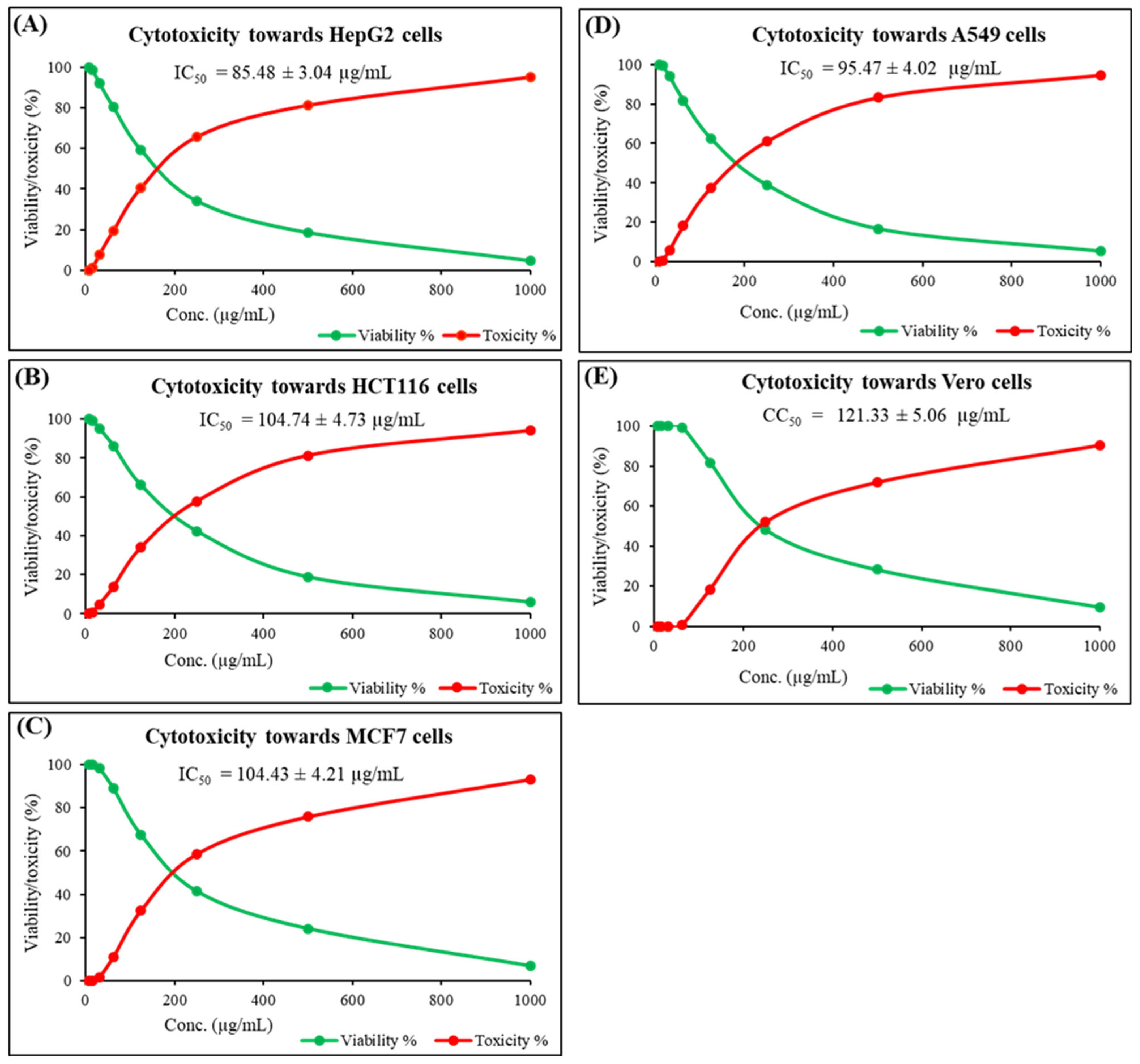
G. lucidum ethyl acetate extract was tested against various cancer cells as well as a normal cell line, as depicted in Figure 2A–E. This combination of cell lines provides a robust platform to evaluate both the anticancer efficacy and safety profile of the tested extracts. It could be noticed that the tested extract had promising antitumor towards hepatocellular carcinoma cells (HepG2), colon carcinoma cells (HCT116), breast carcinoma cells (MCF7), and lung carcinoma cells (A549), with IC50 values of =85.49 ± 3.04, 104.74 ± 4.73, 104.43 ± 4.21, and 95.47 ± 4.02 µg/mL, respectively. Besides, it has a potent impact on mammalian cells from African green monkey kidney (Vero), representing normal cells, with CC50 = 121.33 ± 5.06 µg/mL.
It is anticipated that the discovery of novel drugs to treat human malignancies would make use of mushroom extracts [33]. The present findings indicate that G. lucidum extracts exert varying degrees of cytotoxicity against multiple human cancer cell lines, with notable selectivity when compared with non-cancerous Vero cells. This aligns with a growing body of evidence supporting the anticancer potential of medicinal mushrooms [34,35]. Beyond their historical role in traditional medicine, numerous fungal species have yielded pharmacologically important compounds, including clinically used anticancer agents such as taxanes and bisindole alkaloids [36,37].
In the case of G. lucidum, several classes of bioactive metabolites are implicated in its cytotoxic effects. Polysaccharides, particularly β-D-glucans, have been widely reported to modulate immune responses, enhance host defense mechanisms, and inhibit tumor growth both directly and indirectly [38]. Triterpenoids, such as ganoderic and lucidenic acids, have demonstrated the ability to induce apoptosis, cause cell cycle arrest, and suppress metastatic potential in various cancer cell models. Phenolic compounds and sterols present in G. lucidum may further contribute by exerting antioxidant and pro-oxidant effects that disrupt cancer cell survival pathways.
The differential sensitivity observed among the different cancer cells may reflect differences in their genetic background, receptor expression, and metabolic profiles, suggesting that specific phytochemicals in G. lucidum could target multiple molecular pathways in a tumor-type-dependent manner. Importantly, the relatively low cytotoxicity toward Vero cells highlights the potential therapeutic window and selective cytotoxicity of the extracts.
The cytotoxic effects of G. lucidum ethyl acetate extract observed in this study can be directly linked to the classes of metabolites identified during phytochemical screening. The ethyl acetate extract is rich in polar bioactivities, including carbohydrates/glycosides, flavonoids, tannins/phenolics, alkaloids, and triterpenes. These compound classes have well-documented anticancer mechanisms. For example, polysaccharides such as β-D-glucans can stimulate immune-mediated tumor suppression and induce apoptosis, while phenolics and flavonoids may inhibit cancer cell proliferation through antioxidant, pro-oxidant, and signaling pathway modulation [39,40,41].
It is worth mentioning that the n-butanol and petroleum ether extracts were largely devoid of the key anticancer-associated metabolites identified in ethyl acetate fraction.
The low toxicity of ethyl acetate extract toward Vero cells suggests that these phytochemicals may selectively target cancer cell pathways without significantly affecting normal cells’ viability.
These observations underscore the direct relationship between the phytochemical profile of each extract and its bioactivity, supporting the hypothesis that the anticancer potential of G. lucidum is mediated through a synergistic interplay among polysaccharides, triterpenoids, phenolic compounds, and flavonoids.
Taken together, these findings reinforce the promise of G. lucidum as a source of leading compounds for anticancer drug development, warranting further mechanistic and in vivo studies.

2.3. UHPLC/Q-TOF-MS-MS Analysis

UHPLC/Q-TOF-MS-MS analysis for ethyl acetate extract of G. lucidum reflected the existence of nine different minor compounds, which have been tentatively identified as depsidones using a high-resolution detection (negative mode) in order to determine their molecular formula identification on the basis of their common fragmentation ions in the MS/MS spectra (Figure 3). These molecules were mollicellin G (C22H23O7) [37], simplicildone I (C31H28O8) [38], mollicellin B (C21H18O7) [42], talaromyone B (C23H26O7) [43], simplicildone A (C18H18O6) [44], purpactin C (C23H21O7) [45], emeguisin B (C24H25ClO5) [46], mollicellin E (C22H19ClO8) [37], and simplicildone D (C25H24O7) [38].
According to the LC-MS studies, the ethyl acetate extract of G. lucidum included nine peaks of depsidones. These peaks were accurately recognized by mass measurement, and the molecular structures were subsequently tentatively characterized using MS/MS fragmentation patterns (Supplementary Figure S1 and Table 2). This is the first work to characterize and provisionally identify these groups of compounds in G. lucidum. The results of our study are in line with the findings of an investigation that found that naturally produced depsidones have outstanding biological properties. Given the rapid advancements in studies, the search for novel active depsidones from natural origins may prove to be an effective method for developing new herbal remedies [47].
Depsidone fragmentation ions are produced via cyclization after the straightforward loss of CO2, H2O, and CH2O. A common deprotonated molecule was found in all depsidones, along with a fragment that showed an impairment of 44 mass units. It is assumed that this neutral CO2 loss on the depsidone scaffold’s central core provides a dibenzofuran moiety [47]. The order in which these compounds elute further supports the structural designations of these depsidones [48,49].

2.4. Virtual Screening and Protein–Ligand Interactions

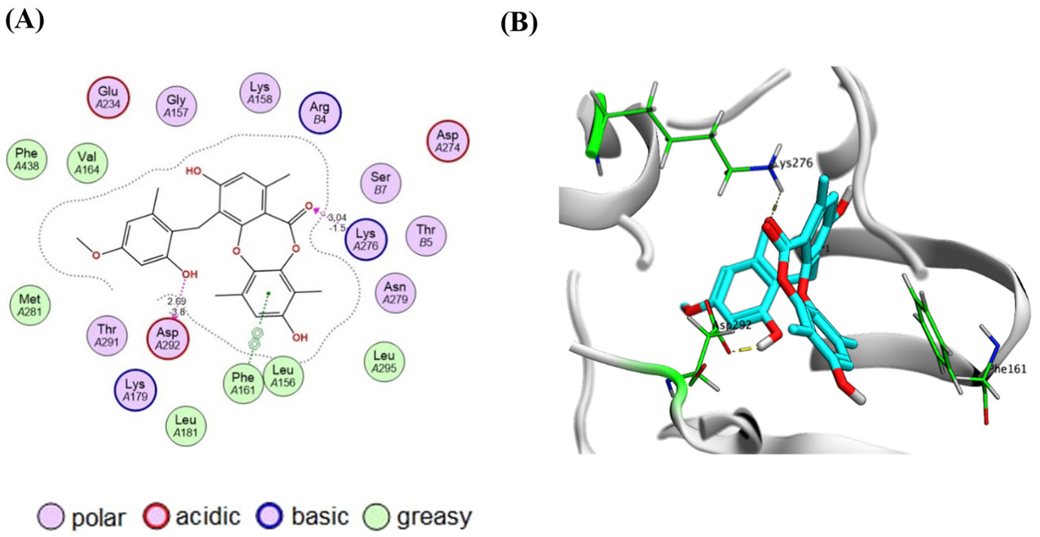
Docking analysis indicated that simplicildone D displayed the highest binding affinity (docking score = −7.77 kcal/mol, RMSD = 1.68 Å) to AKT1 (PDB code: 3OCB, Homo sapiens) [50]. Inspection of the docking poses revealed two key hydrogen bonding interactions that contribute to binding stabilization. The phenolic hydroxy group of the benzyl moiety donates a hydrogen bond (E = −3.8 kcal/mol, distance = 2.69 Å) to the side chain carboxylate group of Asp292 which is a constituent part of the DFG motif. In addition, the lactone carbonyl group accepts a hydrogen bond (E = −1.5 kcal/mol, distance = 3.04 Å) from the side chain amino group of Lys276 located in the catalytic loop of AKT1. On the other hand, the xylenol ring π-π stacks the phenyl side chain of Phe161 located within the G-loop of the kinase domain (Figure 4). It appears that the free rotation offered by the benzyl moiety enables the correct positioning of the hydroxyl group near Asp292 for a strong hydrogen bonding interaction. In simplicildone I, hydrogen bond formation may be sterically hindered by the bulky phenoxy group attached to the benzyl moiety. In other compounds, a phenolic hydroxy group is either absent or directly attached to a rigid tricyclic ring structure, which minimizes the binding flexibility.
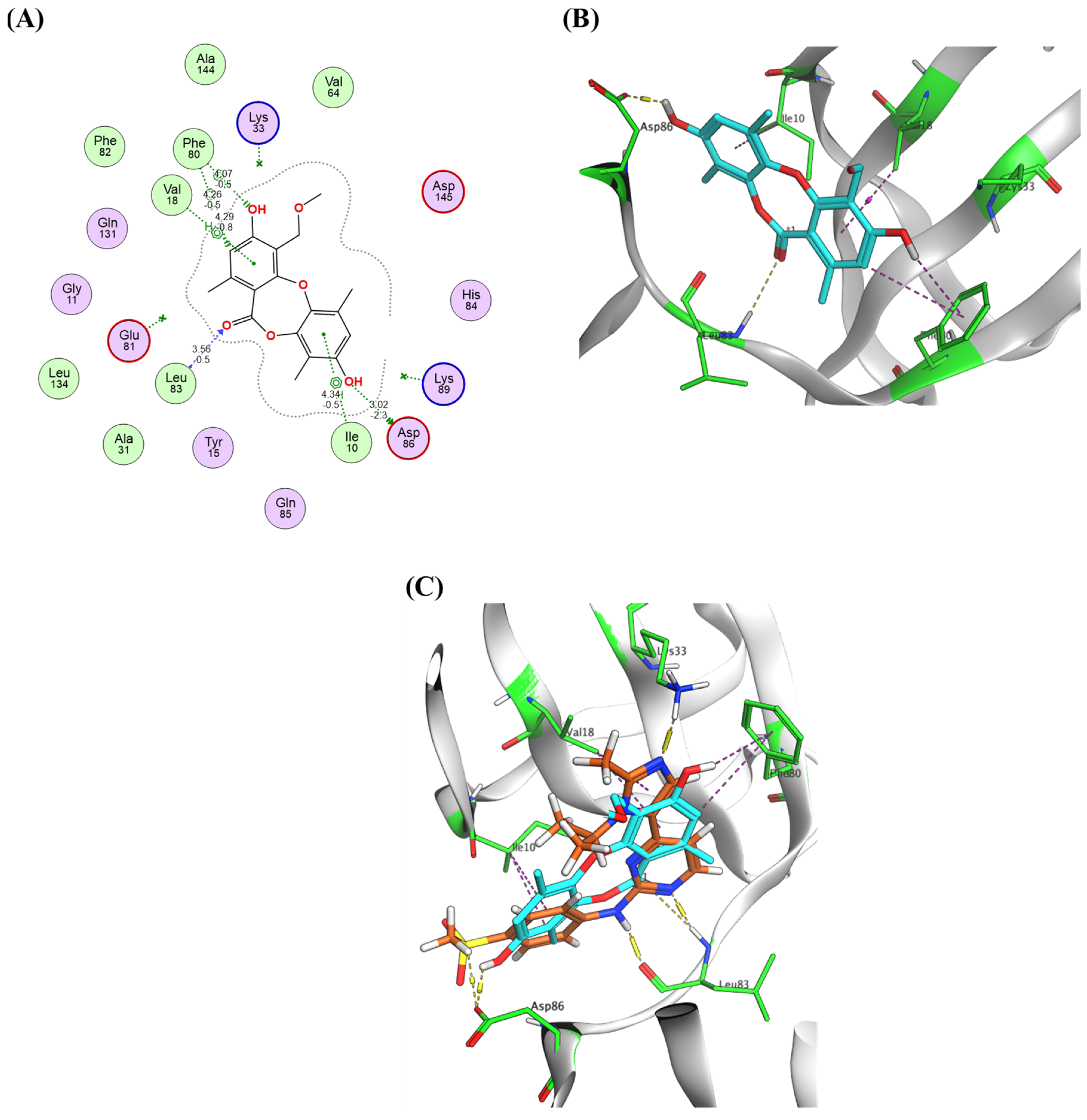
The docking results of the target compounds with CDK2 (PDB code: 6GUE, Homo sapiens) [51] showed that simplicildone A exhibited the highest binding affinity (docking score = −7.72 kcal/mol, RMSD = 1.75 Å) with crucial interactions with the CDK2 hinge residues Asp86 and Leu83. The xylenol hydroxy group donated a hydrogen bond to the side chain carboxylate group of Asp86, and the lactone carbonyl oxygen accepted a hydrogen bond from the backbone NH group of Leu83. It was also observed that Compound 5 establishes several hydrophobic contacts such as the π-H interactions with Ile10 and Val18 located the ATP-binding pocket in the N-terminal lobe of the kinase domain and an H-π interaction with Phe80 located near the hinge region of AKT1, thereby contributing to binding stabilization (Figure 5). Notably, the relatively small molecular size of simplicildone A compared with the other compounds examined indicates that steric parameters may play a role in binding to hotspots of CDK2. This corresponds with previous findings that utilized contour maps to demonstrate the significance of electrostatic, hydrophobic, hydrogen bond donor, and steric fields in CDK2 binding [52].
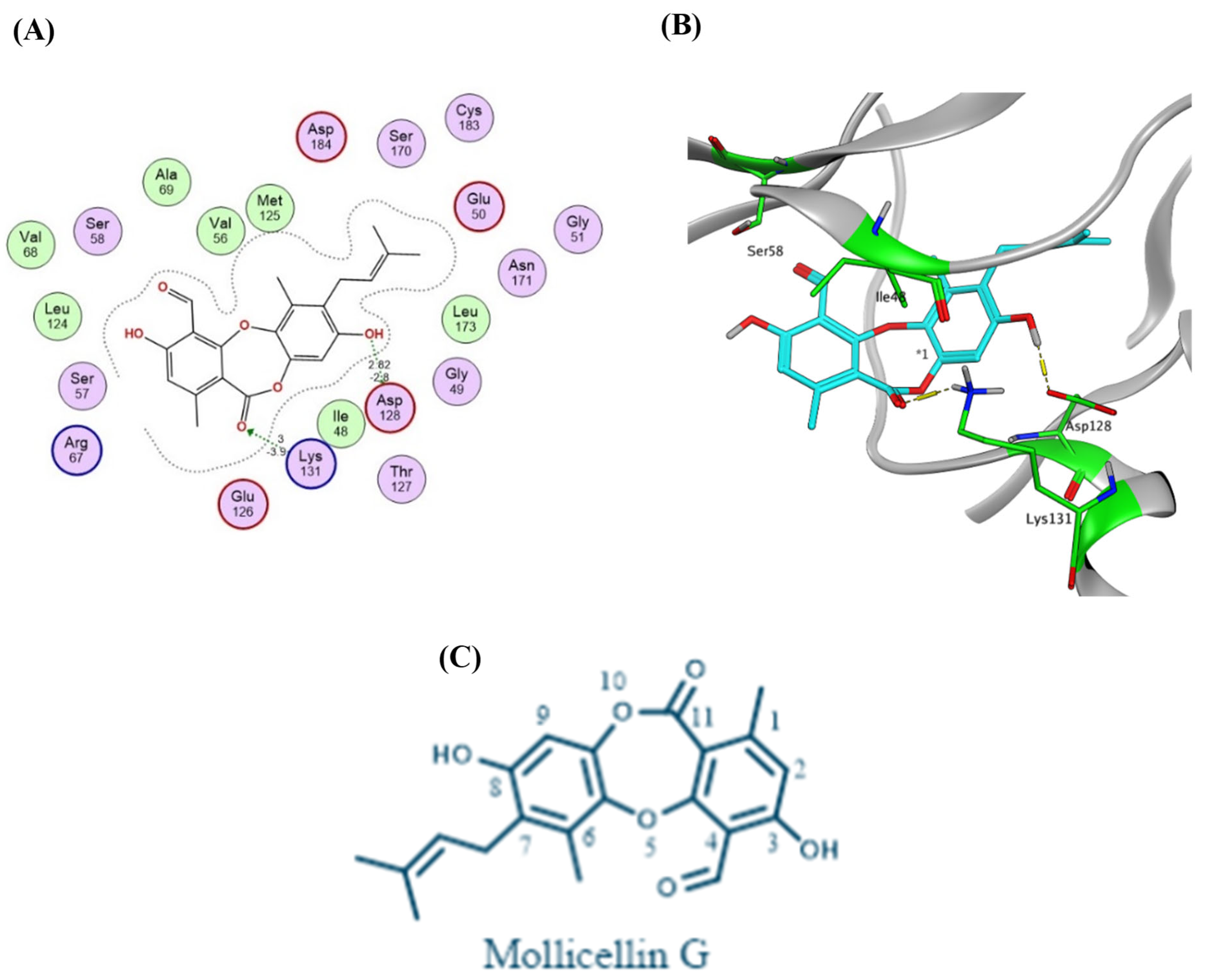
Inspection of the docking poses of the target compounds with ERK1 (PDB code: 2ZOQ, Homo sapiens) [53] revealed that mollicellin G displayed the highest binding affinity (docking score = −7.84 kcal/mol, RMSD = 1.78 Å) establishing two hydrogen bonding interactions. The hydroxy group adjacent to the alkylidene moiety donates a hydrogen bond (E = −2.80 kcal/mol, distance = 2.88 Å) to the carboxylate side chain of Asp128, which is a part of the HRD motif of ERK1. Additionally, the lactone carbonyl group accepts a strong hydrogen bond (E = −3.90 kcal/mol, distance = 3.00 Å) from the side chain amino group of Lys131 in the catalytic loop of the kinase domain (Figure 6A,B). The pentylidene moiety of mollicellin G may influence its conformation in a way that positions the phenolic hydroxyl and lactone carbonyl groups to form strong hydrogen bonds. While mollicellin G; simplicildones A, D, and I; and emeguisin B have a C-8 phenolic hydroxyl group and a carbonyl group at C-11, only mollicellin G can form hydrogen bond interactions with Asp128 and Lys131 of ERK1. This is probably because mollicellin G does not have a C-9 methyl group, which could cause steric hindrance that might interfere with these interactions in the other molecules (Figure 6C).
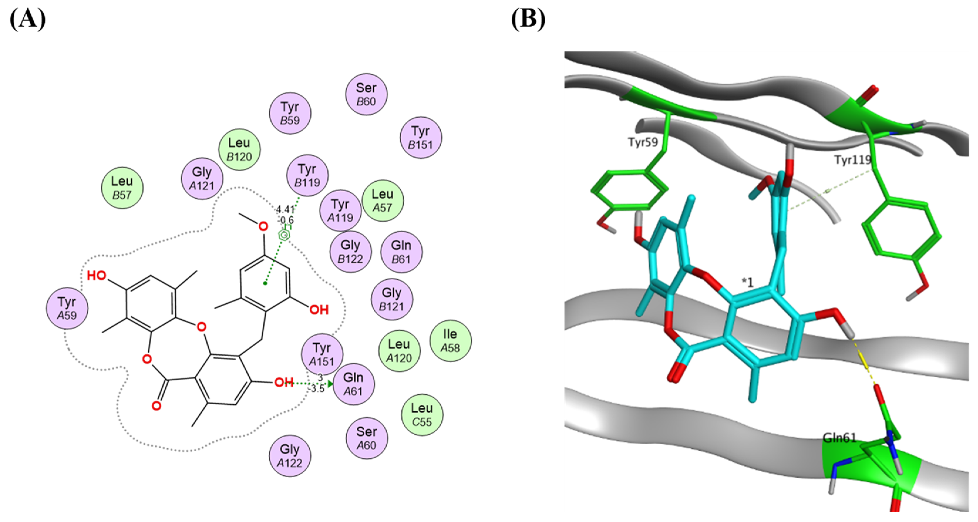
Simplicildone D was found to display the highest binding affinity (docking score = −7.21 kcal/mol, RMSD = 0.79 Å) to TNFα (PDB code: 2AZ5, Homo sapiens) [54]. A key hydrogen bonding interaction is established between the hydroxy group ortho to the benzyl moiety and the side chain carbonyl group of Gln61 located within a β-strand of the “jelly roll” β-sandwich strand. On the other hand, a pi-H interaction is established between the phenyl ring of the benzyl moiety and the methylene group of Tyr119 located in the receptor-binding interface of the TNF-α homotrimer (Figure 7). This pi-H interaction displayed by the benzyl moiety may play a role in stabilizing the hydrogen bonding interaction established by the adjacent hydroxyl group.

3. Materials and Methods

3.1. Acquisition of Mushrooms

G. lucidum fresh fruiting bodies (FFBs) were acquired from the Egyptian Agriculture Research Center in Giza, Egypt. The fungus was classified using established norms and by comparing them to voucher samples (Voucher ID: 0245) from the Tanta University Herbarium’s Microbiology Department (Tanta, Egypt) [55]. Before being kept in a dehydrator at −20 °C until further research, the FFBs were cleaned with deionized water, dried, chopped into pieces (approximately 1.0 cm2), and lyophilized using a FreeZone 2.5 Liter Laboratory Freezing Dry System (Kansas City, MA, USA) [56].

3.2. Production of the Mushroom Extracts

The stored fruiting bodies were extracted using various solvents (petroleum ether, ethyl acetate, n-butanol or methanol) at 10% (w/v) in four separate containers. Each specimen was pulverized using a laboratory mixer (Fisher Scientific, Waltham, MA, USA) and then refrigerated for the whole night. An ultrasonicator (Fisher Model 200, New York, NY, USA) was used to blast down these mushroom cells for 30 min at 30 kHz. After centrifugation of the specimens for 20 min at 10,000× g to exclude debris, the resulting supernatants were obtained in a separator (Beckman, Brea, CA, USA). Each of the specimens were stored at −20 °C, and the extracts were concentrated by drying by air [57].

3.3. Qualitative Screening of Phytochemicals and Total Phenolics in Various Prepared Extracts

The phytochemicals were screened qualitatively in accordance with Nortjie et al. [58]. The total amount of phenolic content was estimated in various samples through mixing 3 mL (70 g/L) of sodium carbonate, 2.0 mL of Folin–Ciocalteu solution, and 100 μL of various extracts. After 20 s of shaking, containers were stored at 37 °C for an hour. At 750 nm, absorbance was determined with a Shimadzu 310 spectrometer (Kyoto, Japan) [59].

3.4. Antioxidant Assay of Different Extracts

In order to assess the evaluated samples’ antioxidant ability, a solution of ethanol with 0.1 mM DPPH was used. The produced solution (1 mL) was mixed with 3 mL of different specimens in ethanol (3.9−1000 µg/mL). Following a vigorous shake, the mixture was allowed to stand for 35 min at room temperature. Absorbance was assessed at 520 nm [60].

3.5. Cytotoxicity Examination

HepG2 (human hepatocellular carcinoma), HCT116 (human colorectal carcinoma), MCF7 (human breast adenocarcinoma), A549 (human lung adenocarcinoma), and Vero (African green monkey kidney epithelial cells) cell lines were used to evaluate the samples’ in vitro cytotoxic impact employing the 3-(4,5-dimethylthiazol-2-yl)-2,5-diphenyl tetrazolium bromide (MTT) test. The American Type Culture VACSERA Company provided the cells. After that, 96-well plates were filled with 1.1 × 10⁵ cells/mL, and they were incubated for 24 h at 37 °C with 5% CO2. After changing the culture medium, six distinct concentrations (1000–32.25 µg/mL) of each extract dissolved in DMSO (dimethyl sulfoxide) were added (the final conc. of DMSO was kept below 0.5% v/v), and the mixture was cultured for 24 h. The culture media were then taken out and cleaned using phosphate-buffered saline (PBS). As explained for the cytotoxicity experiment, the absorbance measurements of formazan crystals utilized in the MTT reagent were used to assess the life span of treated cells and cells that were not treated. Lastly, a microplate reader was used to determine the absorbance at 570 nm [61].

3.6. UHPLC/Q-TOF-MS-MS Testing

Applying the Micromass, Quattro Micro TM (Waters, Milford, MA) technology, high-resolution MS/MS analysis was carried out. After dissolving 50 mg of the sample in 1 mL of the recovered solvent (water–methanol–acetonitrile, 50:25:25 v/v), the mixture was vortexed for 2 min and then ultrasonically sonicated for 10 min. Next, 50 µL of the stock liquid was diluted with 1000 µL of the reconstitution solvent and centrifuged for 10 min at 10,000 rpm. In the end, 2.5 µg/µL was the injected quantity, and 10 µL was injected in negative mode. We used a mobile phase gradient elution (A, negative mode) and 10 µL of the reconstitution solvent as a blank sample. An ammonium formate buffer (pH 8.5) with 1% methanol and a 0.3 mL flow rate on a column (2.1 × 150 mm, 5 µm) was used for the mobile phase C with 100% acetonitrile at 40 °C [62].

3.7. Molecular Docking and Protein–Ligand Interactions

Molecular docking investigations were carried out using the Molecular Operating Environment (MOE) 2024.06 software suite (Chemical Computing Group Inc., Montreal, QC, Canada) [63]. The simulations were run on a Windows 11 workstation with an Intel® Core™ i5 processor, 16 GB RAM, and an NVIDIA RTX GPU. Protein preparation was carried out using the MOE QuickPrep module, which included the addition of hydrogen atoms, the correction of protonation states at physiological pH (7.4), and the allocation of partial charges using the Amber10: EHT forcefield. All ligands, including both the reference compounds and the identified extract components, were produced for molecular modeling using the MOE sketch from their chemical names, which were obtained from the PubChem and Zinc databases. The ligands were prepared using 3D protonation, energy minimization with the MMFF94x forcefield, and partial charge assignment (MMFF94x) along with geometry optimization. The Dock module in the Molecular Operating Environment (MOE) program was used to run molecular docking simulations. The docking methodology utilized the Triangle Matcher method to place the ligands at first, then the London dG function to score them again and finally the GBVI/WSA dG scoring function for refinement. A forcefield-based refinement was used in the binding site and kept the top 10 poses for each ligand for more study. To make sure that the docking method was reliable, the co-crystallized ligand was redocked into its known binding site. The experimental and predicted poses must have a root mean square deviation (RMSD) of ≤2.0 Å for the validation to be successful [63].
Protein–ligand interactions were visualized and analyzed using MOE’s LigX tool, which identified key hydrogen bonds, hydrophobic contacts, salt bridges, and π–π or cation–π interactions. Binding affinity was estimated from the docking score (S score, kcal/mol) [63].

3.8. Statistical Evaluation

The results are expressed as the mean ±SD, and each experiment was run three times. The results were tested using Graph Pad Prism V8 (CA, USA) software, and the Tukey test was used to examine the means before one-way testing of variations (one-way ANOVA). The examination was deemed significant when the p-value was less than 0.05.

4. Conclusions

The present study highlights the value of G. lucidum as a rich source of bioactive compounds, particularly when extracted using ethyl acetate. This solvent not only yielded the highest concentration of phenolic compounds but also demonstrated superior antioxidant and substantial cytotoxic activities. Notably, LC-MS analysis revealed nine unique depsidone derivatives, provisionally identified for the first time in G. lucidum. Molecular docking further substantiated their therapeutic relevance, showing high binding affinity to key cancer-related targets like AKT1, CDK2, ERK1, and TNFα. These interactions suggest the potential of these natural compounds in future anticancer drug development. The study reinforces the importance of solvent selection in phytochemical extraction and provides a foundation for future investigations into the pharmacological utility of depsidones. Overall, the findings support the promising role of G. lucidum in nutraceutical and pharmaceutical applications, particularly as a source of natural depsidone-based therapeutics.

Supplementary Materials

The following supporting information can be downloaded at: https://www.mdpi.com/article/10.3390/molecules30173650/s1; Qualitative phytochemical screening of G. lucidum extracts (Supplementary Table S1) and MS/MS fragmentation patterns of G. lucidum ethyl acetate extract (Supplementary Figure S1).

Author Contributions

Conceptualization, S.H.A.M., Y.A.-G.M., M.Y. and A.A.E.-H.; methodology, S.H.A.M., Y.A.-G.M., M.Y., K.M., N.E.A.-D., M.A. and A.A.E.-H.; software, K.M., N.E.A.-D., M.A. and G.Y.; validation, S.H.A.M., Y.A.-G.M., M.Y., S.E.E. and M.Y.B.; formal analysis, Y.A.-G.M., K.M., N.E.A.-D., M.A. and M.Y.; investigation, S.H.A.M., Y.A.-G.M., M.Y., K.M., N.E.A.-D., M.A. and A.A.E.-H.; resources, K.M., N.E.A.-D., M.A. and G.Y.; data curation, K.M., N.E.A.-D. and G.Y.; writing—original draft preparation, S.H.A.M., Y.A.-G.M., M.Y., K.M., N.E.A.-D., G.Y. and A.A.E.-H.; writing—review and editing, all authors.; visualization, G.Y.; supervision, Y.A.-G.M. and M.Y.; project administration, A.A.E.-H.; funding acquisition, K.M., N.E.A.-D., M.A. and G.Y. All authors have read and agreed to the published version of the manuscript.

Funding

This research received no external funding.

Institutional Review Board Statement

Not applicable.

Informed Consent Statement

Not applicable.

Data Availability Statement

All results included in this work are available with the corresponding author on reasonable request.

Acknowledgments

The authors acknowledge their research institutes for administrative and technical support, and the Deanship of Scientific Research, Vice Presidency for Graduate Studies and Scientific Research, King Faisal University, Saudi Arabia for funding.

Conflicts of Interest

The authors declare no conflicts of interest.

Abbreviations

The following abbreviations are used in this manuscript.
G. lucidumGanoderma lucidum
FFBFresh fruiting bodies
LC-MS/MSLiquid chromatography–mass spectrometry/mass spectrometry
UHPLC/Q-TOF-MS-MSUltra-high-performance liquid chromatography/quadrupole time-of-flight mass spectrometry
MTT3-(4,5-Dimethylthiazol-2-yl)-2,5-diphenyltetrazolium bromide
DPPH2,2-diphenyl-1-picrylhydrazyl
PBSPhosphate-buffered saline
MOEMolecular Operating Environment
RMSDRoot mean square deviation
SDStandard deviation
ANOVAAnalysis of variance
AKT1RAC-alpha serine/threonine-protein kinase 1
CDK2Cyclin-dependent kinase 2
ERK1Extracellular signal-regulated kinase 1
TNFαTumor necrosis factor alpha
VeroAfrican green monkey kidney cells (normal cell line)
IC50Half maximal inhibitory concentration
CC5050% cytotoxic concentration
PDBProtein Data Bank

References

  1. Oke, M.A.; Afolabi, F.J.; Oyeleke, O.O.; Kilani, T.A.; Adeosun, A.R.; Olanbiwoninu, A.A.; Adebayo, E.A. Ganoderma lucidum: Unutilized natural medicine and promising future solution to emerging diseases in Africa. Front. Pharmacol. 2022, 13, 952027. [Google Scholar] [CrossRef]
  2. Galappaththi, M.C.A.; Patabendige, N.M.; Premarathne, B.M.; Hapuarachchi, K.K.; Tibpromma, S.; Dai, D.Q.; Suwannarach, N.; Rapior, S.; Karunarathna, S.C. A Review of Ganoderma Triterpenoids and Their Bioactivities. Biomolecules 2023, 13, 24. [Google Scholar] [CrossRef]
  3. Wu, S.; Zhang, S.; Peng, B.; Tan, D.; Wu, M.; Wei, J.; Wang, Y.; Luo, H. Ganoderma lucidum: A comprehensive review of phytochemistry, efficacy, safety and clinical study. Food Sci. Hum. Wellness 2023, 13, 568–596. [Google Scholar] [CrossRef]
  4. Pascale, C.; Sirbu, R.; Cadar, E. Therapeutical Properties of Bioactive Compounds Extracted from Ganoderma lucidum Species on Acute and Chronic Diseases. Eur. J. Nat. Sci. Med. 2023, 6, 73–86. [Google Scholar] [CrossRef]
  5. Yu, F.; Teng, Y.; Li, J.; Yang, S.; Zhang, Z.; He, Y.; Yang, H.; Ding, C.-F.; Zhou, P. Effects of a Ganoderma lucidum Proteoglycan on Type 2 Diabetic Rats and the Recovery of Rat Pancreatic Islets. ACS Omega 2023, 8, 17304–17316. [Google Scholar] [CrossRef] [PubMed]
  6. Kou, F.; Ge, Y.; Wang, W.; Mei, Y.; Kao, L.; Wei, X.; Xiao, H.; Wu, X. A review of Ganoderma lucidum polysaccharides: Health benefit, structure-activity relationship, modification, and nanoparticle encapsulation. Int. J. Biol. Macromol. 2023, 243, 125199. [Google Scholar] [CrossRef]
  7. Pathak, M.P.; Pathak, K.; Saikia, R.; Gogoi, U.; Ahmad, M.Z.; Patowary, P.; Das, A. Immunomodulatory effect of mushrooms and their bioactive compounds in cancer: A comprehensive review. Biomed. Pharmacother. 2022, 149, 112901. [Google Scholar] [CrossRef]
  8. Ahmad, M.F. Ganoderma lucidum: A rational pharmacological approach to surmount cancer. J. Ethnopharmacol. 2020, 260, 113047. [Google Scholar] [CrossRef]
  9. Ahmad, M.F.; Alsayegh, A.A.; Ahmad, F.A.; Akhtar, M.S.; Alavudeen, S.S.; Bantun, F.; Wahab, S.; Ahmed, A.; Ali, M.; Elbendary, E.Y.; et al. Ganoderma lucidum: Insight into antimicrobial and antioxidant properties with development of secondary metabolites. Heliyon 2024, 10, e25607. [Google Scholar] [CrossRef]
  10. Ekiz, E.; Oz, E.; Abd El-Aty, A.M.; Proestos, C.; Brennan, C.; Zeng, M.; Tomasevic, I.; Elobeid, T.; Çadırcı, K.; Bayrak, M.; et al. Exploring the Potential Medicinal Benefits of Ganoderma lucidum: From Metabolic Disorders to Coronavirus Infections. Foods 2023, 12, 1512. [Google Scholar] [CrossRef]
  11. Campos Ziegenbein, F.; Hanssen, H.P.; König, W.A. Secondary metabolites from Ganoderma lucidum and Spongiporus leucomallellus. Phytochemistry 2006, 67, 202–211. [Google Scholar] [CrossRef]
  12. Cheng, C.R.; Yang, M.; Yu, K.; Guan, S.H.; Wu, X.H.; Wu, W.Y.; Sun, Y.; Li, C.; Ding, J.; Guo, D.A. Metabolite identification of crude extract from Ganoderma lucidum in rats using ultra-performance liquid chromatography-quadrupole time-of-flight mass spectrometry. J. Chromatogr. B Anal. Technol. Biomed. Life Sci. 2013, 941, 90–99. [Google Scholar] [CrossRef]
  13. Murata, C.; Tran, Q.T.; Onda, S.; Usuki, T. Extraction and isolation of ganoderic acid Σ from Ganoderma lucidum. Tetrahedron Lett. 2016, 57, 5368–5371. [Google Scholar] [CrossRef]
  14. Zhao, X.; Zhang, B.-J.; Deng, S.; Zhang, H.-L.; Huang, S.-S.; Huo, X.-K.; Wang, C.; Liu, F.; Ma, X.-C. Isolation and identification of oxygenated lanostane-type triterpenoids from the fungus Ganoderma lucidum. Phytochem. Lett. 2016, 16, 87–91. [Google Scholar] [CrossRef]
  15. Shen, B. A new golden age of natural products drug discovery. Cell 2015, 163, 1297–1300. [Google Scholar] [CrossRef]
  16. Abdel-Razek, A.S.; El-Naggar, M.E.; Allam, A.; Morsy, O.M.; Othman, S.I. Microbial natural products in drug discovery. Processes 2020, 8, 470. [Google Scholar] [CrossRef]
  17. Singh, G.; Armaleo, D.; Dal Grande, F.; Schmitt, I. Depside and Depsidone Synthesis in Lichenized Fungi Comes into Focus through a Genome-Wide Comparison of the Olivetoric Acid and Physodic Acid Chemotypes of Pseudevernia furfuracea. Biomolecules 2021, 11, 1445. [Google Scholar] [CrossRef]
  18. Kulinowska, M.; Dresler, S.; Skalska-Kamińska, A.; Hanaka, A.; Strzemski, M. Methodological Aspects of Green Extraction of Usnic Acid Using Natural Deep Eutectic Solvents. Molecules 2023, 28, 5321. [Google Scholar] [CrossRef]
  19. Baczewska, I.; Strzemski, M.; Feldo, M.; Hanaka, A.; Dresler, S. Green Extraction of Depsidones and Depsides from Hypogymnia physodes (L.) Nyl. Using Natural Deep Eutectic Solvents. Int. J. Mol. Sci. 2024, 25, 5500. [Google Scholar] [CrossRef] [PubMed]
  20. Ibrahim, S.R.; Mohamed, G.A.; Al Haidari, R.A.; El-Kholy, A.A.; Zayed, M.F.; Khayat, M.T. Biologically active fungal depsidones: Chemistry, biosynthesis, structural characterization, and bioactivities. Fitoterapia 2018, 129, 317–365. [Google Scholar] [CrossRef] [PubMed]
  21. Ureña-Vacas, I.; González-Burgos, E.; Divakar, P.K.; Gómez-Serranillos, M.P. Lichen depsidones with biological interest. Planta Medica 2022, 88, 855–880. [Google Scholar] [CrossRef]
  22. Du, Y.; Tian, L.; Wang, Y.; Li, Z.; Xu, Z. Chemodiversity, pharmacological activity, and biosynthesis of specialized metabolites from medicinal model fungi Ganoderma lucidum. Chin. Med. 2024, 19, 51. [Google Scholar] [CrossRef]
  23. Mousavi, S.M.; Hashemi, S.A.; Gholami, A.; Omidifar, N.; Chiang, W.-H.; Neralla, V.R.; Yousefi, K.; Shokripour, M. Ganoderma lucidum methanolic extract as a potent phytoconstituent: Characterization, in-vitro antimicrobial and cytotoxic activity. Sci. Rep. 2023, 13, 17326. [Google Scholar] [CrossRef]
  24. Cadar, E.; Negreanu-Pirjol, T.; Pascale, C.; Sirbu, R.; Prasacu, I.; Negreanu-Pirjol, B.-S.; Tomescu, C.L.; Ionescu, A.-M. Natural Bio-Compounds from Ganoderma lucidum and Their Beneficial Biological Actions for Anticancer Application: A Review. Antioxidants 2023, 12, 1907. [Google Scholar] [CrossRef]
  25. Ahmad, M.F. Ganoderma lucidum: Persuasive biologically active constituents and their health endorsement. Biomed. Pharmacother. 2018, 107, 507–519. [Google Scholar] [CrossRef]
  26. Younis, A.; Stewart, J.; Wu, F.S.; El Shikh, H.; Hassan, F.; Elaasser, M. Effectiveness of different solvents extracts from edible mushrooms in inhibiting the growth of tumor cells. Cancer Biol. 2014, 4, 1–15. [Google Scholar]
  27. Mondal, T.; Some, R.; Dutta, S. Studies on antioxidant and antimicrobial properties of some common mushrooms. J. Today’s Biol. Sci. Res. Rev. 2013, 2, 60–67. [Google Scholar]
  28. Dong, Q.; He, D.; Ni, X. Comparative study on phenolic compounds, triterpenoids, and antioxidant activity of Ganoderma lucidum affected by different drying methods. J. Food Meas. Charact. 2019, 13, 3198–3205. [Google Scholar] [CrossRef]
  29. Kolniak-Ostek, J.; Oszmianski, J.; Szyjka, A.; Moreira, H.; Barg, E. Anticancer and Antioxidant Activities in Ganoderma lucidum Wild Mushrooms in Poland, as Well as Their Phenolic and Triterpenoid Compounds. Int. J. Mol. Sci. 2022, 23, 9359. [Google Scholar] [CrossRef] [PubMed]
  30. Rahman, M.A.; Al Masud, A.; Lira, N.Y.; Shakil, S. Proximate analysis, phytochemical screening and antioxidant activity of different strains of Ganoderma lucidum (Reishi Mushroom). Open J. Biol. Sci. 2020, 5, 24–27. [Google Scholar]
  31. Lin, M.S.; Yu, Z.R.; Wang, B.J.; Wang, C.C.; Weng, Y.M.; Koo, M. Bioactive constituent characterization and antioxidant activity of Ganoderma lucidum extract fractionated by supercritical carbon dioxide. Sains Mal. 2015, 44, 1685–1691. [Google Scholar]
  32. Song, S.; Liu, Y.; Kong, X.; Chang, W.; Song, G. Progress on understanding the anticancer mechanisms of medicinal mushroom: Inonotus obliquus. Asian Pac. J. Cancer Prev. 2013, 14, 1571–1578. [Google Scholar] [CrossRef]
  33. Chen, P.; Qin, H.J.; Li, Y.W.; Ma, G.; Yang, J.S.; Wang, Q. Study on chemical constituents of an edible mushroom Volvariella volvacea and their antitumor activity in vitro. Nat. Prod. Res. 2018, 34, 1417–1422. [Google Scholar] [CrossRef]
  34. Panda, S.K.; Sahoo, G.; Swain, S.S.; Luyten, W. Anticancer Activities of Mushrooms: A Neglected Source for Drug Discovery. Pharmaceuticals 2022, 15, 176. [Google Scholar] [CrossRef]
  35. Park, H.J. Current Uses of Mushrooms in Cancer Treatment and Their Anticancer Mechanisms. Int. J. Mol. Sci. 2022, 23, 10502. [Google Scholar] [CrossRef]
  36. Cör Andrejč, D.; Knez, Ž.; Knez Marevci, M. Antioxidant, antibacterial, antitumor, antifungal, antiviral, anti-inflammatory, and nevro-protective activity of Ganoderma lucidum: An overview. Front. Pharmacol. 2022, 13, 934982. [Google Scholar] [CrossRef] [PubMed]
  37. Ouyang, J.; Mao, Z.; Guo, H.; Xie, Y.; Cui, Z.; Sun, J.; Wu, H.; Wen, X.; Wang, J.; Shan, T. Mollicellins O–R, Four New Depsidones Isolated from the Endophytic Fungus Chaetomium sp. Eef-10. Molecules 2018, 23, 3218. [Google Scholar] [CrossRef] [PubMed]
  38. Zheng, R.; Li, S.; Zhang, X.; Zhao, C. Biological Activities of Some New Secondary Metabolites Isolated from Endophytic Fungi: A Review Study. Int. J. Mol. Sci. 2021, 22, 959. [Google Scholar] [CrossRef]
  39. Liu, R.M.; Zhong, J.J. Ganoderic acid Mf and S induce mitochondria mediated apoptosis in human cervical carcinoma HeLa cells. Phytomedicine 2011, 18, 349–355. [Google Scholar] [CrossRef]
  40. Gurovic, M.S.V.; Viceconte, F.R.; Pereyra, M.T.; Bidegain, M.A.; Cubitto, M.A. DNA damaging potential of Ganoderma lucidum extracts. J. Ethnopharmacol. 2018, 217, 83–88. [Google Scholar] [CrossRef] [PubMed]
  41. Liu, Y.; Ren, S.; Sang, Q.; Cheng, X.; Bi, Y. Potential Active Compounds of Ganoderma lucidum and Their Anticancer Effects: A Comprehensive Review. Food Sci. Nutr. 2025, 13, e70741. [Google Scholar] [CrossRef]
  42. Khumkomkhet, P.; Kanokmedhakul, S.; Kanokmedhakul, K.; Hahnvajanawong, C.; Soytong, K. Antimalarial and cytotoxic depsidones from the fungus Chaetomium brasiliense. J. Nat. Prod. 2009, 72, 1487–1491. [Google Scholar] [CrossRef]
  43. Zeng, Z.; Cai, J.; Chen, Y.; Li, X.; Chen, C.; Liu, Y.; Jayasinghe, L.; Zhou, X. Three New Dipeptide and Two New Polyketide Derivatives from the Mangrove-Derived Fungus Talaromyces sp.: Antioxidant Activity of Two Isolated Substances. Mar. Drugs 2024, 22, 559. [Google Scholar] [CrossRef]
  44. Saetang, P.; Rukachaisirikul, V.; Phongpaichit, S.; Preedanon, S.; Sakayaroj, J.; Borwornpinyo, S.; Seemakhan, S.; Muanprasat, C. Depsidones and an α-pyrone derivative from Simplicillium sp. PSU-H41, an endophytic fungus from Hevea brasiliensis leaf [corrected]. Phytochemistry 2017, 143, 115–123. [Google Scholar] [CrossRef] [PubMed]
  45. Nishida, H.; Tomoda, H.; Cao, J.; Okuda, S.; Omura, S. Purpactins, new inhibitors of acyl-CoA: Cholesterol acyltransferase produced by Penicillium purpurogenum. II. Structure elucidation of purpactins A, B and C. J. Antibiot. 1991, 44, 144–151. [Google Scholar] [CrossRef]
  46. Domingos, L.T.S.; Martins, R.d.S.; Lima LMd Ghizelini, A.M.; Ferreira-Pereira, A.; Cotinguiba, F. Secondary Metabolites Diversity of Aspergillus unguis and Their Bioactivities: A Potential Target to Be Explored. Biomolecules 2022, 12, 1820. [Google Scholar] [CrossRef]
  47. Khayat, M.T.; Ghazawi, K.F.; Samman, W.A.; Alhaddad, A.A.; Mohamed, G.A.; Ibrahim, S.R. Recent advances on natural depsidones: Sources, biosynthesis, structure-activity relationship, and bioactivities. PeerJ 2023, 11, e15394. [Google Scholar] [CrossRef]
  48. Arce-Ramos, L.; Castillo, J.-C.; Becerra, D. Synthesis and Biological Studies of Benzo[b]furan Derivatives: A Review from 2011 to 2022. Pharmaceuticals 2023, 16, 1265. [Google Scholar] [CrossRef] [PubMed]
  49. Ding, Y.; An, F.; Zhu, X.; Yu, H.; Hao, L.; Lu, Y. Curdepsidones B–G, Six Depsidones with Anti-Inflammatory Activities from the Marine-Derived Fungus Curvularia sp. IFB-Z10. Mar. Drugs 2019, 17, 266. [Google Scholar] [CrossRef]
  50. Blake, J.F.; Kallan, N.C.; Xiao, D.; Xu, R.; Bencsik, J.R.; Skelton, N.J.; Spencer, K.L.; Mitchell, I.S.; Woessner, R.D.; Gloor, S.L.; et al. Discovery of pyrrolopyrimidine inhibitors of Akt. Bioorg. Med. Chem. Lett. 2010, 20, 5607–5612. [Google Scholar] [CrossRef] [PubMed]
  51. Wood, D.J.; Korolchuk, S.; Tatum, N.J.; Wang, L.Z.; Endicott, J.A.; Noble, M.E.M.; Martin, M.P. Differences in the Conformational Energy Landscape of CDK1 and CDK2 Suggest a Mechanism for Achieving Selective CDK Inhibition. Cell Chem. Biol. 2019, 26, 121–130.e5. [Google Scholar] [CrossRef]
  52. Zhang, G.; Ren, Y. Molecular Modeling and Design Studies of Purine Derivatives as Novel CDK2 Inhibitors. Molecules 2018, 23, 2924. [Google Scholar] [CrossRef]
  53. Kinoshita, T.; Yoshida, I.; Nakae, S.; Okita, K.; Gouda, M.; Matsubara, M.; Yokota, K.; Ishiguro, H.; Tada, T. Crystal structure of human mono-phosphorylated ERK1 at Tyr204. Biochem. Biophys. Res. Commun. 2008, 377, 1123–1127. [Google Scholar] [CrossRef] [PubMed]
  54. He, M.M.; Smith, A.S.; Oslob, J.D.; Flanagan, W.M.; Braisted, A.C.; Whitty, A.; Cancilla, M.T.; Wang, J.; Lugovskoy, A.A.; Yoburn, J.C.; et al. Small-molecule inhibition of TNF-alpha. Science 2005, 310, 1022–1025. [Google Scholar] [CrossRef]
  55. Loyd, A.L.; Richter, B.S.; Jusino, M.A.; Truong, C.; Smith, M.E.; Blanchette, R.A.; Smith, J.A. Identifying the “Mushroom of Immortality”: Assessing the Ganoderma Species Composition in Commercial Reishi Products. Front. Microbiol. 2018, 9, 1557. [Google Scholar] [CrossRef]
  56. Li, G.; Yu, K.; Li, F.; Xu, K.; Li, J.; He, S.; Cao, S.; Tan, G. Anticancer potential of Hericium erinaceus extracts against human gastrointestinal cancers. J. Ethnopharmacol. 2014, 153, 521–530. [Google Scholar] [CrossRef]
  57. Khunoana, E.T.; Nkadimeng, S.M. Current Advances in the Use of Mushrooms as Therapeutics for Lung Cancer: A Review. Molecules 2025, 30, 1322. [Google Scholar] [CrossRef] [PubMed]
  58. Nortjie, E.; Basitere, M.; Moyo, D.; Nyamukamba, P. Extraction Methods, Quantitative and Qualitative Phytochemical Screening of Medicinal Plants for Antimicrobial Textiles: A Review. Plants 2022, 11, 2011. [Google Scholar] [CrossRef] [PubMed]
  59. Marinova, D.; Ribarova, F.; Atanassova, M. Total phenolic and total flavonoids in ulgarian fruits and vegetables. J. Univ. Chem. Technol. Metall. 2005, 40, 255–260. [Google Scholar]
  60. Calvo, M.M.; Martín-Diana, A.B.; Rico, D.; López-Caballero, M.E.; Martínez-Álvarez, O. Antioxidant, Antihypertensive, Hypoglycaemic and Nootropic Activity of a Polyphenolic Extract from the Halophyte Ice Plant (Mesembryanthemum crystallinum). Foods 2022, 11, 1581. [Google Scholar] [CrossRef]
  61. Zulkipli, N.N.; Rahman, S.A.; Taib, W.R.W.; Razali, R.M.; Ismail, I.; Ahmad, W.A.N.W.; Daud, C.K.D.C.K. The cytotoxicity effect and identification of bioactive compounds of Prismatomeris glabra crude leaf extracts against breast cancer cells. Beni-Suef Univ. J. Basic Appl. Sci. 2024, 13, 33. [Google Scholar] [CrossRef]
  62. Alam, M.M.; Emon, N.U.; Alam, S.; Rudra, S.; Akhter, N.; Mamun, M.M.; Ganguly, A. Assessment of pharmacological activities of Lygodium microphyllum Cav. leaves in the management of pain, inflammation, pyrexia, diarrhea, and helminths: In vivo, in vitro and in silico approaches. Biomed. Pharmacother. 2021, 139, 111644. [Google Scholar] [CrossRef] [PubMed]
  63. Molecular Operating Environment (MOE), 2024.0601 Chemical Computing Group ULC, 910-1010 Sher-brooke St. W., Montreal, QC H3A 2R7, 2025. Available online: http://www.chemcomp.com (accessed on 2 July 2025).
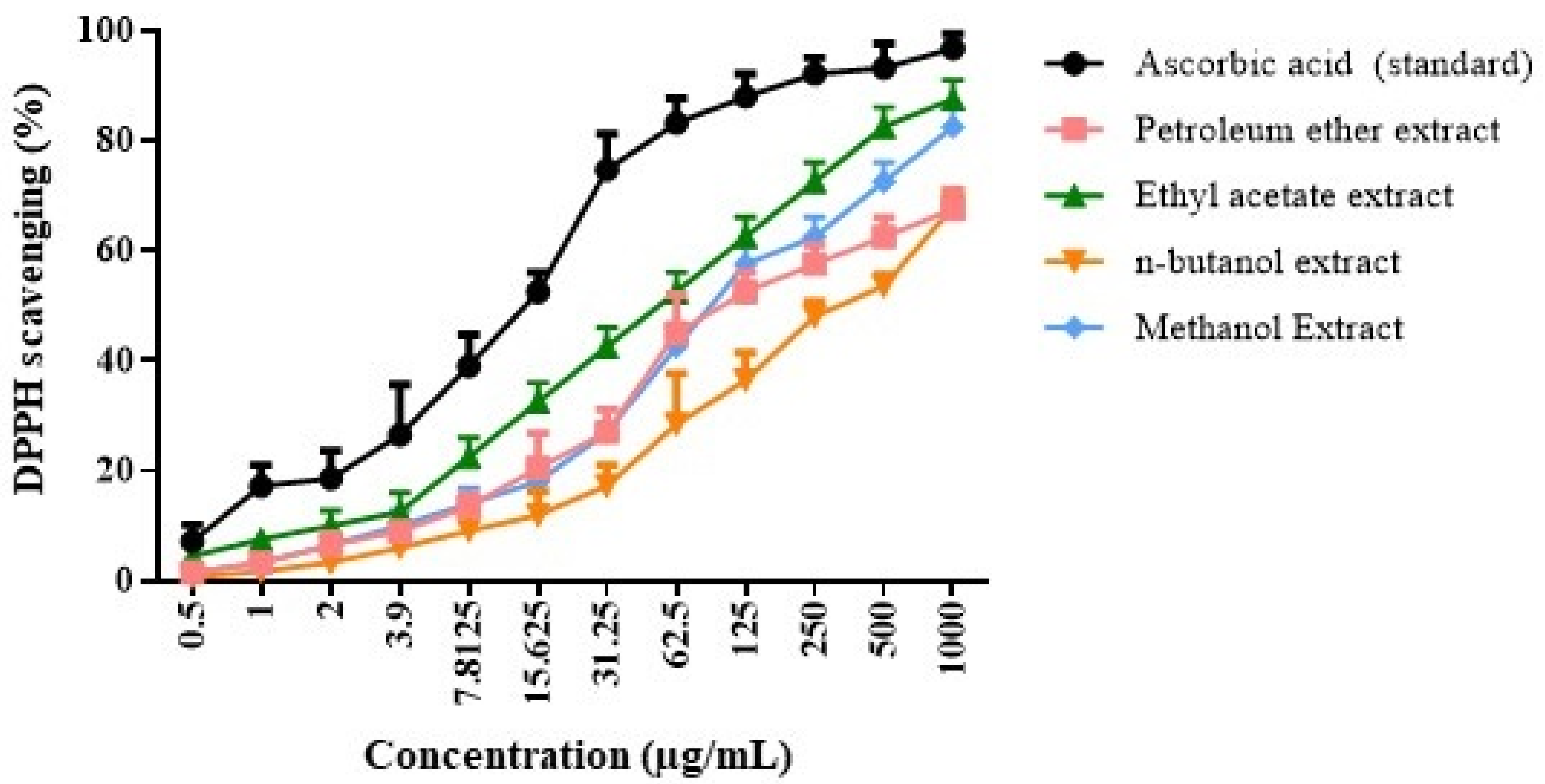
Figure 1. DPPH assay for G. lucidum extract upon using various solvents including petroleum ether, ethyl acetate, n- butanol, and methanol versus the ascorbic acid standard (data are presented as means ± SD).
Molecules 30 03650 g001
Figure 2. Cytotoxicity impact of G. lucidum ethyl acetate extract on (A) HepG2 cells, (B) HCT116 cells, (C) MCF7 cells, (D) A549 cells, and (E) Vero cells. Data are represented as means ± SD.
Molecules 30 03650 g002
Figure 3. Names and structures of depsidones detected in the ethyl acetate extract of G. lucidum.
Molecules 30 03650 g003
Figure 4. Binding interactions of simplicildone D with AKT1 (PDB code: 3OCB). (A) Two-dimensional binding interactions; (B) 3D pose (ligand atoms in cyan and critical amino acid residues in green).
Molecules 30 03650 g004
Figure 5. Binding interactions of simplicildone A with CDK2 (PDB code: 6GUE). (A) Two-dimensional binding interactions; (B) 3D pose (ligand atoms in cyan and critical amino acid residues in green); (C) overlay of Compound 5 with the co-crystallized ligand inside the active site.
Molecules 30 03650 g005
Figure 6. Binding interactions of mollicellin G with ERK1 (PDB code: 2ZOQ). (A) Two-dimensional binding interactions; (B) 3D pose (ligand atoms in cyan and critical amino acid residues in green). (C) Chemical structure of mollicellin G, showing the numbering of carbon atoms.
Molecules 30 03650 g006
Figure 7. Binding interactions of simplicildone D with TNFα (PDB code: 2AZ5). (A) Two-dimensional binding interactions; (B) 3D pose (ligand atoms in cyan and critical amino acid residues in green).
Molecules 30 03650 g007
Table 1. Total phenolics of all extracts of G. lucidum (data are represented as means ± SD).
Sample Code(1) Petroleum Ether Extract(2) Ethyl Acetate Extract(3) N-Butanol Extract(4) Methanol Extract
Total Phenolics (mg/g)0.21 ± 0.129.87 ± 0.71 *0.11 ± 0.014.27 ± 0.11
* p ≤ 0.05 (using one-way ANOVA followed by a post hoc test).
Table 2. The result of depsidones tentatively identified by UHPLC/Q-TOF-MS-MS analysis (negative mode) in the ethyl acetate extract of G. lucidum.
NoName of the
Depsidone Compound
Rt (Min)Mol. Weight
[M-H]-
Error (ppm)Characteristic Frag.References
1Mollicellin G C22H23O71.11235398.13523.5202.037, 238.0756, 396.855, 98.128450, 188.0453.195.0500, 200.0599, 218.0507, 218.105, 278.0984, 308.097, 330.107, 362.144, 398.10425 [37]
2Simplicildone I C31H28O81.21527.17071527.1746, 376.212, 348.402, 177.542[38]
3Mollicellin B C21H18O71.244983381.09770.8381.097, 366.371, 322.804, 179.374, 167.529, 217.1236, 219.04, 219.096, 243.076, 261.0647, 363.1362, 381.102[42]
4Talaromyone B C23H26O71.31413.4565−8.4413.1656, 353.221, 339.391, 283.024[38]
5Simplicildone A C18H18O61.3228329.1031.5329.103, 314.453, 314.409, 85.1782, 285.324, 155.164, 154. 328, 91.443, 76.221[44]
6Purpactin C C23H21O71.43408.12554.6408.125, 393.851, 365.344 [45]
7Emeguisin B C24H25ClO51.72427.8752−13.1112.986, 247.071, 256.93, 265.130, 293.103, 337.1281, 427.167, 381.2082, 71.427,1349, 247.0716, 398.021, 427.1356[46]
8Mollicellin E C22H19ClO83.390283445.0675−3.4445.0675, 312.865, 220.674, 196.034, 191.350, 225.0928, 237.0142, 250.097, 264.9148, 265.108, 279.008, 279.034, 293.046, 309.069, 313.110 361.118, 377.101, 386.1417, 445.069[37]
9Simplicildone D C25H24O77.60215435.14654.8435.1465, 429.432, 381.246, 201.176, 154.369, 91.66, 76.33[38]
Disclaimer/Publisher’s Note: The statements, opinions and data contained in all publications are solely those of the individual author(s) and contributor(s) and not of MDPI and/or the editor(s). MDPI and/or the editor(s) disclaim responsibility for any injury to people or property resulting from any ideas, methods, instructions or products referred to in the content.