Article

Pharmacognosy and Antioxidant Activity of Pruned Leaves from the Unexplored Olea europaea L. ‘Lavagnina’ (Liguria, Italy)

1
Department of Earth, Environment and Life Sciences (DISTAV), University of Genova, Corso Europa 26, 16132 Genova, Italy
2
Department of Pharmacy, University of Salerno, Via Giovanni Paolo II 132, 84084 Fisciano, Italy
3
Consorzio di Tutela del Basilico Genovese DOP, Salita Santa Caterina 50-52R, 16123 Genova, Italy
*
Authors to whom correspondence should be addressed.
Molecules 2025, 30(17), 3605; https://doi.org/10.3390/molecules30173605
Submission received: 22 July 2025 / Revised: 25 August 2025 / Accepted: 2 September 2025 / Published: 3 September 2025
(This article belongs to the Special Issue Chemopreventive Activity of Natural Products)

Abstract

Olea europaea L. ‘Lavagnina’ is cultivated in the Eastern Ligurian coast (Italy), and during the pruning process a huge amount of pruning residues is produced. This by-product is generally disposed of by burning, despite still containing bioactive compounds. In particular, olive leaves are indeed rich in secondary metabolites, which can vary both in quality and quantity in relation to the cultivar considered and the area of cultivation. For this reason, we aimed to carry out a pharmacognostic study of the pruned leaves of the unexplored local cultivar ‘Lavagnina’, evaluating the possibility of reusing this by-product for new health applications. The micromorphological characterization was conducted by light and scanning electron microscopy. ‘Lavagnina’ leaf was micromorphologically similar to that of other olive cultivars; however, it differed in terms of midrib structure. Leaf extracts were obtained using solvents of increasing polarity (petroleum ether, chloroform, methanol) and the food-grade solvent, 70% ethanol. A high antioxidant activity was found only for the methanolic (ME) and hydroalcoholic (HAE) extracts, and, therefore, they were then characterized from a phytochemical point of view by LC-ESI-HR-MS. Such analysis allowed the identification of secondary metabolites belonging mainly to secoiridoids, flavonoids, and iridoids. Overall, the HAE had the highest antioxidant activity (17.3 ± 0.6 μg/mL), and it is, therefore, the best candidate for health applications related to a protective effect on a variety of inflammation-related diseases, also considering that inflammation may play a role in cancer progression.

1. Introduction

In the Mediterranean basin, the olive tree (Olea europaea L. subsp. europaea) is one of the most important agricultural crops [1], with Spain, Italy, and Greece as the leading worldwide producers of olive oil and table olives [2].
In Liguria Region (Italy), the olive growing area is small (about 2000 hectares), but the peculiar pedo-climatic characteristics of the environment and the traditional cultivation methods allow for obtaining a typical extra-virgin olive oil (EVO) certified by the Protected Designation of Origin (PDO). This product is highly valued all over the world [3,4], placing Liguria among the Italian regions excelling in the art of oil-making.
The main cultivars of O. europaea used to make EVO in Liguria are ‘Taggiasca’ and ‘Lavagnina’, both of which owe their name to two towns, Taggia (Imperia province) and Lavagna (Genoa province), respectively. In particular, the ‘Lavagnina’ olive is widespread in the East of Liguria (Province of Genoa, Gulf of Tigullio), and it is used to produce the PDO oil “Riviera Ligure—Riviera di Levante”. According to the PDO Product Specifications [5], ‘Lavagnina’ olives are used alone or mixed with small quantities of other cultivars, such as ‘Razzola’ and ‘Pignola’.
‘Lavagnina’ was already mentioned by G. Gallesio (1817–1839), an agronomist and botanist from Savona (Liguria), in his Pomona Italiana, a treatise on Italian fruit trees [6]. This early documentation highlights the long-standing presence and agricultural relevance of this cultivar in Liguria. Despite this, ‘Lavagnina’ has been little studied. The only reference to O. europaea ‘Lavagnina’ appears in an Italian publication edited by Regione Liguria [7], which reports the phytochemical differences among virgin olive oils obtained from various Ligurian cultivars of O. europaea. A few other works mentioning ‘Lavagnina’ focus on the molecular identification of cultivars [8,9].
In light of its historical and economic significance, investigating and safeguarding ‘Lavagnina’ is essential to preserve the Ligurian olive-growing heritage and maintain the PDO olive oil production.
Nowadays, the cultivation of ‘Lavagnina’ in Eastern Liguria spreads over a few hectares, but olive farming and oil production generate numerous by-products, such as leaves and pomace. In particular, the maintenance of a medium-sized olive grove requires pruning operations to maintain tree health, resulting in a huge amount of leafy branches per year. These pruning residues are sometimes chipped and spread over the soil to reduce erosion but more often are treated as waste and disposed of by burning. A considerable amount of olive leaves is also discarded in the first phase of the oil-making process, when leaves are separated from olives (about 5 kg of leaves per quintal of olives).
Pomace (“sansa”) and fragments of kernels (“nocciolino”) in Eastern Liguria are already valorised: the first is used to produce a low-value oil, while kernels serve as an energy source for heating. In contrast, although they contain a significantly higher percentage of polyphenols than olives or oil, pruned leaves remain underutilized. These compounds could be extracted and used for applications in the medicinal, nutraceutical, and cosmetic sectors [10,11,12,13]. So, exploiting pruning residues of ‘Lavagnina’ would allow the local small companies to open up to new sources of income while promoting a more sustainable olive oil supply chain.
Olive leaves represent a natural source of bioactive compounds with therapeutic effects on health. Indeed, their extracts have been exploited in traditional medicine for centuries, with the first attested use for the treatment of fever, cough, and urinary problems in ancient Egypt [14,15]. Several scientific works have described the ethnobotanical uses of olive leaf preparations in Italy [16] and references therein, and in other Mediterranean countries [17,18], mainly in the form of decoction, for the treatment of various diseases, such as hypertension, stomach problems, kidney stones, hypercholesterolemia, and diabetes.
Many laboratory tests, both in vitro and in vivo, have demonstrated the beneficial properties of olive leaves, including antihypertensive, anticarcinogenic, hypoglycemic, antimicrobial, and hypocholesterolemic effects [11]. All these positive activities can be at least partly ascribed to the antioxidative power of polyphenols [1], such as: (a) secoiridoids (oleuropein and derivatives), (b) flavonoids (apigenin, quercetin, luteolin, luteolin-7-O-glucoside), and (c) simple phenols (hydroxytyrosol and tyrosol, caffeic and vanillic acids) [19].
The qualitative and quantitative phenolic composition of olive leaves can vary significantly according to both genetic and climatic/geographical factors [20,21], highlighting the importance of studying local cultivars.
Considering this, the overall aim of our work was to investigate and valorise the pruned leaves of the ‘Lavagnina’ cultivar growing in Eastern Liguria, to evaluate the potential for recovering bioactive compounds for healthy applications, from a circular economy perspective.
From a scientific point of view, our goal was to chemically characterize pruned leaf extracts and assess their antioxidant activities. In addition, to complete the pharmacognostic study of ‘Lavagnina’, and as a quality control for future applications of the pruned biomasses, we also carried out a morphological and anatomical characterization of the leaves by Light and Scanning Electron Microscopy to highlight their distinctive features, which can help to ensure the purity and safety of this by-product.
From an economic and regulatory perspective, our in-depth study of this unexplored typical Ligurian cultivar is important to support its peculiar intrinsic characteristics that derive from the set of terroir, genetics, and agricultural practices [22]. The whole of the data collected could represent a solid scientific basis that may contribute to a potential PDO or PGI (Protected Geographical Indication) recognition of ‘Lavagnina’ olive.

2. Results

2.1. Micro-Morphological and Anatomical Analysis

The leaf was iso-bilateral and hypostomatic, showing collenchyma layers and scattered sclerenchyma fibers under the epidermis both above and below the midvein (Figure 1A). In particular, in the adaxial surface, these tissues gave rise to a characteristic small protrusion (Figure 1A, arrow). A thick cuticle was present on both surfaces, especially on the abaxial one, as highlighted by the staining with Sudan Black (Figure 1B).
The mesophyll was divided into: (a) three to four layers of palisade parenchyma I (PPI) under the adaxial epidermis, rich in phenolics as indicated by the light blue staining with TBO (Figure 1C); (b) spongy parenchyma (SP) and (c) one layer of palisade parenchyma II (PPII) close to the abaxial epidermis (Figure 1D). The cells of PPII were short, irregularly shaped, and not well distributed compared to the cells of PPI.
Under the adaxial epidermis and among the spongy cells, several filiform sclereids (trichosclereids) were observed, longitudinally oriented in PPI (Figure 1C, arrow) and tangentially oriented in SP (Figure 1D, black arrows).
In both PPI and SP, numerous small calcium oxalate crystals, mainly in the form of raphides, were found. They appeared well visible under polarized light (Figure 1E) and more detailed by Scanning Electron Microscope (SEM) (Figure 1F).
Numerous non-glandular scutiform trichomes were observed on both surfaces (Figure 1B–D), and the histochemical analysis showed in their unicellular stalk cell the presence of phenolic compounds, as highlighted by the green staining with TBO (Figure 1D, red arrow).
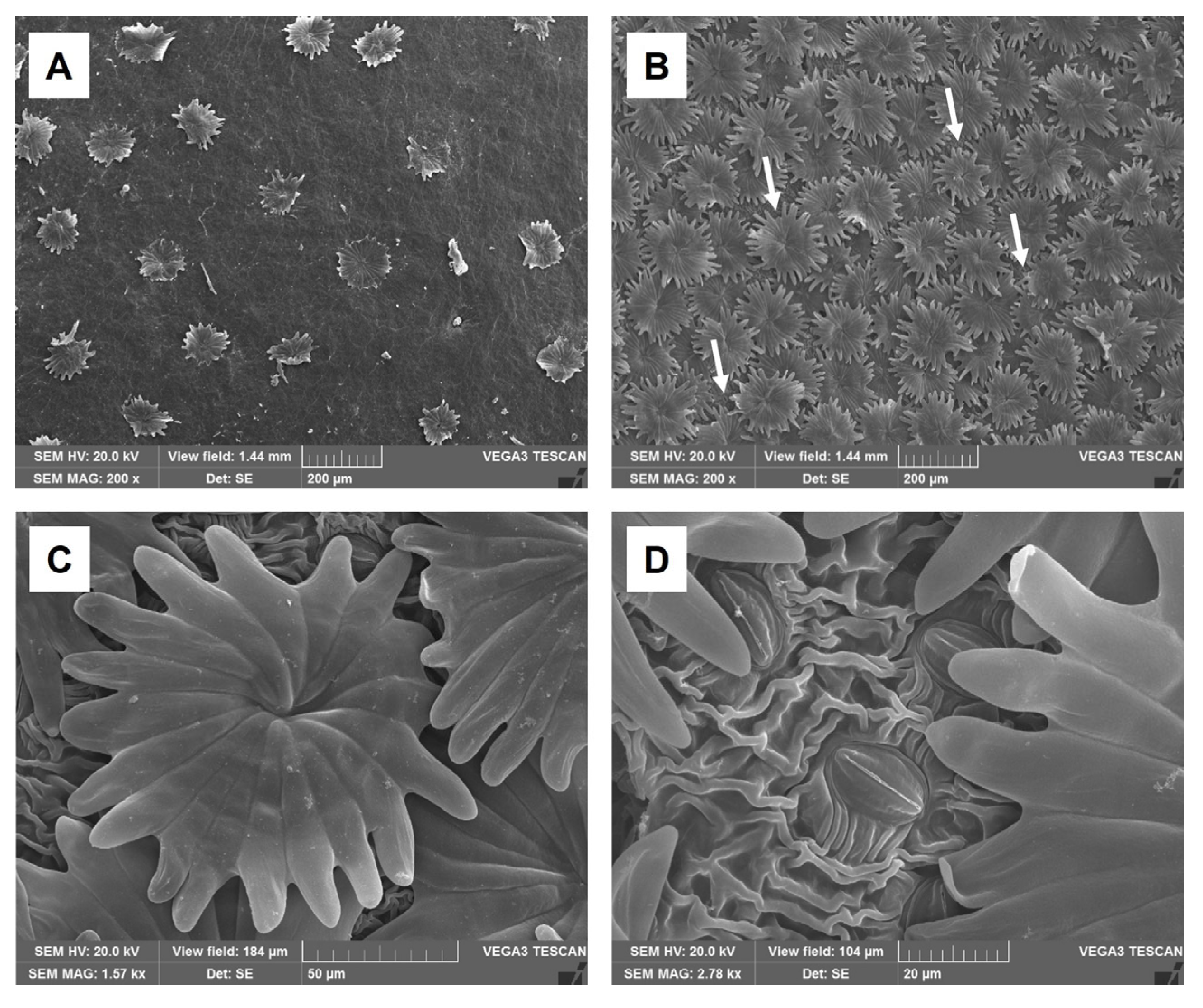
Moreover, SEM investigation revealed that these trichomes were sparse on the adaxial epidermis (Figure 2A), while on the abaxial epidermis they were densely distributed but did not strictly overlap (Figure 2B, arrows). Thus, in some areas, it was possible to observe the underlying striate cuticle and stomata (Figure 2C,D).
All these anatomical traits of olive leaf are conserved among cultivars; however, the structure of the midrib can slightly differ, as shown in Figure S1, where ‘Lavagnina’ (A) is compared to other two cultivars, that are ‘Taggiasca’ (B) and ‘Leccino’ (C).

2.2. Extraction

The extraction yield (%) of petroleum ether extract (PEE), chloroformic extract (CHE), methanolic extract (ME) and hydroalcoholic extract (HAE) was 0.5, 3.3, 3.2, and 7.2, respectively.

2.3. Antioxidant Activity

The antioxidant activity of the extracts evaluated by DPPH, FRAP, and ABTS assays is presented in Table 1.
The PEE did not exhibit any antioxidant activity. On the contrary, CHE, ME, and HAE had some antioxidant activity in all three tests; nevertheless, it was lower with respect to the standards. The HAE showed a significantly higher activity (p < 0.05) than the other extracts only in the DPPH test, with an IC50 value of 17.3 ± 0.6 μg/mL. The least active extract was the CHE (IC50: 79.4 ± 0.7 μg/mL). Instead, considering the other two tests, all extracts showed an excellent antioxidant activity.

2.4. Chemical Analysis

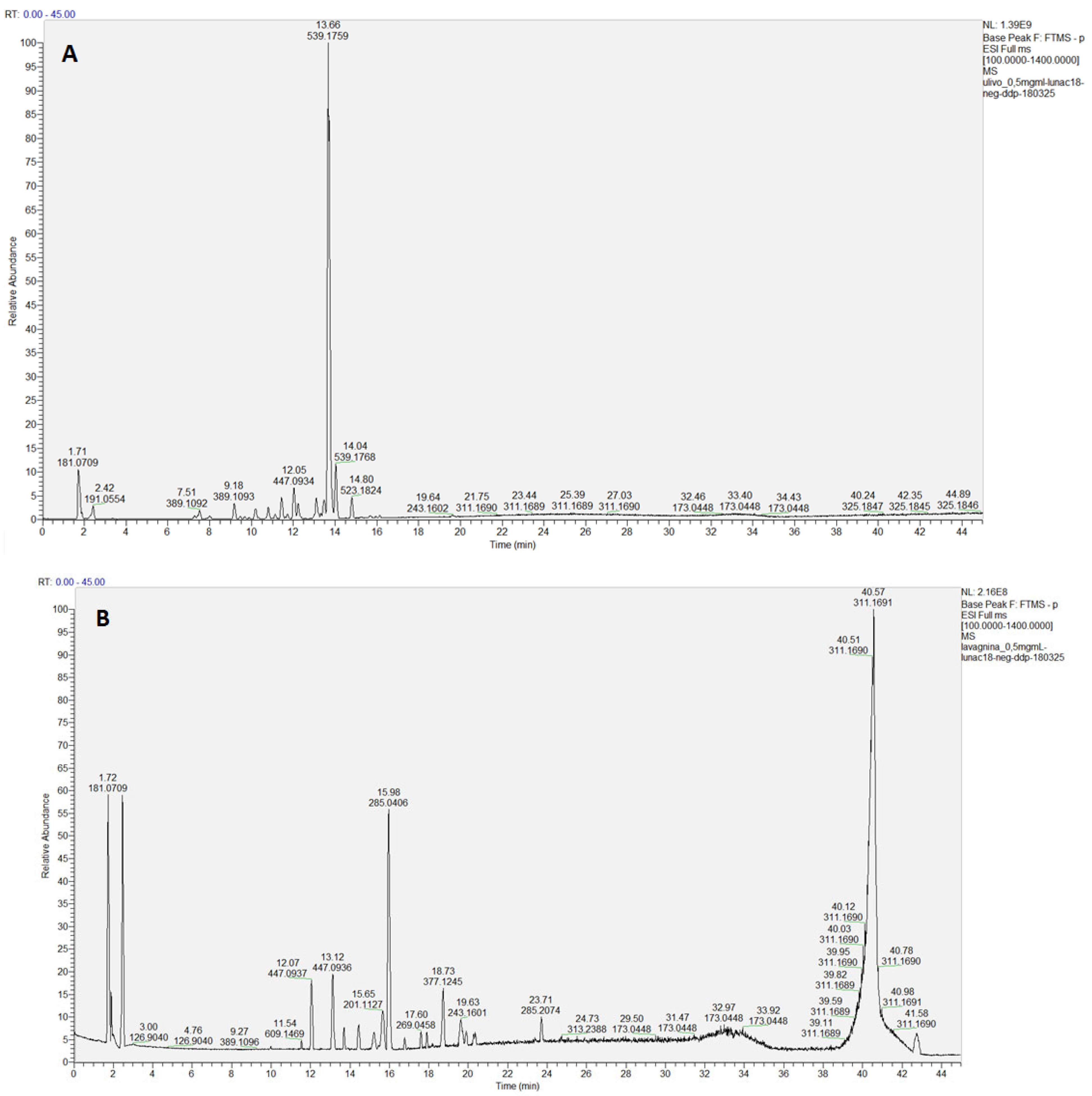
Being the methanolic (ME) and the hydroalcoholic (HAE) extracts the more biologically active, they were analyzed by LC-HRESIMS/MS (Figure 3).
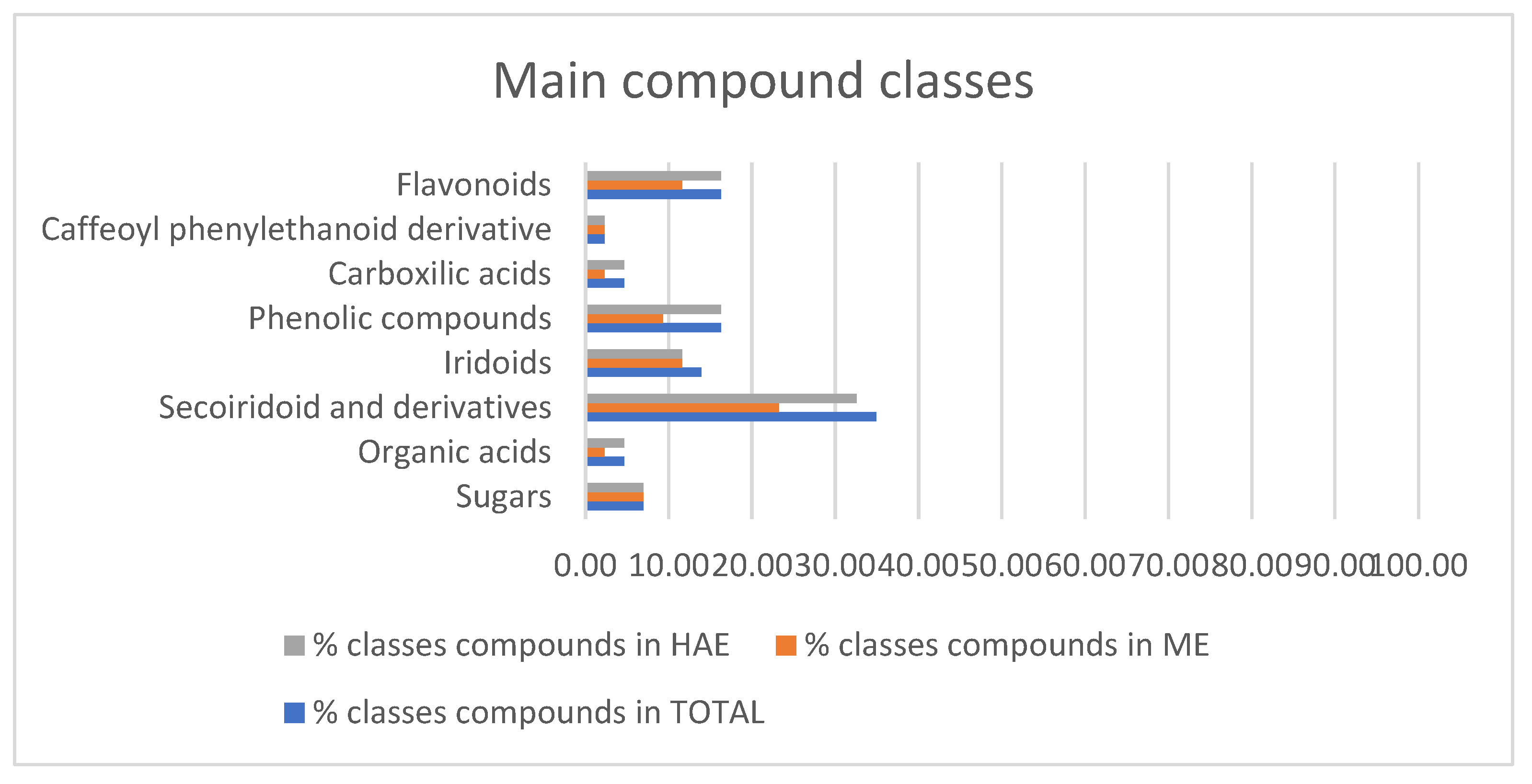
Overall, 43 components (Table 2) were identified, belonging to several representative classes of constituents, mainly secoiridoids and their derivatives (15 compounds, peaks 5, 8, 11, 16–18, 20, 24, 26, 28, 31–34, 40) and flavonoids (7 compounds, peaks 27, 29–30, 36–39) (Figure 4). Most metabolites were common to both extracts (e.g., mannitol, quinic acid, hydroxytyrosol and its glycosides, oleoside, oleuropein, rutin, luteolin, quercetin), but some compounds were specific to each extract, e.g., aucubin and ligstroside only in ME, gluconic acid and vanilloside only in HAE (Table 2).
Compound 1 appeared at tR = 1.72 min and yielded a precursor ion [M-H] at m/z 181.0709, attributed to sugar mannitol, previously reported by Toumi and coworkers [23] in O. europaea roots. MS2 fragmentations revealed characteristic fragments at m/z 163.0604 and 119.0339, corresponding to the loss of water molecules (-H2O), as also reported from the same Authors.
The compound 4 (C8H18O8) was identified in both extracts and could be tentatively annotated as a sugar derivative, as 3-deoxy-D-manno-octulosonate. The major fragment in MS2 spectrum for this compound appeared at m/z 87.0075, already previously reported in O. europaea extract by Serrano-García and coworkers [24].
Another sugar derivative was identified as 1,5-anhydroxylitol, already found in leaves of O. europaea [25], which gave a precursor ion [M-H] at m/z 259.0825: the identification was confirmed by MS2 analysis, which produced the main fragments at m/z 217.0701, 145.0494, and 115.039 [25].
The compounds 2 and 3 were organic acid derivatives: the compound 2 gave a [M-H] ion at m/z 195.0503, corresponding to the deprotonated molecular form of gluconic acid. The substance, already reported in ethanolic leaf extract of O. europaea [26], gave MS2 fragments at m/z 177.0398, 129.0183, 99.0075 and 75.0075, attributable to an organic acid, as previously reported [27,28]. The compounds 3 was identified as quinic acid, previously reported in O. europaea [29]. Also in these case, MS2 fragmentations revealed characteristic fragments at m/z 173.0445 [M-H2O-H], 127.0388 [M-2H2O-CO-H], 111.0439 [M-2H2O-CO2-H], 93.0334 [M-3H2O-CO2-H), 85.0283 [M-H2O-2CO2-H] [29,30].
The peak 5 was attributed to a secoiridoid and identified as 1-b-D-glucopyranosyl acyclodihydroelenolic acid, previously reported in a by-product of olive oil extraction by Rubio-Senent and coworkers [31].
The peaks 8, 11, 16, 17, 18, 20, 24, 25, 26, 28, 31, 32, 33, 34 and 40 were also attributed to the class of secoiridoids and their derivatives. In fact, compound 8 gave a [M-H] ion at m/z 389.1092 and was attributed to oleoside. Its MS2 spectra exhibited a fragment ion peak at m/z 227, related to the loss of a hexose moiety (162 Da). A fragment ion peak at m/z 183 indicated a subsequent loss of CO2. This fragmentation pathway was previously reported in olive leaf extracts by Kabbash et al. [32].
The peak 11 was attributed to loganic acid, that presented deprotonated [M-H] ion at m/z 375.1301. The compound generated the ESI-MS2 ions at m/z 151 [M−Glc−H2O–CO2-H] and m/z 125 [M−Glc−2CO2-H]: the same fragmentation pathway was previously reported [33].
The compound 16, present only in the HAE, was attributed to a derivative of oleoside, oleoside methyl ester, which gave a precursor deprotonated ion [M-H] at m/z 421.1724: the MS2 analysis, with the presence of fragments at m/z 151.075 and 115.0388, was in agreement to Ammar et al. [27].
Compound 17, only present in the HAE, was recognized as a secoiridoid: it showed a precursor ion [M-H] at m/z 257.1032, having a molecular formula C12H18O6. The MS/MS spectrum formed fragment ions at m/z 213.1130 [M-CO2-H] and 151.0758 [M-C3H6O4-H], compatible with 3-hydroxymethyl-2,3-dihydro-5-(methoxycarbonyl)-2-methyl-2Hpyran-4-acetic methyl ester, previously reported in olive oil by products [31].
The secoiridoids 18 and 20, identified in both extracts, were, respectively, recognized as elenolic acid dihexoside isomer (that gave a [M-H] ion at m/z 565.1779) and acyclodihydroelenolic acid (that gave a [M-H] ion at m/z 245.103). Both compounds were previously reported in the literature by Fayek and coworkers [34] in olive leaf metabolome. Acyclodihydroelenolic acid furnished a MS2 spectra with the main fragments at m/z 183.1020, a characteristic of secoiridoid compounds due to the loss of H2O and COOH, as already reported by the same authors.
Compound 24 gave a [M-H] ion at m/z 403.1250 and was attributed to oleoside methyl ester. Ventura and coworkers [35] reported the presence of this compound in the extract of olive leaves. The MS2 analysis confirmed the presence of m/z product ions at 223.0613, 181.0501, 179.0702, and 101.0233, as already reported in the literature [24,35]. Specifically, the m/z 223.0613 corresponded to dehydrated elenolic acid and it is due to the loss of an hexose from the molecule [28,36].
The peaks 26 and 28 were attributed, respectively, to demethyl oleuropein and to 10-hydroxyoleuropein, that presented deprotonated [M-H] ions at m/z 525.1621 and 555.1727: the major fragments in MS/MS spectrum of demethyl oleuropein appeared at m/z 389.1088, 209.0452, 195.0656, 165.0550, and 121.0649. The first major fragment was due to the loss of dehydrated 3-hydroxy-tyrosol; the other fragments were due to the loss of a dehydrated glucose moiety and to multiple fragmentations which caused the break of aglycone molecule [15]. The same MS2 analysis was previously reported by Tarchi and coworkers [37]. The MS2 analysis of 10-hydroxyoleuropein revealed major fragments at m/z 223.0609, corresponding to sequential losses of H2O, dihydroxystyrene and glucose [38]. The substance was previously found in olive tree leaves [39].
The peaks 31–34 were, respectively, identified as 2′′-methoxyoleuropein (at m/z 569.1882), oleuropein (at m/z 539.1759), lucidumoside C (at m/z 583.2039), and ligstroside (deoxy oleuropein—at m/z 523.1826). The latest identified secoiridoid was oleuropein aglycone, that gave deprotonated ion [M-H] at m/z 377.1245, with a retention time of 18.73 min. The MS2 analysis of the above compounds was in agreement with literature. So, the MS2 spectra of the compound 2′′-methoxyoleuropein revealed the fragment at m/z 403,1241, which is due to the cleavage of the phenolic moiety (166 amu), and it could undergo an elimination of the glucose moiety (fragment at m/z 223.0610), as already reported in the literature [40,41]. The MS2 analysis of oleuropein, having a molecular formula C25H32O13, produced the fragment at m/z 377.1253 by the loss of a glucosyl moiety: the fragment ion at m/z 307.0826 was explained by the loss of a C4H6O from the fragment 377.1253, while the fragment at m/z 275.0561 derived from the loss of CH3OH from the fragment at m/z 307.0826. The same fragmentation was previously reported [40,41]. In the MS/MS spectrum of lucidumoside C, found in both extracts, the characteristic fragment ions of [M-C10H12O3-H] at m/z 403.1261 and of [M-M-C10H12O3-Glu-H] at m/z 223.0610, were observed in agreement with Serrano-García and coworkers [24]. As also reported by Quirantes-Piné and coworkers [28], the first fragment was due to cleavage of the phenolic moiety, after which it could undergo elimination of the glucose moiety (fragment 223.0610). Ligstroside, present only in the ME, exhibited a characteristic MS/MS spectrum already reported in the literature: the fragment ions at m/z 361.1298, and 291.0877 could be attributed to the loss of glycosil moiety (162 uma), and of a C4H6O group. The reported fragmentation pathway was in agreement with literature [42].
Oleuropein aglycone, found only in the HAE, presented the molecular formula C19H22O8: the product ion scan of the deprotonated molecule formed characteristic fragment ions at m/z 307.0825 and 275.0571, already reported in the literature [27,39].
The compound 6, identified as a hydroxylated product of the dialdehydic form of decarboxymethyl-elenolic acid, was the first eluted compound belonging to the class of iridoids: it appeared at tR =7.38 min and yielded a precursor ion [M-H] at m/z 199.0608. The compound was only identified in the HAE and produced a MS/MS spectrum with fragment ions at 155.0705, and 111.0804, due to the loss of one and two CO2 molecules, respectively. Recently, López-Salas and co-workers [43] reported the same compound in olive leaf; the same MS2 spectra were found in the literature [44,45].
The peaks 12, 13, 15, 21 and 23 were also attributed to iridoids. Compound 12 gave a [M-H] ion at m/z 345.1195 and was attributed to aucubin. Do and coworkers [46] reported the presence of this compound in various plants, like plantago, vervein, valerian, gentian, and also olive tree. The MS/MS analysis revealed the main fragments at 183.0656 and 165.0552, due to the loss of a glycosil moiety (162 uma) and of H2O, respectively; this fragmentation pathway agrees with the literature [47]. The peaks 13, 15, 21 and 23 were attributed, respectively, to the aldehydic form of decarboxymethyl elenolic acid, 7-deoxyloganic acid, loganin, and lamiol, and presented deprotonated [M-H] ions at m/z 215.0922, 359.1353, 389.1459, and 377.1459, respectively. The major fragments in MS/MS spectrum of aldehydic form of decarboxymethyl elenolic acid appeared at m/z 171.1020, and 153.0912, due to the loss of CO2 and H2O, respectively. Previously, Lozano-Sanchez et al. [48] and Ribeiro et al. [49] reported the same compound in olive by-products. As reported by Ammar and coworkers [27], the fragmentation pathway of 7-deoxyloganic acid with the characteristic ion at m/z 197.0814, due to the loss of a glycosyl moiety, confirmed the identification. The MS2 analysis of loganin, an iridoid with molecular formula of C17H26O10, with the presence of fragments at m/z 345.1531, 119,0343, and 101, 0233 was in agreement with literature [27,50]. Finally, lamiol produced a MS/MS spectrum with main fragments at m/z 197.0815, 153.0913, due to the consequential losses of glucose moiety (180 uma), and of a CO2 molecule, respectively [51].
The peak 7, with a precursor deprotonated ion [M-H] at m/z 315.1091, was attributed to hydroxytyrosol glucoside, already found in olive leaf extracts [35,52]: the corresponding MS/MS spectrum was dominated by the peak of deprotonated hydroxytyrosol (m/z 153.0549), due to the cleavage of the glycosyl moiety and by the peak at 123.0441, resulting from the loss of the CH2OH and CHO groups from the hydroxytyrosol original structure. These two fragments are characteristic for the identification of any conjugated hydroxytyrosol present in olive tree organs. The same fragmentation has been previously reported [53].
The peaks 9–10 were attributed to phenols and were found in both extracts: the compound 9 was identified as hydroxytyrosol-diglucoside, which gave a precursor deprotonated ion [M-H] at m/z 477.1620. In addition, this conjugated hydroxytyrosol gave a MS/MS spectrum dominated by the fragments 153.0549 and 123.0440, as previously reported [53].
Compound 35, recognized as sebacic acid, with molecular formula C10H18O4, is a simple phenol, previously found in olive extract [54]: the MS/MS analysis was dominated by the fragments 183.1020 and 157.1226, resulting from the loss of H2O and CO2 molecules, respectively.
The phenolic shikimic acid (compound 41) was identified at Rt 22.34 min, with a precursor deprotonated ion [M-H] at m/z 173.0448: the MS/MS spectrum revealed the presence of main fragments due to the loss of H2O (m/z = 155.0342), CO (m/z = 145.0496), CO2 (m/z = 129.0547). This compound was previously reported in an extract from olive pomace [53].
Compound 14 and compound 22 are two phenolic aldehydic substances, also linked to the phenolic group: compound 14, with a precursor deprotonated ion [M-H] at m/z 313.0933 was recognized as vanilloside, known also as glucovanillin. The compound, that is the glucoside of vanillin, gave a MS2 spectrum characterized by the fragment ions at 151.0393, and 123.0441, consistent with the leak of a glucose moiety and to the subsequent loss of CH2O group, respectively [55,56]. Compound 22, correlated to the previous compound, was identified as vanillin, a phenolic aldehyde, with the molecular formula C8H8O3: it gave a precursor ion [M-H] at m/z 151.0392. The compound was previously reported in olive leaves [27,28,34]. This assignment was supported by the fragment ions produced in MS/MS spectra: the ion, at m/z 123.0449, was yielded from the loss of the carbonyl group [28].
The compounds 19 and 42 were attributed to two carboxylic acids: specifically, the peak 19, with a precursor deprotonated ion [M-H] at m/z 345.1558, was attributed to 1,1,12,12-dodecanetetracarboxylic acid, acompound that was previously reported in olive leaf extract [54]. The peak 42 was attributed to hexadecanedioic acid, a dicarboxylic acid which gave a deprotonated molecular ion at m/z 285.2074, previously reported in oil mill waste by Marra and coworkers [57]. The MS2 analysis of the compound yielded a main fragment at m/z 267.1969, previously reported [58].
Compound 25 was attributed to decaffeoylverbascoside, which gave a deprotonated molecular ion at m/z 461.1672: the MS2 analysis revealed the presence of the main fragment at 113.0233, previously reported in the literature [27,37].
The peaks 27, 29, 30, 36–39, were attributed to compounds belonging to the class of flavonoids. Compounds 27, 29, and 37 gave [M-H]- ions at m/z 609.1468, m/z 593.1520 and, m/z 301.0358, which were attributed to rutin (quercetin 3-O-rutinoside), luteolin-7-O-rutinoside, and quercetin, respectively. The analysis of the MS2 spectra showed a fragment at m/z 301.0351 for 27, and a fragment at m/z 285.0407 for 29, both consistent with the lack of a rutinoside moiety (308 uma). MS2 fragmentations of the two isomers, in agreement with literature [37] revealed other characteristic fragments at m/z 300.0279, 178.9981, 151.0028 for rutin, and 447.0978, for luteolin-7-O- rutinoside, respectively. Quercetin showed a MS/MS spectrum with main fragments at 273.0406, 178.9980, and 151.0029 [59]: the latest peak was generated after a Retro-Diels-Alder (RDA) cleavage of the A ring [60].
The peaks 30 and 36 were attributed to luteolin-O-hexoside isomer and luteolin, with a precursor deprotonated ion [M-H] at m/z 447.0936 and 285.2074, respectively. This assignment was supported by the fragment ions produced in MS/MS spectra: for luteolin-O-hexoside isomer, the ion at m/z 285.0407 revealed a cleavage of the sugar (−162 m/z) releasing the aglycone form [24]. The MS2 fragmentation of luteolin, with the main fragments at 199.0395, 151.0029, 133.0283, was in agreement with literature [24]: the [M-H]- ion can undergo a RDA fragmentation through the heterocyclic ring, resulting in the formation of product ions with m/z 151.0029 or 133.0283, the latter being the base peak, corresponding to 1,3A and 1,3B, respectively [61]. Moreover, the ion with m/z 175.0392 could correspond to [M-C3O2-C2H2O-H], as already reported [62].
The compounds 38 and 39 were identified as apigenin and diosmetin, only present in the HAE, with the molecular formula C15H10O5 and C16H12O6, respectively. Both compounds were previously reported by Fayek and coworkers [34]. The MS2 analysis revealed the presence of the main fragments at 151.0027 and 117.0332, generated after RDA fragmentation of the [M-H], which can be represented as 1,3A and 1,3B, respectively. The fragment at 107.0127 was due to the loss of CO2 from the 1,3A ring. On the other hand, diosmetin had a different fragmentation pathway. The most abundant product ion was at m/z 284.0329 corresponding to the loss of a methyl group. Subsequent loss of CO involving the formation of a five-membered ring led to the ion with m/z 256.0369 [61].

3. Discussion

In Liguria, due to the sloping nature of the land, olive trees are placed on terraces supported by dry stone walls, reducing the risk of landslides and erosion and, at the same time, shaping the landscape. So, olive groves are of great environmental and cultural importance.
Despite the difficulties related to the geomorphology of the Region, a significant number of farms and olive oil companies can be found throughout the territory. The management of the olive oil chain by-products from a circular perspective is, therefore, a priority objective. The principal contributor to waste generation are the leaves, deriving from both the pruning of trees and olives harvesting [63].
Nowadays, although olive leaves represent a promising source of bioactive compounds, the direct burning of the pruning residues is the preferred method of disposal in Liguria Region, which however entails economic costs and environmental risks.
With a view to recovering these residues, we evaluated the micro-morphological and anatomical features of olive leaves. This analysis represents an important starting point for the quality control process of plant by-products [64].
The micromorphological characterization of ‘Lavagnina’ leaf is in agreement with data obtained on other Olea cultivars by Bacelar et al. [65], Moreno-Alias et al. [66], Rahfeld [67] and Menezes et al. [68]. The diagnostic features of olive leaf are the presence of abundant scutiform trichomes, two zones of palisade parenchyma and individual or grouped elongated sclereids.
These characteristics are linked to the xerophytic nature of olive tree that has to cope with prolonged drought periods during the warm season of the Mediterranean-type climate, as highlighted by Marchioni et al. [69]. Indeed, according to Bosabalidis and Kofidis [70], the presence of the PPII and the high number of trichosclereids, which are involved in the sunlight distribution within the mesophyll, are a response to stress due to water shortage. In addition, the presence of phenolics in the stalk of scutiform trichomes, characteristic previously observed also by Rombaut et al. [71], could be implicated in the protective action of these trichomes against UV radiation.
Although the basic structure of olive leaf is shared between cultivars, according to Menezes et al. [68] and Žuna Pfeiffer et al. [72] there are some anatomical parameters that could differ, such as the midrib structure. For example, ‘Lavagnina’ leaf is similar to that of ‘Grapollo 561’ cultivar described by Menezes et al. [68], as they are both characterized by a bundle of sclerenchyma and collenchyma above the midrib, differently from other cultivars, e.g., ‘Neblina’ that is characterized by a larger bundle dominated by collenchyma [68].
In addition, we noticed that in ‘Lavagnina’ leaf the layer of PPII is not well defined, with loosely arranged cells not easily distinguishable from those of spongy parenchyma, differently from observations on leaves from other Spanish cultivars made by Moreno-Alias et al. [66]. In addition, the abaxial surface of ‘Lavagnina’ does not show a continuous layer of overlapping scutiform trichomes that completely hides the stomata, as instead was observed by Bacelar et al. [65] on two Portuguese cultivars. However, it remains difficult to determine whether these anatomical differences are the result of ecological adaptations, as the Ligurian olive groves facing the sea share similar pedo-climatic conditions.
Extracts of leaves of different Mediterranean olive cultivars have been analyzed in various works, but relevant contributions on antioxidant activity of O. europaea ‘Lavagnina’ are lacking. Therefore, the present research investigated for the first time the phytochemical profile and biological activity of extracts from pruned leaves of this Ligurian olive. However, it is not easy to compare our results with those of other studies because of the heterogeneity in extracts preparation, tests used and data expressions.
In our work, the extraction of bioactive compounds was carried out using solvents of increasing polarity (PEE, CHE and ME) as well as the safe and food grade HAE.
HAE resulted the most suitable solvent for the extraction of phenolics and to ensure a high antioxidant activity. Phenolic compounds, flavonoids, secoiridoids, and secoiridoid glycosides are present in almost all the parts of O. europaea [73], but in significantly higher quantities in olive leaves [13]. Within the leaf, phenolics occur in the mesophyll and trichome stalks, as highlighted by Liakopoulos et al. [74], who also demonstrated that these structures differed chemically. For example, quercetin was detected in trichome layer but not in leaf mesophyll, suggesting that it could be a specific marker of this layer. Our results confirm the presence of phenolic compounds in trichome stalks and parenchyma, particularly in the multilayered palisade, as indicated by the blue-green staining with TBO.
Among the identified compounds, secoiridoids and flavonoids were the most abundant components. Secoiridoids, e.g., oleuropein and ligstroside, are unique to members of the Oleaceae family. They are iridoids derivatives that are generated by cleavage of the cyclopentane ring and present a phenolic or catechol moiety arising from the phenylpropanoid pathway. Oleuropein is the key component of secoiridoids, occurring in high amount particularly in olive leaves from which it can be easily extracted [19,75]. In a previous study, leaves of another Ligurian cultivar, O. europaea ‘Taggiasca’, were extracted with a mixture of 2-propanol:water and then this extract was partitioned to obtain an oleuropein-enriched fraction that was tested on mesothelioma cells. Results showed that this fraction displayed a significant antiproliferative effect, suggesting possible applications in the treatment of malignant mesothelioma [76].
In the literature, secoiridoids have been recognized as antioxidant, anti-inflammatory, and immunomodulatory compounds and, therefore, have been proposed as potential therapeutic agents for diseases driven by inflammation and reactive oxygen species (ROS), which can damage lipids, proteins and DNA [75,77].
Flavonoids, e.g., quercetin and luteolin-7-O-glucoside, consists of two aromatic rings connected by three carbons that typically form an oxygenated heterocycle. They occur either in the aglycone or glycosylated form [78]. These substances were reported for their potent antioxidant activity and protection against cardiovascular and carcinogenic diseases [19,77].
Other phenolic compounds of HAE were represented by both phenolic alcohols, as hydrotyrosol and its derivatives, and phenolic acids, as shikimic acid. They are among the simplest forms of phenolics, containing a phenolic ring and an organic carboxylic acid function, and exert beneficial effects in terms of antioxidation, anti-atherosclerosis, anti-carcinogenic and anti-inflammation [19,77]. Finally, HAE contained iridoids, including a wide group of cyclopentane [c] pyran monoterpenoids which have shown a wide range of bioactivities including anti-inflammatory, antibacterial, anti-carcinogenic, and antiviral [79]. Given the composition of HAE, as well as its antioxidant activities, this study underscores the potential of ‘Lavagnina’ leaves as a valuable source of bioactive compounds with applications in the medicinal, nutraceutical and cosmetic fields. In particular, since oxidative stress and inflammation are closely linked to cancer development and progression, HAE may display chemopreventive properties by protecting cells from oxidative damage and modulating inflammatory pathways. Further investigations, using specific in vitro and in vivo assays, will be useful to confirm and expand the indicated potential.

4. Materials and Methods

4.1. Plant Material

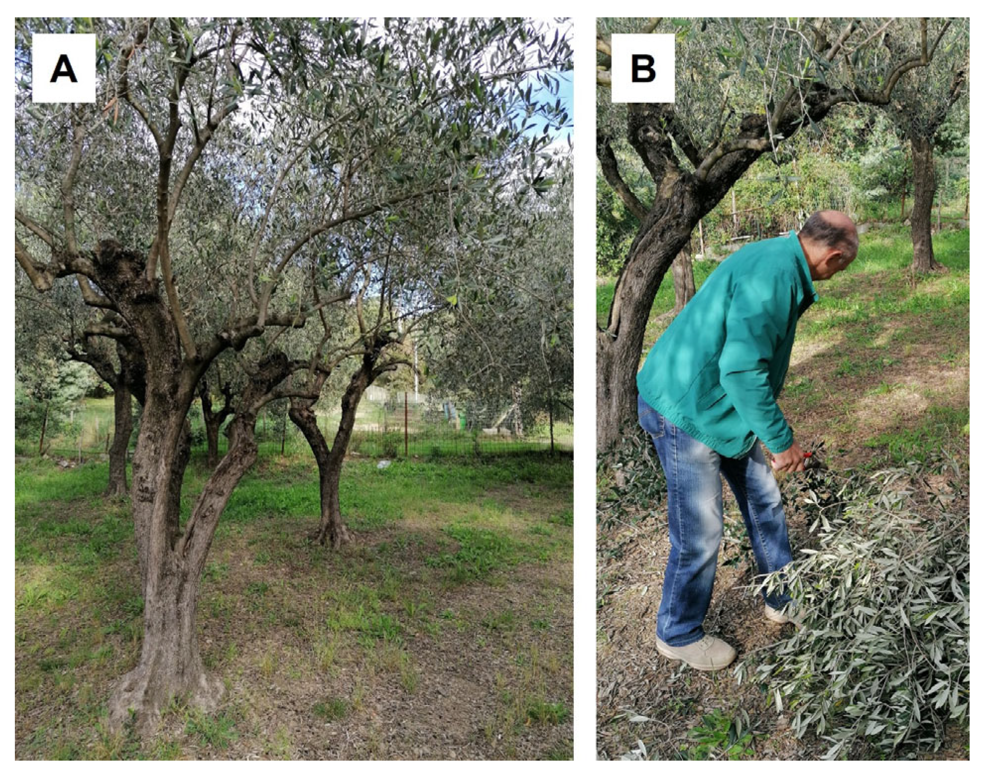
Olive pruning leaves of ‘Lavagnina’ cultivar (Figure 5A,B) were collected in October 2023 and June 2024 from the same trees and provided by “Società Cooperativa Agricola Olivicoltori Sestresi” based in Sestri Levante (Liguria, Italy). The leaves were air-dried before analysis. A voucher specimen (GDOR n. 63471) was deposited in the herbarium of the Natural History Museum Giacomo Doria of Genova (Italy).

4.2. Micro-Morphological and Anatomical Analyses

Mature leaves were fixed in a FineFIX working solution (Milestone SRL, Sorisole, Bergamo, Italy) and left overnight at 4 °C [80]. Then, small fragments deriving from the middle of the leaf were dehydrated in a graded alcohol series (70, 80, 90, and 100%) and embedded in Technovit 7100 (Heraeus Kulzer GmbH, Wehrheim, Germany). The samples obtained were finally cut into semiultrathin sections (8 μm) using a rotary microtome Leica RM 2155 (Leica Microsystems GmbH, Wetzlar, Germany). Sections were stained with the polychromatic dye TBO [81,82] or with Sudan Black [83] and finally observed under a Leica DM 2000 microscope (Leica Microsystems, Wetzlar, Germany) using a ToupCam Digital Camera, CMOS Sensor 3.1 MP resolution (ToupTek Photonics, Hangzhou, China). Polarized light was used to detect the presence of calcium oxalate crystals.
Small portions of the median zone of the leaves, after fixation, were also critical point-dried in CO2 (CPD, K850 2M Strumenti s.r.l., Rome, Italy), mounted on aluminum stubs, covered with a 10 nm layer of gold and observed under a VEGA3-Tescan-type LMU microscope (Apollo, Tescan USA Inc., Cranberry Twp, PA, USA), operating at an accelerating voltage of 20 kV.
For comparison, hand-made leaf sections of ‘Lavagnina’ and other two cultivars, ‘Leccino’ (from Sestri Levante, Eastern Liguria, GDOR. n. 63472) and ‘Taggiasca’ (from Imperia, Western Liguria, GDOR. n. 63473) were prepared and stained with TBO to reveal potential anatomical differences (see Supplementary Materials).

4.3. Extraction

A first sampling of ‘Lavagnina’ pruned leaves (October 2023, 800 g) was extracted using organic solvents of increasing polarity: petroleum ether 40–60° (PEE), chloroform (CHE) and methanol (ME). Since the biological assays indicated that only ME exhibited notable antioxidant properties, we repeated the sample collection (June 2024, 251.4 g) and performed extraction using food-grade 70% ethanol (HAE), given that methanol and ethanol have very similar solubility.
The extraction of olive leaves was carried out by maceration in glass flasks using 10 mL of solvent for each g of leaves. The flasks filled with leaves and solvent were stirred using a magnet. Each extraction cycle lasted 5 days and 3 extraction cycles were carried out to maximize the extraction. Once the extracts, derived from the use of the same solvent, were combined, the solvent was removed using a rotary evaporator and the extract was freeze-dried to remove residual water and stored in hermetically sealed falcons away from heat, light and humidity. The four extracts were then stored at 4 °C in dark glass bottles until analysis. The extraction yield for each extract was calculated as follows: Y (%) = (m/M) × 100, where m is the mass of the extracted residue (g), and M is the initial mass of the plant material (g).

4.4. Antioxidant Activity

4.4.1. DPPH Assay

The antioxidant activity was determined using the stable 1,1-diphenyl-2-picrylhydrazyl (DPPH) radical method as reported by Brand-Williams and Coworkers [84] with some modifications. The analysis was performed in cuvettes by adding 25 μL of a solution of the extracts in MeOH to 975 μL of a DPPH solution (7.6 × 10⁵ M), which was prepared daily and kept in the dark to have a final volume of 1 mL in a straight-sided cuvette. Methanol alone was used as a blank, while 1 mL of DPPH solution (60 μM) was used as a control sample for a baseline measurement. After 45 min of incubation, the absorbance at 515 nm was measured in the spectrophotometer Thermo scientific Multiskan GO (Thermo Fischer Scientific, Vantaa, Finland). The percent inhibition of free radical formation by DPPH (I%) was calculated as follows:
I% = [1 − (A sample/A DPPH)] × 100
where A DPPH is the absorbance of the control reaction (containing all reagents except the test compound) and A sample is the absorbance of the test read at 515 nm after 45 min. The scavenging activity was expressed as the 50% effective concentration (EC50), which is defined as the sample concentration (μg/mL) that causes a 50% decrease in the DPPH absorbance. The experiments were carried out in triplicate, with triplicates obtained from the same extract, and the results were expressed as the mean ± standard deviation.

4.4.2. ABTS Assay

The 2,20-azino-bis 3-ethylbenzothiazoline-6-sulfonic acid (ABTS) test was carried out following the method of Re and coworkers [85]. The radical ABTS (ABTS°+) was generated mixing an aqueous solution of ABTS (7 mM) and potassium persulfate (2.45 mM), that was left in the dark for 16 h at room temperature and then diluted with ethanol to an absorbance of 0.800 at 734 nm. An aliquot of 0.1 mL of each extract was mixed with 0.9 mL of diluted ABTS; then the mixture was incubated for 6 min. The absorbance was read at 734 nm. Trolox (6-hydroxy-2,5,7,8-tetramethylchroman-2-carboxylic acid) was dissolved in methanol at different concentrations and used as a reference standard. The results were expressed as the μM Trolox equivalent antioxidant capacity (TEAC) per gram of samples. The positive control was represented by a solution of the standard antioxidant, ascorbic acid, whose absorbance was measured under the same conditions as the samples studied. The experiments were carried out in triplicate, with triplicates obtained from the same extract, and the results were expressed as the mean ± standard deviation.

4.4.3. FRAP Assay

The FRAP (Ferric Ion Reducing Antioxidant Power) assay was performed following the protocol of Benzie and Strain [86]. A FRAP reagent is a solution consisting of 23 mM acetate buffer (pH 3.6), 10 mM of tripyridyl triazine (TPTZ) in 40 mM of HCl, and 20 mM of FeCl3 (in a 10:1:1 ratio). Different concentrations of ferrous sulfate heptahydrate (FeSO4•7H2O), in the range 1–0.1 mM, were prepared to obtain the calibration curve. The reaction was carried out for each sample in a final volume of 272 μL in wells. The reaction mixture was incubated at 37 °C for 30 min in dark conditions. The absorbance of the blank, consisting of FRAP alone and monitored spectrophotometrically (Thermo scientific Multiskan GO, Thermo Fischer Scientific, Vantaa, Finland) at the wavelength of 593 nm, was subtracted from the absorbance of the FRAP with the sample to determine the FRAP value for each sample. The FRAP values were determined using the FeSO4•7H2O calibration curve and expressed as mmol Fe2+/g of dried extract. The experiments were carried out in triplicate, with triplicates obtained from the same extract, and the results were expressed as the mean ± standard deviation.

4.5. Chemical Analysis

The extracts were examined by LC-ESI-HR-MS, utilizing a Q Exactive: hybrid quadrupole-Orbitrap mass spectrometer (Thermo Fisher, Waltham, MA, USA), operating in negative ionization mode following Crescenzi and colleagues [87], with minor adjustments. LC-MS analysis was performed on a Luna 5 μm C18 100 Å (150 × 2 mm) column (Phenomenex, Aschaffenburg, Germany), at a flow rate of 0.2 mL/min. A binary mobile phase was employed (eluent A: H2O + 0.1% formic acid (99.9:0.1, v/v) and eluent B: H3CN + 0.1% formic acid (99.9:0.1, v/v). The HPLC program started at 5% B, reaching 95% B after 30 min; this level was maintained for an additional 5 min before returning to the starting percentage. The autosampler was set to inject 5 μL of each sample (1 mg/mL). The HESI source settings were as follows: spray voltage −2.5KV; S-Lens RF Level 50%; ion source temperature 300.01 °C; sheath and auxiliary gas flow (N2), 50 and 10; and sweep gas 0. The mass range for MS spectra acquisition was 50–1400 m/z. For the fragmentation study, a data-dependent acquisition was configured, fragmenting the precursor ions of the most intense peaks in the MS analysis with a collision energy of 30%. Xcalibur software version 2.2 was used for system control, data collection, and data processing.
Compound attribution was performed by comparison with the published literature and by evaluating MS/MS (MS2) fragmentation patterns. Specifically, we cross-checked accurate mass, isotopic distribution, plausible adducts, retention behavior, and—crucially—diagnostic product ions against reported spectra and fragmentation pathways for the proposed structures.

4.6. Statistical Analysis

Data were analyzed by a two-way ANOVA followed by Tukey’s post hoc test using Statistical Package for the Social Sciences (SPSS) version 13.0, 2004 (Chicago, IL, United States). The significance level has been set at 0.05.

5. Conclusions

Overall, the data collected in this study highlight the importance of recovering bioactive compounds from the pruned leaves of ‘Lavagnina’ cultivar, which has been scarcely investigated. Indeed, this by-product represents a valuable source of antioxidants potentially suitable for use in the medicinal, nutraceutical, and cosmetic fields. Notably, the high antioxidant activity (17.3 ± 0.6 μg/mL) of the HAE could exert a protective effect against DNA damage induced by free radicals, thereby potentially preventing mutagenesis and the onset of carcinogenesis. HAE is, therefore, a promising candidate for further in-depth studies to explore its potential chemopreventive activity in appropriate biological models and clinical trials. In this perspective, also toxicity tests will be conducted on this extract.
Future research will include pharmacognostic studies of the pruned leaves of different Italian O. europaea cultivars with the aim of better characterizing the anatomical differences among them and their phytochemical profile. This information could direct the harvesting of such residues in order to obtain extracts with high biological activity for new applications in human well-being. This approach also aligns with the principles of circular bioeconomy, promoting the recovery and valorization of agricultural wastes, such as pruned leaves, as a valuable resource for the development of sustainable and innovative natural products.

Supplementary Materials

The following supporting information can be downloaded at: https://www.mdpi.com/article/10.3390/molecules30173605/s1, Figure S1: Anatomical differences in leaves of different cultivars of Olea europaea L. are shown: ‘Lavagnina’ (A), ‘Taggiasca’ (B), ‘Leccino’ (C). The arrangement of vessels in the main vein assumes a different shape depending on the cultivar, and the main differences are represented by the thickening of the bundle of mechanical tissues located above the central vein (white arrows). Indeed, ‘Lavagnina’ (A) and ‘Taggiasca’ (B) are both characterized by a bundle of sclerenchymatic fibers and some collenchymatous cells, although it is slightly less developed in ‘Taggiasca’. In contrast, ‘Leccino’ (C) shows a larger bundle, composed mainly of collenchyma beginning to lignify.

Author Contributions

Conceptualization, L.C., P.M., G.B. and V.D.F.; methodology, L.C., P.M., F.B., F.P., L.D.M. and V.D.F.; software, F.P.; validation, L.C., P.M. and V.D.F.; formal analysis, L.C., P.M., F.B., F.P., L.D.M. and V.D.F.; investigation, L.C., P.M., F.B., F.P., L.D.M. and V.D.F.; plant sampling, L.C., F.B. and G.B.; data curation, L.C., F.B., F.P. and V.D.F.; writing—original draft preparation, L.C., F.B., F.P., L.D.M. and V.D.F.; writing—review and editing, L.C., P.M. and V.D.F.; supervision, L.C., P.M. and V.D.F. All authors have read and agreed to the published version of the manuscript.

Funding

This research was supported by the PhD project of Federica Betuzzi (PhD STAT School, XXXIX cycle, University of Genoa), granted by the Italian Ministry of University and Research, grant number 100022-2024-MS-ALTRIPOSTL-BETUZZI.

Institutional Review Board Statement

Not applicable.

Data Availability Statement

The data presented in this study are available in this published paper.

Acknowledgments

We are grateful to “Consorzio per la Tutela dell’Olio Extra Vergine di Oliva DOP Riviera Ligure” and “Società Cooperativa Agricola Olivicoltori Sestresi” for their support and the provision of the olive leaf samples. We also thank Laura Negretti (DISTAV, University of Genova) for SEM technical assistance.

Conflicts of Interest

The authors declare no conflicts of interest. The funders had no role in the design of the study; in the collection, analyses, or interpretation of data; in the writing of the manuscript; or in the decision to publish the results.

References

  1. Nicolì, F.; Negro, C.; Vergine, M.; Aprile, A.; Nutricati, E.; Sabella, E.; Miceli, A.; Luvisi, A.; De Bellis, L. Evaluation of Phytochemical and Antioxidant Properties of 15 Italian Olea europaea L. Cultivar Leaves. Molecules 2019, 24, 1998. [Google Scholar] [CrossRef]
  2. Lo Giudice, V.; Faraone, I.; Bruno, M.R.; Ponticelli, M.; Labanca, F.; Bisaccia, D.; Massarelli, C.; Milella, L.; Todaro, L. Olive Trees By-Products as Sources of Bioactive and Other Industrially Useful Compounds: A Systematic Review. Molecules 2021, 26, 5081. [Google Scholar] [CrossRef]
  3. Consorzio per la Tutela dell’Olio Extra Vergine di Oliva DOP Riviera Ligure. Available online: https://www.oliorivieraligure.it/ (accessed on 21 February 2025).
  4. Rellini, I.; Demasi, M.; Scopesi, C.; Ghislandi, S.; Salvidio, S.; Pini, S.; Stagno, A. Evaluation of the environmental components of the Taggiasca “terroir” olive (Imperia, Italy). Bull. Environ. Life Sci. 2022, 4, 24–42. [Google Scholar] [CrossRef]
  5. Official Gazette of the Italian Republic 193/1998. Available online: https://www.gazzettaufficiale.it/home (accessed on 21 February 2025).
  6. Quaini, M. Per la Storia del Paesaggio Agrario in Liguria: Note di Geografia Storica Sulle Strutture Agrarie Della Liguria Medievale e Moderna; Camera di Commercio Industria Artigianato e Agricoltura: Savona, Italy, 1973; pp. 75–80. [Google Scholar]
  7. Barichello, R.; Monticelli, A.; Cafici, E.; Guardavilla, A.; Martoni, S.; Ghironi, M.; Rossi, N.; Pini, S.; Spagnoli, F.; Bertucci, A.; et al. Le Varietà di Olivo Liguri; Regione Liguria: Liguria, Italy, 2006. [Google Scholar]
  8. Lombardo, N.; Muzzalupo, I.; Perri, E.; Sajiad, A. Microsatellite markers for olive (Olea europaea L.). In Proceedings of the 12th Simposium Científico-Técnico Expoliva, Jaén, Spain, 12–13 May 2005; Fundación para la promoción y el desarrollo del olivar y el aceite de oliva (ed). pp. 1–8. [Google Scholar]
  9. Bracci, T.; Sebastiani, L.; Busconi, M.; Fogher, C.; Belaj, A.; Trujillo, I. SSR markers reveal the uniqueness of olive cultivars from the Italian region of Liguria. Sci. Hortic. 2009, 122, 209–215. [Google Scholar] [CrossRef]
  10. Carciochi, R.A.; D’Alessandro, L.G.; Vauchel, P.; Rodriguez, M.M.; Nolasco, S.M.; Dimitrov, K. Valorization of Agrifood By-Products by Extracting Valuable Bioactive Compounds Using Green Processes. In Ingredients Extraction by Physicochemical Methods in Food—Handbook of Food Bioengineering; Grumezescu, A.M., Holban, A.M., Eds.; Elsevier Inc.: Amsterdam, The Netherlands, 2017; Volume 4, pp. 191–228. [Google Scholar]
  11. Romani, A.; Ieri, F.; Urciuoli, S.; Noce, A.; Marrone, G.; Nediani, C.; Bernini, R. Health Effects of Phenolic Compounds Found in Extra-Virgin Olive Oil, By-Products, and Leaf of Olea europaea L. Nutrients 2019, 11, 1776. [Google Scholar] [CrossRef]
  12. Chiocchio, I.; Mandrone, M.; Tomasi, P.; Marincich, L.; Poli, F. Plant Secondary Metabolites: An Opportunity for Circular Economy. Molecules 2021, 26, 495. [Google Scholar] [CrossRef]
  13. Magyari-Pavel, I.Z.; Moacă, E.-A.; Avram, Ș.; Diaconeasa, Z.; Haidu, D.; Ștefănuț, M.N.; Rostas, A.M.; Muntean, D.; Bora, L.; Badescu, B.; et al. Antioxidant Extracts from Greek and Spanish Olive Leaves: Antimicrobial, Anticancer and Antiangiogenic Effects. Antioxidants 2024, 13, 774. [Google Scholar] [CrossRef]
  14. Ghanem, M.T.M.; Tawfik, W.A.; Mahdy, E.M.; Abdelgawa, M.E.; Abdel-Azim, N.S.; El-Missiry, M.M. Chemical and biological evaluation of olive leaves as a waste by-product of olive oil industry. Egypt. Pharm. J. 2019, 18, 172–177. [Google Scholar]
  15. Abbattista, R.; Ventura, G.; Calvano, C.D.; Cataldi, T.R.I.; Losito, I. Bioactive Compounds in Waste By-Products from Olive Oil Production: Applications and Structural Characterization by Mass Spectrometry Techniques. Foods 2021, 10, 1236. [Google Scholar] [CrossRef] [PubMed]
  16. Guarrera, P.M. Usi e Tradizioni Della Flora Italiana—Medicina Popolare ed Etnobotanica; Aracne: Rome, Italy, 2006. [Google Scholar]
  17. González, J.A.; García-Barriuso, M.; Amich, F. Ethnobotanical study of medicinal plants traditionally used in the Arribes del Duero, western Spain. J. Ethnopharmacol. 2010, 131, 343–355. [Google Scholar] [CrossRef] [PubMed]
  18. El Khomsi, M.; Dandani, Y.; Chaachouay, N.; Hmouni, D. Ethnobotanical study of plants used for medicinal, cosmetic, and food purposes in the region of Moulay Yacoub, Northeast of Morocco. J. Pharm. Pharmacogn. Res. 2022, 10, 13–29. [Google Scholar] [CrossRef]
  19. Markhali, F.S.; Teixeira, J.A.; Rocha, C.M.R. Olive Tree Leaves—A Source of Valuable Active Compounds. Processes 2020, 8, 1177. [Google Scholar] [CrossRef]
  20. Palmeri, R.; Siracusa, L.; Carrubba, M.; Parafati, L.; Proetto, I.; Pesce, F.; Fallico, B. Olive Leaves, a Promising Byproduct of Olive Oil Industry: Assessment of Metabolic Profiles and Antioxidant Capacity as a Function of Cultivar and Seasonal Change. Agronomy 2022, 12, 2007. [Google Scholar] [CrossRef]
  21. Khelouf, I.; Karoui, I.J.; Lakoud, A.; Hammami, M.; Abderrabba, M. Comparative chemical composition and antioxidant activity of olive leaves Olea europaea L. of Tunisian and Algerian varieties. Heliyon 2023, 9, e22217. [Google Scholar] [CrossRef] [PubMed]
  22. Nenadis, N.; Mastralexi, A.; Tsimidou, M.Z. Physicochemical Characteristics and Antioxidant Potential of the Greek PDO and PGI Virgin Olive Oils (VOOs). Eur. J. Lipid Sci. Technol. 2018, 121, 1800172. [Google Scholar] [CrossRef]
  23. Toumi, K.; S’wiatek, Ł.; Boguszewska, A.; Skalicka-Wo’zniak, K.; Bouaziz, M. Comprehensive Metabolite Profiling of Chemlali Olive Tree Root Extracts Using LC-ESI-QTOF-MS/MS, Their Cytotoxicity, and Antiviral Assessment. Molecules 2023, 28, 4829. [Google Scholar] [CrossRef]
  24. Serrano-García, I.; Martakos, I.C.; Olmo-García, L.; León, L.; de la Rosa, R.; Gómez-Caravaca, A.M.; Belaj, A.; Serrano, A.; Dasenaki, M.E.; Thomaidis, N.S.; et al. Application of Liquid Chromatography-Ion Mobility Spectrometry-Mass Spectrometry-Based Metabolomics to Investigate the Basal Chemical Profile of Olive Cultivars Differing in Verticillium dahliae Resistance. J. Agric. Food Chem. 2024, 72, 27561–27574. [Google Scholar] [CrossRef] [PubMed]
  25. Campeol, E.; Flamini, G.; Cioni, P.L.; Morelli, I.; D’Andrea, F.; Cremonini, R. 1,5-Anhydroxylitol from leaves of Olea europaea. Carbohydr. Res. 2004, 339, 2731–2732. [Google Scholar] [CrossRef]
  26. Villegas-Aguilar, M.D.C.; Cádiz-Gurrea, M.L.; Sánchez-Marzo, N.; Barrajón-Catalán, E.; Arráez-Román, D.; Fernández-Ochoa, Á.; Segura-Carretero, A. The Application of Untargeted Metabolomic Approaches for the Search of Common Bioavailable Metabolites in Human Plasma Samples from Lippia citriodora and Olea europaea Extracts. J. Agric. Food Chem. 2024, 72, 24879–24893. [Google Scholar] [CrossRef]
  27. Ammar, S.; Contreras, M.D.M.; Gargouri, B.; Segura-Carretero, A.; Bouaziz, M. RP-HPLC-DAD-ESI-QTOF-MS based metabolic profiling of the potential Olea europaea by-product “wood” and its comparison with leaf counterpart. Phytochem. Anal. 2017, 28, 217–229. [Google Scholar] [CrossRef]
  28. Quirantes-Piné, R.; Lozano-Sánchez, J.; Herrero, M.; Ibáñez, E.; Segura-Carretero, A.; Fernández-Gutiérrez, A. HPLC-ESI-QTOF-MS as a powerful analytical tool for characterising phenolic compounds in olive-leaf extracts. Phytochem. Analysis. 2013, 24, 213–223. [Google Scholar] [CrossRef]
  29. Beteinakis, S.; Papachristodoulou, A.; Stathopoulos, P.; Mikros, E.; Halabalaki, M. A multilevel LC-HRMS and NMR correlation workflow towards foodomics advancement: Application in table olives. Talanta 2024, 280, 126641. [Google Scholar] [CrossRef]
  30. Divya Priya, A.; Martin, A. UHPLC-MS/MS based comprehensive phenolic profiling, antimicrobial and antioxidant activities of Indian Rhodomyrtus tomentosa fruits. Sci. Rep. 2025, 15, 945. [Google Scholar] [CrossRef] [PubMed]
  31. Rubio-Senent, F.; Rodriguez-Gutierrez, G.; Lama-Munoz, A.; Fernandez-Bolanos, J. New phenolic compounds hydrothermally extracted from the olive oil byproduct alperujo and their antioxidative activities. J. Agric. Food Chem. 2012, 60, 1175–1186. [Google Scholar] [CrossRef] [PubMed]
  32. Kabbash, E.M.; Abdel-Shakour, Z.T.; El-Ahmady, S.H.; Wink, M.; Ayoub, I.M. Comparative metabolic profiling of olive leaf extracts from twelve different cultivars collected in both fruiting and flowering seasons. Sci. Rep. 2023, 13, 612. [Google Scholar] [CrossRef]
  33. Lv, X.; Sun, J.-Z.; Xu, S.-Z.; Cai, Q.; Li, Y.-Q. Rapid Characterization and Identification of Chemical Constituents in Gentiana radix before and after Wine-Processed by UHPLC-LTQ-Orbitrap MSn. Molecules 2018, 23, 3222. [Google Scholar] [CrossRef]
  34. Fayek, N.M.; Zayed, A.; Zahran, H.A.; Ramadan, N.S.; Capanoglu, E.; Li, Z.; Fang, Y.; Khalifa, I.; Farag, M.A. Effect of cultivar type, ontogeny and extraction methods as determinant factors of olive leaf metabolome: A case study in 8 Egyptian cultivars as analyzed using LC/MS-based metabolomics. Ind. Crops Prod. 2024, 222, 120085. [Google Scholar] [CrossRef]
  35. Ventura, G.; Calvano, C.D.; Abbattista, R.; Bianco, M.; De Ceglie, C.; Losito, I.; Palmisano, F.; Cataldi, T.R.I. Characterization of bioactive and nutraceutical compounds occurring in olive oil processing wastes. Rapid Commun. Mass Spectrom. 2019, 33, 1670–1681. [Google Scholar] [CrossRef]
  36. Fu, S.; Arráez-Roman, D.; Segura-Carretero, A.; Menéndez, J.A.; Menéndez-Gutiérrez, M.P.; Micol, V.; Fernández-Gutiérrez, A. Qualitative screening of phenolic compounds in olive leaf extracts by hyphenated liquid chromatography and preliminary evaluation of cytotoxic activity against human breast cancer cells. Anal. Bioanal. Chem. 2010, 397, 643–654. [Google Scholar] [CrossRef]
  37. Tarchi, I.; Olewnik-Kruszkowska, E.; Aït-Kaddour, A.; Bouaziz, M. Innovative Process for the Recovery of Oleuropein-Rich Extract from Olive Leaves and Its Biological Activities: Encapsulation for Activity Preservation with Concentration Assessment Pre and Post Encapsulation. ACS Omega 2025, 10, 6135–6146. [Google Scholar] [CrossRef]
  38. Mushtaq, H.; Piccolella, S.; Cimmino, G.; Ferrara, E.; Brahmi-Chendouh, N.; Petriccione, M.; Pacifico, S. Polyphenols from Olea europaea L. cv. Caiazzana leaf pruning waste for antioxidant and UV-blocking starch-based plastic films. Food Packag. Shelf Life 2025, 47, 101447. [Google Scholar] [CrossRef]
  39. Mir-Cerdà, A.; Granados, M.; Saurina, J.; Sentellas, S. Olive tree leaves as a great source of phenolic compounds: Comprehensive profiling of NaDES extracts. Food Chem. 2024, 456, 140042. [Google Scholar] [CrossRef]
  40. Taamalli, A.; Arráez-Román, D.; Ibañez, E.; Zarrouk, M.; Segura-Carretero, A.; Fernández-Gutiérrez, A. Optimization of microwave-assisted extraction for the characterization of olive leaf phenolic compounds by using HPLC-ESI-TOF-MS/IT-MS(2). J. Agric. Food Chem. 2012, 60, 791–798. [Google Scholar] [CrossRef]
  41. Michel, T.; Khlif, I.; Kanakis, P.; Termentzi, A.; Allouche, N.; Halabalaki, M.; Skaltsounis, A.-L. UHPLC-DAD-FLD and UHPLC-HRMS/MS based metabolic profiling and characterization of different Olea europaea organs of Koroneiki and Chetoui varieties. Phytochem. Lett. 2015, 11, 424–439. [Google Scholar] [CrossRef]
  42. Fayek, N.M.; Farag, M.A.; Saber, F.R. Metabolome classification via GC/MS and UHPLC/MS of olive fruit varieties grown in Egypt reveal pickling process impact on their composition. Food Chem. 2021, 339, 127861. [Google Scholar] [CrossRef]
  43. López-Salas, L.; Díaz-Moreno, J.; Ciulu, M.; Borrás-Linares, I.; Quirantes-Piné, R.; Lozano-Sánchez, J. Monitoring the Phenolic and Terpenic Profile of Olives, Olive Oils and By-Products throughout the Production Process. Foods 2024, 13, 1555. [Google Scholar] [CrossRef]
  44. Lozano-Sánchez, J.; Giambanelli, E.; Quirantes-Piné, R.; Cerretani, L.; Bendini, A.; Segura-Carretero, A.; Fernández-Gutiérrez, A. Wastes generated during the storage of extra virgin olive oil as a natural source of phenolic compounds. J. Agric. Food Chem. 2011, 59, 11491–11500. [Google Scholar] [CrossRef]
  45. Grigoletto, I.; García Salas, P.; Valli, E.; Bendini, A.; Ferioli, F.; Pasini, F.; Sánchez Villasclaras, S.; García-Ruiz, R.; Gallina Toschi, T. HPLC-MS/MS Phenolic Characterization of Olive Pomace Extracts Obtained Using an Innovative Mechanical Approach. Foods 2024, 13, 285. [Google Scholar] [CrossRef] [PubMed]
  46. Do, T.K.T.; De Vaumas, R.; Reich, E. Phytochemical profiling of iridoids by high-performance thin-layer chromatography. J. Planar Chrom. Modern TLC 2021, 34, 361–366. [Google Scholar] [CrossRef]
  47. Zou, W.; Wang, B.; Feng, X.; Lai, C.; Tan, Q. Ultra-High-Performance Liquid Chromatography-Tandem Mass Spectrometry Combined With Network Pharmacology to Elucidate the Bioactive Ingredients and Potential Mechanism of Wu-Teng Decoction for Treatment of Rheumatoid Arthritis. Chem. Biodivers. 2025, 22, e202403454. [Google Scholar] [CrossRef]
  48. Lozano-Sanchez, J.; Bendini, A.; Quirantes-Piné, R.; Cerretani, L.; Segura-Carretero, A.; Fernandez-Gutierrez, A. Monitoring the bioactive compounds status of extravirgin olive oil and storage by-products over the shelf life. Food Control 2013, 30, 606–615. [Google Scholar] [CrossRef]
  49. Ribeiro, T.B.; Costa Célia, M.; Bonifácio–Lopes, T.; Silva, S.; Veiga, M.; Monforte, A.R.; Nunes, J.; Vicente, A.; Pintado, M. Prebiotic effects of olive pomace powders in the gut: In vitro evaluation of the inhibition of adhesion of pathogens, prebiotic and antioxidant effects. Food Hydrocoll. 2021, 112, 106312. [Google Scholar] [CrossRef]
  50. Expósito-Díaz, A.; Miho, H.; Ledesma-Escobar, C.A.; Moral, J.; Díez, C.M.; Priego-Capote, F. Influence of genetic and interannual factors on bioactive compounds of olive pomace determined through a germplasm survey. Food Chem. 2022, 378, 132107. [Google Scholar] [CrossRef]
  51. Romero-Márquez, J.M.; Navarro-Hortal, M.D.; Forbes-Hernández, T.Y.; Varela-López, A.; Puentes, J.G.; Sánchez-González, C.; Sumalla-Cano, S.; Battino, M.; García-Ruiz, R.; Sánchez, S.; et al. Effect of olive leaf phytochemicals on the anti-acetylcholinesterase, anti-cyclooxygenase-2 and ferric reducing antioxidant capacity. Food Chem. 2024, 444, 138516. [Google Scholar] [CrossRef]
  52. Johnson, R.; Melliou, E.; Zweigenbaum, J.; Mitchell, A.E. Quantitation of Oleuropein and Related Phenolics in Cured Spanish-Style Green, California-Style Black Ripe, and Greek-Style Natural Fermentation Olives. J. Agric. Food Chem. 2018, 66, 2121–2128. [Google Scholar] [CrossRef] [PubMed]
  53. Peralbo-Molina, A.; Priego-Capote, F.; Luque de Castro, M.D. Tentative identification of phenolic compounds in olive pomace extracts using liquid chromatography-tandem mass spectrometry with a quadrupole-quadrupole-time-of-flight mass detector. J. Agric. Food Chem. 2012, 60, 11542–11550. [Google Scholar] [CrossRef] [PubMed]
  54. Moudache, M.; Silva, F.; Nerín, C.; Zaidi, F. Olive cake and leaf extracts as valuable sources of antioxidant and antimicrobial compounds: A comparative study. Waste Biomass Valorization 2021, 12, 1431–1445. [Google Scholar] [CrossRef]
  55. Zhao, H.; Kim, Y.; Avena-Bustillos, R.J.; Nitin, N.; Wang, S.C. Characterization of California olive pomace fractions and their in vitro antioxidant and antimicrobial activities. LWT—Food Sci. Technol. 2023, 180, 114677. [Google Scholar] [CrossRef]
  56. Beer, F.; Weinert, C.H.; Wellmann, J.; Hillebrand, S.; Ley, J.P.; Soukup, S.T.; Kulling, S.E. Comprehensive metabolome characterization of leaves, internodes, and aerial roots of Vanilla planifolia by untargeted LC-MS and GC × GC-MS. Phytochem. Anal. 2025, 36, 30–51. [Google Scholar] [CrossRef] [PubMed]
  57. Marra, R.; Vinale, F.; Cesarano, G.; Lombardi, N.; d’Errico, G.; Crasto, A.; Mazzei, P.; Piccolo, A.; Incerti, G.; Woo, S.L.; et al. Biochars from olive mill waste have contrasting effects on plants, fungi and phytoparasitic nematodes. PLoS ONE 2018, 13, e0198728. [Google Scholar] [CrossRef]
  58. Xue, Y.; Guan, T.; Liu, J.; Wang, J.; Yang, Z.; Guan, F.; Li, W.; Wang, Y. Chemical Composition Analysis and Multi-index Component Content Determination of Compounds in Goupi Plaster Based on UPLC–Q-Exactive-MS and UPLC–MS/MS. Chromatographia 2024, 87, 45–57. [Google Scholar] [CrossRef]
  59. Ali, K.; Ali, A.; Khan, M.N.; Rahman, S.; Faizi, S.; Ali, M.S.; Khalifa, S.A.M.; El-Seedi, H.R.; Musharraf, S.G. Rapid Identification of Common Secondary Metabolites of Medicinal Herbs Using High-Performance Liquid Chromatography with Evaporative Light Scattering Detector in Extracts. Metabolites 2021, 11, 489. [Google Scholar] [CrossRef]
  60. Zhang, Y.; Wen, M.; Zhou, P.; Tian, M.; Zhou, J.; Zhang, L. Analysis of chemical composition in Chinese olive leaf tea by UHPLC-DAD-Q-TOF-MS/MS and GC-MS and its lipid-lowering effects on the obese mice induced by high-fat diet. Food Res. Int. 2020, 128, 108785. [Google Scholar] [CrossRef]
  61. Luján, R.J.; Capote, F.P.; Marinas, A.; de Castro, M.D. Liquid chromatography/triple quadrupole tandem mass spectrometry with multiple reaction monitoring for optimal selection of transitions to evaluate nutraceuticals from olive-tree materials. Rapid Commun. Mass Spectrom. 2008, 22, 855–864. [Google Scholar] [CrossRef]
  62. Fabre, N.; Rustan, I.; de Hoffmann, E.; Quetin-Leclercq, J. Determination of flavone, flavonol, and flavanone aglycones by negative ion liquid chromatography electrospray ion trap mass spectrometry. J. Am. Soc. Mass Spectrom. 2001, 12, 707–715. [Google Scholar] [CrossRef]
  63. Espeso, J.; Isaza, A.; Lee, J.Y.; Sörensen, P.M.; Jurado, P.; de Jesús Avena-Bustillos, R.; Olaizola, M.; Arboleya, J.C. Olive Leaf Waste Management. Front. Sustain. Food Syst. 2021, 5, 660582. [Google Scholar] [CrossRef]
  64. WHO. Quality Control Methods for Herbal Materials; WHO Press: Valletta, Malta, 2011; Available online: https://apps.who.int/iris/handle/10665/44479 (accessed on 26 March 2025).
  65. Bacelar, E.A.; Correia, C.M.; Moutinho-Pereira, J.M.; Gonçalves, B.C.; Lopes, J.I.; Torres-Pereira, J.M. Sclerophylly and leaf anatomical traits of five field-grown olive cultivars growing under drought conditions. Tree Physiol. 2004, 24, 233–239. [Google Scholar] [CrossRef]
  66. Moreno-Alías, I.; León, L.; de la Rosa, R.; Rapoport, H.F. Morphological and anatomical evaluation of adult and juvenile leaves of olive plants. Trees 2009, 23, 181–187. [Google Scholar] [CrossRef]
  67. Rahfeld, B. Mikroskopischer Farbatlas Pflanzlicher Drogen, 2nd ed.; Spektrum Akademischer Verlag Gmbh: Heidelberg, Germany, 2011. [Google Scholar]
  68. de Menezes, T.P.; de Oliveira, A.F.; Pasqual, M.; de Souza, A.R.; de Castro, E.M. Conteúdo de DNA e caracterização anatômica de cultivares de oliveiras (Olea europea L.). Agrarian 2018, 11, 196–202. [Google Scholar] [CrossRef]
  69. Marchioni, I.; Rodolfi, M.; Massa, D.; Cacini, S.; Ughini, V.; Bartolini, G.; Fabbri, A.; Petruccelli, R.; Ganino, T. Comparative effects of drought stress on three olive cultivars focusing on older leaves. Sci. Hortic. 2024, 332, 113234. [Google Scholar] [CrossRef]
  70. Bosabalidis, A.M.; Kofidis, G. Comparative effects of drought stress on leaf anatomy of two olive cultivars. Plant Sci. 2002, 163, 375–379. [Google Scholar] [CrossRef]
  71. Rombaut, N.; Chave, T.; Nikitenko, S.I.; Maâtaoui, M.E.; Fabiano-Tixier, A.S.; Chemat, F. Modification of Olive Leaves’ Surface by Ultrasound Cavitation. Correlation with Polyphenol Extraction Enhancement. Appl. Sci. 2021, 11, 232. [Google Scholar] [CrossRef]
  72. Žuna Pfeiffer, T.; Štolfa, I.; Hoško, M.; Žanić, M.; Pavičić, N.; Cesar, V.; Lepeduš, H. Comparative Study of Leaf Anatomy and Certain Biochemical Traits in Two Olive Cultivars. Agric. Conspec. Sci. 2010, 75, 91–97. [Google Scholar]
  73. Hashmi, M.A.; Khan, A.; Hanif, M.; Farooq, U.; Perveen, S. Traditional Uses, Phytochemistry, and Pharmacology of Olea europaea (Olive). Evid. Based Complement. Altern. Med. 2015, 2015, 541591. [Google Scholar] [CrossRef]
  74. Liakopoulos, G.; Stavrianakou, S.; Karabourniotis, G. Trichome layers versus dehaired lamina of Olea europaea leaves: Differences in flavonoid distribution, UV-absorbing capacity, and wax yield. Environ. Exp. Bot. 2006, 55, 294–304. [Google Scholar] [CrossRef]
  75. Filardo, S.; Roberto, M.; Di Risola, D.; Mosca, L.; Di Pietro, M.; Sessa, R. Olea europaea L-derived secoiridoids: Beneficial health effects and potential therapeutic approaches. Pharmacol. Ther. 2024, 254, 108595. [Google Scholar] [CrossRef]
  76. Marchetti, C.; Clericuzio, M.; Borghesi, B.; Cornara, L.; Ribulla, S.; Gosetti, F.; Marengo, E.; Burlando, B. Oleuropein-Enriched Olive Leaf Extract Affects Calcium Dynamics and Impairs Viability of Malignant Mesothelioma Cells. Evid. Based Complement. Alternat. Med. 2015, 2015, 908493. [Google Scholar] [CrossRef]
  77. Charoenprasert, S.; Mitchell, A. Factors influencing phenolic compounds in table olives (Olea europaea). J. Agric. Food Chem. 2012, 60, 7081–7095. [Google Scholar] [CrossRef]
  78. Özcan, M.M.; Matthäus, B. A review: Benefit and bioactive properties of olive (Olea europaea L.) leaves. Eur. Food Res. Technol. 2017, 243, 89–99. [Google Scholar] [CrossRef]
  79. Castejón, M.L.; Montoya, T.; Alarcón-de-la-Lastra, C.; Sánchez-Hidalgo, M. Potential Protective Role Exerted by Secoiridoids from Olea europaea L. in Cancer, Cardiovascular, Neurodegenerative, Aging-Related, and Immunoinflammatory Diseases. Antioxidants 2020, 9, 149. [Google Scholar] [CrossRef]
  80. Chieco, C.; Rotondi, A.; Morrone, L.; Rapparini, F.; Baraldi, R. An ethanol-based fixation method for anatomical and micromorphological characterization of leaves of various tree species. Biotech. Histochem. 2013, 88, 109–119. [Google Scholar] [CrossRef]
  81. Yeung, E. A beginner’s guide to the study of plant structure. In Tested Studies for Laboratory Teaching; Karcher, S.J., Ed.; Purdue University: Lafayatte, IN, USA, 1998; Volume 19, pp. 125–142. [Google Scholar]
  82. Ruzin, S.E. Plant Microtechnique and Microscopy; Oxford University Press: New York, NY, USA, 1999. [Google Scholar]
  83. Badria, F.A.; Aboelmaaty, W.S. Plant Histochemistry: A Versatile and Indispensible Tool in Localization of Gene Expression, Enzymes, Cytokines, Secondary Metabolites and Detection of Plants Infection and Pollution. Acta Sci. Pharm. Sci. 2019, 3, 88–100. [Google Scholar] [CrossRef]
  84. Brand-Williams, W.; Cuvelier, M.E.; Berset, C. Use of a free radical method to evaluate antioxidant activity. LWT—Food Sci. Technol. 1995, 28, 25–30. [Google Scholar] [CrossRef]
  85. Re, R.; Pellegrini, N.; Proteggente, A.; Pannala, A.; Yang, M.; Rice-Evans, C. Antioxidant activity applying an improved ABTS radical cation decolorization assay. Free Radic. Biol. Med. 1999, 26, 1231–1237. [Google Scholar] [CrossRef]
  86. Benzie, I.F.; Strain, J.J. The ferric reducing ability of plasma (FRAP) as a measure of “antioxidant power”: The FRAP assay. Anal. Biochem. 1996, 239, 70–76. [Google Scholar] [CrossRef]
  87. Crescenzi, M.A.; Cerulli, A.; Montoro, P.; Piacente, S. Metabolite Profiling for Typization of “Rucola della Piana del Sele” (PGI), Eruca sativa, through UHPLC-Q-Exactive-Orbitrap-MS/MS Analysis. Foods 2023, 12, 3384. [Google Scholar] [CrossRef]
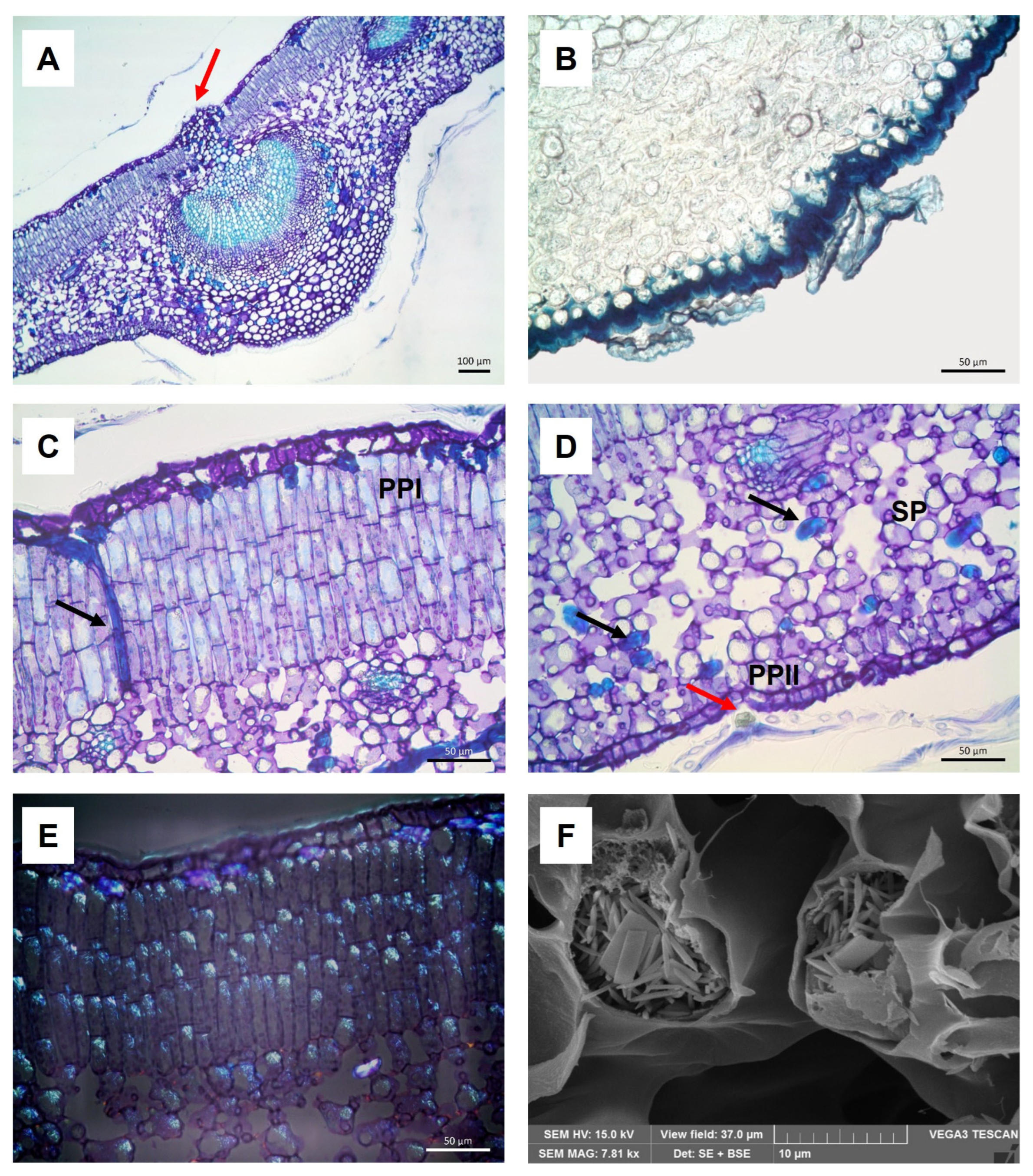
Figure 1. (AE) Light Microscopy micrographs of Olea europaea L. ‘Lavagnina’ leaf sections embedded in Technovit 7100 and stained with TBO (A,C,D) or Sudan Black (B) or observed under polarized light (E): (A) midrib with a small protrusion made up of mechanical tissues (arrow); (B) thick abaxial cuticle and scutiform trichomes; (C) longitudinally oriented trichosclereid through the PPI (arrow); (D) SP, PPII, tangentially oriented trichosclereids (black arrows) and a scutiform trichome with phenolic content in its stalk cell (red arrow); (E) small birefringent calcium oxalate crystals both in PPI and SP. Bar = 100 μm (A), Bars = 50 μm (BE). (F) SEM micrograph of leaf section, showing small calcium oxalate crystals, mainly raphides, in the SP.
Molecules 30 03605 g001
Figure 2. SEM micrographs of Olea europaea L. ‘Lavagnina’: (A) leaf adaxial surface showing sparse scutiform trichomes; (B) leaf abaxial surface with numerous not tightly overlapping trichomes, revealing small areas of the underlying epidermis (arrows); (C) magnification of a scutiform trichome on the abaxial surface; (D) detail of the striated cuticle and of some stomata on the abaxial surface.
Molecules 30 03605 g002
Figure 3. Full scan LC-MS chromatograms (negative ion HRESIMS) of ME (A) and HAE (B) of Olea europaea L. ‘Lavagnina’.
Molecules 30 03605 g003
Figure 4. The main compound classes and their distribution (%) between ME and HAE.
Molecules 30 03605 g004
Figure 5. Trees of Olea europaea L. ‘Lavagnina’ (A), pruning process (B).
Molecules 30 03605 g005
Table 1. Antioxidant activity of Olea europaea L. ‘Lavagnina’ leaf extracts, expressed as mean ± SD of three experiments.
ExtractDPPH
IC50 1 (μg/mL)
FRAP
mmol Fe2+ Equivalents/g Extract
ABTS
mmol TEAC 2/g
PEE>1000->1000
CHE79.4 ± 0.7 a1.4 ± 0.5 b0.9 ± 0.03 b
ME29.7 ± 0.6 b2.6 ± 0.3 b0.7 ± 0.05 b
HAE17.3 ± 0.6 c2.8 ± 0.7 b1.3 ± 0.04 b
Trolox3.21 ± 0.2 d84.2 ± 0.9 a-
Ascorbic acid--39.3 ± 0.8 a
1 IC50 = concentration required to reduce the absorbance of the DPPH solution by 50%; 2 TEAC: trolox equivalent antioxidant activity. Trolox and ascorbic acid were used as reference standards. Means followed by different letters in the same column indicate that they are significantly different at p < 0.05, according to a two-way ANOVA followed by Tukey’s post hoc test. The letters have no intrinsic meaning but serve only to identify groups between which there are significant differences.
Table 2. Composition of ME and HAE of Olea europaea L. ‘Lavagnina’. The x indicates the presence of metabolite in the extracts.
No.FamilyRetention TimeMeasured (m/z)
[M-H]
Molecular FormulaΔppmFragmentFragment FormulaFragment ion (m/z)ΔppmIdentificationMEHAE
1Alcohol sugar1.72181.0709C6H14O61.190[M-H2O-H]C6H11O5163.06041.963Mannitolxx
[M-CH6O2-H]C5H7O4131.0338−0.65
[M-C2H6O2-H]C4H7O4119.03390.04
[M-C2H8O3-H]C4H5O3101.0232−0.797
2Organic acid1.87195.0503C6H12O70.411[M-H2O-H]C6H9O6177.03982.347Gluconic acid x
[M-2H2O-H]C6H7O5159.02890.756
[M-CH6O3-H]C5H5O4129.01830.348
[M-C2H8O4-H]C4H3O399.0075−1.217
[M-C4H8O4-H]C2H3O375.0075−1.606
3Organic acid2.48191.0553C7H12O61.547[M-H2O-H]C7H9O5173.04450.174Quinic acidxx
[M-2H2O-CO-H]C6H7O3127.0388−0.949
[M-2H2O-CO2-H]C6H7O2111.0439−0.955
[M-3H2O-CO2-H]C6H5O93.0334−1 412
[M-C3H6O4-H]C4H5O285.0283−1 599
4Sugar2.59237.0616C8H14O84.582[M-C3H8O4-H]C5H5O4129.01830.193unknown (3-deoxy-D-manno-octulosonate)xx
[M-C5H10O5-H]C3H3O387.0075−1.385
5Secoiridoid7.29407.15652C17H28O114.254[M-C8H16O9-H]C9H11O2151.075360.0261-b-D-glucopyranosyl acyclodihydroelenolic acid x
[M-C9H16O9-H]C8H11O2139.075450.675
[M-C13H20O7-H]C4H7O4119.03346−3.572
6Iridoid7.38199.0608C9H12O53.617[M-CO2-H]C8H11O3155.07051.285Hydroxylated product of the dialdehydic form of decarboxymethyl-elenolic acid x
[M-2CO2-H]C7H11O111.08040.076
[M-C7H8O3-H]C2H3O259.0127−1.286
7Phenolic group7.48315.1091C14H20O85.160[M-C6H10O5-H]C8H9O3153.05491.564Hydroxytyrosol glucosidexx
[M-C6H10O5-H2O-H]C8H7O2135.0440−0.489
[M-C7H12O6-H]C7H7O2123.04410.195
[M-C11H14O5-H]C3H5O389.0232−0.792
8Secoiridoid7.53389.10925C16H22O113.629[M-C6H10O5-H]C10H11O6227.05583.327Oleoside (isomers)xx
[M-C6H10O5-CO2-H]C9H11O4183.06573.085
[M-C8H10O9-H]C8H11O2139.07551.034
[M-C8H12O10-H]C8H9O121.06480.318
[M-C12H16O8-H]C4H5O3101.0233−0.401
[M-C13H16O8-H]C3H5O389.0232−1.129
[M-C14H18O9-H]C2H3O259.0127−1.286
9Phenolic group7.92477.1620C20H30O133.610[M-C14H20O8-H]C6H9O5161.04471.305Hydroxytyrosol-diglucosidexx
[M-C12H20O10-H]C8H9O3153.05491.629
[M-C12H20O10-CH2O-H]-C7H7O2123.0440−0.455
10Phenolic group8.01153.0549C8H10O31.433[M-CH2O-H]C7H7O2123.04410.276Hydroxytyrosolxx
[M-C2H2O2-H]C6H7O95.04920.722
[M-C3H4O2-H]C5H5O81.0333−2.732
11Secoiridoid8.13375.1301C16H24O103.803[M-C6H10O5-H2O-CO2-H]C9H11O2151.07604.063Loganic acidxx
[M-C8H10O9-H]C8H13O125.0958−2.411
[M-C12H16O6-H]C4H7O4119.0334−3.824
[M-C11H18O7-H]C5H5O3113.0232−1.244
[M-C12H18O7-H]C4H5O3101.0223−3.866
[M-C13H18O7-H]C3H5O389.0230−4.275
[M-C13H20O8-H]C3H3O271.0125−3.040
12Iridoid8.50345.1195C15H22O94.263[M-C6H10O5-H]C9H11O4183.06564.232Aucubinx
[M-C6H10O5-H2O-H]C9H9O3165.05523.449
[M-C11H14O5-H]C4H7O4119.0338−0.716
[M-C12H16O6-H]C3H5O389.0233−0.568
13Iridoid8.93215.0922C10H16O53.766[M-CO2-H]C9H15O3171.10202.274Aldehydic form of
decarboxymethyl elenolic acid
xx
[M-CO2-H2O-H]C9H13O2153.09121.266
[M-CO2-H2O-CO-H]C8H13O125.09610.227
14Phenolic aldehyde derivative8.96313.0933C14H18O84.683[M-C6H10O5-H]C8 H7 O3151.03932.048Vanilloside x
[M-C6H10O5-CO-H]C7H7O2123.04410.276
[M-C11H14O5-H]C3H3O387.0075−1.615
15Iridoid8.98359.1353C16H24O94.514[M-C6H10O5-H]C10H13O4197.08142.6627-Deoxyloganic acidxx
[M-C6H10O5- C4H6O-H]C6H7O3127.0389−0.713
16Secoiridoid9.00421.1724C18H30O114.635[M-C9H18O9-H]C9H11O2151.075−2.093Oleoside methyl ester derivative x
[M-C13H22O8-H]C5 H7 O3115.0388−1.309
17Secoiridoid glycoside9.04257.1032C12H18O64.960[M-CO2-H]C11H17O4213.11304.1033-Hydroxymethyl-2.3-dihydro-5-(methoxycarbonyl)-2-methyl-2H-pyran-4-acetic methyl ester x
[M-C3H6O4-H]C9H11O2151.07582.806
[M-C9H14O3-H]C3H3O387.0075−1.730
18Secoiridoid9.10565.1779C23H34O162.847////Elenolic acid dihexoside isomerxx
19 9.27345.1558C16H26O84.131////1,1,12,12-Dodecanetetracarboxylic acid x
20Secoiridoid9.43245.1031C11H18O64.632[M-H2O-CO2-H]C10H15O3183.10202.289Acyclodihydroelenolic acidxx
[M-C3H7O4-H]C8H11O2139.0752−1.123
21Iridoid9.57389.1459C17H26O103.846[M-CO2-H]C16H25O8345.1531−3.547Loganinxx
[M-2CO2-H]C15H25O6301.16542.673
[M-C13H18O6-H]C4H7O4119.0338−0.296
[M-C13H20O7-H]C4H5O3101.0233−0.500
22Phenolic aldehyde9.99151.0392C8 H8O31.717[M-CH2-H]C7H5O3137.02372.623Vanillin x
[M-CO-H]C7H7O2123.04410.764
[M-CHO-H]C7H6O2122.03630.729
23Iridoid10.36377.1459C16H26O104.048[M-C6H12O6-H]C10H13O4197.08153.271Lamiolxx
[M-C6H12O6-CO2-H]C9H13O2153.09131.854
[M-C6H12O6-C6H6-H]C4H7O4119.0340.964
24Secoiridoid10.77403.1250C17H24O113.453[M-C6H12O6-H]C11H11O5223.06135.335Elenolic acid glucoside (oleoside methyl ester)xx
[M-C8H14O7-H]C9H9O4181.05012.898
[M-C6H12O6-CO2-H]C10H11O3179.0702−0.283
[M-C13H18O8-H]C4H5O3101.0233−0.203
[M-C14H18O8 -H]C3H5O389.0232−1.129
25Caffeoyl phenylethanoid derivatives11.20461.1672C20H30O123.962[M-C13H18O6-H]C7H11O6191.05563.169Decaffeoylverbascosidexx
[M-C15H20O7-H]C5H9O5149.04440.001
[M-C15H24O9-H]C5H5O3113.02330.172
26Secoiridoid11.47525.1621C24H30O132119[M-C8H8O2-H]C16H21O11389.10882.473Demethyl oleuropeinxx
[M-C14H20O8-H]C10H9O5209.04523.588
[M-C14H18O9-H]C10H11O4195.06562.331
[M-C14H20O8-CO2-H]C9H9O3165.05502.177
[M-C14H20O8-2CO2-H]C8H9O121.06490.566
27Flavonoid11.55609.1468C27H30O162.920[M-C6H18O2-H]C21H11O14487.01532.030Rutinxx
[M-C11H20O9-H]C16H9O7313.03471.249
[M-C12H20O9-H]C15H9O7301.03512.727
[M-C12H21O9-H]C15H8O7300.02794.786
[M-C13H22O11-H]C14H7O5255.02963.255
[M-C19H26O11-H]-C8H3O5178.99813.298
[M-C20H26O12-H]-C7H3O4151.00281.357
28Secoiridoid11.65555.1727C25H32O143.203[M-C10H16O6-H]C15H15O8323.07713.02110-Hydroxyoleuropeinxx
[M-C14H20O9-H]C11H11O5223.06093.766
[M-C15H24O10-H]C10H7O4191.03421.805
[M-C17H24O11-H]C8H7O3151.03921.717
29Flavonoid11.76593.152C27H30O153.175[M-C6H10O4-H]C21H19O11447.09363.092Luteolin-7-O-rutinosidexx
[M-C12H20O9-H]C15H9O6285.04074.826
30Flavonoid12.05447.0936C21H20O113.226[M-C6H10O5-H]C15H9O6285.04074.931Luteolin-O-hexoside isomerxx
31Secoiridoid12.23569.1882C26H34O143.071[M-C9H10O3-H]C17H23O11403.12411.5432′′-Methoxyoleuropein x
[M-C9H10O3-C6H12O6-H]C11H11O5223.0613.901
32Secoiridoid13.66539.1759C25H32O130.543[M-C6H10O5-H]C19H21O8377.12535.876Oleuropeinxx
[M-C7H16O7-H]C18H15O6327.08650.628
[M-C6H10O5-C4H6O-H]C15H15O7307.08264.594
[M-C6H10O5-C4H6O-CH3OH-H]C14H11O6275.05614.128
[M-C14H20O8-H]C11H11O5223.06103.856
[M-C19H20O7-H]C6H11O6179.0550−0.193
33Secoiridoid14.40583.2039C27H36O143.066[M-C10H12O3-H]C17H23O11403.1261 6.480Lucidumoside Cxx
[M-C10H12O3-C6H12O6-H]C11H11O5223.06103.632
[M-C17H24O11-H]C10H11O3179.07072.621
[M-C19H29O11-H]C8H7O3151.03921.320
34Secoiridoid14.83523.1826C25H32O122.977[M-C6H10O5-H]C19H21O7361.12984.626Ligstroside (deoxy Oleuropein)x
[M-C6H10O5-C4H6O-H]C15H15O6291.08774.897
[M-C6H10O5-C4H6O3-H]C15H15O4259.09795.344
35Simple phenol15.64201.1127C10H18O42.757[M-H2O-H]C10H15O3183.10202.398Sebacic acid x
[M-CO2-H]C9H17O2157.12261.996
[M-H2O-CO2-H]C9H15O139.11190.851
36Flavonoid15.98285.2074C15H10O64.510[M-C3H2O3-H]C12H7O3199.03952.911Luteolinxx
[M-C3O2-C2H2O-H]C10H7O3175.03921.425
[M-C8H6O2-H]C7H3O4151.00292.284
[M-C7H4O4-H]C8H5O2133.0283−0.646
[M-C13H6O4-H]C2H3O259.0126−3.150
37Flavonoid16.14301.0358C15H10O74.953[M-CO-H]C14H9O6273.04064.598Quercetinxx
[M-C7H6O2-H]C8H3O5178.99802.907
[M-C8H6O3-H]C7H3O4151.00291.887
38Flavonoid17.60269.0458C15H10O55.055[M-C8H6O-H]C7H3O4151.00270.695Apigenin x
[M-C7H4O2-H]C8H5O3149.02340.802
[M-C7H4O4-H]C8H5O117.0332−2.233
[M-C8H6O-CO2-H]C6H3O2107.0127−0.148
39Flavonoid17.90299.0562C16H12O64.098[M-CH3-H]C15H8O6284.03294.685Diosmetin x
[M-C2H4-H]C14H7O6271.04504.596
[M-CH3-CO-H]C14H8O5256.03690.958
40Secoiridoid18.73377.1245C19H22O83.834[M-CH3OH-H2O-H]C18H15O6327.08691.912Oleuropein aglycone x
[M-C4H6O-H]C15H15O7307.08254.106
[M-C4H6O-CH3OH-H]C14H11O6275.05717.582
[M-C11H16O5-H]C8H5O3149.02361.674
[M-C12H14O5-H]C7H7O3139.03910.787
[M-C13H14O7-H]C6H7O95.0491−0.857
41Phenolic acid22.34173.0448C7H10O51.850[M-H2O-H]C7H7O4155.03421.772Shikimic acidxx
[M-CO-H]C6H9O4145.04960.515
[M-CO2-H]C6H9O3129.05470.77
42Dicarboxylic acid23.71285.2074C16H30O44.888[M-H2O-H]C16H27O3267.19695.235Hexadecanedioic acidxx
[M-H2O-CO2-H]C15H27O223.20664.158
[M-C6H14O3-H]C10H15O151.1112−3.651
43Sugar28.66259.0825C11H16O74.982[M-C2H2O-H]C9H13O6217.0701−2.7391.5-Anhydroxylitolxx
[M-C5H6O3-H]C6H9O4145.0494−0.588
[M-C6H8O4-H]C5H7O3115.0390.603
Disclaimer/Publisher’s Note: The statements, opinions and data contained in all publications are solely those of the individual author(s) and contributor(s) and not of MDPI and/or the editor(s). MDPI and/or the editor(s) disclaim responsibility for any injury to people or property resulting from any ideas, methods, instructions or products referred to in the content.