Article

An Interpretable Deep Learning and Molecular Docking Framework for Repurposing Existing Drugs as Inhibitors of SARS-CoV-2 Main Protease

School of Biological and Chemical Engineering, Zhejiang University of Science and Technology, Hangzhou 310023, China
*
Author to whom correspondence should be addressed.
These authors contributed equally to this work.
Molecules 2025, 30(16), 3409; https://doi.org/10.3390/molecules30163409
Submission received: 1 July 2025 / Revised: 4 August 2025 / Accepted: 11 August 2025 / Published: 18 August 2025

Abstract

Despite the widespread use of vaccines against SARS-CoV-2, COVID-19 continues to pose global health challenges, requiring efficient drug screening and repurposing strategies. This study presents a novel hybrid framework that integrates deep learning (DL) with molecular docking to accelerate the identification of potential therapeutics. The framework comprises three crucial steps: (1) a previously developed DL model is employed to rapidly screen candidate compounds, selecting those with predicted interaction scores above a cut-off value of 0.8; (2) AutoDock Vina version 1.5.6 and LeDock version 1.0 are used to evaluate binding affinities, with a threshold of <−7.0 kcal·mol−1; and (3) predicted drug–protein binding sites are evaluated to determine their overlap with known active residues of the target protein. We first validated the framework using four experimentally confirmed COVID-19 drug–target pairs and then applied it to identify potential inhibitors of the SARS-CoV-2 main protease (MPro). Among 29 drug candidates selected based on antiviral, anti-inflammatory, or anti-cancer properties, only Enasidenib met all three selection criteria, showing promise as an MPro inhibitor. However, further experimental and clinical studies are required to confirm its efficacy against SARS-CoV-2. This work provides an interpretable strategy for virtual screening and drug repurposing, which can be readily adapted to other DL models and docking tools.

1. Introduction

Persistent infection with SARS-CoV-2 remains a global health concern, following its widespread transmission during the coronavirus disease 2019 (COVID-19) pandemic [1]. Despite the extensive use of vaccines, highly mutated Omicron variants of SARS-CoV-2 continue to emerge, exhibiting immune evasion and posing risks to vulnerable populations [2]. Therefore, developing new therapeutics is still urgently needed to complement vaccination and manage ongoing COVID-19 cases. A key antiviral drug target is the main protease (MPro; also known as 3C-like protease or 3CLPro; PDB ID: 6LU7), which specifically recognizes and cleaves viral polyproteins pp1a and pp1ab at multiple conserved sites to release non-structural proteins essential for viral replication and maturation [3,4]. Figure 1 shows the 3D structure of Mpro and its active site. MPro features a highly conserved substrate-binding pocket across coronaviruses [4]. This conservation ensures that both the substrate recognition sites and the 3D structure of MPro remain nearly unchanged among different coronaviruses, making it an ideal target for the development of broad-spectrum antiviral drugs [5]. Moreover, because human cells lack homologous proteases, the risk of off-target toxicity is minimized [5]. Nirmatrelvir, a component of the oral antiviral Paxlovid (Nirmatrelvir + Ritonavir), was approved by the U.S. Food and Drug Administration (FDA) for emergency use in treating COVID-19. It functions by directly binding to the MPro active site, thereby inhibiting the main protease’s activity and suppressing viral replication within host cells [6].
Several other FDA-approved drugs target different sites within the SARS-CoV-2 virus, including Remdesivir, Baricitinib, and Dexamethasone. Remdesivir targets the RNA-dependent RNA polymerase (RdRp) of SARS-CoV-2 (PDB ID: 6M71), a key enzyme essential for viral RNA replication [7]. Inside host cells, the prodrug Remdesivir is metabolized into its active triphosphate form, which functions as a nucleotide analog. This active metabolite is then incorporated by RdRp into the nascent viral RNA chain, leading to premature termination of RNA synthesis and effectively inhibiting viral replication [8]. Baricitinib, a Janus kinase (JAK2) inhibitor (PDB ID: 6WTO), targets the JAK-STAT signaling pathway to modulate cytokine-driven immune responses [9,10,11]. Suppressing this pathway helps to reduce the hyperinflammation and immune overactivation seen in severe COVID-19 cases, particularly protecting lung function [12]. Dexamethasone is a corticosteroid that binds to intracellular glucocorticoid receptors (PDB ID: 1M2Z) to regulate gene expression and suppress the production of pro-inflammatory cytokines [13]. This mechanism helps mitigate the severity of cytokine storms and acute lung inflammation associated with severe COVID-19 [14].
Despite the availability of approved drugs, the continual emergence of resistant variants and limited efficacy in certain populations reveal a pressing need for novel therapeutics that can robustly inhibit target proteins. Unfortunately, traditional drug development is a resource-intensive endeavor, typically requiring over a decade and approximately USD 3 billion to bring a single new drug to market [15]. Although computational techniques have accelerated early-stage discovery, they do not fundamentally reduce the time needed for preclinical studies and clinical trials [16]. Alternatively, drug repurposing, which involves identifying new uses for existing drugs with established safety profiles, offers a more time-efficient route to drug development [17]. Computational methods enable rapid screening of repurposing candidates, making this approach particularly valuable for urgent health challenges such as ongoing infectious COVID-19. Ambrosio et al. reported a successful case of drug repurposing for an MPro inhibitor using in silico approaches. Through docking analysis, they identified betrixaban as one of the most promising candidates from a pool of approximately 8000 FDA-approved drugs, a finding that was subsequently supported by in vitro experiments [18]. Although repurposed drugs still undergo preclinical and clinical evaluation, the process is generally faster and more cost-effective than developing novel compounds from scratch [19].
In recent years, a wide range of machine learning (ML) models, from classical ML to deep learning (DL), have emerged to improve drug–target interaction (DTI) prediction [20,21,22]. These models are typically trained on large-scale datasets, extract features from sequences and structures of proteins/ligands, and perform binary classification or affinity prediction tasks. Classical ML methods, such as k-nearest neighbor (k-NN), random forest (RF), and support vector machine (SVM), often rely on manual feature engineering and face scalability issues with large datasets [22]. In contrast, deep learning approaches, such as recurrent neural network (RNN) [23], convolutional neural network (CNN) [24], and graph neural network (GNN) [25], can automatically learn high-level representations and capture complex nonlinear relationships between drugs and targets. The introduction of attention mechanisms has further advanced DTI prediction by assigning varying weights to different regions of the input, enabling models to focus on the most relevant features [26,27]. Attention also enhances interpretability by highlighting which atoms or residues contribute most to binding, thereby improving both model transparency and biological insight [28].
While ML-based models are data-driven and effective at capturing complex patterns, they often lack explicit physical interpretations of atomic-level binding events. In contrast, docking-based approaches use the 3D structures of drugs and proteins to simulate binding conformations based on the lock-and-key model and estimate interaction energies using physics-based scoring functions [29]. These methods can reveal specific interaction types in DTIs, including van der Waals (vdW) interactions, electrostatic interactions, and hydrogen bond (H-bond) interactions. As such, docking serves as a valuable complement to ML models in computational drug screening. Commonly used docking tools include AutoDock [30], AutoDock Vina [31,32], LeDock [33], Glide [34,35], and FlexX [36], among others [37], each employing distinct algorithms and scoring strategies to predict binding poses and affinities. Indeed, a growing number of studies have successfully integrated data-driven and physics-based approaches to enhance the accuracy and interpretability of DTI prediction [38,39,40].
In this work, we propose a new framework that combines a DL model with established molecular docking tools to enhance DTI predictions. Our previously developed model, AMMVF-DTI (Drug–Target Interactions Based on Attention Mechanism and Multi-View Fusion), integrates multi-view interaction modeling and attention mechanisms to capture both fine-grained (node-level) and global (graph-level) features via interaction transformers (ITMs) and neural tensor networks (NTNs) [41]. Despite its strong performance, the model has some limitations: first, it relies solely on 1D sequence information for both drugs and proteins, neglecting 3D structural features essential for accurate DTI modeling [42]; second, it lacks atomic-level interpretations of DTIs, hence requiring further visualization of the attention mechanism. To address these issues, we incorporate docking-based approaches, which offer both physics-based insights and explicit 3D structural representations of molecular interactions. This hybrid strategy aims to improve predictive performance and provide mechanistic insights for drug discovery and repurposing.
This paper is organized as follows. Section 2.1 and Section 2.2 present the validation and application of our proposed hybrid framework, respectively. Specifically, Section 2.1 validates the combined DL and docking approach by screening FDA-approved drugs (Remdesivir, Nirmatrelvir, Baricitinib, and Dexamethasone) against their respective target proteins. Section 2.2 discusses drug repurposing results targeting MPro, with subsequent validation through molecular dynamics (MD) simulations. Section 3 outlines our hybrid framework that integrates DL and molecular docking for DTI prediction. Section 3.1 introduces the DL model and compares its performance against several existing models, while Section 3.2 describes the two docking programs employed in this study (AutoDock Vina and LeDock) and the way to prepare input files. Finally, the key findings and implications of this study are summarized in Section 4.

2. Results and Discussion

2.1. Framework Validation with Known Drug–Target Pairs

To validate our proposed DL–docking framework for screening potential drugs, we selected four COVID-19-related drugs (Nirmatrelvir, Remdesivir in its triphosphate form, Baricitinib, and Dexamethasone) with their target proteins, as discussed in the Introduction. Additionally, we included three negative samples: Chloroquine, Amoxicillin, and Cephradine. These drugs have been clinically tested and evaluated through experimental or computational studies but show no meaningful interaction with MPro. Chloroquine, initially developed as an antimalarial agent, has shown no significant clinical benefit in treating COVID-19, despite early interest in its potential antiviral effects [43]. Using the fluorescence resonance energy transfer (FRET)-based enzymatic assay, Ma and Wang explicitly stated that chloroquine is not an inhibitor of the SARS-CoV-2 main protease [44]. Amoxicillin and Cephradine, both antibiotics, have not been shown to improve clinical outcomes in COVID-19 patients [45]. Ghahremanpour et al. identified 42 candidates as inhibitors of Mpro from approximately 2000 approved drugs by a consensus virtual screening protocol, and neither Amoxicillin nor Cephradine was in the list of the selected potential candidates, suggesting weak or no interaction between Mpro and these drugs [46].
For seven known drug–target pairs, we first used our DL model to predict their potential interactions and then employed AutoDock Vina version 1.5.6 and LeDock version 1.0 to calculate the binding affinities of their best-predicted poses. The predicted scores and binding affinities are presented in Table 1. Our DL model demonstrated greater reliability in screening potential drugs than the two docking models. Specifically, for Nirmatrelvir, Remdesivir triphosphate (Remdesivir-TP), Baricitinib, and Dexamethasone, the DL model accurately predicted interactions with their respective experimentally validated target proteins, yielding predicted scores above 0.8. Note that, in a binary classification DL model for DTI, as opposed to a regression model, a higher prediction score typically reflects a higher probability of interaction, not necessarily a stronger binding affinity [47,48]. Conversely, for Chloroquine, Amoxicillin, and Cephradine, the DL model correctly predicted limited interactions with the irrelevant protein MPro. However, the binding affinities calculated by the docking models were highly diverse and less useful for drug screening. For example, AutoDock Vina predicted relatively strong affinities (−7.4 kcal·mol−1) for Amoxicillin and Cephradine with MPro, despite their lack of reported interactions. It also predicted a comparatively weaker affinity (−6.6 kcal·mol−1) for Nirmatrelvir with MPro, which is a known target. LeDock similarly predicted comparable affinities for Dexamethasone and Cephradine with their tested proteins, even though only Dexamethasone is known to interact with its target. Note that we define binding free energies lower than −7.0 kcal·mol−1 as indicative of strong binding affinity in this study. This empirical threshold is commonly regarded as “strong binding” in the field of molecular docking and has been widely adopted in the literature [49,50,51].
The explanation for this discrepancy is straightforward: for complex and flexible proteins, although docking programs can predict drug–target interactions, the predicted binding may not occur at the protein’s active sites and hence may not inhibit its catalytic activity. To investigate this, we further analyzed which residues of MPro are involved in binding Chloroquine, Amoxicillin, and Cephradine as predicted by AutoDock Vina and LeDock. Table 2 presents the binding site predictions from the two docking programs. For the antimalarial Chloroquine, the results predicted from the DL model and the docking programs are in excellent agreement; the binding affinities predicted from the two docking programs were relatively low (greater than −6 kcal·mol−1). For the antibiotics Amoxicillin and Cephradine, AutoDock Vina predicted strong binding affinities (less than −7.0 kcal·mol−1), but none of the identified binding residues were located at or near the well-known active sites of MPro (e.g., His41, Cys145, His164, and Thr190) [52,53]. Conversely, while LeDock did predict some residues (e.g., Gly143, Ser144, Cys145, and Glu166) at or near these active sites, the overall binding affinities were comparatively low (~−5.8 kcal·mol−1). These combined results suggest that Chloroquine, Amoxicillin, and Cephradine are unlikely to be effective MPro inhibitors, which is consistent with the predictions from our DL model.
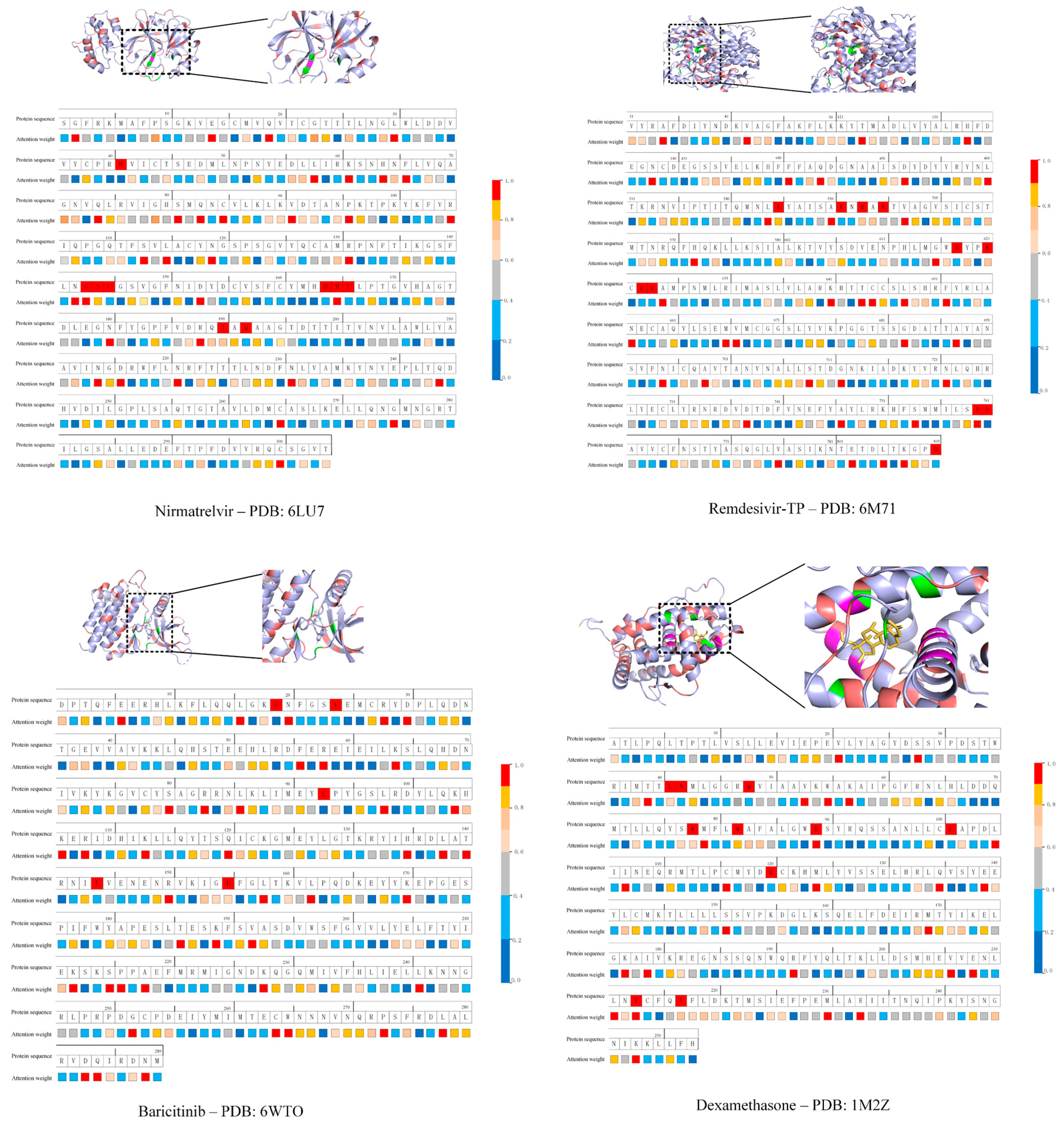
Moreover, we compared the predictions of our framework for the four well-known drug–target pairs with experimental data to further validate its performance. While the DL model successfully identified interactions for all four pairs (see Table 3), it was still necessary to further evaluate whether the attention mechanism assigned significant weights to the actual key residues of these proteins. To this end, we visualized the attention weights learned by our DL model, shown in Figure 2, with detailed data provided in Table S1 of the Supplementary Information. In our analysis, residues with attention weights over 80%, marked in red or bright yellow on the color scale, were considered highly attended. For comparison, experimentally verified binding residues are marked in red in the “Protein residue” row of Figure 2. For MPro (PDB ID: 6LU7), the DL model identified Asn142, Gly143, Ser144, Met165, Ala191, and Ala193, the residues known to be at or adjacent to key active sites [52]. For RdRp (PDB ID: 6M71), the critical residues Leu544, Lys545, Ala550, Ala625, Ser759, and Asp760 were identified [53]. Similarly, for JAK2 (PDB ID: 6WTO), residues Asn859 and Ile982 were identified [54], and for the glucocorticoid receptor (PDB ID: 1M2Z), residues Met601, Arg611, Tyr735, Met565, Met604, and Cys643 were identified [13]. It should be noted that, in this study, we consider the identification of residues adjacent to known binding sites as a successful prediction, given the structural complexity and flexibility of the proteins investigated. Small deviations in residue selection may still reflect meaningful biological relevance, particularly when the binding interface is dynamic or when neighboring residues contribute indirectly to ligand binding. These findings demonstrate that, for all four drug–target pairs, the high-attention residues predicted by our DL model overlap with experimentally validated binding sites. This suggests that our model has basically learned to recognize biologically relevant features at the residue level. However, we acknowledge that the attention mechanism we employed, when limited to 1D sequence or graph features and aimed solely at predicting overall binding probability, tends to produce broad attention distributions. Potential solutions include introducing sparsity constraints to encourage more focused attention and incorporating known binding site annotations as supervision to guide the model toward learning biologically meaningful attention patterns centered on actual binding residues.
Although our DL model seems to have learned key structural patterns underlying DTIs, the large number of residues assigned high attention weights limits its ability to precisely identify binding sites, especially in the absence of experimental data. Furthermore, attention weights reflect only 1D sequence-level information, whereas functional binding regions are typically defined by the protein’s secondary and tertiary structures. In contrast, molecular docking programs provide atomistic, 3D information on binding site residues and predicted affinities. As mentioned earlier, binding affinities below −7.0 kcal·mol−1 are typically considered strong, while values above this threshold may suggest weak or nonspecific interactions [49,50,51]. As shown in Table 4, AutoDock Vina and LeDock both predicted good affinities for four well-known DTIs (i.e., Nirmatrelvir–MPro, Remdesivir triphosphate–RdRp, Baricitinib–JAK2, and Dexamethasone–glucocorticoid receptor), except for AutoDock’s slightly weaker prediction for the first pair and LeDock’s weaker prediction for the last pair (>−7.0 kcal·mol−1).
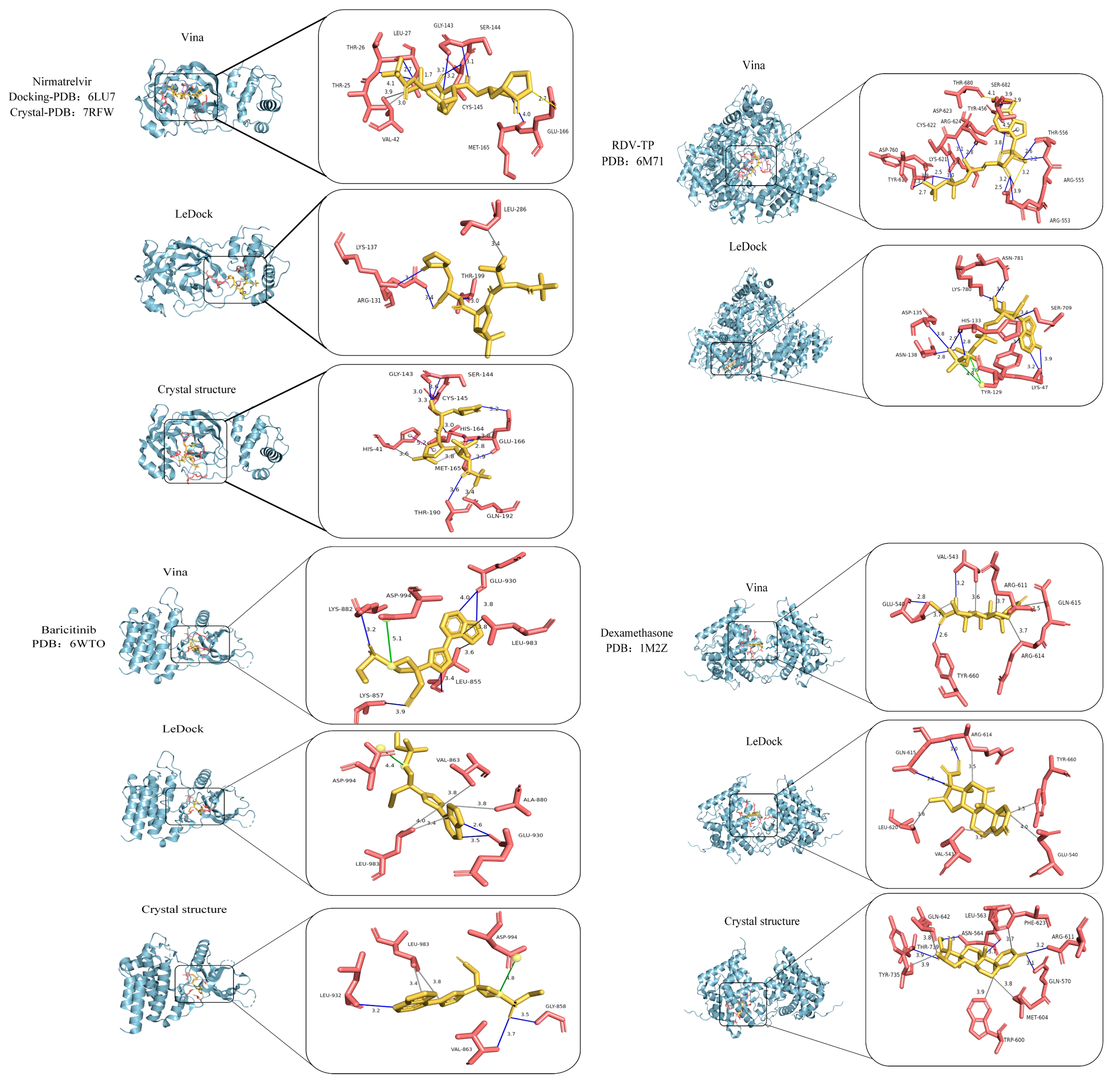
To evaluate whether the docking programs captured experimentally validated binding sites, we compared their predicted residues with known crystal structure data (shown in Figure 3 and Table 3). In Table 3, hydrogen-bonded residues are shown in black; salt-bridge-only residues in red; and hydrophobic-only residues in blue. For Remdesivir triphosphate–RdRp, where direct crystal data are unavailable, we referenced key residues supported by experimental evidence [53]. A glance at Figure 3 clearly reveals that AutoDock Vina generally captured the binding pose of Nirmatrelvir–MPro, while both docking programs correctly predicted the binding conformation of Baricitinib–JAK2. More specifically, in Table 3, for the pair Nirmatrelvir–MPro, AutoDock Vina successfully identified key MPro residues (Val42, Gly143, Ser144, Cys145, and Glu166) at or adjacent to the binding sites for Nirmatrelvir, while LeDock did not. For the pair Remdesivir triphosphate–RdRp, LeDock correctly predicted several key residues (Arg553, Arg555, Thr556, Tyr619, Lys621, Cys622, Asp623, Arg624, and Asp760), whereas AutoDock Vina did not. Both programs captured important binding residues Leu983 and Asp994 for the pair Baricitinib–JAK2, while neither of them fully captured the correct binding sites for the pair Dexamethasone–glucocorticoid receptor, with only AutoDock Vina partially identifying Arg611. These results show a key challenge: no single docking tool consistently outperforms others across all protein targets [55,56]. Since the proteins tested here represent diverse protein families ranging from viral enzymes (e.g., RdRp) to human signaling proteins (e.g., JAK2), the accuracy of docking predictions can vary depending on the protein type and docking algorithm. Therefore, combining multiple docking tools offers a more robust strategy for improving predictive reliability and generating biologically meaningful insights.

2.2. Predictive Application to COVID-19 Main Protease

Based on the results from four positive and three negative drug–protein pairs, we found that DL models are effective for rapidly identifying potential drug candidates for a given target protein, and molecular docking programs can further examine the binding sites and affinities between the protein and selected drugs. Accordingly, we propose three steps to use our hybrid framework that integrates our DL model with molecular docking tools for virtual drug repurposing.
Step 1: DL Filter. Apply the DL model to rapidly screen candidate drugs and retain those with high predicted scores (with a binary default cut-off value of 0.5) based on learned features. This step filters out compounds with little or no predicted interaction with the target protein.
Step 2: Docking Affinity Filter. Use AutoDock Vina and LeDock to calculate binding affinities for the top-scoring poses and select drug–protein pairs with predicted affinities below −7.0 kcal·mol−1. This step eliminates compounds with weak predicted affinity for the target.
Step 3: Binding Pose Validation. Compare the predicted binding residues of the protein to experimentally validate active sites to assess whether the candidate drug is likely to inhibit protein function. This step helps identify candidate drugs that not only bind strongly but also bind to the correct active site of the target protein.
It is important to note that our deep learning model performs a binary classification task, where typically a prediction score above 0.5 indicates a positive interaction, while a score below 0.5 suggests no interaction. However, in this work, we used an empirical cut-off value of 0.8 based on the model’s performance across multiple benchmark datasets, where it consistently demonstrated stable and reliable predictions at or above this level. Despite the adjustment, using this threshold as the initial filter presents a key limitation of the hybrid framework when applied to large-scale drug screening. Specifically, a substantial number of compounds may receive scores above 0.8 for a given target, making subsequent docking computations labor-intensive and time-consuming.
In contrast, our hybrid model is better suited for evaluating a relatively small set of drug candidates, where the proposed three-step process can efficiently find the most promising compounds. Consequently, we selected 29 drug candidates for potential repurposing, most of which are FDA-approved, with the exceptions of Favipiravir, Daclatasvir, Carmofur, and Masitinib, which are approved in Japan or/and Europe. Unlike the three negative control compounds discussed earlier that have been clinically verified ineffective against COVID-19, these drugs exhibit potential therapeutic activity against SARS-CoV-2 but lack direct experimental or clinical evidence confirming their interactions with SARS-CoV-2-related proteins. For consistency and comparison, we focused on evaluating their interactions with SARS-CoV-2 MPro, although several of these drugs may also act on other targets such as RdRp, JAK2, or intracellular glucocorticoid receptors. The selection of these compounds was based on their known pharmacological categories as antiviral, anti-inflammatory, and anti-cancer, as previous studies have suggested that compounds in these categories might demonstrate inhibitory effects on SARS-CoV-2 [57,58,59,60,61]. Among the 16 antiviral drugs, Favipiravir, Oseltamivir, Zanamivir, and Peramivir are used to treat influenza; Lamivudine, Efavirenz, Lopinavir, and Ritonavir are used against human immunodeficiency virus (HIV); Sofosbuvir, Velpatasvir, Voxilaprevir, Daclatasvir, Glecaprevir, Pibrentasvir, and Ribavirin against hepatitis C virus (HCV); and Acyclovir against herpesvirus. Among the seven anti-inflammatory drugs, Tofacitinib, Sirolimus, Imiquimod, and Ibuprofen are classified as anti-inflammatory or immune-modulating agents, while Diclofenac, Naproxen, and Indomethacin are classified as nonsteroidal anti-inflammatory drugs (NSAIDs). Among the six anti-cancer drugs, Enasidenib, Nilotinib, Dasatinib, and Imatinib are primarily approved for the treatment of various forms of leukemia; Carmofur for the treatment of colon cancer; and Masitinib for pancreatic cancer.
We used our proposed three-step framework to evaluate the 29 repurposed drug candidates, with the results summarized in Table 4. In Step 1, our DL model identified 14 drugs with high predicted interaction scores (≥ 0.8), namely, Zanamivir, Peramivir, Lamivudine, Sofosbuvir, Voxilaprevir, Glecaprevir, Pibrentasvir, Tofacitinib, Enasidenib, Nilotinib, Dasatinib, Imatinib, Carmofur, and Masitinib. In Step 2, AutoDock Vina and LeDock identified 8 of these 14 compounds as having binding affinities below −7.0 kcal·mol−1 in at least one docking program. These drugs include Voxilaprevir, Glecaprevir, Pibrentasvir, Enasidenib, Nilotinib, Dasatinib, Imatinib, and Masitinib. The results suggest that three antiviral and five anti-cancer drugs show promising binding potential to MPro, but few anti-inflammatory drugs were successfully selected after Steps 1 and 2. This may be due to the fact that anti-inflammatory drugs are more likely to act on immune-related targets such as JAK1/2 [9].
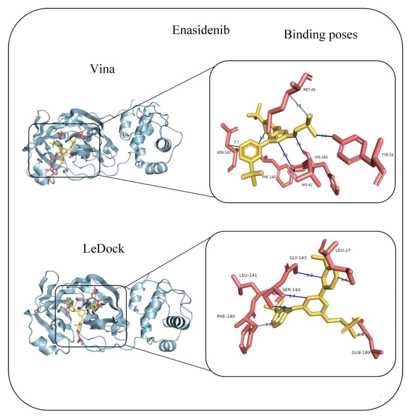
Step 3 is crucial for evaluating whether the selected drug candidates bind to the functionally relevant active sites of MPro by virtual docking programs. According to crystallographic studies (PDB IDs 6LU7 and 7RFW), residues His41, Gly143, Ser144, Cys145, His164, Met165, Glu166, Thr190, and Gln192 form the core of the active site [4,6,52]. Binding to these residues is considered essential for effective inhibition of the main protease’s catalytic activity. We show the binding site predictions for three selected antiviral drugs (Voxilaprevir, Glecaprevir, and Pibrentasvir) with MPro in Figure S1 and those for four selected anticancer drugs (Nilotinib, Dasatinib, Imatinib, and Masitinib) in Figure S2 of the Supplementary Information. The results indicate that none of these compounds bind to the active site or adjacent residues in either AutoDock Vina or LeDock predictions. This means that despite favorable binding affinities, these drugs are unlikely to inhibit MPro function due to their binding positioning outside the protein’s catalytic site. The only compound that passed all three steps of our framework is Enasidenib, an FDA-approved isocitrate dehydrogenase 2 (IDH2) inhibitor used in the treatment of IDH2-mutant acute myeloid leukemia. As shown in Figure 4, AutoDock Vina predicts that Enasidenib interacts with His41, Asn142, and His164—residues located at or near the MPro active site. Similarly, LeDock identifies interactions with Asn142 and Glu166, both at or adjacent to the catalytic core. Our finding is in excellent agreement with another in silico report by Anwaar et al. [62], who also identified Enasidenib as a potential MPro inhibitor in their drug repurposing study for COVID-19. Nevertheless, further experimental validation and clinical trials are essential to confirm its antiviral efficacy and assess its safety profile in treating SARS-CoV-2 infections.
Finally, several other limitations of this study should be discussed. First, the performance of our hybrid framework depends on the predictive reliability of the DL model, which serves as the primary screening filter. Given its “black-box” nature, the DL model may exclude potentially effective drug candidates due to limitations in robustness and generalizability. Enhancing model training on broader, high-quality datasets would be essential to reduce false negatives. Second, molecular docking tools typically neglect protein flexibility and solvent effects, and incorporating molecular dynamics (MD) simulations and binding free energy calculations could substantially improve predictive accuracy [63,64,65,66]. We present additional results from MD simulations of Enasidenib-Mpro complex in Note S1 of the Supplementary Information. Despite these limitations, the proposed hybrid framework offers a promising in silico strategy for drug repurposing and, when complemented with experimental validation, holds potential for accelerating therapeutic discovery.

3. Materials and Methods

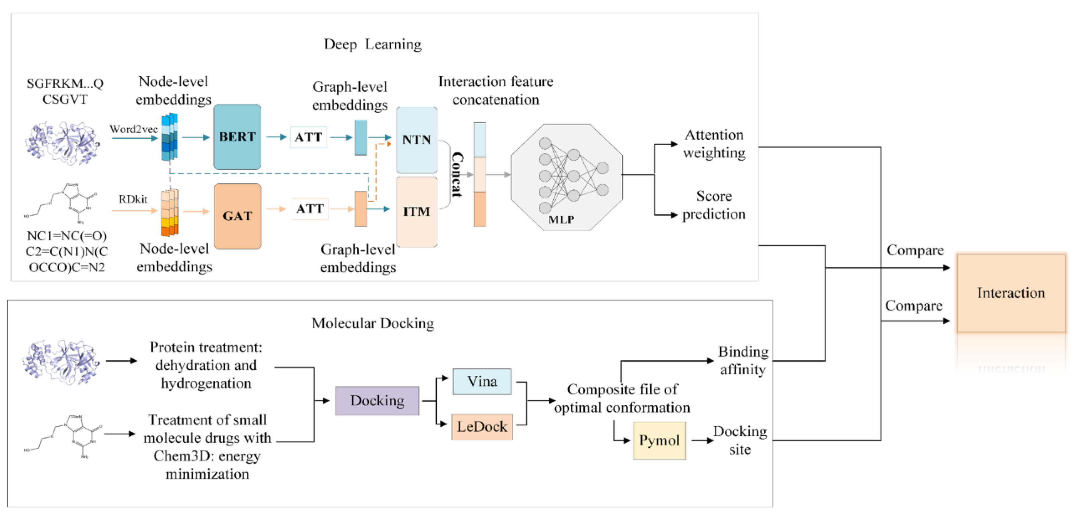
The overall workflow of our interpretable DL and molecular docking framework is illustrated in Figure 5. The framework first employs our DL model to rapidly screen and select candidate ligands with high predicted affinity, followed by molecular docking to evaluate their binding modes and interaction strengths. By integrating these two approaches, the framework aims to reduce the computational cost of docking while improving the predictive accuracy and interpretability of the DL model.

3.1. Deep Learning Model

The DL model architecture is composed of three stages: feature extraction, interaction modeling, and prediction. In the feature extraction stage, raw drug and protein data—namely, SMILES (Simplified Molecular Input Line Entry System) strings [67] and amino acid sequences—are transformed into node-level embeddings, as they cannot be directly processed by the model. Specifically, the cheminformatics toolkit RDKit [68] is used to convert SMILES strings into molecular graphs and extract atom-level features. For protein sequences, a pretrained Word2Vec model [69] is used to encode residues into numerical vectors that capture local sequence patterns. These embeddings form the input features for subsequent stages of the DL model.
In the interaction modeling stage, the node-level embeddings are first processed through two distinct modules: a Graph Attention Network (GAT) [70] for drugs and a Bidirectional Encoder Representations from Transformers (BERT) [71] module for proteins. These modules extract contextualized, high-dimensional features that capture the local structural and sequential information of drugs and proteins, respectively. Next, a multi-head attention mechanism (ATT) [26] is subsequently applied to aggregate the node-level outputs into graph-level representations that capture higher-order structural information. To enhance the model’s ability to capture diverse types of drug–target interactions, both node-level and graph-level features are fed into two specialized interaction modules: interaction transformers (ITMs) and neural tensor networks (NTNs). The ITM is responsible for learning pairwise interactions by modeling complex dependencies between atoms in the drug and protein residues. It applies cross-attention between the two sets of embeddings, enabling the model to capture fine-grained local interactions that are essential for binding specificity. In contrast, the NTN captures global and nonlinear relationships by modeling bilinear interactions between the overall graph-level representations of drugs and proteins. The NTN effectively learns potential interaction patterns that might not be obvious from local attention alone. Together, the ITM and NTN complement each other: ITM emphasizes localized structural and chemical interactions, while NTN focuses on global compatibility between molecules.
In the prediction stage, the learned interaction features (both local atomic-level and global structural-level information from the drug and protein) are combined and input into a multilayer perceptron (MLP) [72] for final DTI prediction. The MLP consists of multiple fully connected layers, each followed by nonlinear activation functions (e.g., ReLU), dropout for regularization, and batch normalization to stabilize training. Through these layers, the model learns complex, nonlinear mappings between interaction patterns and the likelihood of drug–target binding. The final output layer uses a sigmoid activation function to generate a probability score between 0 and 1, representing the predicted likelihood of interaction between the drug and protein. A threshold (typically 0.5 in binary classification tasks) is then applied to determine whether the interaction is predicted to be positive or negative. For more detailed architectural and methodological information, readers are referred to our original work [41].
We evaluated the performance of our DL model via five-fold cross-validation on three benchmark datasets: Human, C. elegans, and DrugBank. The Human and C. elegans datasets are relatively small, while DrugBank is a large-scale dataset [73]. Table S2 of the Supplementary Information summarizes the information in the datasets used for training the DL model. Table S3 of the Supplementary Information outlines the key hyperparameters used to train our deep learning model for DTI prediction. The protein and atom embeddings were set to dimensions of 100 and 34, respectively, while the hidden layer size was 64. The model included three layers of GAT, each with three attention heads, and used eight heads for the multi-head self-attention mechanism. Structural features were captured using a radius of 2- and 3-g tokenization. A dropout rate of 0.1 was applied to prevent overfitting. The model was trained with a batch size of 32, a learning rate of 1 × 10⁻³, and a regularization coefficient of 1 × 10⁻⁴. Additionally, the number of major potential associations (K) was set to 16, and training was carried out over 40 epochs.
The average area under curve (AUC), precision, and recall values for our model and for several benchmark methods are reported in Table 5, Table 6 and Table 7. On the Human and C. elegans datasets, our model outperformed traditional ML baselines such as KNN, RF, L2, and SVM, as well as several DL baselines such as MDL-CPI [74], GNN [75], graph convolutional network (GCN), GraphDTA [76], DrugVQA(VQA-seq) [47], and TransformerCPI [27]. On the DrugBank dataset, our model outperformed several established models, including RWR (ML-based) [77], DrugE-Rank (ML-based) [78], GNN-CPI (DL-based) [28], and DeepCPI (DL-based) [79], and showed performance comparable to that of GraphDTA (DL-based) [76]. These results show the relative strength of our model in DTI prediction. We acknowledge that the performance metrics (AUC, precision, and recall) of our model remain modest when trained on the larger DrugBank dataset. First, in contrast to smaller and more homogeneous human and C. elegans datasets, DrugBank is a large-scale and highly heterogeneous dataset, encompassing a wide variety of compound and protein types. Such a large public databases often contain noise and inconsistencies, which can affect the model’s ability to learn reliable patterns. Second, our current model primarily relies on 1D representations for drugs and amino acid sequences for proteins. Although this approach works well on smaller datasets, it may be insufficient for capturing the more diverse and complex DTI patterns present in DrugBank.
However, it is important to note that the objective of this work is not to outperform all existing models but rather to demonstrate that our model is qualified for DTI prediction and can serve as an important component within the hybrid DL and docking framework illustrated in Figure 5. Moreover, using our DL model is generally convenient and efficient. For model training, smaller datasets such as human and C. elegans take approximately 5 min per training epoch on a standard GPU server. For larger datasets like DrugBank, a full training cycle takes roughly 30 min. Once the model is trained, scoring new drug–target pairs is instantaneous, making it feasible to rapidly evaluate thousands of combinations.

3.2. Molecular Docking Programs

Wang et al. conducted a comprehensive evaluation comparing ten docking programs across diverse protein–ligand complexes [55]. They found that the academic program LeDock ranked among the top two for scoring power following the commercial program GOLD. Moreover, AutoDock Vina demonstrated the best overall scoring performance among all the tested programs. Therefore, we utilized the academic programs LeDock and AutoDock Vina in this work. First, AutoDock [30] and AutoDock Vina [31,32] are two widely used computational tools for molecular docking, both developed by the Olson Laboratory at The Scripps Research Institute. While AutoDock employs a genetic algorithm (GA) and a Lamarckian Genetic Algorithm (LGA) to search for optimal binding conformations of the ligand, AutoDock Vina integrates a stochastic global search with a local optimization method based on the Broyden–Fletcher–Goldfarb–Shanno (BFGS) algorithm for more efficient sampling of the conformational space [31,80]. Moreover, AutoDock Vina features a re-designed scoring function that incorporates a broader range of physicochemical interactions, enhancing its ability to estimate binding affinities. Compared to AutoDock, AutoDock Vina significantly improves docking speed and is particularly effective in high-throughput virtual screening campaigns.
Second, LeDock [33] employs a hybrid optimization algorithm that integrates an LGA with a local search strategy, enabling the program to explore a wide conformational space of ligands within a reasonable computational time. For each sampled conformation, a scoring function is used to estimate the binding free energy, thereby predicting the most favorable binding mode between a ligand and its target protein. The scoring function combines empirical physicochemical parameters to evaluate key intermolecular interactions, such as van der Waals forces and electrostatic interactions. Its robust performance and speed make it particularly well suited for high-throughput virtual screening.
The docking simulation procedure was conducted as follows. Initially, we selected and downloaded the protein crystal structure from the Protein Data Bank (PDB), choosing PDB ID: 6LU7 for the SARS-CoV-2 main protease (Mpro), for example. This structure was selected for two main reasons: first, it provides high-resolution data (2.16 Å), allowing precise characterization of the key active site residues (His41–Cys145) and the surrounding binding pocket; second, it is one of the most widely used Mpro structures in virtual screening, molecular dynamics, and drug design studies [63,65,66]. For protein preparation, AutoDockTools (ADT) version 1.5.6 was used to process the PDB file by removing all bound ligands, cofactors, and water molecules, followed by the addition of polar hydrogens to define the protein as the receptor. For ligand preparation, we downloaded the molecular structures from PubChem in .mol format, performed energy minimization in Chem3D [81], converted them to .pdb format using PyMOL version 2.6 [82], and then used ADT to add hydrogens, automatically detect rotatable bonds, and generate .pdbqt files. During grid generation, we set a grid box encompassing the entire protein, which slightly reduces docking speed but ensures that no potential binding site is overlooked.

4. Conclusions

In this study, we present a hybrid framework that integrates DL and molecular docking to repurpose existing drugs as inhibitors of Mpro. Our framework consists of a three-step virtual screening pipeline. In the first step, our previously developed DL model was employed to rapidly filter drug candidates predicted to interact with the target protein, selecting those with interaction scores above 0.8. We confirmed that the DL model captures biologically relevant features by demonstrating overlaps between the high-attention residues it identified and experimentally verified binding sites. In the second step, two widely used docking programs, AutoDock Vina and LeDock, were used to calculate binding affinities, and drugs with predicted affinities stronger than −0.7 kcal·mol−1 were retained. In the third step, predicted binding sites using two docking tools were evaluated to determine whether the drug candidates could bind to the known active sites of the target protein. This framework is particularly effective for identifying inhibitors of proteins with experimentally resolved 3D structures and their active sites.
To validate our framework, we first applied it to four clinically relevant drug–target pairs associated with COVID-19: Nirmatrelvir–MPro, Remdesivir–RdRp, Baricitinib–JAK2, and Dexamethasone–glucocorticoid receptor. The inclusion of multiple and diverse proteins helps demonstrate the robustness of this approach. We then applied the framework to a set of 29 approved drugs, selected based on their reported antiviral, anti-inflammatory, or anti-cancer properties that may be associated with COVID-19 therapeutic strategies. Through this in silico analysis, an anti-cancer agent Enasidenib was finally identified as a promising MPro inhibitor, with predicted binding at or near the protein’s active site. Overall, our hybrid framework provides a generalizable and interpretable strategy for virtual drug screening and repurposing, particularly in response to emerging infectious diseases.

Supplementary Materials

The following supporting information can be downloaded at: https://www.mdpi.com/article/10.3390/molecules30163409/s1, Figure S1: Predicted binding residues between Mpro and three antivirial drugs; Figure S2: Predicted binding residues between Mpro and four anti-cancer drugs; Table S1: List of highly weighted residues identified by our DL model; Table S2: Summary of the three datasets used in this study; Table S3: Hyperparameter setting for our deep learning model; Note S1: Molecular dynamics simulations of Enasidenib-Mpro complex [83,84,85,86,87]. The Python code for our deep learning model and for visualizing residues with varying attention weights is available as open source and can be downloaded from the following website: https://github.com/frankchenqu/hybrid (accessed on 1 July 2025).

Author Contributions

Conceptualization, J.G. and Q.C.; methodology, Q.C.; investigation, J.G.; writing—original draft preparation, Q.C.; writing—review and editing, Q.C.; supervision, J.H. and Q.C.; funding acquisition, Q.C. All authors have read and agreed to the published version of the manuscript.

Funding

This work was financially supported by the Zhejiang Provincial Natural Science Foundation (grant nos. LY19B060002).

Institutional Review Board Statement

Not applicable.

Informed Consent Statement

Not applicable.

Data Availability Statement

The original contributions presented in this study are included in the article. Further inquiries can be directed to the corresponding author.

Conflicts of Interest

There are no conflicts to declare.

References

  1. Machkovech, H.M.; Hahn, A.M.; Garonzik Wang, J.; Grubaugh, N.D.; Halfmann, P.J.; Johnson, M.C.; Lemieux, J.E.; O’Connor, D.H.; Piantadosi, A.; Wei, W.; et al. Persistent SARS-CoV-2 infection: Significance and implications. Lancet Infect. Dis. 2024, 24, e453–e462. [Google Scholar] [CrossRef] [PubMed]
  2. Chen, X.; Huang, X.; Ma, Q.; Kuzmič, P.; Zhou, B.; Zhang, S.; Chen, J.; Xu, J.; Liu, B.; Jiang, H.; et al. Preclinical evaluation of the SARS-CoV-2 Mpro inhibitor RAY1216 shows improved pharmacokinetics compared with nirmatrelvir. Nat. Microbiol. 2024, 9, 1075–1088. [Google Scholar] [CrossRef] [PubMed]
  3. Narwal, M.; Armache, J.; Edwards, T.J.; Murakami, K.S. SARS-CoV-2 polyprotein substrate regulates the stepwise Mpro cleavage reaction. J. Biol. Chem. 2023, 299, 104697. [Google Scholar] [CrossRef]
  4. Jin, Z.; Du, X.; Xu, Y.; Deng, Y.; Liu, M.; Zhao, Y.; Zhang, B.; Li, X.; Zhang, L.; Peng, C.; et al. Structure of Mpro from SARS-CoV-2 and discovery of its inhibitors. Nature 2020, 582, 289–293. [Google Scholar] [CrossRef]
  5. Ullrich, S.; Nitsche, C. The SARS-CoV-2 main protease as drug target. Bioorg. Med. Chem. Lett. 2020, 30, 127377. [Google Scholar] [CrossRef]
  6. Lamb, Y.N. Nirmatrelvir plus ritonavir: First approval. Drugs 2022, 82, 585–591. [Google Scholar] [CrossRef]
  7. Gao, Y.; Yan, L.; Huang, Y.; Liu, F.; Zhao, Y.; Cao, L.; Wang, T.; Sun, Q.; Ming, Z.; Zhang, L.; et al. Structure of the RNA-dependent RNA polymerase from COVID-19 virus. Science 2020, 368, 779–782. [Google Scholar] [CrossRef]
  8. Yin, W.; Mao, C.; Luan, X.; Shen, D.; Su, H.; Wang, X.; Zhou, F.; Zhao, W.; Gao, M.; Chang, S.; et al. Structural basis for inhibition of the RNA-dependent RNA polymerase from SARS-CoV-2 by remdesivir. Science 2020, 368, 1499–1504. [Google Scholar] [CrossRef]
  9. Zak, M.; Hanan, E.J.; Lupardus, P.; Brown, D.G.; Robinson, C.; Siu, M.; Lyssikatos, J.P.; Romero, F.A.; Zhao, G.; Kellar, T.; et al. Discovery of a class of highly potent Janus Kinase 1/2 (JAK1/2) inhibitors demonstrating effective cell-based blockade of IL-13 signaling. Bioorg. Med. Chem. Lett. 2019, 29, 1522–1531. [Google Scholar] [CrossRef]
  10. Davis, R.R.; Li, B.; Yun, S.Y.; Chan, A.; Nareddy, P.; Gunawan, S.; Ayaz, M.; Lawrence, H.R.; Reuther, G.W.; Lawrence, N.J.; et al. Structural insights into JAK2 inhibition by ruxolitinib, fedratinib, and derivatives thereof. J. Med. Chem. 2021, 64, 2228–2241. [Google Scholar] [CrossRef]
  11. Markham, A. Baricitinib: First global approval. Drugs 2017, 77, 697–704. [Google Scholar] [CrossRef] [PubMed]
  12. Blair, H.A. Remdesivir: A review in COVID-19. Drugs 2023, 83, 1215–1237. [Google Scholar] [CrossRef] [PubMed]
  13. Bledsoe, R.K.; Montana, V.G.; Stanley, T.B.; Delves, C.J.; Apolito, C.J.; McKee, D.D.; Consler, T.G.; Parks, D.J.; Stewart, E.L.; Willson, T.M.; et al. Crystal structure of the glucocorticoid receptor ligand binding domain reveals a novel mode of receptor dimerization and coactivator recognition. Cell 2002, 110, 93–105. [Google Scholar] [CrossRef] [PubMed]
  14. The RECOVERY Collaborative Group. Dexamethasone in hospitalized patients with COVID-19. N. Engl. J. Med. 2021, 384, 693–704. [Google Scholar] [CrossRef]
  15. Deng, J.; Yang, Z.; Ojima, I.; Samaras, D.; Wang, F. Artificial intelligence in drug discovery: Applications and techniques. Brief. Bioinform. 2022, 23, bbab430. [Google Scholar] [CrossRef]
  16. Cavasotto, C.N.; Di Filippo, J.I. Artificial intelligence in the early stages of drug discovery. Arch. Biochem. Biophys. 2021, 698, 108730. [Google Scholar] [CrossRef]
  17. Chong, C.R.; Sullivan, D.J. New uses for old drugs. Nature 2007, 448, 645–646. [Google Scholar] [CrossRef]
  18. Ambrosio, F.A.; Costa, G.; Romeo, I.; Esposito, F.; Alkhatib, M.; Salpini, R.; Svicher, V.; Corona, A.; Malune, P.; Tramontano, E.; et al. Targeting SARS-CoV-2 main protease: A successful story guided by an in silico drug repurposing approach. J. Chem. Inf. Model. 2023, 63, 3601–3613. [Google Scholar] [CrossRef]
  19. Weth, F.R.; Hoggarth, G.B.; Weth, A.F.; Paterson, E.; White, M.P.J.; Tan, S.T.; Peng, L.; Gray, C. Unlocking hidden potential: Advancements, approaches, and obstacles in repurposing drugs for cancer therapy. Br. J. Cancer 2024, 130, 703–715. [Google Scholar] [CrossRef]
  20. He, H.; Chen, G.; Chen, C.Y. Machine learning and graph neural network for finding potential drugs related to multiple myeloma. New J. Chem. 2022, 46, 5188–5200. [Google Scholar] [CrossRef]
  21. Dhakal, A.; McKay, C.; Tanner, J.J.; Cheng, J. Artificial intelligence in the prediction of protein-ligand interactions: Recent advances and future directions. Brief. Bioinform. 2022, 23, bbab476. [Google Scholar] [CrossRef]
  22. Zhang, Y.; Li, S.; Meng, K.; Sun, S. Machine learning for sequence and structure-based protein−ligand interaction prediction. J. Chem. Inf. Model. 2024, 64, 1456–1472. [Google Scholar] [CrossRef]
  23. Velazquez, M.; Anantharaman, R.; Velazquez, S.; Lee, Y. Alzheimer’s Disease Neuroimaging Initiative. RNN-based Alzheimer’s disease prediction from prodromal stage using diffusion tensor imaging. In Proceedings of the 2019 IEEE International Conference on Bioinformatics and Biomedicine (BIBM), San Diego, CA, USA, 18–21 November 2019; pp. 1665–1672. [Google Scholar]
  24. Lee, I.; Keum, J.; Nam, H. DeepConv-DTI: Prediction of drug–target interactions via deep learning with convolution on protein sequences. PLoS Comput. Biol. 2019, 15, e1007129. [Google Scholar] [CrossRef]
  25. Zhang, Z.; Chen, L.; Zhong, F.; Wang, D.; Jiang, J.; Zhang, S.; Jiang, H.; Zheng, M.; Li, X. Graph neural network approaches for drug-target interactions. Curr. Opin. Struc. Biol. 2022, 73, 102327. [Google Scholar] [CrossRef] [PubMed]
  26. Vaswani, A.; Shazeer, N.; Parmar, N.; Uszkoreit, J.; Jones, L.; Gomez, A.N.; Kaiser, Ł.; Polosukhin, I. Attention is all you need. In Proceedings of the 31st Conference on Neural Information Processing Systems (NIPS 2017), Long Beach, CA, USA, 4–9 December 2017; p. 30. [Google Scholar]
  27. Chen, L.; Tan, X.; Wang, D.; Zhong, F.; Liu, X.; Yang, T.; Luo, X.; Chen, K.; Jiang, H.; Zheng, M. TransformerCPI: Improving compound–protein interaction prediction by sequence-based deep learning with self-attention mechanism and label reversal experiments. Bioinformatics 2020, 36, 4406–4414. [Google Scholar] [CrossRef] [PubMed]
  28. Zhao, Q.; Zhao, H.; Zheng, K.; Wang, J. HyperAttentionDTI: Improving drug–protein interaction prediction by sequence-based deep learning with attention mechanism. Bioinformatics 2022, 38, 655–662. [Google Scholar] [CrossRef] [PubMed]
  29. Morrison, J.L.; Breitling, R.; Higham, D.J.; Gilbert, D.R. A lock-and-key model for protein–protein interactions. Bioinformatics 2006, 22, 2012–2019. [Google Scholar] [CrossRef]
  30. Morris, G.M.; Huey, R.; Lindstrom, W.; Sanner, M.F.; Belew, R.K.; Goodsell, D.S.; Olson, A.J. AutoDock4 and AutoDockTools4: Automated docking with selective receptor flexibility. J. Comput. Chem. 2010, 30, 2785–2791. [Google Scholar] [CrossRef]
  31. Trott, O.; Olson, A.J. AutoDock Vina: Improving the speed and accuracy of docking with a new scoring function, efficient optimization, and multithreading. J. Comput. Chem. 2010, 31, 455–461. [Google Scholar] [CrossRef]
  32. Eberhardt, J.; Santos-Martins, D.; Tillack, A.F.; Forli, S. AutoDock Vina 1.2.0: New docking methods, expanded force field, and Python bindings. J. Chem. Inf. Model. 2021, 61, 3891–3898. [Google Scholar] [CrossRef]
  33. Zhao, H.; Dong, J.; Lafleur, K.; Nevado, C.; Caflisch, A. Discovery of a novel chemotype of tyrosine kinase inhibitors by fragment-based docking and molecular dynamics. ACS Med. Chem. Lett. 2012, 3, 834–838. [Google Scholar] [CrossRef]
  34. Friesner, R.A.; Banks, J.L.; Murphy, R.B.; Halgren, T.A.; Klicic, J.J.; Mainz, D.T.; Repasky, M.P.; Knoll, E.H.; Shelley, M.; Perry, J.K.; et al. Glide:  a new approach for rapid, accurate docking and scoring. 1. method and assessment of docking accuracy. J. Med. Chem. 2004, 47, 1739–1749. [Google Scholar] [CrossRef] [PubMed]
  35. Halgren, T.A.; Murphy, R.B.; Friesner, R.A.; Beard, H.S.; Frye, L.L.; Pollard, W.T.; Banks, J.L. Glide:  a new approach for rapid, accurate docking and scoring. 2. enrichment factors in database screening. J. Med. Chem. 2004, 47, 1750–1759. [Google Scholar] [CrossRef] [PubMed]
  36. Rarey, M.; Kramer, B.; Lengauer, T.; Klebe, G. A fast flexible docking method using an incremental construction algorithm. J. Mol. Biol. 1996, 261, 470–489. [Google Scholar] [CrossRef]
  37. Zhao, H.; Jiang, D.; Shen, C.; Zhang, J.; Zhang, X.; Wang, X.; Nie, D.; Hou, T.; Kang, Y. Comprehensive evaluation of 10 docking programs on a diverse set of protein−cyclic peptide complexes. J. Chem. Inf. Model. 2024, 64, 2112–2124. [Google Scholar] [CrossRef]
  38. Sharma, G.; Kumar, N.; Sharma, C.S.; Alqahtani, T.; Tiruneh, Y.K.; Sultana, S.; Rolim Silva, G.V.; de Lima Menezes, G.; Zaki, M.E.A.; Oliveira, J.I.N. Identification of promising SARSCoV-2 main protease inhibitor through molecular docking, dynamics simulation, and ADMET analysis. Sci. Rep. 2025, 15, 2830. [Google Scholar]
  39. Zhang, H.; Liang, B.; Sang, X.; An, J.; Huang, Z. Discovery of potential inhibitors of SARS-CoV-2 main protease by a transfer learning method. Viruses 2023, 15, 891. [Google Scholar] [CrossRef]
  40. Su, Q.; Wang, J.; Gou, Q.; Hu, R.; Jiang, L.; Zhang, H.; Wang, T.; Liu, Y.; Shen, C.; Kang, Y.; et al. Robust protein–ligand interaction modeling through integrating physical laws and geometric knowledge for absolute binding free energy calculation. Chem. Sci. 2025, 16, 5043–5057. [Google Scholar] [CrossRef]
  41. Wang, L.; Zhou, Y.; Chen, Q. AMMVF-DTI: A novel model predicting drug-target interactions based on attention mechanism and multi-view fusion. Int. J. Mol. Sci. 2023, 24, 14142. [Google Scholar] [CrossRef]
  42. Yang, Z.; Zhong, W.; Lv, Q.; Dong, T.; Chen, C.Y. Geometric interaction graph neural network for predicting protein-ligand binding affinities from 3D structures (GIGN). J. Phys. Chem. Lett. 2023, 14, 2020–2033. [Google Scholar] [CrossRef]
  43. Manivannan, E.; Karthikeyan, C.; Moorthy, N.S.H.N.; Chaturvedi, S.C. The rise and fall of chloroquine/hydroxychloroquine as compassionate therapy of COVID-19. Front. Pharmacol. 2021, 12, 584940. [Google Scholar] [CrossRef] [PubMed]
  44. Ma, C.; Wang, J. Dipyridamole, chloroquine, montelukast sodium, candesartan, oxytetracycline, and atazanavir are not SARS-CoV-2 main protease inhibitors. Proc. Natl. Acad. Sci. USA 2021, 118, e2024420118. [Google Scholar] [CrossRef] [PubMed]
  45. Cong, W.; Poudel, A.N.; Alhusein, N.; Wang, H.; Yao, G.; Lambert, H. Antimicrobial use in COVID-19 patients in the first phase of the SARS-CoV-2 pandemic: A scoping review. Antibiotics 2021, 10, 745. [Google Scholar] [CrossRef] [PubMed]
  46. Ghahremanpour, M.M.; Tirado-Rives, J.; Deshmukh, M.; Ippolito, J.A.; Zhang, C.H.; Cabeza de Vaca, I.; Liosi, M.E.; Anderson, K.S.; Jorgensen, W.L. Identification of 14 known drugs as inhibitors of the main protease of SARS-CoV-2. ACS Med. Chem. Lett. 2020, 11, 2526–2533. [Google Scholar] [CrossRef]
  47. Zheng, S.; Li, Y.; Chen, S.; Xu, J.; Yang, Y. Predicting drug–protein interaction using quasi-visual question answering system. Nat. Mach. Intell. 2020, 2, 134–140. [Google Scholar] [CrossRef]
  48. Öztürk, H.; Özgür, A.; Ozkirimli, E. DeepDTA: Deep drug–target binding affinity prediction. Bioinformatics 2018, 34, i821–i829. [Google Scholar] [CrossRef]
  49. Antypenko, L.; Shabelnyk, K.; Antypenko, O.; Arisawa, M.; Kamyshnyi, O.; Oksenych, V.; Kovalenko, S. In silico identification and characterization of spiro[1,2,4]triazolo[1,5-c]quinazolines as diacylglycerol kinase α modulators. Molecules 2025, 30, 2324. [Google Scholar] [CrossRef]
  50. Cosconati, S.; Forli, S.; Perryman, A.L.; Harris, R.; Goodsell, D.S.; Olson, A.J. Virtual screening with AutoDock: Theory and practice. Expert. Opin. Drug Discov. 2010, 5, 597–607. [Google Scholar] [CrossRef]
  51. Fischer, A.; Sellner, M.; Neranjan, S.; Smieško, M.; Lill, M.A. Potential inhibitors for novel coronavirus protease identified by virtual screening of 606 million compounds. Int. J. Mol. Sci. 2020, 21, 3626. [Google Scholar] [CrossRef]
  52. Owen, D.R.; Allerton, C.M.N.; Anderson, A.S.; Aschenbrenner, L.; Avery, M.; Berritt, S.; Boras, B.; Cardin, R.D.; Carlo, A.; Coffman, K.J.; et al. An oral SARS-CoV-2 Mpro inhibitor clinical candidate for the treatment of COVID-19. Science 2021, 374, 1586–1593. [Google Scholar] [CrossRef]
  53. Pant, S.; Jena, N.R. Inhibition of the RNA-dependent RNA polymerase of the SARS-CoV-2 by short peptide inhibitors. Eur. J. Pharm. Sci. 2021, 167, 106012. [Google Scholar] [CrossRef]
  54. Chang, Y.; Min, J.; Jarusiewicz, J.A.; Actis, M.; Bradford, S.Y.; Mayasundari, A.; Yang, L.; Chepyala, D.; Alcock, L.J.; Roberts, K.G.; et al. Degradation of Janus kinases in CRLF2-rearranged acute lymphoblastic leukemia. Blood 2021, 138, 2313–2326. [Google Scholar] [CrossRef] [PubMed]
  55. Wang, Z.; Sun, H.; Yao, X.; Li, D.; Xu, L.; Li, Y.; Tian, S.; Hou, T. Comprehensive evaluation of ten Docking programs on a diverse set of protein–ligand complexes: The prediction accuracy of sampling power and scoring power. Phys. Chem. Chem. Phys. 2016, 18, 12964. [Google Scholar] [CrossRef] [PubMed]
  56. Shen, C.; Wang, Z.; Yao, X.; Li, Y.; Lei, T.; Wang, E.; Xu, L.; Zhu, F.; Li, D.; Hou, T. Comprehensive assessment of nine docking programs on type II kinase inhibitors: Prediction accuracy of sampling power, scoring power and screening power. Brief. Bioinform. 2020, 21, 282–297. [Google Scholar] [CrossRef] [PubMed]
  57. Soy, M.; Keser, G.; Atagündüz, P.; Tabak, F.; Atagündüz, I.; Kayhan, S. Cytokine storm in COVID-19: Pathogenesis and overview of anti-inflammatory agents used in treatment. Clin. Rheumatol. 2020, 39, 2085–2094. [Google Scholar] [CrossRef]
  58. Stebbing, J.; Phelan, A.; Griffinb, I.; Tucker, C.; Oechsle, O.; Smith, D.; Richardson, P. COVID-19: Combining antiviral and anti-inflammatory treatments. Lancet Infect. Dis. 2020, 20, 400–402. [Google Scholar] [CrossRef]
  59. Perico, N.; Cortinovis, M.; Suter, F.; Remuzzi, G. Home as the new frontier for the treatment of COVID-19: The case for anti-inflammatory agents. Lancet Infect. Dis. 2023, 23, e22–e33. [Google Scholar] [CrossRef]
  60. Micallefa, J.; Soeiro, T.; Jonville-Béra, A.P.; French Society of Pharmacology. Non-steroidal anti-inflammatory drugs, pharmacology, and COVID-19 infection. Therapies 2020, 75, 355–362. [Google Scholar] [CrossRef]
  61. Borcherding, N.; Jethava, Y.; Vikas, P. Repurposing anti-cancer drugs for COVID-19 treatment. Drug Des. Dev. Ther. 2020, 14, 5045–5058. [Google Scholar] [CrossRef]
  62. Anwaar, M.U.; Adnan, F.; Abro, A.; Khan, R.A.; Rehman, A.U.; Osama, M.; Rainville, C.; Kumar, S.; Sterner, D.E.; Javed, S.; et al. Combined deep learning and molecular docking simulations approach identifies potentially effective FDA approved drugs for repurposing against SARS-CoV-2. Comput. Biol. Med. 2022, 141, 105049. [Google Scholar] [CrossRef]
  63. Huang, S.; Zhang, Y.; Wang, Z.; Xiong, F. Computational discovery of SARS-CoV-2 main protease inhibitors via a virtual screening, molecular docking, molecular Dynamics and MM/PBSA calculation-driven approach. New J. Chem. 2024, 48, 19650. [Google Scholar] [CrossRef]
  64. Fu, H.; Zhu, Y.; Chen, Q. Free energy calculations in biomolecule-nanomaterial interactions. Front. Phys. 2024, 12, 1469515. [Google Scholar] [CrossRef]
  65. Khan, M.A.; Mutahir, S.; Jabar, G.; Wenwei, Z.; Tariq, M.A.; Almehizia, A.A.; Mustafa, M. DFT, Molecular Docking, ADME, and Cardiotoxicity Studies of Persuasive Thiazoles as Potential Inhibitors of the Main Protease of SARS-CoV-2. Chem. Biodiversity 2024, 21, e202401775. [Google Scholar] [CrossRef]
  66. Sonadevi, S.; Rajaraman, D.; Saritha, M.; Solo, P.; Anthony, L.A. 2,4-dichloro-6-(1,4,5-triphenyl-1 H -imidazol-2-yl) phenol: Synthesis, DFT analysis, Molecular docking, molecular dynamics, ADMET properties against COVID-19 main protease (Mpro: 6WCF/6Y84/6LU7). Mol. Phys. 2025, 123. [Google Scholar] [CrossRef]
  67. Weininger, D. SMILES, a chemical language and information system. 1. Introduction to methodology and encoding rules. J. Chem. Inf. Comput. Sci. 1988, 28, 31–36. [Google Scholar] [CrossRef]
  68. RDKit: Open-Source Cheminformatics Software. Available online: https://www.rdkit.org (accessed on 10 December 2024).
  69. Mikolov, T.; Chen, K.; Corrado, G.; Dean, J. Efficient estimation of word representations in vector space. In Proceedings of the 1st International Conference on Learning Representations, ICLR 2013, Scottsdale, AZ, USA, 2–4 May 2013. [Google Scholar]
  70. Veličković, P.; Cucurull, G.; Casanova, A.; Romero, A.; Lio, P.; Bengio, Y. Graph attention networks. arXiv 2017, arXiv:1710.10903. [Google Scholar]
  71. Devlin, J.; Chang, M.W.; Lee, K.; Toutanova, K. BERT: Pre-training of deep bidirectional transformers for language understanding. In Proceedings of the 2019 Conference of the North American Chapter of the Association for Computational Linguistics: Human Language Technologies, Minneapolis, MN, USA, 2–7 June 2019; Association for Computational Linguistics: Minneapolis, MN, USA, 2019; Volume 1, pp. 4171–4186. [Google Scholar]
  72. Tolstikhin, I.O.; Houlsby, N.; Kolesnikov, A.; Beyer, L.; Zhai, X.; Unterthiner, T.; Yung, J.; Steiner, A.; Keysers, D.; Uszkoreit, J.; et al. Mlp-mixer: An all-mlp architecture for vision. Adv. Neural Inf. Process. Syst. 2021, 34, 24261–24272. [Google Scholar]
  73. Cheng, Z.; Yan, C.; Wu, F.; Wang, J. Drug-target interaction prediction using multi-head self-attention and graph attention network. IEEE/ACM Trans. Comput. Biol. Bioinform. 2022, 19, 2208–2218. [Google Scholar] [CrossRef] [PubMed]
  74. Wei, L.; Long, W.; Wei, L. MDL-CPI: Multi-view deep learning model for compound-protein interaction prediction. Methods 2022, 204, 418–427. [Google Scholar] [CrossRef] [PubMed]
  75. Tsubaki, M.; Tomii, K.; Sese, J. Compound-protein interaction prediction with end-to-end learning of neural networks for graphs and sequences. Bioinformatics 2018, 35, 309–318. [Google Scholar] [CrossRef] [PubMed]
  76. Nguyen, T.; Le, H.; Quinn, T.P.; Nguyen, T.; Le, T.D.; Venkatesh, S. GraphDTA: Predicting drug-target binding affinity with graph neural networks. Bioinformatics 2021, 37, 1140–1147. [Google Scholar] [CrossRef]
  77. Lee, I.; Nam, H. Identification of drug-target interaction by a random walk with restart method on an interactome network. BMC Bioinf. 2018, 19, 208. [Google Scholar] [CrossRef]
  78. Yuan, Q.; Gao, J.; Wu, D.; Zhang, S.; Mamitsuka, H.; Zhu, S. DrugE-rank: Improving drug-target interaction prediction of new candidate drugs or targets by ensemble learning to rank. Bioinformatics 2016, 32, i18–i27. [Google Scholar] [CrossRef]
  79. Wan, F.; Zhu, Y.; Hu, H.; Dai, A.; Cai, X.; Chen, L.; Gong, H.; Xia, T.; Yang, D.; Wang, M.; et al. DeepCPI: A deep learning-based framework for large-scale in silico drug screening. Genom. Proteom. Bioinf. 2019, 17, 478–495. [Google Scholar] [CrossRef]
  80. Morris, G.M.; Goodsell, D.S.; Halliday, R.S.; Huey, R.; Hart, W.E.; Belew, R.K.; Olson, A.J. Automated docking using a Lamarckian genetic algorithm and an empirical binding free energy function. J. Comput. Chem. 1998, 19, 1639–1662. [Google Scholar] [CrossRef]
  81. Li, Z.; Wan, H.; Shi, Y.; Ouyang, P. Personal experience with four kinds of chemical structure drawing software: Review on ChemDraw, ChemWindow, ISIS/Draw, and ChemSketch. J. Chem. Inf. Comput. Sci. 2004, 44, 1886–1890. [Google Scholar] [CrossRef] [PubMed]
  82. DeLano, W. PyMOL, version 2.6; Schrödinger, LLC: New York, NY, USA, 2020; Available online: http://www.pymol.org/pymol (accessed on 3 August 2025).
  83. Huang, J.; Rauscher, S.; Nawrocki, G.; Ran, T.; Feig, M.; de Groot, B.L.; Grubmuller, H.; MacKerell, A.D., Jr. CHARMM36m: An improved force field for folded and intrinsically disordered Proteins. Nat. Methods 2017, 14, 71–73. [Google Scholar] [CrossRef] [PubMed]
  84. Bugnon, M.; Goullieux, M.; Röhrig, U.F.; Perez, M.A.S.; Daina, A.; Michielin, O.; Zoete, V. SwissParam 2023: A Modern Web-Based Tool for Efficient Small Molecule Parametrization. J. Chem. Inf. Model. 2023, 63, 6469–6475. [Google Scholar] [CrossRef]
  85. Allen, M.P.; Tildesley, D.J. Computer Simulation of Liquids, 1st ed.; Clarendon: Oxford, UK, 1989. [Google Scholar]
  86. Darden, T.; York, D.; Pedersen, L. Particle mesh Ewald: An N⋅log(N) method for Ewald sums in large systems. J. Chem. Phys. 1993, 98, 10089–10092. [Google Scholar] [CrossRef]
  87. Van der Spoel, D.; Lindahl, E.; Hess, B.; Groenhof, G.; Mark, A.E.; Berendsen, H.J.C. GROMACS: Fast, flexible, and free. J. Comput. Chem. 2005, 26, 1701–1718. [Google Scholar] [CrossRef]
Figure 1. (a) Three-dimensional structure of SARS-CoV-2 main protease (MPro); (b) close-up view of the MPro active site.
Molecules 30 03409 g001
Figure 2. Visualization of the attention weights learned by our DL model for four drug–protein pairs: Nirmatrelvir and PDB:6LU7, Remdesivir triphosphate (Remdesivir TP) and PDB:6M71, Baricitinib with PDB:6WTO, and Dexamethasone with PDB:1M2Z. In the “Attention weight” row, colors indicate the attention assigned to individual residues (red: high weight; blue: low weight; scale shown on the right). Residues marked in red in the “Protein sequence” row correspond to experimentally validated binding sites. Additional details on high-attention residues are provided in Table S1 of the Supplementary Information.
Molecules 30 03409 g002
Figure 3. Best binding poses for four drug–protein pairs (i.e., Nirmatrelvir–MPro, Remdesivir triphosphate–RdRp, Baricitinib–JAK2, and Dexamethasone–glucocorticoid receptor) using LeDock and AutoDock Vina. For each pair, the overall binding conformation is shown on the left, while a close-up view of the binding site is shown on the right. In the close-up images, protein residues are colored red, drug molecules are yellow, hydrogen bonds are shown as blue lines, and hydrophobic interactions are shown as grey lines.
Molecules 30 03409 g003
Figure 4. Predicted binding poses and binding residues between Enasidenib and Mpro using AutoDock Vina and LeDock.
Molecules 30 03409 g004
Figure 5. Scheme of our interpretable DL and molecular docking framework for DTI predictions.
Molecules 30 03409 g005
Table 1. Predicted scores and binding affinities of seven drug–protein pairs from our DL model and two docking models, respectively. Note that Remdesivir-TP represents its triphosphate form that actually functions.
DrugIDPDBIDDL ScoreDocking (kcal·mol−1)
AutoDock VinaLeDock
Nirmatrelvir6LU70.994−6.6−7.26
Remdesivir-TP6M710.991−8.0−9.26
Baricitinib6WTO0.828−8.8−7.52
Dexamethasone1M2Z0.972−8.0−5.25
Chloroquine6LU70.065−5.6−4.92
Amoxicillin6LU70.001−7.4−5.89
Cephradine6LU70.007−7.4−5.81
Table 2. Predicted binding sites between the main protease (PDB ID: 6LU7) and three drugs (Chloroquine, Amoxicillin, and Cephradine) using AutoDock Vina and LeDock. Note that no results on salt bridges or π-stacking interactions were provided by LeDock.
DrugIDH-Bonding InteractionsHydrophobic InteractionsSalt Bridgeπ-Stacking
VinaLeDockVinaLeDockVinaVina
Chloroquinen/aGln110
Asn151
Phe8
Ile106
Gln110
Asn151
Phe294
Phe8
Asn151
Phe294
Asp153n/a
AmoxicillinGln110
Thr111
Asp295
Thr26
Leu141
Gly143
Ser144
Cys145
Glu166
Val104
Asn151
Thr25
Glu166
n/aPhe294
CephradineGln110
Thr111
Asn151
Asp295
Asn142
Gly143
Ser144
Cys145
His172
Val104
Asn151
Glu166n/aPhe294
Table 3. Predicted binding residues for four drug–target protein pairs using AutoDock Vina and LeDock. Residues shown in black indicate hydrogen bond interactions between proteins and their respective drugs. Residues highlighted in red represent exclusively salt-bridge interactions, while those in blue indicate exclusively hydrophobic interactions.
DrugIDPDBIDAutoDock VinaLeDockCrystal Structure
Nirmatrelvir6LU7Thr24, Thr25, Thr26, Leu27, Val42, Gly143, Ser144, Cys145, Glu166 Arg131, Lys137, Thr199, Leu286His41, Gly143, Ser144, Cys145, His164, Met165, Glu166, Thr190, Gln192
Remdesivir-TP6M71Lys47, Tyr129, His133, Asp135, Asn138, Ser709, Lys780, Asn781Tyr456, Arg553, Arg555, Thr556, Tyr619, Lys621, Cys622, Asp623, Arg624, Thr680, Ser682, Asp760Lys545, Lys551, Arg553, Arg555, Asp618, Lys621, Asp623, Arg624, Asp760, Asp761
His810 (predicted)
Baricitinib6WTOLeu855, Lys857, Lys882, Glu930, Leu983, Asp994Val863, Ala880, Glu930, Leu983, Asp994Gly858, Val863, Leu932, Leu983, Asp994
Dexamethasone1M2ZGlu540, Val543, Arg611, Arg614, Gln615, Leu620, Tyr660 Glu540, Val543, Arg614, Gln615, Leu620, Tyr660Leu563, Asn564, Gln570, Trp600, Met604, Arg611, Phe623, Gln642, Tyr735, Thr739
Table 4. Binding scores predicted by our DL model and binding affinities predicted by two docking programs (AutoDock Vina and LeDock) for the interactions between 29 repurposed drugs and SARS-CoV-2 MPro. Scores over 0.8 are highlighted in red, and affinities below −7 kcal·mol−1 are highlighted in blue.
DrugIDDrug ActionDL ScoreDocking (kcal·mol−1)
AutoDock VinaLeDock
Favipiravirantiviral0.003−5.1−4.5
Oseltamivirantiviral0.115−5.3−4.8
Zanamivirantiviral0.836−6.7−6.8
Peramivirantiviral0.882−6.1−5.3
Lamivudineantiviral0.991−5.7−5.5
Efavirenzantiviral0.028−7.2−4.5
Lopinavirantiviral0.558−7.2−6.9
Ritonavirantiviral0.451−7.2−7.9
Sofosbuvirantiviral0.922−4.8−6.6
Velpatasvirantiviral0.781−8.9−7.7
Voxilaprevirantiviral0.924−8.8−6.8
Daclatasvirantiviral0.223−8.3−8.1
Glecaprevirantiviral0.875−8.6−6.5
Pibrentasvirantiviral0.839−7.7−7.4
Ribavirinantiviral0.003−6.4−5.7
Acyclovirantiviral0.572−5.3−6.1
Tofacitinibanti-inflammatory0.968−6.1−5.8
Sirolimusanti-inflammatory0.388−8.9−6.8
Imiquimodanti-inflammatory0.005−6.8−4.6
Ibuprofenanti-inflammatory0.141−6.0−3.6
Diclofenacanti-inflammatory0.001−6.4−4.7
Naproxenanti-inflammatory0.335−6.5−4.0
Indomethacinanti-inflammatory0.005−6.9−5.0
Enasidenibanti-cancer0.878−9.27.6
Carmofuranti-cancer0.956−5.6−5.2
Masitinibanti-cancer0.990−8.2−7.42
Nilotinibanti-cancer0.979−9.3−7.4
Dasatinibanti-cancer0.997−7.1−7.4
Imatinibanti-cancer0.991−8.3−7.4
Table 5. Comparison of AUC, precision, and recall values for our model and other models on the Human dataset.
ModelAUCPrecisionRecall
KNN0.8600.9270.798
RF0.9400.8970.861
L20.9110.9130.867
SVM0.9100.9660.969
MDL-CPI0.9590.9240.905
GNN0.9700.9230.918
GCN0.9560.8620.928
GraphDTA0.9600.8820.912
DrugVQA(VQA-seq)0.9640.8970.948
TransformerCPI0.9730.9160.925
This work0.9890.9580.987
Table 6. Comparison of AUC, precision, and recall values for our model and other models on the C. elegans dataset.
ModelAUCPrecisionRecall
KNN0.8580.8010.827
RF0.9020.8210.844
L20.8920.8900.877
SVM0.8940.7850.818
MDL-CPI0.9750.9430.929
GNN0.9780.9380.929
GCN0.9750.9210.927
GraphDTA0.9740.9270.912
TransformerCPI0.9880.9520.953
This work0.9890.9810.963
Table 7. Comparison of AUC, precision, and recall values for our model and other models on the DrugBank dataset.
ModelAUCPrecisionRecall
RWR0.7600.7050.651
DrugE-Rank0.7590.7070.629
GNN0.8020.7370.716
DeepCPI0.7000.7000.556
GraphDTA0.8210.7510.769
This work0.8150.7750.760
Disclaimer/Publisher’s Note: The statements, opinions and data contained in all publications are solely those of the individual author(s) and contributor(s) and not of MDPI and/or the editor(s). MDPI and/or the editor(s) disclaim responsibility for any injury to people or property resulting from any ideas, methods, instructions or products referred to in the content.