Article

Synergistic Antibacterial Effect of Eugenol and Biogenic Silver Nanoparticles on Staphylococcus pseudintermedius Isolated from Canine Keratoconjunctivitis Sicca

by
Weslei Roberto Correia Cabral
1,
Caio Ferreira de Oliveira
2,
Rogerio Giuffrida
3,
Lais Fernanda de Almeida Spoladori
1,
Isabela Madeira de Castro
1,
Guilherme Bartolomeu-Gonçalves
4,
Helena Tiemi Suzukawa
1,
Gabriella Maria Andriani
1,
Gerson Nakazato
5,
Eliandro Reis Tavares
1,5,6,
Lucy Megumi Yamauchi
1,5 and
Sueli Fumie Yamada-Ogatta
1,4,5,*
1
Laboratório de Biologia Molecular de Microrganismos, Departamento de Microbiologia, Universidade Estadual de Londrina, Londrina 86055-900, Brazil
2
Departamento de Medicina, Faculdades de Dracena, Dracena 17910-106, Brazil
3
Departamento de Medicina Veterinária, Universidade do Oeste Paulista, Presidente Prudente 19050-920, Brazil
4
Departamento de Patologia, Análises Clínicas e Toxicológicas, Universidade Estadual de Londrina, Londrina 86038-350, Brazil
5
Departamento de Microbiologia, Universidade Estadual de Londrina, Londrina 86055-900, Brazil
6
Departamento de Medicina, Pontifícia Universidade Católica do Paraná, Londrina 86067-000, Brazil
*
Author to whom correspondence should be addressed.
Molecules 2025, 30(16), 3353; https://doi.org/10.3390/molecules30163353
Submission received: 8 July 2025 / Revised: 4 August 2025 / Accepted: 9 August 2025 / Published: 12 August 2025
(This article belongs to the Special Issue Antibacterial Agents from Natural Source, 2nd Edition)

Abstract

Plants are a valuable source of bioactive compounds with therapeutic potential. Antibacterials of natural origin represent a promising and sustainable alternative in the fight against bacterial infections. In addition to being effective against bacterial growth, these natural agents may have lower toxicity and fewer side effects, which reinforces their value in the development of new therapeutic strategies. This study reports on the antibacterial effect of eugenol (EUG) and biogenic silver nanoparticles (bioAgNPs) synthesized using the aqueous extract of Trichilia catigua A. Juss. bark, alone or in combination, against planktonic and sessile cells of multidrug-resistant Staphylococcus pseudintermedius, one of the main opportunistic pathogens in dogs. EUG and bioAgNPs showed a dose- and time-dependent bactericidal effect on planktonic cells, interfering with cell membrane integrity. The interaction between EUG and bioAgNPs was classified as synergistic or indifferent for planktonic cells. Except for one isolate, the combination exhibited a synergistic effect for biofilms previously formed on abiotic surfaces for 24 h. Both bioactive compounds promoted morphological and ultrastructural changes in S. pseudintermedius biofilms. All concentrations of EUG and bioAgNPs in synergistic or indifferent combinations showed reduced toxicity to mammalian cells. These findings suggest that the EUG and bioAgNP combination could be a promising strategy for controlling S. pseudintermedius infections.

1. Introduction

The spread of multidrug-resistant (MDR) bacteria represents a growing threat to animal and public health [1,2] and significantly affects the antimicrobial therapy within veterinary medicine. The reduced efficacy of conventional antibiotics against resistant microorganisms limits therapeutic options and contributes to increased morbidity, treatment costs and the risk of zoonotic transmission [1,2]. In this scenario, the search for new therapeutic approaches that are effective, safe, and sustainable is urgent [1].
Staphylococcus pseudintermedius can colonize the skin, hair, and mucosa of dogs, mainly in the nostrils, mouth, groin, perineum, anus, and reproductive tract [3]. Although less frequently, this bacterial species can also be found as a colonizer or infectious agent of a wide variety of animals, including companion and free-living species of birds and mammals [2]. However, this Gram-positive coagulase-positive coccus is one of the most clinically relevant pathogens in veterinary medicine, being the main etiological agent of dermatological and ophthalmological pathologies in dogs [1,4,5]. In addition, this bacterium has often been associated with infections of the external ear, urinary tract, respiratory tract, reproductive tract, and wounds resulting from surgical procedures [2].
Keratoconjunctivitis sicca (KCS), commonly known as dry eye, is one of the most prevalent corneal disorders in certain dog breeds [6,7]. It is a chronic inflammatory condition of the ocular surface, marked by reduced production of the aqueous portion of the tear film [8]. While the condition has multiple causes, immune-mediated inflammation is the most widely recognized and prevalent underlying factor in affected dogs [8,9].
One of the main challenges in the management of KCS is the occurrence of secondary microbial infections, which often require the inclusion of antimicrobials in therapeutic protocols [9]. However, the growing prevalence of methicillin-resistant S. pseudintermedius (MRSP), often associated with resistance to multiple classes of antimicrobials in addition to β-lactams, has hampered the management of these infections and increased the risk of therapeutic failures [1,10]. Moreover, infections caused by MRSP associated with the formation of biofilms represent an even greater challenge, since biofilms provide protection against the action of antimicrobials [11,12] and host defense mechanisms [13], favoring the persistence of the infection. In addition to its clinical relevance in dogs, S. pseudintermedius is considered an emerging zoonotic pathogen, with the potential to infect humans in close contact with pets [1,2].
Plants have emerged as valuable sources of bioactive compounds with antimicrobial potential [14]. Used for millennia in traditional medicine, plants produce a wide diversity of secondary metabolites—such as alkaloids, flavonoids, terpenoids, and phenolic compounds—many of which exhibit activity against a variety of pathogenic microorganisms, including MDR strains [15,16,17,18,19,20]. In addition, various phytochemicals exhibit different pharmacological effects, combining antimicrobial activity with anti-inflammatory, antioxidant, immunomodulatory, and healing properties, which makes them particularly attractive for the treatment of complex or chronic infections [19]. Among these compounds, eugenol (EUG, 4-allyl-2-methoxyphenol, Figure 1)—a phenolic compound found in several plant species—is widely recognized for its broad spectrum of bioactivity [21]. In the antimicrobial context, EUG has inhibitory activity against planktonic and sessile cells (biofilms) of different bacterial and fungal species [22,23,24].
Plant extracts and their secondary metabolites have also been widely used in the synthesis of metallic nanoparticles [25,26,27,28], a rapidly expanding field in nanotechnology applied to human and animal health. Traditionally, metallic nanoparticle production has been carried out using chemical and physical methods, which, although effective in large-scale production [29], often involve the use of toxic reagents, high temperatures, extreme pressures, and energy-intensive processes. These factors not only increase operating costs but also pose environmental and human health risks. In contrast, biological synthesis, such as that using plant extracts, is emerging as an environmentally friendly and sustainable approach, using natural metabolites such as proteins, enzymes, flavonoids, alkaloids, and phenols as reducing and stabilizing agents [28]. Biogenic silver nanoparticles (bioAgNPs) stand out due to their unique physicochemical properties and multiple pharmacological potential applications, such as antioxidant, anti-inflammatory, antidiabetic, antitumor, and healing [28]. In addition, several studies have shown that bioAgNPs exhibit antimicrobial activity against a wide range of microorganisms, including MDR strains [22,28,30,31,32,33]. Owing to their nanometric size, these particles have a larger contact area and more intense molecular interactions with microbial structures, which contributes to their antimicrobial efficacy even at relatively low concentrations with reduced toxicity to mammalian cells [34].
The antimicrobial activity of EUG and bioAgNPs against S. pseudintermedius is still little explored. Furthermore, to our knowledge there have been no studies to date investigating the antimicrobial activity of the combination of these two compounds against this bacterial species. Therefore, the aim of this study was to evaluate the antimicrobial potential of EUG and bioAgNPs, with the nanoparticles obtained by using the aqueous extract from the bark of Trichilia catigua A. Juss., alone and in combination against planktonic and sessile cells of S. pseudintermedius. The results may contribute to the development of viable alternatives to combat antimicrobial resistance in veterinary medicine.

2. Results and Discussion

2.1. Antimicrobial Susceptibility Profile of Staphylococcus pseudintermedius

In this study, six S. pseudintermedius isolated from dogs with KCS were randomly selected from the bacterial collection of the Clinical Microbiology Laboratory of the Veterinary Hospital of Universidade do Oeste Paulista (UNOESTE). The species identification was confirmed by phenotypic assays [35] and PCR targeting the nuc gene [36]. According to the agar disk diffusion assay, four isolates were resistant to oxacillin and harbored the mecA gene, as detected by PCR [37], and were therefore classified as MRSP. Two isolates were sensitive to oxacillin and negative for the mecA gene, being classified as methicillin-sensitive S. pseudintermedius (MSSP). All isolates also displayed resistance to azithromycin, ciprofloxacin, clindamycin, erythromycin, levofloxacin, and penicillin, and were classified as MDR according to the criteria proposed by Magiorakos et al. [38] (Table 1).
There has been a global increase in the frequency of MDR S. pseudintermedius, including strains isolated from ophthalmic infections [4,5,26,42,43]. However, data on the antimicrobial susceptibility of S. pseudintermedius isolated from KCS are limited in the literature. In a previous study conducted at the same veterinary hospital, S. pseudintermedius was the most frequently ocular isolate in dogs with KCS, with three (4.6%) classified as MDR [42]. Supporting this increase in MDR isolates, some studies have reported a high prevalence of multidrug resistance in S. pseudintermedius isolated from keratitis in dogs. For instance, Park et al. [4] reported a 52.5% prevalence of MDR isolates, mainly among Staphylococcus species, including S. pseudintermedius associated with bacterial keratitis in different dog breeds in Korea. Similar results were reported by Casemiro et al. [5] in dogs treated at the specialized ophthalmology services of the veterinary teaching hospital of the State University of São Paulo (UNESP), Brazil. Among all isolates, Staphylococcus species accounted for 35.55% and S. pseudintermedius was the most prevalent. In addition, approximately 50% of all bacterial isolates were classified as MDR. These findings highlight the urgent need for new strategies to control infections caused by S. pseudintermedius in dogs.

2.2. Eugenol and Biogenic Silver Nanoparticles Exhibit a Dose-Dependent Bactericidal Activity Against Planktonic Cells of MDR Staphylococcus pseudintermedius

The antibacterial activity of EUG and bioAgNPs against planktonic cells of all S. pseudintermedius isolates was initially assessed by the broth microdilution assay [39]. For EUG, the minimum inhibitory concentrations (MICs) were 0.67 mg/mL (two isolates) and 1.33 mg/mL (four isolates). Similarly, bioAgNPs showed an MIC of 3.45 µg/mL against all isolates (Table 1).
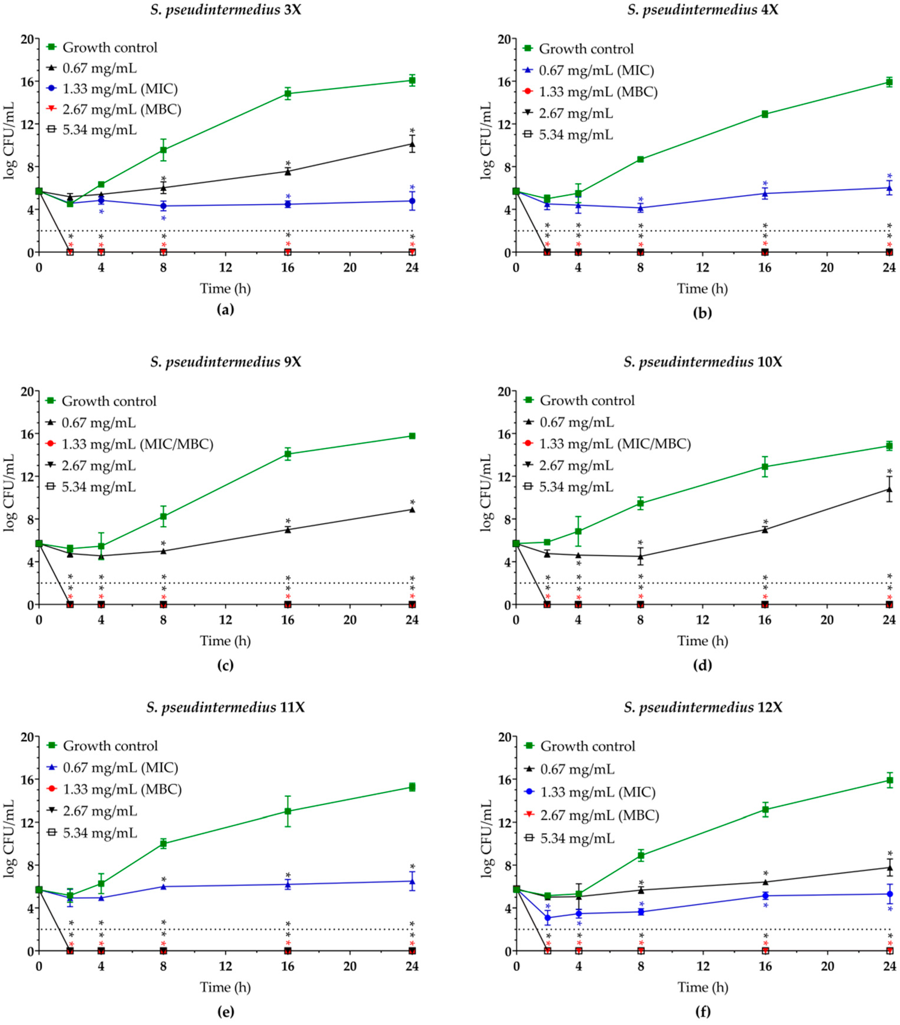
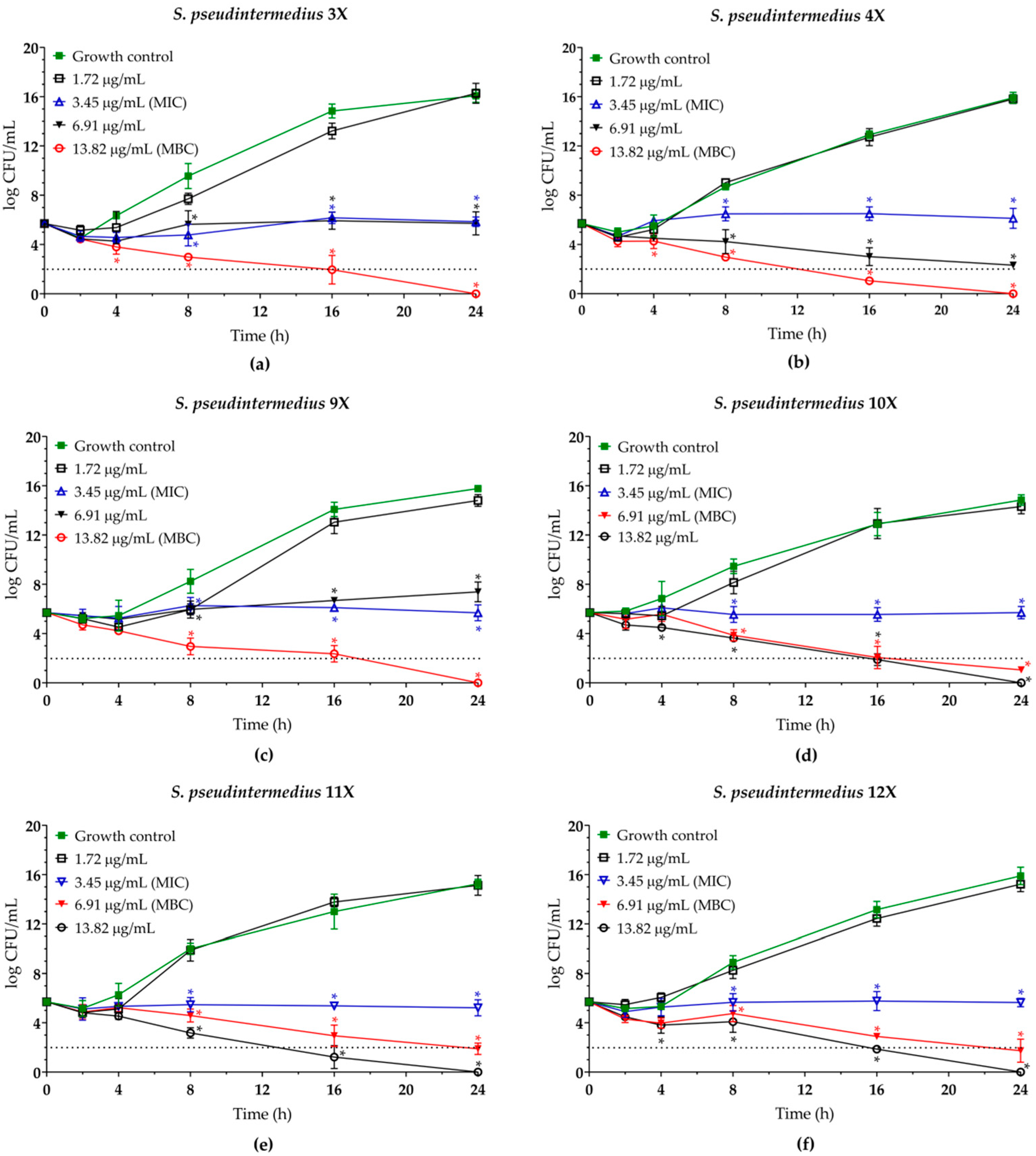
To determine the minimum bactericidal concentration (MBC) of both compounds and assess the kinetics of bacterial killing, planktonic cells of S. pseudintermedius were incubated in the presence of EUG (0.67 to 5.34 mg/mL) and bioAgNPs (1.72 to 13.82 µg/mL) at 37 °C for up to 24 h. Overall, a dose-dependent inhibitory activity of EUG (Figure 2) and bioAgNPs (Figure 3) was observed. Both compounds, at their MIC values, inhibited the growth of all bacterial isolates over time compared to untreated control cells. The MBC values for EUG were 1.33 mg/mL (four isolates) and 2.67 mg/mL (two isolates). These MBC values led to a 100% reduction in colony-forming unit (CFU) counts after 2 h of incubation for all isolates, indicating rapid bactericidal action. In contrast, the MBC values for bioAgNPs were 6.91 µg/mL (three isolates) and 13.82 µg/mL (three isolates), promoting total elimination of bacteria only after 24 h of exposure, exhibiting slower but effective bactericidal activity. The MBC/MIC ratios were 1.0 and 2.0 for EUG, and 2.0 and 4.0 for bioAgNPs, supporting the bactericidal effect [41] (Table 1).
Despite the extensive documentation on the antimicrobial activity of EUG against various microorganisms [23], there are still few studies investigating its inhibitory effect on S. pseudintermedius, as well as on the medicinal plants naturally rich in this phytochemical. The crude ethanolic extract of Piper betle L. leaves, whose main phytochemicals are EUG and hydroxychavicol, showed a bactericidal effect against MSSP and MRSP isolated from canine pyoderma. The average MIC and MBC values were 216.27 and 295.63 µg/mL for the MSSP isolates and 212.37 and 312.50 µg/mL for the MRSP isolates, respectively [17]. These findings are corroborated by the study conducted by Jantorn et al. [16], which also reported the bactericidal activity of the ethanolic extract of the same plant species against MSSP and MRSP isolated from dogs, describing MIC and MBC values of 250 µg/mL. In contrast, Silva et al. [44] reported MIC and MBC values of 2000 µg/mL for EUG against S. pseudintermedius isolated from canine otitis, which were similar to those observed in the present study. The discrepancy between these values and those observed for the crude ethanolic extract of P. betle leaves indicates possible synergistic interactions between EUG and other compounds within the crude extract.
AgNPs have also displayed antimicrobial activity against a variety of microorganisms [28,29,30,31,32,33]. However, there is still a scarcity of studies focused specifically on S. pseudintermedius. The AgNPs, obtained by both chemical [43,45,46] and biological [26,27] synthesis, showed inhibitory activity against S. pseudintermedius, although the MIC and MBC values varied between the studies. The bioAgNPs used in our study were synthesized using an aqueous extract of the bark of T. catigua A. Juss. (Meliaceae family). According to the technical report [25], these nanoparticles present a spherical morphology, with dimensions ranging from 60 to 100 nm and zeta potential of −23.3 mV, as determined by dynamic light scattering analysis. The shape and dispersion of the bioAgNPs were confirmed by transmission electron microscopy. X-ray diffraction characterization revealed a single-phase crystal pattern, evidenced by sharp, well-defined peaks at diffraction angles corresponding to the orthorhombic pattern ICSD 01-074-2076. These results are shown in Supplementary Figure S1.
Previous studies using chemically synthesized AgNPs against S. pseudintermedius reported particle sizes ranging from 4.69 ± 1.56 nm to 15.00 ± 2.70 nm. These nanoparticles exhibited MIC values between 2.00 and 16.00 µg/mL, and MBC values ranging from 2.00 to 128.00 µg/mL [43,45]. Regarding bioAgNPs, Meroni et al. [26] compared the antimicrobial activity of nanoparticles synthesized from the aqueous extract of Curcuma longa (Cl-bioAgNPs) and from the cell-free supernatant of Escherichia coli (Ec-bioAgNPs). In both synthesis methods, the bioAgNPs showed a spherical morphology, with average sizes of 11.1 ± 2.75 nm for Cl-bioAgNPs and 27.28 ± 2.68 nm for Ec-bioAgNPs. The MIC values observed against S. pseudintermedius were 143.7 ± 0 µg/mL for Cl-bioAgNPs and 3.75 ± 3.65 µg/mL for Ec-bioAgNPs. Additionally, bioAgNPs obtained from the methanolic extract of Syzygium cumini leaves also showed a spherical morphology, but with a significantly larger average size (274.6 nm), and MIC and MBC values of 250 and 1000 µg/mL, respectively [27]. These results highlight how the synthesis method, reducing agent, and nanoparticle size affect antimicrobial efficacy against S. pseudintermedius.
Although the mode of action of EUG and bioAgNPs on S. pseudintermedius was not explored in depth in this study, their action on cell membrane integrity was assessed by fluorescence microscopy, using differential staining with SYTO 9™ and propidium iodide probes. The untreated planktonic cells exhibited green fluorescence, characteristic of cells with intact membranes and metabolically active status. In contrast, cells exposed to the MBC of EUG and bioAgNPs showed red fluorescence, indicating loss of membrane integrity and impaired metabolic activity (Supplementary Figure S2).
The interaction mechanisms and mode of action of EUG and AgNPs are complex and multifactorial. Due to its lipophilic nature, EUG disrupts bacterial membranes by reducing the content of unsaturated fatty acids [47], destabilizing the lipid bilayer, and leading to cytoplasmic leakage and loss of homeostasis [15,22].
Direct contact between AgNPs and target cells is essential to increase intracellular bioavailability and potentiate the toxicity of silver ions. In fact, AgNPs accumulate on the cell wall, forming pores and disrupting the cell membrane. This compromises cell integrity, leading to cell death. However, evidence from the literature indicates that the antibacterial activity of AgNPs is influenced by various physicochemical characteristics, such as size, surface charge and concentration [48]. In addition, Gram-positive bacteria tend to be more resistant to AgNPs than Gram-negative bacteria [49]. A recent study integrated data from metalloproteomics, systematic biochemical analysis, and structural biology, identifying 38 proteins with affinity for silver ions in Staphylococcus aureus. Most of these proteins are involved in the oxidative stress response, followed by processes related to energy metabolism. Therefore, silver ions can act primarily on the glycolytic pathway, inhibiting essential enzymes, as well as promoting the accumulation of reactive oxygen species (ROS) through dysfunction of the redox homeostasis system in the final stages of exposure to AgNPs [50]. At high concentrations, ROS can cause DNA and cell membrane damage and induce lipid peroxidation and protein oxidation, culminating in cell death [48].

2.3. Eugenol Displays Synergistic Antibacterial Interaction with Biogenic Silver Nanoparticles Against Planktonic Cells of MDR Staphylococcus pseudintermedius

EUG is widely used in pharmaceutical, cosmetic, food, and hygiene products due to its safety at low concentrations. Actually, EUG is recognized as a GRAS (Generally Recognized as Safe) substance for use as a flavoring agent in food [51]. According to Joint FAO/WHO Expert Committee on Food Additives [52], the maximum permitted daily intake is 2.5 mg/kg of body weight. Despite its proven antimicrobial action, the isolated use of EUG may require relatively high concentrations (≥1600 µg/mL) to achieve efficacy against resistant bacterial strains [23], which may limit its safe application in certain contexts. Therefore, in addition to the search for new molecules with antimicrobial activity, the combined use of drugs has become an effective strategy to optimize therapeutic outcomes and delay the emergence of resistance [53]. The combination of two antimicrobial agents may produce a synergistic effect, thereby enhancing therapeutic efficacy and permitting the use of lower doses of each compound. This strategy can minimize the risk of treatment-associated toxicity [53]. In fact, the combined therapy has consolidated applications in human and veterinary medicine. For example, the combination of the β-lactam antibiotic amoxicillin with the β-lactamase inhibitor clavulanic acid is commonly recommended for the treatment of canine pyoderma [54].
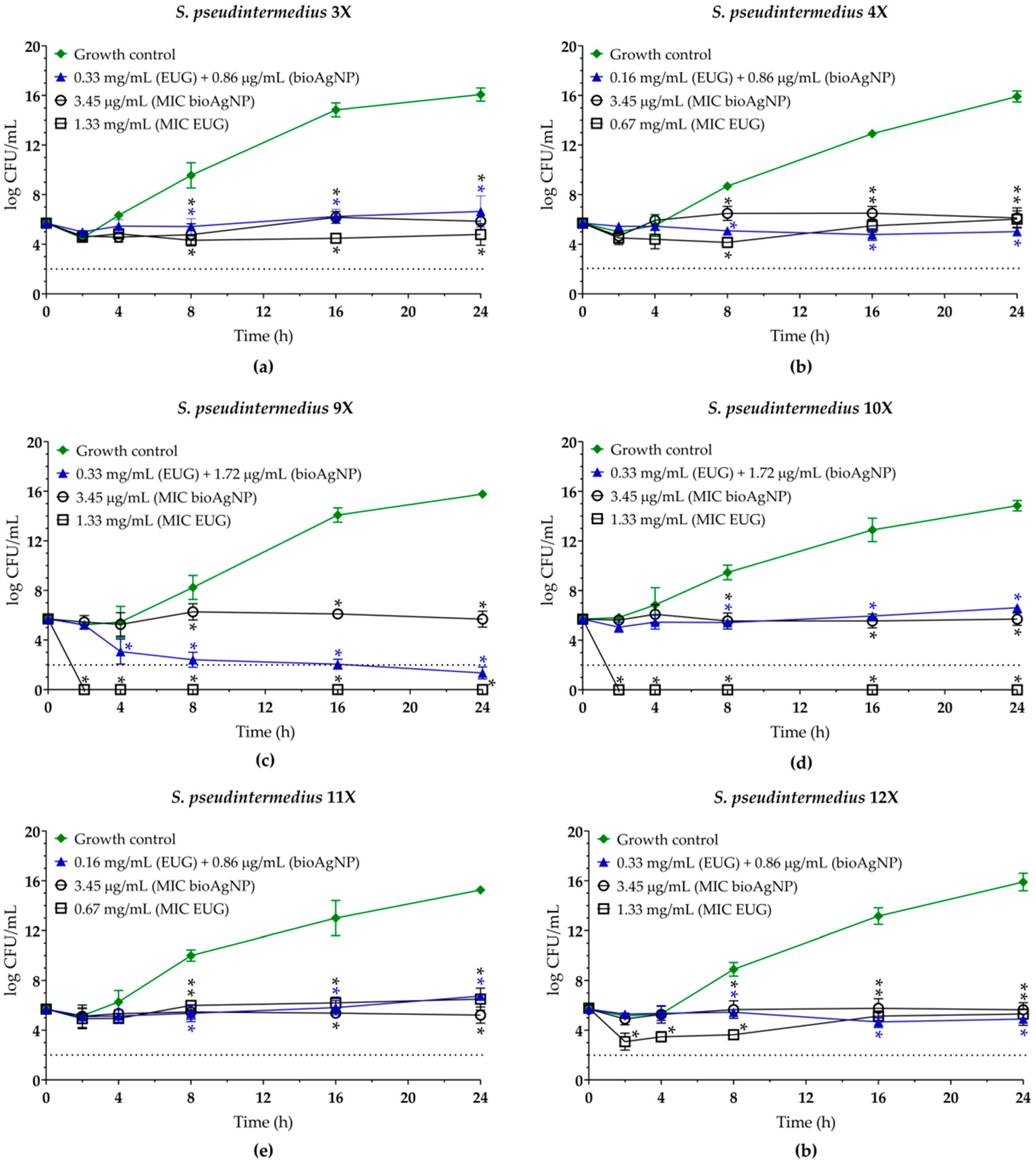
In this study, the antibacterial effect of the EUG/bioAgNPs combination against MDR S. pseudintermedius was evaluated using the checkerboard assay [30], and the results are presented in Table 2. The combination of both compounds resulted in a four-fold reduction in the MIC of EUG, and a two- to four-fold reduction for bioAgNPs. The calculated fractional inhibitory concentration indexes (FICI) values were 0.49, 0.50, and 0.79 across the isolates tested, indicating an indifferent antibacterial effect for two isolates and a synergistic effect for the other four S. pseudintermedius [55]. Simultaneous administration of the two compounds at synergistic or indifferent concentrations inhibited bacterial growth over time, showing a sustained bacteriostatic effect similar to the growth kinetics observed with each compound alone at MIC (Figure 4).
Even though the classification of antibacterial agents as bacteriostatic or bactericidal provides useful information about their in vitro activity, this distinction alone is insufficient to predict their clinical efficacy. For instance, specific bacteriostatic antibacterials, such as linezolid, remain valuable options, particularly in cases of Gram-positive bacteremia [56]. For a more robust assessment of the therapeutic response in vivo, further studies are necessary to integrate these data with pharmacokinetic and pharmacodynamic parameters. These parameters, respectively, reflect the concentrations of the compounds in the organism and their interactions with the infectious agent over time [57].
Although the mechanism underlying the synergistic antibacterial effect of the combination of EUG and bioAgNPs was not the primary focus of this study, some findings may contribute to understanding this interaction: (i) The antimicrobial activity of EUG is widely attributed to the presence of a free hydroxyl group in its chemical structure. This functional group increases the polarity of the molecule, enhancing its interaction with the bacterial plasma membrane bilayer and consequently increasing membrane permeability [22,23]. (ii) The hydroxyl group also contributes to the intracellular generation of reactive oxygen species (ROS), which can lead to oxidative damage within bacterial cells [58]. (iii) BioAgNPs are similarly known to compromise the integrity of bacterial plasma membranes [28,32,33], although their effectiveness may be reduced against Gram-positive bacteria, which typically exhibit greater resistance to this type of nanoparticle [49]. (iv) BioAgNPs have also been shown to promote ROS generation, further contributing to bacterial cell stress and damage through oxidative mechanisms [50,59]. Based on these observations, we hypothesize that EUG enhances membrane permeability to bioAgNPs, thereby increasing the intracellular availability of both agents. This interaction may amplify ROS production, resulting in cumulative damage to bacterial DNA, proteins, and lipids. Nonetheless, further studies are required to elucidate the molecular mechanisms governing this synergistic effect.

2.4. Eugenol, Alone or Combined with Biogenic Silver Nanoparticles, Inhibits the 24 h Biofilms of MDR Staphylococcus pseudintermedius on Abiotic Surfaces

To evaluate the antibiofilm activity of the compounds alone or in combination, the ability of all S. pseudintermedius to form biofilms on the polystyrene surface was initially analyzed. The biomass of the 24 h biofilms was evaluated after staining with crystal violet. The mean optical density at 570 nm (OD570nm) ± standard deviation was 0.296 ± 0.108, ranging from 0.103 ± 0.029 to 0.570 ± 0.036. According to the criteria established by Stepanovic et al. [60], all isolates were classified as strong biofilm formers under the conditions analyzed (Supplementary Figure S3).
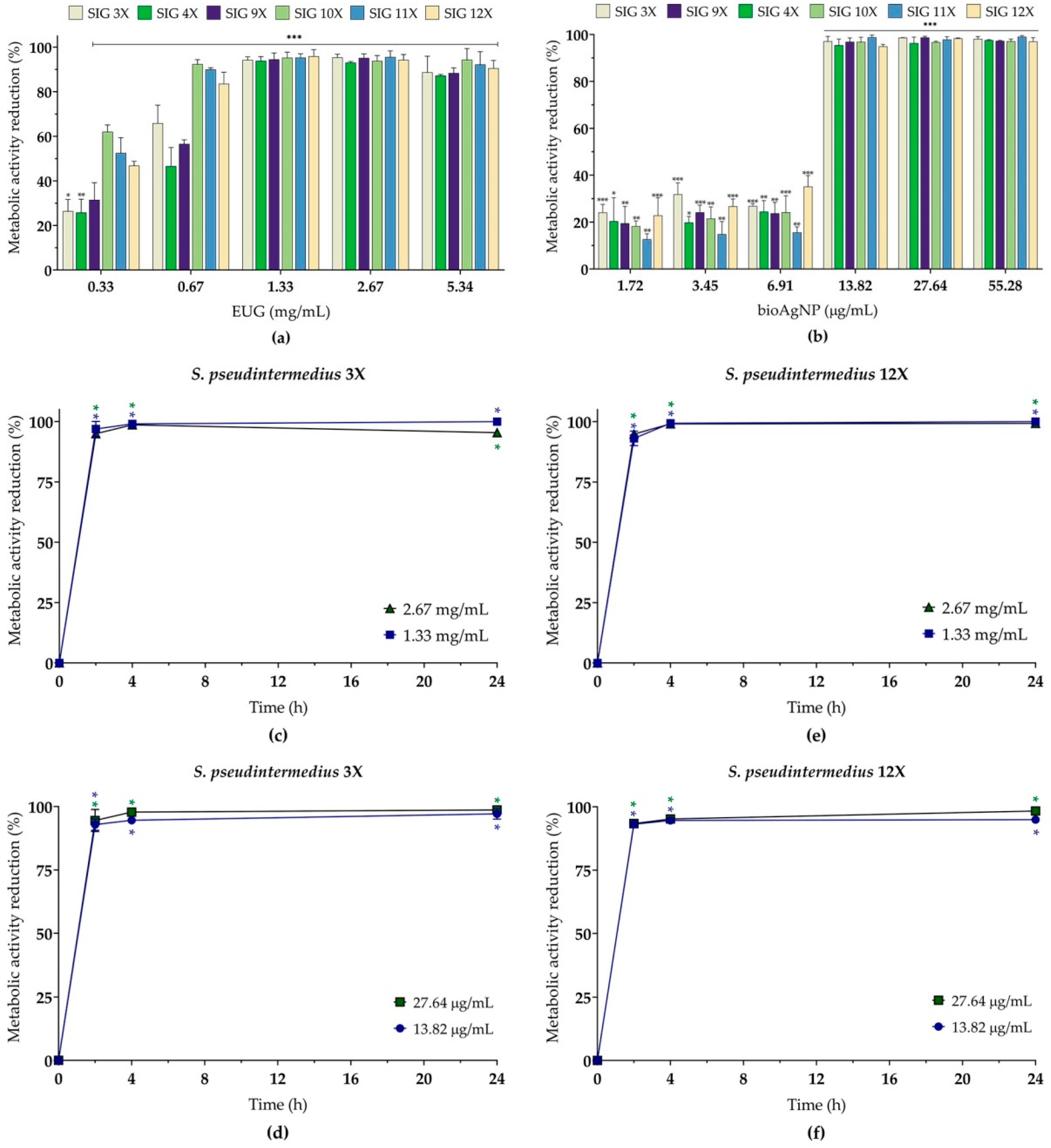
Subsequently, the inhibitory activity of EUG (0.33 to 5.34 mg/mL) and bioAgNPs (1.72 to 55.28 µg/mL) was evaluated individually on pre-formed (24 h) biofilms (sessile cells), with the results presented in Figure 5a (EUG) and Figure 5b (bioAgNPs). All tested concentrations of both compounds significantly reduced the metabolic activity of sessile cells, with inhibition percentages ranging from 13% to 99%, depending on the isolate and concentration tested. The MIC of EUG capable of inhibiting 90% of the metabolic activity of sessile cells (SMIC90) were 0.67 mg/mL (two isolates) and 1.33 mg/mL (four isolates), which were similar to those MICs for planktonic cells. As for bioAgNPs, the SMIC90 was 13.82 µg/mL for all isolates (Table 3).
To evaluate the killing kinetics of sessile cells in the presence of both compounds alone, 24 h biofilms of the S. pseudintermedius SIG 3X (MRSP, Figure 5c,d) and SIG 12X (MSSP, Figure 5e,f) isolates were treated with EUG (1.33 and 2.67 mg/mL) and bioAgNPs (13.82 and 27.64 µg/mL). All concentrations of EUG and bioAgNPs led to a reduction of more than 90% in the metabolic activity of the biofilms in both isolates after 2 h of incubation.
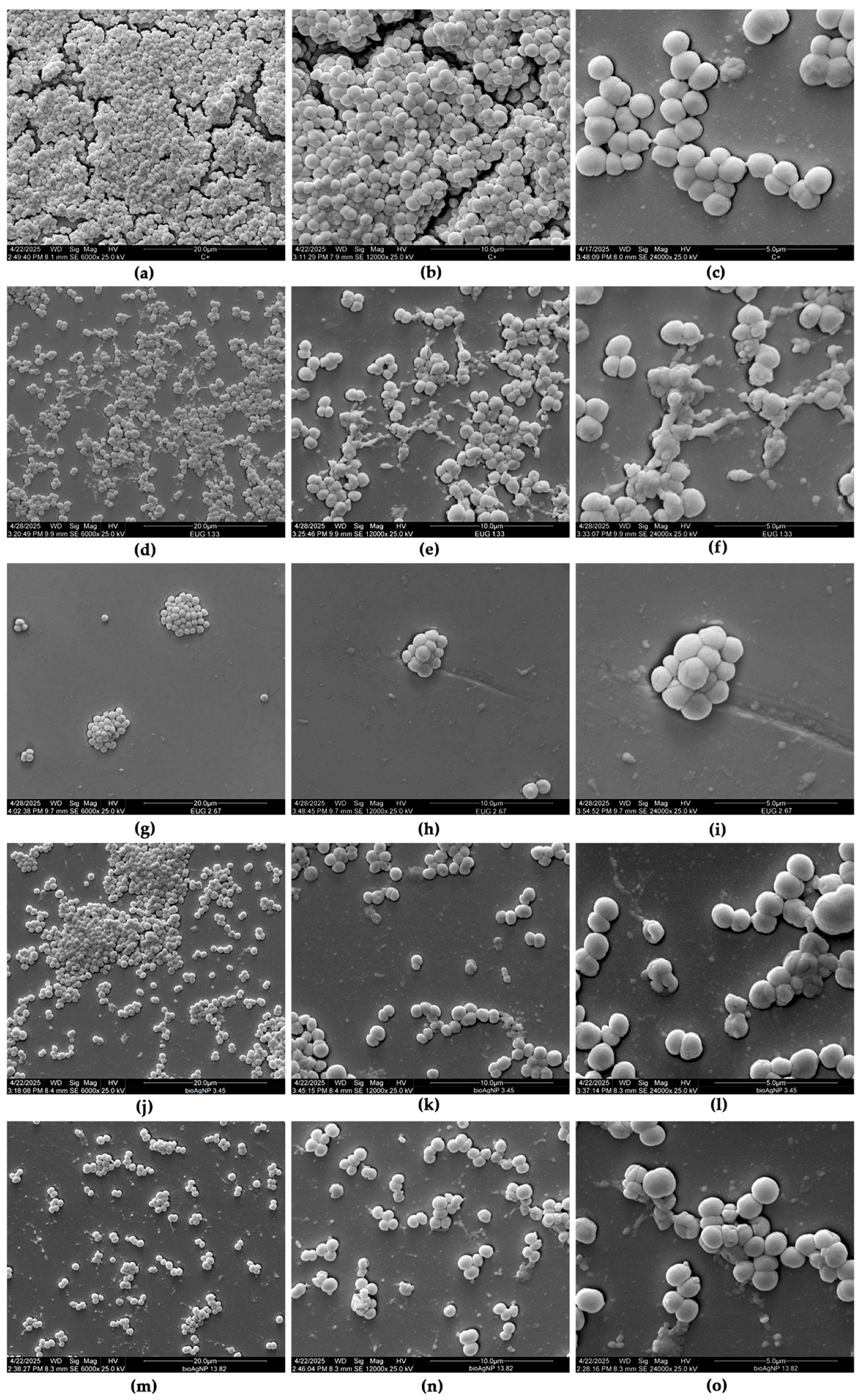
The effect of the compounds on the morphology and ultrastructure of S. pseudintermedius SIG 3X biofilms was analyzed by scanning electron microscopy (SEM). The untreated control biofilm displayed a typical three-dimensional architecture after 24 h of formation on the glass surface characterized by a dense structure, composed of spherical cells organized in multiple layers (Figure 6a–c). Treatment with 1.33 mg/mL (Figure 6d–f) and 2.67 mg/mL (Figure 6g–i) of EUG led to a significant dose-dependent reduction in the biomass of sessile cells. In addition to the reduction in cell density, morphological changes were observed, such as cellular disaggregation, wilted cells, and the presence of amorphous material, suggesting cellular lysis (Figure 6d–i). At the highest concentration tested (2.67 mg/mL), EUG promoted an almost complete reduction in biofilm biomass. Similarly, treatment with bioAgNPs also caused a substantial reduction in biofilm biomass (Figure 6j–o), along with the observation of cells with a distorted and collapsed morphology (Figure 6l,o).
To date, there are no reports in the literature that directly evaluate the inhibitory activity of EUG on S. pseudintermedius biofilms. However, the study conducted by Jantorn et al. [16] demonstrated that the ethanolic extract of P. betle L. leaves—of which eugenol is one of the main constituents—showed significant antibiofilm activity against this species. The extract was able to inhibit biofilm formation and promote the eradication of biofilms previously established on abiotic surfaces. In addition, molecular docking analyses indicated that eugenol has a high binding affinity for the enzyme N-acetylamino glucosamine transferase, an enzyme encoded by the icaA gene. This enzyme is involved in the synthesis of poly-N-acetylglucosamine (PNAG), the main component of the extracellular matrix of biofilms formed by Staphylococcus spp. [61]. Similar results were previously described by Yadav et al. [62], who reported that EUG inhibited biofilm formation and eradicated 24 h biofilms of methicillin-sensitive and methicillin-resistant S. aureus strains, at concentrations corresponding to the MIC (0.04%) and 2 × MIC (0.08%) for planktonic cells. Consistent with our findings, the biofilm cells were eliminated in the early stages of exposure (after 3 to 6 h) to EUG. Moreover, previous studies have shown that EUG [18] and bioAgNPs [31] inhibit the expression of key genes of quorum sensing systems in bacteria, which are fundamental to various cellular processes, including biofilm formation.
The antibiofilm activity of bioAgNPs has been widely reported against biofilms formed by different bacterial species [22,28,30,31,32,33,63]. However, to date, only the study by Seo et al. [46] has investigated the inhibitory activity of chemically synthesized AgNPs on S. pseudintermedius biofilms. In that study, the authors evaluated the effect of AgNPs on 10 clinical isolates obtained from dogs with otitis externa. The results indicated that the spherical nanoparticles, with an average size of 10 nm, significantly inhibited biofilm formation in a dose-dependent manner, at concentrations of 10 and 20 µg/mL. In addition, SEM images confirmed the reduction in biofilm biomass formed on the glass surface, as well as showing the disruption of the extracellular matrix after treatment with AgNPs.
In the present study, the combined effect of EUG and bioAgNPs was also evaluated on pre-formed (24 h) biofilms of S. pseudintermedius. The concurrent addition of EUG and bioAgNPs resulted in a synergistic antibiofilm interaction for most isolates, with FICI values of 0.49 or 0.50. With the exception of SIG 10X, for which the SMIC90 of EUG was reduced two-fold, all combinations resulted in a four-fold reduction in the SMIC90 of both compounds. This is an important finding of this study, as there are no records in the literature describing the synergistic antibiofilm action between EUG and bioAgNPs. The identification of this synergistic interaction offers new possibilities for the development of more effective antimicrobial strategies against infections associated with biofilms.

2.5. Eugenol and Biogenic Silver Nanoparticles Exhibit Reduced Toxicity Toward LLC-MK2 Cells at Concentrations Corresponding to Synergistic or Indifferent Combinations Against Planktonic and Sessile Cells of MDR Staphylococcus pseudintermedius

Evaluating the toxicity of a drug candidate is a fundamental stage in the development of new pharmaceutical formulations. Evidence from the literature, obtained through in silico studies (ADMETox—Absorption, Distribution, Metabolism, Excretion, and Toxicity), indicates that EUG has good oral bioavailability, the ability to cross the blood–brain barrier, and properties compatible with the drug-likeness criteria [64]. In addition, EUG shows good skin permeation and can act as a permeation enhancer [65]. As mentioned before, EUG is recognized as a GRAS substance for use as a flavoring agent in food, as long as the established conditions of use are respected [51]. However, at high doses, it can have adverse effects, including liver toxicity, anesthetic action, and the potential for skin sensitization [65].
Furthermore, one of the main limitations associated with the use of silver nanoparticles (AgNPs) in human and animal health applications is their toxicity. In fact, AgNPs have the ability to translocate and accumulate in various tissues of the body, such as the lungs, heart, liver, kidneys, and nervous tissue, where they can interfere with cell metabolism and induce toxic effects [66]. In view of these, the toxicity of bioAgNPs requires careful evaluation in formulations that combine such nanomaterials with bioactive compounds such as EUG.
In the present study, LLC-MK2 cells (Macaca mulatta kidney epithelial cells) were used as an initial model to evaluate the toxicity of EUG and bioAgNPs to mammalian cells. Therefore, the effect of the compounds on the viability of these cells was assessed using the MTT reduction assay. After 24 h of incubation, it was observed that only around 5.0% of the cells remained metabolically active in the presence of concentrations corresponding to the MIC, MBC, and SMIC90 of EUG (Figure 7a). For bioAgNPs, approximately 63.2%, 53.3%, and 5.3% of viable cells were observed at concentrations 3.45 (MIC), 6.91 (MBC), and 13.82 (MBC/SMIC90) µg/mL, respectively (Figure 7b). On the other hand, most of the cells remained metabolically active after 24 h of treatment with 0.16 mg/mL (90.4%) or 0.33 mg/mL (88.2%) of EUG (Figure 7a), which correspond to the concentrations in the synergistic or indifferent combinations for planktonic and sessile cells. Similarly, at concentrations of 0.86 µg/mL and 1.72 µg/mL of bioAgNPs, around 85.0% and 72.7% of the cells were viable, respectively (Figure 7b).
A limitation of the present study is that the toxic concentrations of the compounds were not evaluated in canine lineage cells. In this context, Zhang et al. [67] reported a cytotoxic concentration for 50% of the cells (CC50) of EUG of 0.165 ± 0.112 mg/mL in primary canine hepatocytes, a value close to the inhibitory concentrations observed in this study, highlighting the importance of more specific investigations in canine cell models. Despite this, evidence indicates that EUG may exert protective effects against toxicity induced by AgNPs. The intraperitoneal administration of these nanoparticles at a dose of 2 mg/kg for 30 days in Rattus norvegicus caused intense oxidative stress in kidney and liver tissues, resulting in significant histopathological changes in these organs. However, concomitant oral administration of EUG at a dose of 100 mg/kg body weight was effective in mitigating most of the toxic effects caused by AgNPs. Furthermore, EUG, when administered alone at the same concentration, did not induce changes in oxidative stress markers, biochemical parameters of kidney and liver function, as well as the morphological and immunohistochemical features of both organs [68,69]. Conversely, a randomized clinical study involving 207 dogs naturally infected with the distemper virus evaluated the efficacy and safety of treatment with 3% AgNPs, administered orally and nasally. No significant adverse reactions were observed in the groups treated with AgNPs. In addition, the use of the nanoparticles resulted in a significant increase in the survival rate, both in dogs with neurological manifestations and in those with non-neurological symptoms of the disease, compared to the animals that received only clinical supportive treatment [70].
Further studies are needed to confirm the safety and therapeutic potential of combining EUG with bioAgNPs in the treatment of infections caused by MDR S. pseudintermedius in dogs.

3. Materials and Methods

3.1. Chemicals and Culture Media

Eugenol (EUG, 4-Allyl-2-methoxyphenol, ≥98%) purity was acquired from Ferquima, Vargem Grande Paulista, Brazil); 3-(4,5-dimethylthiazol-2-yl)-2,5-diphenyltetrazolium bromide (MTT), dimethyl sulfoxide (DMSO), Dulbecco’s Modified Eagle medium (DMEM), glutaraldehyde, hexamethyldisilazane, L-glutamine, menadione, sodium cacodylate, penicillin, streptomycin, and tylosin were acquired from Sigma-Aldrich/Merck (São Paulo, Brazil). LIVE/DEAD® BacLight™ kit, Oxoid™ disks of azithromycin (AZM), ciprofloxacin (CIP), clindamycin (DA), chloramphenicol (CHL), erythromycin (ERY), gentamicin (CN), levofloxacin (LEV), oxacillin (OX), penicillin (P), and tetracycline (TE) were acquired from Thermo Fisher Scientific (São Paulo, Brazil). Fetal bovine serum was acquired from Nova Biotecnologia (São Paulo, Brazil). Cation-Adjusted Muller Hinton Broth (CaMHB) and BD Mannitol Salt Agar (MSA) were acquired from BD BBL™ (São Paulo, Brazil). Mueller Hinton agar (MHA), Tryptone Soya Broth (TSB), and Tryptone Soya Agar (TSA) were acquired from HiMedia (Mumbai, India).
Biogenic silver nanoparticles (bioAgNP) were obtained after AgNO3 reduction by the aqueous extract of T. catigua A. Juss. bark, a process that was patented (BR 102021016375-5) [25] and were acquired from GRAL Bioativos® LDTA (Nano VerdeAg®, Londrina, Brazil). Briefly, air-dried stem barks (200 g) of T. catigua were powdered and mixed with 1000 mL of distilled water. The extract was obtained by turbo-extraction (Ultra-turrax® model UTC115KT, IKA, Campinas, Brazil), and the crude extract was filtered and stored at −20 °C in the dark. After, 1 mL of the plant extract was added into 10 mM AgNO3 solution (1000 mL) and incubated at 30 °C for 72 h in the dark. Morphology and size of bioAgNPs were determined by JEOL JEM 1400 transmission electron microscopy (JEOL, Tokyo, Japan) and dynamic light scattering using the Litesizer DLS 500 (Anton Paar, Graz, Austria). The structural analyses were performed by XRD on a PANalytical X’Pert PRO MPD diffractometer (Malvern Panalytical, Worcestershire, UK) [71]. The bioAgNPs were kept in the dark 4 °C to prevent aggregation and were stable for several months. The characteristics of the bioAgNPs are shown in Supplementary Figure S1.
For all antibacterial assays, EUG was dissolved in 8.0% DMSO to obtain a 42.72 mg/mL stock solution. A stock solution of 1.08 mg/mL bioAgNPs was prepared in ultrapure sterilized water. The stock solutions were maintained at −20 °C and were further diluted in the culture medium to obtain the concentrations used in each assay. DMSO did not exceed 1.0% in all assays. Medium plus 1.0% DMSO, and medium plus 1.0% DMSO plus bacterial cells were used as sterility and growth controls, respectively, in all assays.

3.2. Bacteria and Culture Conditions

S. pseudintermedius (n = 6) isolated from dogs diagnosed with keratoconjunctivitis sicca at the Veterinary Hospital of the Universidade do Oeste Paulista (UNOESTE, Presidente Prudente, Brazil) were included in this study. The study protocol was approved by the Ethics Committee for the use of animals of UNOESTE and was registered with the Research, Development, and Extension Coordination Office under number 3050/2016. The bacteria were stored in TSB containing 20% glycerol at −80 °C and kept in the bacterial collection of the Laboratório de Biologia Molecular de Microrganismos of the Universidade Estadual de Londrina, Londrina, Brazil. Bacterial isolates were inoculated onto MSA and incubated at 37 °C for 24 h. Suggestive colonies of staphylococci were subjected to phenotypic identification based on standard tests, including Gram staining, tube coagulase, catalase, urease, and carbohydrate fermentation assays [35]. The bacterial species was confirmed by multiplex PCR targeting the nuc gene (encoding thermonuclease) as described by Sasaki et al. [36]. The bacterial isolates were cultured in TSA at 37 °C for 24 h. The standard bacterial suspensions were prepared by transferring three to five colonies of each isolate into 0.15 M NaCl solution (saline) until achieving turbidity equivalent to a 0.5 McFarland standard (1.0–2.0 × 10⁸ CFU/mL) using the DensiCHECK™ PLUS colorimeter (bioMérieux, Rio de Janeiro, Brazil). The standard bacterial suspensions were further diluted in culture medium to achieve the inoculum used in each assay.

3.3. Antibacterial Activity Against Planktonic Cells

3.3.1. Disk Diffusion

The antibacterial susceptibility profiles of the isolates were performed and interpreted using the disk diffusion assay as recommended by the Clinical and Laboratory Standards Institute guidelines [39]. Staphylococcus aureus ATCC 25923 was used as quality control. Oxacillin disk and/or the presence of mecA gene, detected as described by Milheiriço et al. [37], were used to define methicillin resistance [39].

3.3.2. Minimum Inhibitory Concentration (MIC)

The MICs of EUG and bioAgNPs were determined by broth microdilution assay in U-bottom 96-well microtiter plates (Techno Plastic Products, Trasadingen, Switzerland) as recommended by the Clinical and Laboratory Standards Institute [39]. The stock solutions of EUG and bioAgNPs were two-fold serially diluted in CaMHB to achieve concentrations ranging from 0.02 to 10.68 mg/mL and 0.22 to 1024 μg/mL in the assay, respectively. MIC was defined as the lowest concentration capable of inhibiting visual bacterial growth after 24 h of incubation at 37 °C compared with growth control. The MBC for both compounds were determined by the time–kill assay, as described in Section 3.3.4. The inhibitory effect was interpreted according to the MBC and MIC ratio as follows: bactericidal MBC/MIC = 1–4; bacteriostatic MBC/MIC > 4 [41].

3.3.3. Checkerboard Microdilution Assay

The antibacterial activity of EUG combined with bioAgNPs against planktonic cells was evaluated using the checkerboard broth microdilution assay, as described in Otaguiri et al. [30], with minor modifications. Two-fold serial dilutions of EUG (0.02–10.68 mg/mL) and bioAgNPs (0.22–1024 µg/mL) in CaMHB were, respectively, added across the rows and columns of the U-bottom 96-well microtiter plates. Around 1.0 × 10⁵ CFU/mL of bacterial cells were inoculated, and the plates were incubated at 37 °C for 24 h. The ratio between the MIC values of the compounds tested in combination and tested individually was used to calculate the fractional inhibitory concentration (FIC) of each compound. The sum of FICEUG and FICbioAgNPs was utilized to calculate the FIC index (FICI), whose values were interpreted as follows: synergistic FICI ≤ 0.5, no interaction 0.5 < FICI ≤ 4.0, or antagonistic FICI > 4.0 [55].

3.3.4. Time–Kill Assay

The MBC and the rate of bacterial killing in the presence of EUG (0.67–5.34 mg/mL) and bioAgNPs (1.72–13.82 µg/mL) alone or in combination were evaluated by the time–kill assay [40]. Planktonic cells (5.0 × 10⁵ CFU/mL) were added to 2 mL of CaMHB containing EUG alone or bioAgNPs alone or EUG/bioAgNPs combination values. Bacterial growth in absence of the compounds was used as control. The cultures were incubated at 37 °C, and at specified time points (0, 2, 4, 8, 16, and 24 h), 20 µL was removed, ten-fold serially diluted, and each dilution was inoculated onto MHA. After incubation at 37 °C for 24 h, the CFU counts were carried out. Averaged data were plotted as log10 CFU/mL versus time (h). The bactericidal effect of the compounds was defined as a 99.9% (3 log10) reduction in CFU/mL of the starting inoculum [72].

3.3.5. Cell Membrane Integrity

The effect of EUG alone or bioAgNPs alone on bacterial membrane integrity was evaluated using the LIVE/DEAD® BacLight™ kit according to the manufacturer’s recommendations. Planktonic cells (1.5 × 10⁷ CFU/mL) were inoculated into 2.0 mL CaMHB containing EUG or bioAgNPs alone and the cultures were incubated at 37 °C for 2 h. Untreated and treated bacteria were incubated with propidium iodide (30 μM) and SYTO™ 9 (6 μM) at room temperature, for 15 min, and observed under a fluorescence microscope (OLYMPUS BX53, Tokyo, Japan) using a fluorescein filter with excitation/emission wavelengths of 480/530 nm, respectively. SYTO™ 9, a green fluorescent nucleic acid dye, stains live and dead bacteria, while propidium iodide, a red fluorescent nucleic acid dye, selectively stains bacteria with permeable (damaged) membranes.

3.4. Antibacterial Effect on Sessile (Biofilms) Cells

3.4.1. Biofilm Production

Biofilm production capacity of all S. pseudintermedius isolates was evaluated in flat-bottomed 96-well polystyrene microtiter plates (Techno Plastic Products, Trasadingen, Switzerland) as described by Stepanovic et al. [60] with minor modifications. Briefly, 1.0 × 10⁷ CFU/mL (20 µL) was placed in each well containing 180 µL of TSB supplemented with 1% glucose, and the plates were incubated statically at 37 °C for 24 h. Afterward, non-adherent cells were removed by washing thoroughly two times with saline. Biofilm biomass was fixed with methanol (200 µL) for 15 min, dried at room temperature, and stained with 2% (v/v) crystal violet for 5 min. The biofilm-adhered stain was removed by the addition of acetic acid (160 µL) and transferred to another plate. The optical density (OD) was measured at 570 nm using a BioTek Synergy™ HT microtiter plate reader (Agilent, Santa Clara, CA, USA). The mean OD values of the negative controls plus 3 × standard deviations of the negative controls were used to define the cut-off OD (ODc). The isolates were classified as follows: ODtest ≤ ODc, non-producer; ODc < ODtest ≤ 2 × ODc, weak producer; 2 × ODc < ODtest ≤ 4 × ODc, moderate producer; 4 × ODc < ODtest, strong producer [60].

3.4.2. Antibiofilm Activity

To determine the antibiofilm activity of the compounds, bacterial biofilms were formed as above. After incubation, the biofilms were washed twice with saline, and fresh medium (100 µL) containing different concentrations of EUG (0.33–5.34 mg/mL) or bioAgNPs (1.72–55.28 µg/mL) was added, and the biofilms were incubated at 37 °C for further 24 h. The biofilms were washed once with saline, and the biofilm biomass was determined as above.
As previous study showed that bioAgNPs do not interfere with the spectrophotometric analysis of the MTT reduction assay [32], this approach was utilized to analyze the effect of compounds on the metabolic activity of sessile cells. Therefore, after incubation, biofilms were washed with saline, and 100 µL of MTT (0.5 mg/mL)/menadione (0.5%) solution was added to each well, and the plates were incubated in the dark at 37 °C for 2 h. Formazan crystals were solubilized with a solution (100 µL) containing 10% Triton X-100, 0.1 N HCl in isopropanol, and then the OD was determined at 550 nm in a BioTek Synergy™ HT microtiter plate reader (Agilent, Santa Clara, USA). The MIC was determined as the lowest concentration capable of inhibiting 90% (SMIC90) of the metabolic activity of sessile cells compared to the untreated controls.
To evaluate the effect of the combination of EUG and bioAgNPs on the 24 h biofilm, it was formed as described above. After removing the non-adherent cells, two-fold serial dilutions of EUG (0.02–10.68 mg/mL) and bioAgNPs (0.22–1024 µg/mL) were added in the rows and in the columns, respectively, of the 96-well microtiter plates. The SMIC90 of the combination was determined by evaluating the metabolic activity of the sessile cells using the MTT reduction assay, as previously described. The FICI was calculated and the values interpreted according to the criteria established for planktonic cells.
Additionally, the viability of sessile cells of S. pseudintermedius SIG 3X and SIG 12X was determined by the CFU counts. Thus, untreated and treated biofilms were removed by scraping with a sterile scalpel. Sessile cells were resuspended in saline, vigorously vortexed for 30 s. An aliquot (20 µL) of the cell suspension was removed, ten-fold serially diluted, and 100 µL of each dilution was inoculated onto MHA. After incubation at 37 °C for 24 h, the CFU counts were carried out to estimate the total number of viable cells.

3.4.3. Scanning Electron Microscopy (SEM)

The effect of compounds on morphology of biofilms was analyzed by SEM. Glass coverslip was immersed in wells of 24-well cell culture plates containing 1 mL TSB plus 1% glucose inoculated with S. pseudintermedius SIG 3X (1 × 106 CFU), and the system was incubated at 37 °C for 24 h. The biofilms were washed twice with PBS and treated with EUG alone or bioAgNPs alone for 24 h at 37 °C. The biofilms were fixed with 2.5% (v/v) glutaraldehyde in 0.1 M sodium cacodylate buffer, pH 7.2 at room temperature for 4 h, dehydrated with serial ethanol washes (30%, 50%, 70%, 80%, 90%, and 100%), critical point dried using hexamethyldisilazane (HMDS) (BAL-TEC, CPD 030), coated with gold, and observed in a FEI Quanta 250 scanning electron microscope (Themo Fisher Scientific, Hillsboro, TX, USA).

3.5. Effect of Eugenol and Biogenic Silver Nanoparticles on Mammalian Cells

The cytotoxicity of EUG and bioAgNPs was evaluated on kidney epithelial cells from Macaca mulatta [LLC-MK2 cells (Merck, Brazil)]. Cells (5 × 10⁴ cells/well) were cultured in DMEM with 10% fetal bovine serum, 2 mM L-glutamine, 100 IU/mL penicillin, 100 µg/mL streptomycin, 1% tylosin in flat-bottomed 96-well microtiter plates, in 5% CO2 at 37 °C for 48 h. The medium was aspirated off and fresh medium containing EUG (0.08–5.34 mg/mL) or bioAgNPs (0.10–13.82 µg/mL) was added. The cells were incubated for further 24 h under the same conditions. Cell viability was analyzed by the MTT reduction assay according to the manufacturer’s recommendation. The concentration of the compound needed to inhibit the viability of 90% of the cells calculated by regression analysis corresponds to the 90% (CC90) cytotoxic concentrations.

3.6. Statistical Analyses

All the experiments were carried out in at least two biological replicates at least three different occasions, and the results are expressed as the mean ± standard deviation. The GraphPad PRISM software version 8.0 (GraphPad Software, San Diego, CA, USA) was used for statistical analyses. Time–kill kinetics and toxicity to LLC-MK2 cells were analyzed using two-way ANOVA followed by Tukey’s multiple comparisons test. The biofilm results were evaluated by one-way ANOVA. For all assays, p < 0.05 was considered significant.

4. Conclusions

This study investigated the antibacterial effect of eugenol (EUG) and biogenic silver nanoparticles (bioAgNPs) synthesized using an aqueous extract of Trichilia catigua A. Juss. bark, both alone and in combination, against planktonic and sessile cells of multidrug-resistant Staphylococcus pseudintermedius isolated from canine keratoconjunctivitis sicca. The main findings include the following: (i) EUG and bioAgNPs showed dose- and time-dependent bactericidal activity against planktonic cells, thereby interfering with cell membrane integrity; (ii) EUG and bioAgNPs showed a synergistic antibacterial effect towards planktonic cells of most isolates tested—although the nature of this interaction was classified as bacteriostatic, it resulted in a four-fold reduction in the MIC values of both compounds; (iii) EUG and bioAgNPs led to a significant dose-dependent reduction in the metabolic activity and biomass of 24 h biofilms formed on the surface of polystyrene and glass, promoting severe morphological changes in sessile cells; (iv) the EUG and bioAgNPs combination also inhibited pre-formed (24 h) biofilms; (v) EUG and bioAgNPs exhibited reduced toxicity toward LLC-MK2 cells at concentrations corresponding to synergistic or indifferent combinations.
This study has some limitations that may restrict the generalizability of the results. The small number of clinical isolates evaluated does not cover the full diversity of genetic and antimicrobial susceptibility profiles of S. pseudintermedius. Furthermore, most of the experiments were conducted under in vitro conditions, which may not accurately reflect the complexity of infectious environments in vivo. Another point to consider is that the mechanism of action of the EUG and bioAgNP combination has not yet been fully elucidated. Despite these limitations, our findings indicate that the combination between EUG and bioAgNPs constitutes a promising and safe strategy, with the potential to be used as an alternative or adjuvant therapy in the control of infections caused by multidrug-resistant strains of S. pseudintermedius. In addition, the use of green routes for the synthesis of nanoparticles contributes to the sustainability of the process by avoiding the use of toxic reagents, which aligns with the principles of green chemistry and current demands for environmentally responsible technologies.

Supplementary Materials

The following supporting information can be downloaded at: https://www.mdpi.com/article/10.3390/molecules30163353/s1, Figure S1: Characteristics of biogenic silver nanoparticles synthesized (bioAgNPs) using the aqueous extract of Trichilia catigua A. Juss. bark. Figure S2: Effect of eugenol (EUG) and biogenic silver nanoparticles (bioAgNPs) on the cell membrane integrity of multidrug-resistant Staphylococcus pseudintermedius planktonic cells; Figure S3: Biofilm biomass of multidrug-resistant Staphylococcus pseudintermedius formed on polystyrene surface after 24 h of incubation at 37 °C.

Author Contributions

Conceptualization, W.R.C.C., C.F.d.O. and S.F.Y.-O.; investigation, W.R.C.C., C.F.d.O., L.F.d.A.S. and I.M.d.C.; validation, G.B.-G., H.T.S., G.M.A. and E.R.T.; formal analysis, W.R.C.C., C.F.d.O., R.G., H.T.S., L.F.d.A.S., G.M.A., L.M.Y. and S.F.Y.-O.; resources, R.G., G.N., L.M.Y. and S.F.Y.-O.; writing—original draft preparation, W.R.C.C., I.M.d.C., H.T.S., G.B.-G., E.R.T. and S.F.Y.-O.; writing—review and editing, W.R.C.C., C.F.d.O., R.G., L.F.d.A.S., I.M.d.C., G.B.-G., H.T.S., G.M.A., G.N., E.R.T., L.M.Y. and S.F.Y.-O.; visualization, W.R.C.C., L.F.d.A.S., G.B.-G., H.T.S., G.M.A. and E.R.T.; supervision, S.F.Y.-O.; project administration, G.N., L.M.Y. and S.F.Y.-O.; funding acquisition, G.N., L.M.Y. and S.F.Y.-O. All authors have read and agreed to the published version of the manuscript.

Funding

This research was funded by Financiadora de Estudos e Projetos (FINEP)-MCTI, grant number 01.22.0200.00; Coordenação de Aperfeiçoamento de Pessoal de Nível Superior (CAPES) Financial Code 01. Conselho Nacional de Desenvolvimento Científico e Tecnológico (CNPq, Process 408547/2024-3).

Institutional Review Board Statement

The animal study protocol was approved by the Ethics Committee for the use of animals of UNOESTE and was registered with the Research, Development, and Extension Coordination Office under number 3050/2016 (data of approval 13 April 2016).

Data Availability Statement

The original contributions presented in this study are included in the article/Supplementary Material. Further inquiries can be directed to the corresponding author.

Acknowledgments

We thank the Laboratório de Microscopia Eletrônica e Microanálise (LMEM–UEL) for the support provided for the microscopy techniques; W.R.C.C. was funded by a post-graduate scholarship from Fundação Araucária-PR; L.F.d.A.S. and G.B.-G. were funded by a post-graduate scholarship from CAPES. H.T.S. was funded by a research fellowship from FINEP-MCTI. R.G., G.N., and S.F.Y.-O. were funded by a research fellowship from CNPq.

Conflicts of Interest

The authors declare no conflicts of interest. The funders had no role in the design of the study; in the collection, analyses, or interpretation of data; in the writing of the manuscript; or in the decision to publish the results.

Abbreviations

The following abbreviations are used in this manuscript:
MDRMultidrug resistant
KCSKeratoconjunctivitis sicca
MRSPMethicillin-resistant Staphylococcus pseudintermedius
EUGEugenol
AgNPsSilver nanoparticles
bioAgNPsBiogenic silver nanoparticles
PCRPolymerase chain reaction
MSSPMethicillin-sensitive Staphylococcus pseudintermedius
MICMinimum inhibitory concentration
MBCMinimum bactericidal concentration
CFUColony forming unit
ROSReactive oxygen species
FICIFractional inhibitory concentration index
ODOptical density
SMICSessile minimum inhibitory concentration
MTT3-(4,5-dimethylthiazol-2-yl)-2,5-diphenyltetrazolium bromide
DMSODimethyl sulfoxide
MSAManitol Salt Agar
TSBTryptone Soya Broth
CaMHBCation-Adjusted Müller Hinton Broth
MHAMüller Hinton Agar
SEMScanning electron microscopy
DMEMDulbecco’s Modified Eagle medium

References

  1. Nocera, F.P.; De Martino, L. Methicillin-resistant Staphylococcus pseudintermedius: epidemiological changes, antibiotic resistance, and alternative therapeutic strategies. Vet. Res. Commun. 2024, 48, 3505–3515. [Google Scholar] [CrossRef]
  2. Roberts, E.; Nuttall, T.J.; Gkekas, G.; Mellanby, R.J.; Fitzgerald, J.R.; Paterson, G.K. Not just in man’s best friend: A review of Staphylococcus pseudintermedius host range and human zoonosis. Res. Vet. Sci. 2024, 174, 105305. [Google Scholar] [CrossRef]
  3. Bannoehr, J.; Guardabassi, L. Staphylococcus pseudintermedius in the dog: taxonomy, diagnostics, ecology, epidemiology and pathogenicity. Vet. Dermatol. 2012, 23, 253–266. [Google Scholar] [CrossRef]
  4. Park, J.; Kim, D.; Kwon, M.; Kwon, H.; Yong, J.; Yoon, H.; Hwang, J.; Jung, J.S.; Park, K.M. Bacterial isolates and antibiotic sensitivity in canine bacterial keratitis in Korea. Vet. Ophthalmol. 2024, 1–12. [Google Scholar] [CrossRef]
  5. Casemiro, P.A.F.; Andrade, A.L.; Cardozo, M.V.; Rodrigues, R.A.; Silva, J.A.; Marinho, M.; Nassar, A.F.C.; Castro, V.; Braz, G.H.R.; Gujanwski, C.A.; et al. Prevalence and antibiotic resistance in bacterial isolates of dogs with ulcerative keratitis in São Paulo State, Brazil. Vet. Ophthalmol. 2025, 28, 37–47. [Google Scholar] [CrossRef]
  6. Engdahl, K.S.; Brodbelt, D.C.; Cameron, C.; Church, D.B.; O’Neill, D.G. English Cocker Spaniels under primary veterinary care in the UK: disorder predispositions and protections. Canine Med. Genet. 2024, 11, 1. [Google Scholar] [CrossRef]
  7. Lau, W.I.; Taylor, R.M. The Prevalence of Corneal Disorders in Pugs Attending Primary Care Veterinary Practices in Australia. Animals 2025, 15, 531. [Google Scholar] [CrossRef]
  8. Williams, D.L. Immunopathogenesis of keratoconjunctivitis sicca in the dog. Vet. Clin. North Am. Small Anim. Pract. 2008, 38, 251–268. [Google Scholar] [CrossRef]
  9. Dodi, P.L. Immune-mediated keratoconjunctivitis sicca in dogs: current perspectives on management. Vet. Med. 2015, 6, 341–347. [Google Scholar] [CrossRef]
  10. Mauer, A.N.; Allbaugh, R.A.; Kreuder, A.J.; Sebbag, L. Impact of multi-drug resistance on clinical outcomes of dogs with corneal ulcers infected with Staphylococcus pseudintermedius. Front. Vet. Sci. 2022, 9, 1083294. [Google Scholar] [CrossRef]
  11. Ferran, A.A.; Liu, J.; Toutain, P.L.; Bousquet-Mélou, A. Comparison of the In vitro Activity of Five Antimicrobial Drugs against Staphylococcus pseudintermedius and Staphylococcus aureus Biofilms. Front. Microbiol. 2016, 7, 1187. [Google Scholar] [CrossRef]
  12. Andrade, M.; Oliveira, K.; Morais, C.; Abrantes, P.; Pomba, C.; Rosato, A.E.; Couto, I.; Costa, S.S. Virulence Potential of Biofilm-Producing Staphylococcus pseudintermedius, Staphylococcus aureus and Staphylococcus coagulans Causing Skin Infections in Companion Animals. Antibiotics. 2022, 11, 1339. [Google Scholar] [CrossRef]
  13. Cangui-Panchi, S.P.; Ñacato-Toapanta, A.L.; Enríquez-Martínez, L.J.; Salinas-Delgado, G.A.; Reyes, J.; Garzon-Chavez, D.; Machado, A. Battle royale: Immune response on biofilms—host-pathogen interactions. Curr. Res. Immunol. 2023, 4, 100057. [Google Scholar] [CrossRef]
  14. Martins, C.H.G. Special Issue Editorial: “Antibacterial Agents from Natural Sources”. Molecules 2024, 29, 644. [Google Scholar] [CrossRef]
  15. Nogueira, J.O.E.; Campolina, G.A.; Batista, L.R.; Alves, E.; Caetano, A.R.S.; Brandão, R.M.; Nelson, D.L.; Cardoso, M.D.G. Mechanism of action of various terpenes and phenylpropanoids against Escherichia coli and Staphylococcus aureus. FEMS Microbiol. Lett. 2021, 368, fnab052. [Google Scholar] [CrossRef]
  16. Jantorn, P.; Tipmanee, V.; Wanna, W.; Prapasarakul, N.; Visutthi, M.; Sotthibandhu, D.S. Potential natural antimicrobial and antibiofilm properties of Piper betle L. against Staphylococcus pseudintermedius and methicillin-resistant strains. J. Ethnopharmacol. 2023, 317, 116820. [Google Scholar] [CrossRef]
  17. Phensri, P.; Thummasema, K.; Sukatta, U.; Morand, S.; Pruksakorn, C. In Vitro Antimicrobial Activity of Piper betle Leaf Extract and Some Topical Agents against Methicillin-Resistant and Methicillin-Susceptible Staphylococcus Strains from Canine Pyoderma. Animals 2022, 12, 3203. [Google Scholar] [CrossRef]
  18. Ribeiro, T.A.N.; Dos Santos, G.A.; Dos Santos, C.T.; Soares, D.C.F.; Saraiva, M.F.; Leal, D.H.S.; Sachs, D. Eugenol as a promising antibiofilm and anti-quorum sensing agent: A systematic review. Microb. Pathog. 2024, 196, 106937. [Google Scholar] [CrossRef]
  19. El-Saadony, M.T.; Saad, A.M.; Mohammed, D.M.; Korma, S.A.; Alshahrani, M.Y.; Ahmed, A.E.; Ibrahim, E.H.; Salem, H.M.; Alkafaas, S.S.; Saif, A.M.; et al. Medicinal plants: bioactive compounds, biological activities, combating multidrug-resistant microorganisms, and human health benefits—a comprehensive review. Front. Immunol. 2025, 16, 1491777. [Google Scholar] [CrossRef]
  20. Bardaji, D.K.R.; Silva, N.B.S.; Miranda, R.R.; Martins, C.H.G.; Savka, M.A.; Hudson, A.O. Unlocking the Potential of Brazilian Plant Terpenes to Combat Antimicrobial Resistance. ACS Bio. Med. Chem. Au. 2025, 5, 365–378. [Google Scholar] [CrossRef]
  21. Lao, Y.; Guo, J.; Fang, J.; Geng, R.; Li, M.; Qin, Y.; Wu, J.; Kang, S.G.; Huang, K.; Tong, T. Beyond flavor: the versatile roles of eugenol in health and disease. Food Funct. 2024, 15, 10567–10581. [Google Scholar] [CrossRef]
  22. Perugini Biasi-Garbin, R.; Saori Otaguiri, E.; Morey, A.T.; Fernandes da Silva, M.; Belotto Morguette, A.E.; Armando Contreras Lancheros, C.; Kian, D.; Perugini, M.R.; Nakazato, G.; Durán, N.; et al. Effect of Eugenol against Streptococcus agalactiae and Synergistic Interaction with Biologically Produced Silver Nanoparticles. Evid. Based Complement. Alternat. Med. 2015, 2015, 861497. [Google Scholar] [CrossRef]
  23. Marchese, A.; Barbieri, R.; Coppo, E.; Orhan, I.E.; Daglia, M.; Nabavi, S.F.; Izadi, M.; Abdollahi, M.; Nabavi, S.M.; Ajami, M. Antimicrobial activity of eugenol and essential oils containing eugenol: A mechanistic viewpoint. Crit. Rev. Microbiol. 2017, 43, 668–689. [Google Scholar] [CrossRef]
  24. Shahina, Z.; Dahms, T.E.S. A Comparative Review of Eugenol and Citral Anticandidal Mechanisms: Partners in Crimes Against Fungi. Molecules 2024, 29, 5536. [Google Scholar] [CrossRef]
  25. Nakazato, G.; Celidonio, A.P.S.; Kobayashi, R.K.T.; Panagio, L.A.; Lonni, A.A.G.S.; De Campos, A.C.L.P.; Goncalves, M.C.; Okino, G.A.K. Carta Patente—Processo de Produção de Nanopartículas Biogênicas de Prata, Nanopartículas Biogênicas de Prata e Usos das Nanopartículas Biogênicas de Prata—GRAL Bioativos LTDA. BR Patent Application No. BR1020210163755, 18 September 2021. Available online: http://www.inpi.gov.br (accessed on 10 February 2025).
  26. Meroni, G.; Soares Filipe, J.F.; Martino, P.A. In Vitro Antibacterial Activity of Biological-Derived Silver Nanoparticles: Preliminary Data. Vet. Sci. 2020, 7, 12. [Google Scholar] [CrossRef]
  27. Wongstitwilairoong, N.; Jermnark, U.; Paochoosak, N.; Limsivilai, O.; Chimnoi, W.; Yohannes, Y.B.; Ikenaka, Y.; Saengtienchai, A. Effect of Java plum (Syzygium cumini) leaves extract and a silver nanoparticles synthesis on pathogens in skin diseases of dogs. Open Vet. J. 2024, 14, 2662–2677. [Google Scholar] [CrossRef]
  28. Akhter, M.S.; Rahman, M.A.; Ripon, R.K.; Mubarak, M.; Akter, M.; Mahbub, S.; Al Mamun, F.; Sikder, M.T. A systematic review on green synthesis of silver nanoparticles using plants extract and their biomedical applications. Heliyon 2024, 10, e29766. [Google Scholar] [CrossRef]
  29. Zahran, M.; Marei, A.H. Innovative natural polymer metal nanocomposites and their antimicrobial activity. Int. J. Biol. Macromol. 2019, 136, 586–596. [Google Scholar] [CrossRef]
  30. Otaguiri, E.S.; Morguette, A.E.B.; Biasi-Garbin, R.P.; Morey, A.T.; Lancheros, C.A.C.; Kian, D.; de Oliveira, A.G.; Kerbauy, G.; Perugini, M.R.E.; Duran, N.; et al. Antibacterial Combination of Oleoresin from Copaifera multijuga Hayne and Biogenic Silver Nanoparticles Towards Streptococcus agalactiae. Curr. Pharm. Biotechnol. 2017, 18, 177–190. [Google Scholar] [CrossRef]
  31. Saeki, E.K.; Martins, H.M.; Camargo, L.C.; Anversa, L.; Tavares, E.R.; Yamada-Ogatta, S.F.; Lioni, L.M.Y.; Kobayashi, R.K.T.; Nakazato, G. Effect of Biogenic Silver Nanoparticles on the Quorum-Sensing System of Pseudomonas aeruginosa PAO1 and PA14. Microorganisms 2022, 10, 1755. [Google Scholar] [CrossRef]
  32. Spoladori, L.F.A.; Andriani, G.M.; Castro, I.M.; Suzukawa, H.T.; Gimenes, A.C.R.; Bartolomeu-Gonçalves, G.; Ishida, K.; Nakazato, G.; Pinge-Filho, P.; Machado, R.R.B.; et al. Synergistic Antifungal Interaction between Pseudomonas aeruginosa LV Strain Metabolites and Biogenic Silver Nanoparticles against Candida auris. Antibiotics 2023, 12, 861. [Google Scholar] [CrossRef]
  33. Castro, I.M.; Antunes, C.; Valentim, C.C.; Spoladori, L.F.A.; Suzukawa, H.T.; Correia, G.F.; Silva-Rodrigues, G.; Borges, P.H.G.; Bartolomeu-Gonçalves, G.; Silva, M.L.; et al. Synergistic Antibacterial Interaction of Geraniol and Biogenic Silver Nanoparticles on Methicillin-Resistant Staphylococcus aureus. Plants 2025, 14, 1059. [Google Scholar] [CrossRef]
  34. Samuggam, S.; Chinni, S.V.; Mutusamy, P.; Gopinath, S.C.B.; Anbu, P.; Venugopal, V.; Reddy, L.V.; Enugutti, B. Green Synthesis and Characterization of Silver Nanoparticles Using Spondias mombin Extract and Their Antimicrobial Activity against Biofilm-Producing Bacteria. Molecules 2021, 26, 2681. [Google Scholar] [CrossRef]
  35. Sasaki, T.; Kikuchi, K.; Tanaka, Y.; Takahashi, N.; Kamata, S.; Hiramatsu, K. Reclassification of phenotypically identified Staphylococcus intermedius strains. J. Clin. Microbiol. 2007, 45, 2770–2778. [Google Scholar] [CrossRef]
  36. Sasaki, T.; Tsubakishita, S.; Tanaka, Y.; Sakusabe, A.; Ohtsuka, M.; Hirotaki, S.; Kawakami, T.; Fukata, T.; Hiramatsu, K. Multiplex-PCR method for species identification of coagulase-positive staphylococci. J. Clin. Microbiol. 2010, 48, 765–769. [Google Scholar] [CrossRef]
  37. Milheiriço, C.; Oliveira, D.C.; de Lencastre, H. Update to the multiplex PCR strategy for assignment of mec element types in Staphylococcus aureus. Antimicrob. Agents Chemother. 2007, 51, 3374–3377, Erratum in Antimicrob. Agents Chemother. 2007, 51, 4537. [Google Scholar] [CrossRef]
  38. Magiorakos, A.P.; Srinivasan, A.; Carey, R.B.; Carmeli, Y.; Falagas, M.E.; Giske, C.G.; Harbarth, S.; Hindler, J.F.; Kahlmeter, G.; Olsson-Liljequist, B.; et al. Multidrug-resistant, extensively drug-resistant and pandrug-resistant bacteria: an international expert proposal for interim standard definitions for acquired resistance. Clin. Microbiol. Infect. 2012, 18, 268–281. [Google Scholar] [CrossRef]
  39. CLSI VET01S. Performance Standards for Antimicrobial Disk and Dilution Susceptibility Tests for Bacteria Isolated From Animals; CLSI Press: Wayne, PA, USA, 2024. [Google Scholar]
  40. CLSI M26. Methods for Determining Bactericidal Activity of Antimicrobial Agents; CLSI Press: Wayne, PA, USA, 1999; Volume 19, p. 18. [Google Scholar]
  41. Levison, M.E.; Levison, J.H. Pharmacokinetics and pharmacodynamics of antibacterial agents. Infect. Dis. Clin. N. Am. 2009, 20, 791–815. [Google Scholar] [CrossRef]
  42. Pereira, C.S.G.; Zulim, L.F.C.; Giuffrida, R.; Cruz, A.G.; Foglia, B.T.D.; Benguella, H.; Batista, A.S.; Andrade, S.F. Antimicrobial susceptibility and minimal inhibitory concentration of bacteria isolated from the eyes of dogs with keratoconjunctivitis sicca. Pesq. Vet. Bras. 2019, 39, 757–763. [Google Scholar] [CrossRef]
  43. Meroni, G.; Laterza, G.; Tsikopoulos, A.; Tsikopoulos, K.; Vitalini, S.; Scaglia, B.; Iriti, M.; Bonizzi, L.; Martino, P.A.; Soggiu, A. Antibacterial Potential of Essential Oils and Silver Nanoparticles against Multidrug-Resistant Staphylococcus pseudintermedius Isolates. Pathogens 2024, 13, 156. [Google Scholar] [CrossRef]
  44. Silva, R.A.; da Silva, B.F.; Pereira, M.S.; Coelho, P.A.T.; Costa, R.A.; Chaves, A.C.; Silva, I.G.N.; Carneiro, V.A. Combinatorial effects between aromatic plant compounds and chlorhexidine digluconate against canine otitis-related Staphylococcus spp. Res. Vet. Sci. 2024, 170, 105182. [Google Scholar] [CrossRef]
  45. Thammawithan, S.; Siritongsuk, P.; Nasompag, S.; Daduang, S.; Klaynongsruang, S.; Prapasarakul, N.; Patramanon, R. A Biological Study of Anisotropic Silver Nanoparticles and Their Antimicrobial Application for Topical Use. Vet. Sci. 2021, 8, 177. [Google Scholar] [CrossRef]
  46. Seo, M.; Oh, T.; Bae, S. Antibiofilm activity of silver nanoparticles against biofilm forming Staphylococcus pseudintermedius isolated from dogs with otitis externa. Vet. Med. Sci. 2021, 7, 1551–1557. [Google Scholar] [CrossRef]
  47. Di Pasqua, R.; Betts, G.; Hoskins, N.; Edwards, M.; Ercolini, D.; Mauriello, G. Membrane toxicity of antimicrobial compounds from essential oils. J. Agric. Food Chem. 2007, 55, 4863–4870. [Google Scholar] [CrossRef]
  48. González-Fernández, S.; Blanco-Agudín, N.; Rodríguez, D.; Fernández-Veja, I.; Merayo-Lloves, J.; Quirós, L.M. Silver Nanoparticles: A Versatile Tool Against Infectious and Non-Infectious Diseases. Antibiotics 2025, 14, 289. [Google Scholar] [CrossRef]
  49. Tripathi, N.; Goshisht, M.K. Recent Advances and Mechanistic Insights into Antibacterial Activity, Antibiofilm Activity, and Cytotoxicity of Silver Nanoparticles. ACS Appl. Bio Mater. 2022, 5, 1391–1463. [Google Scholar] [CrossRef]
  50. Wang, H.; Wang, M.; Xu, X.; Gao, P.; Xu, Z.; Zhang, Q.; Li, H.; Yan, A.; Kao, R.Y.; Sun, H. Multi-target mode of action of silver against Staphylococcus aureus endows it with capability to combat antibiotic resistance. Nat. Commun. 2021, 12, 3331, Erratum in Nat. Commun. 2021, 12, 4140. [Google Scholar] [CrossRef]
  51. FDA, U.S. Food and Drug Administration. Inventory of Food Contact Substances Listed in 21 CFR. Available online: https://www.hfpappexternal.fda.gov/scripts/fdcc/index.cfm?set=IndirectAdditives&id=EUGENOL (accessed on 2 June 2025).
  52. WHO. Evaluations of the Joint FAO/WHO Expert Committee on Food Additives (JECFA). 2021. Available online: https://apps.who.int/food-additives-contaminants-jecfa-database/Home/Chemical/3278. (accessed on 7 July 2025).
  53. Tyers, M.; Wright, G.D. Drug combinations: A strategy to extend the life of antibiotics in the 21st century. Nat. Rev. Microbiol. 2019, 17, 141–155. [Google Scholar] [CrossRef]
  54. Loeffler, A.; Cain, C.L.; Ferrer, L.; Nishifuji, K.; Varjonen, K.; Papich, M.G.; Guardabassi, L.; Frosini, S.M.; Barker, E.N.; Weese, S.J. Synopsis of the antimicrobial use guidelines for canine pyoderma by the International Society for Companion Animal Infectious Diseases (ISCAID). Vet. Dermatol. 2025, 36, 234–282. [Google Scholar] [CrossRef]
  55. Odds, F.C. Synergy, antagonism, and what the chequerboard puts between them. J. Antimicrob. Chemother. 2003, 52, 1. [Google Scholar] [CrossRef]
  56. Kawasuji, H.; Nagaoka, K.; Tsuji, Y.; Kimoto, K.; Takegoshi, Y.; Kaneda, M.; Murai, Y.; Karaushi, H.; Mitsutake, K.; Yamamoto, Y. Effectiveness and Safety of Linezolid Versus Vancomycin, Teicoplanin, or Daptomycin against Methicillin-Resistant Staphylococcus aureus Bacteremia: A Systematic Review and Meta-Analysis. Antibiotics 2023, 12, 697. [Google Scholar] [CrossRef]
  57. Ishak, A.; Mazonakis, N.; Spernovasilis, N.; Akinosoglou, K.; Tsioutis, C. Bactericidal versus bacteriostatic antibacterials: clinical significance, differences and synergistic potential in clinical practice. J. Antimicrob. Chemother. 2025, 80, 1–17. [Google Scholar] [CrossRef] [PubMed]
  58. Das, B.; Mandal, D.; Dash, S.K.; Chattopadhyay, S.; Tripathy, S.; Dolai, D.P.; Dey, S.K.; Roy, S. Eugenol Provokes ROS-Mediated Membrane Damage-Associated Antibacterial Activity Against Clinically Isolated Multidrug-Resistant Staphylococcus aureus Strains. Infect. Dis. 2016, 9, 11–19. [Google Scholar] [CrossRef] [PubMed]
  59. Dharshini, K.S.; Ameen, F.; Anbazhagan, V. Mechanistic Investigation on the Antibacterial Activity of Biogenic Silver Nanoparticles Prepared Using Root Extract of Sarsaparilla and Demonstrated their In Vivo Efficacy in Zebrafish Model. Curr. Microbiol. 2024, 81, 268. [Google Scholar] [CrossRef] [PubMed]
  60. Stepanović, S.; Vuković, D.; Hola, V.; Di Bonaventura, G.; Djukić, S.; Cirković, I.; Ruzicka, F. Quantification of biofilm in microtiter plates: overview of testing conditions and practical recommendations for assessment of biofilm production by staphylococci. APMIS 2007, 115, 891–899. [Google Scholar] [CrossRef]
  61. Arciola, C.R.; Baldassarri, L.; Montanaro, L. Presence of icaA and icaD genes and slime production in a collection of staphylococcal strains from catheter-associated infections. J. Clin. Microbiol. 2001, 39, 2151–2156. [Google Scholar] [CrossRef]
  62. Yadav, M.K.; Chae, S.W.; Im, G.J.; Chung, J.W.; Song, J.J. Eugenol: a phyto-compound effective against methicillin-resistant and methicillin-sensitive Staphylococcus aureus clinical strain biofilms. PLoS One 2015, 10, e0119564. [Google Scholar] [CrossRef]
  63. Santos, L.M.; Rodrigues, D.M.; Alves, B.V.B.; Kalil, M.A.; Azevedo, V.; Barh, D.; Meyer, R.; Duran, N.; Tasic, L.; Portela, R.W. Activity of biogenic silver nanoparticles in planktonic and biofilm-associated Corynebacterium pseudotuberculosis. PeerJ 2024, 12, e16751. [Google Scholar] [CrossRef]
  64. Alsirhani, A.M.; Abu-Almakarem, A.S.; Alwaili, M.A.; Aljohani, S.; Alali, I.; AlRashidi, A.A.; Abuzinadah, N.Y.; Alkhodair, S.A.; Mobasher, M.A.; Alothaim, T.; et al. Syzygium aromaticum Extract Mitigates Doxorubicin-Induced Hepatotoxicity in Male Rats. Int. J. Mol. Sci. 2024, 25, 12541. [Google Scholar] [CrossRef]
  65. Nisar, M.F.; Khadim, M.; Rafiq, M.; Chen, J.; Yang, Y.; Wan, C.C. Pharmacological Properties and Health Benefits of Eugenol: A Comprehensive Review. Oxid. Med. Cell. Longev. 2021, 2021, 2497354. [Google Scholar] [CrossRef]
  66. Ferdous, Z.; Nemmar, A. Health Impact of Silver Nanoparticles: A Review of the Biodistribution and Toxicity Following Various Routes of Exposure. Int. J. Mol. Sci. 2020, 21, 2375. [Google Scholar] [CrossRef]
  67. Zhang, L.W.; Koci, J.; Jeffery, B.; Riviere, J.E.; Monteiro-Riviere, N.A. Safety assessment of potential food ingredients in canine hepatocytes. Food Chem. Toxicol. 2015, 78, 105–115. [Google Scholar] [CrossRef]
  68. Aboelwafa, H.R.; Ramadan, R.A.; Ibraheim, S.S.; Yousef, H.N. Modulation Effects of Eugenol on Nephrotoxicity Triggered by Silver Nanoparticles in Adult Rats. Biology 2022, 11, 1719. [Google Scholar] [CrossRef] [PubMed]
  69. Yousef, H.N.; Ibraheim, S.S.; Ramadan, R.A.; Aboelwafa, H.R. The Ameliorative Role of Eugenol against Silver Nanoparticles-Induced Hepatotoxicity in Male Wistar Rats. Oxid. Med. Cell Longev. 2022, 2022, 3820848. [Google Scholar] [CrossRef] [PubMed]
  70. Gastelum-Leyva, F.; Pena-Jasso, A.; Alvarado-Vera, M.; Plascencia-López, I.; Patrón-Romero, L.; Loera-Castañeda, V.; Gándara-Mireles, J.A.; Lares-Asseff, I.; Leal-Ávila, M.Á.; Alvelais-Palacios, J.A.; et al. Evaluation of the Efficacy and Safety of Silver Nanoparticles in the Treatment of Non-Neurological and Neurological Distemper in Dogs: A Randomized Clinical Trial. Viruses 2022, 14, 2329. [Google Scholar] [CrossRef] [PubMed]
  71. Deonas, A.N.; Souza, L.M.D.S.; Andrade, G.J.S.; Germiniani-Cardozo, J.; Dahmer, D.; de Oliveira, A.G.; Nakazato, G.; Torezan, J.M.D.; Kobayashi, R.K.T. Green Synthesis of Silver Nanoparticle from Anadenanthera colubrina Extract and Its Antimicrobial Action against ESKAPEE Group Bacteria. Antibiotics 2024, 13, 777. [Google Scholar] [CrossRef]
  72. Klepser, M.E.; Ernst, E.J.; Lewis, R.E.; Ernst, M.E.; Pfaller, M.A. Influence of test conditions on antifungal time-kill curve results: proposal for standardized methods. Antimicrob. Agents Chemother. 1998, 42, 1207–1212. [Google Scholar] [CrossRef]
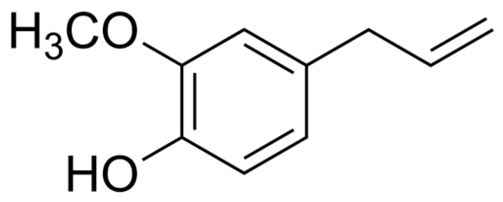
Figure 1. Chemical structure of eugenol (4-allyl-2-methoxyphenol).
Molecules 30 03353 g001
Figure 2. Antibacterial activity of eugenol (EUG) against multidrug-resistant Staphylococcus pseudintermedius. Time–kill kinetics of (a) MRSP SIG 3X, (b) MRSP SIG 4X, (c) MRSP 9X, (d) MRSP 10X, (e) MSSP SIG 11X, and (f) MSSP SIG 12X incubated with different concentrations of EUG. The log10 CFU/mL values represent the mean ± the standard deviation from three independent experiments. The dotted lines indicate a 99.9% (3 log10) reduction in CFU/mL counts. *(p < 0.05) compared to the untreated control cells.
Molecules 30 03353 g002
Figure 3. Antibacterial activity of biogenic silver nanoparticles (bioAgNPs) against multidrug-resistant Staphylococcus pseudintermedius. Time–kill kinetics of (a) MRSP SIG 3X, (b) MRSP SIG 4X, (c) MRSP 9X, (d) MRSP 10X, (e) MSSP SIG 11X, and (f) MSSP SIG 12X incubated with different concentrations of bioAgNPs. The log10 CFU/mL values represent the mean ± the standard deviation from three independent experiments. The dotted lines indicate a 99.9% (3 log10) reduction in CFU/mL counts. *(p < 0.05) compared to the untreated control cells.
Molecules 30 03353 g003
Figure 4. Antibacterial activity of eugenol (EUG) combined with biogenic silver nanoparticles (bioAgNPs) against multidrug-resistant Staphylococcus pseudintermedius. Time–kill kinetics of (a) MRSP SIG 3X, (b) MRSP SIG 4X, (c) MRSP 9X, (d) MRSP 10X, (e) MSSP SIG 11X, and (f) MSSP SIG 12X incubated with the minimum inhibitory concentration (MIC) of each compound alone and at the interaction (synergistic or indifferent) concentrations. The log10 CFU/mL values represent the mean ± the standard deviation from three independent experiments. The dotted lines indicate a 99.9% (3 log10) reduction in CFU/mL counts. *(p < 0.05) compared to the untreated control cells.
Molecules 30 03353 g004
Figure 5. Effect of eugenol (EUG) and biogenic silver nanoparticles (bioAgNPs) on the metabolic activity of sessile (biofilms) cells of multidrug-resistant Staphylococcus pseudintermedius, as assessed by the MTT reduction assay. The inhibitory activity of EUG (a) and bioAgNPs (b) on 24 h biofilms formed on polystyrene surfaces. Time–kill kinetics for (c,d) MRSP SIG 3X and (e,f) MSSP SIG 12X isolates incubated with EUG (c,e) and bioAgNPs (d,f). Values represent the mean ± the standard deviation derived from three independent experiments. *(p < 0.05), **(p < 0.01), ***(p < 0.001) compared to the untreated control cells.
Molecules 30 03353 g005
Figure 6. Effect of eugenol (EUG) and biogenic silver nanoparticles (bioAgNPs) on the morphology and ultrastructure of 24 h biofilms of multidrug-resistant Staphylococcus pseudintermedius SIG 3X formed on polystyrene surfaces. (ac) Untreated biofilms (control). Biofilms incubated with (df) 1.33 mg/mL and (gi) 2.67 mg/mL EUG, and (jl) 3.45 µg/mL and (mo) 13.82 µg/mL bioAgNPs. (a,d,g,j,m) 6000× magnification; (b,e,h,k,n) 12,000× magnification; (c,f,i,l,o) 24,000× magnification.
Molecules 30 03353 g006
Figure 7. Effect of eugenol (EUG) and biogenic silver nanoparticles (bioAgNPs) on LLC-MK2 cell viability, as assessed by MTT reduction assay. LLC-MK2 cells were treated for 24 h with different concentrations of (a) eugenol and (b) bioAgNPs. Values represent the mean ± the standard deviation from three independent experiments. *(p < 0.05), **(p < 0.01), ***(p < 0.001) compared to the untreated control cells.
Molecules 30 03353 g007
Table 1. Characterization of Staphylococcus pseudintermedius: antimicrobial resistance profile and antibacterial activity of eugenol (EUG) and biogenic silver nanoparticles (bioAgNPs).
IsolateResistance profileamecAbEUGBioAgNPsMBC/MICe
MICcMBCdMICcMBCdEUGbioAgNP
SIG 3XOX, P, AZM, ERY, DA*, CIP, LEV, CN*, TE, CHLpositive1.332.673.4513.822.04.0
SIG 4XOX, P, AZM, ERY, DA*, CIP, LEV, CN, CHLpositive0.671.333.4513.822.04.0
SIG 9XOX, P, AZM, ERY, DA*, CIP, LEV, CN*, TE, CHLpositive1.331.333.4513.821.04.0
SIG 10XOX, P, AZM, ERY, DA*, CIP, LEV, CN*, TE, CHLpositive1.331.333.456.911.02.0
SIG 11XP, AZM, ERY, DA*, CIP, LEV, CN*, TEnegative0.671.333.456.912.02.0
SIG 12XP, AZM, ERY, DA*, CIP, LEV, TE, CHLnegative1.332.673.456.912.02.0
aPhenotypic resistance was determined by the disk diffusion method [39]. AZM: azithromycin; CHL: chloramphenicol; CIP: ciprofloxacin; DA: clindamycin; ERY: erythromycin; CN: gentamicin; LEV: levofloxacin; OX: oxacillin; P: penicillin; TE: tetracycline. bThe presence of the mecA gene was determined through PCR [37]. cThe minimum inhibitory concentration (MIC) was determined by the broth microdilution method [39]. dThe minimum bactericidal concentration (MBC) was determined by the time–kill assay [40]. eReference values: 1 to 4 bactericidal effect and > 4 bacteriostatic effect [41]. *Intermediate resistance. EUG values are expressed in mg/mL; bioAgNPs values are expressed in µg/mL.
Table 2. Antibacterial activity of eugenol (EUG) combined with biogenic silver nanoparticles (bioAgNPs) against planktonic cells of MDR Staphylococcus pseudintermedius.
MIC
IsolateEUGabioAgNPsaEUG/bioAgNPsbFICIcInteractiond
SIG 3X1.333.450.33/0.860.50Synergism
SIG 4X0.673.450.16/0.860.49Synergism
SIG 9X1.333.450.33/1.720.75Indifferent
SIG 10X1.333.450.33/1.720.75Indifferent
SIG 11X0.673.450.16/0.860.49Synergism
SIG 12X1.333.450.33/0.860.50Synergism
aThe minimum inhibitory concentration (MIC) determined by the broth microdilution method [39]. bThe MIC of EUG and bioAgNPs in combination determined by checkerboard assay [30]. cThe fractional inhibitory concentration index (FICI) was calculated as the sum of the FICEUG and FICbioAgNPs. dReference values: synergism (FICI ≤ 0.5), no interaction (0.5 < FICI ≤ 4.0), or antagonism (FICI > 4.0) [55]. EUG values are expressed in mg/mL; bioAgNPs values are expressed in µg/mL.
Table 3. Antibiofilm activity of eugenol (EUG) and biogenic silver nanoparticles (bioAgNPs), alone and in combination, against sessile MDR Staphylococcus pseudintermedius.
SMIC90
IsolateEUGabioAgNPsaEUG/bioAgNPsbFICIcInteractiond
SIG 3X1.3313.820.33/3.450.50Synergism
SIG 4X1.3313.820.33/3.450.50Synergism
SIG 9X1.3313.820.33/3.450.50Synergism
SIG 10X0.6713.820.33/3.450.74Indifferent
SIG 11X0.6713.820.16/3.450.49Synergism
SIG 12X1.3313.820.33/3.450.50Synergism
aSMIC90: Sessile minimum inhibitory concentration capable of inhibiting 90% of the metabolic activity of sessile cells. bSMIC90 of EUG and bioAgNPs in combination determined by checkerboard assay [30]. cFICI: fractional inhibitory concentration index was calculated as the sum of the FICEUG and FICbioAgNPs. dReference values: synergism (FICI ≤ 0.5), no interaction (0.5 < FICI ≤ 4.0), or antagonism (FICI > 4.0) [55]. EUG values are expressed in mg/mL; bioAgNPs values are expressed in µg/mL.
Disclaimer/Publisher’s Note: The statements, opinions and data contained in all publications are solely those of the individual author(s) and contributor(s) and not of MDPI and/or the editor(s). MDPI and/or the editor(s) disclaim responsibility for any injury to people or property resulting from any ideas, methods, instructions or products referred to in the content.