Review

Mechanical Properties of Biodegradable Fibers and Fibrous Mats: A Comprehensive Review

Department of Applied Science, University of Quebec at Chicoutimi (UQAC), 555, Boul. de l’Université, Chicoutimi, QC G7H 2B1, Canada
*
Author to whom correspondence should be addressed.
Molecules 2025, 30(15), 3276; https://doi.org/10.3390/molecules30153276
Submission received: 27 June 2025 / Revised: 23 July 2025 / Accepted: 1 August 2025 / Published: 5 August 2025
(This article belongs to the Section Materials Chemistry)

Abstract

The growing demand for sustainable materials has led to increased interest in biodegradable polymer fibers and nonwoven mats due to their eco-friendly characteristics and potential to reduce plastic pollution. This review highlights how mechanical properties influence the performance and suitability of biodegradable polymer fibers across diverse applications. This covers synthetic polymers such as polylactic acid (PLA), polyhydroxyalkanoates (PHAs), polycaprolactone (PCL), polyglycolic acid (PGA), and polyvinyl alcohol (PVA), as well as natural polymers including chitosan, collagen, cellulose, alginate, silk fibroin, and starch-based polymers. A range of fiber production methods is discussed, including electrospinning, centrifugal spinning, spunbonding, melt blowing, melt spinning, and wet spinning, with attention to how each technique influences tensile strength, elongation, and modulus. The review also addresses advances in composite fibers, nanoparticle incorporation, crosslinking methods, and post-processing strategies that improve mechanical behavior. In addition, mechanical testing techniques such as tensile test machine, atomic force microscopy, and dynamic mechanical analysis are examined to show how fabrication parameters influence fiber performance. This review examines the mechanical performance of biodegradable polymer fibers and fibrous mats, emphasizing their potential as sustainable alternatives to conventional materials in applications such as tissue engineering, drug delivery, medical implants, wound dressings, packaging, and filtration.

1. Introduction

Biodegradable polymer fibers and nonwoven mats are increasingly recognized for use in applications such as tissue engineering scaffolds, drug delivery systems, filters, and biosensors because of their high surface-area-to-volume ratio, porosity, and ease of functionalization [1,2,3]. Polymers like polyvinyl alcohol (PVA), polylactic acid (PLA), polyhydroxyalkanoate (PHAs), polycaprolactone (PCL), polyglycolic acid (PGA), and cellulose have gained considerable interest for their use in fiber production because of their sustainable nature [4,5,6,7]. These biodegradable fibers are used in such contexts as agriculture, healthcare, and packaging (Figure 1), help reduce plastic pollution, and are deemed more environmentally friendly than alternatives [8,9,10]. Various production methods for biodegradable polymer fibers and nonwoven mats offer distinct advantages. Electrospinning, a common technique, produces ultrafine fibers with diameters ranging from nanometers to micrometers. Electrospun nanofibers could possess exceptional mechanical properties, high specific surface area, and fine porosity, making them ideal for tissue engineering and drug delivery applications [11]. Although electrospinning is prevalent for polymer nanofiber production, methods like melt blowing, centrifugal spinning, and wet spinning also offer benefits for large-scale manufacturing, cost-effective commercialization, and control of processing [12]. The tensile strength, flexibility, and durability of these materials are vital for demanding applications such as medical implants and protective clothing. Thus, understanding the mechanical behavior of mats and fibers is essential for selecting suitable methods and optimizing process design. Recent advancements include developing biodegradable fibers through copolymerization, crosslinkers, and pre- and post-treatment techniques to enhance mechanical properties [13,14,15,16].
The mechanical properties of fibers can be influenced by various parameters, including (a) the inherent physical properties of the polymer, (b) mat density, (c) average fiber diameter, (d) fiber morphology, and (e) fiber orientation in the mat [17,18]. This review aims to provide a comprehensive overview of the mechanical properties of biodegradable fibers and nonwoven mats, focusing on different production methods. Achieving optimal performance in structure, morphology, strength, and cost-effectiveness requires considering alternative fiber production methods beyond electrospinning. To our knowledge, similar review papers in this area are lacking. This paper gathers and analyzes mechanical reports from both older and recent studies, emphasizing key parameters such as tensile strength, tensile modulus, and elongation. We also discuss specific processing parameters that affect fiber morphology and mechanical properties. Comparing mechanical property data from different researchers is challenging because of variations in production methods, measurement units, and testing conditions. Therefore, we focus on identifying gaps and areas for future improvement in this field.

2. Fiber Manufacturing Methods

This section presents various methods for producing fibers and mats, highlighting their specifications, benefits, and drawbacks. The discussion provides insights into production processes to help guide the selection of the most suitable method on the basis of polymer types, production speed, and fiber quality.

2.1. Electrospinning

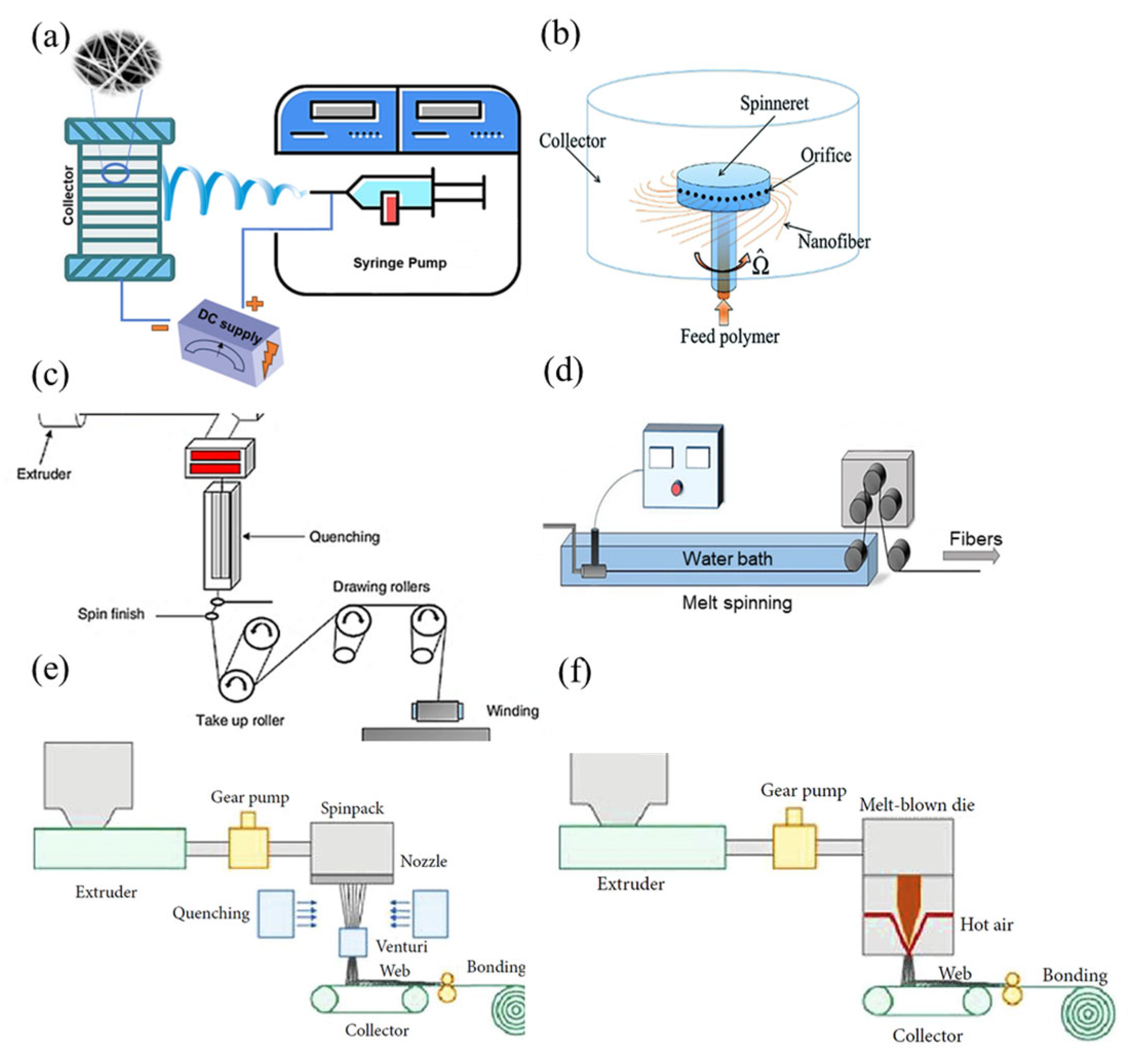
Electrospinning is a versatile and effective method for producing ultrafine fibers with diameters ranging from nanometers to micrometers. The process applies a high voltage to a polymer solution, ejecting it through a thin needle. The charged polymer jet elongates because of electrostatic repulsion until it solidifies and deposits onto a grounded collector, forming a nonwoven fabric of fibers (Figure 2a) [19]. The key parameters affecting electrospinning include solution properties (viscosity, conductivity, surface tension, volatility), process conditions (electric field strength, distance from tip to collector, feed rate, needle tip diameter), and ambient factors (temperature, humidity) [20]. By adjusting these parameters, one can control fiber properties such as morphology, size, and arrangement for various applications [21].
Despite its advantages, including a high surface-to-volume ratio, porosity, and the ability to create composite fibers, electrospinning has drawbacks. It is sensitive to parameter variations, requiring precise control to achieve consistent quality [26,27]. Additionally, its production rate is lower than that of other industrial processes, which limits commercial scalability [28]. Another disadvantage is the need for high voltage, which raises operational safety concerns. However, ongoing research continues to develop solutions that address these limitations and expand the applicability of electrospun materials [29].

2.2. Centrifugal Spinning

Centrifugal spinning is a fiber fabrication technique that uses centrifugal forces to produce microfibers and nanofibers from polymer solutions or melts. In this method, illustrated in Figure 2b, the polymer fluid is loaded into a rotating spinning head with multiple nozzles arranged radially [14]. The rotation generates high centrifugal force, ejecting polymer jets through the nozzles, which are then elongated by air drag and deposited on a collector. The key parameters in this process include rotational speed, solution viscosity and concentration, nozzle geometry and size, spinning distance, and solution feed rate [15]. By adjusting these parameters, fibers with tailored morphologies and dimensions can be produced. Compared to other fiber fabrication methods, centrifugal spinning offers several advantages, such as high production rates, cost-effectiveness, versatility in usable polymer types, and ease of process control and scalability. However, it has limitations, including a relatively broad fiber diameter distribution and challenges in collecting nanofibers because of high-speed rotation [15,16].

2.3. Melt Spinning

Melt spinning is a fiber production process in which molten polymer is extruded through a tiny nozzle and stretched during cooling to form the fiber. As shown in Figure 2c, this process includes a screw extruder barrel, a metering pump, a die head, an air-cooling system, a spin finish, and a winding system [17]. Polymers are fed through the hopper into the extruder, where they are mechanically compressed and melted. The molten polymer is forced through a spinneret as fine jets, with flow speed controlled by the metering pump. The filaments are then cooled and hardened by the quench system to produce fibers [18].
Furthermore, the melt-spinning process is a convenient and straightforward method that does not require coagulation baths or solvents and can produce fibers of varying diameters. Compared to other fiber production methods, it has a simple setup and can be efficiently scaled to create fibers suitable for weaving and usability [19,20,21]. However, this method is primarily used for manufacturing fibers from non-biodegradable synthetic polymers like polyester, polypropylene, and nylon, which can lead to environmental pollution [19,26]. The main reason is that most biodegradable polymers are susceptible to degradation under thermal conditions in this process. On the other hand, fibers cannot be manufactured at a nanoscale via this method, and it is not suitable for applications that need smaller fiber diameters [20].

2.4. Wet Spinning

Wet spinning is widely used in textile manufacturing, filtration, healthcare, and industrial sectors [30,31,32]. It involves dissolving a polymer in a solvent to create a spinning solution. This solution is then extruded through spinnerets into a coagulating bath, where the solvent is removed, solidifying the polymer into a fiber. Wet spinning is commonly used for manufacturing fibers from cellulose, acrylics, rayon, and certain synthetic polymers [27]. The main parameters affecting fiber efficiency and quality include the polymer content, extrusion speed, coagulation bath composition and temperature, and drawdown ratio (Figure 2d) [28]. These parameters influence the final mechanical properties and microstructure of the fiber. Although wet spinning allows for the addition of additives to enhance fiber properties and process high-molecular-weight polymers, its slower production speed, complex solvent recovery, and extensive post-treatments limit its environmental sustainability and economic viability [27].

2.5. Melt Blowing

Melt blowing is used to produce fibers and nonwoven fabric. In this process, polymer resins are melted at high temperatures and are then extruded through tiny nozzles. A high-speed airstream draws the molten polymer into microfibers (Figure 2e) [29]. These microfibers collect on a moving conveyor belt to form a nonwoven web. The ability to create microfibers makes melt-blowing technology essential across various industries, including automotive, railway, aerospace, agriculture, geotextile, health care, and construction industries, for applications like filtration, medical uses, oil absorption, and thermal insulation [30,31]. The main advantage of this method is its higher production speed and quantity compared to other techniques, along with its low cost and versatility in producing nonwoven fabrics [32]. However, melt-blowing technology has some drawbacks. Most importantly, it can only use thermoplastic polymers. Additionally, controlling fiber size and porosity during production is challenging [33].

2.6. Spunbonding

Spunbonding is an efficient method of producing nonwoven fabrics through a continuous process where polymer granules are melted and passed through spinnerets to form fine filaments. These filaments are placed on a conveyor belt and then thermally, chemically, or mechanically bonded to create a strong and flexible fabric (Figure 2f) [29].A key advantage of this technology is its direct conversion from polymer to nonwoven fabric, which eliminates the need for intermediate steps like yarn forming. The main parameters affecting spun fabric quality and properties include polymer type, processing temperatures, spin dimensions, and cooling conditions. Spunbond fabric offers high production speed, cost-effectiveness, and the ability to modify properties such as weight, porosity, and tensile strength to meet specific requirements [22]. However, spunbond fabrics tend to be less soft and flexible than other nonwoven fabrics, making them less suitable for applications that require high drapeability or softness [33]. Other disadvantages include larger average fiber diameters, lack of fiber uniformity, and reduced strength and elongation [22,23,24].

3. Mechanical Property Testing

After the selection of the manufacturing method for the desired fibers or mats on the basis of their application, it is crucial to choose the appropriate apparatus for evaluating their durability and strength. This section examines key testing methods, including tensile testing machines, atomic force microscopy (AFM), and dynamic mechanical analysis (DMA), each offering unique insights into the characteristics of biodegradable polymers.

3.1. Tensile Testing Machines

The tensile testing machine is a cornerstone in evaluating the mechanical properties of fibrous mats. This device measures the force needed to stretch a fiber until it breaks, providing key parameters such as tensile strength, elongation at break, and Young’s modulus. The fundamental formula used in tensile tests is the stress–strain relationship: stress (σ) equals applied force (F) divided by the original cross-sectional area (A) of the fiber (Equation (1)), whereas strain (ε) is the ratio of extension (ΔL) to the original length (L0) (Equation (2)).
σ = F A ,
ε = Δ L L 0
Parameters such as force range, extension rate, and stretching speed can be adjusted with this device. These testing machines feature clamps suitable for small fibers and apply tension without shifting or damaging the sample. Thus, they effectively demonstrate how fibers respond to tensile loading and their mechanical behavior in various applications [34,35].
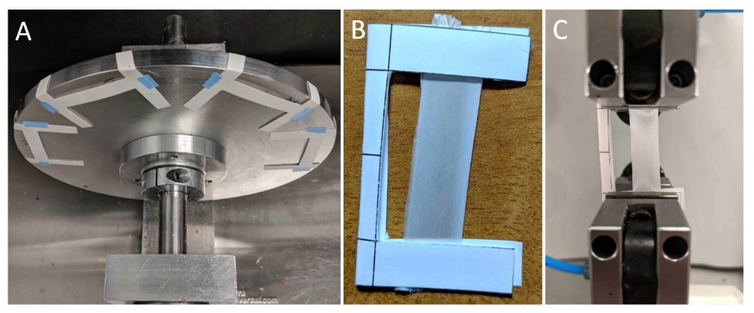
Tensile testing of electrospun fibers and mats typically follows standardized protocols such as ASTM D638, with specimens commonly prepared in rectangular strip or dog-bone shapes to ensure uniform stress distribution and prevent premature failure. For fibrous mats, rectangular strips are often preferred due to ease of handling and consistent load application. In contrast, single fibers require more delicate mounting methods such as adhesive-supported ends or the use of atomic force microscopy.

3.2. Atomic Force Microscopy

AFM is a precise technology for probing the mechanical properties of fibers at the nanoscale. It uses a cantilever with a sharp tip that gently contacts the surface of a fiber sample to measure the forces between the tip and the sample, providing detailed nanoscale topography. In addition to mapping, AFM assesses mechanical properties such as elastic modulus, hardness, and adhesion of the fibers. Cantilever deflection is measured as a function of its position across the fiber, following Hooke’s Law:
F = k x
where F is the force exerted by the tip, k is the cantilever’s spring constant, and x is the cantilever’s displacement. A unique feature of AFM is its ability to image fibers under ambient conditions and in controlled fluid environments, mimicking physiological conditions when necessary. This level of detail makes it an indispensable tool for designing and evaluating advanced fiber materials with tailored mechanical properties [36,37,38].

3.3. Dynamic Mechanical Analysis

DMA is an analytical technique used to characterize the mechanical properties of fibers under oscillating forces. It provides critical information about the material’s viscoelastic behavior by capturing its elastic and viscous characteristics. In DMA, stress is applied to a fiber sample while strain, storage modulus, and loss modulus are measured. These parameters indicate the material’s ability to store and dissipate energy, which is vital for applications ranging from industrial textiles to biomedical implants. DMA covers a wide range of frequencies, temperatures, and controlled environments that simulate practical usage conditions. The device’s sensitivity allows it to detect transitions such as the glass transition temperature (Tg) and melting temperature, which are essential for understanding the thermal behavior of fibers in various environments [39,40].

3.4. Other Mechanical Tests

In addition to tensile testing, AFM, and DMA, fibers undergo other mechanical tests to evaluate their overall performance. One such test is nanoindentation, which measures hardness and modulus at the nanometer scale. Nanoindentation assesses a nanofiber’s resistance to deformation by pressing a hard indentation into its surface under controlled conditions and measuring the resulting depth. This technique is particularly effective for single nanofibers, where other methods may be inadequate [10]. Another important test is microcompression, which compresses a nanofiber to determine its buckling behavior. This test is crucial for applications involving high-pressure loading [10,41].

4. Mechanical Properties of Biodegradable Polymer Fibers and Mats

This section presents a comparative overview of the mechanical properties of various biodegradable fibers and mats, highlighting how different material types and processing methods influence their strength, flexibility, and durability. Key factors discussed include polymer type, fabrication technique, and pretreatment or post-treatment strategies used to enhance alignment, crystallinity, or interfacial bonding. The discussion is organized into sections on natural and synthetic biodegradable polymers to highlight their respective advantages, challenges, and performance trends in fiber-based systems. This section aims to guide researchers in selecting appropriate polymer types and processing techniques to improve the mechanical performance of biodegradable fibers and fibrous mats for specific applications.

4.1. Synthetic-Biodegradable-Polymer-Based Fibers

4.1.1. Polyvinyl Alcohol

Polyvinyl alcohol (PVA) is a synthetic, biodegradable, biocompatible, nontoxic, hydrophilic, and water-soluble polymer obtained by the hydrolysis of polyvinyl acetate (C4H6O2)n. The crystallinity and polarity of PVA are two key factors that can be affected by the degree of hydrolysis of PVA. A higher degree of hydrolysis increases crystallinity because of a greater number of polar hydroxyl groups (–OH) [42,43]. As crystallinity increases, molecules become more tightly packed together in a higher order. This improved distribution and ability to resist applied forces result in higher stiffness and tensile strength [44].
PVA enhances the mechanical properties, biodegradability, biocompatibility, hydrophilicity, and processability of other polymers [42]. Its excellent physical properties, low cost, biodegradability, and biocompatibility make it widely used in medical applications, packaging, adhesives, wound dressings, drug delivery systems, soft contact lenses, and artificial organs [43,45,46]. PVA’s biodegradability stems from its susceptibility to degradation by microbial action, where microorganisms like bacteria and fungi can break down the polymer’s molecular chains using enzymes such as PVAase, hydrolyzing it into simpler, water-soluble compounds. Thus, this microbial susceptibility, combined with its water solubility and hydrolyzable backbone, enables PVA to degrade effectively in a range of natural environments [47,48,49].
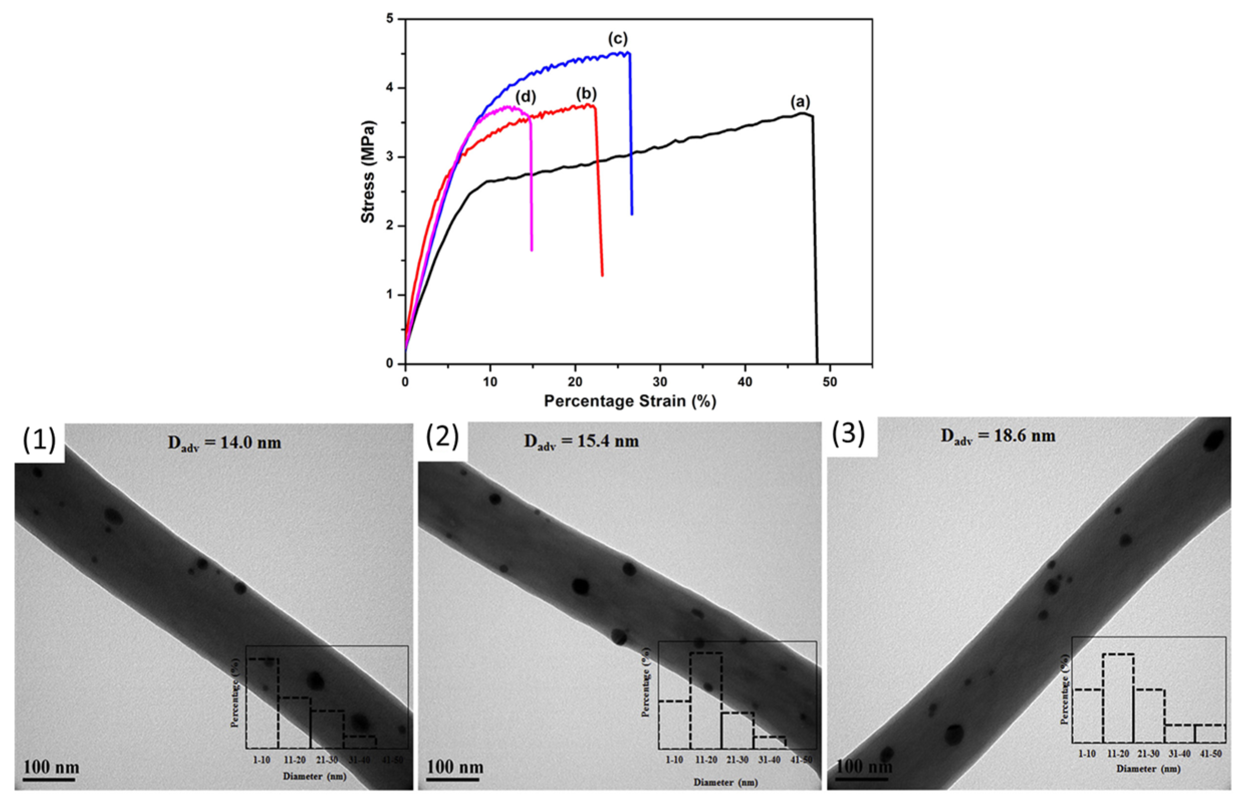
Electrospun PVA-based fibers exhibit tunable mechanical properties that vary depending on nanoparticle incorporation, polymer blending, and processing conditions. For instance, adding cellulose nanowhiskers can improve tensile properties, especially in aligned fibers where the nanowhiskers orient along the fiber axis, enhancing stress transfer and reinforcing the polymer matrix. Tensile strength in aligned fibers increased from 5.37 to 10.5 MPa, demonstrating effective nanowhisker reinforcement [50]. Incorporating silver nanoparticles (AgNPs) can initially strengthen PVA/chitosan fibers because of the strong intermolecular interactions (Figure 3). However, at higher concentrations, AgNPs aggregate, disrupting uniform distribution and reducing mechanical performance. This aggregation hinders polymer chain mobility, increasing stiffness but decreasing flexibility [45]. The highest tensile strength of 4.52 MPa was achieved at 0.5% AgNP content before declining at higher concentrations [51].
Polymer blending also significantly influences mechanical behavior. For example, blending PVA with soy protein isolate (SPI) has been shown to weaken fiber strength, as the globular structure of SPI disrupts the continuity of the PVA matrix and hinders efficient chain entanglement. This incompatibility at the molecular level reduces stress transfer across the fiber network, leading to decreased tensile strength and elongation [52]. In contrast, incorporating silk sericin (SS) and applying thermal treatment strengthens the fibers by promoting hydrogen bonding, which improves molecular cohesion and elasticity. Heated SS/PVA fibers exhibit over five times higher tensile strength than untreated fibers, increasing from 0.7 to 3.7 MPa, highlighting the role of thermal treatment in enhancing mechanical properties [46].
Structural modifications such as porosity control and plasticizer addition further affect fiber properties. Increased porosity in PVA/γ-Fe2O3 nanofibers weakens fiber interactions, reducing tensile strength. Similarly, although plasticizers enhance flexibility by disrupting internal hydrogen bonding, they also reduce stiffness by altering molecular packing. In melt-spinning studies of PVA, the addition of plasticizers has been shown to increase tensile strength by nearly three-fold, while reducing elongation due to alterations in hydrogen bond distribution [53].
The influence of water content in melt-spun PVA fibers has shown how processing parameters control mechanical results. Lower water content restricts molecular movement because of stronger hydrogen bonding, making fibers stiffer but less stretchable. In contrast, higher water content increases chain mobility, enhancing flexibility while reducing overall strength. In one study, the yield stress of PVA fibers rose from 17 to 109 MPa as water content decreased, emphasizing hydrogen bonding’s role in regulating stiffness [54]. Therefore, achieving optimal mechanical performance in PVA fibers requires balancing nanoparticle dispersion, polymer compatibility, structural modifications, and processing conditions to tailor strength, stiffness, and flexibility for specific applications.

4.1.2. Polyethylene Oxide

Poly (ethylene oxide) (PEO) is a water-soluble biodegradable synthetic polymer consisting of a long chain of ethylene oxide units that are chemically linked. Its ability to break down into nontoxic by-products (ethylene glycol and water) makes it suitable for various pharmaceutical and biomedical applications [55]. PEO is a commonly used sacrificial porogen that can be easily electrospun and rapidly dissolved in aqueous solutions [56,57].
Merchiers et al. [17] compared the mechanical properties of PEO fibers produced by centrifugal force spinning (CFS) and electrospinning. CFS fibers exhibited higher tensile strength (1.8–4.2 MPa) and greater elongation at break than electrospun fibers (0.2–2.4 MPa), likely because of increased crystallinity. Blending PEO with carboxymethyl chitosan (CMCS) in centrifugal spinning reduced elongation (from 22.8% to 11.2%) but increased Young’s modulus (from 10.5 to 26.6 MPa), indicating a stiffening effect. However, tensile strength fluctuated (0.8–2.2 MPa), suggesting complex interactions between CMCS and PEO [58]. Incorporating PEO into starch-based fibrous membranes enhanced tensile strength approximately three-fold (from 0.39 to 1.8 MPa) because of improved polymer chain entanglement [59].
Kupka et al. [57] examined electrospun PEO/PCL nanofibers and found that blending hydrophilic PEO with hydrophobic PCL significantly increased Young’s modulus. Mats with 75/25 and 50/50 PCL/PEO compositions showed a 2.5-fold increase in stiffness relative to pure PCL and PEO. The improved mechanical properties may result from interpenetrating networks formed at the nanoscale. Overall, PEO fibers produced via CFS exhibit better tensile strength and elongation than electrospun fibers. Blending with CMCS increases stiffness while reducing flexibility, whereas incorporating PEO into starch-based matrixes strengthens fiber networks. PEO/PCL blends significantly enhance mechanical properties because of nanoscale interpenetrating networks, making them promising for structural applications.

4.1.3. Polyglycolic Acid (PGA) and Poly(lactic-co-glycolic acid) (PLGA)

Polyglycolic acid (PGA) is a synthetic, semicrystalline, biodegradable polyester that degrades into nontoxic by-products, such as carbon dioxide and water, over several weeks under physiological conditions. Its high tensile strength, stiffness, and biocompatibility make it primarily suitable for biomedical sutures, reducing the need for removal surgeries [60,61]. Its copolymer, poly(lactic-co-glycolic acid) (PLGA), derived from lactic acid and glycolic acid, features tunable degradation rates and mechanical properties, making it widely used in drug delivery systems, tissue engineering, and biomedical devices. PLGA’s ability to degrade into nontoxic components enhances its appeal for medical applications [60,62].
Researchers have explored blending PGA with PLA to address PGA’s limitations, including high crystallinity, rapid degradation, and poor processability. Ramdhanie et al. [63] demonstrated that adjusting the PGA-to-PLA ratio in electrospun nanofibers allowed for tuning mechanical properties to match those of collagen and elastin, key structural components in soft tissues. The modulus ranged from 3.4 to 140 MPa, tensile strength from 2.2 to 11 MPa, and strain at break from 70% to 330%, highlighting the potential of PGA/PLA blends for soft tissue engineering.
Comparative studies of synthetic biodegradable polyesters found that PLGA (50/50) exhibited the highest Young’s modulus (144 MPa), followed by PGA (138 MPa) and PLGA (85/15) (114 MPa). PCL and PLLA showed the lowest moduli, around 8.5 MPa, indicating that PGA retains relatively high stiffness among biodegradable polymers [64].
D’Amato et al. [65], as shown in Figure 4, examined the effect of residual electrospinning solvent on PGA fibers and found that solvent retention initially softened the fibers but led to increased brittleness over time. Young’s modulus nearly tripled from 1013.4 to 2989.1 MPa within 14 days as the solvent evaporated, while yield strain decreased from 3.5% to 1.5%, demonstrating that solvent removal significantly affects the mechanical performance of PGA scaffolds.
To enhance porosity, researchers investigated electrospraying sacrificial PEO microparticles into PGA, PLGA, and PCL scaffolds. This technique improved porosity and cell infiltration while maintaining the mechanical integrity of PGA and PLGA. However, the removal of PEO microparticles reduced the strength and stiffness of the more elastic PCL component, indicating its greater sensitivity to porosity modifications [56].
The mechanical properties of PLGA/PCL blends were investigated through coaxial electrospinning. Bazgir et al. [66] reported that coaxial scaffolds with PCL as the core and PLGA as the sheath had lower tensile strength (3.1 vs. 2.89 MPa) and elongation (25.94% vs. 14.61%) compared to pure PLGA membranes. PCL scaffolds exhibited the lowest tensile strength (1.02 MPa) but the highest elongation at break (32.43%). Further studies showed that increasing PLGA content enhanced the tensile strength of PLGA–PCL blends [67,68]. Overall, PGA-based fibers demonstrate high stiffness, with solvent evaporation significantly affecting their mechanical behavior. Blending PGA with PLA can tailor its mechanical and degradation properties, making it more suitable for soft tissue engineering, while PLGA/PCL blends allow for tunable flexibility and strength, which are advantageous in load-bearing or long-term implants.

4.1.4. PLA, Poly(L-lactic acid) (PLLA), and Poly(D-lactic acid) (PDLA)

PLA and PLLA are aliphatic polyesters derived from lactic acid (2-hydroxy propionic acid) monomers. PLA is a copolymer containing L- and D-lactic acid units, whereas PLLA is a homopolymer consisting solely of L-lactic acid units. This difference affects their chain’s structural conformation, properties, and specifications [69]. They can be easily spun and are suitable for packaging, agriculture, and particularly biomedical applications, including sutures, orthopedic implants, and drug delivery systems [70]. The biodegradability of PLA and PLLA stems from their ability to be metabolized by the human body. Their hydrolytic degradation kinetics produce lactic acid, which can be converted to carbon dioxide and water. Their polyester backbone can undergo hydrolytic cleavage under physiological conditions, breaking down into lactic acid, a natural product of human metabolism. This property is highly desirable in the biomedical field, as it eliminates the need for additional surgeries to remove temporary implants [71,72]. Because of their thermoplastic processability, biological properties, biodegradability, and lack of immunogenicity, they are suitable for tissue engineering. However, PLA-based fibers often exhibit low hydrophilicity and inherent brittleness, which can hinder their interaction with cells in soft tissue applications. Therefore, there is considerable interest in increasing hydrophilicity and reducing the brittleness of PLA-fiber-based scaffolds. For example, adding soft and hydrophilic PEG polymer to the PLA solution can enhance surface hydrophilicity [72].
The mechanical properties of electrospun PLLA fibers depend highly on solvent composition and surfactants. Arbeiter et al. [73] demonstrated that adding surfactants, such as TEAC, formic acid, and Triton X-100, influenced tensile strength and crystallinity. TEAC produced the most significant increase, raising tensile strength from 2.7 to 27.1 MPa, whereas TX-100 improved flexibility but slightly reduced elongation, indicating that surfactants alter molecular interactions and fiber structure.
Crosslinking can also enhance mechanical properties. Qiao et al. [74] found that adding dicumyl peroxide (DCP) and triallylisocyanurate (TAIC) to PLLA fibers increased rigidity while maintaining some flexibility, increasing the modulus from 165 to 208 MPa. This improvement demonstrates the potential of chemical modifications in tailoring electrospun PLLA properties for biomedical applications. Additionally, Goreninski et al. [75] investigated iodine and iodine-containing compounds, traditionally used as antiseptics, in PLLA and PCL fibers. Their findings showed that iodine treatment did not significantly affect the scaffolds’ mechanical properties or morphologies.
Furthermore, without altering the underlying chemistry, post-treatment of electrospun PLLA mats can significantly enhance mechanical performance. Immersion in acetone promotes the growth of α-form crystals, approximately doubling the tensile strength and increasing the Young’s modulus, while reducing extensibility [14]. A similar acetone-based rolling treatment applied to tubular PLLA grafts increases the wall strength from sub-megapascal values to over 5 MPa, in line with crystallinity gains confirmed by XRD and DSC [76]. Post-treatment strategies that control porosity yield comparable outcomes. Porous PLLA membranes, prepared by soaking either neat PLLA fibers or PLLA/PCL blends, show steeper stress–strain curves and lower elongation at break. These effects result from a combination of increased crystallinity and fiber–fiber welding [1,77]. Overall, solvent-based treatments that increase the amount and quality of α-phase crystals can effectively improve strength and stiffness, but they often reduce flexibility. This trade-off should be considered when designing PLA-based materials.
Material additives such as bioceramics, essential oils, and nanoparticles also influence fiber properties. Didekhani et al. [78] incorporated oyster shell (OS) particles into PLLA nanofibers, which increased tensile strength while reducing elongation at break. Post-plasma treatment enhanced surface hydrophilicity, making the scaffold more suitable for tissue engineering [78]. Similarly, PLA fibers blended with rosmarinic acid (RosA) and graphite oxide (GO) increased breaking strength from 0.8 to 2.6 MPa but became more brittle because of the reduced fiber diameter and increased crystallinity [79].
Inorganic bioceramic fillers such as hydroxyapatite (HA), amorphous calcium phosphate (ACP), and β-tricalcium phosphate (β-TCP) influence both mechanical properties and bioactivity of PLA/PLLA fibers. For example, adding 5 wt% HA to electrospun PLLA reduces tensile strength from 4.9 to 3.2 MPa due to stress concentration but enhances apatite formation [1]. Similarly, porous PLLA/ACP mats show a three-fold increase in strength after acetone treatment, along with improved modulus and cell response. PLA fibers coated with β-TCP show enhanced osteogenic activity under cyclic loading, although mechanical values are not reported [80]. These examples demonstrate the dual mechanical and biological effects of calcium phosphate-based fillers in PLA systems.
The incorporation of nanocrystalline cellulose (NCC) improved PLA fiber strength, nearly doubling tensile properties at 2% NCC loading, whereas lignin addition enhanced cell proliferation without significantly altering mechanical behavior [81].
Environmental factors, such as hydrolytic degradation, also impact the mechanical properties of PLA and PLLA fibers. Shamsah et al. [82] found that hydrolysis over six months increased crystallinity, making fibers stiffer and stronger but also more brittle (Figure 5). Ultimate tensile strength increased by 75% for PLA-PLLA blends because of polymer chain rearrangement, demonstrating how controlled degradation can enhance mechanical performance in biodegradable materials.
The fabrication method significantly influences the mechanical properties of PLA nonwoven materials. Feng et al. [83] investigated a melt-blowing system for producing PLA nonwoven mats, highlighting how the die-to-collector distance (DCD) affects material properties. Reducing the DCD from 200 to 75 mm decreased the fiber diameter, resulting in a denser structure with improved fiber bonding. Consequently, the tensile strength more than doubled to 0.18 MPa, attributed to increased web density and stronger fiber interactions.
Similarly, Zhou et al. [84] examined the effect of centrifugal melt-spinning speeds on PLA fiber properties. They found that higher spinning speeds refined the fiber structure, enhancing tensile strength and modulus. At 1500 rpm, tensile strength reached 3.31 MPa, and the elastic modulus significantly improved to 66.77 MPa. These results are consistent with previous studies, including that of Boschetto et al. [85], which reported a tensile strength of 3.3 MPa at a higher speed of 2200 rpm, demonstrating a strong correlation between processing speed and mechanical performance.
Additionally, the durability of PLA fibers is often evaluated using tenacity, which measures the breaking force relative to linear density. Chen et al. [86] produced PLA fibers using melt spinning at 800 m/min, reporting a tenacity of 3.7–4.7 cN/tex, making PLA a competitive alternative to other synthetic polymers. Hassan et al. [87] further showed that PLA fibers have an initial modulus of 80.43 cN/tex and an elongation at break of 19%, reinforcing their suitability for various textile applications [19,83].
Beyond PLLA, recent studies have highlighted the distinct mechanical behavior of its enantiomer, PDLA, under similar processing conditions. Published data confirm that PDLA exhibits higher strength and modulus than its L-enantiomer under equivalent spinning conditions. As-spun PDLA nanofiber mats display a tensile modulus of approximately 47 MPa and a maximum tensile strength of around 3 MPa. Brief annealing increases the strength to 4–5 MPa, although stiffness decreases due to simultaneous homocrystallization and chain relaxation [88]. Conjugate electrospinning of continuous yarns further elevates the tensile strength to about 285 MPa, reflecting the higher crystallinity achievable with the D-isomer [89]. When PDLA is involved in stereocomplex formation either as a coaxial shell or as a 1:1 blend with PLLA, the mechanical behavior diverges. Interfacial stereocomplexes tend to soften the fibers (modulus ≈ 20 MPa, elongation at break ≈ 30 percent), whereas hybrid yarns or membranes can exhibit up to a 30 percent increase in tensile strength compared to the homopolymer [88,90]. These observations highlight that mechanical performance is influenced more by the spatial distribution of stereocomplex crystallites than by the overall D-content alone. Optimizing the mechanical properties of PLA and PLLA fibers involves balancing solvent choice, crosslinking, nanoparticle incorporation, degradation behavior, and processing parameters to achieve targeted strength, flexibility, and durability.

4.1.5. PCL

Polycaprolactone (PCL) is a synthetic aliphatic biodegradable polyester approved by the FDA for human use, with a low melting point of around 60 °C and a glass transition temperature of about −60 °C. This semicrystalline polymer can enhance the biodegradability of other polymers and can be processed via methods such as electrospinning, injection molding, extrusion, and 3D printing because of its excellent melt processing and processability. PCL has a high degree of elongation, adequate tensile strength, and very low solubility in water [91], which are essential for controlled release in biomedical applications [92]. These properties make PCL nanofibers suitable for long-term implantable systems and drug delivery [93,94].
PCL’s biodegradability stems from its aliphatic polyester structure, which degrades into nontoxic products under physiological conditions through hydrolysis of its ester bonds. It is well known for its good biocompatibility. Its relatively low degradation rate of up to 3–4 years, compared to other biopolymers like PLA, allows for extended application, making it particularly useful in the medical sector, where prolonged support and gradual biodegradation are desired [93,95].
PCL is widely regarded and frequently combined with PLA or PLLA given its excellent mechanical and viscoelastic properties, high cytocompatibility, and slow degradation rate. These characteristics have made PCL a staple in biomedical engineering, and it has been approved by the FDA for clinical applications [96,97].
Solvent systems, processing techniques, and material modifications highly influence the mechanical properties of PCL fibers. Sharma and Satapathy [98] investigated how solvent conductivity affects electrospun PLA-PCL fibers, finding that PLA fibers spun in a chloroform/DMF system exhibited higher stiffness, whereas PCL fibers showed greater flexibility. A 70/30 PLA-PCL blend in a dichloromethane/DMF system demonstrated an improved strain at break of 155%, highlighting how solvent–polymer interactions impact fiber mechanics. These findings align with trends observed in other studies that have examined the addition of PCL to PLA or PLA composites, further emphasizing the advantages of including PCL in these blends [96,97,99,100].
Also, beyond solvent choice, several additional variables influence the mechanical behavior of PCL-containing fibers. First, the blend ratio plays a key role: adding PCL to PLLA lowers modulus and tensile strength but increases ductility, with elongation rising from 59 percent in neat PLLA to 87–132 percent as the PCL content increases from 10 to 30 weight percent. Second, post-treatment with acetone for five minutes induces recrystallization of PLLA, partially fuses adjacent fibers, and triples the Young’s modulus while reducing elongation. Third, the same treatment creates surface porosity and drives PCL chains toward the fiber surface, lowering the water contact angle from 134° to 112°, enhancing cell affinity, and increasing stiffness through shell-like crosslinks. Together, blend composition, solvent-driven crystallinity, and morphological evolution interact with solvent–polymer compatibility to tune the performance of PCL-rich fiber mats [1]. Other studies in which PCL is blended with other polymers are discussed in other polymer sections.
Surface modifications significantly alter mechanical behavior. Volokhova et al. [101] examined the effects of pulsed electron beam (E-beam) irradiation on PCL nanofibers. Although elongation remained unchanged, the treatment reduced tensile strength from 4.1 to 1.3 MPa and modulus from 48 to 5 MPa because of polymer chain destruction. In contrast, using another surface modification method, Li et al. [102] enhanced PCL fiber mechanics through solvent vapor welding as shown in Figure 6, which restricted nanofiber slippage and increased tensile strength from 11.53 MPa to 21.4 MPa, demonstrating how structural reinforcement improves mechanical performance.
Nanoparticle incorporation further refines fiber properties. Rajzar et al. [103] found that adding hydroxyapatite (HAp) to electrospun PCL scaffolds enhanced bioactivity but slightly reduced tensile strength because of particle-induced brittleness. Similarly, Khunová et al. [104] showed that halloysite nanotubes (HNTs) doubled tensile strength and significantly improved elongation and thus enhanced the interfacial interactions of fiber networks.
Using the AFM method to evaluate the mechanical properties of individual fibers, Baker et al. [36] observed that electrospun PCL nanofibers could stretch up to 98% before slipping, achieving a tensile strength of at least 12 MPa. Similarly, Adrian et al. [105] reported that combining PCL fibers with HAp increased Young’s modulus from 4 to 20 MPa, highlighting the reinforcement effect of ceramic fillers.
Processing parameters, molecular weight, and fabrication methods influence PCL fiber performance. Alharbi et al. [93] found that fiber modulus varied between 257 and 340 MPa, depending on molecular weight and fiber diameters. Loordhuswamy et al. [106] showed that blending PCL with gelatin increased modulus from 40.76 to 84.87 MPa, although tensile strength declined because of phase separation between hydrophobic and hydrophilic polymers. Overall, selecting appropriate solvent systems, reinforcement strategies, and processing techniques can tune the mechanical properties of PCL fibers to balance flexibility, strength, and bioactivity for specific applications.
Table 1 presents a detailed overview of mechanical enhancement strategies applied to synthetic biodegradable polymers. It highlights various approaches, including nanoparticle addition, copolymerization, crosslinking, and processing modifications. For each method, the table specifies the polymer system, improvements in fiber diameter and mechanical performance (such as tensile strength and elongation), and related applications, along with supporting references.

4.2. Natural-Biodegradable-Polymer-Based Fibers

4.2.1. Chitosan

Chitosan (CS), or poly [β-(1-4)-2-amino-2-deoxy-D-glucopyranose], is the second most abundant natural polysaccharide after cellulose, obtained from the deacetylation of chitin, primarily derived from crustacean shells [51,108,109]. Its biodegradability, biocompatibility, nontoxicity, and antibacterial, antifungal, and antioxidant properties make it an excellent biopolymer for pharmaceuticals, nutrition, agriculture, food treatment, cosmetics, smart materials, and biomedicine [110,111].
Chitosan can be easily degraded by chitinase and chitosanase, which are found in nature, breaking it down into simpler, water-soluble compounds that microorganisms can ingest. Consequently, chitosan degrades into nontoxic materials, such as carbon dioxide and water. However, the relatively weak mechanical properties of chitosan fibers remain a primary concern [109]. Chitosan is often combined with other natural or synthetic polymers to address this concern and enhance its mechanical properties and processability [42].
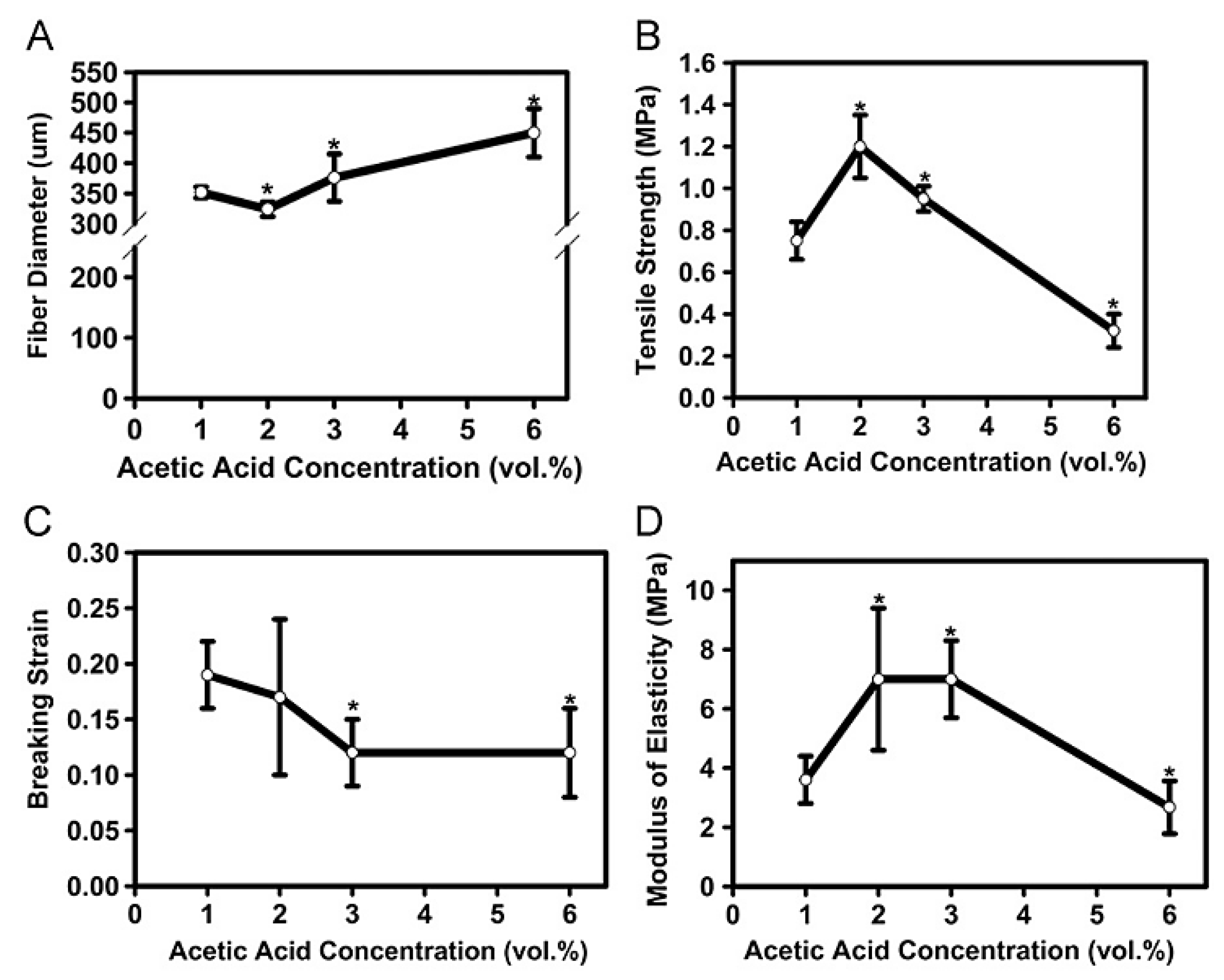
Solvent concentration, thermal treatments, crosslinking, and nanoparticle incorporation influence the mechanical properties of chitosan fibers. Albana et al. [109] examined how acetic acid and ammonia modified fiber strength and modulus (see Figure 7 for the effects of acetic acid). Higher acetic acid concentrations initially enhanced tensile strength because of improved solubility and crystallinity, but concentrations beyond 2 vol.% caused excessive swelling that weakened fiber structure. Ammonia neutralization reinforced fiber rigidity, but excessive concentrations reduced elasticity, demonstrating how solvent interactions alter chitosan’s mechanical behavior.
Barnes et al. [112] incorporated polydioxanone (PDO) with chitosan and found that collagen type I increased stiffness compared to type III. The resulting fibers closely mimicked native extracellular matrix structures, making them promising for vascular graft applications.
Crosslinking with pullulan and cinnamaldehyde further improved chitosan fiber properties. Increasing the chitosan-to-pullulan ratio raised tensile strength from 11.36 to 22 MPa and significantly enhanced modulus, indicating improved polymer network interactions. However, reduced elongation at break suggested that rigid crosslinking limited fiber flexibility [113].
The incorporation of nanoparticles into the fibers has also enhanced their mechanical properties. Lue et al. [51] demonstrated that embedding silver (Ag) nanoparticles in a chitosan–PVA matrix increased tensile strength from 3.65 to 4.52 MPa at optimal loading, attributed to strong intermolecular interactions [42]. In contrast, ceramic nanoparticles like ZnO and TiO2 showed lower dispersion efficiency, emphasizing the importance of nanoparticle–polymer compatibility in optimizing fiber strength [114,115]. Overall, solvent processing, polymer blending, and nanoparticle incorporation provide tunable pathways for enhancing chitosan fiber mechanical properties, balancing strength, flexibility, and bioactivity for biomedical applications.

4.2.2. Collagen

Collagen is a natural polymer present in almost every tissue, maintaining biological and structural integrity [116]. Its biodegradability stems from collagen’s unique amino acid sequence, recognized and processed by various enzymes and microorganisms in the body. Enzymes like collagenases and proteases break down collagen into smaller peptides and amino acids, which can be reabsorbed for metabolic processes or expelled harmlessly [117]. Because of its advantageous properties that match extracellular matrix (ECM) structural proteins, collagen is widely used in biomedical applications, such as tissue-engineered scaffolds, wound dressings, and drug delivery systems [117,118].
The mechanical properties of collagen fibers can be improved through polymer blending, crosslinking, and enzymatic modifications. Blending collagen with PCL enhances fiber strength and stability, resulting in a collagen–PCL fibrous mesh with a tensile strength of 2.5 MPa. Thicker fibers contribute to higher strength because of a better alignment [116]. Chandika et al. [118] developed bilayer scaffolds with fish collagen (FC) and PCL, demonstrating that increasing PCL content significantly improved tensile strength to 12.61 MPa at 90% PCL concentration, attributed to enhanced polymer interactions and structural reinforcement.
Mechanical stability can also be tailored through enzymatic degradation. Panwar et al. [119] investigated how cysteine protease (catK) affected collagen fiber integrity and observed that Young’s modulus decreased from 3.2 to 1.9 GPa because of disrupted fibrillar bridges. This enzymatic weakening illustrates how degradation processes influence collagen’s structural cohesion.
Overall, collagen-based fibers exhibit tunable mechanical properties through polymer reinforcement and biochemical modifications, facilitating applications in tissue engineering and biomaterials.

4.2.3. Cellulose

Cellulose, the most abundant biopolymer, is known for its exceptional biodegradability, hydrophilicity, and extensive chemical modification capacity, formed by a linear polysaccharide. Cellulose is susceptible to enzymatic hydrolysis by cellulases produced by various microorganisms, including bacteria and fungi, that break down cellulose into glucose molecules for energy. Cellulose-based electrospun fibers have been widely explored for applications in tissue engineering scaffolds, wound dressings, filtration, energy storage, and composite reinforcement because of their high surface area, porosity, and structural similarity to the extracellular matrix. Furthermore, cellulose can be chemically modified to produce derivatives such as cellulose esters (e.g., acetate, nitrate), cellulose ethers (e.g., methyl cellulose, carboxymethyl cellulose), cellulose nanocrystals/nanofibrils, and cellulose-based composites, with applications in diverse industries [120].
The mechanical properties of cellulose-based fibers depend on fiber architecture, polymer blending, and nanoparticle incorporation. Coaxial electrospinning can improve fiber strength by creating core–shell structures. For instance, cellulose acetate (CA) fibers reinforced with a PCL core exhibited a four-fold increase in tensile strength, attributed to improved load distribution and polymer compatibility [121].
Cellulose nanocrystals (CNCs) have also been explored for reinforcement. Rahmat et al. [18] incorporated CNCs into PLA fibers and then applied a hot water treatment to improve fiber stiffness and tensile strength because of their enhanced crystallinity. Similarly, Shi et al. [122] demonstrated that increasing CNC concentration to 5% significantly improved tensile stress and modulus; however, concentrations greater than 10% led to CNC aggregation and thus reduced fiber integrity. Incorporating CNCs into PCL fibers also improved mechanical properties, with optimal reinforcement at 10% CNC loading. Beyond this concentration, tensile modulus peaked at 7.2 MPa, but excessive CNC content led to nanoparticle aggregation, reducing fiber flexibility [123].
Surface modifications have also been investigated. PLA fibers coated with cellulose nanowhiskers (CNWs) exhibited up to a 45% increase in tensile modulus because of strong CNW–CNW interactions that improve fiber rigidity without significantly altering tensile strength [124].
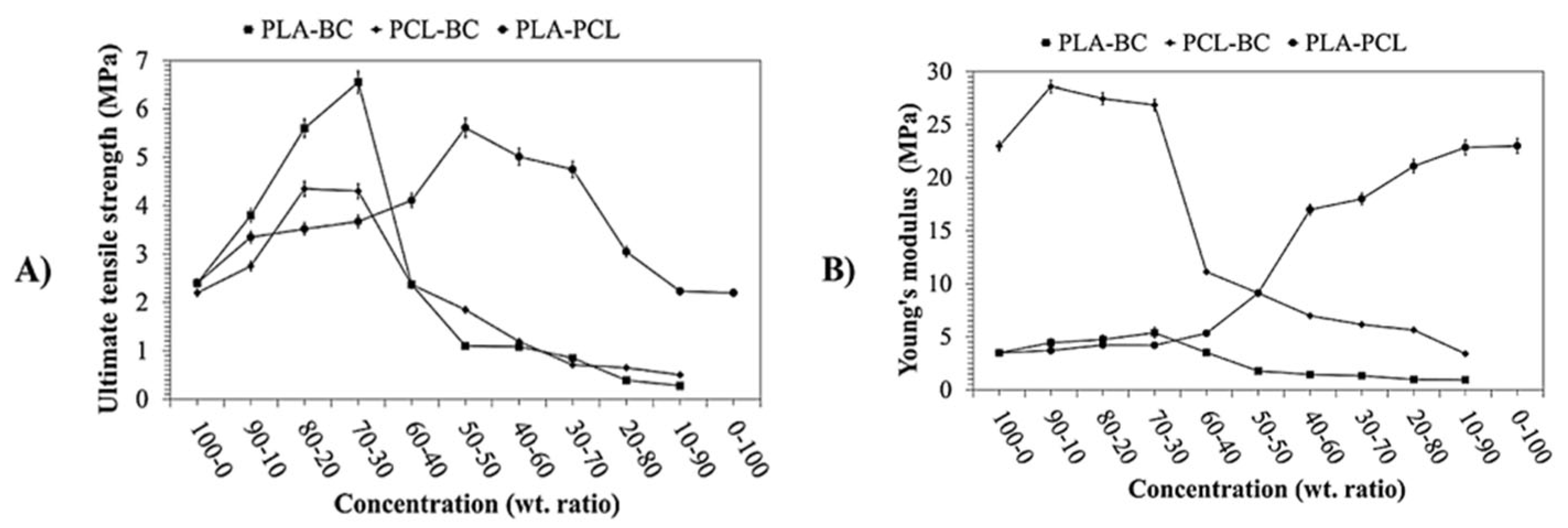
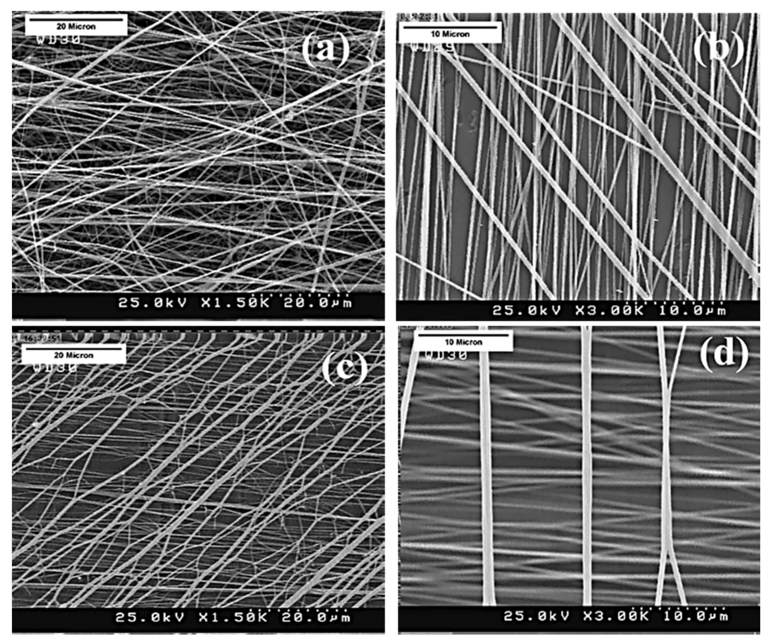
Blending bacterial cellulose (BC) with PCL and PLA via centrifugal spinning demonstrated how composite structures can enhance fiber strength, as shown in Figure 8. The optimized ternary PLA-PCL-BC blend exhibited a tensile strength of 9.1 MPa, balancing the elasticity of PCL with the high mechanical strengths of PLA and BC [125]. In summary, cellulose-based fibers achieve improved mechanical properties through core–shell structures, CNC reinforcement, and BC blending, offering versatile applications in bio-based and high-performance materials.

4.2.4. Starch

Starch, the second most abundant polysaccharide, is found in many plants, including potatoes, as well as tapioca and cornstarch [126,127]. It has diverse applications in food products, packaging, filtration, drug delivery, composites, and textiles, given its cost-effectiveness, biodegradability, and renewability [128,129]. Starch consists of amylose (a linear polymer of α-(1–4) glucose units) and amylopectin (a branched polymer of α-(1–4) glucose units with intermittent divisions of α-(1–6) connections). The presence of intermolecular and intramolecular hydrogen bonds complicates the thermoplastic processing of starch (Figure 9) [129]. Studies on plasticization, as well as physical and genetic modifications, have aimed to enhance the mechanical and functional properties of starch for industrial use. Blending starch with other biodegradable polymers has shown promise in producing materials with improved strength, flexibility, and processability. Its simple structure, composed of glucose units, allows easy breakdown by enzymes from microorganisms, enabling utilization as a carbon and energy source [127].
The mechanical properties of starch-based fibers largely depend on processing techniques and polymer blending. The strong intramolecular hydrogen bonds of starch complicate its processability, but Lancuški et al. [130] demonstrated that formic acid effectively disrupts starch granules, enhancing solubilization and esterification. Electrospun fibers from pure starch in formic acid exhibited a Young’s modulus of 264 MPa, highlighting the role of acid treatment in improving fiber structure.
Blending starch with PVA has been explored to enhance mechanical performance. Afzal et al. [128] showed that increasing PVA content significantly improved tensile strength to 11.54 MPa, owing to PVA’s carbon–carbon backbone and hydroxyl interactions. This approach compensates for starch’s fragility, making it suitable for applications requiring improved mechanical integrity.

4.2.5. Silk Fibroin

Silk fibroin (SF) is a structural protein derived from the silk fibers of domesticated Bombyx mori (B. mori), with a content of the amino acids alanine, glycine, and serine reaching up to 90%, and can be decomposed by enzymes (Figure 9).
Figure 9. The chemical structures of silk fibroin [131] (reprinted with permission).
Molecules 30 03276 g009
Silk fibroin is typically extracted from degummed Bombyx mori silk by dissolving it in either 9.3 M LiBr at 60 °C followed by dialysis or in a hot CaCl2/ethanol/water mixture (1:2:8) followed by dialysis and freeze-drying. These methods yield aqueous silk solutions or powders suitable for further processing [132]. It consists of a fibrous protein called fibroin in the core and glue-like proteins called sericin that cover the fibroin fibers [133]. SF has several beneficial properties, including good biocompatibility, satisfactory toughness, elasticity, minimal inflammatory reaction, and biodegradability, making it suitable for tissue engineering, drug release, and regenerative medicine. It has long been recognized as a high-quality textile fiber and suture material [134].
The mechanical properties of SF stem from its crystallinity and molecular conformation. To enhance the mechanical properties of electrospun silk fibers, researchers use post-processing techniques to induce a conformational transition from random coils to β-sheets [135]. This process converts the random coil conformation of SF into a β-sheet conformation through treatments with organic solvents, such as methanol. Consequently, methanol-treated SF exhibits significantly increased crystallinity and greater dimensional stability [133].
The mechanical properties of silk SF fibers are affected by post-treatment methods, fiber alignment, and polymer blending. Methanol treatment effectively enhances fiber strength by increasing β-sheet content and reinforcing interchain interactions. Amiraliyan et al. [135] reported that methanol-treated SF fibers achieved a tensile strength of 28.53 MPa, whereas elongation at break decreased because of the increased stiffness of the structure.
Further investigations have analyzed the impact of methanol treatment on various fiber configurations. Dodel et al. [134] examined three scaffold structures: random orientation and knitted structures with 60° and 90° interstitial angles. Although methanol treatment did not significantly alter fiber surface morphology, it greatly improved mechanical strength from 4.4 to 15.3 MPa (Figure 10). This improvement was attributed to increased β-sheet formation, which reinforced the fiber structure, and the elongation at break remained largely unchanged in contrast to non-fixed samples.
Fiber alignment also significantly influences mechanical behavior. In a study by Lee et al. [133], increasing the rotational speed of the collector during electrospinning led to improved fiber alignment, which enhanced the orthotropic mechanical properties of both PCL and PCL/SF composite fibers. For instance, adding 2 wt% SF increased the Young’s modulus to 27.47 MPa. However, higher SF content caused aggregation and reduced overall strength and flexibility, showing that both alignment and optimal composition are critical for tuning mechanical response.
Kumkun et al. [46] found that heating significantly enhanced the mechanical properties of SF/PVA scaffolds. Heated fibers exhibited a five-fold increase in tensile strength and a seven-fold improvement in elongation at break because of stronger molecular interactions between SF and PVA, leading to a more flexible polymer network. Overall, SF fibers demonstrate tunable mechanical properties through methanol treatment, fiber orientation control, and polymer blending, making them highly adaptable for biomedical applications such as tissue engineering and wound healing.

4.2.6. PHBV and PHB

The family of polyhydroxyalkanoates (PHAs) has gained significant attention as biocompatible and biodegradable thermoplastic biopolymers. The two most studied polymers in the PHA group are poly(3-hydroxybutyrate) (PHB) and PHBV. Poly(3-hydroxybutyrate-co-3-hydroxyvalerate) (PHBV), a microbial polyester that is biodegradable, non-antigenic, and biocompatible, is a copolymer of polyhydroxybutyrate (PHB) and polyhydroxyvalerate (PHV) [46,136]. PHB is characterized by a high degree of crystallinity and rigidity with a high melting temperature. To enhance its chain flexibility, melting point, and processability, 3-hydroxyvalerate (HV) units are incorporated to create the biopolymer PHBV [137].
PHBV is an environmentally friendly biopolymer that microorganisms can naturally degrade in various environments, depending on the ratio of 3-hydroxybutyrate to 3-hydroxyvalerate. It has a wide range of applications, including in biomedicine, packaging, and agriculture. Because of its natural origin, biocompatibility, and biodegradability, PHBV is ideal for drug delivery systems, tissue engineering, and bioresorbable medical implants.
The mechanical properties of PHBV fibers are influenced by solvent choice, polymer blending, fiber orientation, and processing methods. Kuppan et al. [138] compared electrospun PHBV fibers with solvent-cast 2D films for skin regeneration (Figure 11) and found that whereas the fibers had lower tensile strength (1.42 vs. 1.57 MPa) and Young’s modulus (1028 vs. 5619 MPa), they exhibited superior elasticity, higher porosity, and an accelerated degradation rate, making them more suitable for tissue engineering applications.
Polymer blending has shown significant improvements in the mechanics of PHBV fibers. A study comparing PHB, PHBV, and their blends in chloroform solution revealed that blending PHB with PHBV (75/25 w/w%) increased tensile strength from 1.8 to 2.45 MPa while also enhancing elongation at break. This improvement was attributed to better molecular chain entanglement and reduced brittleness of the polymer network [139].
Solvent choice and fiber orientation also influence mechanical behavior. Xin Lu et al. [136] demonstrated that electrospinning PHBV in 2,2,2-trifluoroethanol (TFE) instead of chloroform significantly improved tensile properties, increasing tensile strain from 3.73% to 25.54% and tensile stress from 1.17 to 1.88 MPa, because of finer fiber diameters (5200 to 690 nm) resulting from the varying solvent conductivity and volatility. Additionally, fiber alignment played a key role: aligned nanofibers exhibited a tensile stress of 8.86 MPa but lower strain (7.77%), whereas randomly oriented fibers showed significantly higher strain (71.49%) but reduced tensile stress (1.62 MPa).
Incorporating PHBV into PLLA fibers has been explored to enhance stiffness and flexibility. Increasing the PHBV content to 30% in PLLA fibers raised Young’s modulus from 41.5 to 73.5 MPa, attributed to stronger molecular interactions and enhanced crystallinity during electrospinning [140].
Alternative fiber fabrication methods, such as centrifugal spinning, have also been investigated. Upson et al. [141] produced PHBV fibers at 9000 rpm, achieving tensile strengths of 3 MPa and a Young’s modulus of 100 MPa, comparable to electrospun PHBV fibers, which typically exhibit strengths of 1.76 MPa and moduli of 125.7 MPa. Although centrifugal spinning resulted in slightly lower stiffness, it offered the advantage of higher production rates.
Nanoparticle incorporation has limited influence on the mechanics of PHBV fibers. Mayorga et al. [142] found that CuO nanoparticles, whether integrated via melt mixing or coating, did not significantly affect mechanical properties because of their low loading levels and uniform dispersion. Similar trends were observed with silver nanoparticles, where finely dispersed AgNPs had minimal impact on fiber strength and flexibility, suggesting that structural modifications may be a more effective strategy for enhancing PHBV fiber mechanics rather than adding nanofillers [137,143].
Therefore, PHBV fibers exhibit tunable mechanical properties through polymer blending, solvent selection, fiber orientation, and alternative processing methods. Although blending and controlled processing significantly enhance strength and elasticity, nanoparticle incorporation appears to have minimal effect, emphasizing the importance of optimizing polymer interactions and fiber morphology for improved mechanical performance.
Table 2 summarizes the mechanical behavior of electrospun fibers derived from natural biopolymers and their composites. It outlines techniques such as blending with synthetic polymers, reinforcement with nanofillers, and post-processing treatments aimed at improving strength, flexibility, or structural integrity.
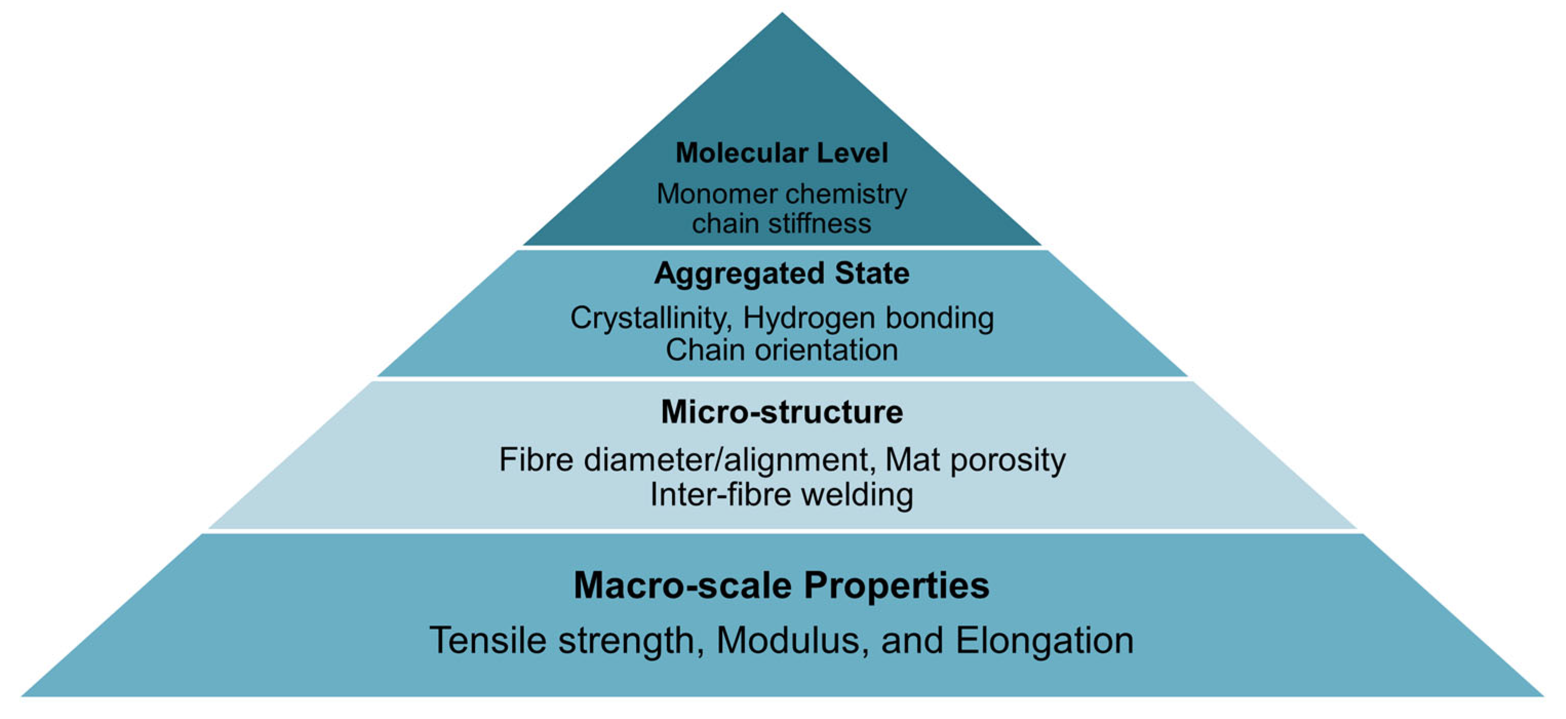
To highlight the multiscale interactions that impact mechanical behavior in biodegradable fiber systems, a schematic structure–property pyramid (Figure 12) has been added. This diagram links molecular features (e.g., monomer chemistry, tacticity), aggregated-state attributes (e.g., crystallinity, hydrogen bonding), and microstructural factors (e.g., fiber alignment, mat porosity) to macroscale performance indicators such as tensile strength, modulus, and elongation [148]. It illustrates how processing strategies and material modifications at different scales collectively shape the final mechanical performance of fiber mats.

5. Future Perspectives

Biodegradable fibers and mats have a promising future as the demand for sustainable materials grows. Research will continue to focus on enhancing the strength, durability, and scalability of these fibers. A key challenge is improving the mechanical properties of natural and synthetic biodegradable polymers to replace traditional materials across various industries. New developments, such as combining different polymers, adding nanoparticles, and using specialized treatments, can enhance fiber strength and flexibility. Simultaneously, finding eco-friendly production methods will be essential to reduce waste and pollution. With further advancements, biodegradable fibers will find greater utility in medicine, packaging, and textiles, contributing to a more sustainable future. On the other hand, looking ahead, artificial intelligence and computational modeling offer exciting opportunities to predict and tailor mechanical behavior in biodegradable electrospun fibers. For instance, a recent study used machine learning models (including GAMLSS, decision trees, and neural networks) to predict fiber diameter, tensile strength, and modulus in gelatin-based fibers spun from green solvent blends, achieving significantly improved accuracy versus traditional regression approaches [149]. In another investigation, machine learning models (CART, SVM) accurately identified the structure–property relationships in crosslinked PVA electrospun scaffolds, reaching prediction R2 values of up to 0.93 for strength and modulus [150].

6. Conclusions

This paper reviews the mechanical properties of biodegradable fibers and mats on the basis of their production methods and whether they are synthetic or natural polymers. It highlights their potential as sustainable alternatives for various applications. The analysis elucidates that the intrinsic properties of biodegradable polymers such as PVA, PEO, PLA, PHBV, PCL, PGA, chitosan, collagen, starch, alginate, silk, and cellulose, alongside their processing techniques, significantly affect the mechanical strengths and limitations of the resulting fibrous structures. It was shown that fibers processed via electrospinning show promise because of their enhanced surface area and porosity, whereas melt-spinning and wet-spinning techniques are advantageous for industrial scalability and customizability. Including nanocomposites, chemical treatments, and fiber orientation strategies are key enhancement methods for tailoring mechanical properties such as tensile strength and elasticity. This review paves the way for future research to bridge the gap between material properties and application requirements. It aims to provide better insight for researchers in selecting the best fiber production methods, mechanical evaluation techniques, and appropriate polymers on the basis of their mechanical properties and applications. It also suggests several techniques for improving these properties. As the demand for bio-based and high-performance materials grows, the optimization of mechanical properties will play a critical role in expanding the applicability of biodegradable fibers.

Funding

The authors wish to acknowledge the financial support provided by the Natural Sciences and Engineering Research Council of Canada, NSERC (ALLRP 584745-23).

Acknowledgments

We thank NSERC (CRSNG), Prima Québec, CIIN, CISD, and CREPEC for their support.

Conflicts of Interest

The authors declare no conflict of interest.

References

  1. Meng, C.; Tang, D.; Liu, X.; Meng, J.; Wei, W.; Gong, R.H.; Li, J. Heterogeneous porous PLLA/PCL fibrous scaffold for bone tissue regeneration. Int. J. Biol. Macromol. 2023, 235, 123781. [Google Scholar] [CrossRef]
  2. Huang, Q.; Meng, C.; Liao, M.; Kou, T.; Zhou, F.; Lu, J.R.; Li, J.; Li, Y. Hierarchically porous, superhydrophobic plla/copper composite fibrous membranes for air filtration. ACS Appl. Polym. Mater. 2024, 6, 2381–2391. [Google Scholar] [CrossRef]
  3. Kaniuk, E.; Lechowska-Liszka, A.; Gajek, M.; Nikodem, A.; Ścisłowska-Czarnecka, A.; Rapacz-Kmita, A.; Stodolak-Zych, E. Correlation between porosity and physicochemical and biological properties of electrospinning PLA/PVA membranes for skin regeneration. Biomater. Adv. 2023, 152, 213506. [Google Scholar] [CrossRef] [PubMed]
  4. Song, J.; Zhao, Q.; Meng, C.; Meng, J.; Chen, Z.; Li, J. Hierarchical porous recycled PET nanofibers for high-efficiency aerosols and virus capturing. ACS Appl. Mater. Interfaces 2021, 13, 49380–49389. [Google Scholar] [CrossRef] [PubMed]
  5. Zhang, Y.; Jian, Y.; Jiang, X.; Li, X.; Wu, X.; Zhong, J.; Jia, X.; Li, Q.; Wang, X.; Zhao, K.; et al. Stepwise degradable PGA-SF core-shell electrospinning scaffold with superior tenacity in wetting regime for promoting bone regeneration. Mater. Today Bio 2024, 26, 101023. [Google Scholar] [CrossRef]
  6. Cao, H.; Chai, S.; Tan, Z.; Wu, H.; Mao, X.; Wei, L.; Zhou, F.; Sun, R.; Liu, C. Recent advances in physical sensors based on electrospinning technology. ACS Mater. Lett. 2023, 5, 1627–1648. [Google Scholar] [CrossRef]
  7. Rahim Labbafzadeh, M.; Vakili, M.H. Application of magnetic electrospun polyvinyl alcohol/collagen nanofibres for drug delivery systems. Mol. Simul. 2022, 48, 1–7. [Google Scholar] [CrossRef]
  8. Idrees, H.; Zaidi, S.Z.J.; Sabir, A.; Khan, R.U.; Zhang, X.; Hassan, S.-U. A Review of Biodegradable Natural Polymer-Based Nanoparticles for Drug Delivery Applications. Nanomaterials 2020, 10, 1970. [Google Scholar] [CrossRef]
  9. Karthik, T.; Rathinamoorthy, R. Nonwovens: Process, Structure, Properties and Applications; CRC Press: Boca Raton, FL, USA, 2017. [Google Scholar]
  10. Leal, A.A.; Deitzel, J.M.; Gillespie, J.W. Compressive Strength Analysis for High Performance Fibers with Different Modulus in Tension and Compression. J. Compos. Mater. 2009, 43, 661–674. [Google Scholar] [CrossRef]
  11. Cleeton, C.; Keirouz, A.; Chen, X.; Radacsi, N. Electrospun nanofibers for drug delivery and biosensing. ACS Biomater. Sci. Eng. 2019, 5, 4183–4205. [Google Scholar] [CrossRef]
  12. Rahim Labbafzadeh, M.; Vakili, M.H. Electrospinning of Polymeric Nanofibers for Tissue Engineering Applications: A Review. Tissue Eng. 2006, 12, 1197–1211. [Google Scholar] [CrossRef]
  13. Ramazani, S.; Karimi, M. Aligned poly (ε-caprolactone)/graphene oxide and reduced graphene oxide nanocomposite nanofibers: Morphological, mechanical and structural properties. Mater. Sci. Eng. C 2015, 56, 325–334. [Google Scholar] [CrossRef]
  14. Meng, C.; Song, J.; Malekmohammadi, S.; Meng, J.; Wei, W.; Li, R.; Feng, J.; Gong, R.H.; Li, J. Hierarchical porous poly (L-lactic acid) fibrous vascular graft with controllable architectures and stable structure. Mater. Des. 2024, 240, 112829. [Google Scholar] [CrossRef]
  15. Pisani, S.; Dorati, R.; Conti, B.; Modena, T.; Bruni, G.; Genta, I. Design of copolymer PLA-PCL electrospun matrix for biomedical applications. React. Funct. Polym. 2018, 124, 77–89. [Google Scholar] [CrossRef]
  16. Nicknejad, E.T.; Ghoreishi, S.M.; Habibi, N. Electrospinning of cross-linked magnetic chitosan nanofibers for protein release. AAPS PharmSciTech 2015, 16, 1480–1486. [Google Scholar] [CrossRef]
  17. Merchiers, J.; Martínez Narváez, C.D.V.; Slykas, C.; Buntinx, M.; Deferme, W.; D’Haen, J.; Peeters, R.; Sharma, V.; Reddy, N.K. Centrifugally spun poly(ethylene oxide) fibers rival the properties of electrospun fibers. J. Polym. Sci. 2021, 59, 2754–2762. [Google Scholar] [CrossRef]
  18. Rahmat, M.; Karrabi, M.; Ghasemi, I.; Zandi, M.; Azizi, H. Silane crosslinking of electrospun poly (lactic acid)/nanocrystalline cellulose bionanocomposite. Mater. Sci. Eng. C Mater. Biol. Appl. 2016, 68, 397–405. [Google Scholar] [CrossRef]
  19. Eatemadi, A.; Daraee, H.; Zarghami, N.; Melat Yar, H.; Akbarzadeh, A. Nanofiber: Synthesis and biomedical applications. Artif. Cells Nanomed. Biotechnol. 2016, 44, 111–121. [Google Scholar] [CrossRef] [PubMed]
  20. Raksa, A.; Numpaisal, P.-o.; Ruksakulpiwat, Y. The effect of humidity during electrospinning on morphology and mechanical properties of SF/PVA nanofibers. Mater. Today Proc. 2021, 47, 3458–3461. [Google Scholar] [CrossRef]
  21. Nadaf, A.; Gupta, A.; Hasan, N.; Ahmad, S.; Kesharwani, P.; Ahmad, F.J. Recent update on electrospinning and electrospun nanofibers: Current trends and their applications. RSC Adv. 2022, 12, 23808–23828. [Google Scholar] [CrossRef] [PubMed]
  22. Šišková, A.O.; Peer, P.; Andicsová, A.E.; Jordanov, I.; Rychter, P. Circulatory management of polymer waste: Recycling into fine fibers and their applications. Materials 2021, 14, 4694. [Google Scholar] [CrossRef]
  23. Maqsood, M.; Langensiepen, F.; Seide, G. Investigation of melt spinnability of plasticized polylactic acid biocomposites-containing intumescent flame retardant. J. Therm. Anal. Calorim. 2020, 139, 305–318. [Google Scholar] [CrossRef]
  24. Mirbaha, H.; Nourpanah, P.; Scardi, P.; D’incau, M.; Greco, G.; Valentini, L.; Bittolo Bon, S.; Arbab, S.; Pugno, N. The impact of shear and elongational forces on structural formation of polyacrylonitrile/carbon nanotubes composite fibers during wet spinning process. Materials 2019, 12, 2797. [Google Scholar] [CrossRef]
  25. Chua, M.H.; Cheng, W.; Goh, S.S.; Kong, J.; Li, B.; Lim, J.Y.; Mao, L.; Wang, S.; Xue, K.; Yang, L.; et al. Face masks in the new COVID-19 normal: Materials, testing, and perspectives. Research 2020, 2020, 7286735. [Google Scholar] [CrossRef]
  26. Esmaeili, J.; Ghoraishizadeh, S.; Farzan, M.; Barati, A.; Salehi, E.; Ai, J. Fabrication and evaluation of a soy protein isolate/collagen/sodium alginate multifunctional bilayered wound dressing: Release of cinnamaldehyde, artemisia absinthium, and oxygen. ACS Appl. Bio Mater. 2024, 7, 5470–5482. [Google Scholar] [CrossRef]
  27. Castro, C.; Juárez, D.A.; Arizmendi-Morquecho, A.; Gonzalez-Perez, G.; Estrada-Villegas, G.M. The effects of relative humidity and salt concentration on the diameter of hydrophilic polymeric nanofibers obtained by electrospinning: Synergistic effect study by Central Composite Design (CCD). React. Funct. Polym. 2024, 203, 106013. [Google Scholar] [CrossRef]
  28. Zhou, Y.; Liu, Y.; Zhang, M.; Feng, Z.; Yu, D.-G.; Wang, K. Electrospun nanofiber membranes for air filtration: A review. Nanomaterials 2022, 12, 1077. [Google Scholar] [CrossRef]
  29. Zulkifli, M.Z.A.; Nordin, D.; Shaari, N.; Kamarudin, S.K. Overview of Electrospinning for Tissue Engineering Applications. Polymers 2023, 15, 2418. [Google Scholar] [CrossRef] [PubMed]
  30. Song, J.; Murillo, L.L.; Yang, K.; Wang, T.; Li, J.; Li, Y.; Chen, Y.; Chen, Z. Revisable and high-strength wheel-spun alginate/graphene oxide based fibrous rods towards a flexible and biodegradable rib internal fixation system. Int. J. Biol. Macromol. 2022, 219, 1308–1318. [Google Scholar] [CrossRef] [PubMed]
  31. Zhuang, J.; Liu, Y.; Bian, D.; He, F.; Li, Y.; Fan, W. Spinning solution viscosity reducing and wet spinning of carbon black-based elastic conductive fibers for sports monitoring and healthcare electrical heating. J. Mater. Res. Technol. 2025, 36, 1–12. [Google Scholar] [CrossRef]
  32. Mejía Suaza, M.L.; Hurtado Henao, Y.; Moncada Acevedo, M.E. Wet electrospinning and its applications: A review. TecnoLógicas 2022, 25, e2223. [Google Scholar] [CrossRef]
  33. Sikdar, P.; S Bhat, G.; Hinchliffe, D.; Islam, S.; Condon, B. Microstructure and physical properties of composite nonwovens produced by incorporating cotton fibers in elastic spunbond and meltblown webs for medical textiles. J. Ind. Text. 2022, 51 (Suppl. 4), 6028S–6050S. [Google Scholar] [CrossRef]
  34. Bunsell, A.R. Handbook of Tensile Properties of Textile and Technical Fibres; Elsevier: Amsterdam, The Netherlands, 2009. [Google Scholar]
  35. kumar Gurajala, N.; Sanjeev, L.; Teja, B.; Kumar, T.S.; Manikanta, J.E. An overview of mechanical properties of biodegradable polymers and natural fibre materials. Mater. Today Proc. 2023. [Google Scholar] [CrossRef]
  36. Baker, S.R.; Banerjee, S.; Bonin, K.; Guthold, M. Determining the mechanical properties of electrospun poly-epsilon-caprolactone (PCL) nanofibers using AFM and a novel fiber anchoring technique. Mater. Sci. Eng. C Mater. Biol. Appl. 2016, 59, 203–212. [Google Scholar] [CrossRef] [PubMed]
  37. Rashid, T.U.; Gorga, R.E.; Krause, W.E. Mechanical Properties of Electrospun Fibers—A Critical Review. Adv. Eng. Mater. 2021, 23, 2100153. [Google Scholar] [CrossRef]
  38. Stachewicz, U.; Bailey, R.J.; Wang, W.; Barber, A.H. Size dependent mechanical properties of electrospun polymer fibers from a composite structure. Polymer 2012, 53, 5132–5137. [Google Scholar] [CrossRef]
  39. Costa, C.; Fonseca, A.; Serra, A.; Coelho, J. Dynamic mechanical thermal analysis of polymer composites reinforced with natural fibers. Polym. Rev. 2016, 56, 362–383. [Google Scholar] [CrossRef]
  40. Saba, N.; Jawaid, M.; Alothman, O.Y.; Paridah, M. A review on dynamic mechanical properties of natural fibre reinforced polymer composites. Constr. Build. Mater. 2016, 106, 149–159. [Google Scholar] [CrossRef]
  41. Li, X.; Bhushan, B.; Takashima, K.; Baek, C.-W.; Kim, Y.-K. Mechanical characterization of micro/nanoscale structures for MEMS/NEMS applications using nanoindentation techniques. Ultramicroscopy 2003, 97, 481–494. [Google Scholar] [CrossRef]
  42. Santiago-Castillo, K.; Torres-Huerta, A.M.; Del Angel-Lopez, D.; Dominguez-Crespo, M.A.; Dorantes-Rosales, H.; Palma-Ramirez, D.; Willcock, H. In Situ Growth of Silver Nanoparticles on Chitosan Matrix for the Synthesis of Hybrid Electrospun Fibers: Analysis of Microstructural and Mechanical Properties. Polymers 2022, 14, 674. [Google Scholar] [CrossRef]
  43. Xu, Y.; Xu, Y.; Sun, C.; Zou, L.; He, J. The preparation and characterization of plasticized PVA fibres by a novel Glycerol/Pseudo Ionic Liquids system with melt spinning method. Eur. Polym. J. 2020, 133, 109768. [Google Scholar] [CrossRef]
  44. Bazzi, M.; Shabani, I.; Mohandesi, J.A. Enhanced mechanical properties and electrical conductivity of Chitosan/Polyvinyl Alcohol electrospun nanofibers by incorporation of graphene nanoplatelets. J. Mech. Behav. Biomed. Mater. 2022, 125, 104975. [Google Scholar] [CrossRef]
  45. Du, L.; Xu, H.; Li, T.; Zhang, Y.; Zou, F. Fabrication of silver nanoparticle/polyvinyl alcohol/polycaprolactone hybrid nanofibers nonwovens by two-nozzle electrospinning for wound dressing. Fibers Polym. 2017, 17, 1995–2005. [Google Scholar] [CrossRef]
  46. Kumkun, P.; Tuancharoensri, N.; Ross, G.; Mahasaranon, S.; Jongjitwimol, J.; Topham, P.D.; Ross, S. Green fabrication route of robust, biodegradable silk sericin and poly(vinyl alcohol) nanofibrous scaffolds. Polym. Int. 2019, 68, 1903–1913. [Google Scholar] [CrossRef]
  47. Alonso-Lopez, O.; Lopez-Ibanez, S.; Beiras, R. Assessment of Toxicity and Biodegradability of Poly(vinyl alcohol)-Based Materials in Marine Water. Polymers 2021, 13, 3742. [Google Scholar] [CrossRef]
  48. Chiellini, E.; Corti, A.; D’Antone, S.; Solaro, R. Biodegradation of poly (vinyl alcohol) based materials. Prog. Polym. Sci. 2003, 28, 963–1014. [Google Scholar] [CrossRef]
  49. Kalia, S.; Dufresne, A.; Cherian, B.M.; Kaith, B.S.; Avérous, L.; Njuguna, J.; Nassiopoulos, E. Cellulose-Based Bio- and Nanocomposites: A Review. Int. J. Polym. Sci. 2011, 2011, 1–35. [Google Scholar] [CrossRef]
  50. Lee, J.; Deng, Y. Increased mechanical properties of aligned and isotropic electrospun PVA nanofiber webs by cellulose nanowhisker reinforcement. Macromol. Res. 2011, 20, 76–83. [Google Scholar] [CrossRef]
  51. Liu, Y.; Liu, Y.; Liao, N.; Cui, F.; Park, M.; Kim, H.Y. Fabrication and durable antibacterial properties of electrospun chitosan nanofibers with silver nanoparticles. Int. J. Biol. Macromol. 2015, 79, 638–643. [Google Scholar] [CrossRef]
  52. Cho, D.; Netravali, A.N.; Joo, Y.L. Mechanical properties and biodegradability of electrospun soy protein Isolate/PVA hybrid nanofibers. Polym. Degrad. Stab. 2012, 97, 747–754. [Google Scholar] [CrossRef]
  53. Ngadiman, N.H.; Yusof, N.M.; Idris, A.; Misran, E.; Kurniawan, D. Development of highly porous biodegradable gamma-Fe(2)O(3)/polyvinyl alcohol nanofiber mats using electrospinning process for biomedical application. Mater. Sci. Eng. C Mater. Biol. Appl. 2017, 70, 520–534. [Google Scholar] [CrossRef]
  54. Wu, Q.; Chen, N.; Li, L.; Wang, Q. Structure evolution of melt-spun poly(vinyl alcohol) fibers during hot-drawing. J. Appl. Polym. Sci. 2011, 124, 421–428. [Google Scholar] [CrossRef]
  55. Hoare, T.R.; Kohane, D.S. Hydrogels in drug delivery: Progress and challenges. Polymer 2008, 49, 1993–2007. [Google Scholar] [CrossRef]
  56. Hodge, J.; Quint, C. The improvement of cell infiltration in an electrospun scaffold with multiple synthetic biodegradable polymers using sacrificial PEO microparticles. J. Biomed. Mater. Res. A 2019, 107, 1954–1964. [Google Scholar] [CrossRef]
  57. Kupka, V.; Dvorakova, E.; Manakhov, A.; Michlicek, M.; Petrus, J.; Vojtova, L.; Zajickova, L. Well-Blended PCL/PEO Electrospun Nanofibers with Functional Properties Enhanced by Plasma Processing. Polymers 2020, 12, 1403. [Google Scholar] [CrossRef]
  58. Li, C.; Huang, Y.; Li, R.; Wang, Y.; Xiang, X.; Zhang, C.; Wang, D.; Zhou, Y.; Liu, X.; Xu, W. Fabrication and properties of carboxymethyl chitosan/polyethylene oxide composite nonwoven mats by centrifugal spinning. Carbohydr. Polym. 2021, 251, 117037. [Google Scholar] [CrossRef] [PubMed]
  59. Li, X.; Lu, Y.; Hou, T.; Zhou, J.; Yang, B. Centrifugally spun ultrafine starch/PEO fibres as release formulation for poorly water-soluble drugs. Micro Nano Lett. 2018, 13, 1688–1692. [Google Scholar] [CrossRef]
  60. Danhier, F.; Ansorena, E.; Silva, J.M.; Coco, R.; Le Breton, A.; Preat, V. PLGA-based nanoparticles: An overview of biomedical applications. J. Control. Release 2012, 161, 505–522. [Google Scholar] [CrossRef]
  61. Aghdam, R.M.; Najarian, S.; Shakhesi, S.; Khanlari, S.; Shaabani, K.; Sharifi, S. Investigating the effect of PGA on physical and mechanical properties of electrospun PCL/PGA blend nanofibers. J. Appl. Polym. Sci. 2011, 124, 123–131. [Google Scholar] [CrossRef]
  62. Hajiali, H.; Shahgasempour, S.; Naimi-Jamal, M.R.; Peirovi, H. Electrospun PGA/gelatin nanofibrous scaffolds and their potential application in vascular tissue engineering. Int. J. Nanomed. 2011, 6, 2133–2141. [Google Scholar] [CrossRef]
  63. Ramdhanie, L.I.; Aubuchon, S.R.; Boland, E.D.; Knapp, D.C.; Barnes, C.P.; Simpson, D.G.; Wnek, G.E.; Bowlin, G.L. Thermal and Mechanical Characterization of Electrospun Blends of Poly(lactic acid) and Poly(glycolic acid). Polym. J. 2006, 38, 1137–1145. [Google Scholar] [CrossRef]
  64. Li, W.J.; Cooper, J.A., Jr.; Mauck, R.L.; Tuan, R.S. Fabrication and characterization of six electrospun poly(alpha-hydroxy ester)-based fibrous scaffolds for tissue engineering applications. Acta Biomater. 2006, 2, 377–385. [Google Scholar] [CrossRef] [PubMed]
  65. D’Amato, A.R.; Bramson, M.T.K.; Puhl, D.L.; Johnson, J.; Corr, D.T.; Gilbert, R.J. Solvent retention in electrospun fibers affects scaffold mechanical properties. Electrospinning 2018, 2, 15–28. [Google Scholar] [CrossRef]
  66. Bazgir, M.; Saeinasab, M.; Zhang, W.; Zhang, X.; Min Tsui, K.; Maasoumi Sarvestani, A.; Nawaz, S.; Coates, P.; Youseffi, M.; Elies, J.; et al. Investigation of Cell Adhesion and Cell Viability of the Endothelial and Fibroblast Cells on Electrospun PCL, PLGA and Coaxial Scaffolds for Production of Tissue Engineered Blood Vessel. J. Funct. Biomater. 2022, 13, 282. [Google Scholar] [CrossRef]
  67. Chou, S.F.; Woodrow, K.A. Relationships between mechanical properties and drug release from electrospun fibers of PCL and PLGA blends. J. Mech. Behav. Biomed. Mater. 2017, 65, 724–733. [Google Scholar] [CrossRef]
  68. Elder, S.; Roberson, J.G.; Warren, J.; Lawson, R.; Young, D.; Stokes, S.; Ross, M.K. Evaluation of Electrospun PCL-PLGA for Sustained Delivery of Kartogenin. Molecules 2022, 27, 3739. [Google Scholar] [CrossRef]
  69. Anderson, J.M.; Shive, M.S. Biodegradation and biocompatibility of PLA and PLGA microspheres. Adv. Drug Deliv. Rev. 2012, 64, 72–82. [Google Scholar] [CrossRef]
  70. Farah, S.; Anderson, D.G.; Langer, R. Physical and mechanical properties of PLA, and their functions in widespread applications—A comprehensive review. Adv. Drug Deliv. Rev. 2016, 107, 367–392. [Google Scholar] [CrossRef] [PubMed]
  71. Gentile, P.; Chiono, V.; Carmagnola, I.; Hatton, P.V. An overview of poly (lactic-co-glycolic) acid (PLGA)-based biomaterials for bone tissue engineering. Int. J. Mol. Sci. 2014, 15, 3640–3659. [Google Scholar] [CrossRef] [PubMed]
  72. Lins, L.C.; Wianny, F.; Livi, S.; Hidalgo, I.A.; Dehay, C.; Duchet-Rumeau, J.; Gerard, J.F. Development of Bioresorbable Hydrophilic-Hydrophobic Electrospun Scaffolds for Neural Tissue Engineering. Biomacromolecules 2016, 17, 3172–3187. [Google Scholar] [CrossRef]
  73. Arbeiter, D.; Kohse, S.; Eickner, T.; Grabow, N.; Schmitz, K.-P. Influence of additives on physico-chemical properties of electrospun poly(L-lactide). Curr. Dir. Biomed. Eng. 2018, 4, 493–496. [Google Scholar] [CrossRef]
  74. Qiao, T.; Song, P.; Guo, H.; Song, X.; Zhang, B.; Chen, X. Reinforced electrospun PLLA fiber membrane via chemical crosslinking. Eur. Polym. J. 2016, 74, 101–108. [Google Scholar] [CrossRef]
  75. Goreninskii, S.I.; Stankevich, K.S.; Nemoykina, A.L.; Bolbasov, E.N.; Tverdokhlebov, S.I.; Filimonov, V.D. A first method for preparation of biodegradable fibrous scaffolds containing iodine on the fibre surfaces. Bull. Mater. Sci. 2018, 41, 100. [Google Scholar] [CrossRef]
  76. Song, J.; Zhang, B.; Lu, Z.; Xin, Z.; Liu, T.; Wei, W.; Zia, Q.; Pan, K.; Gong, R.H.; Bian, L.; et al. Hierarchical porous poly (l-lactic acid) nanofibrous membrane for ultrafine particulate aerosol filtration. ACS Appl. Mater. Interfaces 2019, 11, 46261–46268. [Google Scholar] [CrossRef]
  77. Lu, Z.; Zhang, B.; Gong, H.; Li, J. Fabrication of hierarchical porous poly (l-lactide)(PLLA) fibrous membrane by electrospinning. Polymer 2021, 226, 123797. [Google Scholar] [CrossRef]
  78. Didekhani, R.; Sohrabi, M.R.; Seyedjafari, E.; Soleimani, M.; Hanaee-Ahvaz, H. Electrospun composite PLLA/Oyster shell scaffold enhances proliferation and osteogenic differentiation of stem cells. Biologicals 2018, 54, 33–38. [Google Scholar] [CrossRef] [PubMed]
  79. Liu, C.; Du, G.; Guo, Q.; Li, R.; Li, C.; He, H. Fabrication and Characterization of Polylactic Acid Electrospun Wound Dressing Modified with Polyethylene Glycol, Rosmarinic Acid and Graphite Oxide. Nanomaterials 2023, 13, 2000. [Google Scholar] [CrossRef]
  80. Meng, C.; Liu, X.; Li, J. Hierarchical porous PLLA/ACP fibrous membrane towards bone tissue scaffold. J. Mech. Behav. Biomed. Mater. 2024, 152, 106455. [Google Scholar] [CrossRef]
  81. Kai, D.; Ren, W.; Tian, L.; Chee, P.L.; Liu, Y.; Ramakrishna, S.; Loh, X.J. Engineering Poly(lactide)–Lignin Nanofibers with Antioxidant Activity for Biomedical Application. ACS Sustain. Chem. Eng. 2016, 4, 5268–5276. [Google Scholar] [CrossRef]
  82. Shamsah, A.H.; Cartmell, S.H.; Richardson, S.M.; Bosworth, L.A. Material Characterization of PCL:PLLA Electrospun Fibers Following Six Months Degradation In Vitro. Polymers 2020, 12, 700. [Google Scholar] [CrossRef]
  83. Feng, J. Preparation and properties of poly(lactic acid) fiber melt blown non-woven disordered mats. Mater. Lett. 2017, 189, 180–183. [Google Scholar] [CrossRef]
  84. Zhou, H.; Tang, Y.; Wang, Z.; Zhang, P.; Zhu, Q. Cotton-like micro- and nanoscale poly(lactic acid) nonwoven fibers fabricated by centrifugal melt-spinning for tissue engineering. RSC Adv. 2018, 8, 5166–5179. [Google Scholar] [CrossRef]
  85. Boschetto, F.; Doan, H.N.; Vo, P.P.; Zanocco, M.; Zhu, W.; Sakai, W.; Kinashi, K.; Marin, E.; Pezzotti, G. Effect of BaTiO3 on the aging process of PLA fibers obtained by centrifugal spinning. Mater. Today Chem. 2021, 20, 100461. [Google Scholar] [CrossRef]
  86. Chen, X.-X.; Yu, J.-C.; Chen, K.; Ji, P.; Chen, X.-L.; Pan, Z.-J. Facile and Large-scale Fabrication of Biodegradable Thermochromic Fibers Based on Poly(lactic acid). Chin. J. Polym. Sci. 2022, 40, 1242–1251. [Google Scholar] [CrossRef]
  87. Hassan, E.A.M.; Elarabi, S.E.; Wei, Y.; Yu, M. Biodegradable poly (lactic acid)/poly (butylene succinate) fibers with high elongation for health care products. Text. Res. J. 2017, 88, 1735–1744. [Google Scholar] [CrossRef]
  88. Wu, J.-h.; Hu, T.-g.; Wang, H.; Zong, M.-h.; Wu, H.; Wen, P. Electrospinning of PLA nanofibers: Recent advances and its potential application for food packaging. J. Agric. Food Chem. 2022, 70, 8207–8221. [Google Scholar] [CrossRef] [PubMed]
  89. Meng, X.; Jia, X.; Qi, Y.; Miao, D.; Yan, X. Fabrication of polylactic acid nanofibrous yarns for piezoelectric fabrics. e-Polymers 2023, 23, 20230030. [Google Scholar] [CrossRef]
  90. Neffe, A.T.; Zhang, Q.; Hommes-Schattmann, P.J.; Wang, W.; Xu, X.; Ahmad, B.S.; Williams, G.R.; Lendlein, A. Functionalizable coaxial PLLA/PDLA nanofibers with stereocomplexes at the internal interface. J. Mater. Res. 2021, 36, 2995–3009. [Google Scholar] [CrossRef]
  91. Kheradvar Kolour, A.; Ghoraishizadeh, S.; Zaman, M.S.; Alemzade, A.; Banavand, M.; Esmaeili, J.; Shahrousvand, M. Janus films wound dressing comprising electrospun gelatin/PCL nanofibers and gelatin/honey/curcumin thawed layer. ACS Appl. Bio Mater. 2024, 7, 8642–8655. [Google Scholar] [CrossRef] [PubMed]
  92. Woodruff, M.A.; Hutmacher, D.W. The return of a forgotten polymer—Polycaprolactone in the 21st century. Prog. Polym. Sci. 2010, 35, 1217–1256. [Google Scholar] [CrossRef]
  93. Alharbi, N.; Daraei, A.; Lee, H.; Guthold, M. The effect of molecular weight and fiber diameter on the mechanical properties of single, electrospun PCL nanofibers. Mater. Today Commun. 2023, 35, 105773. [Google Scholar] [CrossRef]
  94. Mobarakeh, A.I.; Ramsheh, A.S.; Khanshan, A.; Aghaei, S.; Mirbagheri, M.S.; Esmaeili, J. Fabrication and evaluation of a bi-layered electrospun PCL/PVA patch for wound healing: Release of vitamins and silver nanoparticle. Heliyon 2024, 10, e33178. [Google Scholar] [CrossRef]
  95. Ray, S.S.; Bousmina, M. Biodegradable polymers and their layered silicate nanocomposites: In greening the 21st century materials world. Prog. Mater. Sci. 2005, 50, 962–1079. [Google Scholar] [CrossRef]
  96. Yalcin Enis, I.; Gok Sadikoglu, T.; Horakova, J.; Lukas, D. The post-morphological analysis of electrospun vascular grafts following mechanical testing. J. Polym. Eng. 2018, 38, 525–535. [Google Scholar] [CrossRef]
  97. Zhang, S.; Yan, D.; Zhao, L.; Lin, J. Composite fibrous membrane comprising PLA and PCL fibers for biomedical application. Compos. Commun. 2022, 34, 101268. [Google Scholar] [CrossRef]
  98. Sharma, D.; Satapathy, B.K. Tuning structural-response of PLA/PCL based electrospun nanofibrous mats: Role of dielectric-constant and electrical-conductivity of the solvent system. J. Biomater. Sci. Polym. Ed. 2022, 33, 1759–1793. [Google Scholar] [CrossRef]
  99. Maleki, H.; Gharehaghaji, A.A.; Moroni, L.; Dijkstra, P.J. Influence of the solvent type on the morphology and mechanical properties of electrospun PLLA yarns. Biofabrication 2013, 5, 035014. [Google Scholar] [CrossRef] [PubMed]
  100. Sharma, D.; Satapathy, B.K. Understanding release kinetics and collapse proof suture retention response of curcumin loaded electrospun mats based on aliphatic polyesters and their blends. J. Mech. Behav. Biomed. Mater. 2021, 120, 104556. [Google Scholar] [CrossRef]
  101. Volokhova, A.A.; Kudryavtseva, V.L.; Spiridonova, T.I.; Kolesnik, I.; Goreninskii, S.I.; Sazonov, R.V.; Remnev, G.E.; Tverdokhlebov, S.I. Controlled drug release from electrospun PCL non-woven scaffolds via multi-layering and e-beam treatment. Mater. Today Commun. 2021, 26, 102134. [Google Scholar] [CrossRef]
  102. Li, H.; Zhu, C.; Xue, J.; Ke, Q.; Xia, Y. Enhancing the Mechanical Properties of Electrospun Nanofiber Mats through Controllable Welding at the Cross Points. Macromol. Rapid Commun. 2017, 38, 1600723. [Google Scholar] [CrossRef]
  103. Rajzer, I.; Menaszek, E.; Castano, O. Electrospun polymer scaffolds modified with drugs for tissue engineering. Mater. Sci. Eng. C Mater. Biol. Appl. 2017, 77, 493–499. [Google Scholar] [CrossRef]
  104. Khunova, V.; Pavlinak, D.; Safarik, I.; Skratek, M.; Ondreas, F. Multifunctional Electrospun Nanofibers Based on Biopolymer Blends and Magnetic Tubular Halloysite for Medical Applications. Polymers 2021, 13, 3870. [Google Scholar] [CrossRef]
  105. Chlanda, A.; Kijeńska, E.; Święszkowski, W. Microscopic Methods for Characterization of Selected Surface Properties of Biodegradable, Nanofibrous Tissue Engineering Scaffolds. Mater. Sci. Forum 2017, 890, 213–216. [Google Scholar] [CrossRef]
  106. Loordhuswamy, A.M.; Krishnaswamy, V.R.; Korrapati, P.S.; Thinakaran, S.; Rengaswami, G.D. Fabrication of highly aligned fibrous scaffolds for tissue regeneration by centrifugal spinning technology. Mater. Sci. Eng. C Mater. Biol. Appl. 2014, 42, 799–807. [Google Scholar] [CrossRef] [PubMed]
  107. Vigani, B.; Rossi, S.; Sandri, G.; Bonferoni, M.C.; Milanesi, G.; Bruni, G.; Ferrari, F. Coated electrospun alginate-containing fibers as novel delivery systems for regenerative purposes. Int. J. Nanomed. 2018, 13, 6531–6550. [Google Scholar] [CrossRef] [PubMed]
  108. Panda, P.K.; Yang, J.M.; Chang, Y.H.; Su, W.W. Modification of different molecular weights of chitosan by p-Coumaric acid: Preparation, characterization and effect of molecular weight on its water solubility and antioxidant property. Int. J. Biol. Macromol. 2019, 136, 661–667. [Google Scholar] [CrossRef] [PubMed]
  109. Albanna, M.Z.; Bou-Akl, T.H.; Blowytsky, O.; Walters, H.L., 3rd; Matthew, H.W. Chitosan fibers with improved biological and mechanical properties for tissue engineering applications. J. Mech. Behav. Biomed. Mater. 2013, 20, 217–226. [Google Scholar] [CrossRef]
  110. Li, Z.; Mei, S.; Dong, Y.; She, F.; Kong, L. High Efficiency Fabrication of Chitosan Composite Nanofibers with Uniform Morphology via Centrifugal Spinning. Polymers 2019, 11, 1550. [Google Scholar] [CrossRef]
  111. Zhu, K.; Wang, Y.; Lu, A.; Fu, Q.; Hu, J.; Zhang, L. Cellulose/Chitosan Composite Multifilament Fibers with Two-Switch Shape Memory Performance. ACS Sustain. Chem. Eng. 2019, 7, 6981–6990. [Google Scholar] [CrossRef]
  112. Barnes, C.P.; Sell, S.A.; Knapp, D.C.; Walpoth, B.H.; Brand, D.D.; Bowlin, G.L. Preliminary investigation of electros pun collagen and polydioxanone for vascular tissue engineering applications. Int. J. Electrospun Nanofibers Appl. 2020, 6, 81. [Google Scholar]
  113. Qin, Z.; Jia, X.; Liu, Q.; Kong, B.; Wang, H. Enhancing physical properties of chitosan/pullulan electrospinning nanofibers via green crosslinking strategies. Carbohydr. Polym. 2020, 247, 116734. [Google Scholar] [CrossRef]
  114. Santiago-Castillo, K.; Del Angel-López, D.; Torres-Huerta, A.M.; Domínguez-Crespo, M.A.; Palma-Ramírez, D.; Willcock, H.; Brachetti-Sibaja, S.B. Effect on the processability, structure and mechanical properties of highly dispersed in situ ZnO:CS nanoparticles into PVA electrospun fibers. J. Mater. Res. Technol. 2021, 11, 929–945. [Google Scholar] [CrossRef]
  115. Abbas, W.A.; Sharafeldin, I.M.; Omar, M.M.; Allam, N.K. Novel mineralized electrospun chitosan/PVA/TiO(2) nanofibrous composites for potential biomedical applications: Computational and experimental insights. Nanoscale Adv. 2020, 2, 1512–1522. [Google Scholar] [CrossRef] [PubMed]
  116. Balasubramanian, P.; Roether, J.A.; Schubert, D.W.; Beier, J.P.; Boccaccini, A.R. Bi-layered porous constructs of PCL-coated 45S5 bioactive glass and electrospun collagen-PCL fibers. J. Porous Mater. 2015, 22, 1215–1226. [Google Scholar] [CrossRef]
  117. Tuan, R.S. Biology of developmental and regenerative skeletogenesis. Clin. Orthop. Relat. Res. 2004, 427, S105–S117. [Google Scholar] [CrossRef]
  118. Chandika, P.; Oh, G.W.; Heo, S.Y.; Kim, S.C.; Kim, T.H.; Kim, M.S.; Jung, W.K. Electrospun porous bilayer nano-fibrous fish collagen/PCL bio-composite scaffolds with covalently cross-linked chitooligosaccharides for full-thickness wound-healing applications. Mater. Sci. Eng. C Mater. Biol. Appl. 2021, 121, 111871. [Google Scholar] [CrossRef]
  119. Panwar, P.; Du, X.; Sharma, V.; Lamour, G.; Castro, M.; Li, H.; Bromme, D. Effects of cysteine proteases on the structural and mechanical properties of collagen fibers. J. Biol. Chem. 2013, 288, 5940–5950. [Google Scholar] [CrossRef] [PubMed]
  120. Klemm, D.; Heublein, B.; Fink, H.P.; Bohn, A. Cellulose: Fascinating biopolymer and sustainable raw material. Angew. Chem. Int. Ed. Engl. 2005, 44, 3358–3393. [Google Scholar] [CrossRef]
  121. Khalf, A.; Singarapu, K.; Madihally, S.V. Cellulose acetate core–shell structured electrospun fiber: Fabrication and characterization. Cellulose 2015, 22, 1389–1400. [Google Scholar] [CrossRef]
  122. Shi, Q.; Zhou, C.; Yue, Y.; Guo, W.; Wu, Y.; Wu, Q. Mechanical properties and in vitro degradation of electrospun bio-nanocomposite mats from PLA and cellulose nanocrystals. Carbohydr. Polym. 2012, 90, 301–308. [Google Scholar] [CrossRef]
  123. Sheng, L.; Jiang, R.; Zhu, Y.; Ji, Y. Electrospun Cellulose Nanocrystals/Polycaprolactone Nanocomposite Fiber Mats. J. Macromol. Sci. Part B 2014, 53, 820–828. [Google Scholar] [CrossRef]
  124. Hossain, K.M.; Hasan, M.S.; Boyd, D.; Rudd, C.D.; Ahmed, I.; Thielemans, W. Effect of cellulose nanowhiskers on surface morphology, mechanical properties, and cell adhesion of melt-drawn polylactic Acid fibers. Biomacromolecules 2014, 15, 1498–1506. [Google Scholar] [CrossRef]
  125. Aydogdu, M.O.; Altun, E.; Ahmed, J.; Gunduz, O.; Edirisinghe, M. Fiber Forming Capability of Binary and Ternary Compositions in the Polymer System: Bacterial Cellulose-Polycaprolactone-Polylactic Acid. Polymers 2019, 11, 1148. [Google Scholar] [CrossRef]
  126. Hemamalini, T.; Giri Dev, V.R. Comprehensive review on electrospinning of starch polymer for biomedical applications. Int. J. Biol. Macromol. 2018, 106, 712–718. [Google Scholar] [CrossRef]
  127. Temesgen, S.; Rennert, M.; Tesfaye, T.; Nase, M. Review on Spinning of Biopolymer Fibers from Starch. Polymers 2021, 13, 1121. [Google Scholar] [CrossRef] [PubMed]
  128. Afzal, A.; Azam, F.; Ahmad, S.; Khaliq, Z.; Shahzad, A.; Qadir, M.B.; Hai, A.M. Development and characterization of biodegradable starch-based fibre by wet extrusion. Cellulose 2021, 28, 2039–2051. [Google Scholar] [CrossRef]
  129. Maqsood, M.; Seide, G. Improved Thermal Processing of Polylactic Acid/Oxidized Starch Composites and Flame-Retardant Behavior of Intumescent Non-Wovens. Coatings 2020, 10, 291. [Google Scholar] [CrossRef]
  130. Lancuski, A.; Vasilyev, G.; Putaux, J.L.; Zussman, E. Rheological Properties and Electrospinnability of High-Amylose Starch in Formic Acid. Biomacromolecules 2015, 16, 2529–2536. [Google Scholar] [CrossRef]
  131. Promnil, S.; Ruksakulpiwat, Y.; Ruksakulpiwat, C.; Numpaisal, P.-O. Effect of silk fibroin content on physical and mechanical properties of electrospun poly (lactic acid)/silk fibroin nanofibers for meniscus tissue engineering scaffold. In Journal of Physics: Conference Series; IOP Publishing: Bristol, UK, 2022; p. 012016. [Google Scholar]
  132. Song, J.; Chen, Z.; Murillo, L.L.; Tang, D.; Meng, C.; Zhong, X.; Wang, T.; Li, J. Hierarchical porous silk fibroin/poly(L-lactic acid) fibrous membranes towards vascular scaffolds. Int. J. Biol. Macromol. 2021, 166, 1111–1120. [Google Scholar] [CrossRef]
  133. Lee, H.; Kim, G. Biocomposites electrospun with poly(epsilon-caprolactone) and silk fibroin powder for biomedical applications. J. Biomater. Sci. Polym. Ed. 2010, 21, 1687–1699. [Google Scholar] [CrossRef]
  134. Dodel, M.; Nejad, N.H.; Bahrami, S.; Soleimani, M.; Hanaee-Ahvaz, H. Modifying the mechanical properties of silk nanofiber scaffold by knitted orientation for regenerative medicine applications. Cell. Mol. Biol. 2016, 62, 16–25. [Google Scholar]
  135. Amiraliyan, N.; Nouri, M.; Haghighat Kish, M. Structural characterization and mechanical properties of electrospun silk fibroin nanofiber mats. Polym. Sci. Ser. A 2010, 52, 407–412. [Google Scholar] [CrossRef]
  136. Lu, L.X.; Wang, Y.Y.; Mao, X.; Xiao, Z.D.; Huang, N.P. The effects of PHBV electrospun fibers with different diameters and orientations on growth behavior of bone-marrow-derived mesenchymal stem cells. Biomed. Mater. 2012, 7, 015002. [Google Scholar] [CrossRef]
  137. Castro Mayorga, J.L.; Fabra Rovira, M.J.; Cabedo Mas, L.; Sánchez Moragas, G.; Lagarón Cabello, J.M. Antimicrobial nanocomposites and electrospun coatings based on poly(3-hydroxybutyrate-co-3-hydroxyvalerate) and copper oxide nanoparticles for active packaging and coating applications. J. Appl. Polym. Sci. 2017, 135, 45673. [Google Scholar] [CrossRef]
  138. Kuppan, P.; Vasanthan, K.S.; Sundaramurthi, D.; Krishnan, U.M.; Sethuraman, S. Development of poly(3-hydroxybutyrate-co-3-hydroxyvalerate) fibers for skin tissue engineering: Effects of topography, mechanical, and chemical stimuli. Biomacromolecules 2011, 12, 3156–3165. [Google Scholar] [CrossRef]
  139. Sombatmankhong, K.; Suwantong, O.; Waleetorncheepsawat, S.; Supaphol, P. Electrospun fiber mats of poly(3-hydroxybutyrate), poly(3-hydroxybutyrate-co-3-hydroxyvalerate), and their blends. J. Polym. Sci. Part B Polym. Phys. 2006, 44, 2923–2933. [Google Scholar] [CrossRef]
  140. Wang, X.; Yan, H.; Shen, Y.; Tang, H.; Yi, B.; Qin, C.; Zhang, Y. Shape Memory and Osteogenesis Capabilities of the Electrospun Poly(3-Hydroxybutyrate-co-3-Hydroxyvalerate) Modified Poly(l-Lactide) Fibrous Mats. Tissue Eng. Part A 2021, 27, 142–152. [Google Scholar] [CrossRef]
  141. Upson, S.J.; O’Haire, T.; Russell, S.J.; Dalgarno, K.; Ferreira, A.M. Centrifugally spun PHBV micro and nanofibres. Mater. Sci. Eng. C Mater. Biol. Appl. 2017, 76, 190–195. [Google Scholar] [CrossRef]
  142. Castro-Mayorga, J.L.; Fabra, M.J.; Cabedo, L.; Lagaron, J.M. On the Use of the Electrospinning Coating Technique to Produce Antimicrobial Polyhydroxyalkanoate Materials Containing In Situ-Stabilized Silver Nanoparticles. Nanomaterials 2017, 7, 4. [Google Scholar] [CrossRef]
  143. Castro-Mayorga, J.; Fabra, M.; Lagaron, J. Stabilized nanosilver based antimicrobial poly (3-hydroxybutyrate-co-3-hydroxyvalerate) nanocomposites of interest in active food packaging. Innov. Food Sci. Emerg. Technol. 2016, 33, 524–533. [Google Scholar] [CrossRef]
  144. Mohamadi, F.; Ebrahimi-Barough, S.; Reza Nourani, M.; Ali Derakhshan, M.; Goodarzi, V.; Sadegh Nazockdast, M.; Farokhi, M.; Tajerian, R.; Faridi Majidi, R.; Ai, J. Electrospun nerve guide scaffold of poly(epsilon-caprolactone)/collagen/nanobioglass: An in vitro study in peripheral nerve tissue engineering. J. Biomed. Mater. Res. A 2017, 105, 1960–1972. [Google Scholar] [CrossRef]
  145. Chakrapani, V.Y.; Gnanamani, A.; Giridev, V.R.; Madhusoothanan, M.; Sekaran, G. Electrospinning of type I collagen and PCL nanofibers using acetic acid. J. Appl. Polym. Sci. 2012, 125, 3221–3227. [Google Scholar] [CrossRef]
  146. Roy, T.; Maity, P.P.; Rameshbabu, A.P.; Das, B.; John, A.; Dutta, A.; Ghorai, S.K.; Chattopadhyay, S.; Dhara, S. Core-Shell Nanofibrous Scaffold Based on Polycaprolactone-Silk Fibroin Emulsion Electrospinning for Tissue Engineering Applications. Bioengineering 2018, 5, 68. [Google Scholar] [CrossRef]
  147. Rajasekaran, R.; Dutta, A.; Ray, P.G.; Seesala, V.S.; Ojha, A.K.; Dogra, N.; Roy, S.; Banerjee, M.; Dhara, S. High fibroin-loaded silk-PCL electrospun fiber with core-shell morphology promotes epithelialization with accelerated wound healing. J. Mater. Chem. B 2022, 10, 9622–9638. [Google Scholar] [CrossRef]
  148. Mamun, A.; Blachowicz, T.; Sabantina, L. Electrospun Nanofiber Mats for Filtering Applications—Technology, Structure and Materials. Polymers 2021, 13, 1368. [Google Scholar] [CrossRef]
  149. Roldán, E.; Reeves, N.D.; Cooper, G.; Andrews, K. Machine learning to predict morphology, topography and mechanical properties of sustainable gelatin-based electrospun scaffolds. Sci. Rep. 2024, 14, 21017. [Google Scholar] [CrossRef]
  150. Roldán, E.; Reeves, N.D.; Cooper, G.; Andrews, K. Machine learning to mechanically assess 2D and 3D biomimetic electrospun scaffolds for tissue engineering applications: Between the predictability and the interpretability. J. Mech. Behav. Biomed. Mater. 2024, 157, 106630. [Google Scholar] [CrossRef]
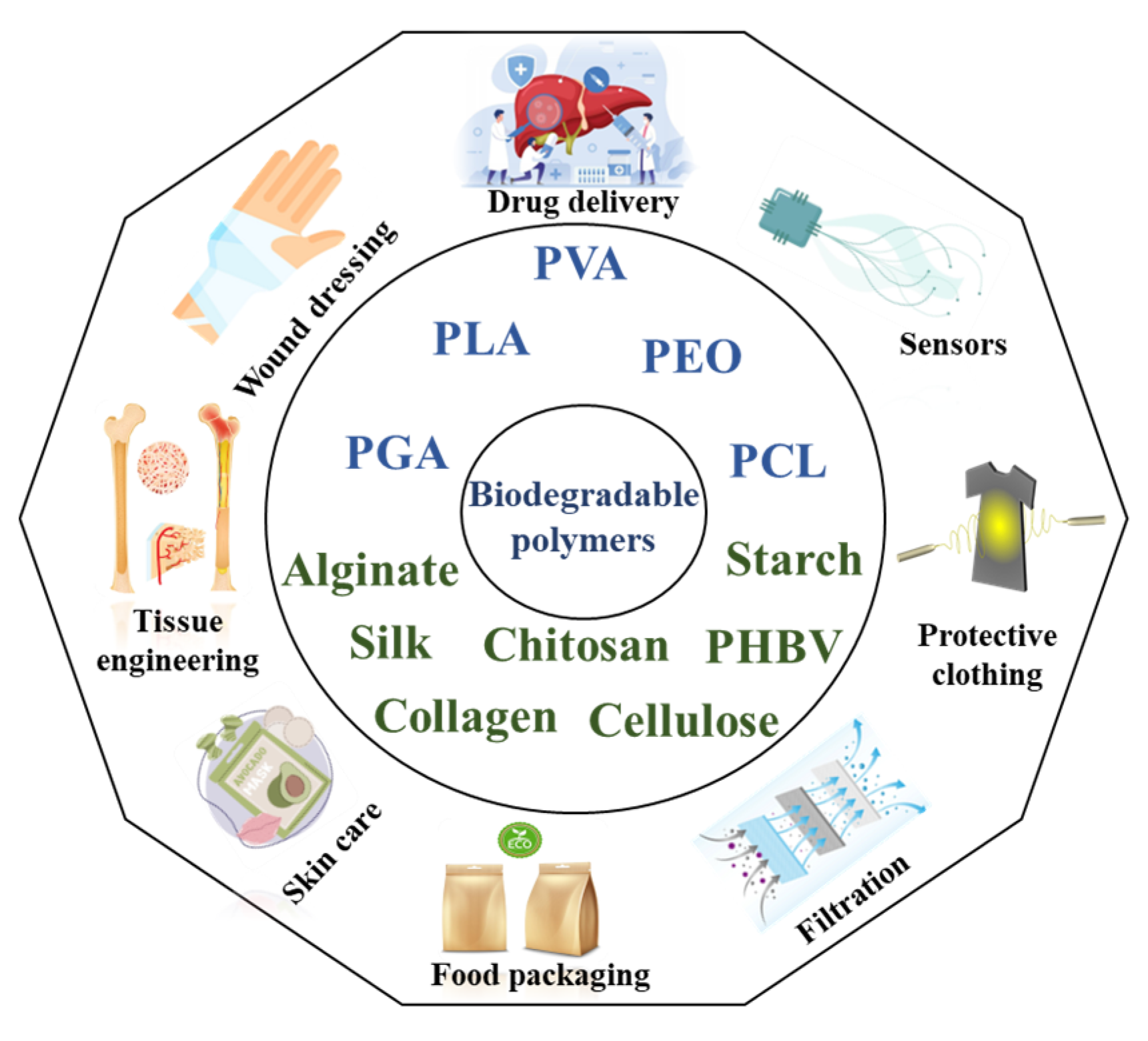
Figure 1. Types of biodegradable polymers and their applications.
Molecules 30 03276 g001
Figure 2. Schematics of (a) electrospinning [19], (b) centrifugal spinning [22] (reprinted with permission), (c) melt spinning [23] (reprinted with permission), (d) wet spinning [24] (reprinted with permission), (e) spunbonding process, and (f) melt blowing [25] (reprinted with permission).
Molecules 30 03276 g002
Figure 3. Stress–strain curves of hybrid nanofibers (PVA/chitosan) with different AgNP contents: (a–d) 0, 0.25, 0.5, and 1 wt.%, respectively. Images (1), (2), and (3) show the TEM morphology of nanofibers containing AgNPs at 0.25, 0.5, and 1 wt.%, respectively [51]. (reprinted with permission).
Molecules 30 03276 g003
Figure 4. Electrospinning and tensile testing setup: (A) rotating mandrel for fiber collection with I-frames; (B) I-frame with fiber mat secured for tensile testing; and (C) a fiber mat in Test Resources universal testing system grips before testing [65] (reprinted with permission).
Molecules 30 03276 g004
Figure 5. Mechanical properties of PCL, 50:50 PCL: PLLA blend, and PLLA electrospun fibers following hydrolytic degradation in phosphate-buffered saline solution at 37 °C; (A) Young’s modulus, (B) ultimate tensile strength, and (C) percentage strain [82]. Stars (*) represent statistical significance compared to non-degraded scaffold (time zero). (reprinted with permission).
Molecules 30 03276 g005
Figure 6. SEM images of electrospun PCL nanofiber mats exposed to 25 μL of DCM vapor in a closed vial for durations of (A,B) 30 min and (C,D) 60 min; (E) cross section of nanofibers before treatment (arrows show no interfiber bonding); (F) cross section of nanofibers after 60 min (arrows indicate welding at cross points) [102] (reprinted with permission).
Molecules 30 03276 g006
Figure 7. Effect of acetic acid concentration on the (A) diameter, (B) strength, (C) elasticity, and (D) stiffness of chitosan fibers [109] (reprinted with permission). “*” indicates p < 0.05 compared the 1 vol% AA.
Molecules 30 03276 g007
Figure 8. Tensile results of the ternary sample (PLA-PCL)-BC: (A) ultimate tensile strength of the binary systems and (B) Young’s modulus of the binary systems [125] (reprinted with permission).
Molecules 30 03276 g008
Figure 10. SEM micrograph of electrospun silk fibroin (SF) fibers: (a) random orientation; (b) SF 60 with knitted orientation (central angle 60°); (c) SF 60 with knitted orientation (central angle 60°), fixed by methanol immersion; (d) SF 90 with knitted orientation (central angle 90°) [134] (reprinted with permission).
Molecules 30 03276 g010
Figure 11. Scanning electron micrograph showing the surface morphology of (A) electrospun nonwoven PHBV fibers and (B) solvent-cast PHBV 2D film [138] (reprinted with permission).
Molecules 30 03276 g011
Figure 12. Structure–processing–property pyramid for biodegradable fibers.
Molecules 30 03276 g012
Table 1. Summary of polymer composition, fiber size, and mechanical properties of electrospun fibers based on synthetic polymers.
Main
Polymer
Fiber
Components
Average Fiber
Diameter (nm)
Tensile Strength (MPa)Elongation at Break (%)Young’s Modulus (MPa)Application
and Processing Technique
Ref.
PVAIsotropic PVA2924.21-64.7Separation filters, wound dressing materials, tissue scaffold
Electrospinning
[50]
PVA/cellulose nanowhisker-7.84-132.8
aligned PVA1665.36-87.7
PVA/cellulose nanowhisker14910.5-190.5
PVA/cellulose nanowhisker-7.84-132.8
PVA/CS1553.65--Antibacterial activity
Electrospinning
[42]
PVA/CS/Ag P (0.25%)1483.78--
PVA/CS/Ag NPs (0.5%)1444.52--
PVA/CS/Ag NPs (1%)1393.74--
PVA3287.3118629.2Wound healing
Electrospinning
[45]
PVA/AgNPs2859.1931182.4
PVA–silk sericin (non-heated)60–5000.75-Biomedical applications (skin)
Electrospinning
[46]
PVA–silk sericin (heated)3.735-
PVA/γ-Fe2O3 porosity: 76.94%30160.31-114.53Soft tissue engineering
Scaffold
Electrospinning
[53]
Porosity: 80.68%29857.48-111.62
Porosity: 85.53%26054.93-108.80
Porosity: 90.85%23551.45-105.97
Porosity: 91.24%22050.28-105.27
PEOPEO670–16201.8–4.23445–5174.88–15.3General applications
Centrifugal spinning
[17]
CMCS-PEO1.91–3.22 µm0.8–2.211.2–22.8%10.5–26.6Wound dressing Centrifugal spinning[58]
Starch–PEO2 µm0.39–1.8--Drug delivery
Centrifugal spinning
[59]
PCL/PEO (75/25)250–4506.5-12–30Surface Functionality[57]
PGA And PLGAPGA-PLA900–13002.2–1170–3303.4–140Biomedical applications
Electrospinning
[63]
PGA300–15002.72.1138Tissue engineering
Electrospinning
[64]
PLGA (50/50)2.82.2144
PLGA (85/15)1.91.95114
PGA480-3.51013.4General application
Electrospinning
[65]
PGA (14 days)--1.52989
PGA6663.4415274Tissue engineering
(cell infiltration)
Electrospinning
[56]
PLGA22115.9276141
PGA (sacrificial PEO)6093.3217166
PLGA (sacrificial PEO)22946.0414486
PCL11657.0545328
PCL (sacrificial PEO)11712.574889
PCL2451.0232.437.75Tissue engineering (blood vessel)
Electrospinning
[66]
PLGA1633.13.113.51
(PCL-PLGA) Coaxial2562.892.899.18
PLA
And
PLA
PLLA8502.72611.15Medical applications
Electrospinning
[73]
PLLA/TEAC27027.2792.8
PLLA/formic acid5209.51501.5
PLLA/TX-1005707.12551.7
PLA-1.17144-Bone tissue engineering
Electrospinning
[78]
PLA-OS 1.72103-
PLA9100.8125-Wound healing
Electrospinning
[79]
PLA/Rosa/GO7202.650-
PLA/PEG/RosA/GO-2.315-
PLA~3601.5--Hard tissue engineering
Electrospinning
[18]
PLA/NCC~2803.3--
PLLA7123.494956.8Cell adhesion and growth
Electrospinning
[107]
PLLA/PLA-Lig104853.252155.2
PLLA/PLA-Lig205252.491956.1
PLLA/PLA-Lig304802.552054.3
PLLA/PLA-Lig404172.581666
PLLA/PLA-Lig503502.561166.8
PLA (CF/DMF (80:20 v/v))-5.829.8305Biomedical applications
Electrospinning
[98]
PCL-1.682.17.5
PLA-PCL (70/30 w/w)-2.515567.5
PLLA (treated at 120 C)7965.656.9165Tissue engineering
Electrospinning
[74]
PLLA (DCP crosslinkers)8958.259.4203
PLLA (DCP-TAIC crosslinkers)-8.7847.3208
PLLA3233.876.191.7
PLLA3233.876.191.7Tissue regeneration (human annulus fibrosus tissue)
Electrospinning
[82]
(PLLA)
After 180 days in PBS
3305.238.9136.4
PCLPCL2192.919016.3
(PCL)2143.581.421.6
PLLA-PCL7204.1148.371.6
PLLA-PCL
After 180 days in PBS
6716.287.3125.6
PLA2500–75000.18--Cell culture
(melt-blown)
[83]
PLA20,0003.77 cN/dtex1880.43 cN/dtexPackaging
Melt spinning
[86,87]
PLA, 350 rpm99801.517.7726.18Tissue engineering Centrifugal spanning[84]
PLA, 900 rpm82702.348.4746.21
PLA, 1500 rpm59903.317.4766.77
PLA (CF/DMF (80:20 v/v))-5.829.8305Biomedical application
Electrospinning
[98]
PCL-1.682.17.5
PLA-PCL (70/30 w/w)-2.515567.5
PCL18304.122248Drug delivery
Electrospinning
[101]
PCL (E-beam treatment)18701.31755
PCL72111.53-8.41General
Electrospinning
[102]
PCL (DCM vapor treatment)69321.4-16.5
PCL/Gel-1.228-Medical application
Electrospinning
[104]
PCL/Gel/HNTs-2.367-
PCL8243.14-40.76Wound dressing Centrifugal spinning[106]
PCL/Gel (70/30)5342.91-84.87
PCL/Gel (50/50)3671.7-82.70
PCL/Gel (30/70)2651.22-72.76
PCL fiber (AFM)440–1040129862Biomedical devices
Electrospinning
[36]
PCL fiber (AFM)---4Tissue engineering Scaffolds
Electrospinning
[105]
PCL fiber/HAp (AFM)---20
PCL fiber (AFM)20–25257–340127–133500–3000General application
Electrospinning
[93]
Table 2. Summary of polymer composition, fiber size, and mechanical properties of electrospun fibers based on natural polymers.
Main PolymerFiber ComponentsAverage
Fiber
Diameter (nm)
Tensile Strength (MPa)Elongation at Break (%)Young’s Modulus (MPa)Application and Processing TechniqueRef.
ChitosanCS (1% acetic acid)3250.7193.6Tissue engineering
Electrospinning
[109]
CS (2% acetic acid)3501.2177
CS (6% acetic acid)4500.3127
Chitosan (10–30%)–PDO 0.26–0.31 µm 4.6–6.3 - 14–68 MPaVascular tissue engineering
Electrospinning
[112]
CS–pullulan-11.364.53344.3Active packaging
Electrospinning
[113]
CS–pullulan–crosslinker (cinnamaldehyde)-222.831150.9
CS-PVA1553.65--Biomedical application
Electrospinning
[51]
CS-PVA-AgNPs (0.25%)1483.78--
CS-PVA-AgNPs (0.5%)1444.52--
CollagenCollagen (AFM) 60–80 µm 650 - 3.2 GPaTissue engineering
Electrospinning
[119]
Collagen–cathepsin K (AFM) 241 39 1.9 Gpa
Collagen–PCL 9:1 318 4.2 4.74-Wound-healing applications
Electrospinning
[118]
Collagen–PCL–crosslinker (5/5) 388 7.65 8.93 -Nerve tissue engineering
Electrospinning
[144]
Collagen–PCL–crosslinker (9/1) 480 12.61 11.76 -Tissue engineering
Electrospinning
[145]
CellulosePLA/nanocrystalline cellulose (NCC) (0–2%)280–3201.5–3.3--Tissue engineering
Electrospinning
[18]
PLA/nanocrystalline cellulose (NCC)600–7000.5–2.1--Tissue engineering
Electrospinning
[121]
PCL–cellulose nanocrystals (CNCs) (0–20%)350–6002.7–7.2110–180%-General application
Electrospinning
[123]
PLA/nanocrystalline cellulose (NCC) 5%6426.370125.6Tissue engineering scaffold
Electrospinning
[122]
PCL–cellulose nanocrystals (CNCs) 65–95% (AFM)11.2 µm705-4.9 GPaTissue engineering Melt spinning[124]
(PLA-PCL)-BC 70/305–18.5 µm9.05-19.61Wound healing Centrifugal spinning[125]
Starch Starch (in formic acid solution) 80–300 9.38 26 264General application
Electrospinning
[130]
Starch–PCL 156–476 µm 13 - 20Tissue engineering
Electrospinning
[109]
Starch–PLA (50)- 27- 33
Starch–PLA (70)- 40- 162
Starch–EVA- 25- 85
Starch–PVA 247–301 µm 0.88–11.54-- Wound dressings
Wet extrusion
[128]
SF SF (10%)
SF after treatment with methanol
85–206 16.03
25.41
21.7
11.9
253 MPa
490 MPa
Air permeability
Electrospinning
[135]
SF (20%) (central angle 60°) 230 4.4 7.99 30Tissue engineering
Electrospinning
[134]
SF after treatment with methanol 15.3 12.3 150
2 wt% PCL/SF - 1.3 28.27 27.67Biomedical application
Electrospinning
[133]
PCL- 1.3 70.47 14.06
PCL/SF (70/30) 539 4.7 62.7 12.73Tissue engineering
Electrospinning
[146]
PCL/SF 242–618 11.2 70Wound healing
Electrospinning
[147]
PLA 10,365 1.14 15.97 70.21Tissue engineering scaffold
Electrospinning
[131]
PLA75/SF25 665 0.47 26.22 20.51
PLA50/SF50 693 1.05 17.51 16.49
SF/PVA 60–500 0.7 5 -Biomedical application
Electrospinning
[46]
SF/PVA (thermal treated)- 3.7 35-
PHBVPHBV fiber7241.4-1028Skin tissue engineering
Electrospinning
[138]
PHBV 2D film-1.57-5619
PHBV105 µm1.6-127Antibacterial fibers
Electrospinning
[139]
PHB383–52001.8-147Bone tissue engineering
Electrospinning
[136]
PHBV-PHB (25/75)2.45 140
PHBV (chloroform solution)1.173.73
PHBV (TFE solution)1.8825.54
PHBV-PLA (30/70)2890 73.5Bone regeneration
Electrospinning
[140]
PLA--41.5
PHBV0.5–3 μm3-100Biomedical application
Centrifugal methods
[141]
PHBV-1.76-125.7Tissue engineering[136]
Disclaimer/Publisher’s Note: The statements, opinions and data contained in all publications are solely those of the individual author(s) and contributor(s) and not of MDPI and/or the editor(s). MDPI and/or the editor(s) disclaim responsibility for any injury to people or property resulting from any ideas, methods, instructions or products referred to in the content.