Article

Anti-Trypanosoma cruzi Potential of New Pyrazole-Imidazoline Derivatives

by
Edinaldo Castro de Oliveira
1,†,
Leonardo da Silva Lara
1,†,
Lorraine Martins Rocha Orlando
1,
Sarah da Costa Lanera
1,
Thamyris Perez de Souza
1,
Nathalia da Silva Figueiredo
1,
Vitoria Barbosa Paes
1,
Ana Carolina Mazzochi
2,
Pedro Henrique Myra Fernandes
2,
Maurício Silva dos Santos
2 and
Mirian Claudia de Souza Pereira
1,*
1
Laboratório de Ultraestrutura Celular, Instituto Oswaldo Cruz, Fiocruz, Av. Brasil 4365 Manguinhos, Rio de Janeiro 21040-900, RJ, Brazil
2
Laboratório de Síntese de Sistemas Heterocíclicos (LaSSH), Instituto de Física e Química (IFQ), Universidade Federal de Itajubá, Avenida BPS, 1303, Pinheirinho, Itajubá, Minas Gerais 37500-903, MG, Brazil
*
Author to whom correspondence should be addressed.
These authors contributed equally to this work.
Molecules 2025, 30(15), 3082; https://doi.org/10.3390/molecules30153082
Submission received: 26 June 2025 / Revised: 19 July 2025 / Accepted: 21 July 2025 / Published: 23 July 2025
(This article belongs to the Special Issue Heterocyclic Compounds for Drug Design and Drug Discovery)

Abstract

Chagas disease, caused by Trypanosoma cruzi, poses a significant public health challenge due to its widespread prevalence, limited therapeutic options, and adverse effects associated with available medications. In this study, we developed 13 novel pyrazole-imidazoline derivatives, inspired by a previously identified cysteine protease inhibitor, and evaluated their antiparasitic activity. Our in silico analyses predicted favorable physicochemical profiles and promising oral bioavailability for these derivatives. Upon phenotypic screening, we observed that these new derivatives exhibited low cytotoxicity (CC50 > 100 µM) and marked efficacy against intracellular amastigotes. Derivative 1k showed high activity (IC50 = 3.3 ± 0.2 µM), selectivity (SI = 73.9), and potency (pIC50 = 5.4). In a 3D cardiac microtissue model, 1k significantly reduced parasite load, matching the efficacy of benznidazole (Bz) even at lower concentrations. Both 1k and Bz effectively prevented parasite recrudescence; however, neither resulted in parasite sterility under the experimental conditions employed. The combination of 1k–Bz yielded an additive interaction, highlighting its potential for in vivo combination therapy. While structural changes abolished cysteine protease inhibition, incorporating a CF3 substituent at the para position and excluding the amino group enhanced antiparasitic activity. These findings reinforce the promise of the pyrazole-imidazoline scaffold and support further structural optimizations to develop innovative candidates for treating Chagas disease.

Graphical Abstract

1. Introduction

Chagas disease (CD), caused by the protozoan parasite Trypanosoma cruzi, represents a significant public health concern with potentially lethal outcomes, particularly in its chronic stages. Current estimates indicate that CD affects 6 to 7 million individuals globally, resulting in around 12,000 deaths annually and placing 75 million people at risk of infection [1]. The World Health Organization (WHO) has classified CD as one of the 20 neglected tropical diseases, with endemic regions in 21 Latin American countries and occurrences in Asia, Australia, Europe, and North America [2,3]. The epidemiology of CD is intricately linked to socioeconomic determinants, such as poverty, substandard housing, and inadequate access to healthcare resources, which disproportionately impact more vulnerable populations [4]. CD is characterized by two phases: acute and chronic, each with distinct symptomatology and severity profiles influenced by the host’s immune response and the stage of the infection [5]. In the chronic phase, the disease may cause severe complications, including cardiac arrhythmias and dilated cardiomyopathy, which pose a risk of sudden death or heart failure [6]. Furthermore, chronic CD can also manifest with gastrointestinal disturbances, vascular accidents, and neurological implications [7,8]. The global burden of CD is substantial, corresponding to an estimated loss of approximately 806,170 disability-adjusted life years (DALYs) and healthcare expenses exceeding US $627.56 million annually [9].
Currently, the only pharmacological treatments for CD are Benznidazole (Bz) and Nifurtimox (Nif), both of which are nitroheterocyclic compounds developed over 50 years ago. These agents demonstrate limited efficacy in the chronic phase of the disease, necessitating treatment durations of more than 60 days [10]. Their use is often complicated by significant adverse effects, frequently leading to treatment discontinuations [11,12]. A randomized clinical trial (BENEFIT) revealed that treatment with Bz could not prevent the clinical progression of chronic Chagas cardiomyopathy [13]. Monotherapy with posaconazole [14], as well as its combination with Bz [15], the prodrug of ravuconazole (E1224) [16], and the nitroimidazole fexinidazole [17], have demonstrated inadequate therapeutic outcomes in clinical trials. The limitations in efficacy and tolerability associated with current treatments, along with the therapeutic failures of potential candidates, highlight the urgent need to discover new bioactive compounds to develop more effective and safer therapeutics for CD.
Pyrazole stands out due to its structural versatility and is regarded as a privileged scaffold in medicinal chemistry. It plays a crucial role in developing various bioactive compounds that exhibit several pharmacological activities [18]. Compounds featuring a pyrazole moiety have demonstrated significant antimicrobial [19], antifungal [20], anti-inflammatory [21], antiviral [22], and antineoplastic [23] properties. Over the past few decades, the Food and Drug Administration (FDA) has approved more than 40 pyrazole-containing therapeutics [24,25], underscoring the pivotal role of this chemical framework in drug optimization strategies.
Recent studies have highlighted the antiparasitic potential of pyrazole derivatives against various infectious agents, elucidating diverse mechanisms of action. For instance, diarylpyrazole derivatives have been identified as inhibitors of CYP121A1 in Mycobacterium tuberculosis [26]. Furthermore, pyrazole aza macrocyclic derivatives specifically inhibit iron superoxide dismutase (FeSOD) when evaluated against Leishmania spp. [27]. A hybrid compound, which integrates oxadiazole and pyrazole acrylic acid, has demonstrated efficacy against Plasmodium falciparum in both chloroquine-resistant (RKL9) and chloroquine-sensitive (3D7) strains. This compound also effectively inhibits falcipain 2, a key enzyme in hemoglobin digestion [28].
In this context, our research group has previously identified a 5-amino pyrazole-imidazoline hybrid compound, identified as a hit compound, exhibiting anti-T. cruzi activity [29]. This compound inhibits the cysteine protease activity of the parasite, showing affinity for the active site of cruzain, as determined through molecular docking studies. Building upon the chemical structure of N-(1H-benzimidazol-2-yl)-1,3-dimethyl-pyrazole-4-carboxamide (3H5), a known cruzain inhibitor [30], we had previously optimized the hit compound by replacing the imidazoline ring at the 4-position of the pyrazole core with a carboxamide group. This alteration reduced antiparasitic activity [31]. However, we observed that the removal of the amino group from the hit compound and the introduction of various substituents on the phenyl ring, including 4-Cl, 4-Br and 3-Cl,4-CH3, enhanced antiparasitic activity [31]. These findings suggest that groups at p-position on the phenyl ring can improve their biological activity. Further optimization efforts included the replacement of the imidazoline moiety with a thiazoline ring, resulting in the identification of three active pyrazole-thiazoline derivatives: 2,4-diCl, 3,4-diCl, and 4-Br, all of which selectively target intracellular amastigotes [32]. Similarly, substituting the imidazoline ring with a thiadiazole yielded two active derivatives, with the 4-NO2 derivative demonstrating antiprotozoal efficacy comparable to Bz in controlling parasite recrudescence in vitro [33]. In this study, our goal is to further improve the activity of the compound 5-amino pyrazole-imidazoline by strategically introducing novel substituents onto the phenyl ring. This approach aims to strengthen the antiparasitic potency of the compound and increase its binding affinity for the active site of T. cruzi cruzain.

2. Results and Discussion

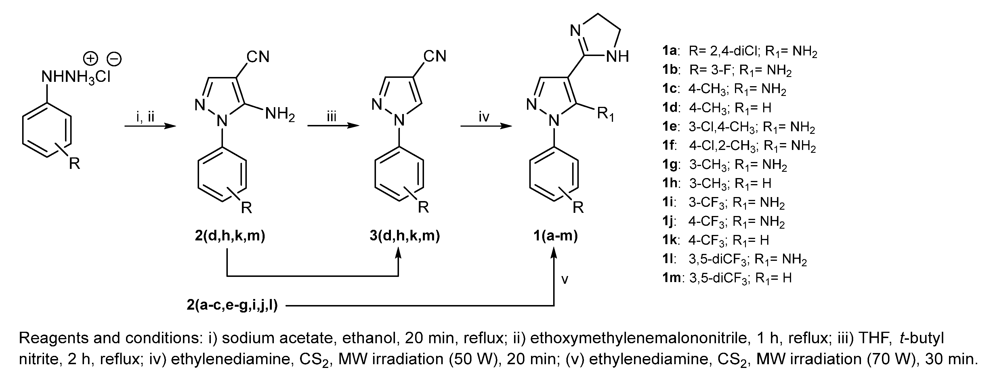
2.1. Synthesis and Chemical Analysis

The desired compounds 1(a–m) were synthesized according to the synthetic pathway outlined in Scheme 1. Firstly, arylhydrazine hydrochlorides were subjected to an acid–base reaction, using sodium acetate and ethanol as solvent, under reflux for 20 min. After that, ethoxymethylenemalononitrile was slowly added to provide a condensation reaction, followed by cyclization, while maintaining reflux for 1 h to afford 2(a–m) in 49–99% yields. In the next step, an aprotic deamination reaction was performed to achieve four key intermediates 3(d,h,k,m) in 87–99% yields, starting from 2(d,h,k,m), t-butyl nitrite, and tetrahydrofuran (THF), under reflux for 2 h. No characteristic bands related to the NH2 group were observed, confirming that the deamination reaction occurred. The syntheses of the final compounds were carried out for 2(a–c,e–g,i,j,l) and 3(d,h,k,m) to achieve 1(a–c,e–g,i,j,l) and 1(d,h,k,m), respectively, using ethylenediamine and carbon disulfide (CS2), under microwave irradiation conditions. In the case of 1(a–c,e–g,i,j,l), the power was 70 W and the reaction time was 30 min, while lower power (50 W) and shorter reaction time (20 min) were required to obtain 1(d,h,k,m). The compounds were isolated in yields ranging from 27 to 95%. They were completely characterized by FT-IR, 1H NMR, 13C NMR, HRMS, and melting point analyses. 1(c,d,g–m) derivatives are being published for the first time, while the synthesis of 1a [34,35], 1b [34,35,36], 1e [35], and 1f [35] have already been reported by our research group.
FT-IR analyses showed characteristic bands due to C=N stretching of the imidazoline ring at 1641–1596 cm−1. Bands at 3115–3064 and 2981–2847 cm−1 were attributed to Csp2-H and Csp3-H stretchings, respectively, while those related to C=C and C=N of aromatic rings were identified at 1583–1464 cm−1. 1H NMR and 13C NMR spectra confirmed that all signals corresponded to the proposed structures. 1H NMR spectra of 1(a−m) revealed a singlet at 7.64–8.08 ppm that was assigned to the proton attached to C3 of the pyrazole ring. It was also identified a singlet at 8.12–8.68 ppm for deaminated derivatives 1(d,h,k,m), R1 = H, corresponding to a proton bonded to C5. Due to prototropic tautomerism in the imidazoline ring, just one signal at 3.49–3.73 ppm for two methylene (CH2) groups was identified. The phenyl protons were assigned according to standard coupling and integration was expected. A broad signal at 6.49–6.91 ppm assigned to protons of the amine group (NH2) was observed in the spectra acquired in DMSO-d6. On the other hand, when the analyses were carried out in methanol-d4, this signal did not appear. The 13C NMR spectra showed a signal at 49.5–50.0 ppm, attributed to two methylene (CH2) groups of the imidazoline ring. In case of 1a and 1i, the signal corresponding to CH2 was superposed by the solvent (methanol) at 49 ppm, as indicated by the HSQC correlation. When DMSO-d6 was used as solvent, the signal related to CH2 was not identified. The carbon atoms of aromatic rings (Csp2) were correctly identified. In derivatives 1(c−h) and 1(i−m), signals related to methyl (CH3) and trifluoromethyl (CF3), respectively, were exhibited. HRMS analyses confirmed the molecular mass of the structures. All NMR spectra are shown in the Supplementary Materials.

2.2. Physicochemical Properties of Pyrazole-Imidazoline Derivatives

Physicochemical properties are critical determinants in the diffusion of molecules across cellular membranes, directly impacting their biological efficacy [37]. Therefore, the physicochemical parameters were analyzed in light of Lipinski’s and Veber’s rules to predict oral bioavailability. Our findings revealed that all derivatives exhibited low molecular weights, ranging from 226.28 to 363.26 g/mol (Table 1). The lipophilicity profile, assessed using the octanol–water partition coefficient (cLogP), revealed a low-to-moderate range of hydrophobicity, with cLogP values varying from 0.21 to 1.89. The exception was derivative 1b, which exhibited polar characteristics with a cLogP of −0.02 (Table 1). Additionally, regarding water solubility (cLogS), the derivatives demonstrated adequate hydrophilicity for systemic distribution in an aqueous environment, as evidenced by cLogS values ranging from −1.9 to −3.71 (Table 1). The derivatives exhibited a hydrogen bond donor (HBD) count of ≤2 and a hydrogen bond acceptor (HBA) count from 4 to 5 (Table 1). The topological polar surface area (tPSA) values, which predict drug diffusion across biological membranes, were recorded at 68.23 Å2, with exceptions noted in derivatives 1f, 1h, 1k, and 1m, which showed reduced tPSA values of 42.21 Å2 (Table 1). The rotatable bond (RB) count varies from 2 to 4, reflecting moderate molecular flexibility. Consequently, all derivatives comply with Lipinski’s and Veber’s rules, suggesting that the structural changes implemented may not adversely impact the drug’s oral bioavailability, thus implying a favorable pharmacokinetic profile. The physicochemical parameters of Bz, the reference drug, were also presented in Table 1.

2.3. Antiparasitic Effect of New Pyrazole-Imidazoline Derivatives

We evaluated thirteen new pyrazole-imidazoline derivatives for their activity against T. cruzi, focusing on both trypomastigotes and intracellular amastigotes. The screening uses T. cruzi Dm28c, genetically modified to express the luciferase enzyme (Dm28c-Luc), in conjunction with Vero cells to assess biological activity and cytotoxicity. Most of the tested pyrazole-imidazoline analogs displayed cytotoxicity values (CC50) exceeding 500 µM, demonstrating low toxicity. However, derivatives 1k (CC50 = 243.8 ± 21.1 µM), 1l (CC50 = 463.6 ± 15.2 µM), and 1m (CC50 = 113.5 ± 11.9 µM) demonstrated lower CC50 values, but these compounds still exhibit a favorable toxicity profile (Table 2).
The new pyrazole-imidazoline derivatives showed limited activity against trypomastigotes, as evidenced by IC50 values exceeding 100 µM. Of the 13 derivatives analyzed, only 1e (IC50 = 76.5 ± 10.3 µM) and 1k (IC50 = 72.2 ± 10.3 µM) achieved values below 100 µM (Table 1), but with less activity than Bz (IC50 = 20.5 ± 1.8 µM). In contrast, most of the derivatives were active against intracellular amastigotes, with 84% exhibiting IC50 values in the range from 88.1 to 3.3 µM. The five most active derivatives were identified as follows: 1a (IC50 = 40.8 ± 3.0 µM), 1d (IC50 = 33.9 ± 0.8 µM), 1h (IC50 = 26.1 ± 3.4 µM), 1k (IC50 = 3.3 ± 0.2 µM), and 1l (IC50 = 27.7 ± 2.0 µM). These compounds exhibited a selectivity index (SI) greater than 10, suggesting a favorable therapeutic window for safety. Among them, derivative 1k stands out as a promising candidate, exhibiting biological activity comparable to Bz (IC50 = 2.2 ± 0.4 µM) and an SI value of 73.9. Additionally, 1k demonstrated an IC90 value of 28.1 ± 0.4 µM and a potency of 5.4, which approaches the potency of Bz (pIC50 = 5.6). The advantageous physicochemical attributes likely contribute to its potent activity against intracellular amastigotes. Key features, including low molecular weight, optimal lipophilicity, and reduced topological polar surface area, enhance cell permeability. These factors are essential for ensuring efficient transport across the host cell membrane, enabling the compounds to reach their molecular targets within the intracellular environment of the amastigotes.
Our findings revealed that the pyrazole-imidazoline derivatives exhibited a distinct efficacy profile against extracellular and intracellular forms of T. cruzi. It is common to observe limited efficacy against trypomastigotes during drug screening trials for this parasite. This low responsiveness likely reflects both the mechanism of action of the compounds and the inherent characteristics of the trypomastigote form, which is marked by its non-replicative nature and low metabolic activity. Conversely, the pronounced potency demonstrated against intracellular amastigotes is particularly significant, as this life stage represents the proliferative and pathogenic stage of the parasite that supports persistent infection and contributes to chronic tissue damage in the mammalian host.
Notably, the deaminated derivative with the 4-CF3 substituent displayed the highest potency against intracellular amastigotes. This finding supports previous results indicating that removing the amino group and introducing electron-withdrawing groups, such as CF3 and halogens (Br and Cl), at the p-position of the phenyl ring typically enhances anti-T. cruzi activity. However, compounds 1a (2,4-diCl), 1f (4-Cl,2-CH3), and 1j (4-CF3), exhibited low anti-T. cruzi activity. This unexpected outcome for these compounds may be due to the electronic and steric effects of the 5-amino group, combined with these particular aryl substituents, which could create a molecular conformation or charge distribution that is less complementary to the biological target(s). The initial optimization of the hit compound yielded three promising derivatives: 4-Cl (IC50 = 6 µM), 4-Br (IC50 = 2.75 µM), and 3-Cl,4-CH3 (IC50 = 3.58 µM) [31]. These candidates advanced to preclinical in vitro and in vivo studies, displaying favorable outcomes in more robust cellular models and in the context of preventing cardiac damage in T. cruzi acute infection (unpublished data). We then proceeded with 1k for more detailed in vitro preclinical analyses.

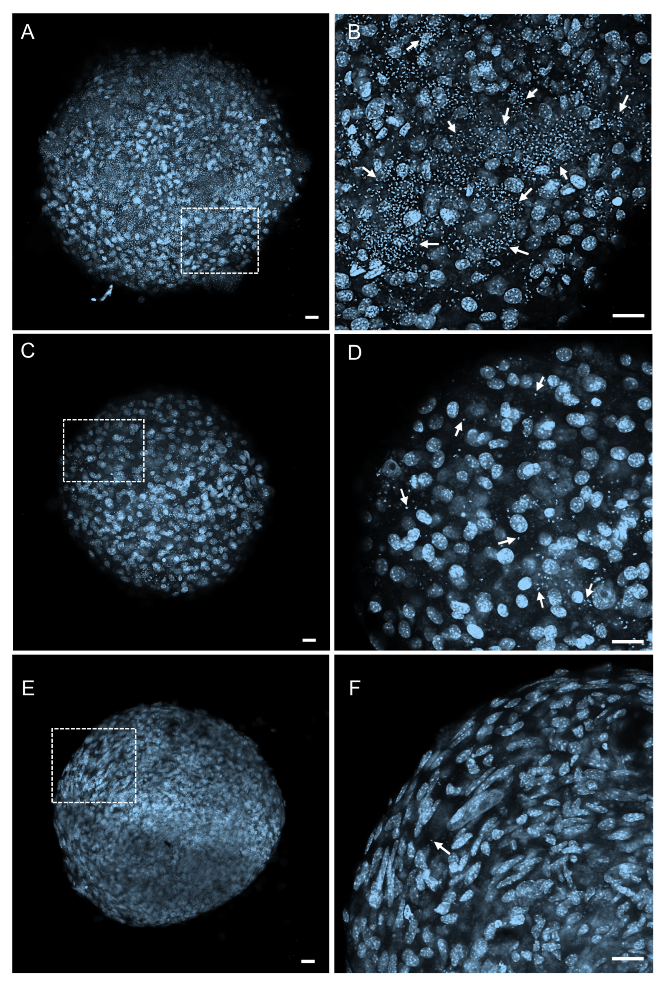
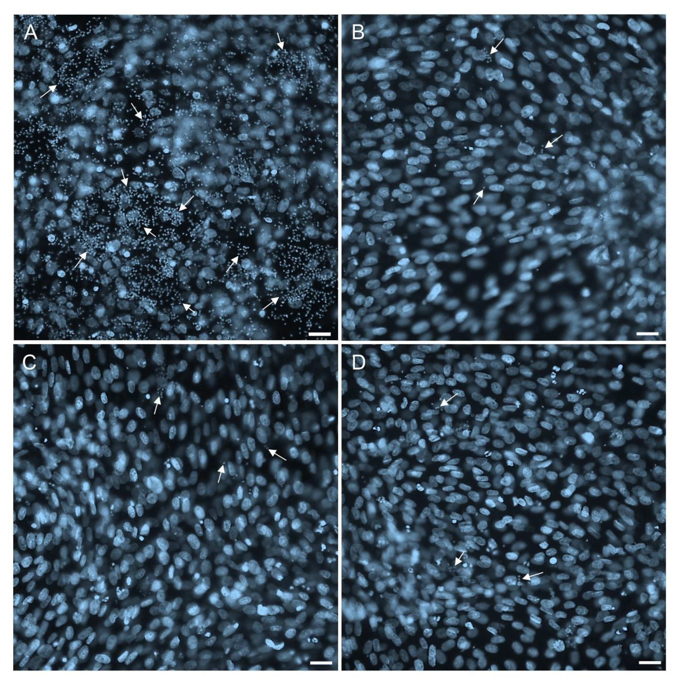
2.4. Permeability and Effectiveness of 1k in T. cruzi-Infected 3D Cardiac Microtissues

The antiparasitic efficacy of the promising 1k derivative was evaluated using a three-dimensional cardiac microtissue model. This 3D model in our screening platform is designed to bridge the gap between in vitro and in vivo preclinical assessments, as it more accurately reflects physiological conditions compared to conventional monolayer cultures [38]. These models promote intricate cellular interactions with the extracellular matrix, which significantly influence vital biological processes, including differentiation, migration, proliferation, and cell survival. Moreover, these interactions have a significant impact on drug uptake, distribution, and cell response [39]. This is particularly relevant regarding T. cruzi infection, given that the heart is one of the primary targets affected in CD pathology.
Therefore, the toxic effect of 1k was assessed using 3D cardiac spheroids, which were incubated for 72 h with various concentrations of 1k, ranging from 15.6 to 500 µM. The low toxicity profile, indicated by a CC50 value of 173.1 ± 6.8 µM, suggests favorable tolerability and a reduced risk of cardiotoxicity, a common reason for drug market withdrawals [40,41]. Subsequently, the antiparasitic efficacy of 1k was investigated in T. cruzi-infected cardiac spheroids. Treatment with 1k led to a significant reduction in parasite load, achieving 90% and 94% inhibition at concentrations of 28.8 µM and 57.6 µM, respectively. The 1k treatment at 57.6 µM showed efficacy comparable to Bz at 100 µM (Figure 1), highlighting the promising potential of the 1k derivative.
The parasite load was assessed using confocal fluorescence microscopy following DAPI staining protocols. Confocal imaging revealed a substantial density of parasites in untreated 3D cardiac spheroids after 96 h of infection, with prominent amastigote nests distributed throughout the spheroid (Figure 2). Treatment with 1k resulted in a remarkable decrease in parasite burden within spheroid cultures. At 57.6 µM concentration, 1k exhibited a marked reduction in parasite load (Figure 2), demonstrating the compound’s effective penetration and efficacy against T. cruzi-infected cardiac microtissue. In spheroids treated with Bz at 100 µM, only a few parasites were observed (Figure 2).
The use of 3D culture systems in drug discovery, particularly for anticancer agents, has gained momentum [42], yet their application in antiparasitic drug screening has not been extensively explored. The translational relevance of 3D models was highlighted in HepG2 and HC-04 hepatic cell spheroids infected with Plasmodium berghei, where the efficacy of the antimalarial M5717 was comparable to that observed in in vivo models [43]. Furthermore, 3D models have provided valuable insights into the process of cardiac fibrosis during T. cruzi infection [44], demonstrating that Posaconazole treatment effectively reduces both parasite load and fibrosis by upregulating tissue inhibitor of metalloproteinases-4 (TIMP-4) [45]. Our previous investigations have also established the potential of various azole hybrids in 3D cultures, including 1,2,3-triazole [46], pyrazole-thiazoline [32], and pyrazole-thiadiazole [33]. These pyrazole derivatives successfully penetrated cardiac spheroids and significantly reduced the parasitic load. These findings emphasize the importance of 3D culture models in advancing the in vitro preclinical phase of drug development for CD.

2.5. Potential of 1k in Inhibiting the Resurgence of the Parasites

Evaluating the impact of potential drug candidates on the reactivation of T. cruzi infection is a relevant approach to tackling drug failure. To assess the trypanocidal efficacy of 1k, we conducted a reversibility assay, which measures the compound’s ability to inhibit the resurgence of parasitism in vitro over extended follow-up periods [47]. Thus, Vero cell cultures infected with Dm28c-Luc were exposed to 1k for 10 days at two concentrations: 28.8 µM (IC90) and 57.6 µM (2-fold IC90). Following this treatment phase, the cultures were kept for an additional 10 days without any compound exposure. Throughout the experiment, we closely monitored the release of trypomastigotes in the culture supernatant using optical microscopy, along with assessments of parasite luminescence (A.L.U). The release of trypomastigotes into the supernatant from infected and untreated cultures (control; DMSO) began at 4 days post-infection (dpi), reaching a peak at 17 dpi (1.4 × 10⁵ A.L.U) (Figure 3A). Treatment with derivative 1k at both 28.8 µM and 57.6 µM, as well as Bz at 100 µM, effectively suppressed parasite release until 11 dpi, with no statistically significant difference among the treatment groups. Compared to the control, the treated groups exhibited a marked reduction in trypomastigote release, maintaining an impressive 96% inhibition even after the treatment was withdrawn (Figure 3A). The infection profile of the cell monolayer was assessed at the end of the experimental assay (21 dpi). The untreated infected cultures exhibited a high parasite load (5 × 10⁵ A.L.U). Treatment with 1k led to a significant reduction in parasite load, achieving 92% and 97.8% inhibition at concentrations of 28.8 µM (4 × 10⁴ A.L.U) and 57.6 µM (1.1 × 10⁴ A.L.U), respectively. As expected, treatment with Bz (100 µM) resulted in a decrease in the parasite load, showing a 98.4% reduction (8 × 10³ A.L.U) (Figure 3B).
Fluorescence microscopy analysis using a high-content imaging system (HCS) demonstrated an abundance of intracellular parasites within infected and untreated Vero cell cultures (Figure 4). DAPI staining highlighted the host cell nucleus as well as the nuclei and kinetoplasts of the parasites. Treatment with 1k at concentrations of 28.8 µM and 57.6 µM led to a substantial reduction in parasite load, with outcomes comparable to those observed with Bz at 100 µM, even after a prolonged 10-day culture without additional treatment. By the 21 dpi mark, a few parasites were observed within the monolayers treated with 1k or Bz (Figure 4).
Treatment with Bz and 1k failed to achieve sterility in cultures under the tested conditions, aligning with findings from previous Bz reversibility assays [47,48]. Complete sterility was only observed at Bz concentrations 25- and 50-fold greater than the IC50 value after a 16-day treatment period [48]. Like Bz, pyrazole derivatives, including pyrazole-thiadiazole [33] and pyrazole-imidazoline [31], significantly reduced both intracellular amastigote and trypomastigote release in the washout assay. However, these pyrazole hybrids did not achieve sterile cure, suggesting that dormant forms may play a role in the persistence of parasitism. Washout assays are increasingly being used in the drug screening process to identify agents with undesirable trypanostatic effects early in the preclinical phases [49]. In vitro studies on infection reversibility demonstrated that posaconazole fails to inhibit parasitic recrudescence, with trypomastigotes released by day 3 post-infection, proving less effective than nitroaromatic drugs such as Bz and nifurtimox [48]. These findings are aligned with the therapeutic failure observed in the CHAGASAZOL and STOP-CHAGAS clinical trials, where posaconazole successfully suppressed parasitemia during the treatment course (60 days) but was followed by reactivation upon drug withdrawal [14,15]. Parasitic recrudescence is likely linked to the persistence and differentiation of dormant amastigote stages, as well as the trypanostatic effects of the therapeutic agents employed. This interplay may significantly influence the effectiveness of etiological treatment, making this approach paramount in the preclinical assays [48,50,51].

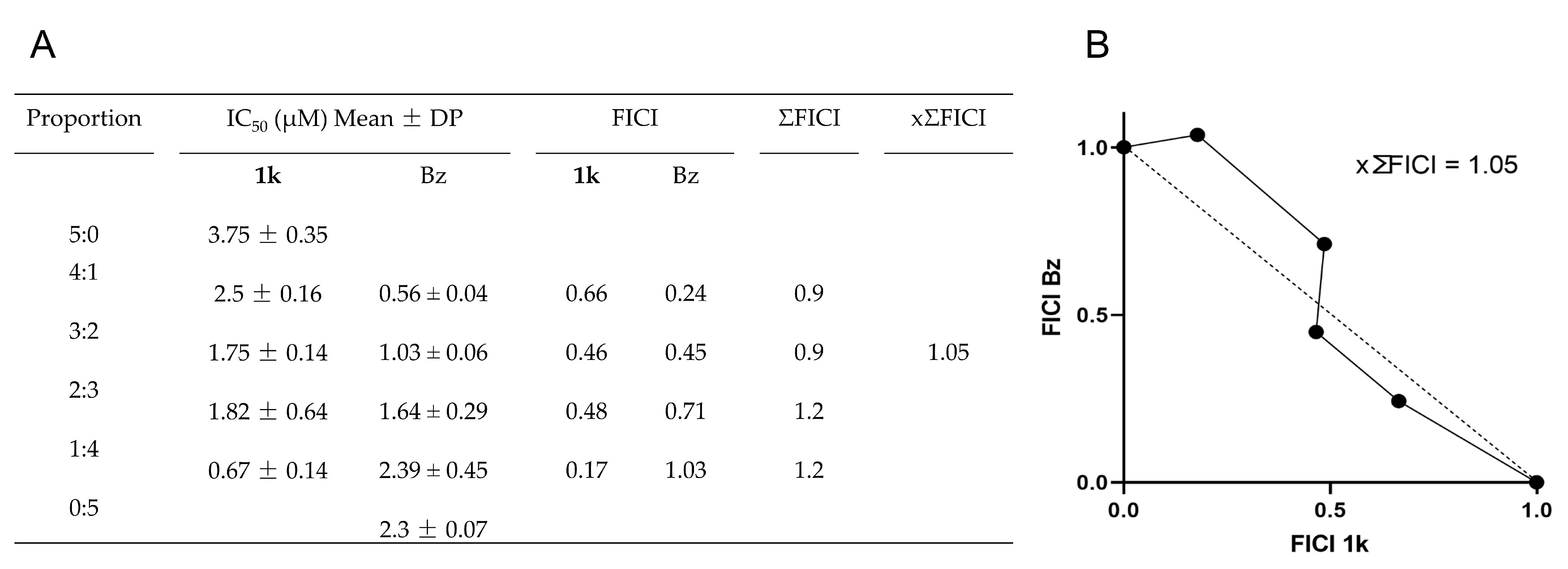
2.6. Drug Combination Effect

Combination drug therapy represents a promising avenue in the pursuit of novel therapeutic options for CD. This strategy has the potential to reduce drug dosage, shorten the overall treatment duration, mitigate adverse effects, and address issues of drug resistance by acting simultaneously on multiple targets in the pathogens [52,53].
To enhance the antiparasitic effect of 1k, we conducted an in vitro synergy assessment with Bz against intracellular amastigote forms of T. cruzi. We first established the maximum concentration for each compound and determined their respective IC50 values, yielding 90 µM for derivative 1k and 61 µM for Bz. Based on these concentrations, we formulated a series of combination ratios: 5:0 (90:0 µM), 4:1 (72:12 µM), 3:2 (54:24 µM), 2:3 (36:36 µM), 1:4 (18:48.8 µM), and 0:5 (0:61 µM). The results demonstrated that the sum of fractional inhibitory concentrations (∑FICI) varied between 0.9 and 1.2, with a mean ∑FICI (x∑FICI) of 1.05, indicating an additive interaction (x∑FICI range for additivity: 0.5–4). The table in Figure 5A details the IC50 values, FICI, ∑FICI, and x∑FICI for each compound tested. Additionally, the isobologram presented in Figure 5B corroborates that all tested compound ratios exhibited additive interactions.
The additive interaction observed with the 1k–Bz combination suggests that their combined effect is equivalent to the cumulative impact of each agent when administered separately [54]. This finding paves the way for further in vivo investigations into their potential in treating T. cruzi infections. This current assessment aims to evaluate whether combination therapy provides greater therapeutic benefits than monotherapy [53].
Previous research validates this combination strategy, demonstrating that an in vitro additive interaction of chloroquine and Bz yielded a x∑FICI of 1.4, translating to an eightfold increase in efficacy compared to Bz administered alone against a Bz-resistant Colombian strain of T. cruzi [55]. Likewise, the in vitro pairing of ravuconazole and amlodipine exhibited enhanced parasitic activity in a murine model, with efficacy observed even at diminished dosage of ravuconazole. This combination also significantly mitigated parasitemia reactivation after immunosuppression compared to monotherapy [56]. Further investigation on the 1k–Bz combination in the in vivo system may contribute to the development of more potent and safer therapeutic regimens for CD.

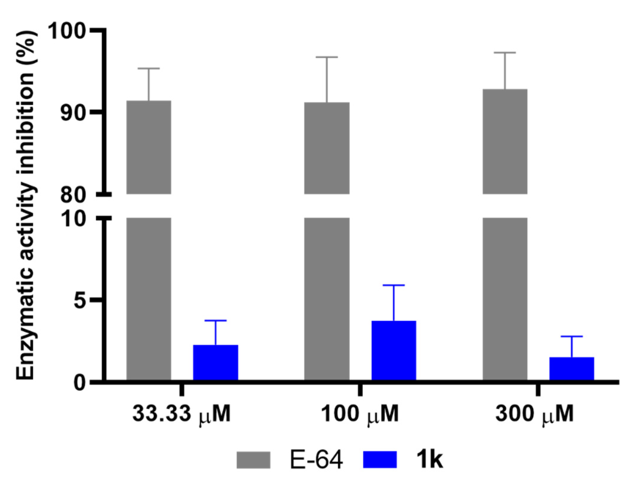
2.7. Effect of 1k on Cysteine Protease Activity

In our investigation of derivative 1k (4-CF3), an optimized variant of the previously identified cysteine protease inhibitor 5-amino-pyrazole-imidazoline [29], we evaluated its inhibitory potential against the cysteine protease from T. cruzi. Total protein extracts of trypomastigotes were incubated with concentrations of 33.3 µM, 100 µM, and 300 µM of 1k, alongside the established cysteine protease inhibitor E-64 as a control. Following a 90 min incubation with the fluorogenic substrate Z-Phe-Arg-AMC, we assessed enzymatic activity. The results showed that 1k exhibited poor inhibition of cysteine protease activity at only 3.76%. In contrast, the known inhibitor E-64 demonstrated a substantial inhibitory effect, reducing enzymatic activity by 92.81% (Figure 6). The incorporation of CF3 substitution, a functional group with a strong electron-withdrawing effect and high lipophilicity, resulted in a 5-fold enhancement in anti-T. cruzi activity relative to the hit compound; however, it fails to inhibit T. cruzi cysteine protease. The overall change in the electronic and steric profile of compound 1k, caused by the CF3 group and absence of the amino group, likely led to an unfavorable interaction with the cruzain active site.
Our previous studies have demonstrated that the deamination of the hit compound, along with the substitution of the 3,5-dichloro moiety with either 4-Cl, 4-Br, or a 3-Cl/4-CH3 configuration, significantly enhanced antiparasitic activity while concurrently abolishing its cysteine protease inhibitory activity [31]. Furthermore, the replacement of the imidazoline ring with benzimidazole, which has been previously identified as a promising scaffold for cruzain inhibition [57], yielded no substantial improvement in enzyme inhibition, achieving only 40% inhibition [58].
Computational studies have shed light on the interactions of potent cruzain inhibitors with the enzyme’s catalytic domain [59]. The active site of cruzain is characterized by seven sub-pockets (S1—S4 and S1′—S3′) [60], with inhibitors typically interacting within the S3 to S1′ sub-pockets. The catalytic triad, comprising Cys25, His162, and Asn182, is located between the S1 and S1′ domains [61]. Furthermore, competitive inhibitors form hydrogen bond interactions with key residues, including Gly66, Asp161, and Gln19 [57]. Non-peptidic, nitrile-based compounds, which incorporate CF3 groups within their molecular structures, were also identified as T. cruzi cruzain inhibitors [62]. Additionally, compounds with a CF3 group at the p-position of the aromatic ring, such as LASSBio-1736 (a member of the hydrazide-N-acylhydrazone class), have demonstrated potent activity against Leishmania cysteine proteases [63]. The potential of CF3 groups in aryl chalcones has also been shown to improve interaction with the active site of cysteine proteinase B from Leishmania (Viannia) braziliensis, as indicated by molecular docking rankings [64]. Thus, further molecular docking studies on compound 1k could provide deeper insights into its binding characteristics and enable structural optimization aimed at enhancing its enzyme inhibitory efficacy.
While our investigations revealed that 1k does not significantly inhibit T. cruzi cysteine protease, its potent anti-amastigote activity strongly suggests an alternative mechanism of action. Given the high replicative demands of this parasitic stage, potential targets likely include enzymes involved in essential biosynthetic pathways for cell growth and division. For instance, compounds related to pyrazole scaffolds, or other nitrogen-based heterocycles, are known to target sterol biosynthesis via CYP51 [65,66]. Moreover, targeting dihydroorotate dehydrogenase (DHODH) for the inhibition of pyrimidine de novo synthesis represents a crucial strategy for managing proliferative stages, as highlighted by the efficacy of tetrahydro-1H,5H-pyrazolo [1,2-a]pyrazole-1-carboxylates against Plasmodium falciparum DHODH [67], an enzyme also essential for T. cruzi proliferation. Furthermore, interference with DNA integrity or other key metabolic processes fundamental to amastigote proliferation also represents a plausible mechanism of action. Further studies are needed to elucidate the molecular target(s) and mechanism(s) of action of 1k, which could lead to the development of new therapeutic strategies for Chagas disease.

3. Materials and Methods

3.1. Chemistry

All commercial reagents and solvents were used as received. The melting points were determined on an Allerbest apparatus (Allerbest, Curitiba, PR, Brazil). FT–IR spectra were recorded on a PerkinElmer Spectrum 100 spectrometer equipped with an ATR diamond-ZnSe apparatus (PerkinElmer, Waltham, MA, USA), with wavenumber expressed in cm−1, involving 16 scans and a resolution of 4 cm−1. NMR spectra were recorded on a Bruker Avance (Bruker, Rheinstetten, Germany), 400 MHz or 500 MHz, at 298 K, in deuterated methanol (MeOD-d4) or deuterated dimethyl sulfoxide (DMSO-d6). Chemical shifts (δ) are expressed in parts per million (ppm) and coupling constants (J) in Hertz (Hz). The HRMS analyses were performed using a Q-TOF Bruker Spectrometer (Bruker, Bremen, Germany), with electrospray ionization (ESI), a capillary of 4.0 kV, an end plate offset of 4.0 kV, a source temperature of 150 °C, a dry gas administered at 4.0 l/min, and a desolvation temperature of 200 °C. The intermediates 5-amino-1-aryl-1H-pyrazole-4-carbonitriles 2(a−m) and 1-aryl-1H-pyrazole-4-carbonitriles 3(d,h,k,m) were synthesized following the experimental procedure previously described by our research group [68].
5-amino-1-(2,4-dichlorophenyl)-4-(4,5-dihydro-1H-imidazol-2-yl)-1H-pyrazole (1a). Yield: 27%; color: beige; mp.: 167–169 °C; FT-IR (cm−1): 3385, 3259, 3115, 2934, 2853, 1608, 1581, 1565, 1532, 1484; 1H NMR (500 MHz, methanol-d4): δ = 7.74 (s, 1H), 7.73 (d, J = 2.2 Hz, 1H), 7.53 (dd, J = 8.5;2.2 Hz, 1H), 7.49 (d, J = 8.5 Hz, 1H), 3.71 (s, 4H); 13C NMR (124 MHz, methanol-d4): δ = 162.2, 150.4, 140.9, 137.8, 135.3, 134.9, 132.5, 131.6, 129.7, 93.3, 49.5; HRMS (ESI): m/z [M + H]+: calcd.: 296.0470; found: 296.0475.
5-amino-1-(3-fluorophenyl)-4-(4,5-dihydro-1H-imidazol-2-yl)-1H-pyrazole (1b). Yield: 47%; color: yellow; mp.: 164–166 °C; FT-IR (cm−1): 3369, 3257, 3162, 2934, 2861, 1601, 1567, 1520, 1492; 1H NMR (400 MHz, DMSO-d6): δ = 7.71 (s, 1H), 7.56–7.52 (m, 1H), 7.49–7.43 (m, 2H), 7.22–7.20 (m, 1H), 6.72 (s, 2H), 3.50 (s, 4H); 13C NMR (100 MHz, DMSO-d6): δ = 162.1 (d, J = 243.0 Hz), 159.9, 147.6, 140.0 (d, J = 10.0 Hz), 138.7, 131.0 (d, J = 9.0 Hz), 118.1 (d, J = 3.0 Hz), 113.2 (d, J = 21.0 Hz), 109.4 (d, J = 25.0 Hz), 94.7; HRMS (ESI): m/z [M + H]+: calcd.: 246.1155; found: 246.1163.
5-amino-1-(4-methylphenyl)-4-(4,5-dihydro-1H-imidazol-2-yl)-1H-pyrazole (1c). Yield: 50%; color: yellow; mp.: 214–216 °C; FT-IR (cm−1): 3258, 3192, 3095, 2928, 2867, 1598, 1561, 1511, 1492; 1H NMR (400 MHz, DMSO-d6): δ = 7.65 (s, 1H), 7.45 (d, J = 8.3, 2H), 7.31 (d, J = 8.3, 2H), 6.49 (s, 2H), 3.49 (s, 4H), 2.35 (s, 3H); 13C NMR (100 MHz, DMSO-d6): δ = 160.0, 147.2, 138.0, 136.1, 136.0, 129.6, 122.5, 94.2, 20.4; HRMS (ESI): m/z [M + H]+: calcd.: 242.1406; found: 242.1412.
1-(4-methylphenyl)-4-(4,5-dihydro-1H-imidazol-2-yl)-1H-pyrazole (1d). Yield: 28%; color: light yellow; mp.: 232–234 °C; FT-IR (cm−1): 3144, 3107, 3077, 2932, 2847, 1631, 1563, 1521, 1482; 1H NMR (500 MHz, methanol-d4): δ = 8.50 (s, 1H), 8.02 (s, 1H), 7.62 (d, J = 8.3 Hz, 2H), 7.31 (d, J = 8.3 Hz, 2H), 3.71 (s, 4H), 2.38 (s, 3H); 13C NMR (125 MHz, methanol-d4): δ = 161.1 140.9, 138.7, 138.6, 131.1, 128.8, 120.5, 115.8, 49.9, 20.9; HRMS (ESI): m/z [M + H]+: calcd.: 227.1297; found: 227.1290.
5-amino-1-(3-chloro-4-methylphenyl)-4-(4,5-dihydro-1H-imidazol-2-yl)-1H-pyrazole (1e). Yield: 85%; color: light yellow; mp.: 238–240 °C; FT-IR (cm−1): 3384, 3261, 3215, 3114, 2924, 2853, 1602, 1581, 1566, 1523, 1501, 1479; 1H NMR (500 MHz, DMSO-d6): δ = 7.68 (s, 1H); 7.62 (s, 1H), 7.49 (s, 1H), 7.48 (s, 1H), 6.64 (s, 2H), 3.50 (s, 4H), 2.37 (s, 3H); 13C NMR (125 MHz, DMSO-d6): δ = 159.9, 147.6, 138.5, 137.4, 133.7, 133.4, 131.5, 122.5, 121.0, 94.6, 19.0; HRMS (ESI): m/z [M + H]+: calcd.: 276.1016; found: 276.1018.
5-amino-1-(4-chloro-2-methylphenyl)-4-(4,5-dihydro-1H-imidazol-2-yl)-1H-pyrazole (1f). Yield: 73%; color: yellow; mp.: 205–206 °C; FT-IR (cm−1): 3410, 3260, 3205, 3123, 2926, 2851, 1596, 1570, 1520, 1482; 1H NMR (500 MHz, DMSO-d6): δ = 7.64 (s, 1H), 7.51 (d, J = 2.3 Hz, 1H), 7.39 (dd, J = 8.4; 2.3 Hz, 1H), 7.28 (d, J = 8.4 Hz, 1H), 6.27 (s, 2H), 3.49 (s, 4H), 2.05 (s, 3H); 13C NMR (125 MHz, DMSO-d6): δ = 160.0, 148.3, 138.1, 138.0, 135.6, 133.1, 130.5, 129.4, 126.5, 92.9, 16.9; HRMS (ESI): m/z [M + H]+: calcd.: 276.1016; found: 276.1026.
5-amino-1-(3-methylphenyl)-4-(4,5-dihydro-1H-imidazol-2-yl)-1H-pyrazole (1g). Yield: 57%; color: light yellow; mp.: 204–205 °C; FT-IR (cm−1): 3243, 3110, 2927, 2854, 1599, 1586, 1560, 1518, 1486, 1464; 1H NMR (400 MHz, methanol-d4): δ = 7.67 (s, 1H), 7.41 (t, J = 7.7 Hz, 1H), 7.35 (s, 1H), 7.31 (d, J = 7.7 Hz, 1H), 7.25 (d, J = 7.7 Hz, 1H), 3.64 (s, 4H), 2.41 (s, 3H); 13C NMR (100 MHz, methanol-d4): δ = 162.7, 148.6, 141.1, 139.8, 139.1, 130.5, 129.9, 125.9, 122.4, 95.5, 49.7, 21.4; HRMS (ESI): m/z [M + H]+: calcd.: 242.1406; found: 242.1414.
1-(3-methylphenyl)-4-(4,5-dihydro-1H-imidazol-2-yl)-1H-pyrazole (1h). Yield: 95%; color: light yellow; mp.: 190–192 °C; FT-IR (cm−1): 3258, 3147, 3064, 2937, 2869, 1620, 1590, 1569, 1493; 1H NMR (400 MHz, methanol-d4): δ = 8.54 (s, 1H), 8.03 (s, 1H), 7.60 (s, 1H), 7.54 (d, J = 7.6 Hz, 1H), 7.38 (t, J = 7.6 Hz, 1H), 7.20 (d, J = 7.6 Hz, 1H), 3.71 (s, 4H), 2.42 (s, 3H); 13C NMR (100 MHz, methanol-d4): δ = 161.1, 141.1, 141.1, 140.8, 130.5, 129.2, 128.8, 121.0, 117.6, 116.0, 49.9, 21.4; HRMS (ESI): m/z [M + H]+: calcd.: 227.1297; found: 227.1302.
5-amino-1-(3-trifluoromethylphenyl)-4-(4,5-dihydro-1H-imidazol-2-yl)-1H-pyrazole (1i). Yield: 38%; color: yellow; mp.: 213–215 °C; FT-IR (cm−1): 3392, 3258, 3205, 3117, 2928, 2860, 1597, 1573, 1523, 1490; 1H NMR (500 MHz, methanol-d4): δ = 7.88 (s, 1H), 7.87–7.85 (m, 1H), 7.76–7.73 (m, 3H), 3.66 (s, 4H); 13C NMR (125 MHz, methanol-d4): δ = 162.5, 149.2, 140.8, 140.0, 132.9 (quartet, J = 32.5 Hz), 131.7, 128.4, 126.2, 125.5 (quartet, J = 3.7 Hz), 125.1 (quartet, J = 270 Hz), 121.9 (quartet, J = 3.7 Hz), 95.8; HRMS (ESI): m/z [M + H]+: calcd.: 296.1123; found: 296.1113.
5-amino-1-(4-trifluoromethylphenyl)-4-(4,5-dihydro-1H-imidazol-2-yl)-1H-pyrazole (1j). Yield: 59%; color: light yellow; mp.: 269–270 °C; FT-IR (cm−1): 3284, 3220, 3083, 2942, 2873, 1606, 1565, 1533, 1513, 1486; 1H NMR (400 MHz, DMSO-d6): δ = 7.89–7.84 (m, 4H), 7.76 (s, 1H), 6.80 (s, 2H), 3.51 (s, 4H); 13C NMR (100 MHz, DMSO-d6): δ = 159.8, 147.9, 141.8, 139.2, 126.4 (quartet, J = 32.0 Hz), 126.4 (quartet, J = 3.7 Hz), 124.0 (quartet, J = 270 Hz), 122.3, 94.9; HRMS (ESI): m/z [M + H]+: calcd.: 296.1123; found: 296.1129.
1-(4-trifluoromethylphenyl)-4-(4,5-dihydro-1H-imidazol-2-yl)-1H-pyrazole (1k). Yield: 76%; color: white; mp.: 253–255 °C; FT-IR (cm−1): 3156, 3110, 3074, 2940, 2876, 1618, 1570, 1529, 1480; 1H NMR (500 MHz, methanol-d4): δ = 8.68 (s, 1H), 8.08 (s, 1H), 8.00 (d, J = 8.5 Hz, 2H), 7.82 (d, J = 8.5 Hz, 2H), 3.72 (s, 4H); 13C NMR (125 MHz, methanol-d4): δ = 160.8, 143.5, 142.0, 129.9 (quartet, J = 33.0 Hz), 128.9, 128.0 (quartet, J = 3.7 Hz), 125.4 (quartet, J = 270.0 Hz), 120.4, 116.9, 50.0; HRMS (ESI): m/z [M + H]+: calcd.: 281.1014; found: 281.1019.
5-amino-1-((3,5-ditrifluoromethyl)phenyl)-4-(4,5-dihydro-1H-imidazol-2-yl)-1H-pyrazole (1l). Yield: 85%; color: yellow; mp.: 192–194 °C; FT-IR (cm−1): 3396, 3262, 3224, 3126, 2942, 2849, 1599, 1583, 1521, 1467; 1H NMR (400 MHz, DMSO-d6): δ = 8.26 (s, 2H), 8.10 (s, 1H), 7.82 (s, 1H), 6.91 (s, 2H), 3.52 (s, 4H); 13C NMR (100 MHz, DMSO-d6): δ = 159.7, 148.2, 140.1, 139.8, 131.2 (quartet, J = 33.0 Hz), 122.8 (quartet, J = 271 Hz), 122.3 (d, J = 3.0 Hz), 119.7 (quintet, J = 3.7 Hz), 95.5; HRMS (ESI): m/z [M + H]+: calcd.: 364.0991; found: 364.0985.
1-((3,5-ditrifluoromethyl)phenyl)-4-(4,5-dihydro-1H-imidazol-2-yl)-1H-pyrazole (1m). Yield: 89%; color: white; mp.: 199–201 °C; FT-IR (cm−1): 3321, 3107, 3034, 2981, 2888, 1641, 1573, 1494; 1H NMR (500 MHz, methanol-d4): δ = 8.81 (s, 1H), 8.45 (s, 2H), 8.12 (s, 1H), 7.96 (s, 1H), 3.73 (s, 4H); 13C NMR (125 MHz, methanol-d4): δ= 160.6, 142.4, 142.1, 134.1 (quartet, J = 33.7 Hz), 129.2, 124.4 (quartet, J = 270 Hz), 121.2 (quintet, J = 3.7 Hz), 120.2 (d, J = 3.7 Hz), 117.3, 49.9; HRMS (ESI): m/z [M + H]+: calcd.: 349.0882; found: 349.0871.

3.2. In Silico Analysis

The physicochemical properties of the compounds were assessed using Osiris DataWarrior software version 5.5.0 [69]. All Simplified Molecular Input Entry Line System (SMILES) representation utilized in the in silico analysis was generated with MarvinSketch software (version 22.22).

3.3. Cell Culture

Vero cells, obtained from the Rio de Janeiro Cell Bank (BCRT code 0245) and maintained through weekly subculturing with a dissociation solution containing 0.25% trypsin-EDTA, initiated once the monolayer reached an 80–100% confluence. Following harvesting, the cells were washed and resuspended in RPMI 1640 medium enriched with 10% fetal bovine serum (FBS). Cultures were maintained at 37 °C in a 5% CO2-humidified incubator. These cultures were used for drug screening assays and for producing culture-derived trypomastigotes of T. cruzi.
Three-dimensional (3D) heart muscle cell cultures were obtained as previously described [44]. Briefly, heart muscle cells were isolated from 18-day-old Swiss Webster mouse fetuses [70]. The ventricular tissue was fragmented and enzymatically dissociated using a trypsin and collagenase type II solution. The isolated cells were seeded at a density of 2.5 × 10⁴ cells/well in 96-well U-bottom microplates previously coated with 1% agarose. The cells were cultured in Dulbecco’s modified Eagle Medium (DMEM) enriched with 10% FBS, 2.5 mM CaCl2, 1 mM L-glutamine, 2% chicken embryo extract, and a cocktail of antibiotics. The cultures were maintained at 37 °C in a 5% CO2 atmosphere. After a 7-day culture period, the resulting fully developed spheroids were used to assess drug-induced cardiotoxicity and evaluate their efficacy against T. cruzi. All animal experimentation protocols were approved by the Animal Care and Use Committee at Instituto Oswaldo Cruz (License L-017-2022-A2).

3.4. Trypanosoma cruzi

The genetically modified Trypanosoma cruzi, clone Dm28c (TcI), expressing luciferase (Dm28c-Luc), was kindly provided by Dr. Cristina Henriques from Oswaldo Cruz Institute—Fiocruz [71]. The parasites were maintained using Vero cell cultures in RPMI 1640 medium enriched with 10% FBS. At 4 days post-infection (dpi), the trypomastigotes released into the culture supernatant were harvested, subjected to centrifugation at 800× g for 20 min at 4 °C, and quantified with a Neubauer chamber. The isolated trypomastigotes were subsequently used for phenotypic screening, drug efficacy assessments employing a 3D cardiac model, washout experiments to evaluate reversibility, drug combination studies, and enzymatic assays.

3.5. Cytotoxicity Assay

Cytotoxicity assays were performed using Vero cells, which were seeded at a density of 1.5 × 10⁴ cells per well in white 96-well plates with clear bottoms. Following a 24 h adhesion period, the cultures were incubated at 37 °C in a 5% CO2 atmosphere. Concentrations of pyrazole-imidazoline compounds or Bz, ranging from 15.6 to 500 µM, were administered. After 72 h of incubation, cell viability was assessed by adding CellTiter-Glo reagent (Promega Corporation, Madison, WI, USA), which induces cell lysis and reacts with the released ATP to produce luminescence. The resulting luminescence was measured using a Glomax Multi-Detection plate reader (Promega Corporation, Madison, WI, USA). The half-maximal inhibitory concentration (CC50) for cell viability was then calculated using GraphPad Prism software version 8.2.1.
The cytotoxicity of the promising candidate was assessed using 3D cardiac spheroids, seeded at 2.5 × 10⁴ cells per well. The cultures were treated with a series of concentrations as previously described, and the cell viability was measured using luminescence following the addition of CellTiter-Glo reagent (Promega Corporation, Madison, WI, USA). A low concentration of dimethyl sulfoxide (DMSO < 1%) served as the negative control. The data shown represents the mean value accompanied by standard deviations, calculated from at least three independent experiments, each performed in duplicate.

3.6. Anti-T. cruzi In Vitro Assay

Drug susceptibility assays were performed on T. cruzi Dm28c-Luc, a genetically engineered parasite expressing luciferase. The drug screening used both trypomastigotes and intracellular amastigotes. Trypomastigotes (1 × 10⁶ parasites/well) were added to white 96-well plates with clear bottoms. Following a 24 h exposure to varying concentrations (ranging from 0.41 to 100 µM) of pyrazole-imidazoline compounds or Bz, D-luciferin solution (300 µg/mL) was added to assess parasite viability via luciferase activity. The luminescence was measured using a Glomax Multi-Detection plate reader (Promega Corporation, Madison, WI, USA).
To evaluate the effect of pyrazole-imidazoline derivatives against intracellular amastigotes, Vero cells (1.5 × 10⁴ cells/well) were cultured in a white 96-well plate with a clear bottom and subsequently infected with T. cruzi Dm28c-Luc at a 10:1 parasite-to-host cell ratio for 24 h. Post-infection, the cultures were treated with serial dilutions of pyrazole-imidazoline compounds or Bz ranging from 0.41 to 100 µM for 72 h at 37 °C. Parasite viability was measured by luminescence following the addition of luciferin (300 µg/mL). Background measurements (non-infected cultures incubated with luciferin, as well as luciferin-only controls) were performed to ensure the signal specifically originates from viable parasites. The IC50 and IC90 values, denoting the concentrations that achieve 50% and 90% reductions in viable parasites, respectively, were calculated using GraphPad Prism 8.2.1 software. Additionally, the selectivity index (SI) was calculated as the cytotoxic concentration (CC50) ratio in mammalian cells to the IC50 against T. cruzi, providing insight into the therapeutic window. Potency against intracellular amastigotes was evaluated using the formula 6—Log10 (IC50).
The efficacy of the promising candidate was assessed using cardiac 3D microtissues. Following a 7-day cultivation period, cardiac spheroids (2.5 × 10⁴ cells/well) were infected with T. cruzi Dm28c-Luc (20:1 parasites/host cell) for 24 h. After a washing step, the spheroids were treated with the promising candidate at concentrations of IC90 and 2-fold IC90 for 72 h at 37 °C, ensuring these concentrations remained within the CC20 range for the compound. Bz was used as a positive control at the concentration of 100 µM (corresponding to 10-fold IC90). At the end of the experiment, luminescence was measured using a Glomax Multi-Detection plate reader (Promega Corporation, Madison, WI, USA) after adding luciferin solution, with results reported in arbitrary luminescence units (A.L.U). To enhance reliability and provide qualitative, orthogonal validation of these luminescence readings, we employed confocal fluorescence microscopy using 4′,6′-diamidino-2-phenylindole (DAPI) staining. Thus, cardiac spheroids were fixed in 4% paraformaldehyde (PFA) in PBS for 20 min at 4 °C and stained with 10 µg/mL of DAPI. Imaging was performed using a Zeiss LSM710 confocal fluorescence microscope (Carl Zeiss, Baden–Württemberg, Germany). For all drug assays, low concentrations of DMSO (<1%) were used as a negative control. Each experimental condition was replicated in duplicates across at least three independent trials.

3.7. Reversibility Assay

Monolayers of Vero cells infected with T. cruzi (Dm28c-Luc) for 24 h were treated with concentrations corresponding to the IC90 and 2-fold IC90 of the promising candidate for 10 days. After treatment, the cultures were thoroughly washed, and parasite recrudescence was evaluated over an extended period of 10 days in RPMI medium supplemented with 10% fetal bovine serum (FBS). Culture supernatants were collected at intervals, and the luminescence signal indicative of trypomastigote release was assessed using a Glomax Multi-Detection plate reader (Promega Corporation, Madison, WI, USA) following the addition of luciferin every 2–3 days. At the experiment’s conclusion, the infection profile of the monolayer was also examined. Cells were fixed with PFA for 20 min at 4 °C, then stained with DAPI for microscopy analysis. Luminescence data were expressed in arbitrary luminescent units (A.L.U), and fluorescence images were acquired using the ImageXpress Micro XLS high-content screening (HCS) system (Molecular Devices, Sunnyvale, CA, USA).

3.8. Drug Combination

The isobologram approach assessed the interaction between Bz and the promising candidate in intracellular amastigotes of T. cruzi [72]. Vero cell cultures infected with T. cruzi (Dm28c-Luc) were treated for 72 h with various concentrations of combinations of Bz and the promising candidate, specifically at ratios of 5:0; 4:1; 3:2; 2:3; 1:4 and 0:5. The maximum concentration of each compound was established based on their IC50 values, ensuring both reached the fourth dilution in the serial dilution setup (1:3), culminating in a total of seven dilutions. After 72 h of treatment, parasite viability was measured by luminescence in a Glomax Multi-Detection plate reader (Promega Corporation, Madison, WI, USA) after incubation with luciferin. The IC50 values of each treatment combination were calculated individually, and the fractional inhibitory concentration index (FICI) was derived using the formula FICI = IC50 of the combination/IC50 of the compound alone. The overall FICI (ΣFICI) was calculated by summing the FICIs for both compounds in each combination group, while the average ΣFICI (xΣFICI) represented the overall mean across all combination groups. The xΣFICI values were then employed to classify the interaction as synergistic (xΣFICI ≤ 0.5), additive (0.5 < xΣFICI < 4), or antagonistic (xΣFICI > 4). The isobologram plot was generated using GraphPad Prism version 8.2.1, with averages of ΣFICI derived from three independent experiments.

3.9. Enzymatic Activity

Total proteins from T. cruzi Dm28c-Luc were extracted using a lysis buffer [100 mM Tris-HCl pH 8.0, 150 mM NaCl, 10% glycerol, 0.5% Triton X-100]. The protein concentration was quantified using the Bradford assay, with bovine serum albumin (BSA) used to establish the standard curve. For the assessment of cysteine protease activity, 5 μg of the protein extract was incubated in an acetate buffer [10 mM sodium acetate, pH 5.0], supplemented with 1 mM dithiothreitol (DTT) in a final volume of 100 μL. The reaction used a fluorogenic peptide substrate, N-benzoyloxycarbonyl-l-phenylalanyl-l-arginine 7-amino-4-methylcoumarin (Z-FR-AMC), at a concentration of 60 μM. Incubation was conducted at 37 °C for 90 min, with relative fluorescence changes measured using a SpectraMax M2e spectrophotometer (Molecular Devices, Sunnyvale, CA, USA). Inhibition assays were performed using three concentrations (33.3, 100, and 300 µM) of the promising candidate and trans-epoxysuccinyl-L-leucylamido(4-guanidino)butane (E-64), each analyzed separately. A control was included using a low DMSO concentration (≤1%). Enzymatic activity was expressed in µmol/min/mg of protein, with residual activity presented as a percentage.

3.10. Statistical Analysis

Statistical analysis was performed using GraphPad Prism version 8.2.1. The dataset underwent a One-Way ANOVA, followed by the Kruskal–Wallis test for non-parametric comparisons. A p-value threshold of 0.05 was established to denote statistical significance.

4. Conclusions

The new pyrazole-imidazoline derivatives (1a−m) were optimized based on a previously identified hit compound described as a cysteine protease inhibitor [29]. This new series exhibited favorable physicochemical properties and was predicted to have oral bioavailability, along with low cytotoxicity in mammalian cells. Overall, they demonstrated activity against the intracellular forms of T. cruzi, with 1k emerging as the most promising candidate, standing out for its activity (IC50 = 3.3 ± 0.2 µM), selectivity (SI = 73.9), and potency (pIC50 = 5.4). Structural modifications, including the removal of the amino group (NH2) and the introduction of a trifluoromethyl (CF3) substituent at the para position of the phenyl ring, positively modulated antiparasitic activity. Compound 1k effectively permeated 3D cardiac microtissues and significantly reduced parasite burden. Although it exhibited an effect similar to Bz in preventing the reactivation of infection, neither compound achieved complete sterilization of cultures after 10 days of treatment. The combination of 1k and Bz revealed an additive interaction, suggesting a beneficial outcome for in vivo combination therapy. Finally, structural alterations in 1k resulted in the loss of its ability to inhibit T. cruzi cysteine protease, implying that a distinct mechanism of action may be responsible for its antiparasitic efficacy. Together, these data underscore the optimization of pyrazole-imidazoline derivatives and suggest their potential as promising candidates in the development of anti-T. cruzi agents.

Supplementary Materials

The following supporting information can be downloaded at: https://www.mdpi.com/article/10.3390/molecules30153082/s1, NMR spectra of the compounds.

Author Contributions

Conceptualization, M.S.d.S., and M.C.d.S.P.; data curation, L.d.S.L., and M.C.d.S.P.; formal analysis, E.C.d.O., L.d.S.L., M.S.d.S., and M.C.d.S.P.; funding acquisition, M.S.d.S. and M.C.d.S.P.; methodology, E.C.d.O., L.d.S.L., L.M.R.O., T.P.d.S., V.B.P., S.d.C.L., N.d.S.F., A.C.M., P.H.M.F., M.S.d.S., and M.C.d.S.P.; project administration, M.S.d.S. and M.C.d.S.P.; resources, M.S.d.S. and M.C.d.S.P.; supervision, M.C.d.S.P.; validation, M.C.d.S.P.; writing—original draft, E.C.d.O., L.d.S.L., and M.C.d.S.P.; writing—review and editing, L.d.S.L., M.S.d.S., and M.C.d.S.P. All authors have read and agreed to the published version of the manuscript.

Funding

This research was funded by Fundação Oswaldo Cruz, Fundação Carlos Chagas Filho de Amparo à Pesquisa do Estado do Rio de Janeiro (FAPERJ) (grant number E-26/202.409/2021, E26/201.001/2022 and E-26/210.613/2023), Conselho Nacional de Desenvolvimento Científico e Tecnológico (CNPq) (grant number 441649/2024-6 and 404212/2023-9), Fundação Coordenação de Aperfeiçoamento de Pessoal de Nível Superior, Fundação de Amparo à Pesquisa do Estado de Minas Gerais (FAPEMIG; Programa Primeiros Projetos-CEX-APQ-01014-14 and RED-00045-23), and Physics and Chemistry Institute-UNIFEI.

Institutional Review Board Statement

The study was conducted according to the guidelines of the Declaration of Helsinki and approved by the Institutional Ethics Committee of Oswaldo Cruz Institute (protocol code L-017-2022-A2, approved on 8 May 2023).

Informed Consent Statement

Not applicable.

Data Availability Statement

The authors confirm that the data supporting the findings of this study are available within the article and its Supplementary Materials.

Acknowledgments

The authors thank the Multi-user Research Facility of the Flow Cytometry Platform of Instituto Oswaldo Cruz, Fiocruz, and the Department of Technical Support and Technological Platforms for the facility in Sterilization and Decontamination Centers. We also thank the Coordenação de Aperfeiçoamento de Pessoal de Nível Superior-Brasil (CAPES)-Finance Code 001. Special thanks are extended to Alanderson Nogueira and Dayse Teixeira Silva Neto for their technical support.

Conflicts of Interest

The authors declare no conflicts of interest.

References

  1. World Health Organization. Chagas Disease (Also Known as American Trypanosomiasis). Available online: https://www.who.int/news-room/fact-sheets/detail/chagas-disease-(american-trypanosomiasis) (accessed on 27 March 2025).
  2. Antinori, S.; Galimberti, L.; Bianco, R.; Grande, R.; Gali, M.; Corbellino, M. Chagas disease in Europe: A review for the internist in the globalized world. Eur. J. Intern. Med. 2017, 43, 6–15. [Google Scholar] [CrossRef] [PubMed]
  3. Coura, J.R.; Viñas, P.A. Chagas disease: A new worldwide challenge. Nature 2010, 465, S6–S7. [Google Scholar] [CrossRef] [PubMed]
  4. Guhl, F.; Ramírez, J.D. Poverty, Migration, and Chagas Disease. Curr. Trop. Med. Rep. 2021, 8, 52–58. [Google Scholar] [CrossRef]
  5. Rassi, A., Jr.; Rassi, A.; Marin-Neto, J.A. Chagas disease. Lancet 2010, 375, 1388–1402. [Google Scholar] [CrossRef] [PubMed]
  6. Simões, M.V.; Romano, M.M.D.; Schmidt, A.; Martins, K.S.M.; Marin-Neto, J.A. Chagas Disease Cardiomyopathy. Int. J. Cardiovasc. Sci. 2018, 31, 173–189. [Google Scholar] [CrossRef]
  7. Lidani, K.C.F.; Andrade, F.A.; Bavia, L.; Damasceno, F.S.; Beltrame, M.H.; Messias-Reason, I.J.; Sandri, T.L. Chagas Disease: From Discovery to a Worldwide Health Problem. Front. Public Health 2019, 7, 166. [Google Scholar] [CrossRef] [PubMed]
  8. Keegan, R.; Yeung, C.; Baranchuk, A. Sudden Cardiac Death Risk Stratification and Prevention in Chagas Disease: A Non-systematic Review of the Literature. Arrhythmia Electrophysiol. Rev. 2020, 9, 175–181. [Google Scholar] [CrossRef] [PubMed]
  9. Gómez-Ochoa, S.A.; Rojas, L.Z.; Echeverría, L.E.; Muka, T.; Franco, O.H. Global, Regional, and National Trends of Chagas Disease from 1990 to 2019: Comprehensive Analysis of the Global Burden of Disease Study. Glob. Heart 2022, 17, 59. [Google Scholar] [CrossRef] [PubMed]
  10. Kratz, J.M.; Garcia, B.F.; Forsyth, C.J.; Sosa-Estani, S. Clinical and pharmacological profile of benznidazole for treatment of Chagas disease. Expert Rev. Clin. Pharmacol. 2018, 11, 943–957. [Google Scholar] [CrossRef] [PubMed]
  11. Pérez-Molina, J.A.; Crespillo-Andújar, C.; Bosch-Nicolau, P.; Molina, I. Trypanocidal treatment of Chagas disease. Enfermedades Infecc. Microbiol. Clin. 2021, 39, 458–470. [Google Scholar] [CrossRef] [PubMed]
  12. Kann, S.; Concha, G.; Frickmann, H.; Hagen, R.M.; Warnke, P.; Molitor, E.; Hoerauf, A.; Backhaus, J. Chagas Disease: Comparison of Therapy with Nifurtimox and Benznidazole in Indigenous Communities in Colombia. J. Clin. Med. 2024, 13, 2565. [Google Scholar] [CrossRef] [PubMed]
  13. Morillo, C.A.; Marin-Neto, J.A.; Avezum, A.; Sosa-Estani, S.; Rassi, A., Jr.; Rosas, F.; Villena, E.; Quiroz, R.; Bonilla, R.; Britto, C.; et al. BENEFIT Investigators. Randomized Trial of Benznidazole for Chronic Chagas’ Cardiomyopathy. N. Engl. J. Med. 2015, 373, 1295–1306. [Google Scholar] [CrossRef] [PubMed]
  14. Molina, I.; Gómez, P.J.; Salvador, F.; Treviño, B.; Sulleiro, E.; Serre, N.; Pou, D.; Roure, S.; Cabezos, J.; Valerio, L.; et al. Randomized trial of posaconazole and benznidazole for chronic Chagas’ disease. N. Engl. J. Med. 2014, 370, 1899–1908. [Google Scholar] [CrossRef] [PubMed]
  15. Morillo, C.A.; Waskin, H.; Sosa-Estani, S.; Del Carmen, B.M.; Cuneo, C.; Milesi, R.; Mallagray, M.; Apt, W.; Beloscar, J.; Gascon, J.; et al. STOP-CHAGAS Investigators. Benznidazole and Posaconazole in Eliminating Parasites in Asymptomatic T. cruzi Carriers: The STOP-CHAGAS Trial. J. Am. Coll. Cardiol. 2017, 69, 939–947. [Google Scholar] [CrossRef] [PubMed]
  16. Torrico, F.; Gascon, J.; Ortiz, L.; Alonso-Veja, C.; Pinazo, M.J.; Schijman, A.; Almeida, I.C.; Alves, F.; Strub-Wourgaft, N.; Ribeiro, I.; et al. Treatment of adult chronic indeterminate Chagas disease with benznidazole and three E1224 dosing regimens: A proof-of-concept, randomised, placebo-controlled trial. Lancet Infect. Dis. 2018, 18, 419–430. [Google Scholar] [CrossRef] [PubMed]
  17. Pinazo, M.J.; Forsyth, C.; Losada, I.; Esteban, E.T.; García-Rodríguez, M.; Villegas, M.L.; Molina, I.; Crespillo-Andújar, C.; Gállego, M.; Ballart, C.; et al. FEXI-12 Study Team. Efficacy and safety of fexinidazole for treatment of chronic indeterminate Chagas disease (FEXI-12): A multicentre, randomised, double-blind, phase 2 trial. Lancet Infect. Dis. 2024, 24, 395–403. [Google Scholar] [CrossRef] [PubMed]
  18. Ravindar, L.; Hasbullah, S.A.; Rakesh, K.P.; Hassan, N.I. Pyrazole and pyrazoline derivatives as antimalarial agents: A key review. Eur. J. Pharm. Sci. 2023, 183, 106365. [Google Scholar] [CrossRef] [PubMed]
  19. Alnufaie, R.; Raj, K.C.H.; Alsup, N.; Whitt, J.; Chambers, S.A.; Gilmore, D.; Alam, M.A. Synthesis and antimicrobial studies of Coumarin-substituted pyrazole derivatives as potent anti-Staphylococcus aureus agents. Molecules 2020, 25, 2758. [Google Scholar] [CrossRef] [PubMed]
  20. Zhao, T.; Sun, Y.; Meng, Y.; Liu, L.; Dai, J.; Yan, G.; Pan, X.; Guan, X.; Song, L.; Lin, R. Design, Synthesis and Antifungal Activities of Novel Pyrazole Analogues Containing the Aryl Trifluoromethoxy Group. Molecules 2023, 28, 6279. [Google Scholar] [CrossRef] [PubMed]
  21. Bekhit, A.A.; Nasralla, S.N.; El-Agroudy, E.J.; Hamouda, N.; El-Fattah, A.A.; Bekhit, S.A.; Amagase, K.; Ibrahim, T.M. Investigation of the anti-inflammatory and analgesic activities of promising pyrazole derivative. Eur. J. Pharm. Sci. 2022, 168, 106080. [Google Scholar] [CrossRef] [PubMed]
  22. Yang, Z.; Li, P.; Gan, X. Novel Pyrazole-Hydrazone Derivatives Containing an Isoxazole Moiety: Design, Synthesis, and Antiviral Activity. Molecules 2018, 23, 1798. [Google Scholar] [CrossRef] [PubMed]
  23. Ashourpour, M.; Mostafavi, H.F.; Amini, M.; Saeedian, M.E.; Kazerouni, F.; Arman, S.Y.; Shahsavari, Z. Pyrazole Derivatives Induce Apoptosis via ROS Generation in the Triple Negative Breast Cancer Cells, MDA-MB. Asian Pac. J. Cancer Prev. 2021, 22, 2079–2087. [Google Scholar] [CrossRef] [PubMed]
  24. Li, G.; Cheng, Y.; Han, C.; Song, C.; Huang, N.; Du, Y. Pyrazole-containing pharmaceuticals: Target, pharmacological activity, and their SAR studies. RSC Med. Chem. 2022, 13, 1300–1321. [Google Scholar] [CrossRef] [PubMed]
  25. Kumar, R.; Sharma, R.; Sharma, D.K. Pyrazole; A Privileged Scaffold of Medicinal Chemistry: A Comprehensive Review. Curr. Top. Med. Chem. 2023, 23, 2097–2115. [Google Scholar] [CrossRef] [PubMed]
  26. Alshabani, L.A.; Kumar, A.; Willcocks, S.J.; Srithiran, G.; Bhakta, S.; Estrada, D.F.; Simons, C. Synthesis, biological evaluation and computational studies of pyrazole derivatives as Mycobacterium tuberculosis CYP121A1 inhibitors. RSC Med. Chem. 2022, 13, 1350–1360. [Google Scholar] [CrossRef] [PubMed]
  27. Martín-Montes, Á.; Martínez-Camarena, Á.; Lopera, A.; Bonastre-Sabater, I.; Clares, M.P.; Verdejo, B.; García-España, E.; Marín, C. The Bioactivity of Xylene, Pyridine, and Pyrazole Aza Macrocycles against Three Representative Leishmania Species. Pharmaceutics 2023, 15, 992. [Google Scholar] [CrossRef] [PubMed]
  28. Verma, G.; Chashoo, G.; Ali, A.; Khan, M.F.; Akhtar, W.; Ali, I.; Akhtar, M.; Alam, M.M.; Shaquiquzzaman, M. Synthesis of pyrazole acrylic acid based oxadiazole and amide derivatives as antimalarial and anticancer agents. Bioorganic Chem. 2018, 77, 106–124. [Google Scholar] [CrossRef] [PubMed]
  29. Monteiro, M.E.; Lechuga, G.; Lara, L.S.; Souto, B.A.; Viganó, M.G.; Bourguignon, S.C.; Calvet, C.M.; Oliveira, F.O.R., Jr.; Alves, C.R.; Souza-Silva, F.; et al. Synthesis, structure-activity relationship and trypanocidal activity of pyrazole-imidazoline and new pyrazole-tetrahydropyrimidine hybrids as promising chemotherapeutic agents for Chagas disease. Eur. J. Med. Chem. 2019, 182, 111610. [Google Scholar] [CrossRef] [PubMed]
  30. Tochowicz, A.; Mckerrow, J.H.; Craik, C.S. Crystal Structure Analysis of Cruzain with Fragment 1 (N-(1H-benimidazole-2-yl)-1,3-dimethyl-pyrazole-4-carboxamide). Available online: https://www.wwpdb.org/pdb?id=pdb_00004w5b (accessed on 31 May 2025).
  31. Orlando, L.M.R.; Lechuga, G.C.; Lara, L.S.; Ferreira, B.S.; Pereira, C.N.; Silva, R.C.; Dos Santos, M.S.; Pereira, M.C.S. Structural Optimization and Biological Activity of Pyrazole Derivatives: Virtual Computational Analysis, Recovery Assay and 3D Culture Model as Potential Predictive Tools of Effectiveness against Trypanosoma cruzi. Molecules 2021, 26, 6742. [Google Scholar] [CrossRef] [PubMed]
  32. Lara, L.D.S.; Lechuga, G.C.; Orlando, L.M.R.; Ferreira, B.S.; Souto, B.A.; Dos Santos, M.S.; Pereira, M.C.S. Bioactivity of Novel Pyrazole-Thiazolines Scaffolds against Trypanosoma cruzi: Computational Approaches and 3D Spheroid Model on Drug Discovery for Chagas Disease. Pharmaceutics 2022, 14, 995. [Google Scholar] [CrossRef] [PubMed]
  33. Souza, T.P.; Orlando, L.M.R.; Lara, L.D.S.; Paes, V.B.; Dutra, L.P.; Dos Santos, M.S.; Pereira, M.C.S. Synthesis and Anti-Trypanosoma cruzi Activity of New Pyrazole-Thiadiazole Scaffolds. Molecules 2024, 29, 3544. [Google Scholar] [CrossRef] [PubMed]
  34. Pereira, C.N.; Rosa, J.O.; Lara, L.S.; Orlando, L.M.R.; Figueiredo, N.S.; Pereira, M.C.S.; Nobuyasu, J.R.S.; Dos Santos, M.S. Synthesis by microwave irradiation of new pyrazole-imidazoline-pyrimidine analogs: Physicochemical and photophysical properties and their biological activity against Trypanosoma cruzi. J. Mol. Struct. 2023, 1290, 135899. [Google Scholar] [CrossRef]
  35. Rosa, G.S.; Souto, B.A.; Pereira, C.N.; Teixeira, B.C.; Dos Santos, M.S. A convenient synthesis of pyrazole-imidazoline derivatives by microwave irradiation. J. Heterocycl. Chem. 2019, 56, 1825. [Google Scholar] [CrossRef]
  36. Dos Santos, M.S.; Oliveira, M.L.; Bernardino, A.M.R.; De Léo, R.M.; Amaral, V.F.; De Carvalho, F.T.; Leon, L.L.; Canto-Cavalheiro, M.M. Synthesis and antileishmanial evaluation of 1-aryl-4-(4,5-dihydro-1H-imidazol-2-yl)-1H-pyrazole derivatives. Bioorganic Med. Chem. Lett. 2011, 21, 7451. [Google Scholar] [CrossRef] [PubMed]
  37. Sharifian, G.M. Recent Experimental Developments in Studying Passive Membrane Transport of Drug Molecules. Mol. Pharm. 2021, 18, 2122–2141. [Google Scholar] [CrossRef] [PubMed]
  38. Picchio, V.; Gaetani, R.; Chimenti, I. Recent Advances in 3D Cultures. Int. J. Mol. Sci. 2024, 25, 4189. [Google Scholar] [CrossRef] [PubMed]
  39. Langhans, S.A. Three-Dimensional in Vitro Cell Culture Models in Drug Discovery and Drug Repositioning. Front. Pharmacol. 2018, 9, 6. [Google Scholar] [CrossRef] [PubMed]
  40. Mamoshina, P.; Rodriguez, B.; Bueno-Orovio, A. Toward a broader view of mechanisms of drug cardiotoxicity. Cell Rep. Med. 2021, 2, 100216. [Google Scholar] [CrossRef] [PubMed]
  41. Qu, Y.; Li, T.; Liu, Z.; Li, D.; Tong, W. DICTrank: The largest reference list of 1318 human drugs ranked by risk of drug-induced cardiotoxicity using FDA labeling. Drug Discov. Today 2023, 28, 103770. [Google Scholar] [CrossRef] [PubMed]
  42. Barbosa, M.A.G.; Xavier, C.P.R.; Pereira, R.F.; Petrikaitė, V.; Vasconcelos, M.H. 3D Cell Culture Models as Recapitulators of the Tumor Microenvironment for the Screening of Anti-Cancer Drugs. Cancers 2021, 14, 190. [Google Scholar] [CrossRef] [PubMed]
  43. Arez, F.; Rebelo, S.P.; Fontinha, D.; Simão, D.; Martins, T.R.; Machado, M.; Fischli, C.; Oeuvray, C.; Badolo, L.; Carrondo, M.J.T.; et al. Flexible 3D Cell-Based Platforms for the Discovery and Profiling of Novel Drugs Targeting Plasmodium Hepatic Infection. ACS Infect. Dis. 2019, 5, 1831–1842. [Google Scholar] [CrossRef] [PubMed]
  44. Garzoni, L.R.; Adesse, D.; Soares, M.J.; Rossi, M.I.; Borojevic, R.; de Meirelles, M.N. Fibrosis and hypertrophy induced by Trypanosoma cruzi in a three-dimensional cardiomyocyte-culture system. J. Infect. Dis. 2008, 197, 906–915. [Google Scholar] [CrossRef] [PubMed]
  45. Nisimura, L.M.; Ferrão, P.M.; Nogueira, A.D.R.; Waghabi, M.C.; Meuser-Batista, M.; Moreira, O.C.; Urbina, J.A.; Garzoni, L.R. Effect of Posaconazole in an in vitro model of cardiac fibrosis induced by Trypanosoma cruzi. Mol. Biochem. Parasitol. 2020, 238, 111283. [Google Scholar] [CrossRef] [PubMed]
  46. Orlando, L.M.R.; Lara, L.D.S.; Lechuga, G.C.; Rodrigues, G.C.; Pandoli, O.G.; de Sá, D.S.; Pereira, M.C.S. Antitrypanosomal Activity of 1,2,3-Triazole-Based Hybrids Evaluated Using in Vitro Preclinical Translational Models. Biology 2023, 12, 1222. [Google Scholar] [CrossRef] [PubMed]
  47. Cal, M.; Ioset, J.R.; Fügi, M.A.; Mäser, P.; Kaiser, M. Assessing anti-T. cruzi candidates in vitro for sterile cidality. Int. J. Parasitol. Drugs Drug Resist. 2016, 6, 165–170. [Google Scholar] [PubMed]
  48. MacLean, L.M.; Thomas, J.; Lewis, M.D.; Cotillo, I.; Gray, D.W.; De Rycker, M. Development of Trypanosoma cruzi in vitro assays to identify compounds suitable for progression in Chagas’ disease drug discovery. PLoS Neglected Trop. Dis. 2018, 12, e0006612. [Google Scholar] [CrossRef] [PubMed]
  49. Soeiro, M.N.C.; Sales-Junior, P.A.; Pereira, V.R.A.; Vannier-Santos, M.A.; Murta, S.M.F.; Sousa, A.S.; Sangenis, L.H.C.; Moreno, A.M.H.; Boechat, N.; Branco, F.S.C.; et al. Drug screening and development cascade for Chagas disease: An update of in vitro and in vivo experimental models. Memórias Inst. Oswaldo Cruz 2024, 119, e240057. [Google Scholar]
  50. Sánchez-Valdéz, F.J.; Padilla, A.; Wang, W.; Orr, D.; Tarleton, R.L. Spontaneous dormancy protects Trypanosoma cruzi during extended drug exposure. Elife 2018, 7, e34039. [Google Scholar] [CrossRef] [PubMed]
  51. Jayawardhana, S.; Ward, A.I.; Francisco, A.F.; Lewis, M.D.; Taylor, M.C.; Kelly, J.M.; Olmo, F. Benznidazole treatment leads to DNA damage in Trypanosoma cruzi and the persistence of rare widely dispersed non-replicative amastigotes in mice. PLoS Pathog. 2023, 19, e1011627. [Google Scholar] [CrossRef] [PubMed]
  52. Alcântara, L.M.; Ferreira, T.C.S.; Gadelha, F.R.; Miguel, D.C. Challenges in drug discovery targeting TriTryp diseases with an emphasis on leishmaniasis. Int. J. Parasitol. Drugs Drug Resist. 2018, 8, 430–439. [Google Scholar] [CrossRef] [PubMed]
  53. Mazzeti, A.L.; Gonçalves, K.R.; Mota, S.L.A.; Pereira, D.E.; Diniz, L.F.; Bahia, M.T. Combination therapy using nitro compounds improves the efficacy of experimental Chagas disease treatment. Parasitology 2021, 148, 1320–1327. [Google Scholar] [CrossRef] [PubMed]
  54. Chaachouay, N. Synergy, Additive Effects, and Antagonism of Drugs with Plant Bioactive Compounds. Drugs Drug Candidates 2025, 4, 4. [Google Scholar] [CrossRef]
  55. Pandey, R.P.; Nascimento, M.S.; Franco, C.H.; Bortoluci, K.; Silva, M.N.; Zingales, B.; Gibaldi, D.; Barrios, L.C.; LannesVieira, J.; Cariste, L.M.; et al. Drug repurposing in Chagas disease: Chloroquine potentiates benznidazole activity against Trypanosoma cruzi in vitro and in vivo. Antimicrob. Agents Chemother. 2022, 15, 0028422. [Google Scholar] [CrossRef] [PubMed]
  56. Machado, Y.A.; Bahia, M.T.; Caldas, I.S.; Mazzeti, A.L.; Novaes, R.D.; Boas, R.V.B.; Santos, L.J.S.; Martins-Filho, O.A.; Marques, M.J.; Diniz, L.F. Amlodipine increases the therapeutic potential of ravuconazole upon Trypanosoma cruzi infection. Antimicrob. Agents Chemother. 2020, 64, e2497–e2519. [Google Scholar] [CrossRef] [PubMed]
  57. Beltran-Hortelano, I.; Alcolea, V.; Font, M.; Pérez-Silanes, S. The role of imidazole and benzimidazole heterocycles in Chagas disease: A review. Eur. J. Med. Chem. 2020, 206, 112692. [Google Scholar] [CrossRef] [PubMed]
  58. Paes, V.B.; Lara, L.S.; Orlando, L.M.R.; Lechuga, G.C.; Souza, T.P.; Ferreira, B.S.; Zago, M.O.; Santos, M.S.; Pereira, M.C.S. Insights into the Antiparasitic Activity of Pyrazole-benzimidazole against Trypanosoma cruzi. Curr. Med. Chem. 2025, 32, e09298673342932. [Google Scholar] [CrossRef] [PubMed]
  59. Santos, V.C.; Ferreira, R.S. Computational approaches towards the discovery and optimisation of cruzain inhibitors. Mem. Inst. Oswaldo Cruz 2022, 117, e210385. [Google Scholar] [CrossRef] [PubMed]
  60. Nery, E.D.; Juliano, M.A.; Meldal, M.; Svendsen, I.; Scharfstein, J.; Walmsley, A.; Juliano, L. Characterization of the substrate specificity of the major cysteine protease (cruzipain) from Trypanosoma cruzi using a portion-mixing combinatorial library and fluorogenic peptides. Biochem. J. 1997, 323, 427–433. [Google Scholar] [CrossRef] [PubMed]
  61. Duschak, V.G.; Couto, A.S. Cruzipain, the major cysteine protease of Trypanosoma cruzi: A sulfated glycoprotein antigen as a relevant candidate for vaccine development and drug target. A review. Curr. Med. Chem. 2009, 16, 3174–3202. [Google Scholar] [CrossRef] [PubMed]
  62. Burtoloso, A.C.; de Albuquerque, S.; Furber, M.; Gomes, J.C.; Gonçalez, C.; Kenny, P.W.; Leitão, A.; Montanari, C.A.; Quilles, J.C.J.; Ribeiro, J.F.; et al. Anti-trypanosomal activity of non-peptidic nitrile-based cysteine protease inhibitors. PLoS Negl Trop Dis. 2017, 11, e0005343. [Google Scholar] [CrossRef] [PubMed]
  63. Moraes, B.S.; Azeredo, F.J.; Izoton, J.C.; Amaral, M.; Barreiro, E.J.; Freddo, R.J.; Costa, T.D.; Lima, L.M.; Haas, S.E. Leishmanicidal candidate LASSBio-1736, a cysteine protease inhibitor with favorable pharmacokinetics: Low clearance and good distribution. Xenobiotica 2018, 48, 1258–1267. [Google Scholar] [CrossRef] [PubMed]
  64. Monteiro, P.Q.; Schaeffer, E.; da Silva, A.J.M.; Alves, C.R.; Souza-Silva, F. A Virtual Screening Approach to Evaluate the Multitarget Potential of a Chalcone Library with Binding Properties to Oligopeptidase B and Cysteine Proteinase B from Leishmania (Viannia) braziliensis. Int J Mol Sci. 2025, 26, 2025. [Google Scholar] [CrossRef] [PubMed]
  65. Sykes, M.L.; Avery, V.M. 3-pyridyl inhibitors with novel activity against Trypanosoma cruzi reveal in vitro profiles can aid prediction of putative cytochrome P450 inhibition. Sci. Rep. 2018, 8, 4901. [Google Scholar] [CrossRef] [PubMed]
  66. Fiuza, L.F.A.; Peres, R.B.; Simões-Silva, M.R.; da Silva, P.B.; Batista, D.D.G.J.; da Silva, C.F.; Nefertiti, A.S.G.; Krishna, T.R.R.; Soeiro, M.N.C. Identification of Pyrazolo[3,4-e][1,4]thiazepin based CYP51 inhibitors as potential Chagas disease therapeutic alternative: In vitro and in vivo evaluation, binding mode prediction and SAR exploration. Eur. J. Med. Chem. 2018, 149, 257–268. [Google Scholar] [CrossRef] [PubMed]
  67. Strašek, N.; Lavrenčič, L.; Oštrek, A.; Slapšak, D.; Grošelj, U.; Klemenčič, M.; Brodnik, H.Ž.; Wagger, J.; Novinec, M.; Svete, J. Tetrahydro-1H,5H-pyrazolo[1,2-a]pyrazole-1-carboxylates as inhibitors of Plasmodium falciparum dihydroorotate dehydrogenase. Bioorganic Chem. 2019, 89, 102982. [Google Scholar] [CrossRef] [PubMed]
  68. Lourenço, A.L.P.G.; Vegi, P.F.; Faria, J.V.; Pinto, G.S.P.; Dos Santos, M.S.; Sathler, P.C.; Saito, M.S.; Santana, M.; Dutra, T.P.P.; Rodrigues, C.R.; et al. Pyrazolyl-tetrazoles and imidazolyl-pyrazoles as potential anticoagulants and their integrated multiplex analysis virtual screening. J. Braz. Chem. Soc. 2019, 30, 33. [Google Scholar] [CrossRef]
  69. Sander, T.; Freyss, J.; von Korff, M.; DataWarrior, R.C. An open-source program for chemistry aware data visualization and analysis. J. Chem. Inf. Model. 2015, 55, 460–473. [Google Scholar] [CrossRef] [PubMed]
  70. Meirelles, M.N.; de Araujo-Jorge, T.C.; Miranda, C.F.; de Souza, W.; Barbosa, H.S. Interaction of Trypanosoma cruzi with heart muscle cells: Ultrastructural and cytochemical analysis of endocytic vacuole formation and effect upon myogenesis in vitro. Eur. J. Cell Biol. 1986, 41, 198–206. [Google Scholar] [PubMed]
  71. Henriques, C.; Henrique-Pons, A.; Meuser-Batista, M.; Ribeiro, A.S.; de Souza, W. In vivo imaging of mice infected with bioluminescent Trypanosoma cruzi unveils novel sites of infection. Parasites Vectors 2014, 7, 89. [Google Scholar] [CrossRef] [PubMed]
  72. Fivelman, Q.L.; Adagu, I.S.; Warhurst, D.C. Modified fixed-ratio isobologram method for studying in vitro interactions between atovaquone and proguanil or dihydroartemisinin against drug-resistant strains of Plasmodium falciparum. Antimicrob. Agents Chemother. 2004, 48, 4097–4102. [Google Scholar] [CrossRef] [PubMed]
Scheme 1. Synthetic pathway to obtain 1(a–m).
Molecules 30 03082 sch001
Figure 1. Efficacy of 1k in three-dimensional cardiac microtissues infected by T. cruzi. The results are expressed in arbitrary luminescence units (A.L.U). The graph presents the mean and standard deviation of at least three independent experiments. Statistical analysis was conducted using the One-Way ANOVA method, followed by the Kruskal–Wallis test, with a significance threshold of p < 0.0001 (****) relative to the control group.
Molecules 30 03082 g001
Figure 2. Anti-T. cruzi effects of 1k and Bz in infected cardiac spheroids. (A,B) Untreated cardiac spheroid infected with T. cruzi for 96 h; (C,D) infected cardiac spheroid treated with 1k at a concentration of 56.7 µM for 72 h; (E,F) treatment of infected cardiac spheroid with Bz at 100 µM for 72 h. The intracellular parasites (indicated by arrows) were visualized after DAPI staining, a DNA dye. The dashed rectangles indicate the magnified region. The spheroids were infected with Dm28c-Luc, and images were obtained using confocal microscopy (Zeiss LSM710). Scale bar = 20 µm.
Molecules 30 03082 g002
Figure 3. Effect of 1k on the in vitro reversibility of infection. T. cruzi-infected Vero cells were treated for 10 days with 1k and Bz, followed by an additional 10 days in culture without the drugs. (A) The release profile of trypomastigotes in the supernatant from untreated T. cruzi-infected Vero cells and those treated with 1k (28.8 µM and 57.6 µM) and Bz (100 µM). (B) The infection profile of the cell monolayer at 21 days post-infection (dpi). The results are expressed in arbitrary luminescence units (A.L.U). Statistical analysis was performed using the One-Way ANOVA test, followed by the Kruskal–Wallis test, where p ≤ 0.0001 (****) was considered statistically significant compared to the untreated control.
Molecules 30 03082 g003
Figure 4. The effect of 1k on preventing the reactivation of infection in vitro is demonstrated by fluorescence images of T. cruzi-infected Vero cells stained with DAPI. (A) Untreated cultures of Vero cells infected by T. cruzi (Dm28c-Luc) for 4 days. All treated groups were infected for 24 h, treated for 10 days, and then the monolayers were fixed at 21 days post-infection (dpi). (B,C) T. cruzi-infected Vero cell monolayers treated with 1k at concentrations of 28.8 µM (B) and 57.6 µM (C). (D) Bz treatment of T. cruzi-infected Vero cells at 100 µM. Arrows indicate intracellular parasites. Images were acquired using the ImageXpress Micro XLS high-content screening (HCS) system (Molecular Devices, Sunnyvale, CA, USA). Bar = 20 µm.
Molecules 30 03082 g004
Figure 5. Combined effect of 1k with Bz on intracellular amastigotes of T. cruzi. (A) The table displays the values corresponding to the fractional inhibitory concentration index (FICI), the sum of the FICI (ΣFICI), and the mean of the ΣFICI (xΣFICI). (B) FICI values are shown on the isobologram graph, where the dashed line indicates the range of additive values.
Molecules 30 03082 g005
Figure 6. Effect of 1k on T. cruzi cysteine protease activity. Protein extracts of trypomastigotes (5 µg) were incubated at 37 °C for 90 min with different concentrations of 1k (33.33, 100, and 300 µM) and the irreversible cysteine protease inhibitor E-64 as a control. Cysteine protease activity was measured after the addition of the substrate Z-Phe-Arg-AMC (30 µM). Results are presented as a percentage of proteinase activity (%) and represent the median of three analyses. Approximately 100% of activity is relative to 56.3 × 10⁵ μmolmin−1 mg of protein−1.
Molecules 30 03082 g006
Table 1. Physicochemical properties of the pyrazole-imidazoline derivatives.
CompoundsMWcLogPcLogSHBAHBDTPSADrug LikenessRB
1a296.161.085−3.635268.236.542
1b245.26−0.02−2.745268.235.22
1c275.740.823−3.235268.236.462
1d275.740.823−3.235268.236.462
1e241.290.21−2.55268.236.452
1f226.280.543−1.94142.216.242
1g241.290.217−2.55268.236.452
1h226.280.543−1.94142.216.242
1i295.260.721−2.935268.23−0.643
1j295.260.721−2.935268.23−0.643
1k280.251.04−2.334142.21−0.833
1l363.261.57−3.715268.23−0.644
1m348.241.89−3.114142.21−0.834
Bz260.25−0.25−1.627192.74−1.715
The physicochemical parameter values of pyrazole-imidazoline derivatives were calculated using the SMILES representation of each molecule using Osiris DataWarrior software (version 5.5.0). MW: molecular weight, cLogP: octanol–water partition coefficient (lipophilicity), cLogS: water solubility, HBA: number of hydrogen bond acceptors, HBD: number of hydrogen bond donors, TPSA: topological polar surface area, RB: rotatable bond.
Table 2. Cytotoxicity and anti-Trypanosoma cruzi activity of pyrazole-imidazoline derivatives.
Compounds(R)Anti-T. cruzi Activity (Mean ± SD)Cytotoxicity (Mean ± SD)
TrypomastigotesIntracellular Amastigotes
IC50 (µM)IC90 (µM)IC50 (µM)IC90 (µM)SIpIC50CC50 (µM)
1a (2,4-diCl)>100>10040.8 ± 3.0 >100>12.24.12>500
1b (3-F)>100>100>100>100ND<4>500
1c (4-CH3)>100>100>100>100ND<4>500
1d (4-CH3) *>100>10033.9 ± 0.8>100>14.74.4>500
1e (3-Cl, 4-CH3)76.5 ± 10.3>10068.9 ± 8.196.6 ± 0.2>7.24.1>500
1f (4-Cl, 2-CH3)>100>10076.1 ± 1.2>100>6.54.1>500
1g (3-CH3)>100>10078.9 ± 2.2>100>6.34.1>500
1h (3-CH3) *>100>10026.2 ± 3.489.7 ± 7.4>19.14.6>500
1i (3-CF3)>100>10051.9 ± 3.6>100>9.64.2>500
1j (4-CF3)>100>10088.1 ± 0.2>100>5.64.0>500
1k (4-CF3) *72.2 ± 10.3>1003.3 ± 0.228.1 ± 0.473.95.4243.8 ± 21.1
1l (3,5-diCF3)>100>10027.7 ± 2.080.1 ± 11.416.74.5463.6 ± 15.2
1m (3,5-diCF3) *>100>10032.4 ± 5.980.7 ± 0.43.54.5113.5 ± 11.9
Bz20.5 ± 1.8>1002.2 ± 0.410.4 ± 0.5>221.25.6>500
The mean values for IC50, IC90, and CC50 were obtained from three independent experiments and are presented as the average ± standard deviation (SD); IC50 refers to the concentration that inhibits 50% of the parasite’s viability, measured at 24 h for trypomastigotes and 72 h for intracellular amastigotes; CC50 indicates the concentration that reduces Vero cell viability by 50%; the selectivity index (SI) = CC50/IC50; not determined (ND). The asterisk (*) represents derivatives that do not contain an amino group (NH2).
Disclaimer/Publisher’s Note: The statements, opinions and data contained in all publications are solely those of the individual author(s) and contributor(s) and not of MDPI and/or the editor(s). MDPI and/or the editor(s) disclaim responsibility for any injury to people or property resulting from any ideas, methods, instructions or products referred to in the content.

