
Progress in Research on Animal Collagen Peptides: Preparation, Bioactivity, and Application

 ,
,
Abstract
1. Introduction
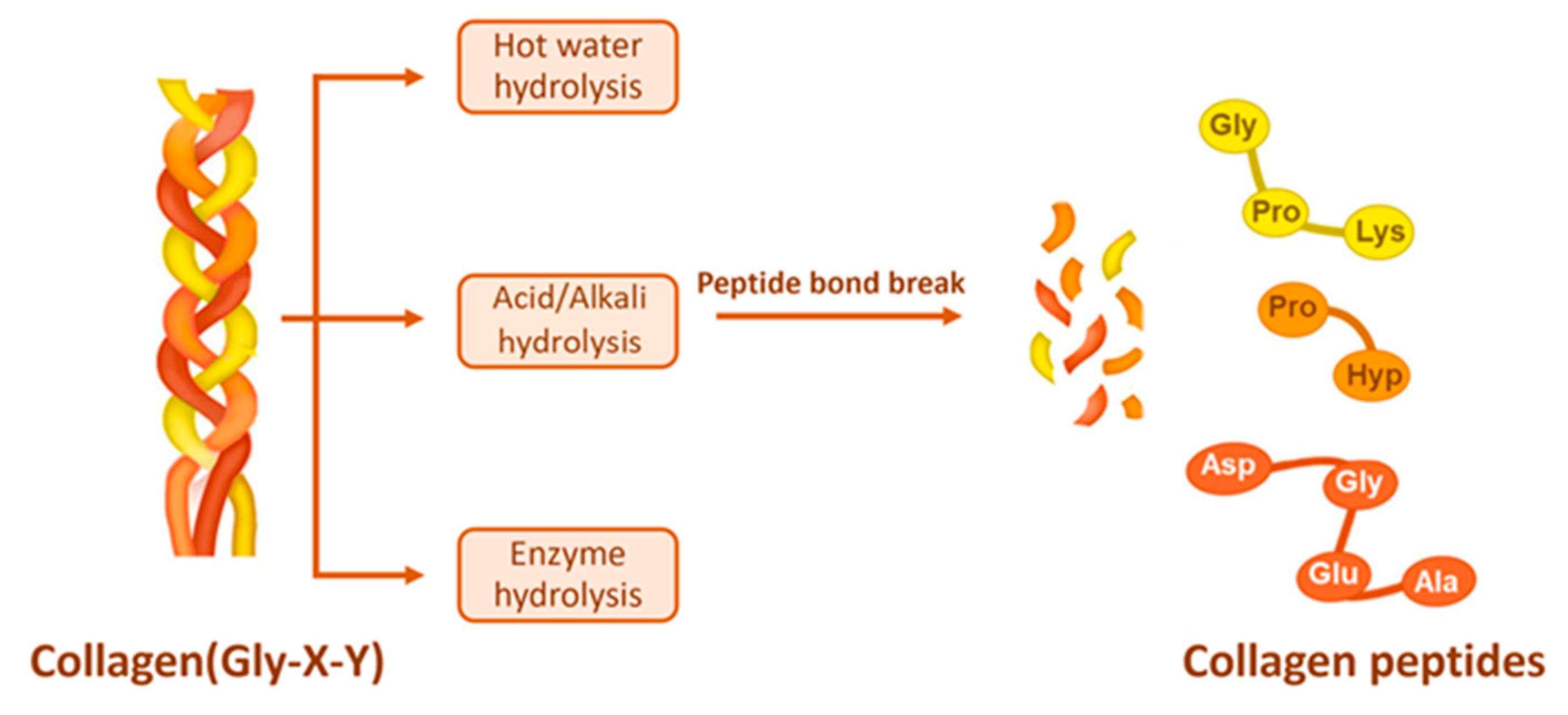
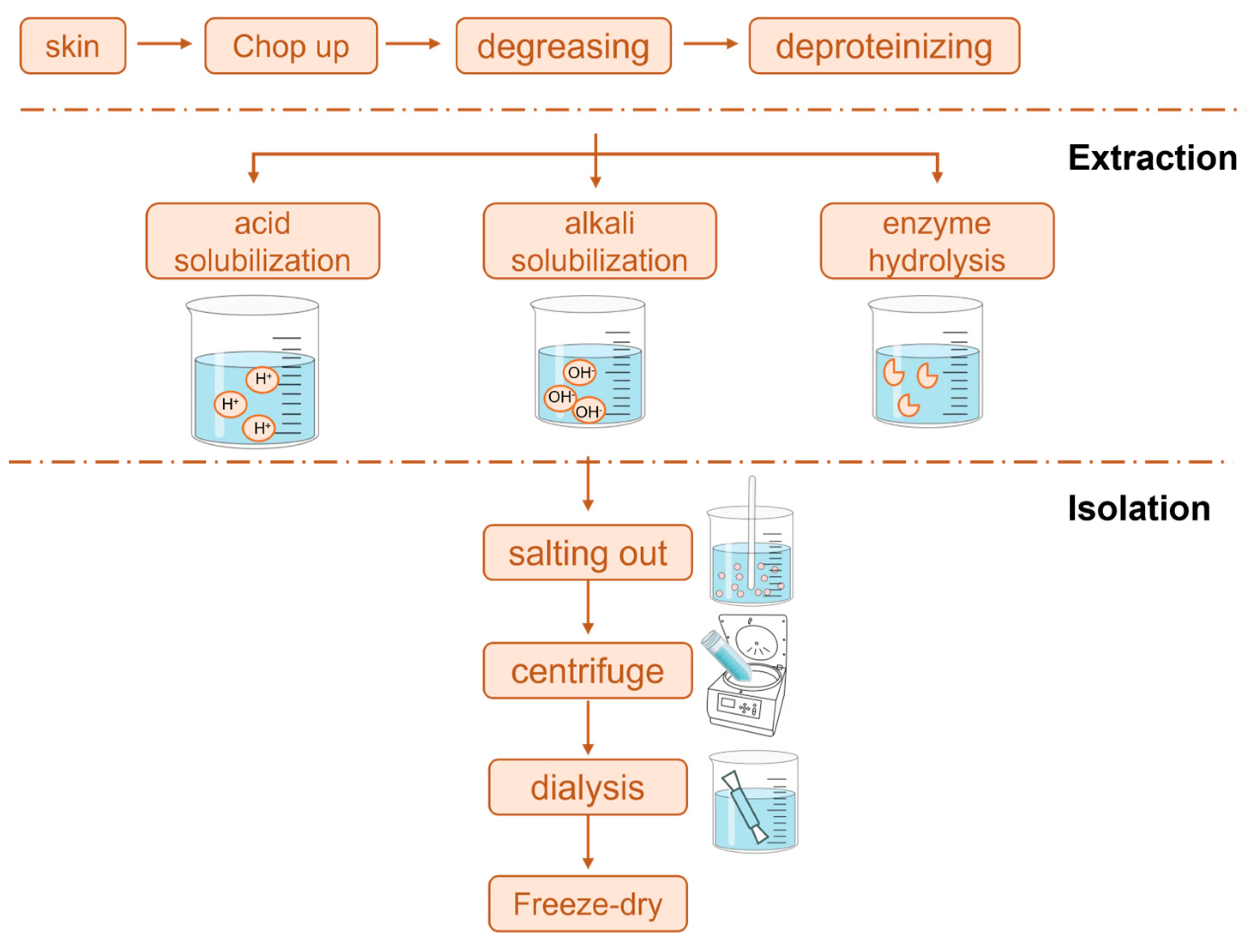
2. Preparation of Collagen Peptides
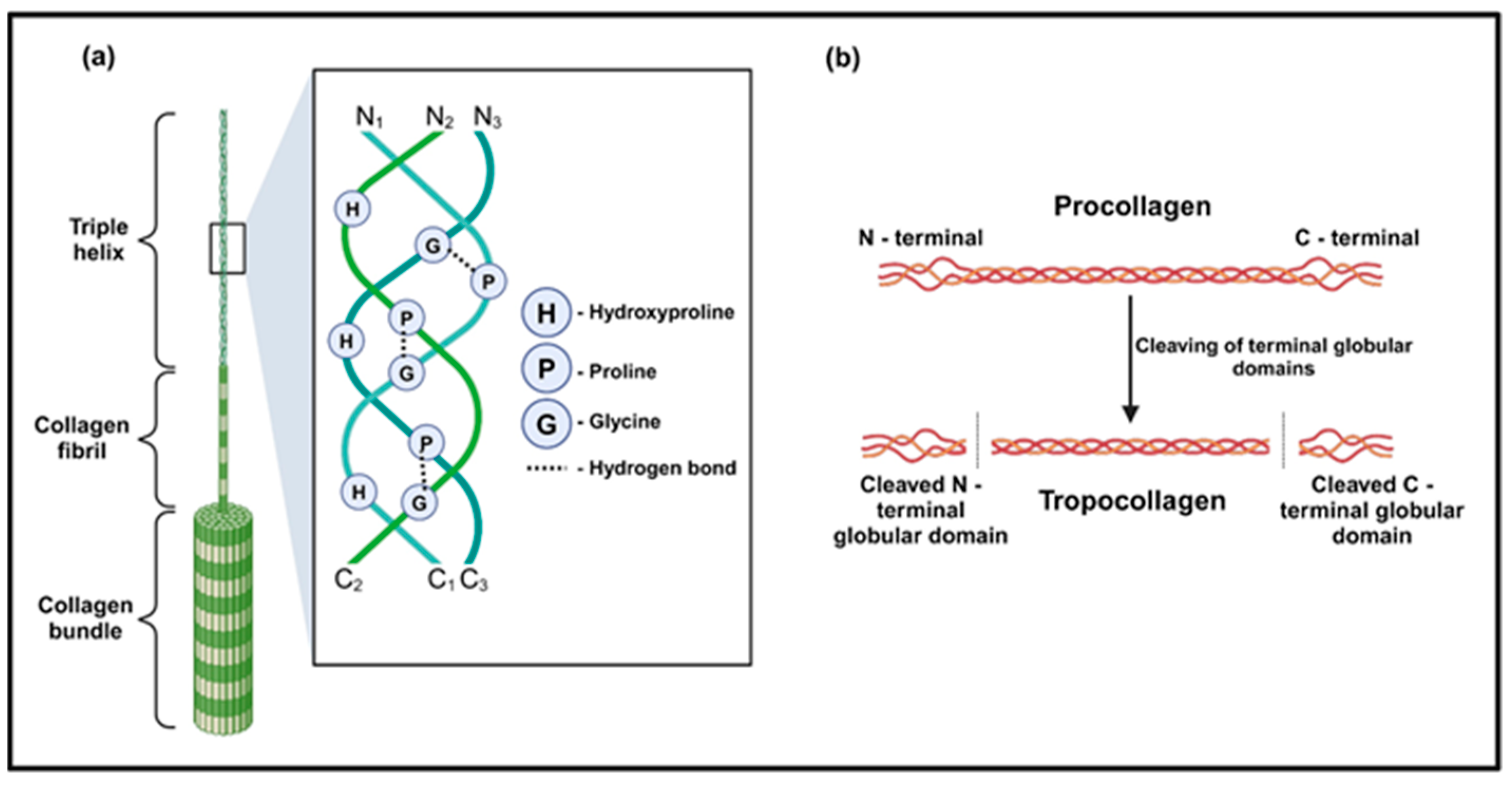
2.1. Extraction and Structural Analysis of Collagen

| Source | Extraction Method | Time (h) | Temperature (°C) | pH | Solid–Liquid Ratio (w/v) | Total Yield (%) a | References |
|---|---|---|---|---|---|---|---|
| Pig skin | 0.5 M acetic acid | 24 h | 4 °C | 2.5 | 1:10 | N/A | [15] |
| 0.5 M acetic acid + 0.1% pepsin | 24 h | 4 °C | 2.5 | 1:10 | 10.8% | [31] | |
| Cow skin | 0.5 M acetic acid | 24 h | 21 °C | 2.60 | 1:9 | 9.0% | [32] |
| 0.5 M acetic acid + 1% pepsin | 24 h | 4 °C | 2.60 | 1:20 | 24.4% | [32,33] | |
| 0.7 M acetic acid | 24 h | 4 °C | 2.35 | 1:9 | 65.7% | ||
| 0.5 M acetic acid + 0.1% pepsin | 24 h | 4 °C | 2.5 | 1:10 | 7.7% | ||
| Chicken skin | 0.5 M acetic acid + 0.1% pepsin | 28 h | 30 °C | N/A | 1:20 | 32.2% | [24] |
| Fish skin | 0.5 M acetic acid | 24 h | 4 °C | N/A | 1:10 | 11.7% | [16] |
| 0.5 M lactic acid | 24 h | 4 °C | N/A | 1:10 | 11.6% | [16] | |
| 0.5 M citric acid | 24 h | 4 °C | N/A | 1:10 | 11.4% | [17] | |
| 0.5 M acetic acid | 24 h | 4 °C | N/A | N/A | 97.7% b | ||
| 0.5 M acetic acid | 72 h | 4 °C | N/A | 1:40 | 27.7% | [18] | |
| alkali protease | 6 h | 50 °C | 10 | 1:24 | 94.8% b | [52] |
2.2. Collagen Hydrolysis
3. Bioactivity Analysis of Collagen Peptides
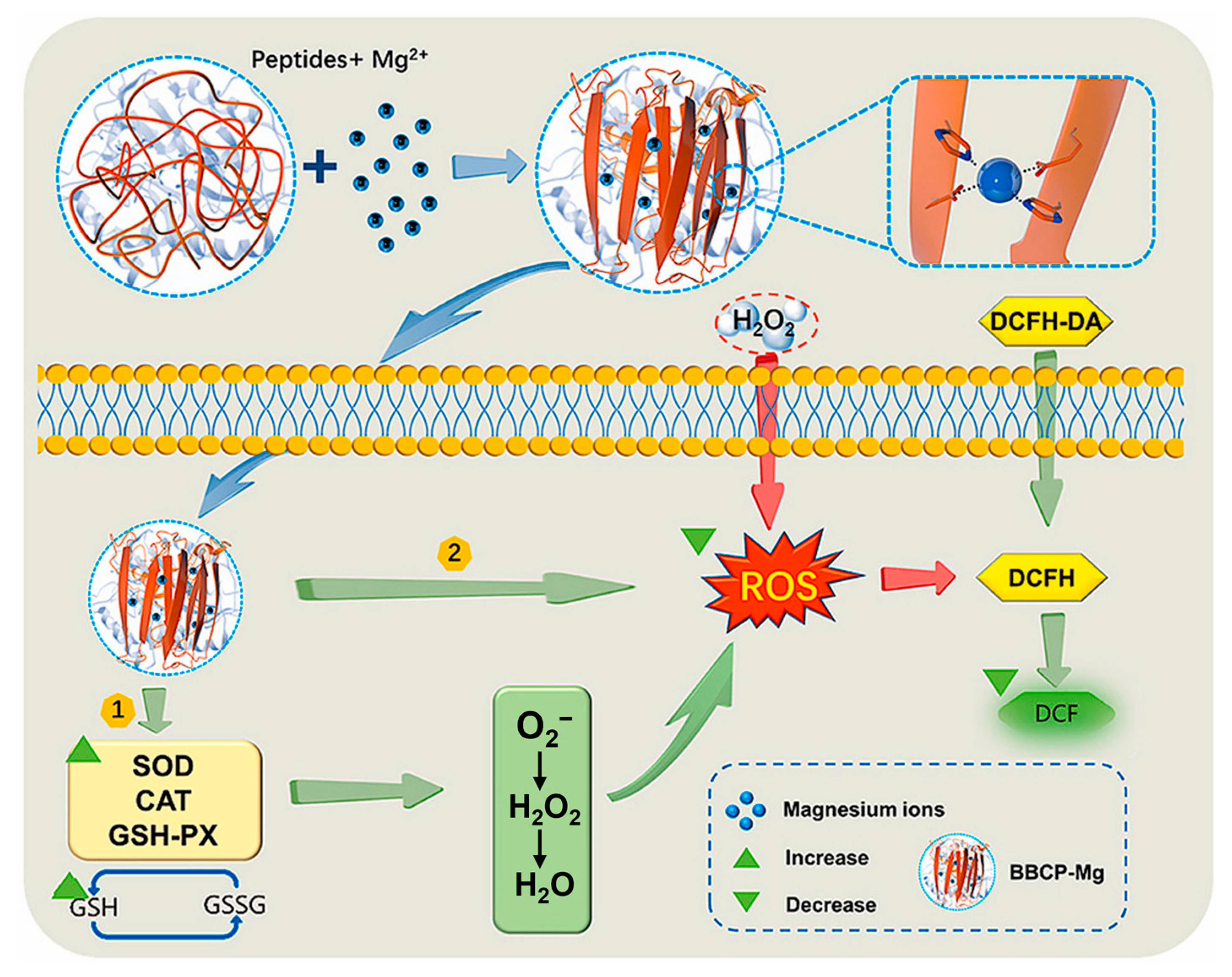
3.1. Antioxidant
3.2. Promote Hematopoiesis
3.3. Promote Osteogenic Differentiation
3.4. Antihypertensive
3.5. Anti-Diabetic
3.6. Other Biological Activities
| Sources | MW | Main Peptide Segment | Effect | References |
|---|---|---|---|---|
| Pork skin | <3 kDa | N/A | Antioxidant | [81] |
| <1 kDa | N/A | Pancreatic lipase inhibition | [31] | |
| N/A | NWYR | Anti-diabetes | [33] | |
| Cowhide | 329–596 Da | GPVG/FGPGP/APGGAP/GPPGPT/GPVGPPG | Anti-diabetes | [82] |
| N/A | ISVPGPM LGPVGNPGPA | ACE inhibition | [83] | |
| Donkey hide | N/A | ACEDAPPSAAHFR/FGFEVGPACFLG | Antioxidant | [64] |
| DGGR/DGD/NAGE/LVGE and GSEG | Tyrosinase inhibition | [84] | ||
| GPAGPIGPV/GPAGPIGPV/LSSPARSGASL | Promoting hematopoiesis | [67] | ||
| Fish skin | 516.5 Da | SEGPK | ACE inhibition | [85] |
| 597.6 Da | FDGPY | |||
| 542.6 Da | SPGPW | |||
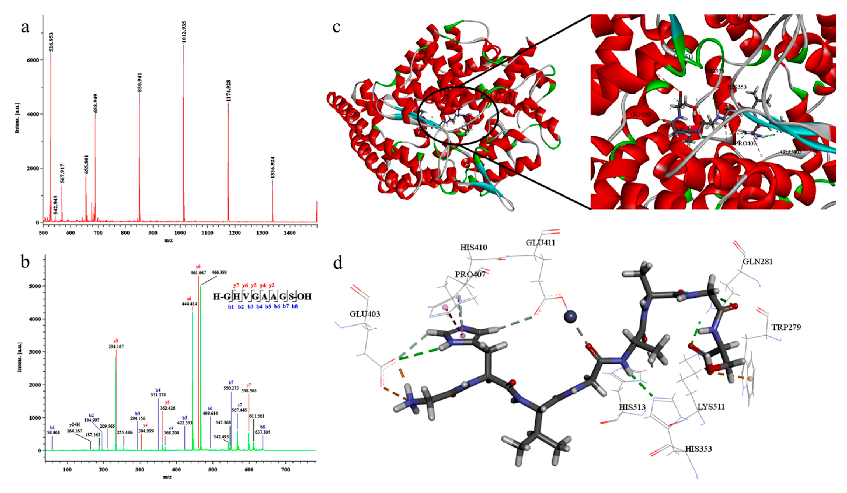
| N/A | GHVGAAGS | [72] | ||
| N/A | SPGSSGPQGFTG/GPVGPAGNPGANGLN/PPGPTGPRGQPGNIGF | Anti-diabetes | [86] | |
| N/A | POGP/POGA/LPO | Contributing to fibrocyte proliferation | [87] | |
| 358.68–863.89 Da | GPPGPPGTPGPQ/SGLPGPIGPPGPR/GLPGPIGPPGPR | Anti-photoaging | [52] | |
| <3 kDa | N/A | Immunoregulation | [88] | |
| 1336 Da | FETLMPLWGNK | Anti-neurodegenerative | [89] | |
| Sea cucumber | 1336 Da | FETLMPLWGNK | Antioxidant | [90] |
| 1389 Da | HEPFYGNEGALR | Antioxidant Promoting wound healing | [90,91] | |
| 961 Da | KMYPVPLN | |||
| N/A | VTPY/VLLY |
4. Applications of Collagen Peptides
4.1. Application in the Food Field
4.2. Cosmetic Applications
4.3. Biomedical Applications
5. Conclusions
Author Contributions
Funding
Institutional Review Board Statement
Informed Consent Statement
Data Availability Statement
Acknowledgments
Conflicts of Interest
References
- Macy, S.; Wall, J.; Kennel, S.; Balachandran, M.; Joseph, W.J.; Heidel, R.; Angela, D.W.; Foster, J.S.; Martin, E.; Tina, R.; et al. Collagen inhibits phagocytosis of amyloid In Vitro and In Vivo and may act as a ‘don’t eat me’ signal. Amyloid J. Protein Fold. Disord. 2022, 30, 249–260. [Google Scholar]
- Sorushanova, A.; Delgado, L.M.; Wu, Z.; Shologu, N.; Kshirsagar, A.; Raghunath, R.; Mullen, A.M.; Bayon, Y.; Pandit, A.; Raghunath, M.; et al. The Collagen Suprafamily: From Biosynthesis to Advanced Biomaterial Development. Adv. Mater. 2019, 31, 1801651. [Google Scholar] [CrossRef] [PubMed]
- Bayramoglu Eke, E.; Kaptanoglu Ikbal, G. Marine collagen. Stud. Nat. Prod. Chem. 2021, 71, 121–139. [Google Scholar]
- Shi, R.; Zhang, Z.; Zhu, A.; Xiong, X.; Zhang, J.; Xu, J.; Sy, M.-S.; Li, C. Targeting type I collagen for cancer treatment. Int. J. Cancer 2022, 151, 665–683. [Google Scholar] [CrossRef] [PubMed]
- Hu, X.; Wu, Z.; Zhang, Z.; Yao, H.; Wang, D.-A. Type II collagen scaffolds for tissue engineering. Commun. Mater. 2024, 5, 149. [Google Scholar] [CrossRef]
- Luo, Y.; Sinkeviciute, D.; He, Y.; Karsdal, M.; Henrotin, Y.; Mobasheri, A.; Önnerfjord, P.; Bay-Jensen, A. The minor collagens in articular cartilage. Protein Cell 2017, 8, 560–572. [Google Scholar] [CrossRef] [PubMed]
- Wang, L.; Zhang, Y.; Zhu, Z.; Zheng, F.; Gao, R. Food-derived collagen peptides: Safety, metabolism, and anti-skin-aging effects. Curr. Opin. Food Sci. 2023, 51, 101012. [Google Scholar] [CrossRef]
- Chen, W.; Xiang, N.; Huang, J.; Xu, H.; Wang, Z.; Ruan, B.; Zhang, J.; Wu, C.; Zhang, J.; Liang, Y. Supramolecular collagen nanoparticles for anti-wrinkle, skin whitening, and moisturizing effects. Colloids Surf. B Biointerfaces 2024, 245, 114275. [Google Scholar]
- Jung, K.; Kim, S.-H.; Joo, K.-M.; Lim, S.-H.; Shin, J.-H.; Roh, J.; Kim, E.; Park, C.W.; Kim, W. Oral Intake of Enzymatically Decomposed AP Collagen Peptides Improves Skin Moisture and Ceramide and Natural Moisturizing Factor Contents in the Stratum Corneum. Nutrients 2021, 13, 4372. [Google Scholar] [CrossRef] [PubMed]
- Żyga, J. Oral collagen supplements intake on improving skin structure and function. J. Educ. Health Sport 2022, 12, 434–440. [Google Scholar] [CrossRef]
- Sheng, Y.; Qiu, Y.-T.; Wang, Y.-M.; Chi, C.-F.; Wang, B. Novel Antioxidant Collagen Peptides of Siberian Sturgeon (Acipenser baerii) Cartilages: The Preparation, Characterization, and Cytoprotection of H2O2-Damaged Human Umbilical Vein Endothelial Cells (HUVECs). Mar. Drugs 2022, 20, 325. [Google Scholar] [CrossRef] [PubMed]
- Xu, S.; Niu, X.; Hou, Z.; Gao, C.; Lu, J.; Pang, Y.; Fang, M.; Lu, Y.; Chen, Y.; S, J.K.; et al. A multifunctional gelatine-quaternary ammonium copolymer: An efficient material for reducing dye emission in leather tanning process by superior anionic dye adsorption. J. Hazard. Mater. 2020, 383, 121142. [Google Scholar] [CrossRef] [PubMed]
- Jayaprakash, S.; Mohamad Abdul Razeen, Z.; Naveen Kumar, R.; He, J.; Milky, M.G.; Renuka, R.; Sanskrithi, M.V. Enriched characteristics of poultry collagen over other sources of collagen and its extraction methods: A review. Int. J. Biol. Macromol. 2024, 273, 133004. [Google Scholar] [CrossRef] [PubMed]
- Cadar, E.; Pesterau, A.-M.; Prasacu, I.; Ionescu, A.-M.; Pascale, C.; Dragan, A.-M.L.; Sirbu, R.; Tomescu, C.L. Marine Antioxidants from Marine Collagen and Collagen Peptides with Nutraceuticals Applications: A Review. Antioxidants 2024, 13, 919. [Google Scholar] [CrossRef] [PubMed]
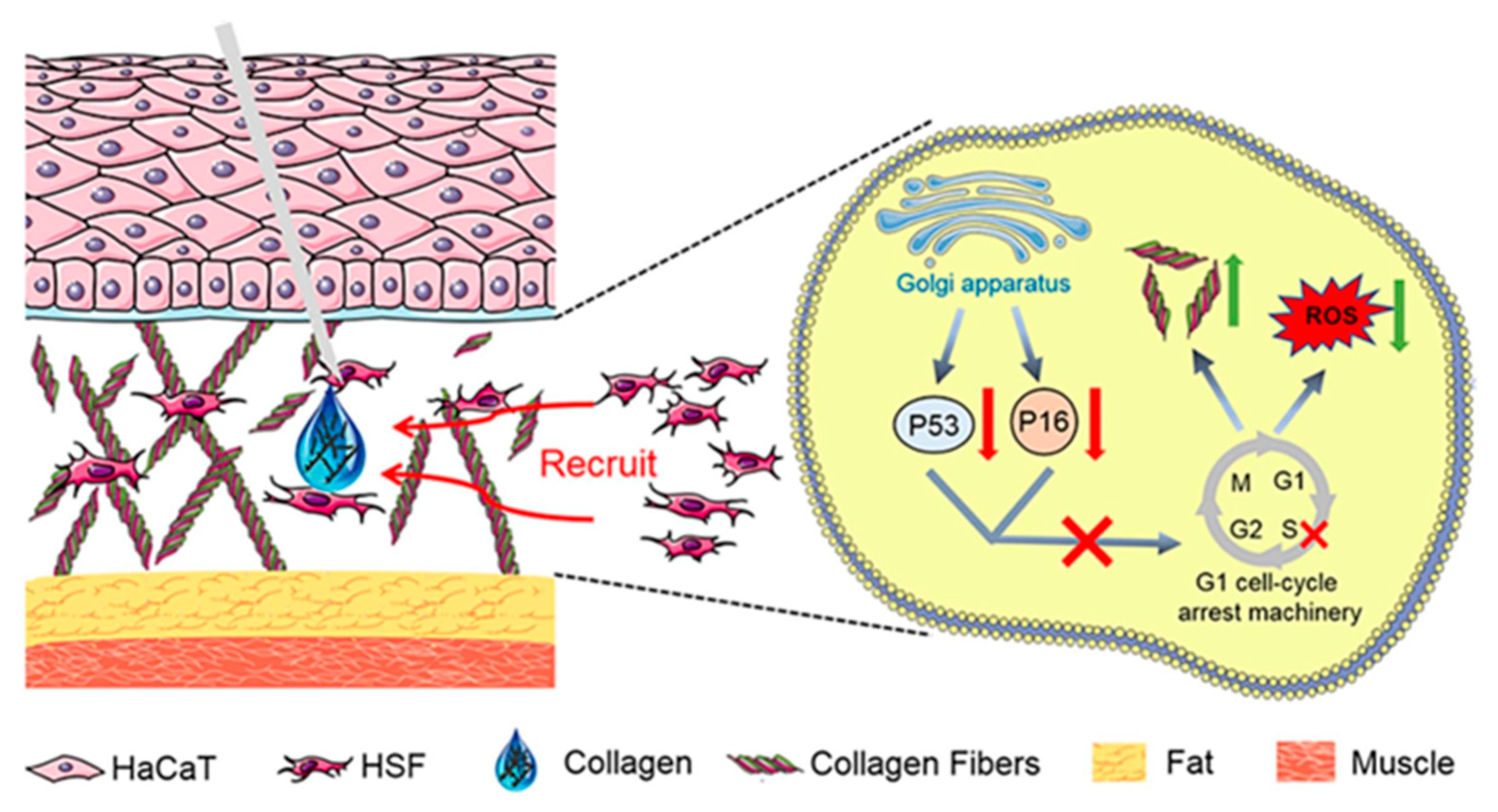
- Ni, H.; Liu, C.; Kong, L.; Zhai, L.; Chen, J.; Liu, Q.; Chen, Z.; Wu, M.; Chen, J.; Guo, Y.; et al. Preparation of injectable porcine skin-derived collagen and its application in delaying skin aging by promoting the adhesion and chemotaxis of skin fibroblasts. Int. J. Biol. Macromol. 2023, 253, 126718. [Google Scholar] [CrossRef] [PubMed]
- Jaziri, A.A.; Shapawi, R.; Mokhtar, R.A.; Noordin, W.N.M.; Huda, N. Microstructural and Physicochemical Analysis of Collagens from the Skin of Lizardfish (Saurida tumbil Bloch, 1795) Extracted with Different Organic Acids. Molecules 2022, 27, 2452. [Google Scholar] [CrossRef] [PubMed]
- Costa, F.T.; Oliveira, T.P.; Droval, A.A.; Marques, L.L.M.; Fuchs, R.H.B.; Cardoso, F.A.R. Evaluation of physicochemical properties of Nile tilapia skin collagen extracted in acid médium. Braz. J. Biol. 2024, 84, e255440. [Google Scholar] [CrossRef] [PubMed]
- Carpio, K.C.R.; Bezerra, R.S.; Cahú, T.B.; do Monte, F.T.D.; Neri, R.C.A.; da Silva, J.F.; dos Santos, P.R.; Carvalho, R.P.; Galeno, D.M.L.; Inhamuns, A.J. Extraction and characterization of collagen from the skin of Amazonian freshwater fish pirarucu. Braz. J. Med. Biol. Res. 2023, 56, e12564. [Google Scholar] [CrossRef] [PubMed]
- Iwen, F.; Baum, J.; Case, D. Dynamic Water-Mediated Hydrogen Bonding in a Collagen Model Peptide. Biochemistry 2015, 54, 6029–6037. [Google Scholar] [CrossRef] [PubMed]
- He, L.; Lan, W.; Zhao, Y.; Chen, S.; Liu, S.; Cen, L.; Cao, S.; Dong, L.; Jin, R.; Liu, Y. Characterization of biocompatible pig skin collagen and application of collagen-based films for enzyme immobilization. RSC Adv. 2020, 10, 7170–7180. [Google Scholar] [CrossRef] [PubMed]
- Asep, A.P.; Rahmi, N.; Annas, D.B. Production and characteristics of fish protein hydrolysate from parrotfish (Chlorurus sordidus) head. PeerJ 2019, 7, e8297. [Google Scholar] [CrossRef] [PubMed]
- Tan, Y.; Chang, S.K.C. Isolation and characterization of collagen extracted from channel catfish (Ictalurus punctatus) skin. Food Chem. 2018, 242, 147–155. [Google Scholar] [CrossRef] [PubMed]
- Cao, C.; Wang, H.; Zhang, J.; Kan, H.; Liu, Y.; Guo, L.; Tong, H.; Wu, Y.; Ge, C. Effects of Extraction Methods on the Characteristics, Physicochemical Properties and Sensory Quality of Collagen from Spent-Hens Bones. Foods 2023, 12, 202. [Google Scholar] [CrossRef] [PubMed]
- Dhakal, D.; Koomsap, P.; Lamichhane, A.; Sadiq, M.B.; Anal, A.K. Optimization of collagen extraction from chicken feet by papain hydrolysis and synthesis of chicken feet collagen based biopolymeric fibres. Food Biosci. 2018, 23, 23–30. [Google Scholar] [CrossRef]
- Huang, C.-Y.; Kuo, J.-M.; Wu, S.-J.; Tsai, H.-T. Isolation and characterization of fish scale collagen from tilapia (Oreochromis sp.) by a novel extrusion–hydro-extraction process. Food Chem. 2016, 190, 997–1006. [Google Scholar] [CrossRef] [PubMed]
- Alexandre, A.B.; Ivo, M.A.; Tiago, H.S.; João, F.M.; Ana Rita, C.D.; Rui, L.R. Water and Carbon Dioxide: Green Solvents for the Extraction of Collagen/Gelatin from Marine Sponges. ACS Sustain. Chem. Eng. 2015, 3, 254–260. [Google Scholar] [CrossRef]
- Vate, N.K.; Undeland, I.; Abdollahi, M. Resource efficient collagen extraction from common starfish with the aid of high shear mechanical homogenization and ultrasound. Food Chem. 2022, 393, 133426. [Google Scholar] [CrossRef] [PubMed]
- Wang, C.; Liu, C.; Guo, X.; Yuan, D.; Teng, Y. Enzymolysis technology optimization on the extraction of collagen from Greenland halibut skin. Shipin Keji 2012, 37, 240–242. [Google Scholar]
- Feng, W.; Zhao, T.; Zhou, Y.; Li, F.; Zou, Y.; Bai, S.; Wang, W.; Yang, L.; Wu, X. Optimization of enzyme-assisted extraction and characterization of collagen from Chinese sturgeon (Acipenser sturio Linnaeus) skin. Pharmacogn. Mag. 2013, 9, 32–37. [Google Scholar] [CrossRef] [PubMed]
- Matinong, A.M.E.; Pickering, K.L.; Waterland, M.R.; Chisti, Y.; Haverkamp, R.G. Gelatin and Collagen from Sheepskin. Polymers 2024, 16, 1563. [Google Scholar] [CrossRef] [PubMed]
- González-Noriega, J.A.; Valenzuela-Melendres, M.; Hernández–Mendoza, A.; Astiazarán-García, H.; Mazorra-Manzano, M.Á.; Peña-Ramos, E.A. Hydrolysates and peptide fractions from pork and chicken skin collagen as pancreatic lipase inhibitors. Food Chem. X 2022, 13 (Suppl. C), 100247. [Google Scholar] [CrossRef] [PubMed]
- Noorzai, S.; Verbeek, C.J.R.; Lay, M.C.; Swan, J. Collagen Extraction from Various Waste Bovine Hide Sources. Waste Biomass Valorization 2020, 11, 5687–5698. [Google Scholar] [CrossRef]
- Zhou, F.; Li, D.; Hou, Y.; Cong, Z.; Li, K.; Gu, X.; Xiao, G. Exploration of hypoglycemic peptides from porcine collagen based on network pharmacology and molecular docking. PLoS ONE 2024, 19, e0298674. [Google Scholar] [CrossRef] [PubMed]
- Valentina, O.; Lorenzo, D.; Federica, T.; Giulia De Negri, A.; Federica, G.; Elena, G.; Raffaella, B. Green Extraction and Preliminary Biological Activity of Hydrolyzed Collagen Peptides (HCPs) Obtained from Whole Undersized Unwanted Catches (Mugil cephalus L.). Molecules 2023, 28, 7637. [Google Scholar] [CrossRef] [PubMed]
- Gulevsky, A.K. Collagen: Structure, Metabolism, Production and Industrial Application. Biotechnol. Acta 2020, 13, 42–61. [Google Scholar] [CrossRef]
- Mahesh, S.; Nishath Sayed, A.; Zeeshan, Q.; Bader Musfer Al, B.; Khalid Zuhair, K.A.G.; Ateet, K.; Khalid Zuhair, K.A.G. Collagen Structure, Synthesis, and Its Applications: A Systematic Review. Cureus 2022, 14, 2168–8814. [Google Scholar] [CrossRef] [PubMed]
- Mienaltowski, M.J.; Gonzales, N.L.; Beall, J.M.; Pechanec, M.Y. Basic Structure, Physiology, and Biochemistry of Connective Tissues and Extracellular Matrix Collagens. Adv. Exp. Med. Biol. 2021, 1348, 5–43. [Google Scholar] [PubMed]
- Balupuri, A.; Son, D.-H.; Sook Kang, N. Investigating the effect of water on collagen triple helix stability. J. Mol. Liq. 2024, 415, 126325. [Google Scholar] [CrossRef]
- He, X.; Lin, L.; Jiang, S.; Lu, J. Characterization of acid-soluble collagen (ASC) derived from walleye pollock and silver carp skin and comparison them with the collagen from pig and duck skin. Food Chem. Adv. 2024, 4, 100746. [Google Scholar] [CrossRef]
- Andayani, A.A.; Harmita, H.; Maggadani, B.P. Isolation, purification, and characterization of porcine skin collagen: Analysis of the glycine, proline, and hydroxyproline components using high-performance liquid chromatography. Int. J. Appl. Pharm. 2018, 10, 294. [Google Scholar] [CrossRef]
- Xu, J.; Li, T.-D.; Tang, X.-L.; Qiao, C.-D.; Jiang, Q.-W. Effect of aggregation behavior of gelatin in aqueous solution on the grafting density of gelatin modified with glycidol. Colloids Surf. B Biointerfaces 2012, 95, 201–207. [Google Scholar] [CrossRef] [PubMed]
- Liu, H.; Li, M.; Tang, K.; Liu, J.; Li, X.; Meng, X. Evolution of conformation and thermal properties of bovine hides collagen in the sodium sulphide solution. J. Mol. Liq. 2022, 367, 120449. [Google Scholar] [CrossRef]
- Gauza-Włodarczyk, M.; Kubisz, L.; Włodarczyk, D. Amino acid composition in determination of collagen origin and assessment of physical factors effects. Int. J. Biol. Macromol. 2017, 104, 987–991. [Google Scholar] [CrossRef] [PubMed]
- Oechsle, A.M.; Akgün, D.; Krause, F.; Maier, C.; Gibis, M.; Kohlus, R.; Weiss, J. Microstructure and physical-chemical properties of chicken collagen. Food Struct. 2016, 7, 29–37. [Google Scholar] [CrossRef]
- Gojkovic, Z.; Marova, I.; Matouskova, P.; Obruca, S.; Miloslav, P. Use of ultrasonic spectroscopy and viscosimetry for the characterization of chicken skin collagen in comparison with collagens from other animal tissues. Prep. Biochem. Biotechnol. 2014, 44, 761–771. [Google Scholar] [CrossRef] [PubMed]
- Liu, T.; Zou, L.; Ji, X.; Xiao, G. Chicken skin-derived collagen peptides chelated zinc promotes zinc absorption and represses tumor growth and invasion In Vivo by suppressing autophagy. Front. Nutr. 2022, 9, 960926. [Google Scholar] [CrossRef] [PubMed]
- Yu, E.; Pan, C.; Luo, X.; Ruan, Q.; Chen, W.; Fang, Y.; Wang, K.; Qin, Y.; Lv, M.; Ma, H. Structural characteristics, component interactions and functional properties of gelatins from three fish skins extracted by five methods. Int. J. Biol. Macromol. 2023, 248 (Suppl. C), 125813. [Google Scholar] [CrossRef] [PubMed]
- Song, Z.; Liu, H.; Chen, L.; Chen, L.; Zhou, C.; Hong, P.; Deng, C. Characterization and comparison of collagen extracted from the skin of the Nile tilapia by fermentation and chemical pretreatment. Food Chem. 2021, 340, 128139. [Google Scholar] [CrossRef] [PubMed]
- Menezes, M.D.L.L.R.; Ribeiro, H.L.; de Oliveira M da Silva Abreu, F.; de Andrade Feitosa, J.P.; de Sá Moreira de S Filho, M. Optimization of the collagen extraction from Nile tilapia skin (Oreochromis niloticus) and its hydrogel with hyaluronic acid. Colloids Surf. B Biointerfaces 2020, 189, 110852. [Google Scholar] [CrossRef] [PubMed]
- Tian, M.; Xue, C.; Chang, Y.; Shen, J.; Zhang, Y.; Li, Z.; Wang, Y. Collagen fibrils of sea cucumber (Apostichopus japonicus) are heterotypic. Food Chem. 2020, 316, 126272. [Google Scholar] [CrossRef] [PubMed]
- Barzkar, N.; Attaran-Fariman, G.; Taheri, A.; Venmathi Maran, B.A. Extraction and characterization of collagen and gelatin from body wall of sea cucumbers Stichopus horrens and Holothuria arenicola. PeerJ 2024, 12, e18149. [Google Scholar] [CrossRef] [PubMed]
- Huang, J.-J.; Li, H.-L.; Xiong, G.-Q.; Cai, J.; Liao, T.; Zu, X.-Y. Extraction, identification and anti-photoaging activity evaluation of collagen peptides from silver carp (Hypophthalmichthys molitrix) skin. LWT Food Sci. Technol. 2023, 173 (Suppl. C), 114384. [Google Scholar] [CrossRef]
- Matinong, A.M.E.; Chisti, Y.; Pickering, K.L.; Haverkamp, R.G. Collagen Extraction from Animal Skin. Biology 2022, 11, 905. [Google Scholar] [CrossRef] [PubMed]
- da Silva, C.M.L.; Spinelli, E.; Rodrigues, S.V. Fast and sensitive collagen quantification by alkaline hydrolysis/hydroxyproline assay. Food Chem. 2015, 173, 619–623. [Google Scholar] [CrossRef] [PubMed]
- Hou, N.-T.; Chen, B.-H. Extraction, purification and characterization of collagen peptide prepared from skin hydrolysate of sturgeon fish. Food Qual. Saf. 2023, 7, fyad033. [Google Scholar] [CrossRef]
- Koliada, M.; Plavan, V. Problems of efficient processing and use of collagen-containing materials. Pure Appl. Chem. 2015, 87, 43–49. [Google Scholar] [CrossRef]
- Zhang, Y.; Olsen, K.; Grossi, A.; Otte, J. Effect of pretreatment on enzymatic hydrolysis of bovine collagen and formation of ACE-inhibitory peptides. Food Chem. 2013, 141, 2343–2354. [Google Scholar] [CrossRef] [PubMed]
- Hu, X.; Yang, Y.; Chang, C.; Li, J.; Su, Y.; Gu, L. The targeted development of collagen-active peptides based on composite enzyme hydrolysis: A study on the structure-activity relationship. Food Funct. 2024, 15, 401–410. [Google Scholar] [CrossRef] [PubMed]
- Li, Y.; Lu, Y.; Zhao, Y.; Zhang, N.; Zhang, Y.; Fu, Y. Deciphering the Wound-Healing Potential of Collagen Peptides and the Molecular Mechanisms: A Review. J. Agric. Food Chem. 2024, 72, 26007–26026. [Google Scholar] [CrossRef] [PubMed]
- León-López, A.; Fuentes-Jiménez, L.; Hernández-Fuentes, A.D.; Campos-Montiel, R.G.; Aguirre-Álvarez, G. Hydrolysed Collagen from Sheepskins as a Source of Functional Peptides with Antioxidant Activity. Int. J. Mol. Sci. 2019, 20, 3931. [Google Scholar] [CrossRef] [PubMed]
- Diego, J.G.-S.; Milad, H.; Matin, V.; Aniseh Zarei, J.; Andres, M.; Jose, M.L. Bioactive Peptide Fractions from Collagen Hydrolysate of Common Carp Fish Byproduct: Antioxidant and Functional Properties. Antioxidants 2022, 11, 509. [Google Scholar] [CrossRef] [PubMed]
- Zhang, C.; Du, B.; Song, Z.; Deng, G.; Shi, Y.; Li, T.; Huang, Y. Antioxidant activity analysis of collagen peptide-magnesium chelate. Polym. Test. 2023, 117, 107822. [Google Scholar] [CrossRef]
- Liang, R.; Xu, L.; Fan, C.; Cao, L.; Guo, X. Structural Characteristics and Antioxidant Mechanism of Donkey-Hide Gelatin Peptides by Molecular Dynamics Simulation. Molecules 2023, 28, 7975. [Google Scholar] [CrossRef] [PubMed]
- Yan, Q.; Li, N.; Li, Y.; Zhao, Z.; Song, Q.; Lu, S.; Wang, J.; Wang, Q. Preparation and identification of novel antioxidant peptides from collagen hydrolysate of sheep hoof assisted by ultrasound. Int. J. Biol. Macromol. 2024, 281, 136415. [Google Scholar] [CrossRef] [PubMed]
- Amaliris, G.; Hamideh, P.; Stefano, R. Novel potential therapeutics to modify iron metabolism and red cell synthesis in diseases associated with defective erythropoiesis. Haematologica 2023, 108, 2582–2593. [Google Scholar] [CrossRef] [PubMed]
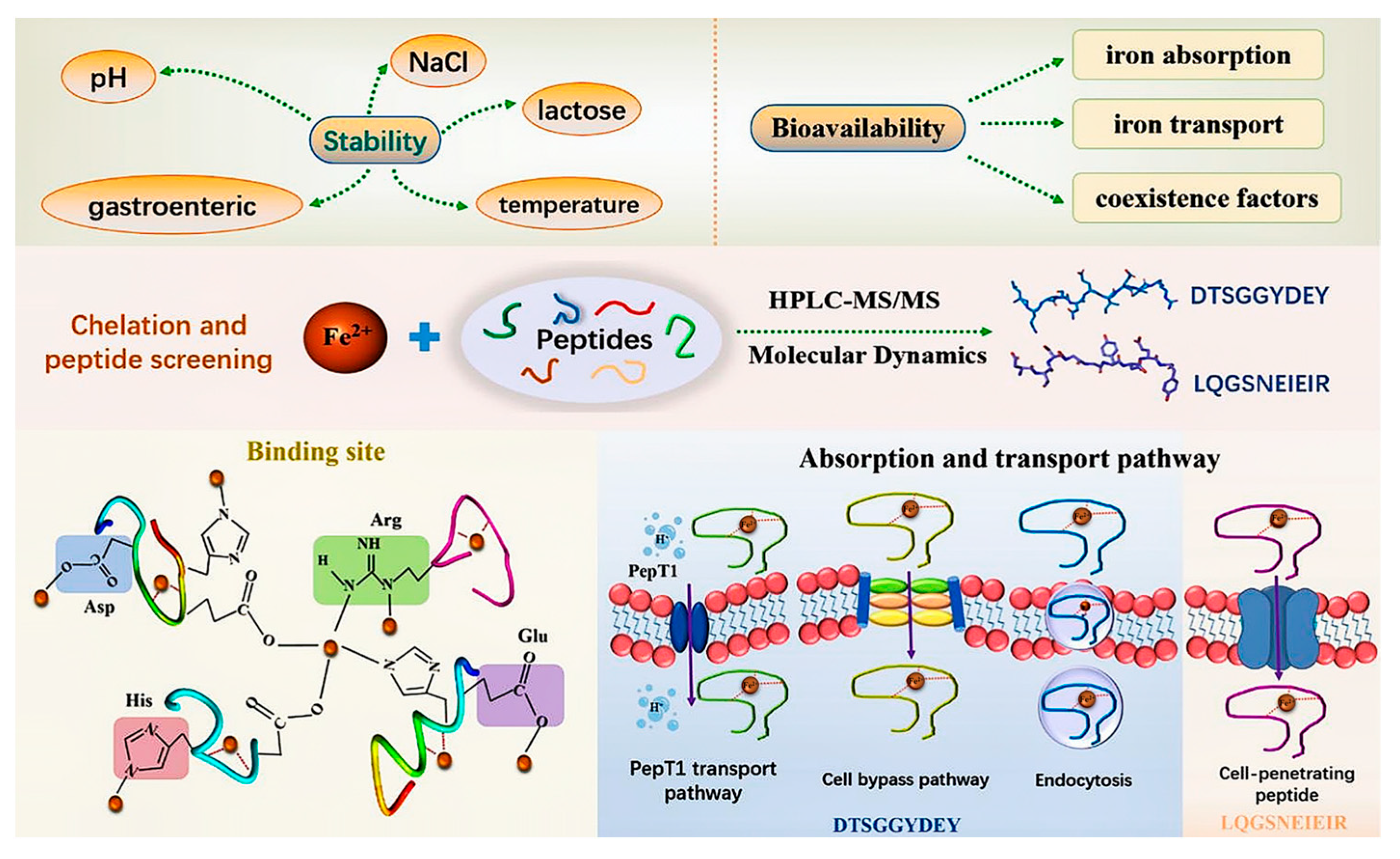
- Zhao, Q.; Liang, W.; Xiong, Z.; Li, C.; Zhang, L.; Rong, J.; Xiong, S.; Liu, R.; You, J.; Yin, T.; et al. Digestion and absorption characteristics of iron-chelating silver carp scale collagen peptide and insights into their chelation mechanism. Food Res. Int. 2024, 190, 114612. [Google Scholar] [CrossRef] [PubMed]
- Wu, H.; Ren, C.; Yang, F.; Qin, Y.; Zhang, Y.; Liu, J. Extraction and identification of collagen-derived peptides with hematopoietic activity from Colla Corii Asini. J. Ethnopharmacol. 2016, 182, 129–136. [Google Scholar] [CrossRef] [PubMed]
- Joseph, B. The Relationship of Peak Bone Mass, Aging, and Bone Loss to Osteoporosis and Fragility Fractures. In Arthroplasty for the Treatment of Fractures in the Older Patient; Springer: Cham, Switzerland, 2018; pp. 3–17. [Google Scholar]
- Li, L.; Yu, Y.; Wu, W.; Wang, P. Extraction, Characterization and Osteogenic Activity of a Type I Collagen from Starfish (Asterias amurensis). Mar. Drugs 2023, 21, 274. [Google Scholar] [CrossRef] [PubMed]
- Hwang, J.M.; Lee, M.-H.; Kwon, Y.; Chung, H.-C.; Kim, D.-U.; Lee, J.-H. In Vitro and In Vivo Bone-Forming Effect of a Low-Molecular-Weight Collagen Peptide. J. Microbiol. Biotechnol. 2024, 34, 415–424. [Google Scholar] [CrossRef] [PubMed]
- Ding, D.; Yu, T.; Du, B.; Huang, Y. Collagen hydrolysate from Thunnus orientalis bone induces osteoblast proliferation and differentiation. Chem. Eng. Sci. 2019, 205, 143–150. [Google Scholar] [CrossRef]
- Yu, Z.; Wu, S.; Zhao, W.; Mi, G.; Ding, L.; Li, J.; Liu, J. Identification of novel angiotensin I-converting enzyme inhibitory peptide from collagen hydrolysates and its molecular inhibitory mechanism. Int. J. Food Sci. Technol. 2020, 55, 3145–3152. [Google Scholar] [CrossRef]
- Fitriyanto, N.; Jamhari, J.; Pratiwi, A.; Erwanto, Y.; Abidin, M.Z.; Matulessy, D.; Rusman, R.; Hakim, T.R. Extraction of Collagen from the Skin of Kacang Goat and Production of Its Hydrolysate as an Inhibitor of Angiotensin Converting Enzyme. Trop. Anim. Sci. J. 2021, 44, 222–228. [Google Scholar] [CrossRef]
- Zhou, M.; Ren, G.; Zhang, B.; Ma, F.; Fan, J.; Qiu, Z. Screening and identification of a novel antidiabetic peptide from collagen hydrolysates of Chinese giant salamander skin: Network pharmacology, inhibition kinetics and protection of IR-HepG2 cells. Food Funct. 2022, 13, 3329–3342. [Google Scholar] [CrossRef] [PubMed]
- Natsir, H.; Dali, S.; Arif, A.R.; Sartika; Leliani. Activity and kinetics of α-glucosidase inhibition by collagen hydrolysate from thunnus albacares bone. J. Phys. Conf. Ser. 2019, 1341, 032015. [Google Scholar] [CrossRef]
- Das, D.; Kabir, M.E.; Sarkar, S.; Wann, S.B.; Kalita, J.; Manna, P. Antidiabetic potential of soy protein/peptide: A therapeutic insight. Int. J. Biol. Macromol. 2022, 194 (Suppl. C), 276–288. [Google Scholar] [CrossRef] [PubMed]
- He, L.; Wang, X.; Wang, Y.; Luo, J.; Zhao, Y.; Han, G.; Han, L.; Yu, Q. Production and identification of dipeptidyl peptidase IV (DPP-IV) inhibitory peptides from discarded cowhide collagen. Food Chem. 2023, 405, 134793. [Google Scholar] [CrossRef] [PubMed]
- Rio, J.K.; Aviria, E.; Salehah, N.H.; Ninda, F.P.; Imroatus, S. Selar (Selar crumenophthalmus) Fish Protein Hydrolysate Has Antidiabetic Properties Possibly through GLP-1. Curr. Nutr. Food Sci. 2021, 17, 516–522. [Google Scholar] [CrossRef]
- Kim, Y.; Lee, J.O.; Lee, J.M.; Lee, M.-H.; Kim, H.-M.; Chung, H.-C.; Kim, D.-U.; Lee, J.-H.; Kim, B.J. Low Molecular Weight Collagen Peptide (LMWCP) Promotes Hair Growth by Activating the Wnt/GSK-3β/β-Catenin Signaling Pathway. J. Microbiol. Biotechnol. 2024, 34, 17–28. [Google Scholar] [CrossRef] [PubMed]
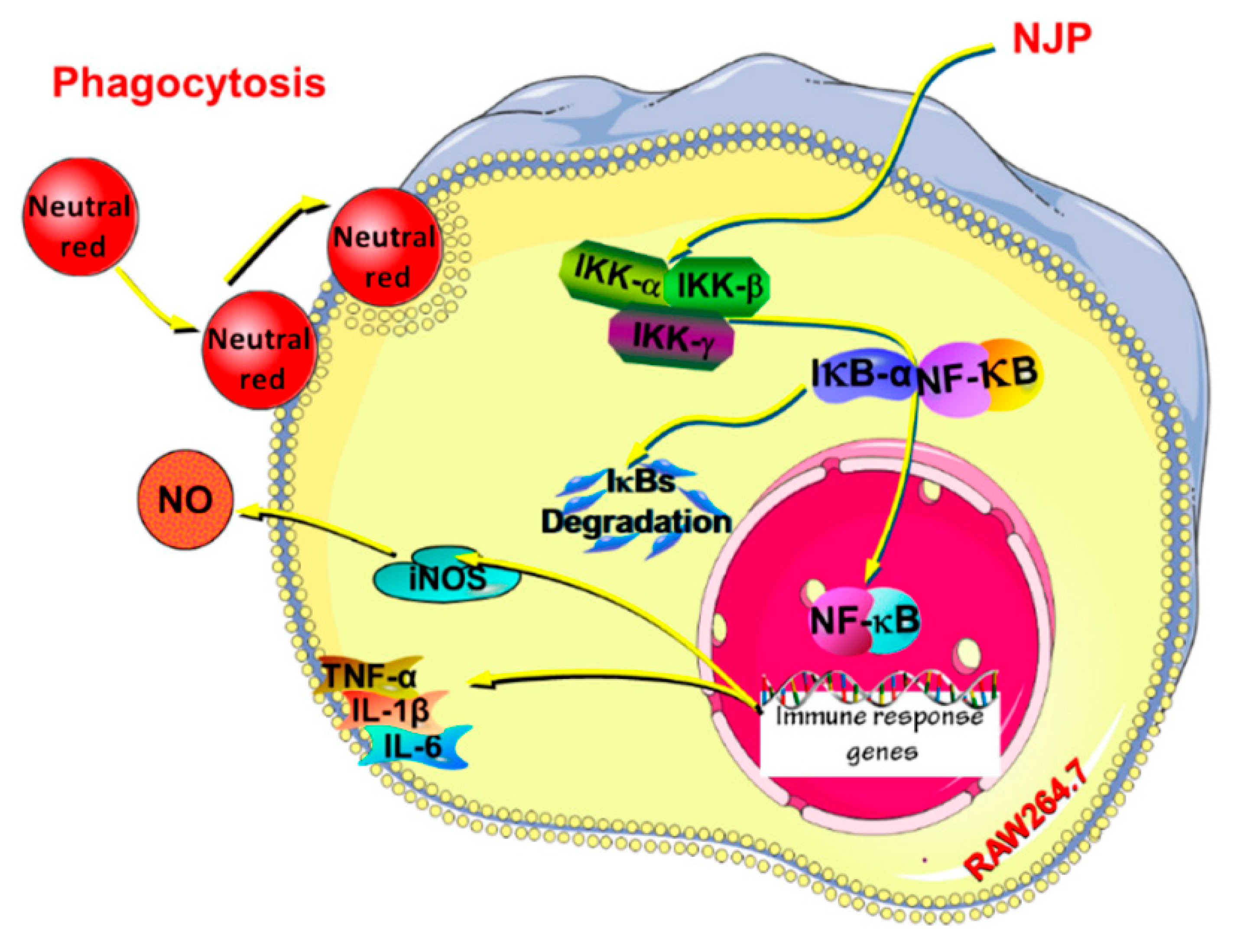
- Zhang, Z.; Hu, X.; Lin, L.; Ding, G.; Yu, F. Immunomodulatory Activity of Low Molecular-Weight Peptides from Nibea japonica in RAW264.7 Cells via NF-κB Pathway. Mar. Drugs 2019, 17, 404. [Google Scholar] [CrossRef] [PubMed]
- Hong, G.-P.; Min, S.-G.; Jo, Y.-J. Anti-Oxidative and Anti-Aging Activities of Porcine By-Product Collagen Hydrolysates Produced by Commercial Proteases: Effect of Hydrolysis and Ultrafiltration. Molecules 2019, 24, 1104. [Google Scholar] [CrossRef] [PubMed]
- Rivero-Pino, F.; Espejo-Carpio, F.J.; Guadix, E.M. Production and identification of dipeptidyl peptidase IV (DPP-IV) inhibitory peptides from discarded Sardine pilchardus protein. Food Chem. 2020, 328, 127096. [Google Scholar] [CrossRef] [PubMed]
- Dong, Y.; Wang, Z.-Q.; Shen, J.-Q.; Fan, J.-y.; Tang, J.; Ma, Z.-Y.; Shen, Y.-L. Two novel ACE inhibitory peptides derived from bovine skin collagen. Sci. Technol. Food Ind. 2017, 38, 135–140. [Google Scholar]
- Ji, H.; Yu, Z.; Zhao, W. Colla corii asini–derived peptides as tyrosinase inhibitors: Identification, inhibitory activity, and molecular mechanism. Int. J. Food Sci. Technol. 2022, 57, 7391–7401. [Google Scholar] [CrossRef]
- Hu, Y.-D.; Xi, Q.-H.; Kong, J.; Zhao, Y.-Q.; Chi, C.-F.; Wang, B. Angiotensin-I-Converting Enzyme (ACE)-Inhibitory Peptides from the Collagens of Monkfish (Lophius litulon) Swim Bladders: Isolation, Characterization, Molecular Docking Analysis and Activity Evaluation. Mar. Drugs 2023, 21, 516. [Google Scholar] [CrossRef] [PubMed]
- Wang, T.-Y.; Hsieh, C.-H.; Hung, C.-C.; Jao, C.-L.; Chen, M.-C.; Hsu, K.-C. Fish skin gelatin hydrolysates as dipeptidyl peptidase IV inhibitors and glucagon-like peptide-1 stimulators improve glycaemic control in diabetic rats: A comparison between warm- and cold-water fish. J. Funct. Foods 2015, 19, 330–340. [Google Scholar] [CrossRef]
- Chang, C.; Ma, Y.; Yang, Y.; Su, Y.; Gu, L.; Li, J. Strategies to Improve Hydrolysis Efficiency of Fish Skin Collagen: Study on ACE Inhibitory Activity and Fibroblast Proliferation Activity. Foods 2024, 13, 3869. [Google Scholar] [CrossRef] [PubMed]
- Yu, F.; He, K.; Dong, X.; Zhang, Z.; Wang, F.; Tang, Y.; Chen, Y.; Ding, G. Immunomodulatory activity of low molecular-weight peptides from Nibea japonica skin in cyclophosphamide-induced immunosuppressed mice. J. Funct. Foods 2020, 68, 103886. [Google Scholar] [CrossRef]
- Wang, Q.; Liang, M.; Xiao, Y.; Li, Z.; Chen, X.; Cheng, P.; Qi, B.; Yu, Y.; Lei, T.; Huang, Z. In Silico and In Vivo discovery of antioxidant sea cucumber peptides with antineurodegenerative properties. Food Funct. 2024, 15, 5972–5986. [Google Scholar] [CrossRef] [PubMed]
- Lu, M.; Mishra, A.; Boschetti, C.; Lin, J.; Liu, Y.; Huang, H.; Kaminski, C.F.; Huang, Z.; Tunnacliffe, A.; Kaminski Schierle, G.S. Sea Cucumber-Derived Peptides Alleviate Oxidative Stress in Neuroblastoma Cells and Improve Survival in C. elegans Exposed to Neurotoxic Paraquat. Oxidative Med. Cell. Longev. 2021, 2021, 8842926. [Google Scholar] [CrossRef] [PubMed]
- Zheng, Z.; Sun, N.; Lu, Z.; Zheng, J.; Zhang, S.; Lin, S. The potential mechanisms of skin wound healing mediated by tetrapeptides from sea cucumber. Food Biosci. 2023, 53, 102742. [Google Scholar] [CrossRef]
- Saleem, K.; Pritha, D.; Charitha, S.; Mayur, B.; Geetika, Y.; Vishwadeep, A.; Pawan, T.; Pradipta, B. Contribution of quasi-fibrillar superstructures in peroxide quenching by collagen peptides derived from fish processing by-products and their application as natural food additives. bioRxiv 2021. [Google Scholar] [CrossRef]
- Cui, P.; Shao, T.; Liu, W.; Li, M.; Yu, M.; Zhao, W.; Song, Y.; Ding, Y.; Liu, J. Advanced review on type II collagen and peptide: Preparation, functional activities and food industry application. Crit. Rev. Food Sci. Nutr. 2023, 64, 11302–17852. [Google Scholar] [CrossRef] [PubMed]
- Han, M.; Zhang, Z.; Li, X.; Tong, H.; Xu, Z.; Ding, Z.; Yang, A.; Xie, M.; Wang, X. Effects of collagen peptides from Micropterus salmoides skin on oxidative damage induced by cyclophosphamide in mice. Front. Nutr. 2022, 9, 1037212. [Google Scholar] [CrossRef] [PubMed]
- Silvestrini, B.; Silvestrini, M. Medical use of hydrolyzed collagen in osteoporosis. Med. Res. Arch. 2023, 11, 4358. [Google Scholar] [CrossRef]
- Kim, H.-R.; Lee, S.-H.; Noh, E.-M.; Choi, B.; Seo, H.-Y.; Jang, H.; Kim, S.-Y.; Park, M.H. Therapeutic Effect of Enzymatically Hydrolyzed Cervi Cornu Collagen NP-2007 and Potential for Application in Osteoarthritis Treatment. Int. J. Mol. Sci. 2023, 24, 11667. [Google Scholar] [CrossRef] [PubMed]
- Luo, J.; Ma, B.; Zhao, Y.; Chen, T. Evolution Model of Health Food Safety Risk Based on Prospect Theory. J. Healthc. Eng. 2018, 2018, 8769563. [Google Scholar] [CrossRef] [PubMed]
- Barbara, J.; Zofia, M.; Tomasz, O. Use of Collagen in Cosmetic Products. Curr. Issues Mol. Biol. 2024, 46, 2043–2070. [Google Scholar] [CrossRef] [PubMed]
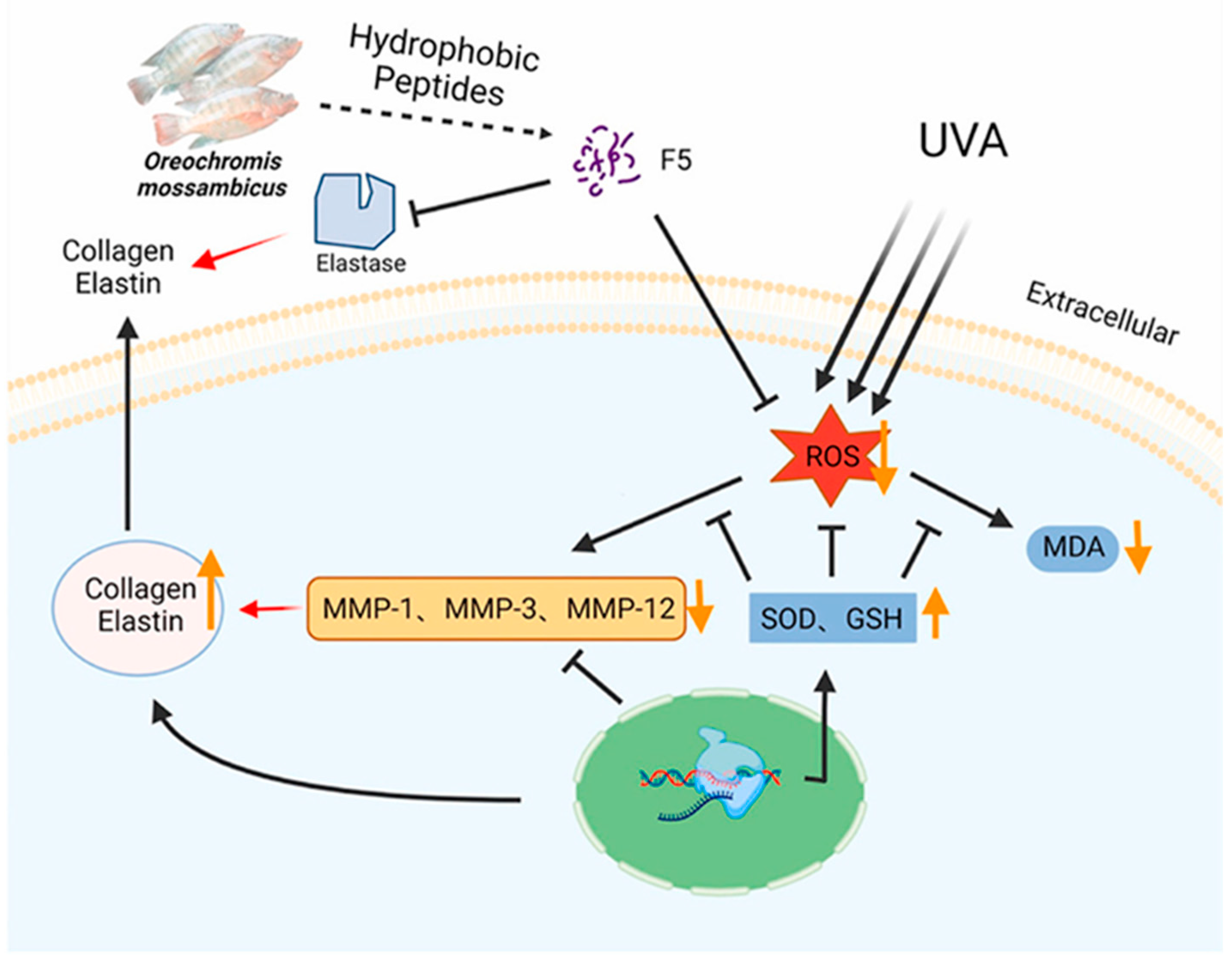
- Yao, H.; Wang, S.; Fu, B.; Xu, X.; Cheng, S.; Du, M. The potential benefits of Oreochromis mossambicus derived hydrophobic peptides in protecting the skin against UVA-induced damage. Food Biosci. 2024, 59, 104120. [Google Scholar] [CrossRef]
- Li, P.-H.; Lu, W.-C.; Chan, Y.-J.; Ko, W.-C.; Jung, C.-C.; Huynh, D.T.L.; Ji, Y.-X. Extraction and characterization of collagen from sea cucumber (Holothuria cinerascens) and its potential application in moisturizing cosmetics. Aquaculture 2020, 515, 734590. [Google Scholar] [CrossRef]
- Elena, D.; Raluca, S.; Mădălina Albu, K.; Georgeta, V.; Maria Minodora, M.; Alina, M.; Irina, T.; Raluca, Ţ. Valorization of Cyprinus Carpio Skin for Biocompatible Collagen Hydrolysates with Potential Application in Foods, Cosmetics and Pharmaceuticals. Waste Biomass Valorization 2021, 13, 917–928. [Google Scholar]
- Martins, E.; Reis, R.L.; Silva, T.H. In Vivo Skin Hydrating Efficacy of Fish Collagen from Greenland Halibut as a High-Value Active Ingredient for Cosmetic Applications. Mar. Drugs 2023, 21, 57. [Google Scholar] [CrossRef] [PubMed]
- Mahdi Ahmed, H.; Manal Mohammed, A. Potential of oxazolidines and zirconium oxalate crosslinkers on the hydrothermal and thermo-mechanical stability of collagen fiber. GSC Adv. Res. Rev. 2023, 14, 001–006. [Google Scholar] [CrossRef]
- Taheri, P.; Jahanmardi, R.; Koosha, M.; Abdi, S. Physical, mechanical and wound healing properties of chitosan/gelatin blend films containing tannic acid and/or bacterial nanocellulose. Int. J. Biol. Macromol. 2020, 154, 421–432. [Google Scholar] [CrossRef] [PubMed]
- Derkach, S.R.; Voron’ko, N.G.; Kuchina, Y.A.; Kolotova, D.S.; Grokhovsky, V.A.; Nikiforova, A.A.; Sedov, I.A.; Faizullin, D.A.; Zuev, Y.F. Rheological Properties of Fish and Mammalian Gelatin Hydrogels as Bases for Potential Practical Formulations. Gels 2024, 10, 486. [Google Scholar] [CrossRef] [PubMed]
- Berechet, M.D.; Gaidau, C.; Miletic, A.; Pilic, B.; Râpă, M.; Stanca, M.; Ditu, L.-M.; Constantinescu, R.; Lazea-Stoyanova, A. Bioactive Properties of Nanofibres Based on Concentrated Collagen Hydrolysate Loaded with Thyme and Oregano Essential Oils. Materials 2020, 13, 1618. [Google Scholar] [CrossRef] [PubMed]
- Li, R.; Xu, Z.; Jiang, Q.; Zheng, Y.; Chen, Z.; Chen, X. Characterization and biological evaluation of a novel silver nanoparticle-loaded collagen-chitosan dressing. Regen. Biomater. 2020, 7, 371–380. [Google Scholar] [CrossRef] [PubMed]
- Nasra, S.; Patel, M.; Shukla, H.; Bhatt, M.; Kumar, A. Functional hydrogel-based wound dressings: A review on biocompatibility and therapeutic efficacy. Life Sci. 2023, 334, 122232. [Google Scholar] [CrossRef] [PubMed]
- Khattak, S.; Ullah, I.; Sohail, M.; Akbar, M.U.; Rauf, M.A.; Ullah, S.; Shen, J.; Xu, H.T. Endogenous/exogenous stimuli-responsive smart hydrogels for diabetic wound healing. Aggregate 2024, 6, e688. [Google Scholar] [CrossRef]
- Juan, J.M.; Carolina Arenas-de, V.; Martín, C.-V.; Lucía, F.C.-S.; Tirso, E.F.-G.; Roberto, E.-N.; Jesús, A.C.-R. Collagen-β-cyclodextrin hydrogels for advanced wound dressings: Super-swelling, antibacterial action, inflammation modulation, and controlled drug release. J. Biomater. Sci. Polym. Ed. 2024, 35, 2170–2203. [Google Scholar]
- Amjad, A.; Abdulwahab, N.; Sahar, H. Characterization of dexamethasone loaded collagen-chitosan sponge and In Vitro release study. J. Drug Deliv. Sci. Technol. 2020, 55, 101449. [Google Scholar] [CrossRef]
- Vichare, R.; Hossain, C.M.; Ali, K.A.; Dutta, D.; Sneed, K.; Biswal, M.R. Collagen-based nanomaterials in drug delivery and biomedical applications. Biopolym.-Based Nanomater. Drug Deliv. Biomed. Appl. 2021, 427–445. [Google Scholar] [CrossRef]
- Bai, D.; Wang, Z.; Xiao, Y.; Liu, T.; Pu, Y.; Sun, H.; Wang, M.; Guo, C.; Zhang, J. Transdermal delivery of elastin peptide assisted by betaine-based deep eutectic solvent to ameliorate skin photoaging. Biomater Adv. 2024, 163, 213965. [Google Scholar] [CrossRef] [PubMed]
- Kohei, Y.; Kazunari, I.; Kouji, S.; Yuu, Y.; Kajiro, Y.; Takeshi, I.; Shizuka, Y.; Yoshihiko, H. Biological Safety of Fish (Tilapia) Collagen. BioMed Res. Int. 2014, 2014, 630757. [Google Scholar] [CrossRef] [PubMed]
- Yang, X.; Han, G.; Pang, X.; Fan, M. Chitosan/collagen Scaffold Containing Bone Morphogenetic Protein-7 DNA Supports Dental Pulp Stem Cell Differentiation In Vitro and In Vivo. J. Biomed. Mater. Res. Part A 2012, 108, 2519–2526. [Google Scholar] [CrossRef] [PubMed]
- Ding, H.; Hu, Y.; Cheng, Y.; Yang, H.; Gong, Y.; Liang, S.; Wei, Y.; Huang, D. Core-Shell Nanofibers with a Shish-Kebab Structure Simulating Collagen Fibrils for Bone Tissue Engineering. Acs Appl. Bio Materlals 2021, 4, 6167–6174. [Google Scholar] [CrossRef] [PubMed]
- Song, H.; Xing, L.; Liu, W.; Wang, X.; Hou, Z.; Wang, Y.; Zhang, Z.; Li, Y.; Li, T.; Wang, X.; et al. Biomimetic and Multifunctional Hemostatic Hydrogel with Rapid Thermoresponsive Gelation and Robust Wet Adhesion for Emergency Hemostasis: A Rational Design Based on Photo-Cross-Linking Coordinated Hydrophilic–Hydrophobic Balance Strategies. Biomacromolecules 2023, 24, 3327–3344. [Google Scholar] [CrossRef] [PubMed]
- Asadpour, S.; Kargozar, S.; Moradi, L.; Ai, A.; Nosrati, H.; Ai, J. Natural biomacromolecule based composite scaffolds from silk fibroin, gelatin and chitosan toward tissue engineering applications. Int. J. Biol. Macromol. 2020, 154, 1285–1294. [Google Scholar] [CrossRef] [PubMed]
- Pei, B.; Hu, M.; Wu, X.; Lu, D.; Zhang, S.; Zhang, L.; Wu, S. Investigations into the effects of scaffold microstructure on slow-release system with bioactive factors for bone repair. Front. Bioeng. Biotechnol. 2023, 11, 1230682. [Google Scholar] [CrossRef] [PubMed]
- Huang, W.-H.; Ding, S.-L.; Zhao, X.-Y.; Li, K.; Guo, H.-T.; Zhang, M.-Z.; Gu, Q. Collagen for neural tissue engineering: Materials, strategies, and challenges. Mater. Today Bio 2023, 20, 100639. [Google Scholar] [CrossRef] [PubMed]
Disclaimer/Publisher’s Note: The statements, opinions and data contained in all publications are solely those of the individual author(s) and contributor(s) and not of MDPI and/or the editor(s). MDPI and/or the editor(s) disclaim responsibility for any injury to people or property resulting from any ideas, methods, instructions or products referred to in the content. |
© 2025 by the authors. Licensee MDPI, Basel, Switzerland. This article is an open access article distributed under the terms and conditions of the Creative Commons Attribution (CC BY) license (https://creativecommons.org/licenses/by/4.0/).
Share and Cite
Ma, X.; Chuang, P.-H.; Tseng, Y.-H.; Wang, X.; Ma, Z.; Chen, H.; Zhai, W.; Yang, W.; Meng, Z.; Xu, J. Progress in Research on Animal Collagen Peptides: Preparation, Bioactivity, and Application. Molecules 2025, 30, 3061. https://doi.org/10.3390/molecules30153061
Ma X, Chuang P-H, Tseng Y-H, Wang X, Ma Z, Chen H, Zhai W, Yang W, Meng Z, Xu J. Progress in Research on Animal Collagen Peptides: Preparation, Bioactivity, and Application. Molecules. 2025; 30(15):3061. https://doi.org/10.3390/molecules30153061
Chicago/Turabian StyleMa, Xuanxuan, Po-Hsiang Chuang, Yu-Hui Tseng, Xiao Wang, Ziteng Ma, Haofei Chen, Wenye Zhai, Wenwen Yang, Zhaoqing Meng, and Jing Xu. 2025. "Progress in Research on Animal Collagen Peptides: Preparation, Bioactivity, and Application" Molecules 30, no. 15: 3061. https://doi.org/10.3390/molecules30153061
APA StyleMa, X., Chuang, P.-H., Tseng, Y.-H., Wang, X., Ma, Z., Chen, H., Zhai, W., Yang, W., Meng, Z., & Xu, J. (2025). Progress in Research on Animal Collagen Peptides: Preparation, Bioactivity, and Application. Molecules, 30(15), 3061. https://doi.org/10.3390/molecules30153061