Article

Photophysical Properties and Protein Binding Studies of Piperazine-Substituted Anthracene-BODIPY Dyads for Antimicrobial Photodynamic Therapy

1
School of Chemical Sciences, Dublin City University, D09W6Y4 Dublin, Ireland
2
Department of Clinical Microbiology, Royal College of Surgeons in Ireland, RCSI Education and Research, Beaumont Hospital, Beaumont, D09YD60 Dublin, Ireland
3
Central Laser Facility, Research Complex at Harwell, STFC Rutherford Appleton Laboratory, Harwell Campus, Didcot OX11 0QX, Oxfordshire, UK
*
Authors to whom correspondence should be addressed.
Molecules 2025, 30(13), 2727; https://doi.org/10.3390/molecules30132727
Submission received: 21 May 2025 / Revised: 13 June 2025 / Accepted: 21 June 2025 / Published: 25 June 2025
(This article belongs to the Special Issue BODIPYs: State of the Art and Future Perspectives)

Abstract

This work presents the synthesis, characterisation, photophysical properties, time-resolved spectroscopic behaviour, and biological evaluation of two structurally distinct heavy-atom-free BODIPY-anthracene dyads (BDP-1) and the newly designed 2,6-bis[1-(tert-butyl) 4-(prop-2-yn-1-yl) piperazine-1,4-dicarboxylate] BODIPY-anthracene (BDP-2), incorporating 2,6-alkynyl-piperazine substituents for potential application in antimicrobial photodynamic therapy. BDP-1 exhibits absorption and emission maxima at 507 nm and 516 nm, respectively, with a Stokes shift of 344 cm−1 in dichloromethane (DCM), characteristic of unsubstituted BODIPYs. In contrast, BDP-2 undergoes a red-shift in the absorption maximum to 552 nm (Stokes shift of 633 cm−1), which is attributed to the extended conjugation from the introduction of the alkyne groups. Time-resolved infrared spectroscopy confirmed efficient spin-orbit charge transfer intersystem crossing, and nanosecond transient absorption studies confirmed the formation of a long-lived triplet state for BDP-2 (up to 138 µs in MeCN). A binding constant (Kb) of 9.6 × 10⁴ M−1 was obtained for BDP-2 when titrated with bovine serum albumin (BSA), which is higher than comparable BODIPY derivatives. BDP-2 displayed improved hemocompatibility compared to BDP-1 (<5% haemolysis of human erythrocytes up to 200 μg·mL−1). Antimicrobial activity of BDP-1 and BDP-2 was most potent when irradiated at 370 nm compared to the other wavelengths employed. However, BDP-2 did not retain the potent (6 log) and rapid (within 15 min) eradication of Staphylococcus aureus achieved by BDP-1 under irradiation at 370 nm. These findings demonstrate the rational design of BDP-2 as a biocompatible, and heavy-atom-free BODIPY offering promise for targeted antimicrobial photodynamic therapeutic applications.

1. Introduction

The high prevalence and severity of infections involving multidrug-resistant (MDR) microbes in recent years is increasingly worrisome. Addressing antimicrobial resistance (AMR) effectively is a global priority given its potentially devastating consequences across healthcare systems in both the developed and developing world. It is predicted that by 2050, 10 million people will die from AMR-related illnesses annually if current trends continue. Developing alternative means of combating infection to conventional antibiotics is among the coordinated strategies being implemented by international agencies to address the AMR challenge [1,2]. Our over-reliance on conventional antibiotics, for which many pathogens have developed resistance, has accelerated their obsolescence. At the same time, the development of antibiotics with new modes of action has not progressed to market in Europe, with the exception of daptomycin in 2006.
Antimicrobial photodynamic therapy (aPDT) has emerged as a promising alternative for the treatment of bacteria-related infections, with an emphasis on open-wound infections [3,4,5,6,7,8]. While photodynamic therapy (PDT) was originally developed for cancer treatment, it has been adapted for treatment of microbial infections with potential for broad-spectrum applications [9,10]. Antibiotics such as penicillin, tetracycline, and vancomycin target specific pathways within the cell, whereas aPDT has a non-specific mode of action whereby a photosensitive molecule or photosensitiser absorbs light and uses that energy to convert oxygen into reactive oxygen species (ROS). These ROS can oxidise a variety of cellular components within the cell wall membranes in addition to nucleic acids, resulting in cell death. Singlet oxygen (1O2) is one ROS, formed by energy transfer (EnT) from the photosensitiser to triplet oxygen (3O2). As aPDT results in oxidative damage to multiple cellular targets, the development of resistance to this therapy is limited. However, 1O2 has a short lifetime in aqueous environments (~3.5 µs), which limits the area of effect by ROS due to the slow nature of diffusion [11]. This highlights the need for PSs that can effectively bind to or penetrate cells to maximise their effectiveness.
Here we described the development of boron dipyrromethene (BODIPY) dyes for aPDT. BODIPYs have been extensively studied in recent years for a wide range of disciplines, although they are primarily used as fluorescent tags due to their high fluorescence quantum yields (ΦF). BODIPYs typically display a λmax at 500 nm corresponding to the S0 → S1 (π → π*) transition. While high fluorescence quantum yields are frequently reported in BODIPYs, this comes at the expense of a low triplet quantum yield (ΦT) and poor ROS generation. However, it is possible to promote spin-forbidden processes via intersystem crossing (ISC), with the most common approach incorporating heavy atoms such as Br, I, or transition metals [12]. While this may result in improved singlet oxygen quantum yields, heavy atoms are frequently associated with mammalian cell cytotoxicity [13,14]. A more appealing, though challenging, strategy involves enhancing ISC via structural modifications that increase molecular twisting or promote charge-transfer interactions. Recent studies have highlighted the role of spin-orbit charge-transfer intersystem crossing (SOCT-ISC) in orthogonal donor–acceptor systems, particularly in meso-anthracenyl-substituted BODIPYs [15,16]. These systems exploit conformational rigidity and orthogonality to decouple singlet and triplet surfaces, which can result in high quantum yields of the triplet state.
The demand for efficient, biocompatible photosensitisers for antimicrobial photodynamic therapy (aPDT) has spurred intense interest in heavy-atom-free (HAF) BODIPY systems. Among these, anthracene-BODIPY dyads have emerged as leading candidates due to their ability to populate triplet states through spin-orbit charge transfer intersystem crossing (SOCT-ISC), without the need for heavy atoms. Seminal studies by Filatov et al. and Wang et al. have established the fundamental role of orthogonal anthracene-BODIPY geometries in achieving high singlet oxygen quantum yields (ΦΔ) via efficient population of the triplet state [17,18,19]. Based on results from time-resolved electron paramagnetic resonance spectroscopy (TR-EPR) and DFT calculations, they proposed spin-orbit charge transfer as the dominating pathway for population of the triplet state in meso-substituted anthracenyl BODIPYs. This was attributed to the compact and orthogonal geometry, which resulted in efficient transitions from the singlet charge transfer state (CTs) to the first triplet excited state (T1). While the triplet quantum yields are low in non-polar solvents such as hexane and toluene, exceptionally high triplet quantum yields for HAF BODIPYs were reported in MeCN for BDP-1T = 0.96 and ΦΔ = 0.86) [19].
Building on these studies, we introduced a novel derivative (BDP-2, Figure 1) that integrates 2,6-alkynyl-piperazine substituents into the BODIPY core. This design enhances π-conjugation, facilitates protein interaction, and improves photophysical properties in polar environments—critical for biological applications. Unlike previously reported anthracene-BODIPY derivatives, BDP-2 exhibits long triplet lifetimes, strong serum protein binding affinity, and excellent hemocompatibility, making it a promising candidate for biomedical applications. The piperazine moiety was incorporated as it is commonly a component in antibacterial drug scaffolds [20,21,22] to further enhance the biofunctional potential of this photosensitiser. Piperazine is a well-established pharmacophore known to improve aqueous solubility, cellular permeability, and protein-binding capacity, all of which are critical properties for effective photosensitiser performance in biological systems [23,24].
The suitability of photosensitisers for aPDT applications is influenced not only by optical and excited-state characteristics but also by their pharmacokinetic behaviour. Binding to serum proteins like bovine serum albumin (BSA) affects circulation time and bioavailability. The strong interaction of BDP-2 with BSA, as evaluated by fluorescence quenching assays, highlights its therapeutic potential. Additionally, the red-shifted absorption and extended excited-state lifetimes of BDP-2 improve its potential for biological targets [25,26,27,28,29].
This study focuses on the photophysical properties and biological evaluation of BDP-1 and BDP-2, with an emphasis on the structural factors that govern triplet-state formation, protein interaction, and antimicrobial efficacy. These findings offer new insights into the rational design of BODIPY-based aPDT agents, thus providing a valuable framework for developing advanced, biocompatible photosensitisers.

2. Results and Discussion

An overall scheme for the synthetic pathways employed in the preparation of the BODIPY compounds is displayed in Scheme 1. The synthesis of 1-(tert-butyl) 4-(prop-2-yn-1-yl) piperazine-1,4-dicarboxylate (Boc-PP) was achieved through the propargylation of Boc-piperazine using propargyl chloroformate, yielding a white solid in 94% yield without the need for further purification (Figure S5). In the final synthetic step, BDP-2 was obtained via Sonogashira coupling of BDP-1a with Boc-PP in the presence of Pd(PPh3)4 and a CuI catalyst, producing a purple solid with a yield of 17%. The structure of BDP-2 was supported by 1H NMR spectroscopy (Figure S6). Mass spectrometry further confirmed the product with a parent ion peak at m/z = 957.4471 for (M + H) + (Figure S10). The efficient iodination leading to BDP-1a facilitated the final coupling reaction, and the incorporation of the piperazine moiety into the BODIPY scaffold suggests potential for biological applications due to its biocompatibility and functional versatility.

2.1. Photophysical Properties

The spectra for both BODIPY compounds are displayed in Figure 2. BDP-1 exhibits the characteristic absorption feature of BODIPYs with a lmax at 507 nm corresponding to the S0 → S1 (π → π*) transition. The lower intensity bands between 320 and 430 nm are assigned to overlapping π → π* transitions on the BODIPY core and the anthracene moiety [30]. The maximum emission is at 552 nm, and a Stokes shift of 344 cm−1 was observed in DCM for BDP-1. The introduction of the alkyne groups at the 2 and 6 positions extends the conjugation in BDP-2, with a 45 nm red-shift in the λmax (552 nm) together with an increased Stokes shift of 633 cm−1 in BDP-2 [31,32,33]. This lower energy absorption band together with the enhanced Stokes’ shift are consistent with theoretical studies (e.g., TD-DFT (Time-Dependent Density Functional Theory)) that predict a bathochromic shift in absorption and emission spectra upon extension of the π-conjugation in BODIPYs [34,35]. These theoretical studies highlight the strong influence of both electron-donating and -withdrawing substituents at the 2,6-positions in tuning the photophysical properties.
An emission lifetime of 4.09 ns was obtained for BDP-2 (in THF), which is consistent with anthracene BODIPYs in the literature [19]. The emission lifetime of BDP-1 was 5.69 in THF; however, in MeCN, biexponential signals were obtained with lifetimes of 3.62 ns (23%) and 5.38 ns (77%) (Table 1). BDP-2 displayed similar characteristics, with a biexponential signal and lifetimes of 2.59 ns (18%) and 5.32 ns (82%), both of which were weak due to the low fluorescence quantum yields. For both compounds, the shorter component is attributed to emission from a CTs state, which is more efficiently populated with increasing solvent polarity. The fluorescence quantum for BDP-1 is reported in the literature as 0.14, and in this study, a similar value was obtained for BDP-2Fl = 0.19) [36]. This observation agrees with theoretical studies of excited-state dynamics in donor–acceptor BODIPYs that predict enhanced charge-transfer character and solvent-dependent emission in these conjugated BODIPY systems [36]. The emission quantum yields of BDP-1 and BDP-2 (Table 1) are consistent with the Reichardt ET (30) scale of solvent polarities, as opposed to the polarity index, which results in significantly higher emission intensity in THF when compared to their emission in DCM in the case of BDP-2 [37]. This solvent-dependent behaviour aligns with TD-DFT-supported studies and the influence of solvent polarity on intramolecular charge transfer (ICT) in BODIPY systems, thus reinforcing the proposed mechanism of solvent-stabilised excited states [38].

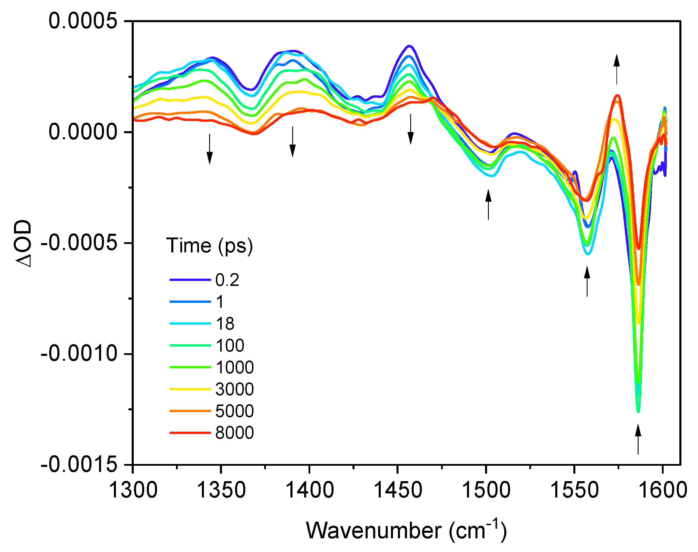
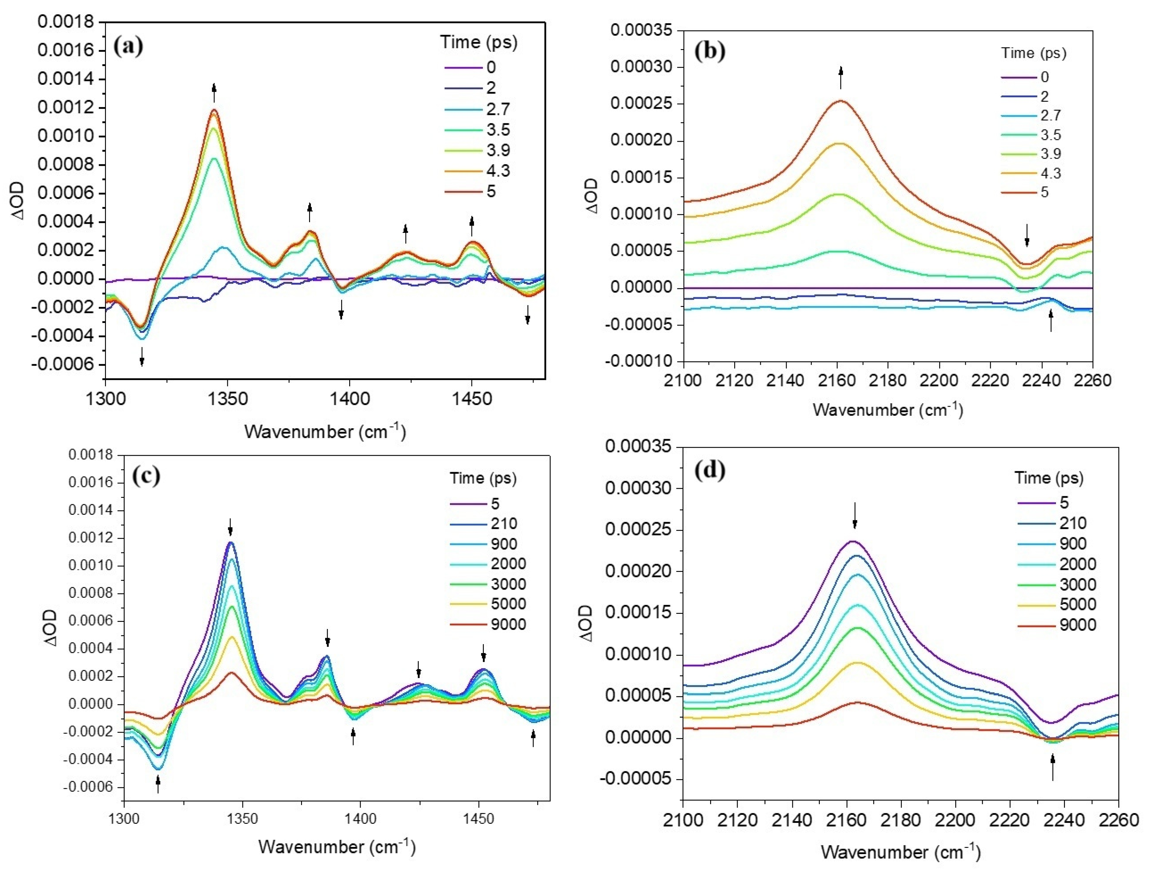
2.2. Time-Resolved Infrared Spectroscopy (TRIR)

In these experiments the BODIPYs were excited at both 400 and 540 nm, and the infrared spectra were recorded on the femtosecond to picosecond timescale in the range 1300–1600 cm−1 (fingerprint region). Following excitation of BDP-1 at 510 nm in CHCl3 the parent features at 1505, 1550, and 1585 cm−1 depleted and were accompanied by excited state absorption (ESA) features at 1350, 1390, and 1455 cm−1 (Figure 3).
The ESA bands decay significantly over 8000 ps, however, on the timescale. These spectral infrared bands are assigned to vibrational modes centred on the BODIPY core and the phenyl moieties and are attributed to π → π* transitions [40]. Global analysis of the spectra yielded two lifetimes, which are tentatively assigned to intersystem crossing and population of the singlet state. The shorter lifetime of 18.93 ps is likely due to intersystem crossing due to SOCT-ISC following photoinduced electron transfer (PeT) from the anthracenyl moiety to the BODIPY core in a πAn → πBDP transition, leading to population of the hole in the BODIPY HOMO following excitation. PeT has previously been reported by Filatov et al. for BDP-1 (2.4 ps), and in this study gives rise to the vibrational bands in the TRIR spectra [41]. The longer lifetime of 7.3 ns is assigned to the S1 state. As features persist in the TRIR spectra beyond the experimental window (8 ns), these bands are attributed to population of one or more triplet states, which is known to occur in this BODIPY [39].
Following excitation of BDP-2 at 540 nm (Figure 4), GSBs occur at 1320 and 1540 cm−1 with the concomitant formation of ESA bands at 1306, 1345, 1380, 1417, 1439, and 1510 cm−1. Global Analysis of the spectra yielded a lifetime of 14.15 ps, which is assigned to ISC, as observed for BDP-1, and is attributed to SOCT-ISC following PeT from the anthracene to the BODIPY core. The longer lifetime of 6.57 ns is assigned to the population of the S1 state. While the spectral features differ between BDP-1 and BDP-2, similar kinetics were obtained. Also, for BDP-2, ESA features persist beyond 9 ns and again are assigned to an excited triplet species.
The TRIR spectra for BDP-2 displayed in Figure 5 display differences following excitation at 400 nm when compared to the results following excitation at 540 nm. Excitation at 540 nm lies in the region of the S1 band, while absorption of a 400 nm photon may populate excited states on the BODIPY core or the anthracene moiety. Each pathway may subsequently result in the population of triplet states via charge transfer intermediates. Following excitation, GSBs were observed at 1314, 1397, and 1475 cm−1, and over the subsequent 5 ps, new bands grew in at 1346, 1385, 1423, and 1452 cm−1 together with an ESA band at 2250 cm−1 in the triple bond region.
The notable change in absorbance of the bands over 5 ps, especially in terms of the growth of the band at 2163 cm−1 highlights a significant change in dipole moment across the photosensitiser. The ESA at 2163 cm−1 has shifted to a lower wavenumber with respect to its corresponding GSB at 2250 cm−1, indicating a loss of electron density in the alkynyl moieties at the 2 and 6 positions. This, in combination with the enhanced amplitude of the ESA, is indicative of a charge transfer from the alkynyl bonds to the BODIPY, which is also evidenced by the large increase in the intensity of the ESA bands in the fingerprint region, particularly at 1345 cm−1 [42]. The enhanced alkynyl ESA band appears at a higher energy than a similar system reported by Cullen et al., who observed the formation of this band at 2073 cm−1 in the BODIPY-diethynylbenzene copolymers, which is 90 cm−1 lower than for BDP-2 [43]. This can be attributed to the lack of the phenyl component conjugated to the ethynyl bond in BDP-2. The decrease in π-conjugation in BDP-2 when compared to Cullen’s copolymer allows less resonance and delocalisation of π-electrons in the triple bond, resulting in vibrational modes at a higher energy [43].

2.3. Nanosecond Transient Absorption Spectroscopy (TAS)

Nanosecond-transient absorption spectroscopy was conducted to determine the triplet lifetimes for BDP-1 and BDP-2 in different solvents. The samples were prepared with an absorption value of ~0.3 at the excitation wavelength (355 or 532 nm) and deaerated using Schlenk technique. This is essential, as any oxygen present can quench the excited triplet states of the photosensitiser. The photosensitisers were irradiated at 355 nm (except BDP-2 in DCM to avoid radical formation in the chlorinated solvent), and spectral changes in the visible region were monitored.
An ns-TAS spectrum is shown in Figure 6 following excitation of BDP-2 at 532 nm in DCM. A ground-state bleach (GSB) occurs in the region 480–580 nm, corresponding to a depopulation of the ground state of the photosensitiser, accompanied by an excited state absorption (ESA) feature between 400 and 470 nm. As the ESA feature decays on the microsecond time scale, the GSB also recovers, indicating a population of S0. The time at which this decay occurs is indicative of relaxation from a triplet state to the ground state [43]. BDP-1 and BDP-2 exhibited two lifetimes in the microsecond time range. The triplet lifetimes for BDP-2 (τ = 31 and 138 µs, in MeCN) were generally longer-lived than those of BDP-1 (τ = 18 and 105 µs, in MeCN) (see Table 2), although all were significantly longer than those reported for heavy-atom-containing BODIPYs [43,44]. The presence of two long-lived components in the ns-TAS results supports the assumption that the population of the T1 triplet state is preceded by charge transfer intermediates. In general, the shorter-lived component contributes to ~30% of the decay curve, and the longer-lived component accounts for 70% of the signal. SOCT-ISC begins to emerge again as a viable pathway for these photosensitisers, with the longer component assigned to the T1 state and the shorter component likely a CTT state. As the population of the CTT state must be preceded by the population of the CTS state, it is feasible that the population of T1 occurs directly from CTS and CTT after charge recombination, with a change in orbital-angular momentum accompanying the charge recombination in the former. This has been extensively reported in work conducted by Filatov and Wang and coworkers and alluded to by TR-EPR [18,39].

2.4. Quantification of Singlet Oxygen

Two well-known methods for quantification of singlet oxygen quantum yields in photosensitisers are by direct measurement of the phosphorescence of singlet oxygen at 1270 nm and by indirect measurement by using singlet oxygen scavengers such as 1,3-diphenylisobenzofuran and monitoring the decay of this scavenger over time under irradiation of the compound. It is important to note that this method is only suitable for irradiation at wavelengths where the scavenger does not absorb, as it is very sensitive to light. It should be noted that anthracene itself is also a singlet oxygen scavenger, and the addition of oxygen to the anthracene rings quenches its triplet excited states [41]. As a result, we found the indirect method to be more suitable as a means of detection, as on its own the anthracene would be the only scavenger available for the direct method; however, when the DPBF scavenger is present, it would be in competition for the singlet oxygen and yield a more accurate estimation of the singlet oxygen quantum yield.
In these experiments I2-BDP was used as a standard, as it has a reported singlet oxygen quantum yield of 0.87 [39,45], its structure is shown in Figure S33. The singlet oxygen quantum yields of BDP-2 were consistent with those of BDP-1, which have previously been reported in the literature [15,17,18]. The lowest yields were seen in THF; however, this was accompanied by the highest fluorescence quantum yield. Both BODIPYs displayed high singlet oxygen quantum yields in DCM and MeCN; however, the fluorescence quantum yields in MeCN were heavily diminished (0.01), whereas they retained a significant amount of fluorescence in DCM (0.10–0.20). These results show overall high singlet oxygen quantum yields upon irradiation in the visible region, making both BDP-1 and BDP-2 suitable candidates for biological testing (Figures S21–S31). A control experiment was conducted by irradiating a solution of DPBF in the absence of the photosensitisers under identical conditions. No significant change in the absorption at 414 nm was observed (Figure S32), confirming that DPBF degradation in the main experiments arises from singlet oxygen generated by the photosensitisers.

2.5. Biological Studies

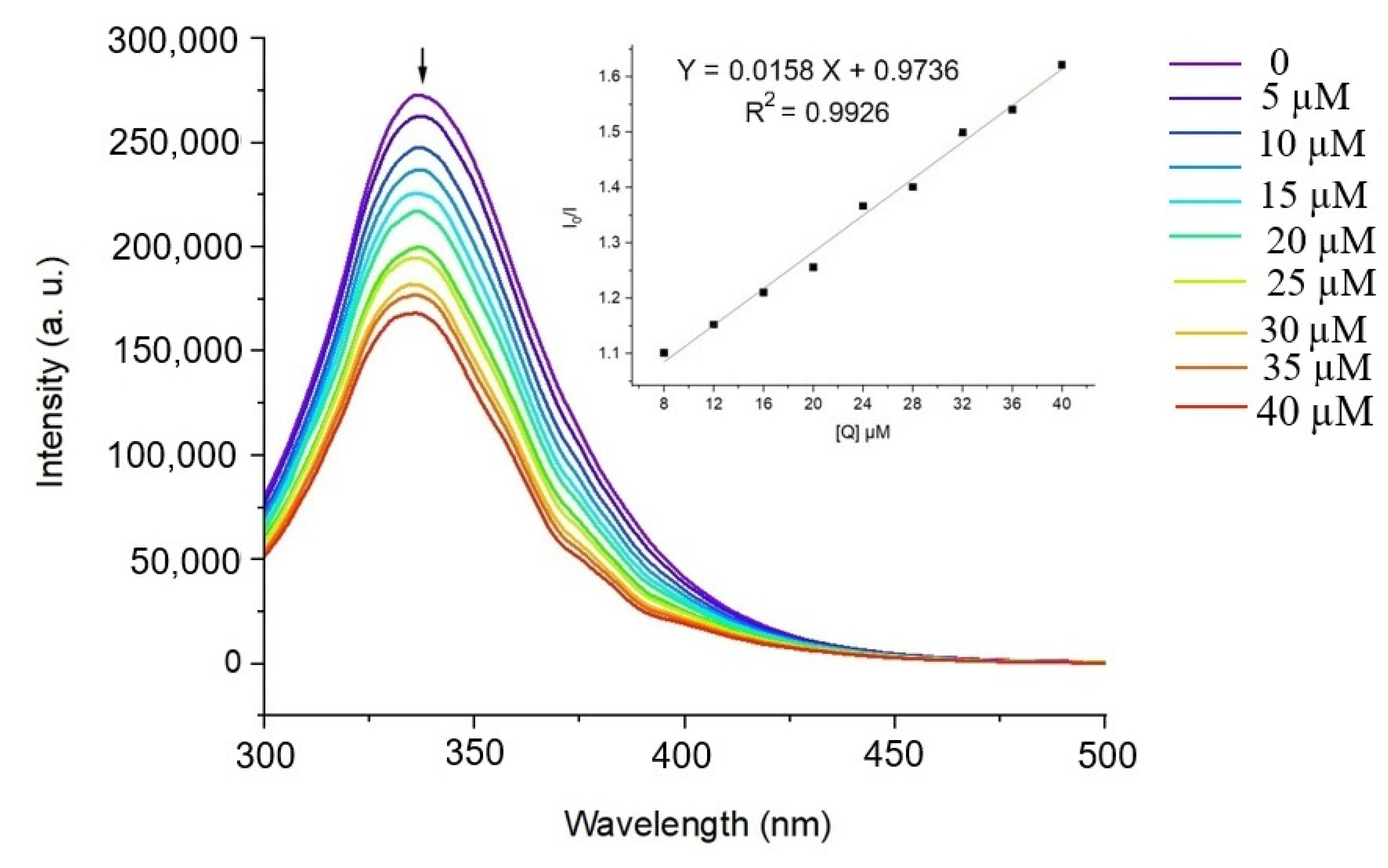
2.5.1. BSA-Binding Studies

BODIPY dyes, known for their high fluorescence, photostability, and environmental sensitivity, are widely used as molecular probes. When conjugated with BSA, a protein with multiple well-defined binding sites, the fluorescence properties of BODIPY can change, providing insights into protein-ligand interactions, structural dynamics, and conformational changes. Understanding this interaction is valuable for applications in drug delivery and biosensor development [46,47].
This study used fluorescence spectroscopy to investigate the binding between BDP-2 and BSA. This technique enables evaluation of binding affinity, protein conformational changes, and environmental effects on the fluorophore. Changes in fluorescence intensity and quenching reveal binding characteristics, site interactions, and structural modifications in BSA upon BDP-2 binding, offering valuable data for biochemical research [48,49].
BSAs intrinsic fluorescence mainly arises from its aromatic residues: tryptophan, tyrosine, and phenylalanine. Tryptophan, particularly Trp-213 located in a hydrophobic pocket, is the dominant contributor due to its high quantum yield and environmental sensitivity, with an emission peak around 330–350 nm. Tyrosine (303 nm) and phenylalanine (282 nm) contribute less due to lower quantum yields and quenching effects [50,51]. These properties make BSA fluorescence a sensitive indicator of conformational changes and molecular interactions [49,52]. The fluorescence emission spectra of BSA in the presence of varying concentrations of BDP-2 are presented in Figure 7. A decrease in fluorescence intensity at 338 nm was observed, indicative of quenching effects caused by BDP-2 binding to BSA. The degree of quenching was quantitatively analysed using the Stern–Volmer equation, from which the Stern–Volmer constants KSV (I0/I= 1+ KSV [Q]) were determined. The KSV value was 1.58 × 10⁴ M−1 for BDP-2 (insert Figure 7). The quenching constant (Kq), where τ0 is the lifetime of the fluorophore (BSA) in the absence of the quencher (10 ns) [53], yielded Kq = 1.58 × 10¹² M−1 S−1 for BDP-2. The fluorescence quenching constant (Kq) for BDP-2, which exceeds 1012 M−1 s−1, suggests the presence of a statistical interaction mechanism at play [54].
For a static quenching interaction, the fluorescence intensity data can also be used to determine the apparent binding constant (Kb) and the number of BSA binding sites (n) for the complex using the following equation [54]:
log ((I0I)/I) = log Kb + n log [Q]
From the plot of log ((I0I)/I) versus log [Q] (Figure S34), the number of binding sites (n) and the binding constant (Kb) values were calculated.
For BDP-2, a linear relationship is observed with binding constants Kb of 9.6 × 10⁴ M−1 S−1. In both compounds, the value of n was determined to be 1.17, which is close to 1. This indicates only one binding site for BDP-2 on the BSA molecule. When compared to literature data for similar compounds, this value is higher than that of a water-soluble meso-thienyl BODIPY derivative (Kb = 4.15 × 10⁴ M−1) [55] and a BODIPY-benzimidazole conjugate (Kb = 1.38 × 10⁴ M−1) [56], suggesting that BDP-2 binds more effectively to BSA. Moreover, naturally derived compounds like curcumin and its piperazine derivative displayed much lower binding constants (Kb = 1.0 × 10⁴ M−1 and 9.1 × 10³ M−1, respectively), further emphasising the superior binding strength of BDP-2 [57].
The quenching constant (Kq) for BDP-2 was calculated as 1.58 × 10¹² M−1 S−1, which is notably higher than typical quenching constants observed in similar systems, generally ranging from 109 to 1011 M−1 S−1. This elevated quenching constant suggests that BDP-2 interacts with BSA through a highly efficient quenching mechanism, likely involving dynamic or statistical processes. The binding interaction between BDP-2 and BSA holds potential for various biomedical applications. Conjugation with BSA can enhance the solubility, biocompatibility, and targeted delivery of BODIPY-based probes. Such complexes are particularly useful in controlled drug delivery, diagnostic imaging, and as biosensors, where changes in fluorescence can signal molecular binding events or environmental shifts. Therefore, although BDP-2 exhibits superior binding affinity and quenching efficiency compared to other BODIPY derivatives, further investigation into its biocompatibility is warranted before considering its use in biological systems.

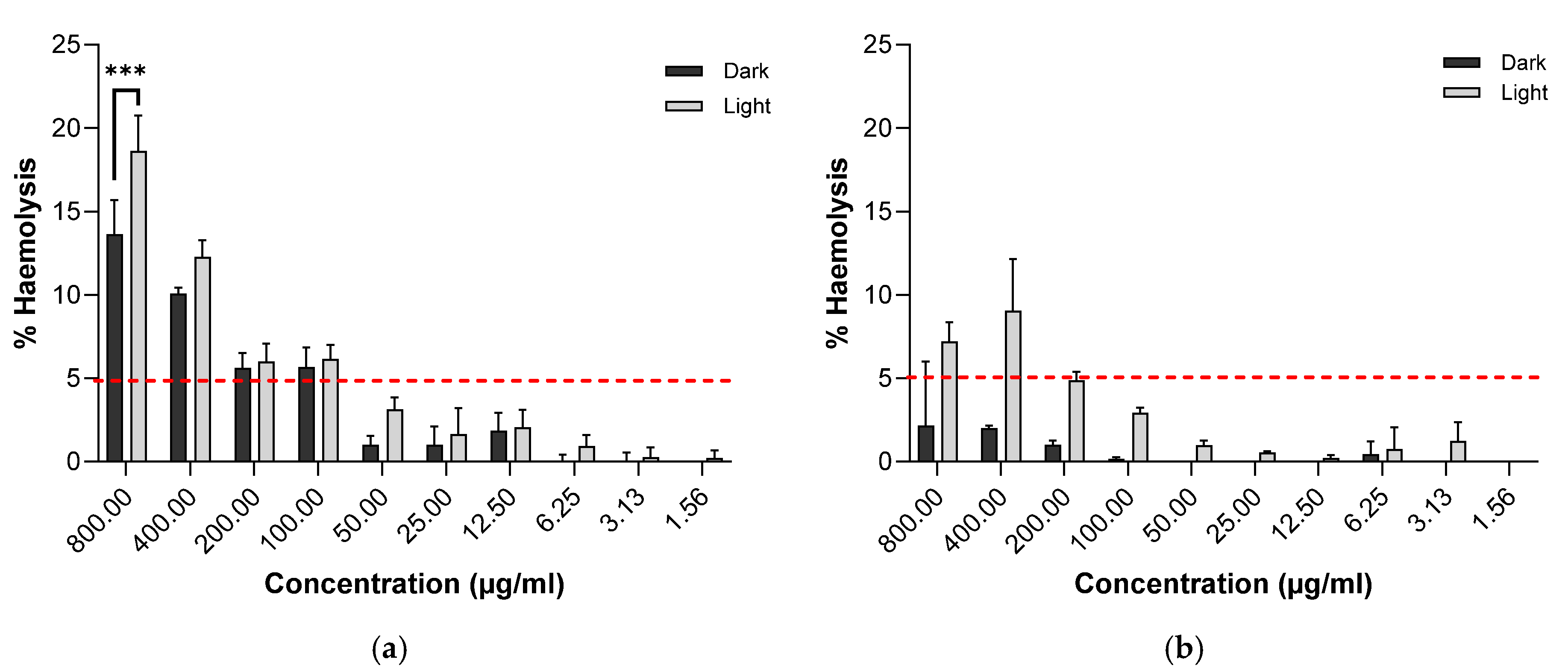
2.5.2. Biocompatibility Investigation

The haemolysis of human erythrocytes in vitro is used as a measure of biocompatibility, with a threshold of 5% haemolysis or less applied. Under irradiation conditions (370 nm, 1 h), BDP-1 displayed greater than 5% haemolysis at concentrations of 50 μg·mL−1. However, improved biocompatibility was found for BDP-2 under the same conditions, with haemolysis remaining below the 5% threshold up to 200 μg·mL−1 (Figure 8).

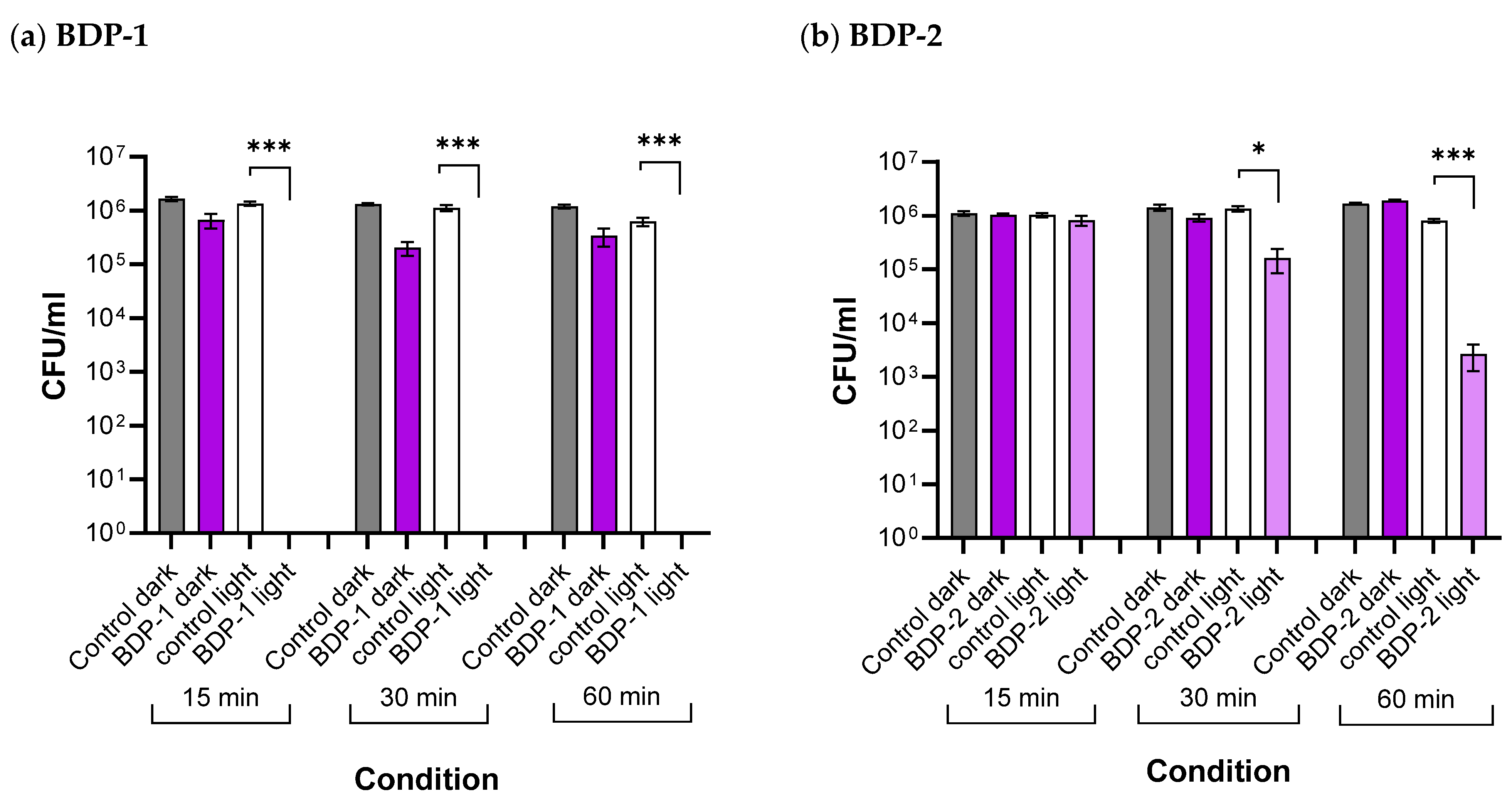
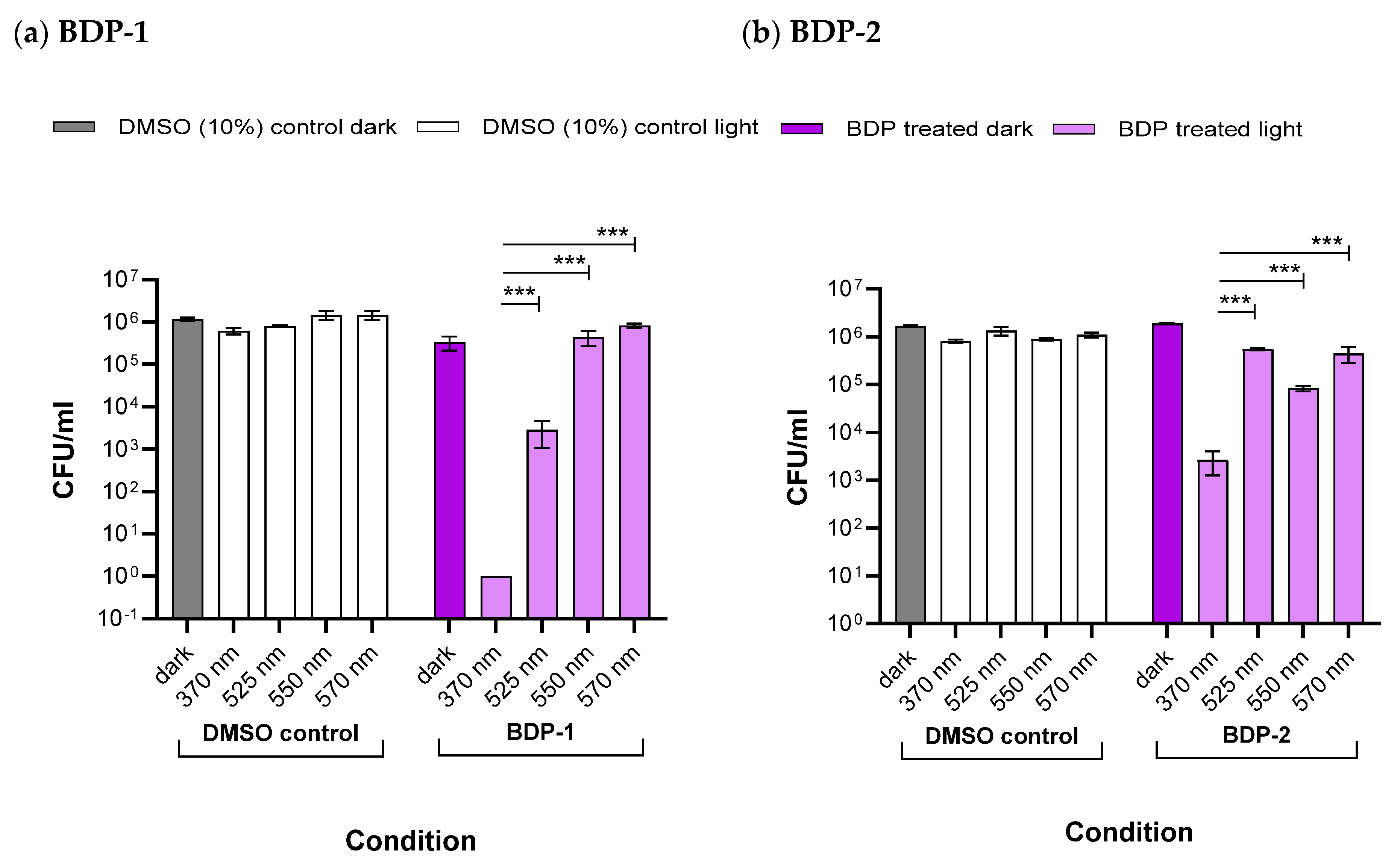
2.5.3. Antibacterial Studies

Under irradiation at 370 nm for 1 h, the highest level of killing was seen in S. aureus, a Gram-positive bacterial species, where a 6 log reduction in CFU/mL was found for BDP-1 compared to only 2.68 log for BDP-2. Both BDP-1 and BDP-2 had lower activity against Gram-negative E. coli, with killing activity of 1.74 and 1.30 log, respectively (Figure 9). An investigation of the time course of S. aureus killing showed differences between BDP-1 and BDP-2. For BDP-1, 6 log killing was achieved, even at the lowest period of irradiation (15 min), but for BDP-2, this pattern was not observed; rather, time dependence was apparent, with at least 1 h of irradiation required to reach 2.7 log (Figure 10). The differences in bactericidal activity for BDP-1 and BDP-2 could be due to the structural differences and interactions with the bacterial cells and also the light dose from the LED lamps used (Table 3, Figure 11). Control experiments with irradiation at 370 nm alone for 60 min had a negligible effect on bacterial viability, and therefore, killing required the combination of 370 nm and BDPs.

3. Materials and Methods

All chemicals and solvents were supplied by Aldrich Chemicals Co. (Wicklow, Ireland) and anhydrous solvents containing Sure/Seal were used under the flow of nitrogen. The NMR spectra were recorded on a Bruker 600 MHz spectrometer (Tospin 2.1, Bruker BioSpin srl, Milan, Italy). Mass spectroscopy was measured on a maXis HD (Bruker, Bremen, Germany) with an Ion trap detector interfaced to an Agilent 1100 series HPLC system (Agilent, Santa Clara, CA, USA). The BODIPYs were synthesised by methods previously reported in the literature. BDP-1 was synthesised using standard procedures reported in the literature [39]. BDP-1a was synthesised by iodination of BDP-1 under similar conditions reported by Cullen et al. with minor modifications [43]. BDP-2 was synthesised by Sonogashira coupling of BDP-1a with phenylacetylene under similar conditions to those reported by Zhang et al. [58,59]. Absorption, excitation, and emission spectra were recorded using a Horiba Fluorimeter (Kyoto, Japan). Samples were aerated and prepared to an optical density of <0.2 at the excitation wavelength in quartz cuvettes. Emission lifetimes and fluorescence quantum yields for the BODIPY samples were recorded on the Time-correlated Single Photon Counter (TCSPC) from Edinburgh Instruments. Samples were aerated and prepared to an optical density of <0.2 at the excitation wavelength in quartz cuvettes. Nanosecond transient absorption spectroscopy spectra were recorded on the LP980 transient absorption spectrometer from Edinburgh Instruments (Edinburgh, Scotland). Samples were prepared to an optical density of < 0.4 at the excitation wavelength in the absence of oxygen, by performing three freeze-pump-thaw cycles under the Schlenk technique. Singlet oxygen quantum yields were recorded using a Shimadzu Spectrometer (Shimadzu, Kyoto, Japan) and 1,3-Diphenylisobenzofuran (DPBF) from Merck (Arklow, Ireland) as the scavenger. A lamp with an irradiation wavelength of 528 nm was used.

3.1. Protein Binding Studies

The binding of BDP-2 to BSA was investigated using fluorescence spectroscopy recorded at a fixed excitation wavelength (280 nm) corresponding to BSA while monitoring the emission at 335 nm at 298 K. The stock solution of BSA was prepared in 50 mM phosphate buffer (pH = 7.2) and stored in the dark at 4 °C for further use. Concentrated stock solutions of each test compound were prepared by dissolving the compound in phosphate buffer and subsequently diluted with phosphate buffer to the required concentrations. Aliquots of 0–40 µM test solutions were added to BSA (50 µM). A 3 mL solution containing BSA (50 µM) was titrated by successive additions of 0–40 µM test solutions (BDP-2) for fluorescence measurements.

3.2. Singlet Oxygen Studies

Singlet oxygen quantum yields were determined using 1,3-Diphenylisobenzofuran (DPBF) as a scavenger. DPBF reacts with singlet oxygen, resulting in a decrease in its absorption maxima at 414 nm. The rate of this decrease can be correlated to the amount of 1O2 produced by the PS using the formula [41]:
ɸ Δ = ɸ s t d m B D P m s t d α s t d α B D P
where ɸΔ is the singlet oxygen quantum yield of the analyte, ɸstd is the singlet oxygen quantum yield of the standard (I2-BDP, Figure S32) [31,41], m is the normalised slope of the curve monitoring changes in absorption at 414 nm over time (s), and α = 1–10−A, where A is the average absorption of the sample or standard at the irradiation wavelength (528 nm). Samples were irradiated using a 528 nm LED light source with a measured power output of 0.152 mW/cm2. The samples were placed in a 1 cm path length quartz cuvette and irradiated for up to 180 s, resulting in a total light dose of 28 mJ/cm2. The distance from the LED source to the sample surface was maintained at 30 cm. Absorbance changes were recorded at fixed intervals using a UV–vis spectrophotometer.

3.3. Antimicrobial Studies
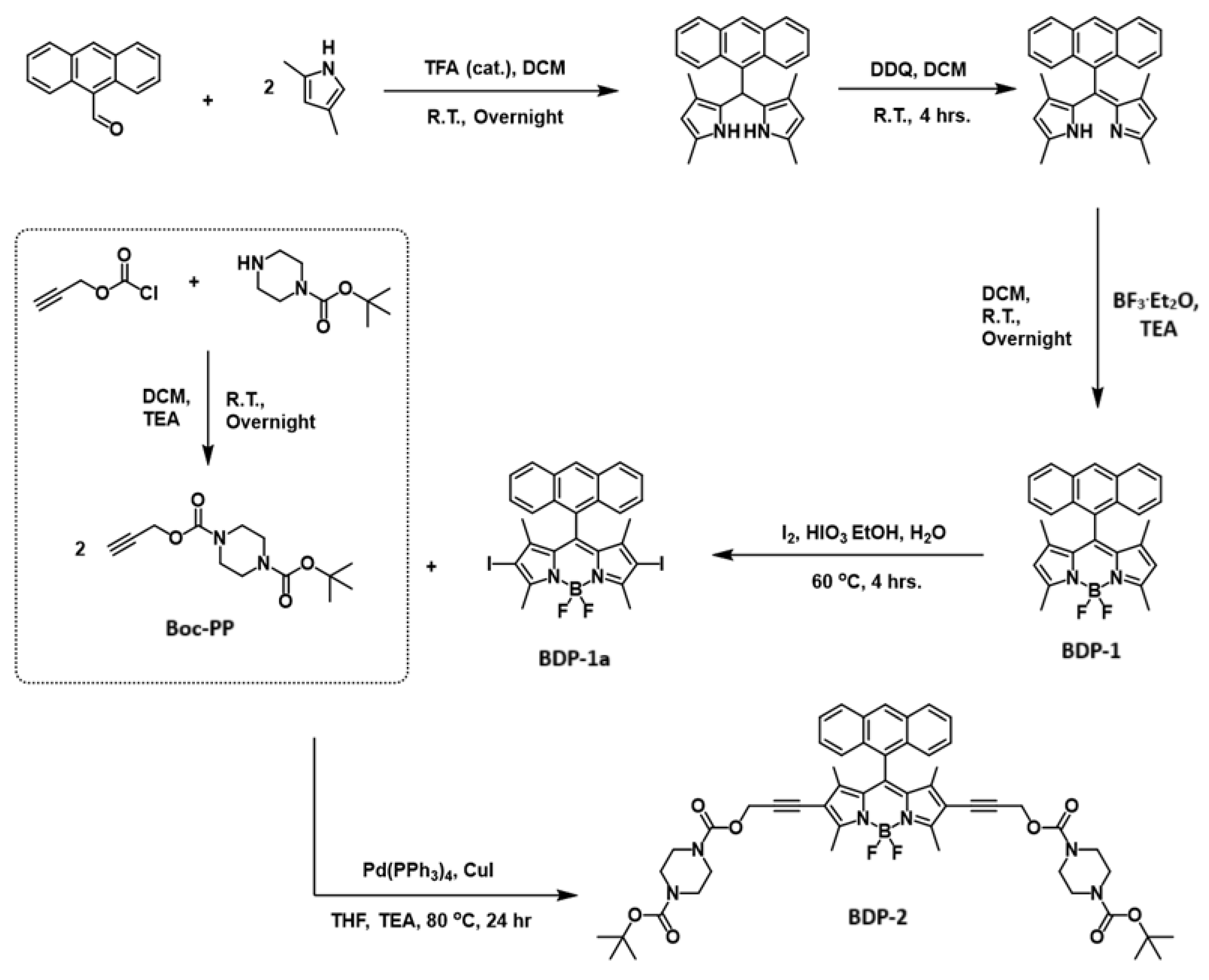
Stock solutions of the BODIPYs were prepared freshly on the day of testing by dissolving in 100% (v/v) Dimethyl sulfoxide (DMSO) at 1 mg·mL−1. All stocks were stored in the dark until required. Bacterial strains Staphylococcus aureus (ATCC25922) and Escherichia coli (ATCC25922) were purchased from the Health Protection Agency, UK. Pure colonies were isolated by subculturing on Mueller–Hinton agar and overnight aerobic growth at 37 °C in a static incubator. A bacterial suspension was prepared from single colonies with phosphate buffered saline (PBS) to the density of a 0.5 McFarland standard using a pre-calibrated turbidometer (Densichek TM, Biomerieux, Marcy-L’Etoile, France). The suspension was further diluted 1/100 in PBS. Assays were prepared in the wells of 96-well plates by adding 20 μL of stock BODIPY solution to triplicate wells and 180 μL of diluted bacterial suspension, giving a final assay concentration of 100 μg·mL−1 and approximately 106 colony-forming units/mL (CFU·mL−1) of bacteria. Two control series were prepared in which BODIPYs were replaced by either diluent (PBS) or 10% DMSO. Duplicate plates were prepared in this way: One was incubated at room temperature in the dark for various times (15–60 min), and the other was irradiated with light for the same time period. Therefore, each experiment included bacteria in the dark with no BDPs, irradiated bacteria with no BDPs, bacteria in the dark with BDPs, and irradiated bacteria with BDPs. The lamps used were suspended above the plate at a distance of 30 cm. After irradiation, 1/10 serial dilutions were prepared from treated and control wells under light and dark conditions in PBS from 10−1 to 10−4. Aliquots (20 μL) from these dilutions were applied in triplicate to four quadrants of Mueller–Hinton agar plates. The plates were incubated overnight at 37 °C in a static incubator. CFU·mL−1 was determined by counting resulting colonies at each dilution in treated and control plates.

3.4. Synthesis

3.4.1. Synthesis of 1-(tert-Butyl) 4-(Prop-2-yn-1-yl) Piperazine-1,4-Dicarboxylate (Boc-PP)

Approximately 500 mg of 1-Boc-piperazine was added to anhydrous dichloromethane (DCM, 30 mL) and TEA (1 mL). The mixture was purged under N2 for 15 min and allowed to react overnight under an N2 atmosphere with stirring. The mixture was initially placed in an ice bath when adding propargyl chloroformate (395 µL, 1.5 eq.) to control the reaction rate. The crude mixture was dried under reduced pressure and redissolved in DCM (40 mL) and washed with saturated Na2CO3 solution (30 mL (×3)). The organic phase was then dried over MgSO4 and filtered by gravity filtration and then dried under reduced pressure. No further purification was required. This afforded a white solid (94%). 1H NMR (600 MHz): 4.64 (s, 2H, 4J = 2.3 Hz), 3.38 (m, 8H), 2.45 (t, 1H, 4J = 2.3 Hz), 1.40 (s, 9H).

3.4.2. Synthesis of BDP-2

The product was synthesised through Sonogashira. Coupling with palladium as co-catalysts, as per previously reported methods with minor modifications [43]. BDP-1a (200 mg, 0.32 mmol) and Boc-piperazine-propargyl (BPP) (188 mg, 0.70 mmol) were added to a dry Schlenk tube with a magnetic stir bar. In addition to this, tetrakis(triphenylphosphine)palladium(0) Pd(PPh3)4 (37 mg, 10 mol%) and copper(I) iodide (CuI) (7 mg, 10 mol%) were added to the Schlenk tube. Tetrahydrofuran (THF, 10 mL) and triethylamine (TEA, 10 mL) were added to the Schlenk tube. The mixture was then freeze-pump-thawed under high vacuum strength and nitrogen flow using the Schlenk technique. It was then brought to a temperature of 80 °C while stirring for 24 h. The crude mixture was dried under reduced pressure and redissolved in DCM (40 mL) and washed with saturated Na2CO3 solution (30 mL (×3)). The organic phase was then dried over MgSO4 and filtered by gravity filtration. The product was then purified by silica column chromatography with a gradient mobile phase of DCM and ethyl acetate (starting at 95:5) to afford a purple solid (17%). 1H NMR (600 MHz, CDCl3): = 8.60 (s, 1H), 8.04 (d, 2H, 3J = 8.6 Hz), 7.76 (d, 2H, 3J = 8.6 Hz), 7.48 (t, 2H, 3J = 7.5 Hz), 7.40 (t, 2H, 3J = 7.7 Hz), 4.82 (s, 4H), 3.36 (m, 16H), 2.68 (s, 6H), 1.43 (s, 18H), 0.70 (s, 6H). 13C NMR (CDCl3): δ = 159.23, 154.69, 154.52, 145.21, 140.83, 132.09, 131.44, 129.60, 129.00, 128.68, 127.45, 127.32, 126.02, 124.78, 115.35, 90.80, 80.32, 78.53, 54.13, 43.81, 29.81, 28.49, 13.87, 12.44. ESI-MS = 957.4471 m/z, [M + H]+.

4. Conclusions

In this study, a piperazine-containing side chain integrated into the BODIPY structure (BDP-2) was synthesised and assessed for antibacterial activity. The high-yield iodination of BDP-1 to form BDP-1a and the subsequent Sonogashira coupling to generate BDP-2 highlight the synthetic adaptability of the BODIPY framework. A significant redshift and increased Stokes shift were observed for BDP-2, which is attributed to structural modifications. The BODIPYs presented in this work exhibit long-lived excited states and charge transfer characteristics, providing strong evidence that SOCT-ISC is the predominant pathway leading to population of the triplet state. Incorporation of the piperazine-containing side chain into the BODIPY structure not only resulted in a significant red-shift in absorption but also longer-lived triplet states compared to its parent anthracene BODIPY. Time-resolved techniques, such as TRIR and TAS, confirmed the efficient formation of these triplet states while also highlighting the influence of solvent polarity and structural modifications on the photophysical behavior. These findings offer valuable insights into how these compounds can be optimised for specific applications, such as antimicrobial photodynamic therapy, by fine-tuning their emission lifetimes, quantum yields, and triplet-state formation. This study lays the groundwork for the development of BODIPY-based photosensitisers with enhanced efficacy for photophysical and photobiological applications.
Biological studies demonstrated strong binding interactions between BDP-2 and BSA, indicating its potential for drug delivery systems. Furthermore, both compounds exhibited high singlet oxygen yields in polar solvents, validating their utility in photodynamic therapy and other biological applications.
The antibacterial assays further highlighted the potential of BDP-1 and BDP-2 as photosensitisers, particularly against gram-positive S. aureus, where potent bactericidal activity was observed. This S. aureus-specific activity informs the potential clinical applications of these compounds. S. aureus-specific eradication capability would favour their development for S. aureus aPDT decolonisation strategies, for example, prior to surgery. Currently, decolonisation (for methicillin-resistant S. aureus—MRSA) relies on nasal mupirocin application or chlorhexidine bathing. Furthermore, targeted treatments are central to antimicrobial stewardship programs to avoid the development of AMR associated with broader-spectrum agents, and in this regard, particularly for skin and soft tissue infection, where S. aureus predominates as the infecting pathogen, aPDT using specific BDPs to target S. aureus would be impactful. However, under the experimental condition used in our studies, the efficacy of BDP-1 and BDP-2 was wavelength-dependent, with superior antibacterial performance at 370 nm compared to 525 nm. This could be attributed to several factors, including population of the triplet state and light dosage.
In conclusion, BDP-1 and BDP-2 are promising candidates for antibacterial applications where S. aureus targeted eradication is required clinically, and future work will focus on exploring structure-property relationships and more extensive antibacterial studies under varying conditions.

Supplementary Materials

The following supporting information can be downloaded at: https://www.mdpi.com/article/10.3390/molecules30132727/s1, Synthesis of BDP-1, Synthesis of BDP-1a, Synthesis and Characterization of BDP-1 and BDP-1a, Figure S1. 1H NMR for BDP-1, CDCl3. Figure S2. 13C NMR for BDP-1, CDCl3. Figure S3. 1H NMR for BDP-1a, CDCl3. Figure S5. 1H NMR Boc-piperazine propargyl. Figure S6. 1H NMR for BDP-2, CDCl3. Figure S7. 13C NMR for BDP-2, CDCl3. Figure S8. Mass spec. analysis of BDP-1. Figure S9. Mass spec. analysis of BDP-1a. Figure S10. Mass spec. analysis of BDP-2. Figure S11. Emission lifetime for BDP-1 in MeCN, excited at 510 nm, recorded at 520 nm. Figure S12. Emission lifetime for BDP-1 in THF, excited at 510 nm, recorded at 520 nm. Figure S14. Emission lifetime for BDP-2 in MeCN, excited at 510 nm, recorded at 572 nm. Figure S15. Emission lifetime for BDP-2 in THF, excited at 510 nm, recorded at 572 nm. Figure S16. ns-lifetime decay trace for BDP-1 in THF (355nm). Figure S17. ns-lifetime decay trace for BDP-1 in MeCN (355nm). Figure S18. ns-lifetime decay trace of BDP-2 in THF (355 nm). Figure S19. ns-lifetime decay trace for BDP-2 in MeCN (355 nm). Figure S20. ns-lifetime decay trace for BDP-2 in DCM (532 nm). Figure S21. Indirect singlet oxygen detection for determination of the singlet oxygen quantum yield of BDP-2 in MeCN, excited at 528 nm (0 – 180 s). DPBF was used as the singlet oxygen scavenger and the rate of its decrease in absorption at 414 nm monitored to determine the singlet oxygen quantum yield, with I2-BDP as the standard (ɸΔ = 0.87). Figure S22. Linear regression analysis of the depletion of absorption features of BDP-2 and an I2-BDP standard, monitored at 414 nm from the indirect singlet oxygen detection using DPBF as a scavenger (12.5 µM) and BDP-2 as a PS (0.5 µM), with irradiation at 528 nm, in MeCN. Figure S23. Indirect singlet oxygen detection using DPBF as a scavenger (12.5 µM) and BDP-2 as a PS (0.5 µM), with irradiation at 528 nm, in DCM. Figure S24. Indirect singlet oxygen detection using DPBF as a scavenger (12.5 µM) and I2-BDP (std) as a PS (0.5 µM), with irradiation at 528 nm, in DCM. Figure S25. Indirect singlet oxygen detection using DPBF as a scavenger (12.5 µM) and BDP-2 as a PS (0.5 µM), with irradiation at 528 nm, in THF. Figure S26. Indirect singlet oxygen detection using DPBF as a scavenger (12.5 µM) and I2-BDP (std) as a PS (0.5 µM), with irradiation at 528 nm, in THF. Figure S27. Indirect singlet oxygen detection using DPBF as a scavenger (12.5 µM) and BDP-2 as a PS (0.5 µM), with irradiation at 528 nm, in MeCN. Figure S28. Indirect singlet oxygen detection using DPBF as a scavenger (12.5 µM) and I2-BDP (std) as a PS (0.5 µM), with irradiation at 528 nm, in MeCN. Figure S29. Normalized standard curves displaying the decrease in absorption of the BODIPY and DPBF mixtures at 414 nm after irradiation with 528 nm light, in DCM. Figure S30. Normalized standard curves displaying the decrease in absorption of the BODIPY and DPBF mixtures at 414 nm after irradiation with 528 nm light, in THF. Figure S31. Normalized standard curves displaying the decrease in absorption of the BODIPY and DPBF mixtures at 414 nm after irradiation with 528 nm light, in MeCN. Figure S32. A control experiment was conducted by irradiating a solution of DPBF (12.5 µM) in the absence of photosensitiser with irradiation at 528 nm, in MeCN during 180 s. Figure S33. Structure of I2-BDP (std). Figure S33. Scatchard plots of the fluorescence titrations of BDP-2 (0-40 µM) with BSA (50 µM). Figure S34. Decay trace for the emission signal of BDP-2 at 572 nm, in DCM (following excitation at 375 nm). Fluorescence quantum yield determined using the TCSPC integrating sphere.

Author Contributions

Conceptualization, M.T.P. and D.F.-H.; methodology, S.O., L.T., K.T., I.P.C., D.F.-H. and M.T.P.; validation, D.F.-H. and M.T.P.; formal analysis, S.O., L.T., K.T., I.P.C., D.F.-H. and M.T.P.; investigation, S.O., L.T., K.T., I.P.C., D.F.-H. and M.T.P.; resources, D.F.-H. and M.T.P.; writing—original draft preparation, S.O., L.T., K.T., D.F.-H. and M.T.P.; visualization, S.O., L.T., K.T., I.P.C., D.F.-H. and M.T.P.; supervision, D.F.-H. and M.T.P.; project administration, D.F.-H. and M.T.P. funding acquisition, D.F.-H. and M.T.P. All authors have read and agreed to the published version of the manuscript.

Funding

The authors gratefully acknowledge funding provided by Research Ireland (19/FFP/6882) and the EU Commission Recovery and Resilience Facility under the Research Ireland Healthy Environment for All Challenge Grant Number 22/NCF/HE/11252.

Institutional Review Board Statement

The study was conducted in accordance with the Declaration of Helsinki. The use of human blood for the study was approved by the Ethics Committee of The Royal College of Surgeons in Ireland (protocol code 202203014, 24 August 2022).

Informed Consent Statement

Informed consent was obtained from all subjects involved in the study.

Data Availability Statement

Not applicable.

Conflicts of Interest

The authors declare no conflict of interest.

References

  1. WHO|No Time to Wait: Securing the Future from Drug-Resistant Infections. Available online: https://www.who.int/publications/i/item/no-time-to-wait-securing-the-future-from-drug-resistant-infections (accessed on 21 October 2019).
  2. Home|AMR Review. Available online: https://amr-review.org/ (accessed on 19 March 2020).
  3. Abrahamse, H.; Hamblin, M.R. New Photosensitizers for Photodynamic Therapy. Biochem. J. 2016, 473, 347–364. [Google Scholar] [CrossRef] [PubMed]
  4. Dolmans, D.E.J.G.J.; Fukumura, D.; Jain, R.K. Photodynamic Therapy for Cancer. Nat. Rev. Cancer 2003, 3, 380–387. [Google Scholar] [CrossRef] [PubMed]
  5. Tabrizi, L.; Hughes, D.F.; Pryce, M.T. Covalent Organic Frameworks: Advancing Antimicrobial Photodynamic Therapy for next-Generation Treatments. Coord. Chem. Rev. 2025, 528, 216424. [Google Scholar] [CrossRef]
  6. Fallon, M.; Lalrempuia, R.; Tabrizi, L.; Brandon, M.P.; McGarry, R.; Cullen, A.; Fernández-Alvarez, F.J.; Pryce, M.T.; Fitzgerald-Hughes, D. Novel Cyclometalated Iridium (III) Complexes as Antibacterial Agents for Photodynamic Inactivation. J. Photochem. Photobiol. Chem. 2025, 462, 116218. [Google Scholar] [CrossRef]
  7. Tabrizi, L.; McGarry, R.; Turzanska, K.; Varvarezos, L.; Fallon, M.; Brannigan, R.; Costello, J.T.; Fitzgerald-Hughes, D.; Pryce, M.T. Porphyrin-Polymer as a Photosensitizer Prodrug for Antimicrobial Photodynamic Therapy and Biomolecule Binding Ability. Biomacromolecules 2024, 25, 7736–7749. [Google Scholar] [CrossRef] [PubMed]
  8. Rajagopal, A.; Biddulph, J.; Tabrizi, L.; Fitzgerald-Hughes, D.; Pryce, M.T. Chapter Eleven—Photoactive Organometallic Compounds as Antimicrobial Agents. In Advances in Inorganic Chemistry; Ford, P.C., van Eldik, R., Eds.; Biomedical Applications of Inorganic Photochemistry; Academic Press: Cambridge, MA, USA, 2022; Volume 80, pp. 381–409. [Google Scholar]
  9. Hamblin, M.R.; Hasan, T. Photodynamic Therapy: A New Antimicrobial Approach to Infectious Disease? Photochem. Photobiol. Sci. 2004, 3, 436–450. [Google Scholar] [CrossRef]
  10. Hamblin, M.R. Antimicrobial Photodynamic Inactivation: A Bright New Technique to Kill Resistant Microbes. Curr. Opin. Microbiol. 2016, 33, 67–73. [Google Scholar] [CrossRef]
  11. Skovsen, E.; Snyder, J.W.; Lambert, J.D.C.; Ogilby, P.R. Lifetime and Diffusion of Singlet Oxygen in a Cell. J. Phys. Chem. B 2005, 109, 8570–8573. [Google Scholar] [CrossRef]
  12. Piskorz, J.; Porolnik, W.; Kucinska, M.; Dlugaszewska, J.; Murias, M.; Mielcarek, J. BODIPY-Based Photosensitizers as Potential Anticancer and Antibacterial Agents: Role of the Positive Charge and the Heavy Atom Effect. ChemMedChem 2021, 16, 399–411. [Google Scholar] [CrossRef]
  13. Gorman, A.; Killoran, J.; O’Shea, C.; Kenna, T.; Gallagher, W.M.; O’Shea, D.F. In Vitro Demonstration of the Heavy-Atom Effect for Photodynamic Therapy. J. Am. Chem. Soc. 2004, 126, 10619–10631. [Google Scholar] [CrossRef]
  14. Nguyen, V.-N.; Yim, Y.; Kim, S.; Ryu, B.; Swamy, K.M.K.; Kim, G.; Kwon, N.; Kim, C.-Y.; Park, S.; Yoon, J. Molecular Design of Highly Efficient Heavy-Atom-Free Triplet BODIPY Derivatives for Photodynamic Therapy and Bioimaging. Angew. Chem. Int. Ed. 2020, 59, 8957–8962. [Google Scholar] [CrossRef]
  15. Dong, Y.; Dick, B.; Zhao, J. Twisted Bodipy Derivative as a Heavy-Atom-Free Triplet Photosensitizer Showing Strong Absorption of Yellow Light, Intersystem Crossing, and a High-Energy Long-Lived Triplet State. Org. Lett. 2020, 22, 5535–5539. [Google Scholar] [CrossRef] [PubMed]
  16. Hu, W.; Liu, M.; Zhang, X.-F.; Shi, M.; Jia, M.; Hu, X.; Liu, L.; Wang, T. Minimizing the Electron Donor Size of Donor–Acceptor-Type Photosensitizer: Twisted Intramolecular Charge-Transfer-Induced Triplet State and Singlet Oxygen Formation. J. Phys. Chem. C 2020, 124, 23556–23558. [Google Scholar] [CrossRef]
  17. Callaghan, S.; Filatov, M.A.; Savoie, H.; Boyle, R.W.; Senge, M.O. In Vitro Cytotoxicity of a Library of BODIPY-Anthracene and -Pyrene Dyads for Application in Photodynamic Therapy. Photochem. Photobiol. Sci. 2019, 18, 495–504. [Google Scholar] [CrossRef]
  18. Filatov, M.A. Heavy-Atom-Free BODIPY Photosensitizers with Intersystem Crossing Mediated by Intramolecular Photoinduced Electron Transfer. Org. Biomol. Chem. 2019, 18, 10–27. [Google Scholar] [CrossRef] [PubMed]
  19. Wang, Z.; Zhao, J. Bodipy–Anthracene Dyads as Triplet Photosensitizers: Effect of Chromophore Orientation on Triplet-State Formation Efficiency and Application in Triplet–Triplet Annihilation Upconversion. Org. Lett. 2017, 19, 4492–4495. [Google Scholar] [CrossRef]
  20. Halimehjani, A.Z.; Dehghan, F.; Tafakori, V.; Amini, E.; Hooshmand, S.E.; Nosood, Y.L. Synthesis of Novel Antibacterial and Antifungal Dithiocarbamate-Containing Piperazine Derivatives via Re-Engineering Multicomponent Approach. Heliyon 2022, 8, e09564. [Google Scholar] [CrossRef]
  21. Chaudhary, P.; Kumar, R.; Verma, A.K.; Singh, D.; Yadav, V.; Chhillar, A.K.; Sharma, G.L.; Chandra, R. Synthesis and Antimicrobial Activity of N-Alkyl and N-Aryl Piperazine Derivatives. Bioorg. Med. Chem. 2006, 14, 1819–1826. [Google Scholar] [CrossRef]
  22. Salem, M.E.; Fares, I.M.Z.; Ghozlan, S.A.S.; Abdel-Aziz, M.M.; Abdelhamid, I.A.; Elwahy, A.H.M. Facile Synthesis and Antimicrobial Activity of Bis(Fused 4H-Pyrans) Incorporating Piperazine as Novel Hybrid Molecules: Michael’s Addition Approach. J. Heterocycl. Chem. 2022, 59, 1907–1926. [Google Scholar] [CrossRef]
  23. Faizan, M.; Kumar, R.; Mazumder, A.; Salahuddin; Kukreti, N.; Kumar, A.; Chaitanya, M.V.N.L. The Medicinal Chemistry of Piperazines: A Review. Chem. Biol. Drug Des. 2024, 103, e14537. [Google Scholar] [CrossRef]
  24. Romanelli, M.N.; Manetti, D.; Braconi, L.; Dei, S.; Gabellini, A.; Teodori, E. The Piperazine Scaffold for Novel Drug Discovery Efforts: The Evidence to Date. Expert Opin. Drug Discov. 2022, 17, 969–984. [Google Scholar] [CrossRef] [PubMed]
  25. He, X.M.; Carter, D.C. Atomic Structure and Chemistry of Human Serum Albumin. Nature 1992, 358, 209–215. [Google Scholar] [CrossRef]
  26. Khaibrakhmanova, D.; Nikiforova, A.; Sedov, I. Binding Constants of Substituted Benzoic Acids with Bovine Serum Albumin. Pharmaceuticals 2020, 13, 30. [Google Scholar] [CrossRef] [PubMed]
  27. Fossum, C.J.; Johnson, B.O.V.; Golde, S.T.; Kielman, A.J.; Finke, B.; Smith, M.A.; Lowater, H.R.; Laatsch, B.F.; Bhattacharyya, S.; Hati, S. Insights into the Mechanism of Tryptophan Fluorescence Quenching Due to Synthetic Crowding Agents: A Combined Experimental and Computational Study. ACS Omega 2023, 8, 44820–44830. [Google Scholar] [CrossRef]
  28. Patra, S.; Santhosh, K.; Pabbathi, A.; Samanta, A. Diffusion of Organic Dyes in Bovine Serum Albumin Solution Studied by Fluorescence Correlation Spectroscopy. RSC Adv. 2012, 2, 6079–6086. [Google Scholar] [CrossRef]
  29. Yallur, B.C.; Katrahalli, U.; Krishna, P.M.; Hadagali, M.D. BSA Binding and Antibacterial Studies of Newly Synthesized 5,6-Dihydroimidazo[2,1-b]Thiazole-2-Carbaldehyde. Spectrochim. Acta A Mol. Biomol. Spectrosc. 2019, 222, 117192. [Google Scholar] [CrossRef] [PubMed]
  30. Çetindere, S. Photophysics of BODIPY Dyes: Recent Advances. In Photophysics, Photochemical and Substitution Reactions—Recent Advances; IntechOpen: London, UK, 2020; ISBN 978-1-83968-224-7. [Google Scholar]
  31. Agazzi, M.L.; Ballatore, M.B.; Reynoso, E.; Quiroga, E.D.; Durantini, E.N. Synthesis, Spectroscopic Properties and Photodynamic Activity of Two Cationic BODIPY Derivatives with Application in the Photoinactivation of Microorganisms. Eur. J. Med. Chem. 2017, 126, 110–121. [Google Scholar] [CrossRef]
  32. Chen, K.; Dong, Y.; Zhao, X.; Imran, M.; Tang, G.; Zhao, J.; Liu, Q. Bodipy Derivatives as Triplet Photosensitizers and the Related Intersystem Crossing Mechanisms. Front. Chem. 2019, 7, 821. [Google Scholar] [CrossRef]
  33. Dong, Y.; Sukhanov, A.A.; Zhao, J.; Elmali, A.; Li, X.; Dick, B.; Karatay, A.; Voronkova, V.K. Spin–Orbit Charge-Transfer Intersystem Crossing (SOCT-ISC) in Bodipy-Phenoxazine Dyads: Effect of Chromophore Orientation and Conformation Restriction on the Photophysical Properties. J. Phys. Chem. C 2019, 123, 22793–22811. [Google Scholar] [CrossRef]
  34. Stranius, K.; Iashin, V.; Nikkonen, T.; Muuronen, M.; Helaja, J.; Tkachenko, N. Effect of Mutual Position of Electron Donor and Acceptor on Photoinduced Electron Transfer in Supramolecular Chlorophyll–Fullerene Dyads. J. Phys. Chem. A 2014, 118, 1420–1429. [Google Scholar] [CrossRef]
  35. Petrushenko, I.K.; Petrushenko, K.B. Effect of Meso-Substituents on the Electronic Transitions of BODIPY Dyes: DFT and RI-CC2 Study. Spectrochim. Acta A Mol. Biomol. Spectrosc. 2015, 138, 623–627. [Google Scholar] [CrossRef] [PubMed]
  36. Wang, Z.; Sukhanov, A.A.; Toffoletti, A.; Sadiq, F.; Zhao, J.; Barbon, A.; Voronkova, V.K.; Dick, B. Insights into the Efficient Intersystem Crossing of Bodipy-Anthracene Compact Dyads with Steady-State and Time-Resolved Optical/Magnetic Spectroscopies and Observation of the Delayed Fluorescence. J. Phys. Chem. C 2019, 123, 265–274. [Google Scholar] [CrossRef]
  37. Wei, Z.; Sharma, S.; Philip, A.M.; Sengupta, S.; Grozema, F.C. Excited State Dynamics of BODIPY-Based Acceptor–Donor–Acceptor Systems: A Combined Experimental and Computational Study. Phys. Chem. Chem. Phys. 2021, 23, 8900–8907. [Google Scholar] [CrossRef] [PubMed]
  38. Shakerizadeh-Shirazi, F.; Hemmateenejad, B.; Mehranpour, A.M. Determination of the Empirical Solvent Polarity Parameter ET(30) by Multivariate Image Analysis. Anal. Methods 2013, 5, 891–896. [Google Scholar] [CrossRef]
  39. Dai, Y.; Dellai, A.; Bassan, E.; Bellatreccia, C.; Gualandi, A.; Anselmi, M.; Cozzi, P.G.; Ceroni, P.; Negri, F. Solvent and Alkyl Substitution Effects on Charge-Transfer Mediated Triplet State Generation in BODIPY Dyads: A Combined Computational and Experimental Study. Photochem. Photobiol. Sci. 2024, 23, 451–462. [Google Scholar] [CrossRef]
  40. Suhina, T.; Amirjalayer, S.; Woutersen, S.; Bonn, D.; Brouwer, A.M. Ultrafast Dynamics and Solvent-Dependent Deactivation Kinetics of BODIPY Molecular Rotors. Phys. Chem. Chem. Phys. 2017, 19, 19998–20007. [Google Scholar] [CrossRef]
  41. Filatov, M.A.; Karuthedath, S.; Polestshuk, P.M.; Savoie, H.; Flanagan, K.J.; Sy, C.; Sitte, E.; Telitchko, M.; Laquai, F.; Boyle, R.W.; et al. Generation of Triplet Excited States via Photoinduced Electron Transfer in Meso-Anthra-BODIPY: Fluorogenic Response toward Singlet Oxygen in Solution and in Vitro. J. Am. Chem. Soc. 2017, 139, 6282–6285. [Google Scholar] [CrossRef]
  42. Balanikas, E.; Reymond-Joubin, M.; Vauthey, E. Excited-State Symmetry Breaking in Solvent Mixtures. J. Phys. Chem. Lett. 2024, 15, 2447–2452. [Google Scholar] [CrossRef] [PubMed]
  43. Cullen, A.A.; Rajagopal, A.; Heintz, K.; Heise, A.; Murphy, R.; Sazanovich, I.V.; Greetham, G.M.; Towrie, M.; Long, C.; Fitzgerald-Hughes, D.; et al. Exploiting a Neutral BODIPY Copolymer as an Effective Agent for Photodynamic Antimicrobial Inactivation. J. Phys. Chem. B 2021, 125, 1550–1557. [Google Scholar] [CrossRef]
  44. Cullen, A.A.; Heintz, K.; O’Reilly, L.; Long, C.; Heise, A.; Murphy, R.; Karlsson, J.; Gibson, E.; Greetham, G.M.; Towrie, M.; et al. A Time-Resolved Spectroscopic Investigation of a Novel BODIPY Copolymer and Its Potential Use as a Photosensitiser for Hydrogen Evolution. Front. Chem. 2020, 8, 584060. [Google Scholar] [CrossRef]
  45. Zhang, C.; Zhao, J.; Wu, S.; Wang, Z.; Wu, W.; Ma, J.; Guo, S.; Huang, L. Intramolecular RET Enhanced Visible Light-Absorbing Bodipy Organic Triplet Photosensitizers and Application in Photooxidation and Triplet–Triplet Annihilation Upconversion. J. Am. Chem. Soc. 2013, 135, 10566–10578. [Google Scholar] [CrossRef] [PubMed]
  46. Raskolupova, V.I.; Popova, T.V.; Zakharova, O.D.; Nikotina, A.E.; Abramova, T.V.; Silnikov, V.N. Human Serum Albumin Labelling with a New BODIPY Dye Having a Large Stokes Shift. Molecules 2021, 26, 2679. [Google Scholar] [CrossRef]
  47. Das, S.; Dey, S.; Patra, S.; Bera, A.; Ghosh, T.; Prasad, B.; Sayala, K.D.; Maji, K.; Bedi, A.; Debnath, S. BODIPY-Based Molecules for Biomedical Applications. Biomolecules 2023, 13, 1723. [Google Scholar] [CrossRef] [PubMed]
  48. El Gammal, R.N.; Elmansi, H.; El-Emam, A.A.; Belal, F.; Elzahhar, P.A.; Belal, A.S.F.; Hammouda, M.E.A. Insights on the In-Vitro Binding Interaction between Donepezil and Bovine Serum Albumin. BMC Chem. 2023, 17, 31. [Google Scholar] [CrossRef] [PubMed]
  49. Kummur, K.N.; Panda, S.M.; Patil, M.B.; Tripathy, U.; Sidarai, A.H. Revealing the Interaction Mechanism between Bovine Serum Albumin (BSA) and a Fluorescent Coumarin Derivative: A Multispectroscopic and in Silico Approach. Spectrochim. Acta A Mol. Biomol. Spectrosc. 2024, 318, 124466. [Google Scholar] [CrossRef]
  50. Theoretical Research on Excited States: Ultraviolet and Fluorescence Spectra of Aromatic Amino Acids|Interdisciplinary Sciences: Computational Life Sciences. Available online: https://link.springer.com/article/10.1007/s12539-020-00395-3 (accessed on 20 January 2025).
  51. Verma, R.; Pyreddy, S.; Redmond, C.E.; Qazi, F.; Khalid, A.; O’Brien-Simpson, N.M.; Shukla, R.; Tomljenovic-Hanic, S. Detection and Identification of Amino Acids and Proteins Using Their Intrinsic Fluorescence in the Visible Light Spectrum. Anal. Chim. Acta 2023, 1282, 341925. [Google Scholar] [CrossRef]
  52. Minamide, M.; Tsurushima, M.; Koga, R.; Hasegawa, K.; Kurosawa, Y.; Tsuchida, T.; Goto, S. Distinguishing the Transitions of Fluorescence Spectra of Tryptophan-134 and 213 in BSA Induced by Bindings of UV Filters, Oxybenzone-3, and Avobenzone. Bull. Chem. Soc. Jpn. 2024, 97, uoae058. [Google Scholar] [CrossRef]
  53. Ahmad, B.; Parveen, S.; Khan, R.H. Effect of Albumin Conformation on the Binding of Ciprofloxacin to Human Serum Albumin: A Novel Approach Directly Assigning Binding Site. Biomacromolecules 2006, 7, 1350–1356. [Google Scholar] [CrossRef]
  54. Zornić, S.; Simović Marković, B.; Franich, A.A.; Janjić, G.V.; Jadranin, M.B.; Avdalović, J.; Rajković, S.; Živković, M.D.; Arsenijević, N.N.; Radosavljević, G.D.; et al. Characterization, Modes of Interactions with DNA/BSA Biomolecules and Anti-Tumor Activity of Newly Synthesized Dinuclear Platinum(II) Complexes with Pyridazine Bridging Ligand. J. Biol. Inorg. Chem. JBIC Publ. Soc. Biol. Inorg. Chem. 2024, 29, 51–73. [Google Scholar] [CrossRef]
  55. Karanlık, C.C.; Karanlık, G.; Erdoğmuş, A. Water-Soluble Meso-Thienyl BODIPY Therapeutics: Synthesis, Characterization, Exploring Photophysicochemical and DNA/BSA Binding Properties. J. Photochem. Photobiol. Chem. 2023, 438, 114581. [Google Scholar] [CrossRef]
  56. Chakraborty, G.; Ray, A.K.; Singh, P.K.; Pal, H. Non-Covalent Interaction of BODIPY-Benzimidazole Conjugate with Bovine Serum Albumin–A Photophysical and Molecular Docking Study. J. Photochem. Photobiol. Chem. 2019, 377, 220–227. [Google Scholar] [CrossRef]
  57. Pang, W.; Lv, J.; Du, S.; Wang, J.; Wang, J.; Zeng, Y. Preparation of Curcumin-Piperazine Coamorphous Phase and Fluorescence Spectroscopic and Density Functional Theory Simulation Studies on the Interaction with Bovine Serum Albumin. Mol. Pharm. 2017, 14, 3013–3024. [Google Scholar] [CrossRef] [PubMed]
  58. Chinchilla, R.; Nájera, C. The Sonogashira Reaction:  A Booming Methodology in Synthetic Organic Chemistry. Chem. Rev. 2007, 107, 874–922. [Google Scholar] [CrossRef] [PubMed]
  59. Zhang, D.; Wen, Y.; Xiao, Y.; Yu, G.; Liu, Y.; Qian, X. Bulky 4-Tritylphenylethynyl Substituted Boradiazaindacene: Pure Red Emission, Relatively Large Stokes Shift and Inhibition of Self-Quenching. Chem. Commun. 2008, 39, 4777–4779. [Google Scholar] [CrossRef]
Figure 1. BODIPY structures in this work.
Molecules 30 02727 g001
Scheme 1. Synthetic pathway to BDP-2.
Molecules 30 02727 sch001
Figure 2. Absorption and emission spectra for BDP-1 and BDP-2 in DCM.
Molecules 30 02727 g002
Figure 3. TRIR spectra of BDP-1 in CDCl3, excited at 510 nm (time range, 0.2–8000 ps).
Molecules 30 02727 g003
Figure 4. TRIR of BDP-2 in CDCl3, excited at 540 nm (time range, 0–250 ns).
Molecules 30 02727 g004
Figure 5. TRIR spectra following excitation of BDP-2 in CDCl3 at 400 nm (0–5 ps): (a) In the fingerprint region, (b) in the alkynyl region, and (c) the fingerprint region from 5 to 9000 ps. (d) In the alkynyl region from 5 to 9000 ps.
Molecules 30 02727 g005
Figure 6. Nanosecond transient absorption (ns-TAS) of BDP-2 in DCM, following excitation at 532 nm (0–750 µs).
Molecules 30 02727 g006
Figure 7. Fluorescence quenching curves of BSA (50 µM) in PBS solution in the presence of different amounts (0–40 µM) of BDP-2. Insert: Stern–Volmer plot of the fluorescence titrations.
Molecules 30 02727 g007
Figure 8. In vitro haemolysis studies of human erythrocytes for (a) BDP-1 and (b) BDP-2 at varying concentrations under dark conditions and when irradiated at 370 nm. Values shown are the mean ± SEM for triplicate assays. Statistically significant differences between the light and dark series are shown: *** p ≤ 0.0001. The red dotted line indicates 5% haemolysis.
Molecules 30 02727 g008
Figure 9. Photodynamic bactericidal activity of BDP-1 (a) and BDP-2 (b) against two bacterial species. Assays containing 10% DMSO (controls) or compounds (100 μg·mL−1) and approximately 106 CFU·mL−1 bacteria were incubated in the dark or irradiated for 60 min at 370 nm. Values shown are mean ± SEM for three separate assays performed in triplicate. The Y axis is logarithmic scale. Statistically significant differences between the reduction in CFU/mL for light-treated series (control minus BDP-treated) against E. coli vs. S. aureus are shown: * p ≤ 0.05, *** p ≤ 0.0001. For BDP1 with light treatment, bars representing zero CFU/mL are not visible.
Molecules 30 02727 g009
Figure 10. Photodynamic bactericidal activity of (a) BDP-1 and (b) BDP-2 against S. aureus (ATCC 25923). Assays containing 10% DMSO (controls) or compounds (100 μg·mL−1) and approximately 106 CFU·mL−1 bacteria were incubated in the dark or irradiated at 370 nm for 15, 30, and 60 min. Values shown are mean ± SEM for three separate assays performed in triplicate. The Y axis is a logarithmic scale. Statistically significant differences between the reduction in CFU/mL for the light-treated series (control minus BDP-treated) are shown: *** p ≤ 0.001, * p ≤ 0.05. For BDP1 with light treatment, bars representing zero CFU/mL are not visible.
Molecules 30 02727 g010
Figure 11. Effect of irradiation wavelength on antimicrobial activity of (a) BDP-1 and (b) BDP-2. Bacterial killing activity at 100 μg/mL is shown against S. aureus (ATCC 25923). Assays were incubated in the dark or irradiated for 60 min at 370, 525, 550, and 570 nm. Values shown are mean ± SEM for three separate assays performed in triplicate. Statistically significant differences between the reduction in CFU/mL for light-treated series (control minus BDP-treated) are shown: *** p ≤ 0.001.
Molecules 30 02727 g011
Table 1. Photophysical results for BDP-1 and BDP-2 in various solvents.
Solventabs (nm)em (nm)Δ υ ¯ (cm−1)ɸFlτ1 (ns)τ2 (µs)ɸΔ
BDP-1DCM5075163440.14 a4.20 (510 nm)82 a0.82 a
THF5055184970.155.6948/166 b0.31 a
MeCN5035123490.01 a3.62/5.3818/105 b0.86 a
BDP-2DCM5525726330.194.0644/230 c0.85
THF5525685100.304.0949/213 b0.25
MeCN5495717020.012.59/5.3531/138 b0.81
a Figure taken from work conducted by Wang et al. [39], b λexc = 355 nm. c λexc = 532 nm.
Table 2. ns-TAS lifetimes calculated from kinetic decay curves produced upon excitation of BDP-1 and BDP-2.
Solventabs (nm)τ1 (µs)τ2 (µs)ɸΔ
BDP-1DCM507- b82 a0.82 a
THF50548 c166 c0.31 a
MeCN50318 c105 c0.86 a
BDP-2DCM55244 d230 d0.85
THF55249 c213 c0.25
MeCN54931 c138 c0.81
a Figure taken from work conducted by Wang et al. [39], b Not applicable. c λexc = 355 nm. d λexc = 532 nm.
Table 3. S. aureus killing activity of BDP-1 and BDP-2 (100 μg·mL−1) at varying wavelengths.
Wavelength (nm)Light Intensity
(mW/cm2)
Light Dose
(mJ/cm2)
Log Reduction in CFU·mL−1 a
BDP-1BDP-2
3700.7025235.182.68
5250.051962.110.37
5506.1222,0180.610.75
5700.02640.230.44
a Log reduction in CFU·mL−1 for BDP-treated compared to DMSO controls, where activity in the dark has been subtracted.
Disclaimer/Publisher’s Note: The statements, opinions and data contained in all publications are solely those of the individual author(s) and contributor(s) and not of MDPI and/or the editor(s). MDPI and/or the editor(s) disclaim responsibility for any injury to people or property resulting from any ideas, methods, instructions or products referred to in the content.

