Article

Bioactive Compounds from Dodonaea viscosa Flowers: Potent Antibacterial and Antiproliferative Effects in Breast Cancer Cells

1
Department of Pharmaceutical Sciences, The Daniel K. Inouye College of Pharmacy, University of Hawai‘i at Hilo, Hilo, HI 96720, USA
2
Preclinical Core, Cancer Biology Program, University of Hawai‘i Cancer Center, Honolulu, HI 96813, USA
3
Department of Pharmacy Practice, The Daniel K. Inouye College of Pharmacy, University of Hawai‘i at Hilo, Hilo, HI 96720, USA
4
Georgia Institute of Science and Research Incorporation, Duluth, GA 30096, USA
5
Hawaiian Language/Performing Arts, Kamehameha Schools, Hawai‘i Campus, Kea’au, HI 96749, USA
6
Daniel K. Inouye U.S. Pacific Basin Agricultural Research Center, Hilo, HI 96720, USA
*
Authors to whom correspondence should be addressed.
Molecules 2025, 30(11), 2274; https://doi.org/10.3390/molecules30112274
Submission received: 31 December 2024 / Revised: 7 May 2025 / Accepted: 15 May 2025 / Published: 22 May 2025

Abstract

:
Dodonaea viscosa (Sapindaceae), a Hawaiian local medicinal plant, has been traditionally used to treat rashes and skin diseases. The study aimed to discover and characterize bioactive compounds from D. viscosa flowers extract with antimicrobial and antitumor properties. Thirteen compounds were isolated from the methanol extract of D. viscosa flowers, and their structures were characterized using spectroscopy data, comparing their NMR spectroscopic profiles with previously reported data. Subsequent antibacterial assays revealed that one particular compound, 12, exhibited significant antimicrobial activity against Gram-positive bacteria. Notably, it demonstrated a minimum inhibitory concentration (MIC) of 2 μg/mL, indicating its potent antibacterial potential. In addition to antimicrobial properties, the isolated compounds demonstrated dose-dependent antiproliferative effects in breast cancer cell lines. Notably, inflammatory breast cancer (IBC) cell lines, a highly aggressive subtype, were more sensitive to compound 6, with IC50 values of 4.22 μM (BCX-010), 6.74 μM (SUM190), and 7.73 (SUM149), compared to non-IBC cell line. These findings highlight the dual antibacterial and anticancer potential of compounds from D. viscosa, emphasizing their promise as candidates for therapeutic development.

Graphical Abstract

1. Introduction

The integration of traditional Hawaiian medicine and modern scientific research underscores the considerable therapeutic potential of Dodonaea viscosa, commonly known as ‘A‘ali’i. For centuries, Native Hawaiians have harnessed the resilience of this plant to treat various ailments, including toothaches, fever, and skin irritations [1]. This historical application suggests the presence of bioactive compounds with analgesic, antipyretic, and anti-inflammatory properties. Contemporary phytochemical studies have revealed a diverse array of constituents in D. viscosa, including alkaloids, flavonoids, glycosides, phenols, terpenoids, and steroids [2,3,4,5,6,7]. These varied chemical compounds are recognized for their extensive biological activities, positioning D. viscosa as a promising candidate in the fight against significant health challenges such as antimicrobial resistance and cancer.
The escalating threat of antibiotic-resistant bacteria, particularly methicillin-resistant Staphylococcus aureus (MRSA), is a critical public health crisis that requires urgent global attention, including within the Hawaiian Islands [8,9]. The disproportionately high rates of community-acquired MRSA infections among Native Hawaiian, Pacific Islander, and pediatric populations in Hawai‘i underscore the urgent need for novel antimicrobial agents [9,10,11]. Intriguingly, a specific clerodane-type diterpenoid isolated from D. viscosa, 6β-hydroxy-15,16-epoxy-5β,8β,9β,10α-cleroda-3,13(16),14-trien-18-oic acid, has demonstrated bacteriostatic activity against certain bacterial strains [12]. This discovery suggests that D. viscosa may serve as a valuable natural reservoir of antibacterial compounds capable of combating multidrug-resistant microorganisms. Further research into isolating and characterizing other antibacterial constituents from this plant could lead to the development of innovative therapeutic strategies against persistent bacterial infections.
In parallel, breast cancer, the most prevalent malignancy in women worldwide, remains a major clinical challenge due to its heterogeneity and resistance to therapy [13,14]. Metastatic breast cancer (mBC), particularly HER2-negative subtypes such as triple-negative breast cancer (TNBC), inflammatory breast cancer (IBC), and hormone receptor-positive (HR+) breast cancer, is associated with poor prognoses and limited treatment options. TNBC, characterized by the absence of hormone receptors and HER2 expression, exhibits high recurrence rates, metastasis, and poor overall survival [15,16]. Similarly, resistance to endocrine therapy in HR+ breast cancer [17] and the aggressive nature of IBC underscore the urgent need for innovative therapeutic strategies [18].
Preliminary investigations have indicated that D. viscosa flower extracts possess anti-proliferative effects in breast cancer cell lines. This highlights their potential as dual-action agents, possessing both antibacterial and anticancer properties [19,20,21]. The current study aimed to isolate and characterize bioactive compounds from the flowers of D. viscosa, evaluate their antimicrobial efficacy, and investigate their potential to inhibit the proliferation of breast cancer cells. These findings could pave the way for developing novel therapeutics to address both infectious disease and cancer challenges.

2. Results and Discussion

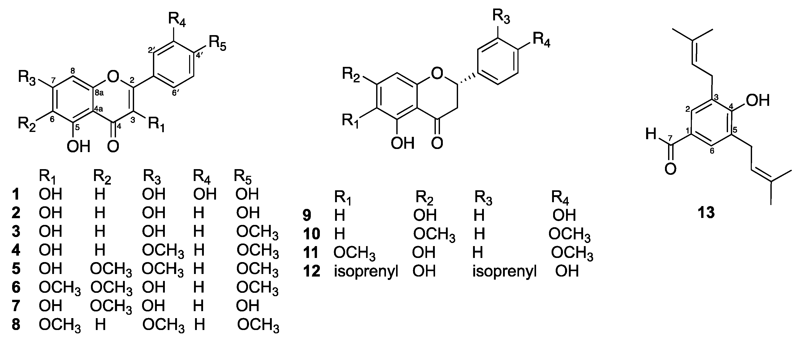
2.1. Isolated Compounds from D. viscosa Flowers

The MeOH extract of fresh flowers of D. viscosa was fractionated by silica gel and Sephadex LH-20 column chromatography to afford thirteen compounds identified as quercetin (1) [22], kaempferol (2) [22], kaempferide (3) [23], 3,5-dihydroxy-7,4′-dimethoxyflavone (4) [24], mikanin (5) [25], santin (6) [26], 6-methoxykaempferol (7) [27], 5-hydroxy-3,7,4′-trimethoxyflavone (8) [28], naringenin (9) [29], 5-hydroxy-7,4′-dimethoxyflavone (10) [30], 2,3-dihydro-5,7-dihydroxy-6-methoxy-2-(4-methoxyphenyl)-4H-1-benzopyran-4-one (11) [31], macarangaflavanone B (12) [32], and 3,5-diprenyl-4-hydroxybenzaldehyde (13) [33] (Figure 1). The structures were elucidated using spectroscopic data, including NMR results and literature comparisons (can be found in Supplementary Materials).
Compounds 18 were obtained as yellow solids. The 1H NMR spectrum of compound 1 displayed the presence of a set of hydrogen-bonded hydroxy proton at δH 12.18 (1H, s, 6-OH) and a trisubstituted benzene ring at δH 7.80 (1H, d, J = 2.0 Hz, H-2′), 7.68 (1H, dd, J = 8.4, 2.0 Hz, H-6′), and 6.97 (1H, d, J = 8.4 Hz, H-5′). A pair of meta-coupled doublets observed at δH 6.25 (1H, d, J = 2.0 Hz) and 6.52 (1H, d, J = 2.0 Hz) were assigned as H-6 and H-8, respectively. Therefore, structure 1 has a flavonol skeleton, which is a type of flavonoid and was identified as quercetin (1) [22]. The 1H NMR spectrum of compound 2, an analog of compound 1. Notably, compound 2 exhibited a pair of para-substituted benzene rings, with signals at δH 8.08 (2H, d, J = 8.9 Hz, H-2′ and H-6′) and 6.91 (2H, d, J = 8.9 Hz, H-3′ and H-5′). A comparison of the NMR data of compound 2 with those of kaempferol [22] confirmed its structure. In addition, the 1H NMR spectra of compounds 38 showed structural similarities to compound 2. Compound 3 displayed a methoxy group at δH 3.89 (3H, s), situated at C-4′. In contrast, compound 4 had two methoxy groups at δH 3.88 (6H, s), located at C-7 and C-4′. Furthermore, compound 5 contained three methoxy groups at δH 3.97 (3H, s), 3.93 (3H, s), and 3.89 (3H, s), positioned at C-7, C-6, and C-4′, respectively. Compound 6 displayed three methoxy groups at δH 4.07 (3H, s), 3.92 (3H, s), and 3.88 (3H, s), situated at C-6, C-4′, and C-3, respectively. Compound 7 had one methoxy group at δH 3.90 (3H, s), located at C-6. Compound 8 contained three methoxy groups, which were located as C-3, C-7, and C-4′ at δH 3.88 (3H, s), 3.92 (3H, s), and 3.90 (3H, s), respectively. Based on the spectral data and a comparison with literature [23,24,25,26,27,28], compounds 38 were identified as kaempferide (3), 3,5-dihydroxy-7,4′-dimethoxyflavone (4), mikanin (5), santin (6), 6-methoxykaempferol (7), and 5-hydroxy-3,7,4′-trimethoxyflavone (8).
Compounds 912 were obtained as pale-yellow solids. The 1H NMR spectroscopic data of compound 9 displayed characteristic resonances for a ABX spin system of a flavanone at δH 5.30 (1H, dd, J = 13.0, 3.0 Hz, H-2), 3.08 (1H, dd, J = 16.8, 13.0 Hz, H-3b) and 2.65 (1H, dd, J = 16.8, 3.0 Hz, H-3a), and a para-substituted benzene ring at δH 7.29 (2H, d, J = 8.5 Hz, H-2′ and H-6′) and 6.82 (2H, d, J = 8.5 Hz, H-3′ and H-5′). A pair of meta-coupled doublets observed at δH 5.89 (1H, d, J = 2.0 Hz) and 5.88 (1H, d, J = 2.0 Hz) were assigned as H-6 and H-8, respectively. From the above and literature data, compound 9 was identified as naringenin (9) [29]. The 1H NMR data of compound 10 were similar to those of 9, except that the two hydroxy groups of compound 9 at C-7 and C-4′ were replaced by methoxy groups at δH 3.86 (3H, s) and 3.83 (3H, s), respectively. A comparison of the NMR data of compound 10 with those of 5-hydroxy-7,4′-dimethoxyflavone [30] confirmed its structure. The 1H NMR data of compound 11 indicate a structural similarity to compound 10. However, a pair of meta-coupled doublets observed at δH 6.06 (1H, d, J = 2.0 Hz, H-6) and 6.09 (1H, d, J = 2.0 Hz, H-8) in compound 10 disappeared. In compound 10, the proton at C-6 was replaced by a methoxy group that appeared at δH 3.83 (3H, s). Additionally, the methoxy group at C-7, which was noted at δH 3.86 (3H, s) in compound 10, was substituted with a hydroxy group in compound 11. Additionally, a singlet resonance at δH 6.11 (1H, s) was assigned to H-8. The identity of compound 11 was established by comparison of its 1H NMR data with those of 2,3-dihydro-5,7-dihydroxy-6-methoxy-2-(4-methoxyphenyl)-4H-1-benzopyran-4-one [31]. The 1H NMR data of compound 12 were similar to those of 11, except that the methoxy group at C-6 and the proton at C-3′ in compound 11 were substituted by isoprenyl units, the first unit at δH 5.19 (1H, t, J = 7.2 Hz), 3.21 (2H, d, J = 7.2 Hz), 1.75 (3H, s), and 1.70 (3H, s), and the second unit located at C-3′ [δH 5.33 (1H, t, J = 7.2 Hz), 3.31 (2H, d, J = 7.2 Hz), 1.72 (3H, s), and 1.63 (3H, s)]. Compound 12 was identified as macarangaflavanone B by comparing its 1H NMR data with the existing literature [32].
Compound 13 was obtained as a white solid. The 1H NMR spectrum exhibited only one aromatic singlet at δH 7.56 (2H, s, H-2, and H-6), suggesting a specific arrangement due to the molecule’s symmetry. Additionally, a singlet at δH 9.82 (1H, s) indicated the presence of an adjacent aldehyde group located at C-1. This definitively identified the isoprenyl substituents at C-3 and C-5, with resonances observed at δH 5.34 (2H, t, J = 7.2 Hz), δH 3.43 (4H, d, J = 7.2 Hz), δH 1.82 (6H, s), and δH 1.80 (6H, s). Additionally, a singlet resonance at δH 6.07 (1H, s) was assigned to the hydroxy group at C-4. From the above data, compound 13 has a hydroxybenzaldehyde derivative and was identified from comparison with literature data as 3,5-diprenyl-4-hydroxybenzaldehyde [33].
The crude extract of D. viscosa has demonstrated significant biological activity, exhibiting properties such as antioxidant, antimicrobial, antidiabetic, gastroprotective, and hepatoprotective effects, as evidenced by both in vitro and in vivo studies [34,35]. Notably, the methanol extract from flowers of D. viscosa inhibited the growth of Mycobacterium tuberculosis (TB) in vitro. TB is a serious infectious disease that primarily affects the lungs and is transmitted through airborne droplets. The inhalation of even a small number of these droplets can lead to infection [36].

2.2. Antibacterial Activity

This investigation examined several bacterial strains, including nine Gram-positive (MRSA USA-300, MSSA LUU7, MSSA 8325-4, S. aureus 8384, MSSA ONE6, MSSA RI27, MSSA LUE1, MRSA N315 and MSSA Newman). In addition, three Gram-negative bacteria were tested: Escherichia coli (9637), Klebsiella pneumoniae, and Pseudomonas aeruginosa. The minimum inhibitory concentrations (MICs) for the crude extract, each compound, and positive controls (vancomycin and gentamycin) were assessed. Vancomycin, acting as a positive control for Gram-positive strains, displayed MICs ranging from 0.5 to 1 µg/mL for all MRSA and MSSA strains tested. Gentamycin exhibited MICs between 0.25 and 1 µg/mL against the Gram-negative bacteria. The D. viscosa flowers methanol extract showed weak antibacterial activity against all Gram-positive and Gram-negative tested strains (MIC 320–1280 μg/mL). Compound 12 exhibited potent activity against Gram-positive bacteria, with MIC values of 2 μg/mL, and showed moderate activity against Gram-negative bacteria with MIC values of 128 μg/mL. The remaining compounds displayed moderate antibacterial activity (MIC 64–128 μg/mL) or no activity against all tested strains, as summarized in Table 1. The literature indicates that three isolated flavonoids—kaempferol (2), kaempferide (3), and santin (6)—were found to inhibit the growth of Staphylococcus aureus, with MIC values of 63, 250, and 63 mg/mL, respectively. For E. coli, the MIC values were 16, 125, and 63 mg/mL; and for P. aeruginosa, the values were 63, 250, and 125 mg/mL, respectively [37].
In our investigation, quercetin (1) showed weak antibacterial activity against all tested strains, with MIC values of 128 µg/mL. Kaempferol (2) inhibited the growth of all Gram-positive strains, including MRSA and MSSA, with MIC values of 64 µg/mL, and also weak activity against all Gram-negative strains, with MICs of 128 µg/mL. In contrast, kaempferide (3) was inactive against these strains. The presence of hydroxyl groups on flavonoids positively influences their antibacterial effectiveness, while methoxy substitutions tend to reduce activity against Gram-positive bacteria [38,39]. For example, kaempferol (2) contains hydroxyl groups at the C-4′ position, whereas kaempferide (3) has a methoxy substitution at the same position, which renders it inactive.
Among the subclass of flavonoids, compounds 912 (Figure 1) are categorized as flavanones. Notably, macarangaflavanone B (12) is the only flavanone that contains two isoprenyl units: one at the C-6 position in the A ring and another at the C-3 position in the B ring. These isoprenyl units may enhance antibacterial activity by improving membrane penetration for lipophilic groups (compound 12 exhibited potent activity against Gram-positive bacteria, with MIC values of 2 μg/mL, and showed weak activity against Gram-negative bacteria with MIC values of 128 μg/mL), whereas compounds 911 were inactive. In parallel, the literature indicated macarangaflavanone B (12) was previously isolated from the leaves of Macaranga pleiostemona, a shrub that is endemic to New Guinea. It exhibited significant antibacterial activity against Gram-negative bacteria E. coli and Micrococcus luteus, with MIC values of 0.5 μg as determined by thin-layer chromatography (TLC) using the Bioautographic method. In comparison, the MIC value for chloramphenicol was 0.05 μg [40]. These findings suggest that macarangaflavanone B (12) may have potential as a lead compound for further evaluation and development as an antibacterial agent.

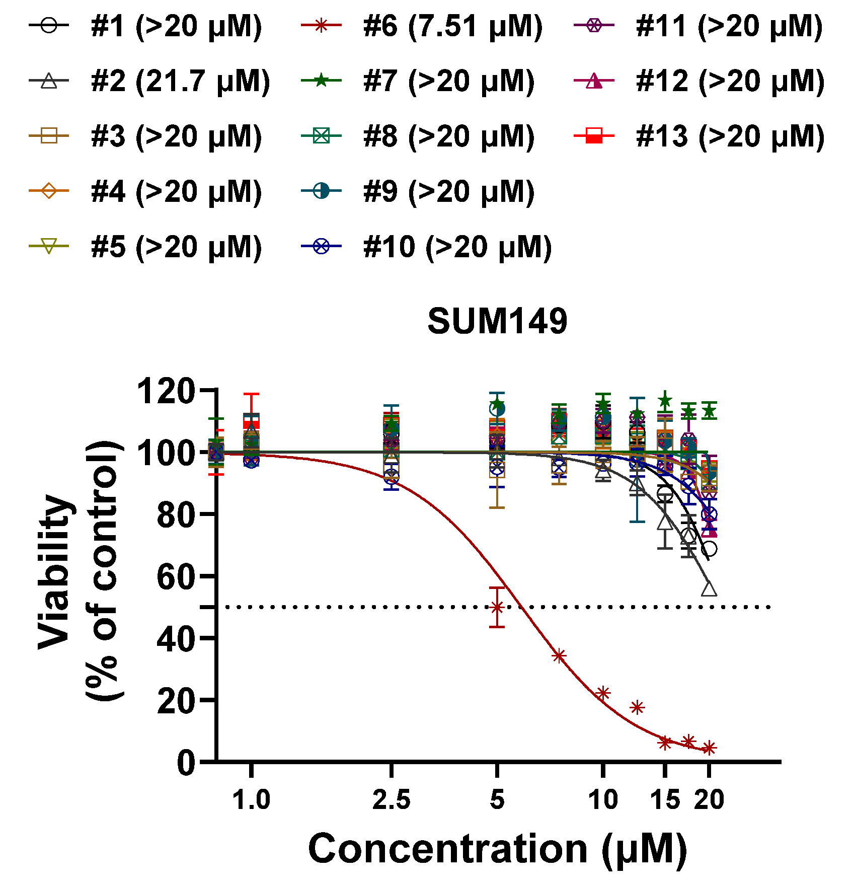
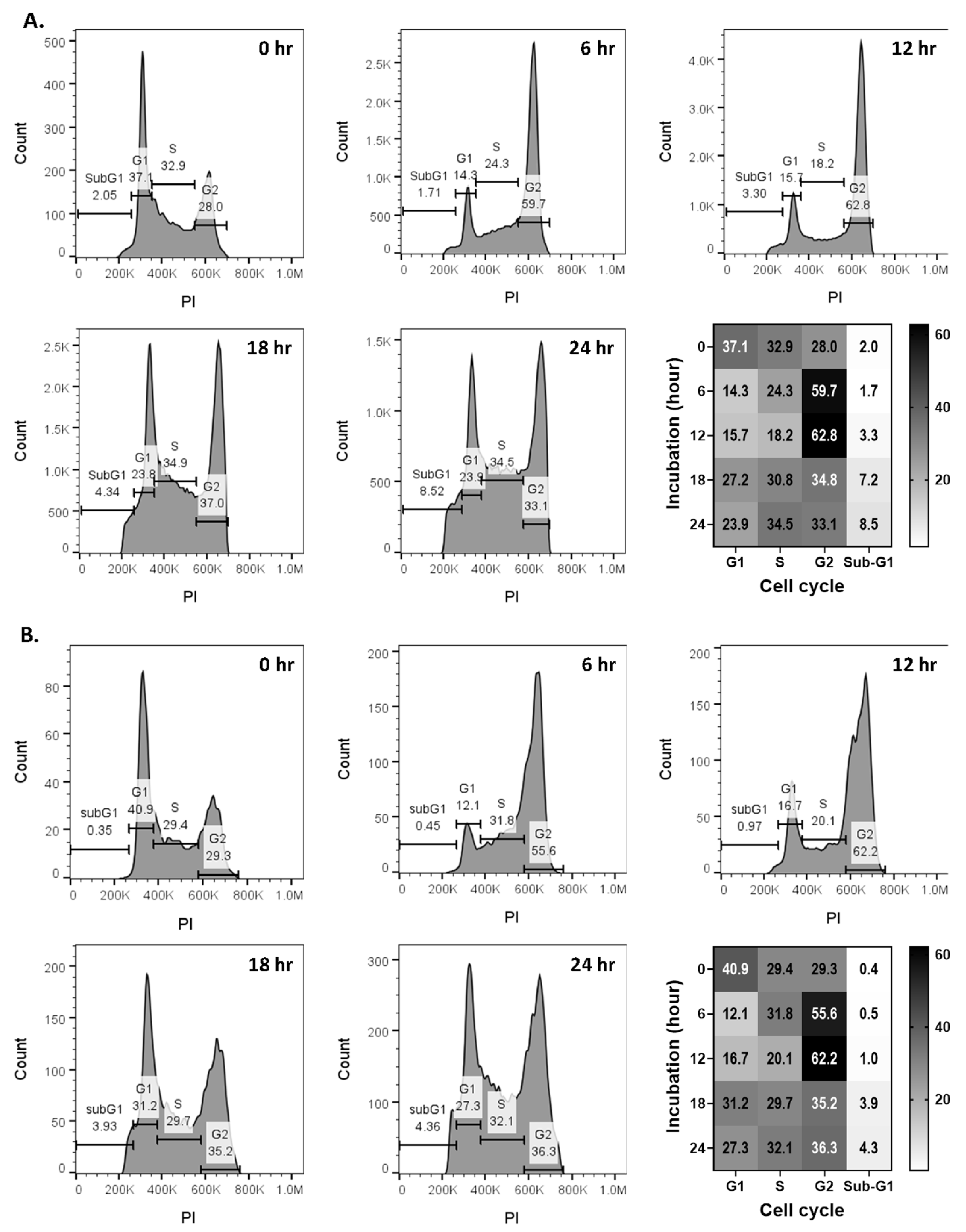
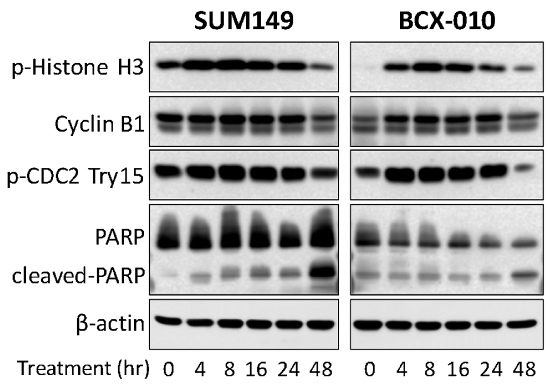
2.3. Compound 6 (Santin) from D. viscosa Flowers Inhibits Breast Cancer Cell Growth via G2-M Cell Cycle Arrest

Flavonoids, a diverse group of plant-derived polyphenolic compounds, are well documented for their broad-spectrum biological activities, including antioxidant, anti-inflammatory, antimicrobial, and anticancer properties [41,42,43]. To evaluate the antitumor potential of thirteen compounds extracted from D. viscosa flowers, we performed in vitro proliferation assays using the SUM149 triple-negative breast cancer (TNBC) cell line. Among the tested compounds, compound 6 (santin) exhibited the dose-dependent inhibition of cell growth (Figure 2), while the remaining twelve compounds showed no significant inhibitory effect at concentrations up to 20 μM. We further assessed the therapeutic potential of compound 6 across various breast cancer subtypes and other cancer cell lines. Compound 6 demonstrated dose-dependent growth inhibition in breast cancer cell lines, including TNBC (IC50: 4.22–26.71 µM), HER2-positive (HER2+) (IC50: 6.74–25.61 µM), and hormone receptor-positive (HR+) (IC50: 14.71–24.35 µM) cell lines (Figure 3A). Notably, inflammatory breast cancer (IBC) cell lines, which represent the most aggressive and lethal subtypes, showed heightened sensitivity to compound 6. The lowest IC₅₀ values were observed in BCX-010 (4.22 µM), SUM190 (6.74 µM), and SUM149 (7.73 µM), compared to non-IBC cell lines. In addition to breast cancer models, compound 6 also showed antitumor activity in non-small cell lung cancer (A549, IC50 = 10.73 µM), colon cancer (HT29, IC50 = 27.88 µM; HT116, IC50 = 17.03 µM), and hepatocellular carcinoma (SNU398, IC50 = 7.39 µM) (Figure 3B). Importantly, compound 6 displayed minimal cytotoxicity in the normal mammary epithelial cell line MCF-10A (IC50 > 20 µM), suggesting a favorable therapeutic index and minimal toxicity to normal tissues (Figure 3C). The IC50 values for all tested cell lines are summarized in Table 2.
To investigate the mechanism of action, we examined cell cycle progression in SUM149 and BCX-010 IBC cell lines. Compared to control cells, compound 6 treatment led to a marked accumulation of cells in the G2-M phase arrest within 6–12 h, ranging from 59.7% to 62.8% in SUM149 cells (Figure 4A) and 55.6% to 62.2% in BCX-010 cells (Figure 4B). To further validate G2–M checkpoint arrest, we performed a Western blot analysis to assess the expression of key cell cycle regulatory proteins. As shown in Figure 5, compound 6 induced a time-dependent increase in phospho-Histone H3 (a mitotic marker indicating metaphase accumulation) and phospho-CDC2 (Tyr15), a negative regulator of CDK1 activity. These findings confirm that compound 6 causes arrest in both the G2 phase and mitosis. Additionally, we observed an increase in the apoptosis marker cleaved PARP, suggesting that G2–M arrest induced by compound 6 ultimately leads to apoptotic cell death.
Flavonoids, specifically the subclass flavanones, are characterized by the absence of a double bond between the C–2 and C–3 positions. This structural difference results in losing the planar configuration of the rings in the benzo-γ-pyrone molecules, which is associated with a decrease in cytotoxicity [44]. Supporting this structure–activity relationship (SAR), flavanones 912, isolated from the flower extract of D. viscosa, showed no activity against all tested inflammatory (IBC) breast cancer cell lines.
Quercetin and kaempferol are well-studied flavonols that exhibit anticancer and antioxidant properties in several cancer cell lines. Studies in the literature have shown that flavanols differ from flavones, in which the presence of a hydroxy group at the C–3 position is linked to reduced cytotoxicity. For example, kaempferol and quercetin exhibited less pronounced cytotoxicity compared to apigenin [45]. Supporting this SAR, flavanols quercetin (1), kaempferol (2), kaempferide (3), 3,5-dihydroxy-7,4′-dimethoxyflavone (4), mikanin (5), and 6-methoxykaempferol (7), which were isolated from the flowers extract of D. viscosa, demonstrated little to no activity against the tested IBC breast cancer cell lines, highly aggressive subtypes.
Flavones isolated from D. vicosa flowers, namely makinin (5), santin (6), and 5-hydroxy-3,7,4-trimethoxyflavone (8), demonstrate varying levels of activity. Notably, compounds 5 and 8 show lower or no activity when compared to compound 6, which possesses a hydroxyl group at position C7. The presence of a hydroxyl group at C7 is essential for the cytotoxic activity observed in cancer cell lines [33]. The incorporation of methoxy groups in flavones decisively enhances their lipophilicity and membrane permeability, facilitating greater accumulation within cells. Our structure–activity relationship (SAR) analysis clearly reveals that 3,6-dimethoxyflavanone from santin (6) exhibits significant antiproliferative activity against IBC cell lines. While methoxy groups may contribute to metabolic challenges, they also confer selectivity and potent anti-inflammatory effects in flavone analogs. Specifically, santin effectively promotes TRAIL-mediated apoptosis in colon cancer cells, highlighting its potential as a therapeutic agent [34].

3. Materials and Methods

3.1. General Experimental Procedures

NMR spectra were recorded on a Bruker AVANCE DRX400 NMR spectrometer (Bruker, Billerica, MA, USA) at 400 MHz for 1H and 100 MHz for 13C. Data processing was performed using MestReNova 14.2.1-27684, with CDCl3 (δH 7.23, δC 77.16) or MeOD (δH 3.31, δC 49.0) as solvents. Silica gel (230–400 mesh, 480–800 mesh, Sorbent Technologies, Atlanta, GA, USA), C-18 reversed-phase (particle size 40–63 μm, Sorbent technologies, USA), and Sephadex LH-20 (GE Healthcare, Piscataway, NJ, USA) were used for column chromatography (CC). ACS grade solvent (acetone, hexane, ethyl acetate, methanol) used for extraction, isolation was analytical grade (VWR, Radnor, PA, USA). An amount of 10% cerium (IV) sulfate solution (Cat. No. 13590-82-4, Aldrich, St. Louis, MO, USA) in 15% sulfuric acid (Cat. No. 339741, Aldrich, St. Louis, MO, USA) was used as a TLC reagent.

3.2. Plant Materials

On 31 May 2023, fresh flowers of D. viscosa were collected from Hawaii Volcanoes National Park, located at 19.45482′ N, 155.33612′ W, along Mauna Loa Road, Hawai‘i, USA. The collected flowers were cleaned to remove dust and dirt before extraction. The plant was identified by Kumu Piilani Kaawaloa, and a voucher specimen (No. MQF05) was deposited at the Natural Product Chemistry Laboratory in the Department of Pharmaceutical Sciences at Daniel K. Inouye College of Pharmacy, University of Hawai‘i at Hilo.

3.3. Extraction and Isolation

The fresh flowers of D. viscosa (203.4 g) were finely ground and extracted with three times two liters of methanol for three days at room temperature. The extract was filtered through filter paper (qualitative 2, Cat. No. 1002110, Whatman, Buckinghamshire, UK). After solvent removal using a rotary evaporator, a methanol crude extract weighing 25.39 g was obtained. The methanol extract (25.39 g) was subjected to reversed-phase C-18 column chromatography (CC) [particle size 40–63 μm, Cat. No. 52546M-05, Sorbent technologies, USA] using a gradient elution of H2O-MeOH-acetone (1:0:0 to 0:0:1, v/v), yielding 21 fractions (A-W). Compounds 3 (184.4 mg), 10 (148.6 mg), and 13 (8.7 mg) were isolated directly. Fractions D, E, F, and G were combined (7.8 g) and further separated by Sephadex-LH20 CC (100% MeOH), yielding compounds 1 (6.4 mg), 2 (6.1 mg), 7 (282.3 mg), 9 (15.3 mg), and 11 (108.4 mg). Fraction K (264.8 mg) was purified by Sephadex-LH20 CC (100% MeOH) followed by silica gel CC (20% acetone-hexanes), yielding compounds 5 (1.6 mg), 6 (8.4 mg), and 14 (3.4 mg). Fraction N (1.8 g) was purified by Sephadex-LH20 CC (100% MeOH) followed by silica gel CC (20% ethyl acetate-hexanes), yielding compounds 3 (3.1 mg), 4 (12.4 mg), 8 (3.5 mg), and 12 (132.3 mg).
  • Quercetin (1): yellow solids, mp 315–316 °C; 1H NMR (acetone-d6, 400 MHz) δ 12.18 (1H, s, 5-OH), 7.80 (1H, d, J = 2.0 Hz, H-2′), 7.68 (1H, dd, J = 8.4, 2.0 Hz, H-6′), 6.97 (1H, d, J = 8.4 Hz, H-5′), 6.52 (1H, d, J = 2.0 Hz, H-8), and 6.25 (1H, d, J = 2.0 Hz, H-6).
  • Kaempferol (2): yellow solids, mp 277–278 °C; 1H NMR (MeOD, 400 MHz) δ 8.08 (2H, d, J = 8.9 Hz, H-2′ and H-6′), 6.91 (2H, d, J = 8.9 Hz, H-3′ and H-5′), 6.38 (1H, d, J = 2.0 Hz, H-8), and 6.17 (1H, d, J = 2.0 Hz, H-6).
  • Kaempferide (3): yellow solids, mp 228–229 °C; 1H NMR (acetone-d6, 400 MHz) δ 12.16 (1H, br s, 5-OH), 8.21 (2H, d, J = 8.8 Hz, H-2′ and H-6′), 7.10 (2H, d, J = 8.8 Hz, H-3′ and H-5′), 6.53 (1H, d, J = 2.0 Hz, H-8), 6.26 (1H, d, J = 2.0 Hz, H-6), and 3.89 (3H, s, CH3-4′).
  • 3,5-Dihydroxy-7,4′-dimethoxyflavone (4): yellow solids, mp 158–159 °C; 1H NMR (CDCl3, 400 MHz) δ 11.74 (1H, br s, 5-OH), 8.28 (2H, d, J = 8.8 Hz, H-2′ and H-6′), 7.04 (2H, d, J = 8.8 Hz, H-3′ and H-5′), 6.48 (1H, d, J = 2.0 Hz, H-8), 6.37 (1H, d, J = 2.0 Hz, H-6), and 3.89 (6H, s, CH3-7 and CH3-4′).
  • Mikanin (5): yellow solids, 1H NMR (CDCl3, 400 MHz) δ 11.83 (1H, br s, 5-OH), 8.19 (2H, d, J = 8.7 Hz, H-2′ and H-6′), 7.03 (2H, d, J = 8.7 Hz, H-3′ and H-5′), 6.55 (1H, s, H-8), 3.97 (3H, s, CH3-7), 3.93 (3H, s, CH3-6), and 3.89 (3H, s, CH3-4′).
  • Santin (6): yellow solids, mp 157–158 °C; 1H NMR (CDCl3, 400 MHz) δ 12.90 (1H, br s, 5-OH), 8.08 (2H, d, J = 9.0 Hz, H-2′ and H-6′), 7.04 (2H, d, J = 9.0 Hz, H-3′ and H-5′), 6.58 (1H, s, H-8), 4.07 (3H, s, CH3-6), 3.92 (3H, s, CH3-4′), and 3.88 (3H, s, CH3-3).
  • 6-Methoxykaempferol (7): yellow solids, mp 239–240 °C; 1H NMR (MeOD, 400 MHz) δ 8.09 (2H, d, J = 9.0 Hz, H-2′ and H-6′), 6.91 (2H, d, J = 9.0 Hz, H-3′ and H-5′), 6.52 (1H, s, H-8), and 3.90 (3H, s, CH3-6).
  • 5-Hydroxy-3,7,4′-trimethoxyflavone (8): yellow solids, mp 140–141 °C; 1H NMR (CDCl3, 400 MHz) δ 12.67 (1H, br s, 5-OH), 8.11 (2H, d, J = 9.0 Hz, H-2′ and H-6′), 7.05 (2H, d, J = 9.0 Hz, H-3′ and H-5′), 6.46 (1H, d, J = 2.0 Hz, H-8), 6.37 (1H, d, J = 2.0 Hz, H-6), 3.92 (3H, s, CH3-7), 3.90 (3H, s, CH3-4′), and 3.88 (3H, s, CH3-3).
  • Naringenin (9): pale-yellow solids, mp 250–251 °C; 1H NMR (MeOD, 400 MHz) δ 7.29 (2H, d, J = 8.5 Hz, H-2′ and H-6′), 6.82 (2H, d, J = 8.5 Hz, H-3′ and H-5′), 5.89 (1H, d, J = 2.0 Hz, H-8), 5.88 (1H, d, J = 2.0 Hz, H-6), 5.30 (1H, dd, J = 13.0, 3.0 Hz, H-2), 3.08 (1H, dd, J = 16.8, 13.0 Hz, H-3b), 2.65 (1H, dd, J = 16.8, 3.0 Hz, H-3a).
  • 5-Hydroxy-7,4′-dimethoxyflavone (10): pale-yellow solids, mp 164–165 °C; 1H NMR (CDCl3, 400 MHz) δ 12.06 (1H, s, 5-OH), 7.42 (2H, d, J = 8.5 Hz, H-2′ and H-6′), 6.97 (2H, d, J = 8.5 Hz, H-3′ and H-5′), 6.09 (1H, d, J = 2.0 Hz, H-8), 6.07 (1H, d, J = 2.0 Hz, H-6), 5.40 (1H, dd, J = 13.0, 3.0 Hz, H-2), 3.86 (3H, s, CH3-7), 3.83 (3H, s, CH3-4′), 3.09 (1H, dd, J = 16.8, 13.0 Hz, H-3b), and 2.79 (1H, dd, J = 16.8, 3.0 Hz, H-3a).
  • 2,3-Dihydro-5,7-dihydroxy-6-methoxy-2-(4-methoxyphenyl)-4H-1-benzopyran-4-one (11): pale-yellow solids, mp 98–99 °C; 1H NMR (MeOD, 400 MHz) δ 7.26 (2H, d, J = 8.5 Hz, H-2′ and H-6′), 6.79 (2H, d, J = 8.5 Hz, H-3′ and H-5′), 6.10 (1H, d, J = 2.0 Hz, H-8), 5.27 (1H, dd, J = 13.0, 3.0 Hz, H-2), 3.83 (3H, s, CH3-6), 3.72 (3H, s, CH3-4′), 3.05 (1H, dd, J = 16.8, 13.0 Hz, H-3b), and 2.64 (1H, dd, J = 16.8, 3.0 Hz, H-3a).
  • Macarangaflavanone B (12): pale-yellow solids, mp 159–160 °C; 1H NMR (MeOD, 400 MHz) δ 7.14 (1H, br s, H-2′), 7.09 (1H, br d, J = 8.0 Hz, H-6′), 6.77 (1H, d, J = 8.0 Hz, H-5′), 5.93 (1H, s, H-8), 5.16 (1H, dd, J = 13.0, 3.0 Hz, H-2), 3.02 (1H, dd, J = 16.8, 13.0 Hz, H-3b), 2.60 (1H, dd, J = 16.8, 3.0 Hz, H-3a), isoprenyl unit at C-6 [δH 5.19 (1H, t, J = 7.2 Hz), 3.21 (2H, d, J = 7.2 Hz), 1.75 (3H, s), and 1.70 (3H, s)], and isoprenyl unit at C-3′ [δH 5.33 (1H, t, J = 7.2 Hz), 3.31 (2H, d, J = 7.2 Hz), 1.72 (3H, s), and 1.63 (3H, s)].
  • 3,5-Diprenyl-4-hydroxybenzaldehyde (13): white solid, mp 84–85 °C; 1H NMR (CDCl3, 400 MHz) δ 9.82 (1H, s, H-7), 7.56 (2H, s, H-2 and H-6), 6.07 (1H, s, 4-OH), 2× isoprenyl unit [δH 5.34 (2H, t, J = 7.2 Hz), 3.43 (4H, d, J = 7.2 Hz), 1.82 (6H, s), and 1.80 (6H, s)].

3.4. Minimum Inhibition Concentration (MIC)

Bacteria strains: The antibacterial activity was tested using selected pathogens and commensal strains from the American Type Culture Collection (ATCC) (Manassas, MD, USA): MRSA USA-300 (LAC, ATCC BAA1756), MRSA N315 (NR-45898), MSSA Newman (ATCC 25904), MSSA 8325-4, E. coli (ATCC 9637), K. pneumoniae NCTC 9633 (ATCC 13883), and P. aeruginosa Boston 41,501 (ATCC 27853). MSSA LUU7, MSSA ONE6, S. aureus 8384, MSSA RI27, and MSSA LUE1 were isolated from Hawai’i [46]. S. aureus N315 was an MRSA pathogen strain causing hospital-acquired infections in 1982. This strain is easy to acquire antibiotic resistance, such as vancomycin. MSSA strains isolated from the Island of Hawaii include the following: Wailuku River (LUU7); Wailuku River—Estuary (LUE1); Richardson’s Beach Park—Back (RI27); Onekahakaha Beach Park (ONE6) [46]. Mueller–Hinton Broth (MHB, SKU:275730, GTIN:00382902757306, Becton, Dickinson and Company, Franklin Lakes, NJ, USA) and Mueller–Hinton Agar (MHA, SKU:225250, GTIN:00382902252504, Becton, Dickinson and Company, Franklin Lakes, NJ, USA) were used to prepare bacterial inocula. All the inoculate were prepared with fresh cultures plated the day before the test.
The MIC of the samples was determined using a standard two-fold serial dilution method in Mueller–Hinton Broth (MHB, Cat. No. 70192, Sigma-Aldrich, USA), according to guidelines from the Clinical and Laboratory Standards Institute (CLSI) [47,48]. In brief, serial dilutions of the compounds in DMSO were prepared and added to 96-well microplates containing MHB. Different concentrations of the compound (ranging from 0.25 to 128 μg/mL) were then added to each well. Following this, bacterial suspensions (1 × 10⁴ CFU/well) were then inoculated to each well. As a negative control, wells containing 50 μL of MHB broth and 50 μL of bacterial strains without added samples. The plates were incubated at 37 °C for 18–24 h. After incubation, 10 µL of 0.018% resazurin (Cat. No. R7017, Sigma Aldrich, USA) was added to each well. The MIC values were determined after 2–3 h of incubation with resazurin. All antimicrobial assays were performed in triplicate, with vancomycin (Cat. No. 1709007, Sigma Aldrich, USA) and gentamicin (Cat. No. 46305, Sigma Aldrich, USA) used as standard controls.

3.5. Cell Lines and Reagents

BT20, HCC1428, HCC3153, HCC1937, HCC1806, MDA-MB-175 VII, MDA-MB-231, MDA-MB-436, MDA-MB-468, A549, HT29, HT116, SNU398, MCF-10A, and ZR-75-1 cells were purchased from American Type Culture Collection (Manassas, VA, USA). SUM149, SUM159, and SUM190 cells were purchased from Asterand Bioscience (now BioIVT, Hicksville, NY, USA). CAL51 cell line was purchased from DSMZ (Braunschweig, Germany). KPL4 cell line was provided by Kawasaki Medical School (Okayama, Japan). A3250 cell line was provided by Icahn School of Medicine at Mount Sinai (New York, NY, USA). FC-IBC02, IBC3, and BCX-010 cell lines were provided by MD Anderson Cancer Center (Houston, TX, USA). HCC1428, HCC3153, HCC1937, HCC1806, MDA-MB-175 VII, A549, HT29, HT116, SNU398, and ZR-75-1 cells were maintained in Roswell Park Memorial Institute 1640 medium (Sigma-Aldrich, St. Louis, MO, USA). BT-20, MDA-MB-231, MDA-MB-436, and MDA-MB-468 cells were maintained in Dulbecco’s modified Eagle’s medium/F-12 medium (DMEM/F-12, Sigma-Aldrich). FC-IBC02, IBC3, BCX-010, SUM190, and KPL4 cells were maintained in Ham’s F-12 medium (Sigma-Aldrich) supplemented with 5 µg/mL of insulin (Thermo Fisher Scientific Inc., Waltham, MA, USA), and 1 µg/mL of hydrocortisone (Sigma-Aldrich). MCF-10A was maintained in DMEM/F-12 medium supplemented with 5% horse serum, 10 µg/mL human insulin, 20 ng/mL EGF, 100 ng/mL Cholera toxin, and 0.5 µg/mL Hydrocortisone (Sigma-Aldrich). All media were supplemented with 10% fetal bovine serum (GenDEPOT, Katy, TX, USA) and 1% antibiotic/antimycotic (Sigma-Aldrich). All cell lines were validated by DNA typing at the MD Anderson Characterized Cell Line Core and confirmed to be free of Mycoplasma using the MycoAlert Mycoplasma Detection Kit (Lonza, Morristown, NJ, USA).

3.6. Sulforhodamine B Cell Proliferation Assay: Anticancer Activity

The antiproliferative effects of the extract of and compounds isolated from D. viscosa on breast cancer cells were evaluated using sulforhodamine B staining assays [49]. Briefly, cells (3 to 10 × 103 cells/well) were seeded in 96-well plates and allowed to adhere overnight. On the following day, cells were treated with samples for 5 days. After the treatment period, cells were fixed with 5% trichloroacetic acid (Cat. No. T6399, Sigma-Aldrich) at 4 °C for 2 h and washed with water three times. Fixed cells were stained with 0.03% sulforhodamine B (Cat. No. 230162, Sigma-Aldrich) solution in 1% acetic acid (Cat. No. 64-19-7, Sigma-Aldrich) for 30 min at room temperature. Excess stain was removed by washing the wells three times with 1% acetic acid. The stained cells were dissolved in 10 mM Tris buffer (Cat. No. T8101-010, GenDEPOT), and the optical density was measured at the wavelength of 560 nm using a Spark enhanced microplate reader (TECAN, Männedorf, Switzerland). The cell proliferation rate was calculated and summarized using descriptive statistics (mean) with the GraphPad Prism software program (version 10; GraphPad Software, Boston, MA, USA).

3.7. Cell Cycle Analyses

Breast cancer cells (5 × 10⁵ cells) were placed in a 6 cm culture dish and cultured overnight. The cells were treated with the compound 6 with an IC50 concentration for the indicated time points. After incubation, cells were harvested and fixed for DNA content analysis. Adherent cells were detached using trypsin-EDTA and collected into 5 mL conical tubes, followed by centrifugation at 1500 rpm for 4 min at 4 °C. The cell pellet was washed once with 3 mL of ice-cold PBS and centrifuged under the same conditions. The cell pellet was gently resuspended in 350 μL of cold PBS, and then ice-cold 99.9% ethanol (1 mL, 200 proof) was added dropwise to the cell suspension while gently vortexing to avoid cell clumping. Fixed cells were incubated at 4 °C for at least 2 h. The ethanol-fixed cells were centrifuged at 1500 rpm for 4 min and washed twice with 3 mL of cold PBS. The cell pellet was then resuspended in 200 μL of PBS containing 100 μg/mL RNase A and 50 μg/mL propidium iodide (#P4864; Sigma-Aldrich) and incubated at 37 °C for 30 min in the dark. After incubation, samples were analyzed using a Thermo Fisher Attune NxT BRYV6 device (Thermo Fisher Scientific Inc.), and then the cell cycle was determined using FlowJo software v10 (BD Biosciences, Milpitas, CA, USA).

3.8. Western Blotting

The effect of compound 6 on the expression of G2–M phase cell cycle-related proteins in breast cancer cell lines was analyzed by Western blotting. Cells were seeded in 6 cm culture dishes and incubated overnight. The following day, cells were treated with compound 6 at its IC50 concentration for the indicated time points. Whole-cell lysates were prepared using M-PER lysis buffer (Fisher Scientific) supplemented with protease and phosphatase inhibitors (GenDepot). Protein concentrations were determined, and 20 µg of denatured protein per sample was resolved by SDS-PAGE and transferred onto PVDF membranes (Bio-Rad, Hercules, CA, USA). Membranes were probed with the following primary antibodies at 1:1000 dilution: cleaved PARP (9542S, Cell Signaling Technology, Danvers, MA, USA), phospho-Histone H3 (Ser10; 382159, Cell Signaling Technology), Cyclin B1 (122231, Cell Signaling Technology), phospho-CDC2 (Tyr15; 9114S, Cell Signaling Technology), and β-actin (A5316, Sigma-Aldrich) as a loading control. After washing, membranes were incubated with HRP-conjugated secondary antibodies (1:10,000; Life Technologies, Waltham, MA, USA) and developed using enhanced chemiluminescence. Chemiluminescent signals were detected using the Azure 500 Imaging System (Azure Biosystems, Dublin, CA, USA).

3.9. Statistical Analysis

Cell proliferation data were summarized using descriptive statistics (mean), and the IC50 values were determined by fitting a dose–response curve using nonlinear regression analysis in GraphPad Prism (version 10, GraphPad Software, Boston, MA, USA).

4. Conclusions

D. viscosa flowers yielded bioactive compounds with antibacterial and anticancer properties. Macarangaflavanone B (12) showed potent antibacterial effects, while santin (6) demonstrated selective antiproliferative activity against IBC cell lines. These findings highlight the therapeutic potential of D. viscosa and underscore the need for further studies to explore its mechanisms and optimize its use in translational applications. Compounds 5 and 8 are inactive, etc. Santin demonstrated notable antiproliferative activity against IBC cell lines, highlighting its potential as a therapeutic candidate for aggressive cancer subtypes. Its structural features, particularly the hydroxyl group at the C7 position, may play a key role in its cytotoxic effects. While flavonoids such as compounds 5 and 8 exhibited minimal or no activity, the structural characteristics of santin enhance its therapeutic potential. These findings support further investigation of santin in preclinical breast cancer models.

Supplementary Materials

The following supporting information can be downloaded at: https://www.mdpi.com/article/10.3390/molecules30112274/s1. Figure S1: 1H NMR spectrum (400 MHz, Acetone-d6) of compound 1. Figure S2: 1H NMR spectrum (400 MHz, MeOD) of compound 2. Figure S3: 1H NMR spectrum (400 MHz, Acetone-d6) of compound 3. Figure S4: 13C NMR spectrum (100 MHz, Acetone-d6) of compound 3. Figure S5: HSQC spectrum of compound 3. Figure S6: HMBC spectrum of compound 3. Figure S7: 1H NMR spectrum (400 MHz, CDCl3) of compound 4. Figure S8: 13C NMR spectrum (100 MHz, CDCl3) of compound 4. Figure S9: HSQC spectrum of compound 4. Figure S10: HMBC spectrum of compound 4. Figure S11: 1H NMR spectrum (400 MHz, CDCl3) of compound 5. Figure S12: 13C NMR spectrum (100 MHz, CDCl3) of compound 5. Figure S13: HSQC spectrum of compound 5. Figure S14: HMBC spectrum of compound 5. Figure S15: 1H NMR spectrum (400 MHz, CDCl3) of compound 6. Figure S16: 13C NMR spectrum (100 MHz, CDCl3) of compound 6. Figure S17: HSQC spectrum of compound 6. Figure S18: HMBC spectrum of compound 6. Figure S19: 1H NMR spectrum (400 MHz, MeOD) of compound 7. Figure S20: 13C NMR spectrum (100 MHz, MeOD) of compound 7. Figure S21: HSQC spectrum of compound 7. Figure S22: HMBC spectrum of compound 7. Figure S23: 1H NMR spectrum (400 MHz, CDCl3) of compound 8. Figure S24: 13C NMR spectrum (100 MHz, CDCl3) of compound 8. Figure S25: HSQC spectrum of compound 8. Figure S26: HMBC spectrum of compound 8. Figure S27: 1H NMR spectrum (400 MHz, MeOD) of compound 9. Figure S28: 13C NMR spectrum (100 MHz, MeOD) of compound 9. Figure S29: 1H NMR spectrum (400 MHz, CDCl3) of compound 10. Figure S30: 13C NMR spectrum (100 MHz, CDCl3) of compound 10. Figure S31: HSQC spectrum of compound 10. Figure S32: HMBC spectrum of compound 10. Figure S33: 1H NMR spectrum (400 MHz, MeOD) of compound 11. Figure S34: 13C NMR spectrum (100 MHz, MeOD) of compound 11. Figure S35: HSQC spectrum of compound 11. Figure S36: HMBC spectrum of compound 11. Figure S37: 1H NMR spectrum (400 MHz, MeOD) of compound 12. Figure S38: 13C NMR spectrum (100 MHz, MeOD) of compound 12. Figure S39: HSQC spectrum of compound 12. Figure S40: HMBC spectrum of compound 12. Figure S41: 1H NMR spectrum (400 MHz, CDCl3) of compound 13. Figure S42: 13C NMR spectrum (100 MHz, CDCl3) of compound 13.

Author Contributions

Conceptualization, L.C.C. and K.P.K.; methodology, A.R., J.L., Y.J.G. and D.Y.; formal analysis, A.R., J.L., D.Y., Y.J.G. and L.C.C.; investigation, A.R., J.L., Y.J.G. and D.Y.; resources, L.C.C., K.P.K. and M.M.W.; data curation, A.R., J.L., D.Y. and L.C.C.; writing—original draft preparation, A.R., J.L., L.C.C., M.M.W. and K.P.K.; writing—review and editing, A.R., L.C.C., K.P.K., L.K.C., M.M.W. and S.W.; funding acquisition, L.C.C. and J.L. All authors have read and agreed to the published version of the manuscript.

Funding

The research was supported by a cooperative agreement with the Agricultural Research Service, U.S. Department of Agriculture (No. NACA 58-2040-0-011) and RTRF grants from the National Institutes of Health (NIH), National Institute of General Medical Sciences (NIGMS), IDeA Networks of Biomedical Research Excellence (INBRE), Award number: P20GM103466. The DKICP provided support for the NMR facility. The content is solely the authors’ responsibility and does not necessarily represent the official views of the National Institutes of Health. Anticancer screening was supported by the University of Hawaii Cancer Center Support Grant (P30CA071789; Preclinical Core Shared Resources).

Institutional Review Board Statement

Not applicable.

Informed Consent Statement

Not applicable.

Data Availability Statement

The data are included in this article.

Acknowledgments

The authors wish to thank Tyler Gerken and Tracy Wiegner for providing Hawaiian bacteria strains.

Conflicts of Interest

The authors declare no conflicts of interest.

References

  1. Nanea Valeros, Public Affairs Specialist, U.S. Fish and Wildlife Service. 17 September 2021. Available online: https://medium.com/usfwspacificislands/l%C4%81%CA%BBau-lapa%CA%BBau-reclaiming-traditional-practices-of-medicine-9d5ebfe281c1 (accessed on 26 October 2024).
  2. Almarfadi, O.M.; Siddiqui, N.A.; Shahat, A.A.; Fantoukh, O.I.; El Gamal, A.A.; Raish, M.; Bari, A.; Iqbal, M.; Alqahtani, A.S. Isolation of a novel isoprenylated phenolic compound and neuroprotective evaluation of Dodonaea viscosa extract against cerebral ischaemia–reperfusion injury in rats. Saudi Pharm. J. 2024, 32, 101898. [Google Scholar] [CrossRef] [PubMed]
  3. Karim, N.; Irshad, S.; Khan, I.; Mohammad, A.; Anis, I.; Shah, M.R.; Khan, I.; Chebib, M. GABA(A) receptor modulation and neuropharmacological activities of viscosine isolated from Dodonaea viscosa (Linn). Pharmacol. Biochem. Behav. 2015, 136, 64–72. [Google Scholar] [CrossRef] [PubMed]
  4. Lei, C.; Wang, X.H.; Liu, Y.N.; Zhao, T.; Hu, Z.; Li, J.Y.; Hou, A.J. Clerodane diterpenoids from Dodonaea viscosa and their inhibitory effects on ATP citrate lyase. Phytochemistry 2021, 183, 112614. [Google Scholar] [CrossRef] [PubMed]
  5. Zhao, X.T.; Lei, C.; You, J.Q.; Zhao, T.; Yu, M.H.; Shi, X.L.; Hu, X.; Hou, A.J. Dimeric clerodane diterpenoids and antiviral constituents of Dodonaea viscosa. Bioorg. Chem. 2021, 112, 104916. [Google Scholar] [CrossRef]
  6. Siddiqui, N.A.; Almarfadi, O.M.; Shahat, A.A.; Alqahtani, A.S.; El Gamal, A.A.; Raish, M.; Iqbal, M. Isolation of new compound and neuroprotective studies from Dodonaea viscosa. J. King Saud Univ. Sci. 2023, 35, 102704. [Google Scholar] [CrossRef]
  7. Yu, H.; Liu, B.; Zhao, Y.; Li, J.; Wu, G.; Ma, J.; Gui, F.; Tao, F.; Hao, X.; Ding, X.; et al. Combined activity of saponin B isolated from Dodonaea viscosa seeds with pesticide azadirachtin against the pest Spodoptera litura. Metabolites 2023, 14, 15. [Google Scholar] [CrossRef]
  8. Liang, W.; Yu, Q.; Zheng, Z.; Liu, J.; Cai, Q.; Liu, S.; Lin, S. Design and synthesis of phenyl sulfide-based cationic amphiphiles as membrane-targeting antimicrobial agents against Gram-positive pathogens. J. Med. Chem. 2022, 65, 14221–14236. [Google Scholar] [CrossRef]
  9. Li, F.; Arnsberger, P.; Miller, F.D. Profile of methicillin-resistant Staphylococcus aureus among nursing home residents in Hawai ‘i. Hawaii Med. J. 2010, 6, 126. [Google Scholar]
  10. Estivariz, C.F.; Park, S.Y.; Hageman, J.C.; Dvorin, J.; Melish, M.M.; Arpon, R.; Coon, P.; Slavish, S.; Kim, M.; McDougal, L.K. Emergence of community-associated methicillin-resistant Staphylococcus aureus in Hawaii, 2001–2003. J. Infect. 2007, 54, 349–357. [Google Scholar] [CrossRef]
  11. Li, F.; Park, S.Y.; Ayers, T.L.; Miller, F.D.; MacFadden, R.; Nakata, M.; Lee, M.C.; Effler, P.V. Methicillin-resistant Staphylococcus aureus, Hawaii, 2000–2002. Emerg. Infect. Dis. 2005, 11, 1205–1210. [Google Scholar] [CrossRef]
  12. Khurram, M.; Lawton, L.A.; Edwards, C.; Iriti, M.; Hameed, A.; Khan, M.A.; Khan, F.A.; Rahman, S.U. Rapid bioassay-guided isolation of antibacterial clerodane type diterpenoid from Dodonaea viscosa (L.) Jaeq. Int. J. Mol. Sci. 2015, 16, 20290–20307. [Google Scholar] [CrossRef] [PubMed]
  13. Rivenbark, A.G.; O’Connor, S.M.; Coleman, W.B. Molecular and cellular heterogeneity in breast cancer: Challenges for personalized medicine. Am. J. Pathol. 2013, 183, 1113–1124. [Google Scholar] [CrossRef]
  14. Luond, F.; Tiede, S.; Christofori, G. Breast cancer as an example of tumour heterogeneity and tumour cell plasticity during malignant progression. Br. J. Cancer 2021, 125, 164–175. [Google Scholar] [CrossRef] [PubMed]
  15. O’Reilly, D.; Sendi, M.A.; Kelly, C.M. Overview of recent advances in metastatic triple negative breast cancer. World J. Clin. Oncol. 2021, 12, 164–182. [Google Scholar] [CrossRef]
  16. Al-Mahmood, S.; Sapiezynski, J.; Garbuzenko, O.B.; Minko, T. Metastatic and triple-negative breast cancer: Challenges and treatment options. Drug Deliv. Transl. Res. 2018, 8, 1483–1507. [Google Scholar] [CrossRef]
  17. Elfgen, C.; Bjelic-Radisic, V. Targeted therapy in HR+ HER2- metastatic breast cancer: Current clinical trials and their implications for CDK4/6 inhibitor therapy and beyond treatment options. Cancers 2021, 13, 5994. [Google Scholar] [CrossRef]
  18. Lim, B.; Woodward, W.A.; Wang, X.; Reuben, J.M.; Ueno, N.T. Inflammatory breast cancer biology: The tumour microenvironment is key. Nat. Rev. Cancer 2018, 18, 485–499. [Google Scholar] [CrossRef]
  19. Herrera-Calderon, O.; Herrera-Ramírez, A.; Cardona-G, W.; Melgar-Merino, E.J.; Chávez, H.; Pari-Olarte, J.B.; Loyola-Gonzales, E.; Kong-Chirinos, J.F.; Almeida-Galindo, J.S.; Peña-Rojas, G.; et al. Dodonaea viscosa Jacq. induces cytotoxicity, antiproliferative activity, and cell death in colorectal cancer cells via regulation of caspase 3 and p53. Front. Pharmacol. 2023, 14, 1197569. [Google Scholar] [CrossRef]
  20. Anglana, C.; Rojas, M.; Girelli, C.R.; Barozzi, F.; Quiroz-Troncoso, J.; Alegría-Aravena, N.; Montefusco, A.; Durante, M.; Fanizzi, F.P.; Ramírez-Castillejo, C.; et al. Methanolic extracts of D. viscosa specifically affect the cytoskeleton and exert an antiproliferative effect on human colorectal cancer cell lines, according to their proliferation rate. Int. J. Mol. Sci. 2023, 24, 14920. [Google Scholar] [CrossRef]
  21. Kheyar-Kraouche, N.; Boucheffa, S.; Bellik, Y.; Farida, K.; Brahmi-Chendouh, N. Exploring the potential of Inula viscosa extracts for antioxidant, antiproliferative and apoptotic effects on human liver cancer cells and a molecular docking study. BioTechnologia 2023, 104, 183–198. [Google Scholar] [CrossRef]
  22. Ren, G.; Hou, J.; Fang, Q.; Sun, H.; Liu, X.; Zhang, L.; Wang, P.G. Synthesis of flavonol 3-O-glycoside by UGT78D1. Glycoconj. J. 2012, 29, 425–432. [Google Scholar] [CrossRef] [PubMed]
  23. Schmidt, T.J.; Willuhn, G. Sesquiterpene lactone and flavonoid variability of the Arnica angustifolia aggregate (Asteraceae). Biochem. Syst. Ecol. 2000, 28, 133–142. [Google Scholar] [CrossRef]
  24. Lien, T.T.P.; Razumovskaya, R.G.; Spiridovich, E.V.; Tan, L.Q.; Hoa, N.T.; Hue, C.T. Chemical Composition and Biological Activity of Vietnamese Amaranthus spinosus. Chem. Nat. Compd. 2019, 55, 1164–1166. [Google Scholar] [CrossRef]
  25. El-Sayed, N.H.; Norris, J.A.; Ahmed, A.A.; Mabry, T.J. Flavonoids of Brickellia longifolia. Phytochemistry 1990, 29, 2364–2365. [Google Scholar] [CrossRef]
  26. Martinez, J.; Silván, A.M.; Abad, M.J.; Bermejo, P.; Villar, A.; Söllhuber, M. Isolation of two flavonoids from Tanacetum microphyllum as PMA-induced ear edema inhibitors. J. Nat. Prod. 1997, 60, 142–144. [Google Scholar] [CrossRef] [PubMed]
  27. Poonam, V.; Kumar, G.; Reddy, L.C.S.; Jain, R.; Sharma, S.K.; Prasad, A.K.; Parmar, V.S. Chemical constituents of the genus Prunus and their medicinal properties. Curr. Med. Chem. 2011, 18, 3758–3824. [Google Scholar] [CrossRef]
  28. Yenjai, C.; Prasanphen, K.; Daodee, S.; Wongpanich, V.; Kittakoop, P. Bioactive flavonoids from Kaempferia parviflora. Fitoterapia 2004, 75, 89–92. [Google Scholar] [CrossRef]
  29. Nagarajan, G.R.; Parmar, V.S. Chemical examination of the heartwood of Prunus domestica. Planta Med. 1977, 31, 146–150. [Google Scholar] [CrossRef]
  30. Kamperdick, C.; Van, N.H.; Van Sung, T. Constituents from Miliusa balansae (Annonaceae). Phytochemistry 2002, 61, 991–994. [Google Scholar] [CrossRef]
  31. Pisutthanan, N.; Liawruangrath, B.; Liawruangrath, S.; Bremner, J.B. A new flavonoid from Chromolaena odorata. Nat. Prod. Res. 2006, 20, 1192–1198. [Google Scholar] [CrossRef]
  32. Shirataki, Y.; Yokoe, I.; Endo, M.; Komatsu, M. Determination of C-6 or C-8 substituted flavanone using 13C-1H long range coupling and the revised structures of some flavanones. Chem. Pharm. Bull. 1985, 33, 444–447. [Google Scholar] [CrossRef]
  33. Tip-Pyang, S.; Veerachato, G.; Phuwapraisirisan, P.; Sathanasaowapak, S. Antibacterial component from Arfeuillea arborescens Pierre. Chem. Res. Commun. 1999, 9, 15–19. [Google Scholar]
  34. Ali, H.; Kabir, N.; Muhammad, A.; Shah, M.R.; Musharraf, S.G.; Iqbal, N.; Nadeem, S. Hautriwaic acid as one of the hepatoprotective constituents of Dodonaea viscosa. Phytomedicine 2014, 21, 131–140. [Google Scholar] [CrossRef] [PubMed]
  35. Sastry, K.N.S.; Nayudamma, Y. Leucocyanidin from Dodonaea viscosa bark. Leather Sci. 1966, 13, 174–176. [Google Scholar]
  36. Tong, Z.W.; Gul, H.; Awais, M.; Saddick, S.; Khan, F.S.; Gulfraz, M.; Afzal, U.; Nazir, K.; Malik, M.Y.; Khan, S.U.; et al. Determination of in vivo biological activities of Dodonaea viscosa flowers against CCL4 toxicity in albino mice with bioactive compound detection. Sci. Rep. 2021, 11, 13336. [Google Scholar] [CrossRef]
  37. Teffo, L.S.; Aderogba, M.A.; Eloff, J.N. Antibacterial and antioxidant activities of four kaempferol methyl ethers isolated from Dodonaea viscosa Jacq. var. angustifolia leaf extracts. S. Afr. J. Bot. 2010, 76, 25–29. [Google Scholar] [CrossRef]
  38. Wu, T.; He, M.; Zang, X.; Zhou, Y.; Qiu, T.; Pan, S.; Xiaoyun, X. A structure-activity relationship study of flavonoids as inhibitors of E. coli by membrane interaction effect. Biochim. Biophys. Acta Biomembr. 2013, 1828, 2751–2756. [Google Scholar] [CrossRef]
  39. Xiao, J. Dietary flavonoid aglycones and their glycosides: Which show better biological significance? Crit. Rev. Food Sci. Nutr. 2017, 57, 1874–1905. [Google Scholar] [CrossRef]
  40. Schütz, B.A.; Wright, A.D.; Rali, T.; Sticher, O. Prenylated flavanones from leaves of Macaranga pleiostemona. Phytochemistry 1995, 40, 1273–1277. [Google Scholar] [CrossRef]
  41. Liga, S.; Paul, C.; Peter, F. Flavonoids: Overview of Biosynthesis, Biological Activity, and Current Extraction Techniques. Plants 2023, 12, 2732. [Google Scholar] [CrossRef]
  42. Gupta, T.; Kataria, R.; Sardana, S. A Comprehensive Review on Current Perspectives of Flavonoids as Antimicrobial Agent. Curr. Top. Med. Chem. 2022, 22, 425–434. [Google Scholar] [CrossRef]
  43. Ullah, A.; Munir, S.; Badshah, S.L.; Khan, N.; Ghani, L.; Poulson, B.G.; Emwas, A.H.; Jaremko, M. Important flavonoids and their role as a therapeutic agent. Molecules 2020, 25, 5243. [Google Scholar] [CrossRef] [PubMed]
  44. Plochmann, K.; Korte, G.; Koutsilieri, E.; Richling, E.; Riederer, P.; Rethwilm, A.; Schreier, P.; Scheller, C. Structure–activity relationships of flavonoid-induced cytotoxicity on human leukemia cells. Arch. Biochem. Biophys. 2007, 460, 1–9. [Google Scholar] [CrossRef]
  45. Touil, Y.S.; Fellous, A.; Scherman, D.; Chabot, G.G. Flavanoid-induced morphological modifications of endothelial cells through microtubule stabilization. Nutr. Cancer 2009, 61, 310–321. [Google Scholar] [CrossRef]
  46. Gerken, T.J.; Roberts, M.C.; Dykema, P.; Melly, G.; Lucas, D.; De Los Santos, V.; Gonzalez, J.; Butaye, P.; Wiegner, T.N. Environmental surveillance and characterization of antibiotic-resistant Staphylococcus aureus at coastal beaches and rivers on the island of Hawaiʻi. Antibiotics 2021, 10, 980. [Google Scholar] [CrossRef]
  47. Reller, L.B.; Weinstein, M.; Jorgensen, J.H.; Ferraro, M.J. Antimicrobial susceptibility testing: A review of general principles and contemporary practices. Clin. Infect. Dis. 2009, 49, 1749–1755. [Google Scholar]
  48. Elshikh, M.; Ahmed, S.; Funston, S.; Dunlop, P.; McGaw, M.; Marchant, R.; Banat, I.M. Resazurin-based 96-well plate microdilution method for the determination of minimum inhibitory concentration of biosurfactants. Biotechnol. Lett. 2016, 38, 1015–1019. [Google Scholar] [CrossRef]
  49. Vichai, V.; Kirtikara, K. Sulforhodamine B colorimetric assay for cytotoxicity screening. Nat. Protoc. 2006, 1, 1112–1116. [Google Scholar] [CrossRef]
Figure 1. Structures of compounds 113 from D. viscosa flowers extract.
Molecules 30 02274 g001
Figure 2. Compound 6 has the most effective antiproliferative effect compared to other compounds in SUM149 breast cancer cell lines. Sulforhodamine B assay was conducted for 5 days of incubation.
Molecules 30 02274 g002
Figure 3. Compound 6 has an antiproliferative effect in cancer cell lines. Sulforhodamine B assay was conducted for 5 days of incubation. (A,C) Breast cancer cell lines, (B) lung (A549), colon (HT29, HT116), and liver (SNU398) cancer cell lines.
Molecules 30 02274 g003
Figure 4. Compound 6 induces G2-M cell cycle arrest. Cells were treated with compound 6 for the indicated time and then analyzed by flow cytometry. Heatmaps were generated using the Graph Pad Prism software package (V10). (A) SUM149 and (B) BCX-010.
Molecules 30 02274 g004
Figure 5. Activation of G2-M Checkpoint and induction of apoptosis by compound 6. Western blotting: The breast cancer cell lines were treated with IC50 concentration of compound 6 for the indicated time points, and then whole cell lysates were collected for Western blotting.
Molecules 30 02274 g005
Table 1. MIC values (µg/mL) of compounds from D. viscosa extract.
CompoundsGram-PositiveGram-Negative
MRSA USA-300MSSA
LUU7
MSSA 8325-4S. aureus 8384MSSA ONE6MSSA RI27MSSA
LUE1
MRSA N315MSSA
Newman
E. coliK.
pneumoniae
P. aeruginosa
D. viscosa
flowers extract
640320640640320320320320320128012801280
1128128128128128128128128128128128128
2646464646464646464128128128
3----------128-
4----------128128
5----------128-
6----------128-
7----------128128
8-----------128
9-----------128
10----------128128
11---------128128128
12222222222128128128
13----------128128
Vancomycin0.510.50.50.510.250.50.5ntntnt
Gentamicinntntntntntntntntnt0.2510.25
“-” = Inactive indicated a MIC value > 128 µg/mL; nt = not tested.
Table 2. IC50 values of compound 6 from D. viscosa extract.
Cell LinesSubtypeIC50 ± S.D
(µM)
Cell LinesTumor TypeIC50 ± S.D
(µM)
Cell LinesTumor TypeIC50 ± S.D
(µM)
SUM149TNBC7.73 ± 0.24HCC1806TNBC21.96 ± 1.29SUM159TNBC14.23 ± 0.48
IBC3HER2+25.61 ± 3.36HCC1937TNBC26.28 ± 3.43MDA-MB-468TNBC12.3 ± 0.30
FC-IBC02TNBC17.81 ± 1.96MDA-MB-175 VIIHR+ BC21.48 ± 1.48MDA-MB-231TNBC4.96 ± 0.44
A3250TNBC17.63 ± 1.84HCC3153TNBC15.46 ± 0.66A549NSCLC10.73 ± 0.81
BCX-010TNBC4.22 ± 0.37ZR-75-1HR+ BC14.71 ± 0.58HT29Colon27.88 ± 5.43
SUM190HER2+6.74 ± 0.32CAL51TNBC9.16 ± 0.40HT116Colon17.03 ± 0.81
KPL4HER2+11.69 ± 0.36HCC1428HR+ BC24.35 ± 1.52SNU398HCC7.39 ± 0.54
BT20TNBC26.71 ± 3.56MDA-MB-436TNBC23.48 ± 1.16MCF-10ANormal>20
Disclaimer/Publisher’s Note: The statements, opinions and data contained in all publications are solely those of the individual author(s) and contributor(s) and not of MDPI and/or the editor(s). MDPI and/or the editor(s) disclaim responsibility for any injury to people or property resulting from any ideas, methods, instructions or products referred to in the content.

