Article

Constituents of Chimaphila japonica and Their Diuretic Activity

1
School of Pharmaceutical Sciences, Yanbian University, Yanji 133000, China
2
School of Pharmaceutical Sciences, Jilin University, Changchun 130021, China
3
Key Laboratory of Natural Medicines of the Changbai Mountain, Ministry of Education, Yanbian University, Yanji 133000, China
4
Center of Medical Functional Experiment, Yanbian University College of Medicine, Yanji 133000, China
*
Authors to whom correspondence should be addressed.
Molecules 2024, 29(5), 1092; https://doi.org/10.3390/molecules29051092
Submission received: 11 January 2024 / Revised: 23 February 2024 / Accepted: 27 February 2024 / Published: 29 February 2024

Abstract

:
Three new phenols (13), one new cyclohexanol (4), two known phenols (56), and six known flavonoids (712) were isolated from the n-butanol of the 75% ethanol extract of all plants of Chimaphila japonica Miq. Among them, compound 5 was named and described in its entirety for the first time, and compounds 9 and 10 were reported in C. japonica for the first time. The structures of all compounds were confirmed using a comprehensive analysis of 1D and 2D NMR and HRESIMS data. Biological results show that compounds 4, 7, and 11 exhibited potent diuretic activity. The modes of interaction between the selected compounds and the target diuretic-related WNK1 kinase were investigated in a preliminary molecular docking study. These results provided insight into the chemodiversity and potential diuretic activities of metabolites in C. japonica.

1. Introduction

Diuretics play a crucial role in enhancing urine production and facilitating the excretion of water and electrolytes from the body, making them indispensable for the management of a wide spectrum of medical conditions, such as hypertension, congestive heart failure, kidney disorders, and certain edematous states [1,2]. Diuretics are classified into different types, such as loop, thiazide, potassium-sparing, and carbonic anhydrase inhibitors, based on their site of action in the kidneys [3]. Nevertheless, the utilization of these pharmacological agents is coupled with potential side effects or adverse consequences. Improper or excessive use of diuretics can lead to electrolyte and fluid loss, triggering compensatory mechanisms such as the renin–angiotensin system (RAS), which increases renal sodium retention throughout the nephron [4,5]. Therefore, there is an urgent need to develop alternative drugs that are more effective and have fewer side effects. With the growing understanding of the physiology of renal salt, water reabsorption, and their regulation, new possibilities have been spawned for diuretic development. With-No-Lysine kinase 1 (WNK1), a member of the serine/threonine kinase family, was first identified in 2000. It is characterized by the unusual location of lysine in kinase subdomain I, as opposed to subdomain II [6]. To date, WNK1 has been found to be involved in a wide range of physiological and pathological processes, particularly in the control of ion transport and electrolyte balance in the kidney [7,8]. The diuretic impact of WNK1 inhibitors such as WNK 463 has been verified in vivo, which makes WNK1 kinase the emerging target for screening novel diuretics [9].
Natural products have a long history of being used as medicines to treat a variety of human diseases and are a valuable source of safe and extremely effective diuretics. The discovery and development of novel diuretic agents from natural products represent an attractive avenue [10]. The genus Chimaphila is a typical member of the Ericaceae family, which grows naturally in Bhutan, China, Japan, Korea, and Russia; it comprises about five species around the world, of which three species (one of which is endemic) can be found in China. Chimaphila japonica Miq. is a perennial herbaceous plant that has diuretic, astringent, analgesic, and other effects; and it can treat various conditions such as edema, hydrops, etc. [11]. At present, little research has been carried out on the chemical composition of the plant; the biological activity is mainly directed towards crude extracts, and the pharmacodynamic material basis is unclear. To date, only a few terpenoids, flavonoids, sterols, quinoids, and phenolic glycosides have been reported [12,13]. Therefore, an in-depth study of the active ingredients of C. japonica is essential. In our continuing search for potent diuretic agents from medicinal plants, petroleum ether (PE), ethyl acetate (EtOAc), and n-butyl alcohol (n-BuOH) soluble fractions from the 75% ethanol extract of whole-plant C. japonica were evaluated. Herein, the diuretic bioguided isolation of the active n-BuOH constituents of the soluble fraction, together with the diuretic activity of some of the isolated compounds, is evaluated and the possible diuretic mechanisms of the active compounds are investigated.

2. Results and Discussion

2.1. Structure Elucidation

The 75% ethanol extract from the whole-plant C. japonica was partitioned into three fractions via liquid–liquid partition using PE, EtOAc, and n-BuOH. The crude extracts and fractions were tested for cytotoxicity in vitro. The results show that the ethanol extract and EtOAc and n-BuOH fractions showed no or minor toxicity relative to MDCK cells, and could be used for subsequent activity testing. In vitro assays were performed on the crude extract and selected fractions against a new type of cell screening model for diuretic agents, in which the transport of Na+ and Cl is an essential indicator for the study of diuretic activity. Compared with the ethanol extract (9.23%) and the EtOAc fractions (10.11%), the n-BuOH fraction showed better inhibitory activity on Na+ transport, with an inhibition rate of 15.40%. The effect of these fractions on the suppression of Cl transport is not obvious. The n-BuOH fraction was further chemically investigated to obtain 12 compounds including three previously unreported phenols: 3′-O-β-d-glucopyranosyl-isohomoarbutin (1), 4′-O-β-d-glucopyranosyl-isohomoarbutin (2), and 5-5′-dehydro-di (2-methyl-4-hydroxy-phenyl-1-O-β-d-glucopyranoside) (3); one new cyclohexanol: (1R,3R,4R)-3-methylcyclohexanol β-d-glucopyranoside (4); and one first named new phenol: 3-[(E)-4-hydroxy-3-methyl-2-butenyl]-4-hydroxy-2-methylphenyl-O-β-d-glucopyranoside (5) [14]; together with: isohomoarbutin (6) [15], quercetin (7) [16], quercitrin (8) [17], isoquercitrin (9) [17], hyperoside (10) [18], kaempferol (11) [19], and juglanin (12) [20] (Figure 1).
Compound 1 was obtained as a white amorphous powder. Its molecular formula was determined as C19H28O12 via HR-ESI-MS at m/z 447.1505 [M − H] (calcd. 447.1508). The 1H NMR spectrum of 1 (Table 1) displayed signals for three aromatic protons for an ABX spin system at δH 6.98 (1H, d, J = 8.7 Hz, H-6), 6.57 (1H, d, J = 2.7 Hz, H-3), and 6.52 (1H, dd, J = 8.6, 3.0 Hz, H-5); two anomeric protons at δH 4.74 (1H, d, J = 7.6 Hz, H-1′) and 4.59 (1H, d, J = 7.7 Hz, H-1″); and one methyl group at δH 2.21 (3H, s, H-7). The 13C NMR (Table 1) together with HMQC spectrums exhibited a total of 19 carbon signals, which were assigned to six aromatic carbons at δC 153.6 (C-4), 150.6 (C-1), 130.6 (C-2), 118.7 (C-6), 118.1 (C-3), and 113.8 (C-5); two sets of hexose groups at δC 105.3 (C-1″), 103.8 (C-1′), 88.0 (C-3′), 78.2 (C-3″), 77.8 (C-5′), 77.7 (C-5″), 75.5 (C-2″), 74.4 (C-2′), 71.6 (C-4″), 69.9 (C-4′), 62.6 (C-6″), and 62.5 (C-6′); and one methyl group at δC 16.6 (C-7). The 1H and 13C NMR spectra indicated that the skeleton of compound 1 was a phenol with two sugar moieties. According to the overall analysis, compound 1 was similar to compound 6 (isohomoarbutin) based on their similar NMR data [15], with the exception that compound 1 had one more hexose group, which resulted in the conclusion that 1 was the analogue of isohomoarbutin. After the acid hydrolysis of 1, the two sugar residues were confirmed to be D-glucose via the HPLC assay, and the retention time was consistent with the standard substance of sugar (retention time: 20.112 min). The coupling constant (J = 7.6 Hz, H-1′ and J = 7.7 Hz, H-1″) of the anomeric protons revealed that D-glucopyranosyl units were both in the β-configuration. The sugar linkages were established based on the key HMBC correlations (Figure 2) from H-1″ (δH 4.59) to C-3′ (δC 88.0) and H-1′ (δH 4.74) to C-1 (δC 150.6), which indicated that the two glucopyranosyl groups were located at the C-3′ and C-1 positions, respectively. The HMBC correlations (Figure 2) between H-7 (δH 2.21) and C-1 (δC 150.6), C-2 (δC 130.6) and C-3 (δC 118.1) indicate that the methyl group was linked to C-2. Therefore, the structure of compound 1 was established as 3′-O-β-d-glucopyranosyl-isohomoarbutin.
Compound 2 was obtained as a white amorphous powder. Its molecular formula was determined as C19H28O12 via HR-ESI-MS at m/z 447.1507 [M − H] (calcd. 447.1508). By comparing both the NMR data (Table 1) of compounds 2 and 1, nearly identical data indicated that 2 was a glycosylated phenol, and the only significant difference between the two compounds was the sugar moiety. The acid hydrolysis of 2 produced two D-glucose as sugar residues via the HPLC assay. The coupling constants (J = 7.0 Hz, H-1′ and J = 7.7 Hz, H-1″) of the anomeric protons revealed that two D-glucopyranosyl units were both in the β-configuration. The sugar linkages were established based on the key HMBC correlations (Figure 2) between H-1″ (δH 4.60) and C-4′ (δC 80.4), and between H-1′ (δH 4.76) and C-1 (δC 150.5), which indicated that the two glucopyranosyl groups were located at the C-4′ and C-1 positions. On the basis of this evidence and the spectral data, the structure of compound 2 was 4′-O-β-d-glucopyranosyl-isohomoarbutin.
Compound 3 was obtained as colorless needles. Its molecular formula was determined as C26H34O14 via HR-ESI-MS at m/z 569.1875 [M − H] (calcd. 569.1875). According to the 1H- and 13C-NMR spectral data (Table 2), the signals at δH 7.12 (1H, s, H-6) and 6.70 (1H, s, H-3) defined a 1,2,4,5-tetrasubstituted aromatic ring, which was confirmed in the 13C-NMR spectrum by four quaternary aromatic carbon peaks at δC 151.3 (C-1), 149.8 (C-4), 130.4 (C-2), and 125.4 (C-5); and two methine peaks at δC 120.9 (C-6) and 119.4 (C-3). The high field of the 1H NMR spectrum showed one aromatic methyl at δH 2.26 (3H, s, H-7). According to the overall analysis, compound 3 was similar to 5-5′-dehydro-di(3-methyl-4-hydroxy-phenyl-1-O-β-d-glucopyranoside) [21], an homoarbutin analogue, except that the methyl group position was different. The linkage between the methyl group and aromatic ring, established by an HMBC experiment (Figure 2), concluded that 3 was the analogue of isohomoarbutin. Further analysis of the 13C NMR data for a quaternary carbon at C-5 (δC 125.4) revealed that compound 3 should be a symmetric dehydro isohomoarbutin dimer with a linkage via the C-5/C-5′ bond. It was also supported by HR-ESI-MS data analysis. The glucosyl moiety was identified via the acid hydrolysis of 3 and comparisons with authentic sample (D-glucose). The β-configuration of anomeric carbon was determined via the coupling constant (J = 7.4 Hz) of anomeric protons. With all this evidence, compound 3 was identified as 5-5′-dehydro-di (2-methyl-4-hydroxy- phenyl-1-O-β-d-glucopyranoside).
Compound 4 was obtained as colorless oil. Its molecular formula was determined as C13H24O7 via HR-ESI-MS at m/z 293.1596 [M + H]+ (calcd. for 293.1595). The 1H NMR spectrum (Table 3) of 4 displayed signals for: one anomeric proton at δH 4.45 (1H, d, J = 7.8 Hz, H-1′); two oxygenated methines at δH 3.78 (1H, m, H-1), and 3.71 (1H, m, H-4); three methylenes at δH 1.77, 1.51 (2H, m, H-2), 1.91, 1.61 (2H, m, H-5), and 1.90, 1.55 (2H, m, H-6); one methine proton at δH 1.64 (1H, m, H-3); and one methyl group at δH 1.03 (3H, d, J = 6.7 Hz, H-7). The 13C NMR (Table 3) and DEPT 135 and HMQC spectrums (Figure 2) exhibited a total of 13 carbon signals. These included: one hexose at δC102.3 (C-1′), 78.0 (C-5′), 77.9 (C-3′), 75.1 (C-2′), 71.7 (C-4′), and 62.8 (C-6′); two olefinic carbons at δC78.4 (C-1) and 69.8 (C-4); one methine carbon at δC 36.5 (C-3); three methylene carbons at δC 36.5 (C-2), 32.3 (C-6), and 26.3 (C-5); and one methyl group at δC 18.7 (C-7). The 1H-1H COSY correlations (Figure 2) were observed relative to H-7 (δH 1.03), H-3 (δH 1.64), and H-4 (δH 3.71), and the HMBC correlations were observed from H-7 (δH 1.03) to C-2 (δC 36.5), C-3 (δC 36.5), and C-4 (δC 69.8), which indicated that the methyl group was located at the C-3 position. According to the overall analysis, compound 4 was similar to (1R,2R)-2-methylcyclohexanol [22], except for having an extra hexose group at C-1 position in 4. After the acid hydrolysis of 4, the sugar residue was confirmed to be D-glucose by the HPLC assay. The coupling constant (J = 7.8 Hz, H-1′) of the anomeric protons revealed that D-glucopyranosyl unit comprised the β-configuration. The HMBC correlations (Figure 2) from H-1′ (δH 4.45) to C-1 (δC 150.5) indicated that the glucopyranosyl group was linked to C-1. According to the report [23], the configuration of C-1 could be identified based on the chemical shift of the sugar’s anomeric carbon. Anomeric carbon occurs around δC 103.5 where it comprises the R configuration, while it comprises the S configuration at around δC 106.3. The anomeric carbon at δC 102.3 (C-1′) suggested that the configuration of C-1 in 4 was R. The NOESY correlations between H-4 (δH 3.71) and H-7 (δH 1.03) were detected, and this suggested that these protons were co-facial positions. In contrast, the absence of correlations between H-4 (δH 3.71)/H-7 (δH 1.03) and H-1 (δH 3.78) indicated that H-1 (δH 3.78) comprised a different orientation. Therefore, the structure of compound 4 was established as (1R,3R,4R)-3- methylcyclohexanol β-d-glucopyranoside.
Compound 5 was isolated as a white amorphous powder. The 1H NMR spectrum (Table 4) of 5 displayed signals for a pair of AB-type aromatic protons at δH 6.87 (1H, d, J = 8.8 Hz, H-6) and 6.55 (1H, d, J = 8.8 Hz, H-5); a cis-coupled olefinic proton at δH 5.33 (1H, dd, J = 6.8, 5.6 Hz, H-2′); oxygenated methylene at δH 3.89 (2H, s, H-4′); a methylene group at δH 3.40 (each 2H, m, H-1′); two methyl groups at δH 2.20 (3H, s, H-7) and 1.80 (3H, s, H-5′); a set of signals assignable to one hexose group; an anomeric proton signal at δH 4.66 (1H, d, J = 7.4 Hz, H-1″); and six other proton signals at δH 3.86–3.32. Consistent with the 1H NMR spectral analysis, the 13C NMR spectrum of 5 (Table 4) also revealed the presence of: an aromatic ring at δC 151.5 (C-4), 150.6 (C-1), 129.1 (C-2), 128.3 (C-3), 116.2 (C-6), and 113.3 (C-5); two olefinic carbons at δc 135.3 (C-3′) and 125.4 (C-2′); oxygenated methylene at δc 69.0 (C-4′); methylene at δc 26.2 (C-1′); two methyls at δc 14.0 (C-5′) and 12.6 (C-7); and one hexose, including an anomeric carbon at δC 104.3 (C-1″) and five other carbons at δc 78.2–62.6. The acid hydrolysis of 5 produced D-glucose, which was determined to have a β-configuration based on the large coupling constants of the anomeric proton [δH 4.72 (d, J = 7.6 Hz)].
The connections of the functional groups were determined mainly by the HMBC spectrum. In the HMBC spectrum (Figure 2), correlations between H-2′/C-4′, C-5′; H-5′/C-2′, C-3′, and C-4′; and H-1′/C-2′ were observed, indicating the presence of a 4-hydroxy-3-methyl-2-butenyl moiety. This group was located at C-3, as deduced from the cross peaks between the methylene protons H-1′ (δH 3.40) and C-4 (δc 151.5), C-2 (δc 129.1), and C-3 (δc 128.3) separately. The glucose moiety located at C-1 was evidenced by correlations between the proton signal at δH 4.66 (H-1″) and the carbon signal at δc 150.6 (C-1). The proton signal at δH 2.20 (H-7) correlated with the carbon signal at δc 150.6 (C-1), δc 129.1 (C-2), and δc 128.3 (C-3), indicating that the methyl group was located at C-2. Therefore, in a comparison of NMR data with the literature, the structure of compound 5 was established as 3-[(E)-4-hydroxy-3-methyl-2-butenyl]-4-hydroxy-2- methylphenyl-O-β-d-glucopyranoside [14].

2.2. In Vitro Cytotoxicity

First, we used the MTT method to examine the cytotoxicity of the compounds (112) on MDCK cells at a concentration of 100 µmol/L, and the results are shown in Table 5. Based on the experimental results, all compounds, with the exception of 2 and 3, showed no or minor toxicity relative to MDCK cells and could be used for subsequent activity testing.

2.3. In Vitro Diuretic Activity

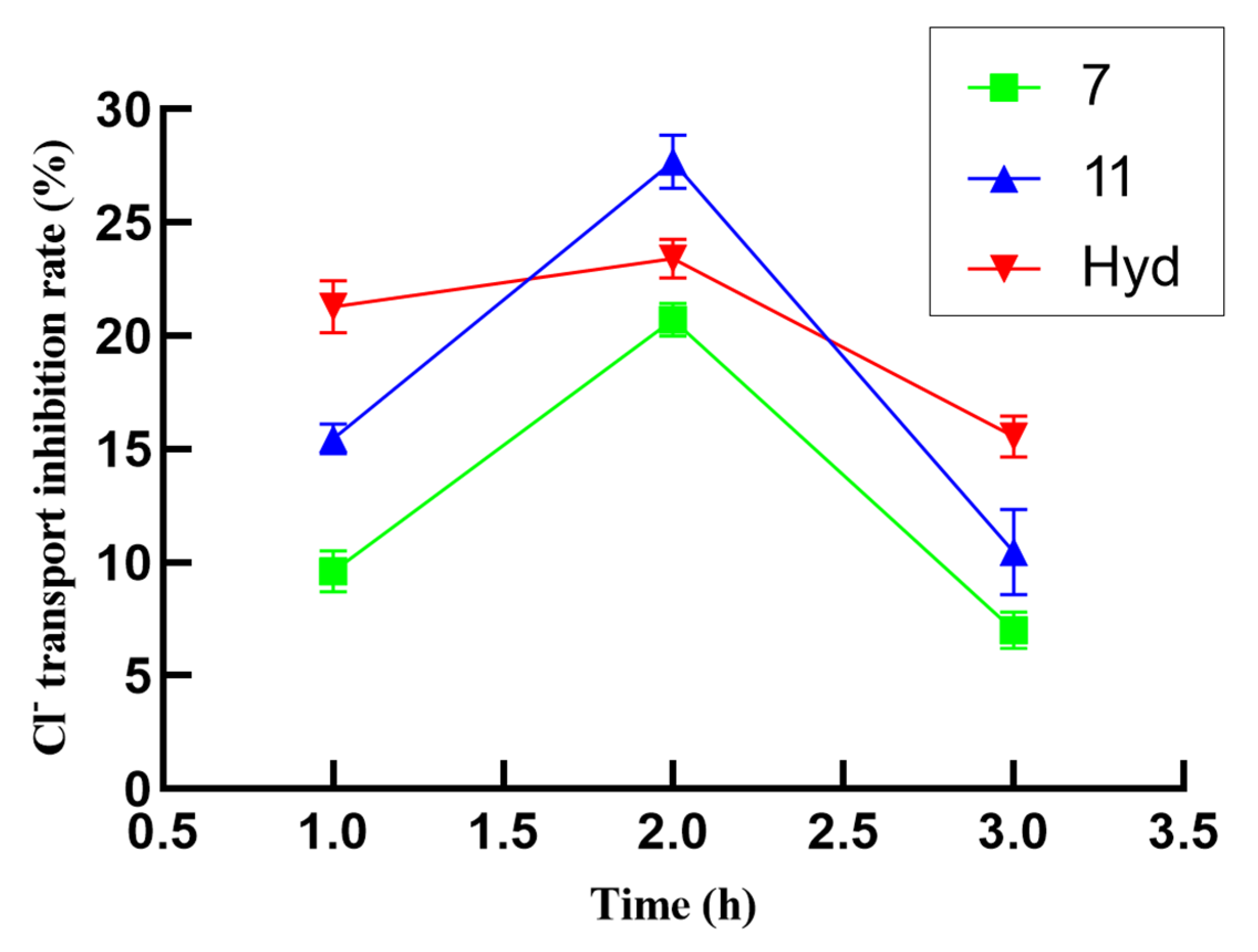
Generally, the transport of Na+ and Cl plays an essential role in glomerular filtration and tubular reabsorption. In this study, a Transwell chamber seeded with MDCK cells was used to simulate the renal tubules and investigate the inhibitory effect of compounds (1, 412) on NaCl transport in the renal tubules at 100 µmol/L. As shown in Table 6, all compounds, except compound 10, exhibited highly inhibitory activity on Na+ transport (p < 0.0001) compared to the blank group. As for the transport of Cl, compounds 1, 4, 67, and 911 exhibited extremely inhibitory activity (p < 0.0001), compound 5 exhibited good inhibitory activity (p < 0.001), and compound 12 has a general inhibitory activity (p < 0.5). The results showed that some compounds exhibited excellent inhibitory activity on Na+ transport, particularly 4, 7, and 11, with inhibition rates higher than 20%. On the other hand, compounds 7 and 11 exhibited a strong inhibitory effect on Cl transport (20.72% and 27.68%), which was significantly higher than or close to the positive control hydrochlorothiazide (23.42%).
In order to explore the relationship between the Na+ and Cl transport inhibition activity of potential compounds and time, the inhibitory activities of compounds with inhibition rates that were higher than 20% were further evaluated at 1, 2, and 3 h. It can be observed in the data shown in Figure 3 that the transport inhibition rates of these compounds reached a peak (greater than 20%) during the second hour. The inhibition rate of compound 7 on Na+ transport inhibition was more than 30.24% at 1 h, which was better than that of positive control hydrochlorothiazide (27.91%). The suppression rate of Na+ transport inhibitory activity in compound 11 exceeds that of hydrochlorothiazide at 3 h, and the suppression rate may be more stable. As shown in Figure 4, the inhibition rates of compounds 7 and 11 on Cl transport reached a peak (greater than 20%) during the second hour, and the inhibitory rate of compound 11 for Cl transport inhibition activity exceeded 27.68%, which exceeded the positive control (hydrochlorothiazide, 23.42%). Unfortunately, the stability of these compounds is relatively weak. In short, the preliminary test results demonstrated that compounds 4, 7, and 11 have potential for application in diuretic activity.

2.4. Molecular Docking

Based on the results obtained in previous in vitro experiments, compounds 4, 7, and 11, which have better inhibitory activity on Na+ and Cl transports, were selected for molecular docking studies to further explore the diuretic mechanisms of the selected compounds.
WNK1 kinase was employed to evaluate the diuretic effects of selected compounds by docking them into the active site of the WNK1 kinase domain (PDB ID 5DRB) [24]. WNK463 was re-docked to the active site to validate docking reliability. The results indicated the binding mode of co-crystallized and re-docked WNK463 was almost the same in the active site of the WNK1 kinase domain in Figure 5A (binding energy: −7.47 kcal/mol).
Compared with WNK463, compound 4 mainly formed two hydrogen bonds with Asp368 and Thr301, which were key for Na+ and Cl transport. In addition, the hydrophobic interaction with Phe356 was essential for the binding of compound 4 and the WNK1 kinase domain, with a binding energy of −6.12 kcal/mol (Figure 5B). In this study, compound 7 formed two hydrogen bonds with Asp368 and Met304, and hydrophobic interactions with Val235, Ala248, and Phe356, in Figure 5C (binding energy: −5.87 kcal/mol). Detailed interaction analyses of compound 11 revealed hydrogen bond interactions with Met304, Val281, and Cys250, and hydrophobic interactions with the Ala248, Thr301, and Val281 of WNK1, with a binding energy of −6.34 kcal/mol (Figure 5D).
It is commonly believed that the lower the binding energy, the stronger the binding force of the two molecules. In addition, if the binding energy is below −5 kcal/mol, the two molecules are considered to be strongly bound [25]. Molecular docking results showed that the selected compounds have a certain binding capacity. Thus, it can be observed that the diuresis potential of the active compounds can be realized by inhibiting the activity of the WNK1 kinase domain. Their binding driving forces comprise hydrophobic and hydrogen bond interactions.

3. Materials and Methods

3.1. Chemistry

The NMR spectra were obtained using a Bruker AV 300 MHz spectrometer (Burker, Fallanden, Switzerland) in CD3OD and TMS (tetramethylsilane) as the internal standard. High-resolution electrospray ionization mass spectra (HR-ESIMS) were recorded using a UHPLC–Q Exactive Orbitrap–MS (Thermo Fisher Scientific, Boston, MA, USA). Optical rotations were measured with a Rudolph Autopol I automatic polarimeter. Column chromatography was performed using normal-phase silica gel (200–300 mesh, Branch of Qingdao Haiyang Chemical Co., Ltd., Qingdao, China). Thin-layer chromatography (TLC) was conducted on pre-coated silica gel GF254 glass plates (200 × 200 mm, Qingdao Haiyang Chemical Co., Ltd., Qingdao, China) and RP-18 F254S (Merck KGaA, Darmstadt, Germany). All reagents and solvents were of reagent grade or purified according to standard methods before use.

3.2. Plant Material and Identification

The C. japonica was harvested from the Changbai Mountain area, Jilin Province, China, in July 2018, and it was identified by Prof. Ming-shan Zheng (School of Pharmaceutical Sciences, Yanbian University, China). A voucher specimen (20180705-XDC) was deposited at the Department of Pharmacognosy, School of Pharmaceutical Sciences, Yanbian University, China.

3.3. Extraction and Isolation

The air-dried whole plants of C. japonica (3.3 kg) were extracted with 75% ethanol (3 × 40 L) via reflux. The extract was freed from the solvent using a rotavapor to yield 642.7 g of EtOH extract. Part of the crude extract (627.2 g) was suspended with distilled water and successively partitioned into PE (3 × 1.4 L), EtOAc (3 × 1.4 L), and n-BuOH (3 × 1.4 L) sequentially to obtain PE, EtOAc, n-BuOH, and aqueous fractions.
The n-BuOH fraction (160.2 g) was subjected to silica gel column chromatography (CC) with CH2Cl2–MeOH–H2O (40:1:0–3:1:0.1, v/v/v) to afford eight fractions (Fr. 1–8). Fr. 2 (12.0 g) was subjected to silica gel CC using CH2Cl2–MeOH (30:1–0:100, v/v) as the mobile phase to give eight subfractions (Fr. 2-1–Fr. 2-8). Fr. 2-2 was separated successively using a Sephadex LH-20 column by eluting with CH2Cl2–MeOH (3:7, v/v) and a reverse-phase (RP) column by eluting with MeOH–H2O (30:1–1:0, v/v) to give compounds 7 (5.0 mg), 8 (7.0 mg), and 9 (7.0 mg). Fr. 2-5 was subjected to RP CC by eluting with MeOH–H2O (4:6–1:0, v/v) to yield compounds 10 (11.1 mg), 11 (17.5 mg), and 12 (8.2 mg), successively. Fr. 3 (13.0 g) was subjected to silica gel CC using CH2Cl2–MeOH (30:1–0:100, v/v) as the mobile phase to give six subfractions (Fr. 3-1–Fr. 3-6). Fr. 3-4 was subjected to RP CC by gradient elution with MeOH–H2O (4:6–1:0, v/v) to yield compounds 6 (118.1 mg). Fr. 5 (18.6 g) was fractionated using silica gel CC by gradient elution with CH2Cl2–MeOH (20:1–3:1, v/v) to give five subfractions (Fr. 5-1–Fr. 5-5). Fr. 5-3 was purified using a Sephadex LH-20 column by eluting with 100% MeOH to afford compound 1 (12.9 mg) and compound 2 (15.3 mg). Fr. 5-4 (3.9 g) was subjected to silica gel CC using CH2Cl2–MeOH (3:1-0:100, v/v) as the mobile phase to give seven subfractions (Fr. 5-4-1–Fr. 5-4-7). Fr. 5-4-3 was subjected to RP CC by gradient elution with MeOH–H2O (1:9–1:0, v/v) to yield compounds 3 (7.3 mg), 4, (16.1 mg) and 5 (6.1 mg).

3.4. Characterization of the Isolates

3′-O-β-d-glucopyranosyl-isohomoarbutin (1): colorless needles; [ α ] D 25 −18.05 (c 0.20, MeOH); UV (MeOH) λmax (nm; log ε): 287 (3.46); HR-ESI-MS m/z 447.1505 [M − H] (calcd. for C19H27O12, 447.1508). For 1H and 13C NMR data (CD3OD, 300 and 75 MHz), we refer the reader to Table 1. All significant data are provided in the electronic Supplementary Materials (Figures S1–S8).
4′-O-β-d-glucopyranosyl-isohomoarbutin (2): colorless needles, [ α ] D 25 −20.11 (c 0.22, MeOH); UV (MeOH) λmax (nm; log ε): 294 (3.46); HR-ESI-MS m/z 447.1507 [M − H] (calcd. for C19H27O12, 447.1508). For 1H and 13C NMR data (CD3OD, 300 and 75 MHz), we refer the reader to Table 1. All significant data are provided in the electronic Supplementary Materials (Figures S9–S16).
5-5′-dehydro-di(2-methyl-4-hydroxy-phenyl-1-O-β-d-glucopyranoside) (3): colorless needles, [ α ] D 25 −20.36 (c 0.25, MeOH); UV (MeOH) λmax (nm; log ε): 219 (3.35), 250 (3.11), 300 (3.10); HR-ESI-MS m/z 569.1875 [M − H] (calcd. for C26H33O14, 569.1875). For 1H and 13C NMR data (CD3OD, 300 and 75 MHz), we refer the reader to Table 2. All significant data are provided in the electronic Supplementary Materials (Figures S17–S23).
(1R,3R,4R)-3-methylcyclohexanol β-d-glucopyranoside (4): colorless needles, [ α ] D 25 −32.16 (c 0.10, MeOH); UV (MeOH) λmax (nm; log ε): 287 (3.10); HR-ESI-MS m/z 293.1596 [M + H]+ (calcd. for C13H25O7, 293.1595). For 1H and 13C NMR data (CD3OD, 300 and 75 MHz), we refer the reader to Table 3. All significant data are provided in the electronic Supplementary Materials (Figures S24–S33).

3.5. In Vitro Cytotoxicity Assays

Each compound was evaluated for cytotoxicity on DMCK cells using MTT assays. A 96-well plate containing 1 × 10⁴ cells per well was seeded with logarithmic growth-phase DMCK cells, which were then cultured for 24 h in a cell culture incubator. After that, the culture medium was changed to one that included medication (100 µmol/L for the treatment groups, 100 µmol/L for the control group, or 100 µmol/L for the positive control group). In total, 100 µL of the appropriate solution was added to each well, and they were then incubated for a further 24 h. After that, culture media were taken out and changed for a DMEM solution that included 20 µL of MTT (5 mg/mL). After incubating for 4 h, the liquid in each well was removed and replaced with 150 µL of DMSO, which was shaken for 10 min to ensure thorough mixing. The absorbance at 490 nm was measured to determine the optical density (OD) of each well. The cell growth inhibition rate was calculated as follows: cell growth inhibition rate (%) = (ODBlank − ODExperimental)/ODBlank × 100%.

3.6. In Vitro Diuretic Activity Assay

Log-phase MDCK cells were seeded in the upper chambers of Transwell plates (4 × 10⁴ cells/well), and 800 µL of complete culture medium was added to the lower chambers. After 24 h of incubation at 37 ℃, the electrical resistance of the upper chamber cells was measured one by one (R = (Rcell − Rblank) × 0.04π). When the electrical resistance of the upper chamber cells reached ≥ 300 Ω cm2, the upper chamber medium was removed and replaced with the drug solution. The blank group (given normal saline), experimental group (1, 412, 100 µmol/L), and hydrochlorothiazide group (200 µmol/L) were set, and each well was treated with 200 µL of the corresponding solution. The cells were then further incubated for 24 h. After that, the upper and lower chamber fluids were removed, and 200 µL of NaCl solution (21 mg/mL) was added to the upper chamber, while 800 µL of DMEM was added to the lower chamber for continued incubation. At 1, 2, and 3 h, 50 µL of the lower chamber fluid was taken, and the OD values were measured using Na+ and Cl detection kits. The transport inhibition rate (%) was calculated as follows: transport inhibition rate (%) = (ODblank − ODexperimental)/ODblank × 100%.

3.7. Molecular Docking Study

To explore the interaction between different compounds and the WNK1 kinase, the crystal structure of the WNK1 kinase domain in complex with WNK463 (PDB ID 5DRB) was selected to perform molecular docking studies [24]. Using the PubChem database (http://pubchem.ncbi.nlm.nih.gov/, accessed on 5 January 2024) for the 2D structure of small molecule ligands, the 2D structures were fed into Chem Office 2022 software to produce their 3D structures. Then, the RCSB PDB database (http://www.rcsb.org/, accessed on 5 January 2024) was used to screen the protein targets, and the crystal structure with high resolution was used as the molecular docking receptor. The PyMOLWin 2.6 software was used to dewater and dephosphate the protein. The Molecular Operating Environment 2019 software was used to minimize the energy of the compounds, pretreat the target proteins, and find the active pockets. Finally, MOE 2019 was run for molecular docking. The results were visualized using PyMOL and Discovery Studio 2019 software.

3.8. Statistical Analyses

Statistical analyses were performed by Graphpad Prism 6.0. Data were expressed as mean ± standard deviation (SD) based on at least three independent experiments. And differences between groups were analyzed by one-way analysis of variance and Student’s t-test. A p < 0.05 was considered to be statistically significant, p < 0.01 was considered as a significant difference, p < 0.0001 considered as an extremely significant differences.

4. Conclusions

In summary, to identify compounds with potent diuretic activity, the components of C. japonica were separated, resulting in the identification of 12 compounds (112), including three previously undescribed phenols (13) and one new cyclohexanol (4). Bioassays demonstrated that compounds 4, 7, and 11 possess potent diuretic activity. The docking study further revealed that the diuresis potential of the active compounds could be realized by the WNK1 kinase domain. Their binding driving forces comprise hydrophobic and hydrogen bond interactions. However, in order to improve the bioavailability of active compounds, further structural modification and mechanism analysis are necessary in the future.

Supplementary Materials

The following supporting information can be downloaded at: https://www.mdpi.com/article/10.3390/molecules29051092/s1, Figure S1 1H NMR spectrum of compound 1 (300 MHz, methanol-d4). Figure S2. 13C NMR spectrum of compound 1 (75 MHz, methanol-d4). Figure S3. 1H-13C HMQC spectrum of compound 1 (methanol-d4). Figure S4. 1H-13C HMBC spectrum of compound 1 (methanol-d4). Figure S5. 1H-1H COSY spectrum of compound 1 (methanol-d4). Figure S6. UV spectrum of compound 1. Figure S7. HRESIMS spectrum of compound 1. Figure S8. The HPLC spectrum of the compound 1 of D-glucopyranose. Figure S9. 1H NMR spectrum of compound 2 (300 MHz, methanol-d4). Figure S10. 13C NMR spectrum of compound 2 (75 MHz, methanol-d4). Figure S11. 1H-13C HMQC spectrum of compound 2 (methanol-d4). Figure S12. 1H-13C HMBC spectrum of compound 2 (methanol-d4). Figure S13. 1H-1H COSY spectrum of compound 2 (methanol-d4). Figure S14. UV spectrum of compound 2. Figure S15. HRESIMS spectrum of compound 2. Figure S16. The HPLC spectrum of the compound 2 of D-glucopyranose. Figure S17. 1H NMR spectrum of compound 3 (300 MHz, methanol-d4). Figure S18. 13C NMR spectrum of compound 3 (75 MHz, methanol-d4). Figure S19. 1H-13C HMQC spectrum of compound 3 (methanol-d4). Figure S20. 1H-13C HMBC spectrum of compound 3 (methanol-d4). Figure S21. UV spectrum of compound 3. Figure S22. HRESIMS spectrum of compound 3. Figure S23. The GC spectrum of the compound 3 of D-glucopyranose. Figure S24. 1H NMR spectrum of compound 4 (300 MHz, methanol-d4). Figure S25. 13C NMR spectrum of compound 4 (75 MHz, methanol-d4). Figure S26. DEPT spectrum of compound 4 in methanol-d4. Figure S27. 1H-1H COSY spectrum of compound 4 (methanol-d4). Figure S28. 1H-13C HMQC spectrum of compound 4 (methanol-d4). Figure S29. 1H-13C HMBC spectrum of compound 4 (methanol-d4). Figure S30. NOESY spectrum of compound 4 in methanol-d4. Figure S31. UV spectrum of compound 4. Figure S32. HRESIMS spectrum of compound 4. Figure S33. The HPLC spectrum of the compound 4 of D-glucopyranose. Figure S34. 1H NMR spectrum of compound 5 (300 MHz, methanol-d4). Figure S35. 13C NMR spectrum of compound 5 (75 MHz, methanol-d4). Figure S36. 1H-1H COSY spectrum of compound 5 (methanol-d4). Figure S37. 1H-13C HMQC spectrum of compound 5 (methanol-d4). Figure S38. 1H-13C HMBC spectrum of compound 5 (methanol-d4). Figure S39. UV spectrum of compound 5. Figure S40. 1H-NMR spectrum of compound 6 (300 MHz, methanol-d4). Figure S41. 13C-NMR spectrum of compound 6 (75 MHz, methanol-d4). Figure S42. 1H-NMR spectrum of compound 7 (300 MHz, methanol-d4). Figure S43. 13C-NMR spectrum of compound 7 (75 MHz, methanol-d4). Figure S44. 1H-NMR spectrum of compound 8 (300 MHz, methanol-d4). Figure S45. 13C-NMR spectrum of compound 8 (75 MHz, methanol-d4). Figure S46. 1H-NMR spectrum of compound 9 (300 MHz, methanol-d4). Figure S47. 13C-NMR spectrum of compound 9 (75 MHz, methanol-d4). Figure S48. 1H-NMR spectrum of compound 10 (300 MHz, methanol-d4). Figure S49. 13C-NMR spectrum of compound 10 (75 MHz, methanol-d4). Figure S50. 1H-NMR spectrum of compound 11 (300 MHz, methanol-d4). Figure S51. 13C-NMR spectrum of compound 11 (75 MHz, methanol-d4). Figure S52. 1H-NMR spectrum of compound 12 (300 MHz, methanol-d4). Figure S53. 13C-NMR spectrum of compound 12 (75 MHz, methanol-d4).

Author Contributions

Conceptualization, writing—original draft, Y.Y.; methodology, D.H., J.L., C.W., Y.S., X.H. and M.L.; supervision, M.Z.; resources, J.Z.M.; writing—review and editing, H.C., J.Z.M., D.K. and M.Z. All authors have read and agreed to the published version of the manuscript.

Funding

This work was supported by the National Natural Sciences Foundation of China (NNSFC, Grant Nos. 81660646), the National Natural Science Foundation of China (No. 82260002, No. 81460001), the Natural Science Research Foundation of Jilin Province for Sciences and Technology (No. 20220101353JC), and the Education Department Project of Jilin (No. JJKH20220546KJ).

Institutional Review Board Statement

Not applicable.

Informed Consent Statement

Not applicable.

Data Availability Statement

Data are included within the manuscript or the Supplementary Materials.

Conflicts of Interest

The authors declare no conflicts of interest.

References

  1. Ngamlai, E.V.; Pradhan, R.B.; Lalbiaknii, P.C.; Ralte, V.; Lalnunmawia, F.; Vanlalhluna, P.C.; Mehta, S.K. Diuretic activity evaluation and chemical composition analysis of Hedyotis scandens extract from Mizoram, India, in rat models. J. Ethnopharmacol. 2024, 319, 117079. [Google Scholar] [CrossRef]
  2. Li, X.X.; Liao, J.; Jiang, Z.L.; Liu, X.Y.; Chen, S.; He, X.; Zhu, L.; Duan, X.M.; Xu, Z.Y.; Qi, B.W.; et al. A concise review of recent advances in anti-heart failure targets and its small molecules inhibitors in recent years. Eur. J. Med. Chem. 2020, 186, 111852. [Google Scholar] [CrossRef]
  3. Kehrenberg, M.C.A.; Bachmann, H.S. Diuretics: A contemporary pharmacological classifcation? Naunyn-Schmiedeberg’s Arch. Pharmacol. 2022, 395, 619–627. [Google Scholar] [CrossRef] [PubMed]
  4. Tetiana, T.; Lina, P.; Iryna, D.; Yevgen, T. Modern trends in diuretics development. Eur. J. Med. Chem. 2020, 208, 112855. [Google Scholar]
  5. He, H.; Sui, Y.; Yu, X.; Luo, G.; Xue, J.; Yang, W.; Long, Y. Potential low toxic alternative for Na-Cl cotransporter inhibition: A diuretic effect and mechanism study of Pyrrosia petiolosa. Ann. Pharm. Fr. 2024, 82, 44–52. [Google Scholar] [CrossRef] [PubMed]
  6. Lang, T.Q.; Zhang, Y.; Chen, F.; Luo, G.Y.; Yang, W.D. Characterization of chemical components with diuretic potential from Pyrrosia petiolosa. J. Asian Nat. Prod. Res. 2021, 23, 764–771. [Google Scholar] [CrossRef] [PubMed]
  7. Saha, B.; Leite-Dellova, D.C.A.; Demko, J.; Soerensen, M.V.; Takagi, E.; Gleason, C.E.; Shabbir, W.; Pearce, D. WNK1 is chloride-stimulated scaffold that regulates mTORC2 activity and ion transport. J. Cell Sci. 2022, 135, jcs260313. [Google Scholar] [CrossRef] [PubMed]
  8. Rodan, A.R.; Jenny, A. WNK Kinases in Development and Disease. Curr. Top. Dev. Biol. 2017, 123, 1–47. [Google Scholar] [PubMed]
  9. Argaiz, E.R.; Chavez-Canales, M.; Ostrosky-Frid, M.; Rodriguez-Gama, A.; Vazquez, N.; Gonzalez-Rodriguez, X.; Garcia-Valdes, J.; Hadchouel, J.; Ellison, D.; Gamba, G. Kidney-specific WNK1 isoform (KS-WNK1) is a potent activator of WNK4 and NCC. Am. J. Physiol. 2018, 315, F734–F745. [Google Scholar] [CrossRef]
  10. Sui, Y.; Teng, M.G.; Chai, H.F.; Yu, X.; Long, Y.; Luo, G.Y.; Yang, W.D. Leaching effect of babaric acid in Pyrrosia petiolosa. Chin. Tradit. Herb. Drugs 2021, 52, 1026–1030. [Google Scholar]
  11. Yu, Y.; Liu, D.; Elshafei, A.; Lang, M.Y.; Hu, D.R.; Sun, Y.H.; Kang, D.Z.; Zheng, M.S. Chemical constituents from Chimaphila japonica Miq. and their chemotaxonomic significance. Biochem. Syst. Ecol. 2023, 109, 104664. [Google Scholar] [CrossRef]
  12. Yu, Y.; Elshafei, A.; Zheng, X.D.; Cheng, S.Y.; Wang, Y.X.; Piao, M.H.; Wang, Y.M.; Jin, M.; Li, G.; Zheng, M.S. Chemical constituents of Chimaphila japonica Miq. Biochem. Syst. Ecol. 2021, 95, 104219. [Google Scholar] [CrossRef]
  13. Yu, Y.; Cheng, S.Y.; Alaa, E.; Zheng, X.D.; Piao, M.H.; Zheng, M.S. Chemical constituents of Chimaphila japonica herb. J. Chin. Med. Mater. 2021, 44, 1124–1127. [Google Scholar]
  14. Ren, F.X.; Zhao, Y.M.; Zhang, A.J.; Yang, Y.; Zhang, Y. Extraction Method of Effective Components from Chinese Medicine Pyrola and Its Application for Treating Hemorrhage and/or Pain-Related Disease. CN102204937A, 5 October 2011. [Google Scholar]
  15. Raja, S.; Ravindranadh, K.; Keerthi, T. A complete profile on Chimphila umbellata-traditional uses, pharmacological activites and phytoconstituents. Int. J. Phytomed. 2014, 6, 464–470. [Google Scholar]
  16. Dang, Q.L.; Vu, H.D.; Nguyen, V.M.; Choi, G.J.; Hoa, L.T.P.; Dung, D.T.; Kiem, P.V.; Nhiem, N.X.; Tran, Q.D.; Nguyen, Q.C.; et al. Desmodinosides A-E: New flavonoid C-glycosides from Desmodium heterocarpon var. stigosum with hepatoprotective and antifungal activity. Fitoterapia 2023, 169, 10609. [Google Scholar] [CrossRef]
  17. Qin, Y.Q.; Liu, W.; Yin, R.; Xiao, P.T.; Wang, Z.Y.; Huang, T.Q.; Liu, E.H. New 4′,5′-methylenedioxyflavone derivatives from the whole plant of Sarcandra glabra. Nat. Prod. Res. 2022, 38, 177–185. [Google Scholar] [CrossRef]
  18. Zedet, A.; Kanga, Y.; Pudlo, M.; Senejoux, F.; Girard, C. Arginase inhibitory activities of chemical constituents from Macaranga hurifolia Beille leaves. Nat. Prod. Res. 2023, 1, 1–6. [Google Scholar] [CrossRef]
  19. Yu, Y.; Liu, G.J.; Piao, M.H.; Lang, M.Y.; Wang, Y.M.; Jin, M.; Li, G.; Zheng, M.S. Chemical constituents of Polygonum aviculare L. and their chemotaxonomic significance. Biochem. Syst. Ecol. 2022, 105, 104529. [Google Scholar] [CrossRef]
  20. Gao, Y.; Wang, B.; Yang, J.; Zhang, R.; Liu, N.N.; Wang, X.; Yu, C.P.; Rong, Z.J.; Zhang, H.L.; Long, Q.Z. Chemical investigation and anti-inflammatory activities of the aerial part of Filipendula palmata. J. Ethnopharmacol. 2022, 287, 114959. [Google Scholar] [CrossRef]
  21. Chang, J.; Inui, T. Novel phenolic glycoside dimer and trimer from the whole herb of Pyrola rotundifolia. Chem. Pharm. Bull. 2005, 53, 1051–1053. [Google Scholar] [CrossRef]
  22. Kasai, Y.; Taji, H.; Fujita, T.; Yamamoto, Y.; Akagi, M.; Sugio, A.; Kuwahara, S.; Watanabe, M.; Harada, N.; Ichikawa, A.; et al. MαNP acid, a powerful chiral molecular tool for preparation of enantiopure alcohols by resolution and determination of their absolute configurations by the 1H NMR anisotropy method. Chirality 2004, 16, 569–585. [Google Scholar] [CrossRef] [PubMed]
  23. Itano, K.; Yamasaki, K.; Kihara, C.; Tanaka, O. Stereospecific preparation of monoglucosides of optically active trans-1,2-cyclohexanediols by enzymic trans-D-glucosylation and carbon-13NMR spectroscopy of the resulting mono-D-glucopyranosides. Carbohyd. Res. 1980, 87, 27–34. [Google Scholar] [CrossRef]
  24. Yu, X.; Sui, Y.; Xi, Y.K.; Zhang, Y.; Luo, G.Y.; Long, Y.; Yang, W.D. Semisynthesis, biological evaluation and molecular docking studies of barbatic acid derivatives as novel diuretic candidates. Molecules 2023, 28, 4010. [Google Scholar] [CrossRef] [PubMed]
  25. Wang, Y.; Han, W.; Chen, X.; Lu, K. A study on the mechanism of aloperine on breast cancer based on network pharmacology and molecular docking. Clin. J. Chin. Med. 2023, 15, 28–34. [Google Scholar]
Figure 1. Structures of isolated compounds 112.
Molecules 29 01092 g001
Figure 2. Key 1H–1H COSY, NOESY, and HMBC correlations of compounds 15.
Molecules 29 01092 g002
Figure 3. Na+ transport inhibition activity of compounds 4, 7, and 11 at 1, 2, and 3 h.
Molecules 29 01092 g003
Figure 4. Cl transport inhibition activity of compounds 7 and 11 at 1, 2, and 3 h.
Molecules 29 01092 g004
Figure 5. Interaction modes of WNK463 (A), compounds 4 (B), 7 (C), and 11 (D) with WNK1 kinase domain (A).
Molecules 29 01092 g005
Table 1. 1H (300 MHz) and 13C (75 MHz) NMR data of compound 12 in CD3OD (δ in ppm).
Position12
δH, Mult (J in Hz)δCδH, Mult (J in Hz)δC
1-150.6-150.5
2-130.6-130.7
36.57 (d, 2.7)118.16.57 (d, 2.8)118.2
4-153.6-153.6
56.52 (dd, 8.6, 3.0)113.86.52 (dd, 8.6, 2.9)113.8
66.98 (d, 8.7)118.76.96 (d, 8.7)118.7
72.21 (s)16.62.21 (s)16.7
1′4.74 (d, 7.6)103.84.76 (d, 7.0)103.8
2′3.60 (m)74.43.22 (m)74.9
3′3.49 (m)88.03.62 (m)76.5
4′3.49 (m)69.93.64 (m)80.4
5′3.32 (m)77.83.64 (m)76.5
6′3.88 (d, 12.0)
3.71 (dd, 12.0, 5.2)
62.53.89 (dd, 11.2, 3.2)
3.73 (dd, 12.0, 5.2)
61.7
1″4.59 (d, 7.7)105.34.60 (d, 7.7)104.6
2″3.30 (1H, m)75.53.54 (m)74.8
3″3.34 (1H, m)78.23.35 (m)78.1
4″3.44 (1H, m)71.63.35 (m)71.4
5″3.34 (1H, m)77.73.35 (m)77.8
6″3.88 (d, 12.0)
3.71 (dd, 12.0, 5.2)
62.63.89 (dd, 11.2, 3.2)
3.73 (dd, 12.0, 5.2)
62.4
Table 2. This 1H (300 MHz) and 13C (75 MHz) NMR data of compound 3 in CD3OD (δ in ppm).
PositionδH, Mult (J in Hz)δCPositionδH, Mult (J in Hz)δC
1 (1′)-151.3Glc-1 (1′) 4.80 (d, 7.4)104.4
2 (2′)-130.4Glc-2 (2′)3.44 (m)75.1
3 (3′)6.70 (s)119.4Glc-3 (3′)3.39 (m)78.1
4 (4′)-149.8Glc-4 (4′)3.37 (m)71.6
5 (5′)-125.4Glc-5 (5′)3.39 (m)78.0
6 (6′)7.12 (s)120.9Glc-6 (6′)3.86 (d, 11.8)
3.66 (d, 10.5)
62.6
7 (7′)2.26 (s)16.4
Table 3. 1H (300 MHz) and 13C (75 MHz) NMR data of compound 4 in CD3OD (δ in ppm).
PositionδH, Mult (J in Hz)δCPositionδH, Mult (J in Hz)δC
13.78 (m)78.41′4.45 (d, 7.8)102.3
21.77 (d, 11.7)
1.51 (m)
36.52′3.60 (m)75.1
31.64 (m)36.53′3.39 (m)77.9
43.71 (m)69.84′3.37 (m)71.7
51.91 (d, 8.5)
1.61 (m)
26.35′3.39 (m)78.0
61.90 (m), 1.55 (m)32.36′3.86 (d, 11.8)
3.66 (d, 10.5)
62.8
71.03 (d, 6.7)18.7
Table 4. 1H (300 MHz) and 13C (75 MHz) NMR data of compound 5 in CD3OD (δ in ppm).
PositionδH, Mult (J in Hz)δCPositionδH, Mult (J in Hz)δC
1-150.63′-135.3
2-129.14′3.89 (s)69.0
3-128.35′1.80 (s)14.0
4-151.51″4.66 (d, 7.4)104.3
56.55 (d, 8.8)113.32″3.41 (m)75.1
66.87 (d, 8.8)116.23″3.30 (m)77.9
72.20 (s)12.64″3.38 (m)71.5
1′3.40 (m)26.25″3.40 (m)78.2
2′5.33 (dd, 6.8, 5.6)125.46″3.68 (dd, 12.0, 5.1)
3.89 (m)
62.6
Table 5. Cytotoxicity of compounds 112 on MDCK Cells at 100 µmol/L.
CompoundGrowth Inhibition Rate (%) aCompoundGrowth Inhibition Rate (%) a
14.89 ± 0.057/
229.48 ± 0.128/
323.31 ± 0.139/
44.89 ± 0.0510/
54.08 ± 0.0311/
6/ b12/
Hyd c/
a Values are the mean ± SD of three replicates. b No inhibition action. c Hyd represents hydrochlorothiazide.
Table 6. Na+ and Cl transport inhibition activity of compounds 1, 412 at 100 µmol/L.
CompoundTransport Inhibition Rate (%) aCompoundTransport Inhibition Rate (%) a
Na+ClNa+Cl
1−11.70 ± 1.14 e9.44 ± 0.78 e818.52 ± 1.02 e0.65 ± 1.45
421.13 ± 0.89 e12.23 ± 0.18 e96.22 ± 0.53 e−8.36 ± 2.60 e
57.61 ± 0.71 e4.57 ± 0.65 d101.82 ±0.81−13.16 ± 1.89 e
610.22 ± 1.24 e12.17 ± 1.33 e1128.81± 1.04 e27.68 ± 1.18 e
735.95 ± 1.42 e20.72 ± 0.74 e1214.18 ± 0.69 e3.27 ± 0.31 c
Hyd b39.74 ± 0.64 e23.42 ± 0.87 e
a Values are the mean ± SD of three replicates. b Hyd (hydrochlorothiazide) was used as the positive control. c * p < 0.05 vs. blank group. d ** p < 0.01 vs. blank group. e **** p < 0.0001 vs. blank group.
Disclaimer/Publisher’s Note: The statements, opinions and data contained in all publications are solely those of the individual author(s) and contributor(s) and not of MDPI and/or the editor(s). MDPI and/or the editor(s) disclaim responsibility for any injury to people or property resulting from any ideas, methods, instructions or products referred to in the content.