Review

Influence of Spider Silk Protein Structure on Mechanical and Biological Properties for Energetic Material Detection

1
Toxicology Research Center, Institute for Hygiene of Ordnance Industry, NO. 12 Zhangbadong Road, Yanta District, Xi’an 710065, China
2
Xi’an Key Laboratory of Toxicology and Biological Effects, NO. 12 Zhangbadong Road, Yanta District, Xi’an 710065, China
*
Author to whom correspondence should be addressed.
Molecules 2024, 29(5), 1025; https://doi.org/10.3390/molecules29051025
Submission received: 31 January 2024 / Revised: 21 February 2024 / Accepted: 22 February 2024 / Published: 27 February 2024
(This article belongs to the Section Molecular Structure)

Abstract

:
Spider silk protein, renowned for its excellent mechanical properties, biodegradability, chemical stability, and low immune and inflammatory response activation, consists of a core domain with a repeat sequence and non-repeating sequences at the N-terminal and C-terminal. In this review, we focus on the relationship between the silk structure and its mechanical properties, exploring the potential applications of spider silk materials in the detection of energetic materials.

1. Introduction

Natural spider silk fibers, having evolved over 400 million years, serve crucial functions in feeding, reproduction, and survival [1]. With a remarkable diversity of over 45,000 species, spiders exhibit the ability to produce task-specific silks with diverse mechanical properties originating from their unique silk glands [2,3]. Several types of spidroins, with different structures, determine their specific physical properties corresponding to their biological roles (Figure 1). Major ampullate silk (MaSp) (dragline silk) and minor ampullate silk (MiSp), known for their high strength, together with flagelliform silk (Flag), known for its high elasticity, contribute to the intricate production of a spider’s orb web, facilitating both its formation and effective capture of prey insects [4]. Aggregate silk (AgSp) (aggregate glue) provides spider silk with its renowned stickiness [5]. Tubuliform silk (TuSp) (cylindriform spidroin (CySp)) is the main constituent used by female spiders to manufacture egg cases [6]. Aciniform silk (AcSp) is applied for wrapping prey and protecting the egg sac from predators and environmental fluctuations, such as temperature and humidity changes [7]. Pyriform silk (PySp) is utilized in creating the attachment disc, enabling the web to adhere to different surfaces [8].
Exhibiting an intriguing blend of light weight, high strength, and exceptional elasticity, spider silk stands out as a stellar biomaterial. Its mechanical properties are on par with the best synthetic fibers manufactured by state-of-the-art technology. Its low toxicity and immunogenicity, coupled with its slow biodegradability and an apparent propensity for cell adhesion and growth, make it a compelling selection for biomedical applications [10,11]. In addition, silk threads also demonstrate interesting properties, featuring torsional memory, supercontraction, and self-healing [12,13,14].
Spider silk has been used by humans since ancient times. During the Middle Ages, and even as far back as the ancient Greek and Roman cultures, spider webs were employed for wound dressing [15]. The first pair of gloves and stockings made of spider silk were displayed at the Paris Academy of Sciences in 1710. Another pair of stockings was made by an American [16]. During the past years, dragline silk has been the most researched object of all the spider silks, while studies on minor ampullate silk, flagelliform silk, pyriform silk, aciniform silk, aggregate glue, and tubuliform silk have all been reported [3,9,10,11,12]. Nowadays, recombinant spider silk proteins have gained widespread employment in various fields, including biomedical, cosmetic, and technical applications.
However, owing to the territorial and cannibalistic nature of spiders and the challenge of generating substantial quantities of silk proteins at an economical cost, spider farming has been deemed impractical. Consequently, the best alternative is the biotechnological production of spider silk proteins. This process comprises two strategies: One involves the use of prokaryotic hosts, like E. coli, leveraging the well-established techniques of cell disruption and protein purification [17,18,19]. The disadvantage of prokaryotic expression systems is their inherent limitation in the size of synthetic genes. Another approach employs eukaryotic hosts, such as yeast, Bombyx mori, transgenic plants, bovine mammary epithelial alveolar cells, etc., which produce recombinant proteins most closely resembling natural silks [20,21,22,23,24,25,26,27,28]. Unlike the above-mentioned genetic engineering methods, chemical synthesis only replaces some regions of the silk protein with organic polymers [28]. However, the mechanical properties of the new fiber material are worse than those of natural silk.
In this review article, our focus is on how the silk structure determines its mechanical properties and its potential applications in detecting energetic materials. The repetitive domain is important for the mechanical properties of spider silk. Each repeat consists of crystalline stretches and amorphous areas, which collectively influence the strength and elasticity of the spider silk. The assembly of spidroins is controlled by non-repetitive domains, and some spider silk proteins feature a linker region bridging the terminal and repetitive regions of the silk protein. The structure and influence of these regions have yet to be discussed. Spider glues, microscopic glycoprotein nodules on capture silk that do not stick to each other, contribute to the stickiness of spider silk. Apart from the primary structure of spider silk, spinning techniques also have an important influence on the silk properties. Different forms of silk proteins have unique mechanical properties, rendering them suitable for various applications in different areas. Recombinant spider silks have found applications in diverse areas, particularly as exceptional candidates for biomedical applications, including tissue engineering, as well as in functionalized textiles, electronic engineering, cosmetics, etc. In response to the growing need for high-performance and eco-friendly materials in the sensor industry, it is essential to engineer green sensors capable of identifying explosive vapors at ambient temperature. Spider silk protein, which has shown promise in bioelectronic materials, could function as an eco-compatible sensory material for detecting the vapors of standard nitroaromatic explosives.

2. Relationship between Structural and Mechanical Properties of Spider Silk Proteins and Their Structural Benefits in Biosensor Applications

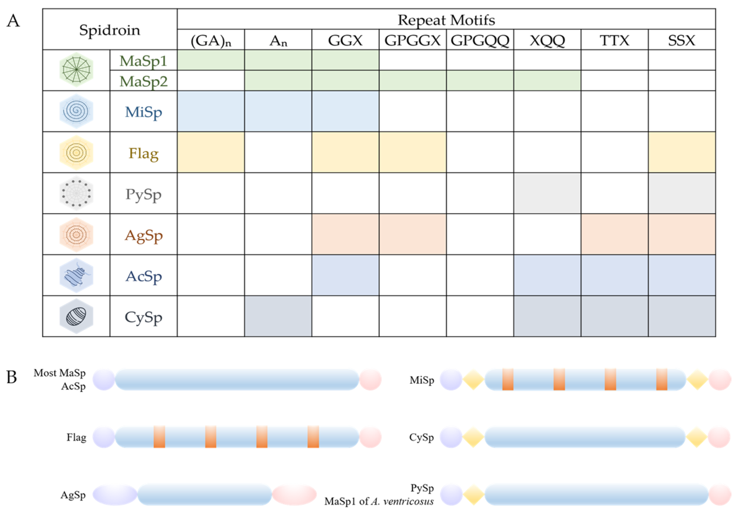
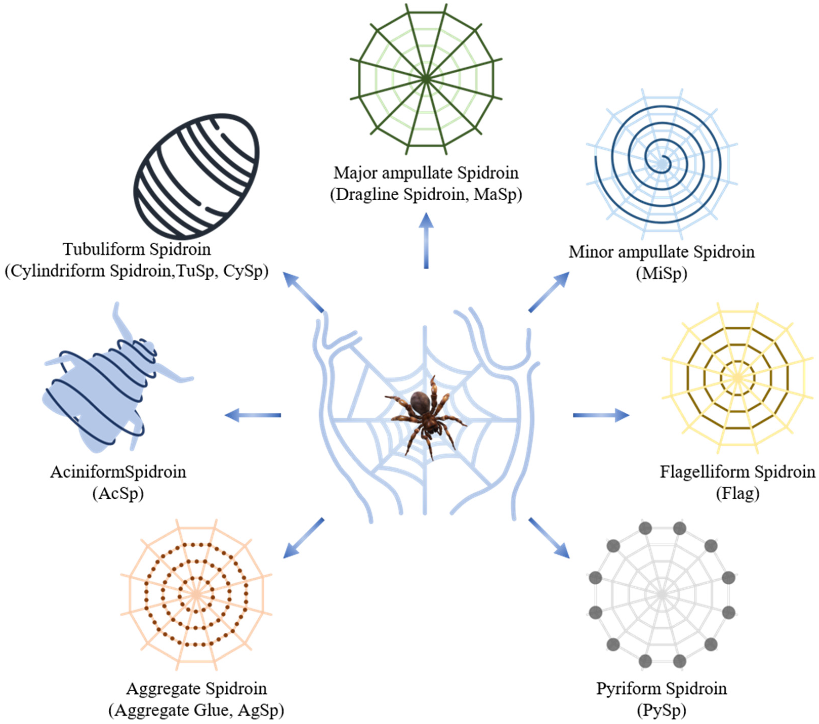
2.1. Structure of Spider Silk Proteins

Spider silk proteins exhibit a broad molecular-weight range, spanning from 70 to 700 kDa. They have a specific amino acid composition, which significantly deviates from that of structural globular proteins (Figure 2, Table 1). Structurally, spider silk proteins involve three distinct domains: a repetitive central core domain, organized with highly repetitive crystalline regions periodically interrupted by less repetitive crystalline peptides, and the conserved amino-terminal (N-terminal) and carboxy-terminal (C-terminal) domains (Figure 2B) [29,30,31]. All native spider silk proteins exhibit remarkably high levels of alanine, glycine, serine, and glutamine, while the contents of charged acidic and basic amino acids are relatively low. The repetitive regions of spider silk proteins contain sequence motifs, such as polyalanine stretches and glycine-rich areas [31], leading to different secondary structures that vary based on the spider species, the silk type, and the solid state of the silk fibers (Figure 2A, Table 1) [32,33].
Most silk proteins, including major ampullate, minor ampullate, and flagelliform silks, consist of short repeated sequence motifs in their repetitive regions. In contrast, aciniform and tubuliform proteins possess long and complex repeats without obvious simple internal motifs. The core domain of pyriform silk is composed of six repeated α-helix globular domains, each flanked by two disordered regions [34]. Regions of intrinsic disorder link the helical bundles, creating a beads-on-a-string architecture [34]. While the core domains differ, the N-terminal and C-terminal domains remain highly conserved across the silk types (Figure 2B). Furthermore, the semi-crystalline phase, along with the crystalline and amorphous phases, play a crucial role in the mechanical properties of spider silk fibers [35]. However, limited insights have been gained into the functions of each domain in PySps protein forms that utilize protein as cement in their attachment disks.
Figure 2. Structural motifs and correlated secondary structures of spidroins [36]. (A) The variety of motifs in the repetitive domains of different spidroins directly reflect their mechanical properties, allowing them to perform different tasks. A: alanine, G: glycine, P: proline, Q: glutamine, S: serine, T: threonine; and X: one of tyrosine (Y); leucine (L); alanine, serine, and arginine (R); valine (V); or glutamine (Q). (B) Spider silk proteins (spidroins) are mainly composed of a repetitive domain (blue) flanked by the non-repetitive and highly conserved N-terminal (purple) and C-terminal (pink). Some specific types of spidroins have a linker region (yellow) and spacers (orange) in addition.
Molecules 29 01025 g002
Table 1. Molecular weight, mainly containing amino acids and motifs, of each type of spider silk protein.
Spider SilkMolecular WeightMain
Amino Acids
Main
Motifs of Repeat Domain
References
MaSp>250Alanine
Glycine
polyA or An
polyGA or (GA)n
GGX
GPGXX
[37,38]
MiSp>250Alanine
Glycine
polyA
polyGA
GGX
Spacer
N-linker
[37,39,40]
Flag>250Alanine
Glycine
polyGA
GGX
GPGXX
Spacer
[37]
PySp200–400Proline
Glutamine
polyPX or (PX)n
QQ-containing regions
N-linker
[8,37,41]
AgSp450–1400GlutamineDistinct repeat motifs
Q-rich regions
[37]
AcSp300–500/Long and complex motifs[37]
CySp180Alanine
Glutamine
Serine
polyA
QQ-containing regions
S-rich motifs
Linkers
[37,42]
Spider silk fibers produced in the silk gland exhibit a combination of spidroins with different structures. For instance, a silk fiber produced by the major ampullate gland is composed of two main proteins: the highly ordered hydrophobic major ampullate spidroin 1 (MaSp1), and the less-ordered hydrophilic major ampullate spidroin 2 (MaSp2) [43]. Other major ampullate spidroins are also found in specific spider species, which play important roles in their extraordinary mechanical properties. Major ampullate spidroin 3 (MaSp3), recently identified in the Araneus genus, is hypothesized to contribute to silk formation during the spinning process, although the precise relationship between its protein sequence and properties remains unclear [36]. Moreover, the discovery of major ampullate spidroins 4 (MaSp4) and 5 (MaSp5) in Darwin’s bark spider (Caerostris darwini) potentially explains the Darwini spidroin’s extraordinary toughness [44]. This suggests that several types of spidroins can be produced by a single spider species. The spidroin number in the velvet spider (Stegodyphus mimosarum) is 19, and in Nephila clavipes, the number is 28 [41].

2.2. Relationship between Different Functional Domains of Spider Silk Proteins and Their Mechanical Properties

Spider silk, recognized for its exceptional mechanical properties, which surpass those of high-performance manufactured materials like Kevlar and carbon fibers, also exhibits remarkable optical characteristics, such as nearly perfect transparency in the visible spectrum [45]. The structure of the silk fiber determines its mechanical properties. Through the exploration of recombinant protein block copolymers, we have obtained a crucial understanding that resolves essential problems in polymer science, including the thermophysical traits, self-assembly processes, and sequence–structure relationships of silk protein polymers. These insights have further inspired novel applications in biomedical engineering, such as gene transfer therapy and drug delivery [46,47,48].

2.2.1. Repetitive Domains Mostly Influence Mechanical Properties of Spidroins

The crystalline domains play a dominant role in biomechanical studies of spider silk, with a crystal volume fraction ranging from 30% to 40%, depending on the spider silk species and measurement technique [49]. These domains are composed of polyA and polyGA repeat motifs, forming β-sheet crystalline domains with dimensions typically ranging between 1 and 10 nm during fiber formation, laying the groundwork for the tensile strength and stiffness of the silk fiber [31,50,51,52,53]. The nucleation and fibril growth are affected by the formation of β-sheets, with each β-sheet crystalline structure containing 6–12 alanine residues. Stable β-sheet nanocrystals result from the antiparallel alignment and stacking of polyalanine domains along the direction of hydrogen bonding. The mechanical properties of spider silk may experience improvement by increasing the number of alanine residues. However, beyond six alanine residues, the improvement in the mechanical properties becomes less significant [54]. The significantly enhanced mechanical properties of β-sheet nanocrystals are attributed to the utilization of hydrogen bonds and the emergence of dissipative molecular stick–slip deformation, given that hydrogen bonds are among the weakest chemical bonds [55]. Furthermore, hydrogen bonds serve as an optimal choice for establishing stable and robust bonds between the nanocrystalline and amorphous domains in silk [35]. Unlike MaSp1 and MaSp2, both MaSp3 and MaSp4 lack typical polyA motifs, which are replaced by serine-rich motifs [56].
Glycine-rich (G-rich) areas are mainly generated by (GGX)n, (GPGXX)n (X = Y, L, A, S, R, V, or Q, 43 ≤ n ≤ 63) and are thus responsible for elasticity and flexibility [31,51,57,58,59,60,61]. Special G-rich motifs, such as GPX, GGGX, GGX-GGX-GX, (GX)noligoA-(GX)n, GGX-GGX-GGGX, and (GX)n, are also found in minor ampullate silk, flagelliform silk, and aciniform silk [37]. Interestingly, GPGXX motifs are not iterated within each G-rich repeat, as is the case of MaSp2 [38]. The amorphous structural analysis reveals that the regions consist of different contents of α-helix, 31-helix, β-sheets, and β-turns, with fewer dense hydrogen bonds [33,62]. The outstanding elasticity of silk fiber is attributed to the β-turns generated by (GPGXX)n repeat motifs, positioned adjacent to (GGX)n motifs and spacer motifs that form helices and β-sheets [63,64]. These weakly oriented and unaggregated β-sheets in amorphous domains are the primary contributors to the strain stiffening observed in spider silks [65]. Nanoscale mechanical deformation simulations trace the initial suppleness of the spider silk matrix back to the rupture of hydrogen bonds within domains rich in glycine [62]. Recent research indicates an abundance of post-translational modifications, predominantly phosphorylation and deamidation, that occur in GGX motifs. These alterations are projected to influence the conformation of the spidroins and, by extension, the physical properties of the resultant protein [66]. Due to the difficulty of detecting the amorphous phase in diffraction experiments, investigations into the molecular mechanics of this phase are limited [67]. In addition to the investigation into proline concerning its distinct impacts on peptide backbone structures and its contribution to supercontraction, recent research has established a potential association between tyrosine residues and supercontraction. The repetitive region of MaSp4 lacks GGX motifs and instead features unique GPGPQ motifs, increasing the flexibility of the spidroins by incorporating more β-turn conformation content within the fibers [56].
Several non-repetitive “spacer” regions are present within minor ampullate silk and flagelliform silk (Figure 3) [39,40]. The consensus repeat unit of the spacer consists of several GGX and GA/GGX/GPGGX domains, and these repetitive units alternate with the spacer regions. Sequences of spacers display near-identical patterns across different silk proteins [39]. Interestingly, the research on the spacer’s functions shows that the motif G’F’Y’ outperforms the motifs G’ (G’ is short for the motif (GGX)n), F’ (F’ is short for the spacer), and Y’ (Y’ is short for the motif (GPGGX)n) and their pairwise combinations in terms of toughness, strength, and moderate extensibility, while G’Y’ and G’F’ only present limited extensibility and decreased strength [64]. This result implies that the silk fiber’s strength is enhanced by the spacer motif, thanks to optimal molecular organization, β-sheet formation, and varied sequence complexity [64]. The unique property of the spacer allows the protein to safely revert to its original state post-elongation, without detriment to the silk’s mechanical or physical properties [60]. Furthermore, the spacer region might contribute to the β-sheet formation in silk fibers [60]. However, the exact structural nature of the spacer motif remains elusive, and additional research is required to pinpoint how the spacer motif influences the mechanical attributes of silk fibers.
In addition to the three regions mentioned above, there are some other specific regions in specific spidroins. The pyriform spidroin’s core repetitive region contains two distinct motifs: proline-rich motifs (PX)n and QQ-containing motifs [8]. The (PX)n motifs are characterized by their tendency to form random coils, in contrast to the QQ-containing regions, which are often shaped into α-helix or β-sheet structures [37]. Glutamine segments, also found in tubuliform silk, are thought to be involved in spidroin self-assembly [37]. X residues play a role in influencing the structure and function of dragline silk [37]. During periods of high metabolic costs for spiders, particularly when they need to produce a substantial amount of material for constructing the egg sac, there is an argument that the presence of serine-rich (S-rich) motifs in tubuliform spidroins serves to safeguard against the depletion of the serine content [42]. However, it is worth noting that S-rich motifs are linked to a slight decrease in the mechanical performance of tubuliform silk [42].
Silk comprises not only the well-recognized crystalline and amorphous phases but also a third phase referred to as the semi-crystalline phase. This phase can contain isolated β-sheets or other structures indicated by NMR, yet it appears disordered when analyzed via larger-scale X-ray diffraction [58,68]. It has been suggested that the semi-crystalline phase contributes significantly to the outstanding toughness observed in natural spider silk due to its superior energy absorption abilities [69]. The semi-crystalline phase develops at crystalline–amorphous interfaces, with the semi-crystal quality relying on the β-sheet nanocrystal length [35]. Yet, a comprehensive understanding of the features, stability, and composition of these semi-crystalline domains is still lacking.
The successful formation of spider silk-like copolymers relies not only on the β-sheet content but also significantly on the block repeats in the structure. An increase in the number of these repeats induces a transformation in the proteins from disordered structures into lamellar ones. The synthetic dragline-like proteins, produced through an E. coli expression system, demonstrated that reducing the number of repeats culminates in significantly shorter fibers. So far, construction with eight repetitions appears to be the minimum requirement for fiber polymerization, leading to flexible fibers that form free coils inside cells, featuring a constant diameter and chemical resistance [70]. Reducing this number to four leads to a loss of fiber flexibility and the emergence of unique “bird beak” structures, not observed in fibers with eight repeats [70]. The crystallization of the protein becomes easier as the number of hydrophobic block repeats increases [71]. This increase also boosts both the nucleation and growth rates, supporting the nucleation-growth mechanism typically seen in cross-β-fibril formation [72]. The assembly kinetics of spider silk fibers vary depending on the number of repeats [72].
Apart from their impact on the silk mechanical properties, repetitive sequences during silk self-assembly may also be designed for solubility and/or phase transitions; however, further studies have not been promoted [73].

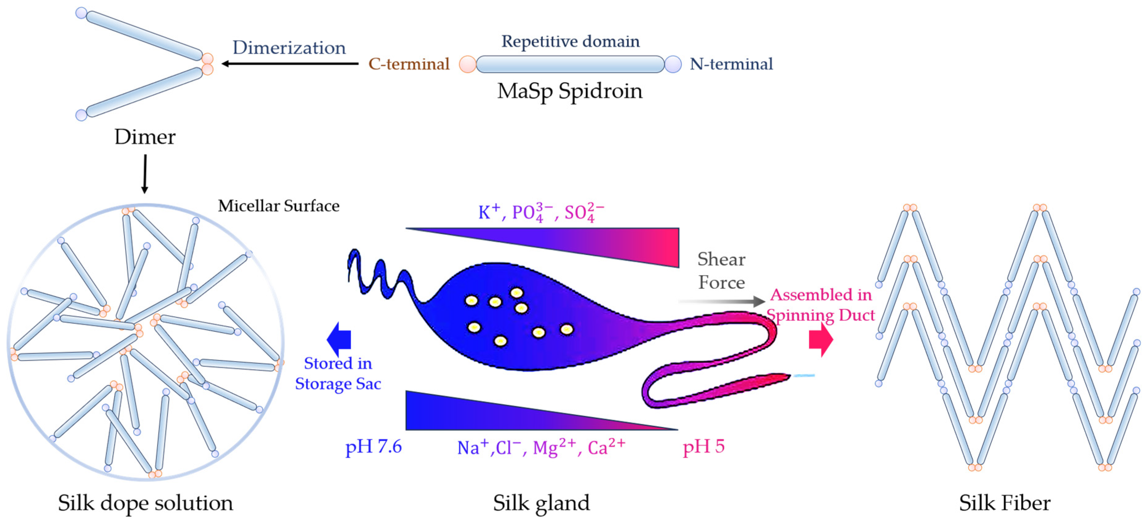
2.2.2. Non-Repetitive Terminal Domain Controls Spidroin Assembly

Spider silk proteins’ terminal domains, in comparison to the repetitive domain with varying extended sequences among spider species, are small-sized (~110–130 residues) and exhibit high conservation across all types of spiders [74,75]. Both domains of MaSp show a homodimer of dipolar, antiparallel, five-helix bundle subunits, consisting of α-helical and loop elements [33,76]. They serve a critical function in regulating chain assembly, governed by changes in the ionic composition, mechanical stimuli, and pH during transit within the glands [18,33]. Moreover, the terminal domains are instrumental in preserving the solubility of spidroins at elevated concentrations (30–50% w/v) in the silk gland [9]. The pH-dependent dimerization is conserved between MaSp, MiSp, and Flag, although the amino acid residues mediating this process may differ [77]. CySp and AcSp have a different pH dependency mechanism than other silk proteins. CySp’s unique N-terminal amino acid sequence prevents full stabilization conformation at neutral pH and in low-salt conditions, while AcSp exists as a mixture of dimers, trimers, and larger oligomers under these conditions, unlike MaSp, which forms stable dimers [77,78]. However, further research on the terminals of CySp and AcSp is necessary, and the assembly mechanisms of AgSp and PySp assembly remain unclear. Thus, this review mainly focuses on the extensively discussed structure and function of the MaSp terminal domains in previous studies. While stored in the spider’s gland, silk proteins form micelle-like structures, with the terminal domains on the micelle surface preventing the non-specific aggregation of disordered, long, repetitive regions in solution and premature fiber formation [79,80]. The dimerization of proteins enhances the spinning-dope properties and fiber formation [81]. Notably, the presence of the C-terminal slightly destabilizes the structure of the N-terminal [82]. During silk assembling, there seems to be no interaction between the N- and C-terminal domains, simplifying the control of the spidroin assembly and minimizing unproductive side reactions (Figure 4) [82].
The C-terminal domain, instrumental in controlling the solubility, is of paramount importance in regulating the transition between the storage and assembly forms of silk proteins [79,83]. It also significantly influences the alignment of secondary structural features formed by the repetitive elements in the backbone of spider silk proteins, an essential critical factor in determining the mechanical properties of the resulting fiber [83]. Characterized by its dual thermo-sensitive behavior, the C-terminal domain predominantly undergoes a “gel-sol-gel” transition in response to varying temperatures [84]. Substituting the native C-terminal domain with non-native C-terminal domains from MaSp1 and MaSp2 results in fibers with reduced stiffness and strength, underscoring the significance of the native C-terminal domain [3]. In the examined silk types and species, an analysis of 19 families within the Araneae order demonstrates the consistent preservation of a pH-sensitive salt bridge, typically consisting of an arginine–glutamic acid pairing [85]. This notable conservation strongly supports the essential involvement of this characteristic in the accurate assembly of silk fibers.
Under conditions in the gland reservoir of a pH of 7 and 300 mM of NaCl, the N-terminal exists as stable monomers. However, in conditions resembling the spinning duct characterized by a lower pH (pH 5.5) and reduced NaCl concentrations, the N-terminal domain undergoes dimerization in an antiparallel orientation [86]. Its role includes enhancing the solubility of the spidroins during storage and facilitating self-assembly as the pH decreases along the spider’s silk extrusion duct, preventing premature aggregation [76]. The functionality of the N-terminal depends on the interchange between its monomeric and dimeric forms [87]. The dimerization of the N-terminal domain is pH-responsive [88]. With a drop in pH from 7 to 5, the N-terminal domain transforms its conformation into an antiparallel homodimer, a molecular detail that remains to be fully elucidated [89,90,91]. Acidification induces an instability in the C-terminal structural domain, prompting it to unravel into β-sheet formations. Remarkably, N-terminal dimers can transition into silk fibers at an exceedingly high pace (>1 m/s), all while being confined to the most distant part of the spinning ducts [90,92].

2.2.3. Linker Regions Influence Silk Fiber Properties and Assembly

Some spider silk proteins, such as MaSp1 and MaSp3 of Araneus ventricosus, pyriform spidroins, and minor spidroins, possess uniquely long and repetitive “linker” regions that bridge the terminal and repetitive regions of the silk protein (Figure 5) [39,93,94]. The linker sections vary in their amino acid counts, ranging from 83 to 174, and are characterized by a significant abundance of serine [39,94]. These “linkers” typically tend to be short in most spidroins, with pyriform spidroins being a notable exception, serving as transitional structures from the terminal regions to the medially repetitive region [41]. Each domain, including the terminal, repetitive, and linker domains, is characterized by a distinct stability and solubility, playing specific roles in ensuring the stable storage of silk protein fragments [94]. Due to their propensity for oligomerization, the linker regions and repetitive region in minor ampullate spidroins can initiate oligomerization through weak hydrophobic interactions, subsequently forming the core region of the oligomers [94]. Serving as a nucleation site, the linker domain facilitates the assembly of diverse molecules, while the terminal domains ensure their highly ordered arrangement under shear force [94].
Pyriform silk is distinguished by its long N-linkers and short repetitive region [95]. In each of these repeats, there is a more hydrophilic, glutamine-rich segment that contrasts with the rest of the repeat sequence [93]. Insights into the MaSp1 spidroin’s alternative splicing phenomena highlight the crucial role of the N-linker region in facilitating the self-assembly, stability, and mechanical properties [96]. The research also introduces an approach to devise recombinant spider silk proteins with superior mechanical properties. On the contrary, lacking a unique structure, the C-linker in pyriform silk marginally surpasses others in length [97].
Proteins with glutamine-rich regions alone have shown the ability to self-assemble into fibers, and their flexibility is enhanced with the addition of a proline-rich region [93]. It is hypothesized that the N-linker repeat region alone can independently form fibers, and incorporating flanking N-linker sequences might influence the self-assembly rate or mechanical attributes of the fibers [93]. However, the structure and influence of the linker region still need to be discussed. A deeper understanding of these mechanisms necessitates in-depth studies on the structures of the assembled forms [93,94].

2.2.4. Spider Silk Glues and Viscidity of Silk Fiber

Spiders spin their capture threads in a distinctive manner, forming a central flagelliform fiber interspersed regularly with glue droplets. This configuration enables the spider to move freely within its web, ensuring that any prey making contact with the web encounters numerous nodules simultaneously, leading to effective entrapment [98]. The water-soluble silk glue, primarily produced by the aggregate gland, and considered one of the most effective biological glues, manifests as microscopic glycoprotein nodules that avoid mutual adhesion [99,100,101]. The extensive, rough endoplasmic reticulum forms small vacuoles, which are then synthesized into secretory vesicles, travel through the Golgi complex, and release aqueous glue through the mechanism of apocrine secretion into the lumen [101]. Unlike fibrous silk production, the Golgi complex plays an important role in glue production, involving either a condensing or packaging step [101]. The glycoprotein-rich glue tightly surrounds the twin internal axial fibers produced by the flagelliform gland.
In contrast to other spider silk proteins, silk glue is a viscoelastic, amorphous, wet material that responds to environmental conditions [102]. It is noteworthy that the glue droplets of cobweb weavers demonstrate greater firmness and resilience compared to those of most of their orb web-weaving counterparts [103]. The aqueous nature of the glue enables it to absorb moisture from the surrounding humid environment, thereby reducing the surface tension of spider silks and enhancing their distinctive elasticity [104].
The coding sequences of proteins in aqueous silk glue represent one of the largest groups of silk protein genes documented to date [102]. Predominantly, the sequence is composed of two distinct repeat motifs, measuring 387 bp and 339 bp, interspersed by glutamine-rich regions [102]. In contrast to other spider silk proteins, the glue proteins exhibit pentameric QPGSG repeats, bearing a striking resemblance to preserved modular elements in mammalian elastin, an elastomeric protein interacting with collagen [105]. The precise locations of galactose, mannose, glucosamine, caramel, glucose, N-acetylgalactosamine, γ-Aminobutyramide, N-acetyltaurine, choline, betaine, and ethionic acid are also identified within these nodules [99,106].
The glue droplets consist of three regions, incorporating these glycoproteins, low-molecular-mass compounds (LMMCs), and organic salts [107]. These droplets comprise a central opaque anchoring granule, a surrounding middle region of larger transparent glycoprotein, and an outermost fluid and viscous coating. The adhesion of a single glue droplet relies on a delicate balance between two factors: the interfacial adhesion and bulk dissipation. Proteins present in aggregate glues are characterized by higher levels of glycosylation and phosphorylation compared to proteins in other silk fibers. These modifications likely contribute to the stickiness of aggregate glue and may also enhance its cohesion. The glycoprotein core’s adhesion is influenced by the presence of low-molecular-mass compounds (LMMCs) in the surrounding aqueous layer. These LMMCs solvate the adhesive glycoprotein, imparting hygroscopic properties, softening the glue, and enhancing its adhesion. Additionally, salts play a direct role in facilitating adhesion by solvating the glycoproteins and increasing their mobility. However, the specific proteins comprising the cores of aggregate glue droplets remain unknown at present. The precise mechanism by which spiders assemble glue proteins into fibrous-like materials is a subject of considerable debate [105].
Glue drops function as a viscoelastic material that enhances adhesion through capillary forces [108]. The viscosity of the glue is believed to respond to humidity, aiding spiders in adapting to their local environments [109]. While high humidity typically inhibits adhesion, spider glues become stickier with increasing humidity levels [110]. Atmospheric water uptake acts as a plasticizer, enhancing glycoprotein plasticity rather than adhesion. Consequently, the viscosity decreases as the humidity increases [111,112]. Despite significant variation in the glue viscosity across different species, spanning five orders of magnitude in response to humidity, the viscosity at maximum adhesion remains remarkably consistent for each species [109]. The distinct humidity response exhibited by the glue components, distinct from the pristine glue, reinforces the hypothesis that the synergistic association among the spider glue constituents plays a crucial role in its adhesive functionality [113]. In addition to humidity, factors such as the strain rate, temperature, ultraviolet radiation B (UVB) radiation, and spider’s protein intake level also influence the adhesive properties of viscid spider silks [104].

2.3. Benefits of Spider Silk Structure When Silk Is Utilized in Biosensors

The interest in flexible sensing materials has increased in recent decades due to their technological potential. These materials provide stretchability, compressibility, twistability, bendability, and, in general, deformability to sensors [114]. Spider silk protein, especially the dragline silk protein, is one of the most attractive flexible materials that is applied in sensors, due to its excellent biological and mechanical abilities, light waveguide ability, and humidity sensitivity [115]. Their unique structures, which cause specific conformations and excellent properties, are the most important influencing factors for their utilizations in biosensors.
In addition to the outstanding mechanical and biological properties that we mentioned above, the structure of spider silk threads with round cross sections and, thus, good light-guiding properties, make them a candidate for use as optical fiber sensors [116,117]. In addition, spidroins are ideal humidity sensors because of their “supercontraction” properties when wetted or saturated in a high-relative-humidity (RH) atmosphere [118]. When the spidroins are laid in wet conditions, H2O molecules infiltrate the silk fiber and disrupt the hydrogen bonding of the amorphous region, resulting in the rearrangement of the configurations [119]. These rearrangements of the protein structures change the silk modulus and cause the reversible supercontraction phenomenon. Moreover, spider silk proteins act as natural pH-sensitive materials that can be integrated to develop pH sensors without further processing [120]. When in an acid or alkaline solution, the N- and C-terminals of the silk proteins react with hydrogen ions and hydroxide ions, and the hydrogen bonds are modified by the acid or base, resulting in the reversible denaturing of the spidroins [121]. These changes in the SDS protein structure lead to the change in the refractive index (RI), with the wavelength red-shifting when the pH increases and blue-shifting when the pH decreases [120].
Metal nanostructures as well as quantum dots and dyes can be easily attached to the surfaces of spider silk fibers without complicated surface treatment [122]. Reversible hydrogen bonds, holding the repetitive crystalline and amorphous regions together in the fiber, can be modified by different molecules, making the silk ideal for detecting modifying agents such as water molecules, acids, bases, etc., allowing for potential applications in silk-based biosensing [123]. Because even a 10 mm spider silk thread is composed of quantities of spidroin proteins, it is theoretically possible to detect a multitude of compounds with only a single silk thread with the benefits of high sensitivity and a quick response time.
Though the properties of spider silks utilized in biosensors should be further analyzed from the perspective of their molecular compositions and structures, the application of spider silk materials in the biosensing field has broad prospects. Moreover, with the continuous deepening of spider silk biosensing research, the mysterious veil over the structure and properties of spider silk protein is gradually being lifted.

3. Influence of Production and Spinning of Spider Silk Proteins on Their Properties and Their Influences in Spidroin Biosensing Research

3.1. Properties of Recombinant Spider Silk Do Not Compare with Those of Native Silk Fibers

The properties of synthetic spider silk, derived from recombinant processes, do not match those of native silk fibers. The challenges associated with commercial spider farming, such as the aggressive and cannibalistic nature of spiders and the difficulty in producing sufficient silk proteins cost-effectively, make it an unviable option [124]. Therefore, biotechnological production offers the best solution for generating silk proteins. However, the properties of recombinant spider silk proteins produced by various production strategies, especially their mechanical properties and water solubility, show significant deterioration compared to those of native silk.
Extensive exploration of recombinant spider silk production has focused on bacteria and other prokaryotes as potential host systems for producing spider silk proteins. These microorganisms offer advantages in terms of gene manipulation and metabolic engineering, making them cost-effective for fermentation production [125]. The utilization of E. coli for spider silk production has gained prominence in recent years (Table 2). To establish commercially viable production platforms, studies investigating the cost-effectiveness and environmental implications of manufacturing synthetic spider silk have been initiated [126]. However, the effectiveness of employing E. coli for producing spider silk proteins is hindered by the silk gene’s instability and the sluggish production rate, which further influence the mechanical properties as well as other properties of spidroins [19]. The obtained proteins may possess affinity domains, which may alter the protein properties, disrupt their function, and even cause protein cytotoxicity [127]. Furthermore, recombinant spider silk fibers with specific affinity domains (such as His-tag) are brittle and difficult to manipulate [128]. Moreover, recombinant spider silk proteins are generally insoluble in aqueous solutions, while natural spider silk proteins remain soluble in a concentrated state (approximately 50% w/v) within inorganic salt solutions in the silk glands [129]. To help dissolve the recombinant protein, a large number of organic solvents and additives proven to be hazardous to human health and the environment are used, the residues of which inevitably increase the cytotoxic effects in organisms [130]. Although several ways to obtain recombinant spider silk protein aqueous solutions have been researched in previous studies for biomaterial development, it is still very difficult to access high-molecular-weight, water-soluble spidroin proteins [129].
Employing eukaryotic hosts is an alternative approach to spider silk production, enabling the production of recombinant proteins closely resembling their natural counterparts (Table 3). Yeast, owing to its robust research background and unparalleled proficiency in expressing heterologous proteins, is extensively employed for producing spider silk proteins. Notably, proteins expressed in Pichia pastoris closely mimic the amino acid sequence and structure of native proteins, establishing it as a promising expression host for recombinant silk proteins. Numerous plants, such as tobacco, potato, and Arabidopsis thaliana, have also served as expression hosts for spider silk protein genes. While spider silk proteins expressed in animal hosts lack size restrictions and exhibit a low rate of gene recombination, challenges arise in efficiently purifying the protein due to organism aggregation. Moreover, the time-consuming nature and high costs are inherent issues in eukaryotic expression systems.
Chemical synthesis is also a potential method of spider silk production. In contrast to the aforementioned genetic engineering methods, chemical synthesis involves replacing regions of silk protein with organic polymers. Sogah’s discovery of a bioinspired synthetic material, consisting of poly(ethylene glycol) (PEG) and (AlaGly)2 segments, allows for self-assembly into nanostructures, mimicking B. mori silk’s solid-state properties [28]. However, the mechanical properties of the new fiber material fall significantly short of those found in natural silk.
An essential determinant in matching the mechanical properties of natural fibers is the high molecular weights of natural spider silk proteins. Consequently, a challenge in recombinant spider silk production lies in expressing silk proteins with molecular weights equivalent to those of natural silk proteins. Despite extensive efforts and encouraging progress, biomimetic approaches to fabricate native-like fibers with comparable mechanical resistance have seen limited success so far [134]. Full control over the fiber structure and mechanics remains incomplete, and the utilization of fusion or hybrid proteins frequently serves as an alternative route for the creation of innovative biomaterials.

3.2. Spinning Process Has Important Influence on Silk Properties

3.2.1. Influences of Natural Spinning Process of Native Spider Silk Proteins on Silk Properties

The spider silk properties are greatly influenced not only by the primary structure of the silk, but also by the processing of the protein solution [134]. Spiders convert this aqueous solution into a solid fiber under ambient conditions [135]. The lack of a defined tertiary structure in the core domain of spider silk protein in the solution results in its transformation into insoluble cross-β fibrils after spinning [136,137]. Two models, the liquid crystal model and micellar model, describe the organization of this process. These models dictate that protein molecules establish specific structures in the solution, either forming liquid crystalline structures or generating micellar structures. This sequence formation lessens the fluid’s viscosity, grants it non-Newtonian traits, and prepares the proteins for subsequent structural changes, resulting in solidification.
In the spider gland, both the Na+ and Cl concentrations progressively decrease as the silk dope travels down the duct, while the K+ concentration rises [138]. The pH of the dope also falls from 6.9 to 6.3 during its passage through the duct [138]. During native spinning, shear forces, water segregation, the pH, and ion exchange control the structural transition from the spidroins’ storage state to the assembled state [139,140]. The transformation of cylindrical, aciniform, and piriform silk proteins from solution to solid-state fiber involves a change in structure from globular α-helical domains to mixed α-helix/β-sheet fibers. The mechanical properties of spider major ampullate silks significantly change with variations in the speed and temperature of the drawing. The re-alignment of protein chains in spider silk, facilitated by stretching, is achieved by restoring the network of hydrogen bonds [141]. Spider silk’s mechanical properties can be effectively and consistently modified through a simple technique called wet stretching [141]. The mechanical properties and supercontraction of spider silk could also be adjusted using reeling conditions [142]. Evidence is mounting that the upper structures in the hierarchy are not just shaped by the primary structure but are also notably influenced by the process of spinning itself. A thorough investigation of the silk-spinning process and its associated material properties could offer exceptional insights into the structure–property–function relationship of this extraordinary biomaterial.

3.2.2. Artificial Spinning Technology of Recombinant Spider Silk Proteins and Their Properties

There are three main strategies for producing artificial spidroin fibers: wet spinning, dry spinning, and electrospinning. Silk fiber production predominantly relies on wet-spinning techniques [81]. Yet, this method faces challenges when applied to the spinning of bioinspired or regenerated fibers due to its complexity, the time and energy intensity, and the substantial amount of solvent required [143]. In contrast, dry-spun silk fibers could potentially exhibit superior toughness compared to their natural counterparts [144]. Notably, natural silk spinning is fundamentally an anisotropic or liquid crystal-based process, occurring under ambient physiological conditions without the need for additional immobilization or post-processing steps [139]. Electrospinning, which shares similarities with dry spinning, is another widely adopted fiber-spinning technology that allows for the creation of more delicate fibers [145]. Nevertheless, the fibers spun using these methods often display brittleness and inferior mechanical properties, necessitating complex post-processing treatments to yield practical fibers [143]. Critically, the current spinning techniques, including both wet and dry spinning, mainly aim to emulate the mechanical attributes of natural silks, neglecting to maintain the unique hierarchical structures that significantly contribute to the properties of these natural protein fibers [143].
By harnessing the inherent anisotropic dry-spinning features of natural spinning, it is possible to utilize a bioinspired spinning approach for producing polymorphic hierarchical silk fibers, and even for constructing intricate 2D and 3D structures [143]. The method of Straining Flow Spinning (SFS), employed for the spinning of fibroin fibers, has promising potential owing to its remarkable versatility and capability of spinning fibers, reaching lengths over 1000 m at present [146]. Previous research has shown that the number of crystalline and amorphous regions in the fiber, one of the most important influencing factors on the spidroin mechanical properties, can be tuned by changing the spinning speed and environmental conditions of the silk [147,148,149]. These attempts may facilitate the production of high-performance composites with superior mechanical characteristics.
However, the endeavor to produce the spinning of spider silk poses substantial technological hurdles. Not only does it involve the intricate process of synthesizing an insoluble fiber from an aqueous solution teeming with large proteins, but it also demands the ability to adjust the fiber’s tensile strength in split seconds [150,151]. Further complicating the challenge, most attempts to artificially manufacture spider silk fibers have relied on harsh conditions, such as toxic solvents or coagulants, or exposure to high-temperature processes [146]. Consequently, achieving the spinning of artificial silk fibers under mild conditions (aqueous dopes, low-toxicity coagulants, and ambient temperature) remains an ambitious scientific goal that has yet to be achieved [146].

3.3. Influence of Production and Spinning on Spidrion Biosensor Fabrication

As mentioned above, the production and spinning of spider silk proteins influence their mechanical and biological properties and water solubility. An adequate mechanical performance, biocompatibility, and water solubility are important to their long-term use in practical applications, especially applications in biomedicine [152]. In addition, spidroin materials can be functionalized by genetic engineering. The production of functionalized recombinant protein constructed at the DNA level combines the sequence encoding bioengineered spider silk, which is responsible for the biomaterial structure, with sequences encoding the polypeptides for functionalization [153]. Ideally, these bioengineered silk proteins would be fabricated as combinations of functionalized fibers and selective sensors to specific chemical compounds. Moreover, sensitivities to target molecules of spider silk proteins with known numbers of crystalline and amorphous regions can be custom-made, such as by tuning the percentage of the water-accessible β-sheets of the spidroin fibers, which can influence their humidity [154].
While natural spidroin proteins are suitable for concept exploration in laboratories, artificially fabricated spidroin silks need to upscaled to the industry level. There are still many barriers to overcome to fully exploit this highly promising material for the biosensing field. The attractive properties achieved in the production and spinning strategies influencing artificial spidroin materials will allow for the production of tailor-made, flexible biosensors. These would be the first building blocks towards the conception of a new generation of functionalized, biocompatible, selective sensitive devices.

4. Applications of Spider Silk Proteins

Spiders have been exceptional silk producers for the past 400 million years [1]. In recent decades, significant advancements have been made in comprehending the gene organization, assembly, and applications of spider silk. The production of silk is an environmentally friendly process, and the remarkable mechanical properties, lightness, biocompatibility (biosafety and biofunctionality), and biodegradation of these biomaterials contribute to their ability to produce customized primary structures for various novel functionalities. Due to these biocompatible and mechanical features, spider silk shows potential for the development of technical textiles, healthcare products, biomedical devices, biosensors, and more [155]. Spider silk proteins can assemble in various forms, providing versatility to meet diverse demands.
Regarding potential applications, spider silk proteins exhibit strong potential, especially in technical applications, like biosensing devices [156,157]. Biosensing is a multidisciplinary field that integrates biology, chemistry, electronics, and mechanics. Biosensors are devices that integrate components such as transduction mechanisms, biological recognition elements, and analytes. They are specifically engineered for the detection of particular analytes in environmental or healthcare contexts [158]. Upon interaction with the intended analyte, the biological recognition element yields a stimulus, which is then channeled and amplified through the transduction mechanism for analyte detection. Depending on the diverse transduction approaches, biosensors can be categorized into mass-based (piezoelectric and magnetoelectric) sensors, optical sensors, and electrochemical sensors (potentiometric, amperometric, impedimetric, conductometric, etc.) [159]. Over the years, wearable, flexible nanoengineered sensor materials, such as spider silk sensors, have seen a surge in development for the detection of a wide range of analytes with low detection limits [158].
Energetic materials (EMs), which are also known as high-energy-density materials (HEDMs), are special materials with high chemical energy [160]. Their applications in the aerospace, military, mining, and construction industries, and especially in explosives production, are widely recognized [161]. In the context of our dynamically changing global order, characterized by a rise in regional and ethnic conflicts, the persistent threat of terrorism and explosive attacks looms large. Exposure to explosives like nitroaromatic compounds (NACs) beyond safe levels poses various health risks, including eye and skin irritation, respiratory disorders, liver dysfunction, anemia, and others [162]. Therefore, detecting explosives has gained prominence as a crucial research focus, particularly within the realm of material science, for its essential role in ensuring homeland security, monitoring environmental health, and mitigating health hazards [163]. Many conventional detection techniques, such as gas chromatography–electron capture detection, surface-enhanced Raman spectroscopy, and energy-dispersive X-ray diffraction, are currently widely applied in the detection of explosive materials [164]. However, most of the devices used in these methods are expensive and not easy to carry, and their eco-incompatibility and sensitivity are not ideal. With the sensor market witnessing substantial growth, the demand for efficient, environmentally friendly materials for sensor development has reached new heights. Consequently, the quest for cost-effective, selective, and eco-friendly sensors for detecting explosive vapors at room temperature has become critical. The properties of biomaterials are increasingly being exploited in the approaches to fabricating physical and chemical sensors [165]. In particular, spider silk proteins have unique properties that can be used in sensors.
Singh et al. found that, upon exposure to methanol and chloroform vapors, a gold nanoparticle–spidroin bioconjugate material underwent a shift in the electrical transport of its nanoparticles [166]. This intriguing outcome initially suggests the feasibility of creating a biosensor predicated on spider silk. These sensors can detect small molecules (<900 Da), metal ions, macromolecules (>900 Da), entire cells, and specific environmental conditions [158]. Since then, spider silk proteins have been widely employed in various biosensors and have served as flexible substrates for biomedical applications in silicon nanomaterial-based electronic devices [123].
Spider silk, as an inherently moisture-sensitive material, requires no additional sensitization [118]. It is well suited for crafting humidity sensors, offering the benefits of reversibility, consistent repeatability, and eco-friendliness [167]. The remarkable supercontraction attribute of spider silk allows for the adjustment of its length with changes in relative humidity (RH), exhibiting considerable changes in high-humidity conditions [168]. This underscores the potential of spider silk to detect RH in high-humidity scenarios with great sensitivity, serving as a humidity alarm to shield storage spaces from moisture. Spider silk is also a naturally pH-sensitive material, needing no additional processing, and it can be integrated with optical fibers to construct pH sensors [114]. Its refractive index (RI) undergoes reversible adjustments based on the pH of the surrounding liquid environment, fluctuating within a reasonable range.
Except for their use as bio-recognition elements, spider silk materials are primarily used as stable substrates for attaching the recognition element, or as the basic structure upon which sensors are built [158]. Self-assembled spider silk proteins can self-assemble with inorganic particles like metals or quantum dots, offering a possible approach to the more effective functionalization of spider silk protein. For example, the interaction between the spider silk thread and FeCo layer strongly affects the magnetic behavior of hybrid material consisting of spider silk protein and the FeCo coating, resulting in changes in its magnetic signal when subjected to tensile strain [114]. Therefore, this spider silk composite material is suitable for magneto-electronic sensors. Apart from FeCo, a number of silk fibers containing inorganic materials, such as Ag, Au, Al, SiO2, Fe2O3, Fe3O4, CdTe, CdS, TiO2, and ZrO2, have appeared and are used as templates in synthesized biopolymers, resulting in new smart structural materials, all of which have potential in biosensor development [122,169,170,171,172,173,174,175,176,177]. In these biosensors, spider silk serves as a robust substrate to anchor the bio-recognition elements with minimal loss of their bioactivity. Therefore, their biocompatibilities allow for their use in living tissues without any immune reactions.
Spidroin materials can provide a platform upon which the sensor is fabricated and the recognition elements are immobilized. Their biocompatibility and bioactivity help maintain the activity of any biomolecule for longer durations, enabling the use of the biosensors in tissues without eliciting any immunological response. The use of easily obtainable flexible spider silk fibers as textile substrates in wearable sensing devices preserves the sensing functionalities [165]. Through recombinant production strategies, spider silk proteins with different molecular masses can be easily designed for specific aims. Their mechanical properties can be readily controlled as needed, and they can be effortlessly dissolved or mixed with other simple or complex molecules to form polymers and fusion proteins with the combined properties of all the molecules.
However, numerous challenges need to be addressed in order to overcome obstacles such as achieving native-like silk production, controlling the structure and properties of the biomaterials, and minimizing their ecological impact [134]. The limited number of biosensors developed using spider silk, in comparison to other biomaterials, could be linked to insufficient research and awareness. Hence, novel biomaterial designs should prioritize the intended purpose while considering factors like the appropriate mechanical, optical, biocompatibility, and biodegradability properties, as well as the ecological footprint throughout the lifecycle.

5. Conclusions and Outlook

Various spider species produce specific silk proteins in morphologically distinct glands. Since 1990, numerous silk genes have been identified for different silk proteins, accompanied by a plethora of physical studies. The spider silk protein comprises a core domain with a repeat sequence and non-repeating N-terminal and C-terminal sequences. The structures, properties, and functions of spider silk proteins depend on the arrangement of different amino acids. Spider silk fibers exhibit mechanical properties superior to the best synthetic fibers produced by modern technology, such as Kevlar and nylons. Additionally, spider silk exhibits excellent biological properties, making it attractive for biomedical applications. Apart from fibers, spider silk proteins possess exceptional versatility for processing in various colloidal forms, such as particles, foams, gels, films, sponges, porous systems, capsules, non-woven fiber mats, and emulsions. Their ability to be processed in aqueous solutions under ambient conditions, coupled with their favorable characteristics, makes them an excellent candidate for applications in electronic and photonic devices.
Researchers have shifted their attention from conventional substrates, like glass and silicon, to exploring the potential offered by biopolymer silk fibroin scaffolds, applicable for material development, device fabrication, and, notably, the detection of nitroaromatic explosives [178]. These sensors exhibit a significant change in resistivity when exposed to the vapors of nitroaromatic compounds, such as TNP and TNT. The similarity between spider silk and silk fibroin makes the use of spider silk materials and impedimetric sensing techniques an ideal choice for detecting explosive vapors, aligning with the ambitious goal of sustainability. It is, however, disappointing that spider silk materials have not been actively utilized in the field of explosives detection. Exploring this aspect could prove to be a significant avenue for future research and development.

Author Contributions

Investigation and writing—original draft preparation, X.P.; writing—review and editing, Z.L. and J.G.; data curation, Y.Z. and H.W.; funding acquisition, C.L., X.L. and H.D.; visualization, Y.G., B.Z., T.G. and H.L. All authors have read and agreed to the published version of the manuscript.

Funding

This research was funded by the Construction on GLP Laboratory Platform of Toxicological Safety Assessment, grant number 2023-CX-PT-05.

Institutional Review Board Statement

Not applicable.

Informed Consent Statement

Not applicable.

Data Availability Statement

Not applicable.

Acknowledgments

We thank the Institute for Hygiene of Ordnance Industry for the administrative and technical support.

Conflicts of Interest

The authors declare no conflicts of interest.

References

  1. Vendrely, C.; Scheibel, T. Biotechnological production of spider-silk proteins enables new applications. Macromol. Biosci. 2007, 7, 401–409. [Google Scholar] [CrossRef]
  2. Lamont, S.M.; Vink, C.J.; Seldon, D.S.; Holwell, G.I. Spider diversity and community composition in native broadleaf-podocarp forest fragments of northern Hawke’s Bay, New Zealand. N. Z. J. Zool. 2017, 44, 129–143. [Google Scholar] [CrossRef]
  3. Xu, L.; Lefevre, T.; Orrell, K.E.; Meng, Q.; Auger, M.; Liu, X.-Q.; Rainey, J.K. Structural and Mechanical Roles for the C-Terminal Nonrepetitive Domain Become Apparent in Recombinant Spider Aciniform Silk. Biomacromolecules 2017, 18, 3678–3686. [Google Scholar] [CrossRef]
  4. Li, X.; Qi, X.M.; Cai, Y.M.; Sun, Y.; Wen, R.; Zhang, R.; Johansson, J.; Meng, Q.; Chen, G.F. Customized Flagelliform Spidroins Form Spider Silk-like Fibers at pH 8.0 with Outstanding Tensile Strength. ACS Biomater. Sci. Eng. 2022, 8, 119–127. [Google Scholar] [CrossRef] [PubMed]
  5. Amarpuri, G.; Dhopatkar, N.; Blackledge, T.A.; Dhinojwala, A. Molecular Changes in Spider Viscid Glue As a Function of Relative Humidity Revealed Using Infrared Spectroscopy. ACS Biomater. Sci. Eng. 2022, 8, 3354–3360. [Google Scholar] [CrossRef]
  6. Shanafelt, M.; Larracas, C.; Dyrness, S.; Hekman, R.; La Mattina-Hawkins, C.; Rabara, T.; Wu, W.; Vierra, C.A. Egg Case Protein 3: A Constituent of Black Widow Spider Tubuliform Silk. Molecules 2021, 26, 10. [Google Scholar] [CrossRef]
  7. Wen, R.; Wang, K.K.; Yang, D.; Yu, T.T.; Zan, X.J.; Meng, Q. The novel aciniform silk protein (AcSp2-v2) reveals the unique repetitive domain with high acid and thermal stability and self-assembly capability. Int. J. Biol. Macromol. 2022, 202, 91–101. [Google Scholar] [CrossRef] [PubMed]
  8. Zhu, H.N.; Rising, A.; Johansson, J.; Zhang, X.H.; Lin, Y.; Zhang, L.; Yi, T.; Mi, J.P.; Meng, Q. Tensile properties of synthetic pyriform spider silk fibers depend on the number of repetitive units as well as the presence of N- and C-terminal domains. Int. J. Biol. Macromol. 2020, 154, 765–772. [Google Scholar] [CrossRef]
  9. Bittencourt, D.M.D.; Oliveira, P.; Michalczechen-Lacerda, V.A.; Rosinha, G.M.S.; Jones, J.A.; Rech, E.L. Bioengineering of spider silks for the production of biomedical materials. Front. Bioeng. Biotechnol. 2022, 10, 16. [Google Scholar] [CrossRef]
  10. Kozlowska, A.K.; Florczak, A.; Smialek, M.; Dondajewska, E.; Mackiewicz, A.; Kortylewski, M.; Dams-Kozlowska, H. Functionalized bioengineered spider silk spheres improve nuclease resistance and activity of oligonucleotide therapeutics providing a strategy for cancer treatment. Acta Biomater. 2017, 59, 221–233. [Google Scholar] [CrossRef]
  11. Kuhbier, J.W.; Coger, V.; Mueller, J.; Liebsch, C.; Schlottmann, F.; Bucan, V.; Vogt, P.M.; Strauss, S. Influence of direct or indirect contact for the cytotoxicity and blood compatibility of spider silk. J. Mater. Sci.-Mater. Med. 2017, 28, 127. [Google Scholar] [CrossRef] [PubMed]
  12. De Tommasi, D.; Puglisi, G.; Saccomandi, G. Damage, Self-Healing, and Hysteresis in Spider Silks. Biophys. J. 2010, 98, 1941–1948. [Google Scholar] [CrossRef] [PubMed]
  13. Das, R.; Kumar, A.; Patel, A.; Vijay, S.; Saurabh, S.; Kumar, N. Biomechanical characterization of spider webs. J. Mech. Behav. Biomed. Mater. 2017, 67, 101–109. [Google Scholar] [CrossRef]
  14. Lin, S.; Zhu, J.; Li, X.; Guo, Y.; Fang, Y.; Cheng, H.; Zhu, H. Water-driven actuation of Ornithoctonus huwena spider silk fibers. Appl. Phys. Lett. 2017, 110, 053103. [Google Scholar] [CrossRef]
  15. Scheibel, T. Spider silks: Recombinant synthesis, assembly, spinning, and engineering of synthetic proteins. Microb. Cell Factories 2004, 3, 14. [Google Scholar] [CrossRef]
  16. Yuan, X. The development and application of spider silk. Beijing Text. J. 2005, 26, 30–32. [Google Scholar]
  17. Prince, J.T.; McGrath, K.P.; Digirolamo, C.M.; Kaplan, D.L. Construction, cloning, and expression of synthetic genes encoding spider dragline silk. Biochemistry 1995, 34, 10879–10885. [Google Scholar] [CrossRef]
  18. Xia, X.-X.; Qian, Z.-G.; Ki, C.S.; Park, Y.H.; Kaplan, D.L.; Lee, S.Y. Native-sized recombinant spider silk protein produced in metabolically engineered Escherichia coli results in a strong fiber. Proc. Natl. Acad. Sci. USA 2010, 107, 14059–14063. [Google Scholar] [CrossRef]
  19. Fahnestock, S.R.; Irwin, S.L. Synthetic spider dragline silk proteins and their production in Escherichia coli. Appl. Microbiol. Biotechnol. 1997, 47, 23–32. [Google Scholar] [CrossRef]
  20. Fahnestock, S.R.; Bedzyk, L.A. Production of synthetic spider dragline silk protein in Pichia pastoris. Appl. Microbiol. Biotechnol. 1997, 47, 33–39. [Google Scholar] [CrossRef]
  21. Miao, Y.; Zhang, Y.; Nakagaki, K.; Zhao, T.; Zhao, A.; Meng, Y.; Nakagaki, M.; Park, E.Y.; Maenaka, K. Expression of spider flagelliform silk protein in Bombyx mori cell line by a novel Bac-to-Bac/BmNPV baculovirus expression system. Appl. Microbiol. Biotechnol. 2006, 71, 192–199. [Google Scholar] [CrossRef] [PubMed]
  22. Zhang, Y.S.; Zhao, T.F.; Zhao, A.C.; Nakagaki, M. Stably express spider flagelliform silk protein in Bombyx mori cell line by piggyBac transposon-derived vector. In Advanced Textile Materials, Pts 1–3; Qian, X.M., Liu, H.W., Eds.; Trans Tech Publications Ltd.: Tianjin, China, 2011; Volume 332–334, pp. 779–782. [Google Scholar]
  23. Piruzian, E.S.; Bogush, V.G.; Sidoruk, K.V.; Goldenkova, I.V.; Mysiychuk, K.A.; Debabov, V.G. Construction of synthetic genes for analogs of spider silk spidroin 1 and their expression in tobacco plants. Mol. Biol. 2003, 37, 554–560. [Google Scholar] [CrossRef]
  24. Barr, L.A.; Fahnestock, S.R.; Yang, J.J. Production and purification of recombinant DP1B silk-like protein in plants. Mol. Breed. 2004, 13, 345–356. [Google Scholar] [CrossRef]
  25. Peng, C.A.; Russo, J.; Gravgaard, C.; McCartney, H.; Gaines, W.; Marcotte, W.R. Spider silk-like proteins derived from transgenic Nicotiana tabacum. Transgenic Res. 2016, 25, 517–526. [Google Scholar] [CrossRef]
  26. Casem, M.L.; Turner, D.; Houchin, K. Protein and amino acid composition of silks from the cob weaver, Latrodectus hesperus (black widow). Int. J. Biol. Macromol. 1999, 24, 103–108. [Google Scholar] [CrossRef] [PubMed]
  27. Lazaris, A.; Arcidiacono, S.; Huang, Y.; Zhou, J.F.; Duguay, F.; Chretien, N.; Welsh, E.A.; Soares, J.W.; Karatzas, C.N. Spider silk fibers spun from soluble recombinant silk produced in mammalian cells. Science 2002, 295, 472–476. [Google Scholar] [CrossRef]
  28. Rathore, O.; Sogah, D.Y. Self-assembly of beta-sheets into nanostructures by poly(alanine) segments incorporated in multiblock copolymers inspired by spider silk. J. Am. Chem. Soc. 2001, 123, 5231–5239. [Google Scholar] [CrossRef]
  29. Kluge, J.A.; Rabotyagova, U.; Leisk, G.G.; Kaplan, D.L. Spider silks and their applications. Trends Biotechnol. 2008, 26, 244–251. [Google Scholar] [CrossRef]
  30. Thamm, C.; Scheibel, T. Recombinant Production, Characterization, and Fiber Spinning of an Engineered Short Major Ampullate Spidroin (MaSp1s). Biomacromolecules 2017, 18, 1365–1372. [Google Scholar] [CrossRef] [PubMed]
  31. Ayoub, N.A.; Garb, J.E.; Tinghitella, R.M.; Collin, M.A.; Hayashi, C.Y. Blueprint for a High-Performance Biomaterial: Full-Length Spider Dragline Silk Genes. PLoS ONE 2007, 2, e514. [Google Scholar] [CrossRef] [PubMed]
  32. Blackledge, T.A.; Hayashi, C.Y. Silken toolkits: Biomechanics of silk fibers spun by the orb web spider Argiope argentata (Fabricius 1775). J. Exp. Biol. 2006, 209, 2452–2461. [Google Scholar] [CrossRef]
  33. Aparecido dos Santos-Pinto, J.R.; Arcuri, H.A.; Priewalder, H.; Salles, H.C.; Palma, M.S.; Lubec, G. Structural Model for the Spider Silk Protein Spidroin-1. J. Proteome Res. 2015, 14, 3859–3870. [Google Scholar] [CrossRef]
  34. Simmons, J.R.; Gasmi-Seabrook, G.; Rainey, J.K. Structural features, intrinsic disorder, and modularity of a pyriform spidroin 1 core repetitive domain. Biochem. Cell Biol. 2023, 13, 271–283. [Google Scholar] [CrossRef]
  35. Sintya, E.; Alam, P. Self-assembled semi-crystallinity at parallel beta-sheet nanocrystal interfaces in clustered MaSp1 (spider silk) proteins. Mater. Sci. Eng. C-Mater. Biol. Appl. 2016, 58, 366–371. [Google Scholar] [CrossRef]
  36. Kono, N.; Nakamura, H.; Ohtoshi, R.; Moran, D.; Shinohara, A.; Yoshida, Y.; Fujiwara, M.; Mori, M.; Tomita, M.; Arakawa, K. Orb-weaving spider Araneus ventricosus genome elucidates the spidroin gene catalogue. Sci. Rep. 2019, 9, 13. [Google Scholar] [CrossRef]
  37. Ramezaniaghdam, M.; Nahdi, N.D.; Reski, R. Recombinant Spider Silk: Promises and Bottlenecks. Front. Bioeng. Biotechnol. 2022, 10, 18. [Google Scholar] [CrossRef]
  38. Gatesy, J.; Hayashi, C.; Motriuk, D.; Woods, J.; Lewis, R. Extreme diversity, conservation, and convergence of spider silk fibroin sequences. Science 2001, 291, 2603–2605. [Google Scholar] [CrossRef]
  39. Colgin, M.A.; Lewis, R.V. Spider minor ampullate silk proteins contain new repetitive sequences and highly conserved non-silk-like “spacer regions”. Protein Sci. 1998, 7, 667–672. [Google Scholar] [CrossRef] [PubMed]
  40. Lefevre, T.; Pezolet, M. Unexpected beta-sheets and molecular orientation in flagelliform spider silk as revealed by Raman spectromicroscopy. Soft Matter 2012, 8, 6350–6357. [Google Scholar] [CrossRef]
  41. Babb, P.L.; Lahens, N.F.; Correa-Garhwal, S.M.; Nicholson, D.N.; Kim, E.J.; Hogenesch, J.B.; Kuntner, M.; Higgins, L.; Hayashi, C.Y.; Agnarsson, I.; et al. The Nephila clavipes genome highlights the diversity of spider silk genes and their complex expression. Nat. Genet. 2017, 49, 895–903. [Google Scholar] [CrossRef] [PubMed]
  42. Jorge, I.; Ruiz, V.; Lavado-Garcia, J.; Vazquez, J.; Hayashi, C.; Rojo, F.J.; Atienza, J.M.; Elices, M.; Guinea, G.V.; Perez-Rigueiro, J. Expression of spidroin proteins in the silk glands of golden orb-weaver spiders. J. Exp. Zool. Part B 2022, 338, 241–253. [Google Scholar] [CrossRef]
  43. Bergmann, F.; Stadlmayr, S.; Millesi, F.; Zeitlinger, M.; Naghilou, A.; Radtke, C. The properties of native Trichonephila dragline silk and its biomedical applications. Biomater. Adv. 2022, 140, 213089. [Google Scholar] [CrossRef] [PubMed]
  44. Htut, K.Z.; Alicea-Serrano, A.M.; Singla, S.; Agnarsson, I.; Garb, J.E.; Kuntner, M.; Gregoric, M.; Haney, R.A.; Marhabaie, M.; Blackledge, T.A.; et al. Correlation between protein secondary structure and mechanical performance for the ultra-tough dragline silk of Darwin’s bark spider. J. R. Soc. Interface 2021, 18, 10. [Google Scholar] [CrossRef] [PubMed]
  45. Omenetto, F.G.; KapLan, D.L. A new route for silk. Nat. Photonics 2008, 2, 641–643. [Google Scholar] [CrossRef]
  46. Xing, C.; Munro, T.; White, B.; Ban, H.; Copeland, C.G.; Lewis, R.V. Thermophysical properties of the dragline silk of Nephila clavipes spider. Polymer 2014, 55, 4226–4231. [Google Scholar] [CrossRef]
  47. Krishnaji, S.T.; Bratzel, G.; Kinahan, M.E.; Kluge, J.A.; Staii, C.; Wong, J.Y.; Buehler, M.J.; Kaplan, D.L. Sequence-Structure-Property Relationships of Recombinant Spider Silk Proteins: Integration of Biopolymer Design, Processing, and Modeling. Adv. Funct. Mater. 2013, 23, 241–253. [Google Scholar] [CrossRef]
  48. Tokareva, O.S.; Lin, S.; Jacobsen, M.M.; Huang, W.; Rizzo, D.; Li, D.; Simon, M.; Staii, C.; Cebe, P.; Wong, J.Y.; et al. Effect of sequence features on assembly of spider silk block copolymers. J. Struct. Biol. 2014, 186, 412–419. [Google Scholar] [CrossRef] [PubMed]
  49. Anton, A.M.; Heidebrecht, A.; Mahmood, N.; Beiner, M.; Scheibel, T.; Kremer, F. Foundation of the Outstanding Toughness in Biomimetic and Natural Spider Silk. Biomacromolecules 2017, 18, 3954–3962. [Google Scholar] [CrossRef] [PubMed]
  50. Simmons, A.; Ray, E.; Jelinski, L.W. Solid-state 13C nmr of nephila-clavipes dragline silk establishes structure and identity of crystalline regions. Macromolecules 1994, 27, 5235–5237. [Google Scholar] [CrossRef]
  51. Kummerlen, J.; van Beek, J.D.; Vollrath, F.; Meier, B.H. Local structure in spider dragline silk investigated by two-dimensional spin-diffusion nuclear magnetic resonance. Macromolecules 1996, 29, 2920–2928. [Google Scholar] [CrossRef]
  52. Guerette, P.A.; Ginzinger, D.G.; Weber, B.H.F.; Gosline, J.M. Silk properties determined by gland-specific expression of a spider fibroin gene family. Science 1996, 272, 112–115. [Google Scholar] [CrossRef] [PubMed]
  53. Cohen, N. The underlying mechanisms behind the hydration-induced and mechanical response of spider silk. J. Mech. Phys. Solids 2023, 172, 13. [Google Scholar] [CrossRef]
  54. Bratzel, G.; Buehler, M.J. Sequence-structure correlations in silk: Poly-Ala repeat of N. clavipes MaSp1 is naturally optimized at a critical length scale. J. Mech. Behav. Biomed. Mater. 2012, 7, 30–40. [Google Scholar] [CrossRef] [PubMed]
  55. Keten, S.; Xu, Z.; Ihle, B.; Buehler, M.J. Nanoconfinement controls stiffness, strength and mechanical toughness of beta-sheet crystals in silk. Nat. Mater. 2010, 9, 359–367. [Google Scholar] [CrossRef]
  56. Wen, R.; Wang, S.Y.; Wang, K.K.; Yang, D.; Zan, X.J.; Meng, Q. Complete gene sequence and mechanical property of the fourth type of major ampullate silk protein. Acta Biomater. 2023, 155, 282–291. [Google Scholar] [CrossRef] [PubMed]
  57. Park, J.; Kim, D.; Lee, S.-M.; Choi, J.-u.; You, M.; So, H.-M.; Han, J.; Nah, J.; Seol, J.H. Effects of beta-sheet crystals and a glycine-rich matrix on the thermal conductivity of spider dragline silk. Int. J. Biol. Macromol. 2017, 96, 384–391. [Google Scholar] [CrossRef]
  58. Sintya, E.; Alam, P. Localised semicrystalline phases of MaSp1 proteins show high sensitivity to overshearing in beta-sheet nanocrystals. Int. J. Biol. Macromol. 2016, 92, 1006–1011. [Google Scholar] [CrossRef]
  59. Heim, M.; Roemer, L.; Scheibel, T. Hierarchical structures made of proteins. The complex architecture of spider webs and their constituent silk proteins. Chem. Soc. Rev. 2010, 39, 156–164. [Google Scholar] [CrossRef]
  60. Bittencourt, D.M.D.; Oliveira, P.F.; Souto, B.M.; de Freitas, S.M.; Silva, L.P.; Murad, A.M.; Michalczechen-Lacerda, V.A.; Lewis, R.V.; Rech, E.L. Molecular Dynamics of Synthetic Flagelliform Silk Fiber Assembly (vol 306, 2000530, 2021). Macromol. Mater. Eng. 2022, 307, 2200368. [Google Scholar] [CrossRef]
  61. Onofrei, D.; Stengel, D.; Jia, D.; Johnson, H.R.; Trescott, S.; Soni, A.; Addison, B.; Muthukumar, M.; Holland, G.P. Investigating the Atomic and Mesoscale Interactions that Facilitate Spider Silk Protein Pre-Assembly. Biomacromolecules 2021, 22, 3377–3385. [Google Scholar] [CrossRef]
  62. Keten, S.; Buehler, M.J. Nanostructure and molecular mechanics of spider dragline silk protein assemblies. J. R. Soc. Interface 2010, 7, 1709–1721. [Google Scholar] [CrossRef]
  63. Li, X.; Shi, C.-H.; Tang, C.-L.; Cai, Y.-M.; Meng, Q. The correlation between the length of repetitive domain and mechanical properties of the recombinant flagelliform spidroin. Biol. Open 2017, 6, 333–339. [Google Scholar] [CrossRef]
  64. Adrianos, S.L.; Teule, F.; Hinman, M.B.; Jones, J.A.; Weber, W.S.; Yarger, J.L.; Lewis, R.V. Nephila clavipes Flagelliform Silk-Like GGX Motifs Contribute to Extensibility and Spacer Motifs Contribute to Strength in Synthetic Spider Silk Fibers. Biomacromolecules 2013, 14, 1751–1760. [Google Scholar] [CrossRef]
  65. Du, N.; Yang, Z.; Liu, X.Y.; Li, Y.; Xu, H.Y. Structural Origin of the Strain-Hardening of Spider Silk. Adv. Funct. Mater. 2011, 21, 772–778. [Google Scholar] [CrossRef]
  66. Aparecido dos Santos-Pinto, J.R.; Lamprecht, G.; Chen, W.-Q.; Heo, S.; Hardy, J.G.; Priewalder, H.; Scheibel, T.R.; Palma, M.S.; Lubec, G. Structure and post-translational modifications of the web silk protein spidroin-1 from Nephila spiders. J. Proteom. 2014, 105, 174–185. [Google Scholar] [CrossRef] [PubMed]
  67. van Beek, J.D.; Hess, S.; Vollrath, F.; Meier, B.H. The molecular structure of spider dragline silk: Folding and orientation of the protein backbone. Proc. Natl. Acad. Sci. USA 2002, 99, 10266–10271. [Google Scholar] [CrossRef]
  68. Grubb, D.T.; Ji, G.D. Molecular chain orientation in supercontracted and re-extended spider silk. Int. J. Biol. Macromol. 1999, 24, 203–210. [Google Scholar] [CrossRef] [PubMed]
  69. Korley, L.T.J.; Pate, B.D.; Thomas, E.L.; Hammond, P.T. Effect of the degree of soft and hard segment ordering on the morphology and mechanical behavior of semicrystalline segmented polyurethanes. Polymer 2006, 47, 3073–3082. [Google Scholar] [CrossRef]
  70. Ittah, S.; Barak, N.; Gat, U. A Proposed Model for Drag line Spider Silk Self-Assembly: Insights from the Effect of the Repetitive Domain Size on Fiber Progenies. Biopolymers 2010, 93, 458–468. [Google Scholar] [CrossRef] [PubMed]
  71. Huang, W.; Krishnaji, S.; Tokareva, O.R.; Kaplan, D.; Cebe, P. Tunable crystallization, degradation, and self-assembly of recombinant protein block copolymers. Polymer 2017, 117, 107–116. [Google Scholar] [CrossRef]
  72. Humenik, M.; Magdeburg, M.; Scheibel, T. Influence of repeat numbers on self-assembly rates of repetitive recombinant spider silk proteins. J. Struct. Biol. 2014, 186, 431–437. [Google Scholar] [CrossRef]
  73. Malay, A.D.; Craig, H.C.; Chen, J.M.; Oktaviani, N.A.; Numata, K. Complexity of Spider Dragline Silk. Biomacromolecules 2022, 23, 1827–1840. [Google Scholar] [CrossRef]
  74. Bauer, J.; Scheibel, T. Dimerization of the Conserved N-Terminal Domain of a Spider Silk Protein Controls the Self-Assembly of the Repetitive Core Domain. Biomacromolecules 2017, 18, 2521–2528. [Google Scholar] [CrossRef]
  75. Otikovs, M.; Chen, G.; Nordling, K.; Landreh, M.; Meng, Q.; Joernvall, H.; Kronqvist, N.; Rising, A.; Johansson, J.; Jaudzems, K. Diversified Structural Basis of a Conserved Molecular Mechanism for pH-Dependent Dimerization in Spider Silk N-Terminal Domains. ChemBioChem 2015, 16, 1720–1724. [Google Scholar] [CrossRef]
  76. Askarieh, G.; Hedhammar, M.; Nordling, K.; Saenz, A.; Casals, C.; Rising, A.; Johansson, J.; Knight, S.D. Self-assembly of spider silk proteins is controlled by a pH-sensitive relay. Nature 2010, 465, 236-U125. [Google Scholar] [CrossRef]
  77. Sede, M.; Fridmanis, J.; Otikovs, M.; Johansson, J.; Rising, A.; Kronqvist, N.; Jaudzems, K. Solution Structure of Tubuliform Spidroin N-Terminal Domain and Implications for pH Dependent Dimerization. Front. Mol. Biosci. 2022, 9, 13. [Google Scholar] [CrossRef]
  78. Chakraborty, R.; Fan, J.S.; Lai, C.C.; Raghuvamsi, P.V.; Chee, P.X.; Anand, G.S.; Yang, D.W. Structural Basis of Oligomerization of N-Terminal Domain of Spider Aciniform Silk Protein. Int. J. Mol. Sci. 2020, 21, 15. [Google Scholar] [CrossRef]
  79. Jin, H.J.; Kaplan, D.L. Mechanism of silk processing in insects and spiders. Nature 2003, 424, 1057–1061. [Google Scholar] [CrossRef] [PubMed]
  80. Finnigan, W.; Roberts, A.D.; Ligorio, C.; Scrutton, N.S.; Breitling, R.; Blaker, J.J.; Takano, E. The effect of terminal globular domains on the response of recombinant mini-spidroins to fiber spinning triggers. Sci. Rep. 2020, 10, 13. [Google Scholar] [CrossRef] [PubMed]
  81. Heidebrecht, A.; Eisoldt, L.; Diehl, J.; Schmidt, A.; Geffers, M.; Lang, G.; Scheibel, T. Biomimetic Fibers Made of Recombinant Spidroins with the Same Toughness as Natural Spider Silk. Adv. Mater. 2015, 27, 2189–2194. [Google Scholar] [CrossRef] [PubMed]
  82. Bauer, J.; Scheibel, T. Conformational Stability and Interplay of Helical N- and C-Terminal Domains with Implications on Major Ampullate Spidroin Assembly. Biomacromolecules 2017, 18, 835–845. [Google Scholar] [CrossRef] [PubMed]
  83. Hagn, F.; Eisoldt, L.; Hardy, J.G.; Vendrely, C.; Coles, M.; Scheibel, T.; Kessler, H. A conserved spider silk domain acts as a molecular switch that controls fibre assembly. Nature 2010, 465, 239–242. [Google Scholar] [CrossRef]
  84. Zhou, M.; Qian, Z.; Song, W.; Xia, X. Smart hydrogels assembled from the genetically engineered C-terminal domain of spider silk. J. Control. Release 2017, 259, E95–E96. [Google Scholar] [CrossRef]
  85. Strickland, M.; Tudorica, V.; Rezac, M.; Thomas, N.R.; Goodacre, S.L. Conservation of a pH-sensitive structure in the C-terminal region of spider silk extends across the entire silk gene family. Heredity 2018, 120, 574–580. [Google Scholar] [CrossRef]
  86. Hagn, F.; Thamm, C.; Scheibel, T.; Kessler, H. pH-Dependent Dimerization and Salt-Dependent Stabilization of the N-terminal Domain of Spider Dragline Silk-Implications for Fiber Formation. Angew. Chem.-Int. Ed. 2011, 50, 310–313. [Google Scholar] [CrossRef]
  87. Jaudzems, K.; Askarieh, G.; Landreh, M.; Nordling, K.; Hedhammar, M.; Jornvall, H.; Rising, A.; Knight, S.D.; Johansson, J. pH-Dependent Dimerization of Spider Silk N-Terminal Domain Requires Relocation of a Wedged Tryptophan Side Chain. J. Mol. Biol. 2012, 422, 477–487. [Google Scholar] [CrossRef]
  88. Oktaviani, N.A.; Malay, A.D.; Matsugami, A.; Hayashi, F.; Numata, K. Unusual pKa Values Mediate the Self-Assembly of Spider Dragline Silk Proteins. Biomacromolecules 2023, 24, 1604–1616. [Google Scholar] [CrossRef]
  89. Kronqvist, N.; Otikovs, M.; Chmyrov, V.; Chen, G.; Andersson, M.; Nordling, K.; Landreh, M.; Sarr, M.; Jornvall, H.; Wennmalm, S.; et al. Sequential pH-driven dimerization and stabilization of the N-terminal domain enables rapid spider silk formation. Nat. Commun. 2014, 5, 3254. [Google Scholar] [CrossRef]
  90. Andersson, M.; Chen, G.; Otikovs, M.; Landreh, M.; Nordling, K.; Kronqvist, N.; Westermark, P.; Jornvall, H.; Knight, S.; Ridderstrale, Y.; et al. Carbonic Anhydrase Generates CO2 and H+ That Drive Spider Silk Formation Via Opposite Effects on the Terminal Domains. PLoS Biol. 2014, 12, e1001921. [Google Scholar] [CrossRef] [PubMed]
  91. Bauer, J.; Schaal, D.; Eisoldt, L.; Schweimer, K.; Schwarzinger, S.; Scheibel, T. Acidic Residues Control the Dimerization of the N-terminal Domain of Black Widow Spiders’ Major Ampullate Spidroin 1. Sci. Rep. 2016, 6, 34442. [Google Scholar] [CrossRef] [PubMed]
  92. Ortlepp, C.S.; Gosline, J.M. Consequences of forced silking. Biomacromolecules 2004, 5, 727–731. [Google Scholar] [CrossRef] [PubMed]
  93. Chaw, R.C.; Saski, C.A.; Hayashi, C.Y. Complete gene sequence of spider attachment silk protein (PySp1) reveals novel linker regions and extreme repeat homogenization. Insect Biochem. Mol. Biol. 2017, 81, 80–90. [Google Scholar] [CrossRef] [PubMed]
  94. Gao, Z.; Lin, Z.; Huang, W.; Lai, C.C.; Fan, J.-S.; Yang, D. Structural Characterization of Minor Ampullate Spidroin Domains and Their Distinct Roles in Fibroin Solubility and Fiber Formation. PLoS ONE 2013, 8, e56142. [Google Scholar] [CrossRef] [PubMed]
  95. Wang, K.K.; Wen, R.; Wang, S.Z.; Tian, L.Y.; Xiao, J.H.; Meng, Q. The molecular structure of novel pyriform spidroin (PySp2) reveals extremely complex central repetitive region. Int. J. Biol. Macromol. 2020, 145, 437–444. [Google Scholar] [CrossRef] [PubMed]
  96. Wang, K.K.; Wen, R.; Meng, Q. Properties of two spliceoforms of major ampullate spidroin 1 reveal unique functions of N-linker region. Int. J. Biol. Macromol. 2020, 157, 67–74. [Google Scholar] [CrossRef] [PubMed]
  97. Wang, K.K.; Wen, R.; Jia, Q.P.; Liu, X.Q.; Xiao, J.H.; Meng, Q. Analysis of the Full-Length Pyriform Spidroin Gene Sequence. Genes 2019, 10, 12. [Google Scholar] [CrossRef] [PubMed]
  98. Wolff, J.O.; van der Meijden, A.; Herberstein, M.E. Distinct spinning patterns gain differentiated loading tolerance of silk thread anchorages in spiders with different ecology. Proc. Biol. Sci. 2017, 284, 20171124. [Google Scholar] [CrossRef]
  99. Vollrath, F.; Tillinghast, E.K. Glycoprotein glue beneath a spider webs aqueous coat. Naturwissenschaften 1991, 78, 557–559. [Google Scholar] [CrossRef]
  100. Denny, M. Physical-properties of spiders silk and their role in design of orb-webs. J. Exp. Biol. 1976, 65, 483–506. [Google Scholar] [CrossRef]
  101. Sun, Y.; Lee, S.M.; Ku, B.J.; Moon, M.J. Fine structural aspects on the web glue production in the golden orb-web spider Trichonephila clavata. Anim. Cells Syst. 2023, 27, 10–18. [Google Scholar] [CrossRef] [PubMed]
  102. Stellwagen, S.D.; Renberg, R.L. Toward Spider Glue: Long Read Scaffolding for Extreme Length and Repetitious Silk Family Genes AgSp1 and AgSp2 with Insights into Functional Adaptation. G3-Genes Genomes Genet. 2019, 9, 1909–1919. [Google Scholar] [CrossRef] [PubMed]
  103. Ayoub, N.A.; Friend, K.; Clarke, T.; Baker, R.; Correa-Garhwal, S.M.; Crean, A.; Dendev, E.; Foster, D.; Hoff, L.; Kelly, S.D.; et al. Protein Composition and Associated Material Properties of Cobweb Spiders’ Gumfoot Glue Droplets. Integr. Comp. Biol. 2021, 61, 1459–1480. [Google Scholar] [CrossRef]
  104. Liu, X.; Shi, L.X.; Wan, X.Z.; Dai, B.; Chen, Y.P.; Wang, S.T. Recent Progress of Spider-Silk-Inspired Adhesive Materials. ACS Mater. Lett. 2021, 3, 1453–1467. [Google Scholar] [CrossRef]
  105. Vasanthavada, K.; Hu, X.; Tuton-Blasingame, T.; Hsia, Y.; Sampath, S.; Pacheco, R.; Freeark, J.; Falick, A.M.; Tang, S.; Fong, J.; et al. Spider Glue Proteins Have Distinct Architectures Compared with Traditional Spidroin Family Members. J. Biol. Chem. 2012, 287, 35986–35999. [Google Scholar] [CrossRef] [PubMed]
  106. Vollrath, F.; Fairbrother, W.J.; Williams, R.J.P.; Tillinghast, E.K.; Bernstein, D.T.; Gallagher, K.S.; Townley, M.A. Compounds in the droplets of the orb spiders viscid spiral. Nature 1990, 345, 526–528. [Google Scholar] [CrossRef]
  107. Kelly, S.D.; Opell, B.D.; Correa-Garwhal, S.M. Correlated evolution between orb weaver glue droplets and supporting fibres maintains their distinct biomechanical roles in adhesion. J. Evol. Biol. 2022, 35, 879–890. [Google Scholar] [CrossRef]
  108. Sahni, V.; Blackledge, T.A.; Dhinojwala, A. Viscoelastic solids explain spider web stickiness. Nat. Commun. 2010, 1, 19. [Google Scholar] [CrossRef]
  109. Amarpuri, G.; Zhang, C.; Diaz, C.; Opell, B.D.; Blackledge, T.A.; Dhinojwala, A. Spiders Tune Glue Viscosity to Maximize Adhesion. ACS Nano 2015, 9, 11472–11478. [Google Scholar] [CrossRef]
  110. Opell, B.D.; Karinshak, S.E.; Sigler, M.A. Environmental response and adaptation of glycoprotein glue within the droplets of viscous prey capture threads from araneoid spider orb-webs. J. Exp. Biol. 2013, 216, 3023–3034. [Google Scholar] [CrossRef]
  111. Opell, B.D.; Karinshak, S.E.; Sigler, M.A. Humidity affects the extensibility of an orb-weaving spider’s viscous thread droplets. J. Exp. Biol. 2011, 214, 2988–2993. [Google Scholar] [CrossRef]
  112. Qiao, C.D.; Ma, X.G.; Zhang, J.L.; Yao, J.S. Effect of hydration on water state, glass transition dynamics and crystalline structure in chitosan films. Carbohydr. Polym. 2019, 206, 602–608. [Google Scholar] [CrossRef]
  113. Jain, D.; Amarpuri, G.; Fitch, J.; Blackledge, T.A.; Dhinojwala, A. Role of Hygroscopic Low Molecular Mass Compounds in Humidity Responsive Adhesion of Spider’s Capture Silk. Biomacromolecules 2018, 19, 3048–3057. [Google Scholar] [CrossRef]
  114. Spizzo, F.; Greco, G.; Del Bianco, L.; Coïsson, M.; Pugno, N.M. Magnetostrictive and Electroconductive Stress-Sensitive Functional Spider Silk. Adv. Funct. Mater. 2022, 32, 14. [Google Scholar] [CrossRef]
  115. Zhang, M.; Liu, Z.H.; Zhang, Y.; Zhang, Y.X.; Zhang, J.Z.; Yang, X.H.; Yuan, L.B. Spider Silk as a Flexible Light Waveguide for Temperature Sensing. J. Light. Technol. 2023, 41, 1884–1889. [Google Scholar] [CrossRef]
  116. Monks, J.N.; Yan, B.; Hawkins, N.; Vollrath, F.; Wang, Z.B. Spider Silk: Mother Nature’s Bio-Superlens. Nano Lett. 2016, 16, 5842–5845. [Google Scholar] [CrossRef] [PubMed]
  117. Huby, N.; Vié, V.; Renault, A.; Beaufils, S.; Lefèvre, T.; Paquet-Mercier, F.; Pézolet, M.; Bêche, B. Native spider silk as a biological optical fiber. Appl. Phys. Lett. 2013, 102, 3. [Google Scholar] [CrossRef]
  118. Lin, L.Q.; Zhong, Y.; Lin, H.Y.; Wang, C.L.; Yang, Z.F.; Wu, Q.; Zhang, D.; Zhu, W.G.; Zhong, Y.C.; Pan, Y.W.; et al. Spider Silk-Improved Quartz-Enhanced Conductance Spectroscopy for Medical Mask Humidity Sensing. Molecules 2022, 27, 11. [Google Scholar] [CrossRef] [PubMed]
  119. Mohammadi, P.; Zemke, F.; Wagermaier, W.; Linder, M.B. Interfacial Crystallization and Supramolecular Self-Assembly of Spider Silk Inspired Protein at the Water-Air Interface. Materials 2021, 14, 13. [Google Scholar] [CrossRef]
  120. Zhang, Y.; Guo, H.G.; Zhang, M.; Ning, Y.G.; Liu, Z.H.; Zhang, Y.X.; Ji, X.H.; Zhang, J.Z.; Yuan, L.B. Spider dragline silk for PH sensing. Opt. Commun. 2022, 506, 4. [Google Scholar] [CrossRef]
  121. Zhang, G.M.; Xu, T.; Du, H.Z.; Qiao, Y.Y.; Guo, X.H.; Shi, L.H.; Zhang, Y.; Shuang, S.M.; Dong, C.; Ma, H.M. A reversible fluorescent pH-sensing system based on the one-pot synthesis of natural silk fibroin-capped copper nanoclusters. J. Mater. Chem. C 2016, 4, 3540–3545. [Google Scholar] [CrossRef]
  122. Wang, J.L.; Zhang, H.; Tang, Y.J.; Wen, M.C.; Yao, B.J.; Yuan, S.; Zhang, W.N.; Lei, H.X. Metal-Nanostructure-Decorated Spider Silk for Highly Sensitive Refractive Index Sensing. ACS Biomater. Sci. Eng. 2022, 8, 1060–1066. [Google Scholar] [CrossRef] [PubMed]
  123. Tow, K.H.; Chow, D.M.; Vollrath, F.; Dicaire, I.; Gheysens, T.; Thévenaz, L. Exploring the Use of Native Spider Silk as an Optical Fiber for Chemical Sensing. J. Light. Technol. 2018, 36, 1138–1144. [Google Scholar] [CrossRef]
  124. Yang, Y.-X.; Qian, Z.-G.; Zhong, J.-J.; Xia, X.-X. Hyper-production of large proteins of spider dragline silk MaSp2 by Escherichia coli via synthetic biology approach. Process Biochem. 2016, 51, 484–490. [Google Scholar] [CrossRef]
  125. Chung, H.; Kim, T.Y.; Lee, S.Y. Recent advances in production of recombinant spider silk proteins. Curr. Opin. Biotechnol. 2012, 23, 957–964. [Google Scholar] [CrossRef]
  126. Edlund, A.M.; Jones, J.; Lewis, R.; Quinn, J.C. Economic feasibility and environmental impact of synthetic spider silk production from Escherichia coli. New Biotechnol. 2018, 42, 12–18. [Google Scholar] [CrossRef]
  127. Jastrzebska, K.; Felcyn, E.; Kozak, M.; Szybowicz, M.; Buchwald, T.; Pietralik, Z.; Jesionowski, T.; Mackiewicz, A.; Dams-Kozlowska, H. The method of purifying bioengineered spider silk determines the silk sphere properties. Sci. Rep. 2016, 6, 28106. [Google Scholar] [CrossRef]
  128. Dams-Kozlowska, H.; Majer, A.; Tomasiewicz, P.; Lozinska, J.; Kaplan, D.L.; Mackiewicz, A. Purification and cytotoxicity of tag-free bioengineered spider silk proteins. J. Biomed. Mater. Res. Part A 2013, 101, 456–464. [Google Scholar] [CrossRef]
  129. Peng, X.Y.; Cui, Y.T.; Chen, J.H.; Gao, C.A.; Yang, Y.; Yu, W.F.; Rai, K.; Zhang, M.; Nian, R.; Bao, Z.X.; et al. High-Strength Collagen-Based Composite Films Regulated by Water-Soluble Recombinant Spider Silk Proteins and Water Annealing. ACS Biomater. Sci. Eng. 2022, 8, 3341–3353. [Google Scholar] [CrossRef]
  130. Tucker, C.L.; Jones, J.A.; Brinshurst, H.N.; Copeland, C.G.; Addison, J.B.; Weber, W.S.; Mou, Q.S.; Yarger, J.L.; Lewis, R.V. Mechanical and Physical Properties of Recombinant Spider Silk Films Using Organic and Aqueous Solvents. Biomacromolecules 2014, 15, 3158–3170. [Google Scholar] [CrossRef]
  131. Lewis, R.V.; Hinman, M.; Kothakota, S.; Fournier, M.J. Expression and purification of a spider silk protein: A new strategy for producing repetitive proteins. Protein Expr. Purif. 1996, 7, 400–406. [Google Scholar] [CrossRef]
  132. Simmons, J.R.; Xu, L.; Rainey, J.K. Recombinant Pyriform Silk Fiber Mechanics Are Modulated by Wet Spinning Conditions. ACS Biomater. Sci. Eng. 2019, 5, 4985–4993. [Google Scholar] [CrossRef]
  133. Jansson, R.; Lau, C.H.; Ishida, T.; Ramstrom, M.; Sandgren, M.; Hedhammar, M. Functionalized silk assembled from a recombinant spider silk fusion protein (Z-4RepCT) produced in the methylotrophic yeast Pichia pastoris. Biotechnol. J. 2016, 11, 687–699. [Google Scholar] [CrossRef]
  134. Lefevre, T.; Auger, M. Spider silk as a blueprint for greener materials: A review. Int. Mater. Rev. 2016, 61, 127–153. [Google Scholar] [CrossRef]
  135. Giesa, T.; Perry, C.C.; Buehler, M.J. Secondary Structure Transition and Critical Stress for a Model of Spider Silk Assembly. Biomacromolecules 2016, 17, 427–436. [Google Scholar] [CrossRef]
  136. Lefevre, T.; Boudreault, S.; Cloutier, C.; Pezolet, M. Diversity of Molecular Transformations Involved in the Formation of Spider Silks. J. Mol. Biol. 2011, 405, 238–253. [Google Scholar] [CrossRef]
  137. Jahn, T.R.; Makin, O.S.; Morris, K.L.; Marshall, K.E.; Tian, P.; Sikorski, P.; Serpell, L.C. The Common Architecture of Cross-beta Amyloid. J. Mol. Biol. 2010, 395, 717–727. [Google Scholar] [CrossRef]
  138. Knight, D.P.; Vollrath, F. Changes in element composition along the spinning duct in a Nephila spider. Naturwissenschaften 2001, 88, 179–182. [Google Scholar] [CrossRef]
  139. Vollrath, F.; Knight, D.P. Liquid crystalline spinning of spider silk. Nature 2001, 410, 541–548. [Google Scholar] [CrossRef] [PubMed]
  140. Lefevre, T.; Boudreault, S.; Cloutier, C.; Pezolet, M. Conformational and orientational transformation of silk proteins in the major ampullate gland of Nephila clavipes spiders. Biomacromolecules 2008, 9, 2399–2407. [Google Scholar] [CrossRef] [PubMed]
  141. Guinea, G.V.; Elices, M.; Perez-Rigueiro, J.; Plaza, G.R. Stretching of supercontracted fibers: A link between spinning and the variability of spider silk. J. Exp. Biol. 2005, 208, 25–30. [Google Scholar] [CrossRef] [PubMed]
  142. Liu, Y.; Shao, Z.Z.; Vollrath, F. Relationships between supercontraction and mechanical properties of spider silk. Nat. Mater. 2005, 4, 901–905. [Google Scholar] [CrossRef]
  143. Ling, S.; Qin, Z.; Li, C.; Huang, W.; Kaplan, D.L.; Buehler, M.J. Polymorphic regenerated silk fibers assembled through bioinspired spinning. Nat. Commun. 2017, 8, 1387. [Google Scholar] [CrossRef] [PubMed]
  144. Luo, J.; Zhang, L.; Peng, Q.; Sun, M.; Zhang, Y.; Shao, H.; Hu, X. Tough silk fibers prepared in air using a biomimetic microfluidic chip. Int. J. Biol. Macromol. 2014, 66, 319–324. [Google Scholar] [CrossRef]
  145. Greiner, A.; Wendorff, J.H. Electrospinning: A fascinating method for the preparation of ultrathin fibres. Angew. Chem.-Int. Ed. 2007, 46, 5670–5703. [Google Scholar] [CrossRef]
  146. Madurga, R.; Guinea, G.V.; Elices, M.; Perez-Rigueiro, J.; Ganan-Calvo, A.M. Straining flow spinning: Simplified model of a bioinspired process to mass produce regenerated silk fibers controllably. Eur. Polym. J. 2017, 97, 26–39. [Google Scholar] [CrossRef]
  147. Shao, Z.Z.; Vollrath, F. Materials: Surprising strength of silkworm silk. Nature 2002, 418, 741. [Google Scholar] [CrossRef] [PubMed]
  148. Madsen, B.; Shao, Z.Z.; Vollrath, F. Variability in the mechanical properties of spider silks on three levels: Interspecific, intraspecific and intraindividual. Int. J. Biol. Macromol. 1999, 24, 301–306. [Google Scholar] [CrossRef]
  149. Holland, C.; O’Neil, K.; Vollrath, F.; Dicko, C. Distinct structural and optical regimes in natural silk spinning. Biopolymers 2012, 97, 368–373. [Google Scholar] [CrossRef]
  150. Xu, M.; Lewis, R.V. Structure of a protein superfiber-spider dragline silk. Proc. Natl. Acad. Sci. USA 1990, 87, 7120–7124. [Google Scholar] [CrossRef]
  151. Garrido, M.A.; Elices, M.; Viney, C.; Perez-Rigueiro, J. Active control of spider silk strength: Comparison of drag line spun on vertical and horizontal surfaces. Polymer 2002, 43, 1537–1540. [Google Scholar] [CrossRef]
  152. Jia, Z.R.; Gong, J.L.; Zeng, Y.; Ran, J.H.; Liu, J.; Wang, K.F.; Xie, C.M.; Lu, X.; Wang, J. Bioinspired Conductive Silk Microfiber Integrated Bioelectronic for Diagnosis and Wound Healing in Diabetes. Adv. Funct. Mater. 2021, 31, 13. [Google Scholar] [CrossRef]
  153. Florczak, A.; Mackiewicz, A.; Dams-Kozlowska, H. Functionalized Spider Silk Spheres As Drug Carriers for Targeted Cancer Therapy. Biomacromolecules 2014, 15, 2971–2981. [Google Scholar] [CrossRef]
  154. Paquet-Mercier, F.; Lefèvre, T.; Auger, M.; Pézolet, M. Evidence by infrared spectroscopy of the presence of two types of β-sheets in major ampullate spider silk and silkworm silk. Soft Matter 2013, 9, 208–215. [Google Scholar] [CrossRef]
  155. Guo, C.; Zhang, J.; Wang, X.; Nguyen, A.T.; Liu, X.Y.; Kaplan, D.L. Comparative Study of Strain-Dependent Structural Changes of Silkworm Silks: Insight into the Structural Origin of Strain-Stiffening. Small 2017, 13, 1702266. [Google Scholar] [CrossRef]
  156. Jeon, J.W.; Cho, S.Y.; Jeong, Y.J.; Shin, D.S.; Kim, N.R.; Yun, Y.S.; Kim, H.-T.; Choi, S.B.; Hong, W.G.; Kim, H.J.; et al. Pyroprotein-Based Electronic Textiles with High Stability. Adv. Mater. 2017, 29, 1605479. [Google Scholar] [CrossRef]
  157. Bai, J.; Ma, T.; Chu, W.; Wang, R.; Silva, L.; Michal, C.; Chiao, J.-C.; Chiao, M. Regenerated spider silk as a new biomaterial for MEMS. Biomed. Microdevices 2006, 8, 317–323. [Google Scholar] [CrossRef]
  158. Sammi, A.; Divya; Mahapatra, S.; Kumar, R.; Chandra, P. Nano-bio-engineered silk matrix based devices for molecular bioanalysis. Biotechnol. Bioeng. 2022, 119, 784–806. [Google Scholar] [CrossRef] [PubMed]
  159. Kumar, A.; Purohit, B.; Maurya, P.K.; Pandey, L.M.; Chandra, P. Engineered Nanomaterial Assisted Signal-amplification Strategies for Enhancing Analytical Performance of Electrochemical Biosensors. Electroanalysis 2019, 31, 1615–1629. [Google Scholar] [CrossRef]
  160. Yu, C.P.; Gu, B.N.; Bao, M.H.; Chen, J.H.; Shi, W.; Ye, J.H.; Zhang, W.C. In Situ Electrochemical Construction of CuN3@CuCl Hybrids for Controllable Energy Release and Self-Passivation Ability. Inorg. Chem. 2024, 63, 1642–1651. [Google Scholar] [CrossRef] [PubMed]
  161. Srivastava, V.; Boczkaj, G.; Lassi, U. An Overview of Treatment Approaches for Octahydro-1, 3, 5, 7-tetranitro-1, 3, 5, 7-tetrazocine (HMX) Explosive in Soil, Groundwater, and Wastewater. Int. J. Environ. Res. Public Health 2022, 19, 21. [Google Scholar] [CrossRef] [PubMed]
  162. Xu, F.H.; Ma, S.X.; Zhao, C.J.; Dong, D.M. Application of Molecular Emissions in Laser-Induced Breakdown Spectroscopy: A Review. Front. Phys. 2022, 10, 7. [Google Scholar] [CrossRef]
  163. Lee, W.; Lee, K. Recent Research Trends in Explosive Detection through Electrochemical Methods. Appl. Chem. Eng. 2019, 30, 399–407. [Google Scholar]
  164. Wang, J.M.; Yu, R.H.; Tao, F.R.; Cui, Y.Z.; Li, T.D. Determination of Nitroaromatics Using a Double-Layer of Gelatin Nanofibers and a Pyrene-Doped Polystyrene Membrane. Anal. Lett. 2018, 51, 2878–2894. [Google Scholar] [CrossRef]
  165. Guidetti, G.; d’Amone, L.; Kim, T.; Matzeu, G.; Mogas-Soldevila, L.; Napier, B.; Ostrovsky-Snider, N.; Roshko, J.; Ruggeri, E.; Omenetto, F.G. Silk materials at the convergence of science, sustainability, healthcare, and technology. Appl. Phys. Rev. 2022, 9, 50. [Google Scholar] [CrossRef]
  166. Singh, A.; Hede, S.; Sastry, M. Spider silk as an active scaffold in the assembly of gold nanoparticles and application of the gold-silk bioconjugate in vapor sensing. Small 2007, 3, 466–473. [Google Scholar] [CrossRef] [PubMed]
  167. Zhang, Y.; Yang, J.D.; Zhang, M.; Li, S.; Liu, Z.H.; Zhang, Y.X.; Zhang, J.Z.; Yuan, L.B. Long-period fiber grating humidity sensor based on spider silks. Sens. Actuator A-Phys. 2022, 342, 7. [Google Scholar] [CrossRef]
  168. Zhang, M.; Liu, Z.H.; Zhang, Y.; Zhang, Y.X.; Yang, X.H.; Zhang, J.Z.; Yang, J.; Yuan, L.B. Spider dragline silk-based humidity alarm sensor with ultra-high sensitivity. Opt. Commun. 2022, 519, 6. [Google Scholar] [CrossRef]
  169. Gong, Z.Y.; Wu, T.L.; Chen, X.X.; Guo, J.H.; Zhang, Y.; Li, Y.C. Upconversion Nanoparticle Decorated Spider Silks as Single-Cell Thermometers. Nano Lett. 2021, 21, 1469–1476. [Google Scholar] [CrossRef]
  170. Zhang, X.; Liu, W.F.; Sun, D.T.; Huang, J.H.; Qiu, X.Q.; Li, Z.X.; Wu, X.X. Very Strong, Super-Tough, Antibacterial, and Biodegradable Polymeric Materials with Excellent UV-Blocking Performance. ChemSusChem 2020, 13, 4974–4984. [Google Scholar] [CrossRef]
  171. Kucharczyk, K.; Rybka, J.D.; Hilgendorff, M.; Krupinski, M.; Slachcinski, M.; Mackiewicz, A.; Giersig, M.; Dams-Kozlowska, H. Composite spheres made of bioengineered spider silk and iron oxide nanoparticles for theranostics applications. PLoS ONE 2019, 14, 20. [Google Scholar] [CrossRef]
  172. Chu, M.Q.; Sun, Y. Self-assembly method for the preparation of near-infrared fluorescent spider silk coated with CdTe nanocrystals. Smart Mater. Struct. 2007, 16, 2453–2456. [Google Scholar] [CrossRef]
  173. Chang, S.Q.; Kang, B.; Dai, Y.D.; Chen, D. A novel route to synthesize CdS quantum dots on the surface of silk fibers via gamma-radiation. Mater. Lett. 2008, 62, 3447–3449. [Google Scholar] [CrossRef]
  174. Pan, H.; Zhang, Y.P.; Shao, H.L.; Hu, X.C.; Li, X.H.; Tian, F.; Wang, J. Nanoconfined crystallites toughen artificial silk. J. Mat. Chem. B 2014, 2, 1408–1414. [Google Scholar] [CrossRef] [PubMed]
  175. Chen, W. Index to Data of Agricultural. Agric. Archaeol. 1991, 1, 373–377. [Google Scholar]
  176. Butulija, S.; Spasojevic, V.; Brankovic, G.; Dordevic, M.P.; Radulovic, T.; Zarubica, A.; Matovic, B. Synthesis, characterization and magnetic properties of spider silk coated with maghemite (gamma-Fe2O3) nanoparticles. Mater. Lett. 2022, 314, 4. [Google Scholar] [CrossRef]
  177. Trigona, C.; Cunsolo, C.; Cardillo, G.D.; Rizza, M.; Baglio, S. Exploitation of a Spider Silk based Sensing Element. In Proceedings of the IEEE International Instrumentation and Measurement Technology Conference (I2MTC), Ottawa, ON, Canada, 16–19 May 2022; IEEE: Ottawa, ON, Canada, 2022. [Google Scholar]
  178. Chakravarty, S.; Bhardwaj, N.; Mandal, B.B.; Sen Sarma, N. Silk fibroin-carbon nanoparticle composite scaffolds: A cost effective supramolecular ‘turn off’ chemiresistor for nitroaromatic explosive vapours. J. Mater. Chem. C 2016, 4, 8920–8929. [Google Scholar] [CrossRef]
Figure 1. Schematic overview of silk produced by spider [9].
Molecules 29 01025 g001
Figure 3. Molecular organization of spidroins with spacers. Spacer sequences were aligned using Jalview. Blue box in graphic represents GPGXX domain, green box represents GGX domain, gray box represents polyGA, and orange box represents spacer, the purple circle is N-terminal and pink circle is C-terminal. Sequences of spacer in MiSp are shaded in blue, and those in Flag are shaded in red. Gaps (-) indicate missing amino acid residues that have been inserted to align amino acid residues.
Molecules 29 01025 g003
Figure 4. Schematic depictions of spider major ampullate silk gland and MaSp spidroin polypeptide chains [73]. Precursor spidroins, primarily adopting intrinsically disordered structures, can persist in a soluble state for long durations within the gland sac, serving as a concentrated liquid resource (silk dope). During the fiber creation process, spidroins traverse the serpentine spinning duct, undergoing extensive transformation. Conditions encountered in the duct include a pH shift (from neutral to acidic), an ion transition (from chaotropic to kosmotropic ions), dehydration, and the application of elongational and shear forces (due to the gradually narrowing duct structure). These collective circumstances trigger quick and precisely synchronized structural alterations in the distinct spidroin domains (N-terminal, repetitive domain, C-terminal), ultimately leading to the generation of large-scale silk fibers with a unique hierarchical arrangement.
Molecules 29 01025 g004
Figure 5. Molecular organization of spidroins with linkers.
Molecules 29 01025 g005
Table 2. Production of spider silk proteins in prokaryote host.
ProteinProtein Weight (kDa)ProductionReference
MaSp24510 mg/g[131]
MaSp1284.92.7 g/L[18]
MaSp22013.6 g/L[124]
PySp147.930–60 mg/L[132]
Table 3. Production of spider silk proteins in eukaryotic host.
ProteinHostProductionReference
DP-1B (MaSp)Pichia pastoris1 g/L[20]
Z-4RepCT
(fusion protein)
Pichia pastoris/[133]
Flagelliform silkBombyx mori0.08 mg/flask (2 × 10⁶ cells, 6 mL)[21]
MaSp 1Nicotiana tabacum0.5% TSP[23]
DP1B (MaSp)Arabidopsis0.34% TSP (64 kDa)
0.03% TSP (127 kDa)
[24]
MaSp1Nicotiana tabacum0.7% TSP[25]
MaSp2Nicotiana tabacum1.9% TSP[25]
ADF-3 (MaSp)Goat mammary epithelial cell2.8–8.3%[26,27]
ADF-3 (MaSp)Bovine mammary epithelial alveolar cells10–28%[26,27]
Disclaimer/Publisher’s Note: The statements, opinions and data contained in all publications are solely those of the individual author(s) and contributor(s) and not of MDPI and/or the editor(s). MDPI and/or the editor(s) disclaim responsibility for any injury to people or property resulting from any ideas, methods, instructions or products referred to in the content.

