Article

Construction of Porphyrin-Based Bimetallic Nanomaterials with Photocatalytic Properties

1
School of Civil Engineering, Yantai University, Yantai 264005, China
2
School of Environmental and Materials Engineering, Yantai University, Yantai 264005, China
*
Authors to whom correspondence should be addressed.
These authors contributed equally to this work.
Molecules 2024, 29(3), 708; https://doi.org/10.3390/molecules29030708
Submission received: 14 December 2023 / Revised: 25 January 2024 / Accepted: 1 February 2024 / Published: 3 February 2024

Abstract

:
The efficient synthesis of nanosheets containing two metal ions is currently a formidable challenge. Here, we attempted to dope lanthanide-based bimetals into porphyrin-based metal-organic skeleton materials (MOFs) by microwave-assisted heating. The results of the EDX, ICP, and XPS tests show that we have successfully synthesized porphyrin-based lanthanide bimetallic nanosheets (Tb-Eu-TCPP) using a household microwave oven. In addition, it is tested and experimentally evident that these nanosheets have a thinner thickness, a larger BET surface area, and higher photogenerated carrier separation efficiency than bulk porphyrin-based bimetallic materials, thus exhibiting enhanced photocatalytic activity and n-type semiconductor properties. Furthermore, the prepared Tb-Eu-TCPP nanomaterials are more efficient in generating single-linear state oxygen under visible light irradiation compared to pristine monometallic nanosheets due to the generation of bimetallic nodes. The significant increase in catalytic activity is attributed to the improved separation and transfer efficiency of photogenerated carriers. This study not only deepens our understanding of lanthanide bimetallic nanosheet materials but also introduces an innovative approach to improve the photocatalytic performance of MOFs.

Graphical Abstract

1. Introduction

Metal–organic frameworks (MOFs) [1], also referred to as coordination polymers, are hybrid materials featuring intramolecular pores formed through the self-assembly of organic ligands and metal ions or clusters via coordination bonds [2]. These MOFs exhibit remarkable potential as multifunctional materials, owing to their high porosity and extensive specific surface area, and have been extensively explored for applications in gas storage and separation [3], sensing [4], catalysis [5], and biomedicine [6]. Lanthanide metal–organic framework materials are an important branch of MOFs; they are characterized by their sharp emissions, large choke offset, and relatively long luminescence lifetime [7,8]. However, these unique advantages also bring limitations to photocatalysis. When a lanthanide metal–organic skeleton is irradiated by light and enters the excited state, electrons absorb energy and move out of the orbitals, leaving behind holes. Due to a certain coordination pattern between the lanthanide metal and the organic ligand, the photogenerated electrons and holes recombine rapidly, which limits its photocatalytic performance [9,10]. Therefore, the choice of organic ligands is crucial in the synthesis of lanthanide metal–organic framework materials. Porphyrin [11] is an excellent organic ligand with a unique macrocyclic structure and efficient visible light-trapping ability. In recent years, there has been a strong level of interest in utilizing porphyrin-based MOF photocatalysts for various applications, such as photocatalytic hydrogen evolution [12,13], the photocatalytic reduction of carbon dioxide [14,15,16], and photocatalytic organic reactions [17]. For example, Qi et al. [18] developed Co-Nd and Ni-Nd PMOF materials for the effective photocatalytic oxidation of benzyl alcohol and benzylamine under moderate air conditions. ARR et al. [19] reviewed the use of porphyrin-based or porphyrin-containing MOFs and COFs, including nanomaterials as heterogeneous single-linear state oxygen photosensitizers, for antimicrobial applications in recent times.
Despite the promise that MOFs hold in terms of structure tuning and composite materials for property enhancement, there remains a need for significant improvements in their performance. On the other hand, since the discovery of materials like graphene [20,21,22], it has become evident that 2D materials, characterized by their extensive surface areas and relatively high surface energies, possess unique physicochemical properties unrivaled by bulk crystalline materials. Two-dimensional metal backbone materials [23,24], a recent addition to the realm of 2D materials, have captured significant attention due to their unique attributes. Compared to the typical three-dimensional MOF crystals, 2D MOFs offer wider planar dimensions and ultra-small thicknesses, granting 2D materials an expanded specific surface area and more exposed active sites, particularly beneficial for catalytic and sensing applications [25,26,27]. Up until now, 2D MOF-based catalysts have attracted widespread research interest in electrocatalysis, photocatalysis, and thermocatalysis.
According to the relevant literature reports, we know that when certain lanthanide ions are doped in the catalysts, they can realize the up-conversion from visible to ultraviolet light of the substances [28], the extension of the response region [29], as well as the improvement of the separation efficiency of the photogenerated electron–hole pairs [30,31], which can improve the catalytic activity of the catalysts. Inspired by ultra-thin nanomaterials [32,33] and the MOFs of bimetallic porphyrins [34], we attempted to incorporate two lanthanide elements into porphyrin nanosheets to investigate their effects on photocatalytic performance. In this study, porphyrin-based nanosheet materials containing two lanthanide metal ions were synthesized using microwave-assisted heating, with acetic acid serving as a moderator. The prepared bimetallic nanosheets exhibit a large BET specific surface area and unique metal sites. Notably, the synthesized lanthanide bimetallic porphyrin nanomaterials demonstrated superior photocatalytic properties compared to both mono-metallic and bulk bimetallic materials. The results indicate that incorporating multi-component metal ions in nanomaterials can effectively enhance the catalytic efficiency of nanomaterial catalysts, offering a promising avenue for improving the photocatalytic performance of nanomaterials.

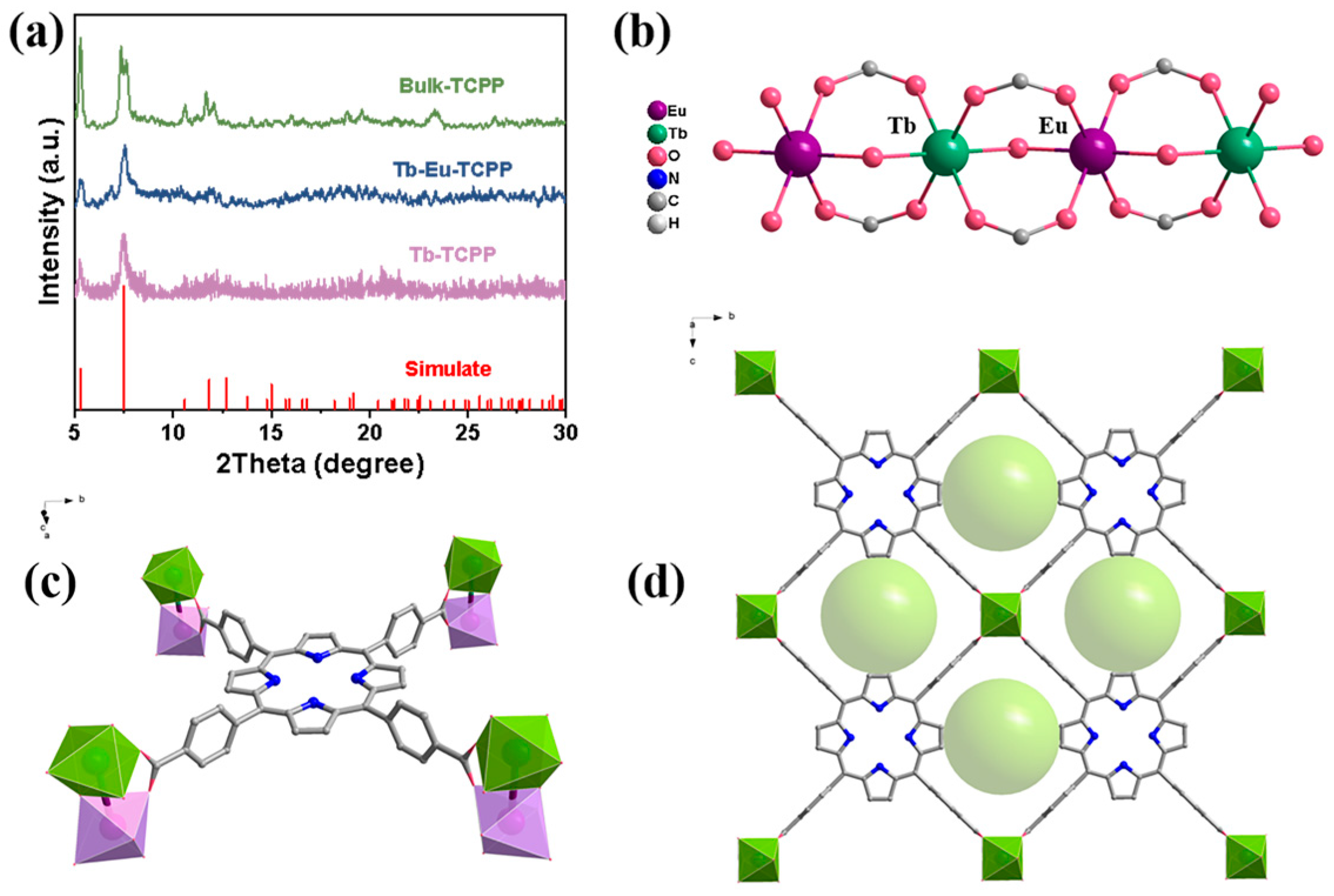
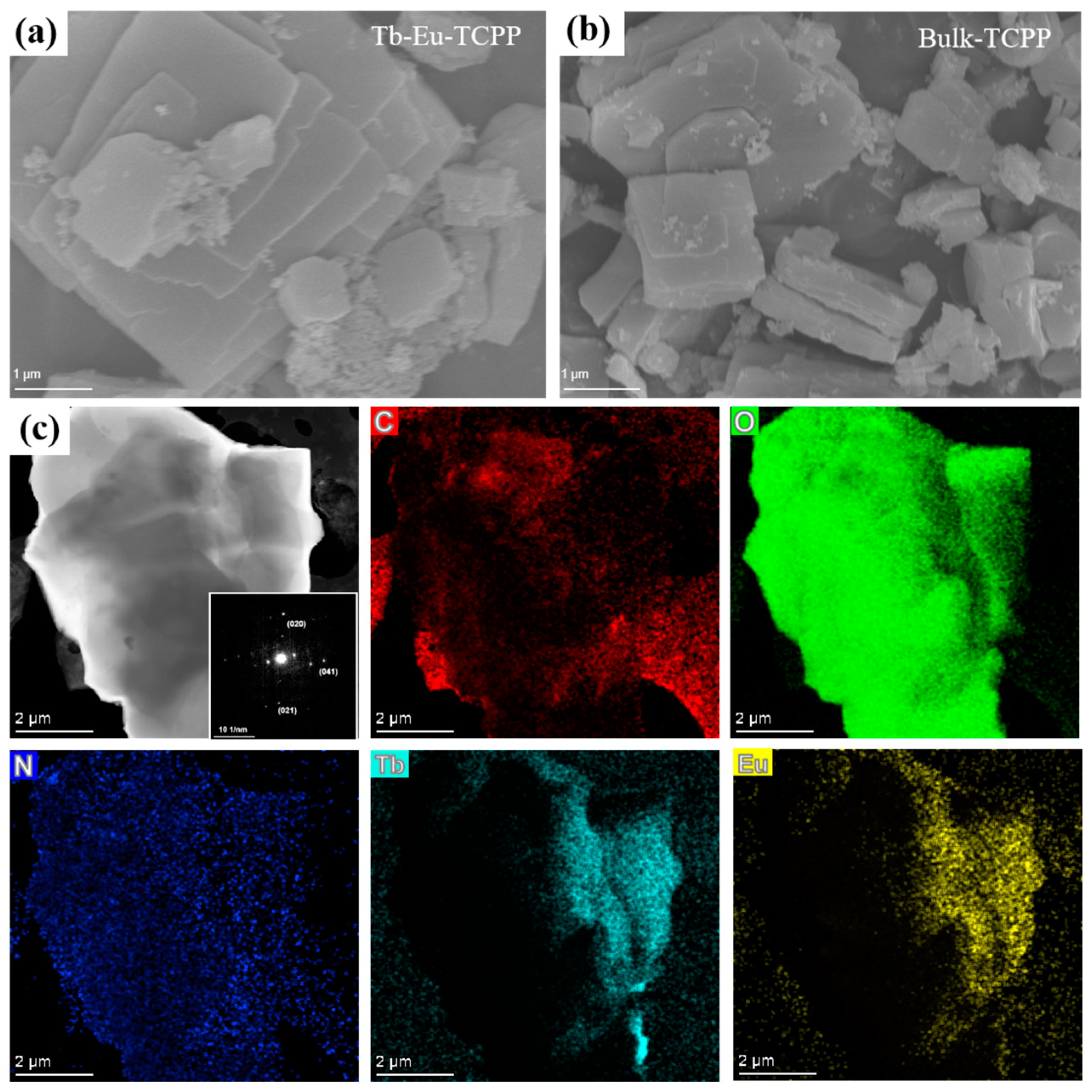
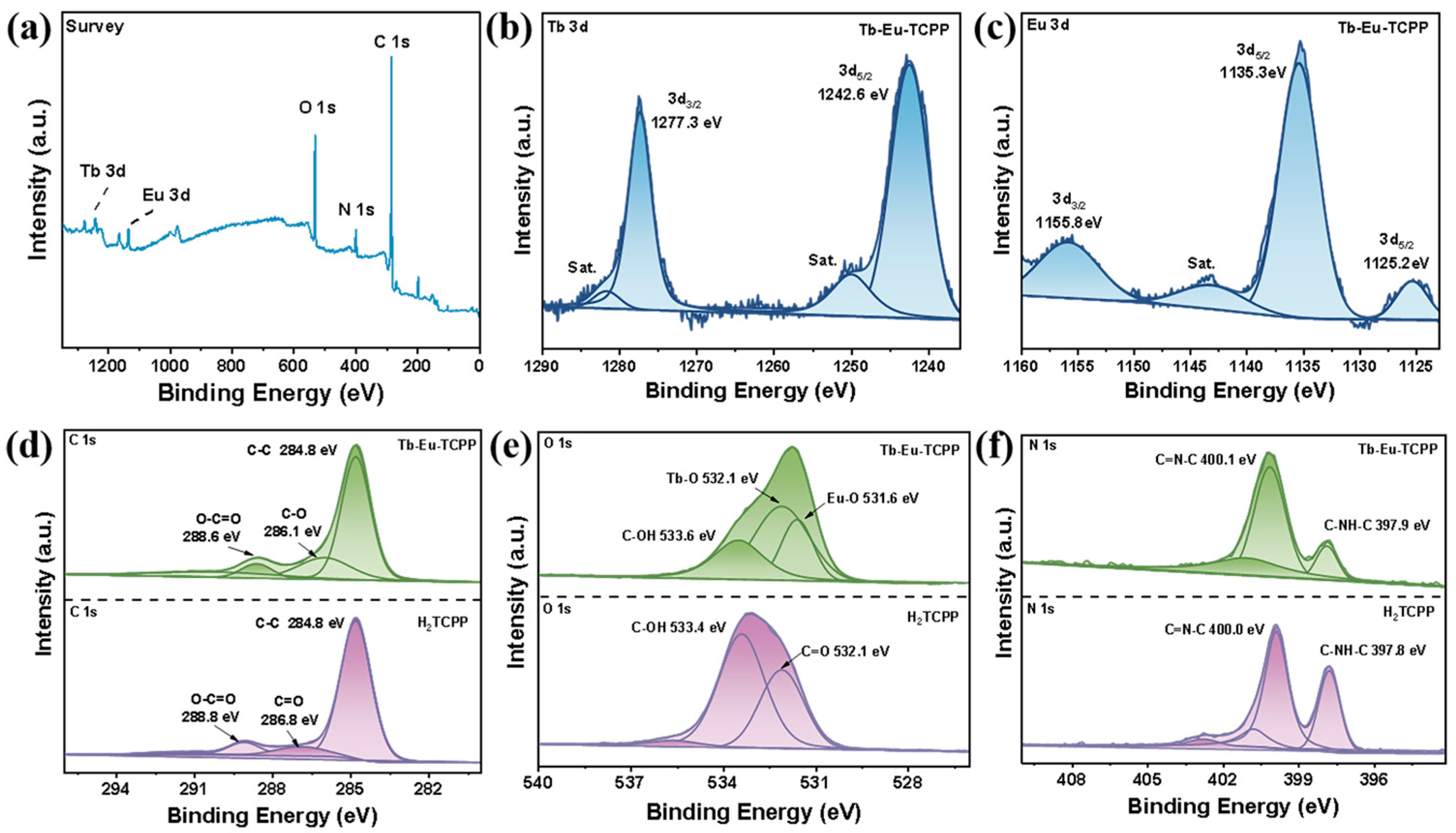
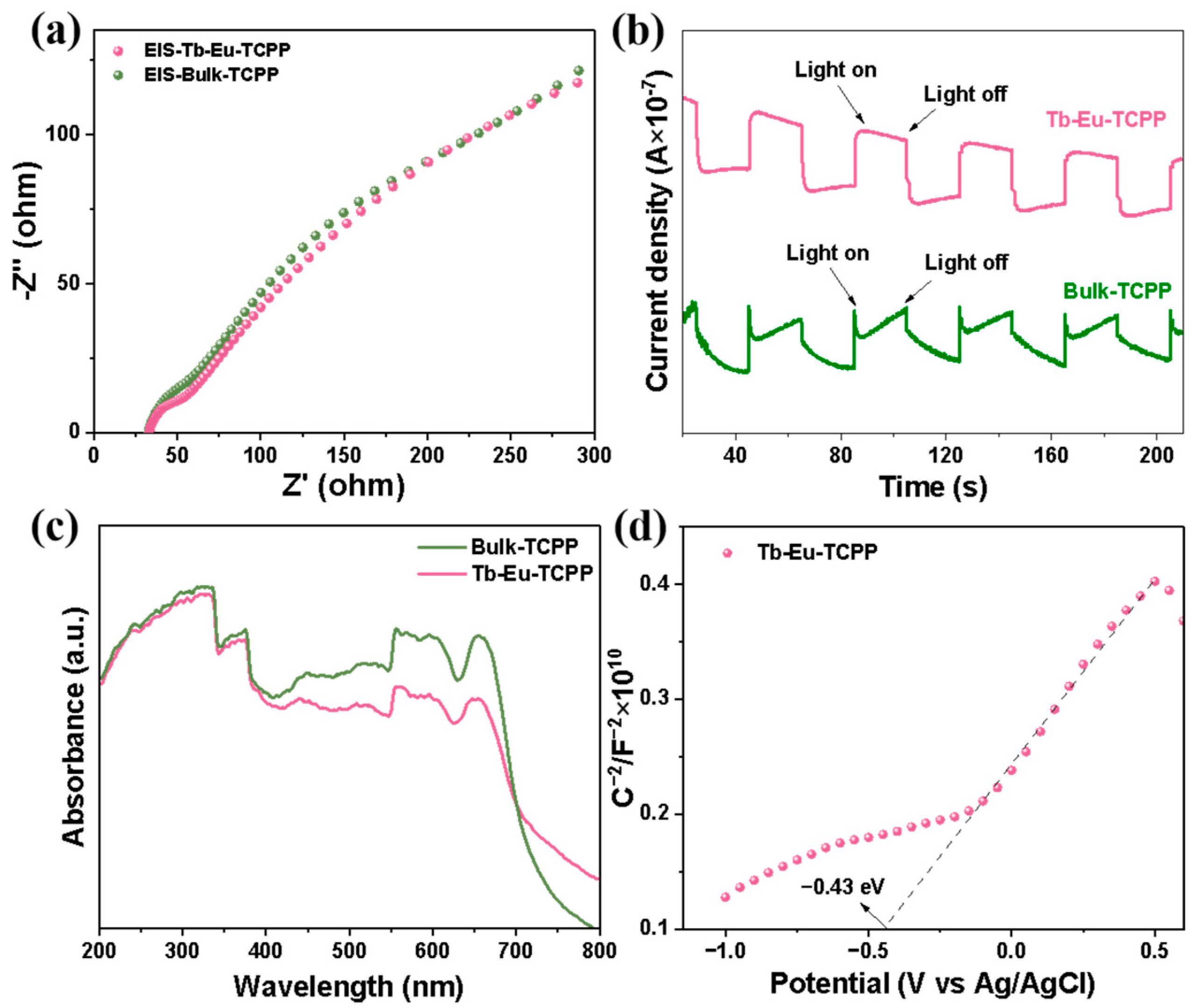
2. Results and Discussion

The crystal structures of the synthesized samples were measured by powder XRD spectroscopy. As shown in Figure 1a, all the samples showed better crystallinity and similar diffraction peaks in the 2θ (5°–30°) range. Furthermore, comparing them with the XRD of the known crystal Tb-TCPP (CCDC: 1899682), it was found that all the materials have similar diffraction peaks, which suggests that these materials have a similar structure to Tb-TCPP. Since Tb and Eu belong to the same lanthanide metal and have similar chemical properties, they have similar coordination environments during the synthesis of crystals, and during the synthesis process of doping two metals, Tb and Eu may be coordinated at the same position [34,35,36]. In addition, the concentrations of Tb and Eu were obtained through ICP testing. The ratio of the two lanthanide elements was calculated to be Tb:Eu = 1.1:1 (Table S1). This result is close to the ratio of the preparation process (Tb:Eu = 1:1). Therefore, when studying the structure, a 1:1 ratio of Tb and Eu was adopted. On this basis, the possible crystal structure formed by Tb-Eu-TCPP is simulated by the known crystal structure [37], as shown in Figure 1. The Tb/Eu atom adopts a pseudo-octahedral coordination with the oxygen atoms of four bridging carboxyl groups and two μ2-OH groups of four different TCPP linkers to form a 1D chain along the a-axis direction (Figure 1b). The porphyrin centers are metal-free, nearly planar, and have no apparent out-of-plane deformation (Figure 1c). A one-dimensional channel constructed from each carboxylate in TCPP and two Tb/Eu atoms from the 1D chain is shown in Figure 1d (the channel is highlighted with a green ball).
Subsequently, the morphology and structure of Tb-Eu-TCPP were measured by scanning electron microscopy (SEM) and transmission electron microscopy (TEM). The bright spots were clearly presented in a selected area electron diffraction (SAED) pattern, which were attributed to the (020), (021), and (041) planes of the Tb-Eu-TCPP nanosheets, confirming the crystal structure (inset in Figure 2a). This result is in excellent agreement with the three typical peaks (020), (021), and (041) in the XRD and simulated pattern of Tb-Eu-TCPP and Bulk-TCPP (Figure 1a). The contents shown in Figure 2a,b are the SEM images of Tb-Eu-TCPP and Bulk-TCPP. Unlike the hydrothermal method [38,39], microwave radiation induces localized superheating. The selective adsorption of solvent molecules or moderators on the crystal surface under microwave radiation plays a crucial role in the anisotropic growth of 2D nanosheets, leading to the formation of these distinct 2D nanosheets [40,41,42]. Simultaneously, acetic acid was used as a moderating conditioner, which was effective in inhibiting the deprotonation of carboxyl groups within the ligand [43,44]. Therefore, an increase in the amount of acetic acid in the hydrothermal process during the reaction results in a reduced number of nucleation events and increased inhibition of lateral growth in the nanosheets, leading to the thickening of the prepared nanosheets and the formation of a bulkier structure. TEM characterization further revealed the morphology of Tb-Eu-TCPP, with C, O, N, Tb, and Eu elements evenly distributed on the octahedron (Figure 2c). In addition, the results of elemental analysis testing further show that the material contains C, H, and N elements (Table S2). According to the test results of EDX (Figure S3) and ICP (Table S1), both Tb and Eu lanthanide metals were present in Tb-Eu-TCPP. The results of TEM and ICP indicate that the bimetallic Tb-Eu-TCPP was successfully synthesized.
X-ray photoelectron spectroscopy (XPS) was utilized to explore the chemical state and bonding configuration of H2TCPP and Tb-Eu-TCPP. Figure 3a shows the presence of N, O, and C elements in H2TCPP and Tb, Eu, N, O, and C elements in Tb-Eu-TCPP. The binding energy data have been corrected for C-C at 284.8 eV (Figure 3d) [45]. The high-resolution spectrum of Tb 3d was deconvoluted into four peaks (Figure 3b). The peaks at 1281.6 eV and 1249.9 eV were satellite signals (abbreviated as “Sat.”). The two peaks at 1242.6 eV and 1277.3 eV were attributed to Tb 3d5/2 and Tb 3d3/2, respectively [35]. The high-resolution spectrum of Eu 3d was deconvoluted into four peaks (Figure 3c). The peak at 1143.1 eV was a satellite signal. The peak at 1135.3 eV was attributed to Eu3+. Two smaller Eu 3d5/2 and Eu 3d3/2 peaks were observed at 1125.2 eV and 1155.8 eV, respectively, attributed to the Eu2+ oxidation state [46,47]. The corresponding O 1s XPS profiles were deconvoluted into three peaks of 531.6 eV, 532.1 eV, and 533.6 eV, among which the peak at 533.6 eV was associated with C-OH bonds, the peak at 532.6 eV was ascribed to Tb-O bonds, and the peak located at 531.6 eV came from Eu-O bonds [48,49]. The above results all indicated the successful preparation of Tb-Eu-TCPP. Figure 3f displays the N 1s energy spectrum of the Tb-Eu-TCPP nanosheets alongside the H2TCPP ligand. Among the characteristic energy spectrum peaks of the H2TCPP ligand, the peaks at 400.0 and 397.8 eV correspond to the C=N-C and C-NH-C groups, respectively [50,51]. Notably, the N 1s energy spectrum of the Tb-Eu-TCPP nanosheets did not exhibit significant changes when compared to the characteristic N 1s peaks of H2TCPP. In the characteristic energy spectrum peaks of the H2TCPP ligand, the peaks corresponding to the C=N-C and C-NH-C groups are 400.1 and 397.9 eV, respectively. This indicates that the porphyrin ring is not coordinated with Tb and Eu. In addition, the results of XPS demonstrate that during the formation of Tb-Eu-TCPP, Tb/Eu only coordinates with the O in the carboxyl group to form a Tb/Eu-O bond. Meanwhile, Tb/Eu does not bond with the N in the center of the porphyrin ring. Therefore, the results of XPS further demonstrate that in the crystal structure of Tb-Eu-TCPP, Tb/Eu forms a strong coordination with the carboxyl group in the porphyrin, forming a one-dimensional chain structure.
Moreover, the Raman spectra of Tb-Eu-TCPP nanosheets and Bulk-TCPP show typical characteristics of H2TCPP except for the peaks at 713 cm−1 and 776 cm−1, which are assigned to Tb-O and Eu-O, respectively (Figure 4a) [52,53]. Figure 4b shows the FTIR spectroscopy results acquired for the aforementioned materials. By analyzing the results obtained from Tb-Eu-TCPP, it can be observed that there are significant peaks at 3321 and 2920 cm−1 in the range of 4000–2000 cm−1. Among these peaks, the peak at 3321 cm−1 corresponds to the OH-stretching vibration [54,55]. This is consistent with the peak position in the ligand, indicating that there may be less TCPP in the substance in the free state. Notably, the band at 2920 cm−1 was attributed to intergranular water and the intracavity physical absorption of water [56,57]. In the range of 500–2000 cm−1, the shoulder peaks at 1689 cm−1 and 1604 cm−1 assigned to the asymmetric stretching vibration of C=O in H2TCPP were shifted to 1581 cm−1 and 1539 cm−1, respectively, and significantly decreased in their intensity for the Tb-Eu-TCPP nanosheets. The disappearance of the stretching vibration of C-O at 1261 cm−1 can be also observed in the spectra of Bulk-TCPP and Tb-Eu-TCPP nanosheets [7,55,58]. All these results demonstrate that the Tb-Eu-TCPP nanosheets are constructed by the strong coordination of Tb3+ (Eu3+) with the carboxyl groups in H2TCPP.
The relative changes in the specific surface area of the Tb-Eu-TCPP nanosheets and Bulk-TCPP were investigated through N2 adsorption–desorption isotherm experiments. As illustrated in Figure 4c, the nanosheet material exhibited a significantly higher specific surface area compared to the bulk material. Specifically, the BET specific surface area of the Tb-Eu-TCPP nanosheets was 332.13 m2/g, whereas that of Bulk-TCPP was 313.86 m2/g. This exceptional specific surface area suggests that these materials have excellent catalytic performance potential [59,60,61]. The pore size distribution curves (Figure S4) indicate that the Tb-Eu-TCPP nanosheets have similar micropores as Bulk-TCPP. The thermal stability of the Tb-Eu-TCPP nanosheets and Bulk-TCPP was assessed using thermogravimetric analysis (TGA). As illustrated in Figure 4d, the TGA curves of the Tb-Eu-TCPP nanosheets and Bulk-TCPP exhibit a high degree of similarity. Within the temperature range of 30–350 °C, the weight loss is approximately 20%. Subsequently, a sharp decline in weight is observed at 500 °C, indicating the onset of nanosheet backbone collapse. These results indicate that both Tb-Eu-TCPP nanosheets and Bulk-TCPP possess high thermal stability, maintaining the integrity of the backbone under nitrogen, and do not undergo thermal decomposition up to 500 °C.
Porphyrin-based MOFs are well-established for their applications in singlet oxygen (1O2) generation within the realms of photocatalysis and photodynamic therapy [62,63,64]. To determine the production of 1O2 species in the photocatalytic process, 9,10-dibenzanthracene (DPA) was used as a probe for 1O2 to assess the ability of Tb-Eu-TCPP nanosheets to generate 1O2 under visible light irradiation. DPA functions as an 1O2-trapping agent, with the concentration of DPA decreasing as it traps 1O2.
The concentration of DPA was monitored by measuring its absorbance at 374 nm using a UV–visible spectrophotometer. Figure 5 illustrates the concentration curves of DPA over time in different solution systems. Subsequently, Figure S5a–d display the absorption spectra of DPA over time for the Tb-TCPP nanosheets, Tb-Eu-TCPP nanosheets, Bulk-TCPP, and the system without added catalyst, respectively. The analysis of the data in Figure 5 leads to the following conclusions: In the presence of the Tb-Eu-TCPP nanosheets, the concentration of DPA consistently decreases over time, indicating the consumption of DPA during the process and providing evidence for the generation of singlet oxygen (1O2). Furthermore, compared to Tb-TCPP, Bulk-TCPP systems, or systems without added catalyst, there is no significant decrease in the concentration of DPA. This result emphasizes the enhanced photocatalytic activity of thinner Tb-Eu-TCPP nanosheets. Additionally, the results demonstrate that bimetallic nanosheets exhibit better catalytic effects compared to monometallic nanosheets.
In order to gain a deeper understanding of the mechanism of photocatalytic activity depending on the thickness and metal nodes, photocurrent tests were conducted to evaluate their charge separation efficiency. In Figure 6a, the plots display electrical impedance data for nanosheets with varying thicknesses. The test results reveal that the radius of the arc in the EIS plots for the Tb-Eu-TCPP nanosheets is smaller than that of Bulk-TCPP. This smaller arc radius suggests that the separation efficiency of photogenerated electron pairs is higher in the Tb-Eu-TCPP nanosheets, indicating an enhanced photocatalytic effect. Figure 6b presents photocurrent data for nanosheets with different thicknesses. Based on the experimental data, it is evident that the photocurrent density of Tb-Eu-TCPP nanosheets exceeds that of Bulk-TCPP, signifying that thinner nanosheets exhibit a higher separation efficiency of electrons and holes. This phenomenon suggests the potential for increased production of ·O2 during the experimental process. Taking into account the previously conducted characterization, it is shown that thinner Tb-Eu-TCPP nanosheets have a greater BET specific surface area, light-trapping capacity, carrier density, and hole separation efficiency, all of which boost photocatalytic activity [65,66,67]. Additionally, the UV–Vis diffuse reflectance spectra reveal a similar band gap between the Tb-Eu-TCPP nanosheets and Bulk-TCPP, as depicted in Figure 6c. This suggests that the electron-leaping ability of both materials is comparable.
To determine the conduction band bottom potential (ECB) of the prepared Tb-Eu-TCPP, Mott–Schottky analysis was employed [68,69,70]. The results in Figure 6a,d reveal that Tb-Eu-TCPP exhibits n-type semiconductor behavior with a flat-band potential Efb= −0.43 V (Ag/AgCl). For n-type semiconductors, Efb is typically 0.1 or 0.2 V more positive than the conduction band bottom potential (ECB). Consequently, the ECB of Tb-Eu-TCPP is −0.53 V (Ag/AgCl). By applying the conversion equation for the standard hydrogen potential (NHE) and Ag/AgCl potential (ENHE = EAg/AgCl + 0.197 V), the ENHE of Tb-Eu-TCPP is calculated to be −0.34 V (NHE). From Figure S6, the band gap (Eg) of the Tb-Eu-TCPP photocatalyst is 2.8 eV with bimetallic 2D properties. The valence band top potential (EVB) of Tb-Eu-TCPP is calculated to be 2.27 V (NHE) using the equation EVB = ENHE + Eg. This establishes a high oxidation–reduction potential for electron/hole pairs, allowing them to engage with dissolved oxygen and generate a substantial quantity of reactive oxygen species [71], notably 1O2.
In the system of bimetallic composites, the combined effect of the bimetals accelerates the charge transfer and inhibits electron and hole complexation, which improves the photocatalytic activity of this catalyst. Meanwhile, in the photocatalytic system of Tb-Eu-TCPP, light energy mainly drives the charge transfer, which leads to the generation of hydrated electrons upon photoexcitation. These hydrated electrons are subsequently trapped by oxygen to produce superoxide anion radicals (·O2), and ·O2 can further react with holes to produce 1O2 [72]. This principle is the same as outlined by Demyanenko et al. [73], who proposed an alternative mechanism of the photocatalytic generation of singlet oxygen through the photodetachment of an electron from the superoxide radical anion. In other words, once generated through the single-electron reduction of oxygen (Equation (1)), the superoxide radical anion absorbs light, giving rise to singlet oxygen and releasing an electron (Equation (2)), which, in turn, can recombine or reduce another oxygen molecule. The possible reactions are schematically shown in Figure 7.
O 2 + e · O 2
· O 2 + h v O 2 1 + e

3. Materials and Methods

3.1. Materials

Tb(NO3)3·6H2O, Eu(NO3)3·6H2O, meso-Tetra(4-carboxyphenyl) porphyrin (H2TCPP), 9,10-diphenylanthracene (DPA), acetonitrile (MeCN), acetic acid (HAc), and N-N dimethylformamide (DMF) were all employed in this study. All the reagents used in the experiments were of commercial grade and did not require additional purification. Unless specified otherwise, ultrapure water was used in the experiments.

3.2. Characterization and Instruments

The Midea brand home microwave oven M1-L213B (Midea, Shanghai, China) was used to prepare Tb-Eu-TCPP nanosheet samples. A JSM-7610F (JEOL Ltd., Tokyo, Japan) scanning electron microscope was used to characterize the morphology and size of the prepared samples. A transmission electron microscope was used for the morphological and selected area electron diffraction characterization of the samples with the FEI Tecnai G2 instrument model. An atomic force microscope (Dimension ICON) (Bruker Corporation, NASDAQ, Billerica, MA, United States) was used to characterize the thickness of the samples. A Smartlab 3 X-ray (JEOL Ltd., Tokyo, Japan) diffractometer was used to collect the powder diffraction data of the samples. Nitrogen adsorption–desorption isotherms of the samples were examined using an ASAP 2460 (Micromeritics, Norcross, Norcross, GA, United States) fully automated gas adsorption analyzer. Thermogravimetric analysis of the samples was carried out on an NETZSCH STA 2500 (Netzsch, Selb, Germany) thermogravimetric analyzer. The material was tested for Tb and Eu using an Agilent 7800 ICP-MS tool (Agilent, Santa Clara, CA, United States). The TEM and EDX of the material were tested using Talos F200X G2 (Thermo scientific, Waltham, MA, United States) to characterize the material’s morphology and elemental information. The elemental analysis of the materials was conducted using an Elementar Unicube (Elementar, Langenselbold, Germany). A Thermo ESCELAB 250 XI X-ray photoelectron spectrometer (Thermo scientific, Massachusetts, United States) was used to characterize the X-ray photoelectron spectra of the samples. A RENISHAW InVia Raman microscope (Renishaw, London, England) was used to test the Raman spectra of the samples. The Fourier transform infrared spectrometer FTIR Prestige-21 (Shimadzu, Kyoto, Japan) was used to characterize the infrared spectra of the samples. The UV–Vis absorption spectra and solid diffuse reflectance spectra of the samples were tested and recorded using a UV2600 spectrophotometer (Techcomp, Shanghai, China). A 300 W xenon lamp (CEL-HXF300, China Education Au-light Co., Ltd., Beijing, China) was used to provide the light source. All electrochemical measurements were performed on a CHI 760E (CH Instruments, Shanghai, China) electrochemical workstation.

3.3. Synthesis of Tb-Eu-TCPP Nanosheets

First, 0.045 mmol Tb(NO3)3·6H2O (20.4 mg), 0.045 mmol Eu(NO3)3·6H2O (20.1 mg), and 0.03 mmol H2TCPP (23.7 mg) were dissolved in 9 mL of DMF. Then, 100 μL of each of the 100 μL of the above-mixed solutions were taken into a 3-mL high-temperature-resistant glass vial. After that, 5 μL of a glacial acetic acid solution was added to it, which was labeled as Tb-Eu-TCPP. The mixed solution was diluted to 1 mL with DMF and then sealed tightly with the cap of the vial and placed in a household microwave oven at medium–low heat (its power was about 231 W) and microwaved for 10 min. After cooling to room temperature, the reaction was washed twice with DMF and anhydrous ethanol by centrifugation at 5000 rpm for 10 min. After washing, they were dried at room temperature. The specific schematic diagram of the synthesis process is shown in Figure S1.

3.4. Synthesis of Bulk-TCPP

Firstly, 0.019 mmol Tb(NO3)3·6H2O (8.5 mg) plus 0.013 mmol Eu(NO3)3·6H2O (8.6 mg) and 0.03 mmol H2TCPP (23.7 mg) were dissolved in 10 mL of DMF; then, 5 mL of the above-mixed was transferred into a PTFE-lined stainless steel autoclave, following which 400 μL of acetic acid was added, and it was placed in a 120 °C blast drying oven for 12 h. After being cooled down to room temperature, the Tb-Eu-TCPP block was washed by centrifugation with DMF and anhydrous ethanol two times. Centrifugation conditions: 5000 rpm for 10 min; after washing, it was dried at room temperature. The specific schematic diagram of the synthesis process is shown in Figure S2.

3.5. Monitoring of Singlet Oxygen Using DPA as a Probe

To validate the generation of singlet oxygen (1O2) under exposure to Tb-Eu-TCPP light, 9,10-diphenylanthracene (DPA) was employed as an 1O2-trapping agent. The experimental procedure was as follows: a solution of DPA (100 μg/mL) was prepared using an oxygen-saturated acetonitrile/water (4:1) solvent mixture. Additionally, a dispersion of Tb-Eu-TCPP nanosheets (10 μg/mL) was prepared. Subsequently, 10 mL of the DPA (100 μg/mL) solution and Tb-Eu-TCPP nanosheet (10 μg/mL) dispersion were mixed in a tube. The mixture was then exposed to a 300 W xenon lamp equipped with a UV cut-off filter, and samples were collected at different exposure times for testing UV–visible absorption spectra.

4. Conclusions

In conclusion, we have successfully synthesized nanosheets of porphyrin-based bimetallic compositions, Tb-Eu-TCPP, using a microwave-assisted method, as well as bulk porphyrin-based bimetallic materials, Bulk-TCPP, using a hydrothermal method. In this study, we succeeded in preparing ultrathin bimetallic nanosheets, and compared with the thicker bimetallic Bulk-TCPP, the prepared Tb-Eu-TCPP nanosheets have a thinner thickness, a larger BET surface area, higher photogenerated carrier separation efficiency, and better photocatalytic activity, thus exhibiting n-type semiconductor properties. This study marks a significant progress in the controlled synthesis of bimetallic-doped nanosheet materials and opens up new possibilities for synthesizing bimetallic nanosheet materials.

Supplementary Materials

The following supporting information can be downloaded at: www.mdpi.com/article/10.3390/molecules29030708/s1, Figure S1: Tb-Eu-TCPP schematic diagram of nanosheet synthesis; Figure S2: Bulk-TCPP schematic diagram of nanosheet synthesis; Figure S3: EDX of Tb-Eu-MOF obtained by TEM; Figure S4: Pore size distribution curves corresponding to Tb-Eu-TCPP (a) as well as Bulk-TCPP (b); Figure S5: The absorption spectra of DPA over time for Tb-TCPP nanosheets (a), Tb-Eu-TCPP nanosheets (b), Bulk-TCPP (c), and the system without added catalyst (d), respectively; Figure S6: VB-XPS spectra of Tb-Eu-TCPP nanosheets in different states; Table S1: ICP MS of Tb-Eu-TCPP; Table S2: Elemental analytics of Tb-Eu-TCPP.

Author Contributions

M.Y.: Conceptualization, experiment, data curation, writing—original manuscript, writing—review and editing, writing—revised manuscript. Z.H. and H.W.: experiment. X.W. and J.S.: experiment guidance. L.J.: Ideas, modification guidance, experiment guidance, supervision, funding acquisition. Z.J.: supervision, funding acquisition. All authors have read and agreed to the published version of the manuscript.

Funding

This work was supported by the National Natural Science Foundation of China (No. 41771070, No. 42071080, and No. 41201070), the Open Fund of State Key Laboratory of Frozen Soil Engineering (No. SKLFSE201508), the Natural Science Foundation of Shandong Province (ZR2023QB099), the College Students’ Innovative Entrepreneurial Training Plan Program of Yantai University, and the Laboratory Open Fund of Yantai University.

Institutional Review Board Statement

Not applicable.

Informed Consent Statement

Not applicable.

Data Availability Statement

Data are contained within the article and Supplementary Materials.

Acknowledgments

We wish to thank Wei Liu from Yantai University for their advice on the revisions.

Conflicts of Interest

The authors declare no conflicts of interest.

References

  1. Skrabalak, S.E.; Vaidhyanathan, R. The Chemistry of Metal Organic Framework Materials. Chem. Mater. 2023, 35, 5713–5722. [Google Scholar] [CrossRef]
  2. Razavi, S.A.A.; Morsali, A. Linker functionalized metal-organic frameworks. Coord. Chem. Rev. 2019, 399, 213820. [Google Scholar]
  3. Lin, R.-B.; Xiang, S.; Zhou, W.; Chen, B. Microporous Metal-Organic Framework Materials for Gas Separation. Chemistry 2020, 6, 337–363. [Google Scholar] [CrossRef]
  4. Lustig, P.W.; Mukherjee, S.; Rudd, N.D.; Desai, A.V.; Li, J.; Ghosh, S.K. Metal-organic frameworks: Functional luminescent and photonic materials for sensing applications. Chem. Soc. Rev. 2017, 46, 3242–3285. [Google Scholar] [CrossRef]
  5. Chen, L.; Xu, Q. Metal-Organic Framework Composites for Catalysis. Matter 2019, 1, 57–89. [Google Scholar] [CrossRef]
  6. Zheng, Y.; Zhang, X.; Su, Z. Design of metal–organic framework composites in anti-cancer therapies. Nanoscale 2021, 13, 12102–12118. [Google Scholar] [CrossRef]
  7. Parrino, F.; D’Arienzo, M.; Mostoni, S.; Dirè, S.; Ceccato, R.; Bellardita, M.; Palmisano, L. Electron and Energy Transfer Mechanisms: The Double Nature of TiO2 Heterogeneous Photocatalysis. Top. Curr. Chem. 2021, 380, 2. [Google Scholar] [CrossRef]
  8. Skorupskii, G.; Trump, B.A.; Kasel, T.W.; Brown, C.M.; Hendon, C.H.; Dincă, M. Efficient and tunable one-dimensional charge transport in layered lanthanide metal–organic frameworks. Nat. Chem. 2019, 12, 131–136. [Google Scholar] [CrossRef]
  9. Sahoo, S.; Mondal, S.; Sarma, D. Luminescent Lanthanide Metal Organic Frameworks (Ln MOFs): A Versatile Platform towards Organomolecule Sensing. Coord. Chem. Rev. 2022, 470, 214707. [Google Scholar] [CrossRef]
  10. Pei, K.; Wu, J.; Zhao, M.; Feng, X.; Li, Y.; Ma, Y.; Li, H.; Zhai, T. Polarized Emission of Lanthanide Metal–Organic Framework (Ln-MOF) Crystals for High-Capacity Photonic Barcodes. Adv. Opt. Mater. 2021, 10, 2102143. [Google Scholar] [CrossRef]
  11. Younis, S.A.; Lim, D.-K.; Kim, K.-H.; Deep, A. Metalloporphyrinic metal-organic frameworks: Controlled synthesis for catalytic applications in environmental and biological media. Adv. Colloid Interface Sci. 2020, 277, 102108. [Google Scholar] [CrossRef] [PubMed]
  12. Chen, M.; Umer, K.; Li, B.; Li, Z.; Li, K.; Sun, W.; Ding, Y. Metalloporphyrin based MOF-545 coupled with solid solution ZnxCd1-xS for efficient photocatalytic hydrogen production. J. Colloid Interface Sci. 2024, 653, 380–389. [Google Scholar] [CrossRef] [PubMed]
  13. Leng, F.; Liu, H.; Ding, M.; Lin, Q.-P.; Jiang, H.-L. Boosting Photocatalytic Hydrogen Production of Porphyrinic MOFs: The Metal Location in Metalloporphyrin Matters. ACS Catal. 2018, 8, 4583–4590. [Google Scholar] [CrossRef]
  14. Wang, L.; Jin, P.; Duan, S.; She, H.; Huang, J.; Wang, Q. In-situ incorporation of Copper(II) porphyrin functionalized zirconium MOF and TiO2 for efficient photocatalytic CO2 reduction. Sci. Bull. 2019, 64, 926–933. [Google Scholar] [CrossRef]
  15. Kong, X.J.; He, T.; Zhou, J.; Zhao, C.; Li, T.C.; Wu, X.Q.; Wang, K.; Li, J.R. In Situ Porphyrin Substitution in a Zr(IV)-MOF for Stability Enhancement and Photocatalytic CO2 Reduction. Small 2021, 17, 2005357. [Google Scholar]
  16. Shi, Q.; Chen, M.-H.; Xiong, J.; Li, T.; Feng, Y.-Q.; Zhang, B. Porphyrin-Based Two-Dimensional Metal-Organic framework nanosheets for efficient photocatalytic CO2 transformation. Chem. Eng. J. 2024, 481, 148301. [Google Scholar] [CrossRef]
  17. Shang, S.; Xiong, W.; Yang, C.; Johannessen, B.; Liu, R.; Hsu, H.-Y.; Gu, Q.; Leung, M.K.H.; Shang, J. Atomically Dispersed Iron Metal Site in a Porphyrin-Based Metal–Organic Framework for Photocatalytic Nitrogen Fixation. ACS Nano 2021, 15, 9670–9678. [Google Scholar] [CrossRef]
  18. Qi, Y.; Cai, Z.; Zheng, C.; Cheng, Z.; Fan, S.; Feng, Y.-S. Bimetallic synergy significantly enhances the photocatalytic performance of lanthanide porphyrin-based MOFs: Efficient photocatalytic oxidation of benzyl alcohol and benzylamine under mild conditions in air. J. Catal. 2024, 429, 115226. [Google Scholar] [CrossRef]
  19. Schlachter, A.; Asselin, P.; Harvey, P.D. Porphyrin-Containing MOFs and COFs as Heterogeneous Photosensitizers for Singlet Oxygen-Based Antimicrobial Nanodevices. ACS Appl. Mater. Interfaces 2021, 13, 26651–26672. [Google Scholar] [CrossRef]
  20. Yu, W.; Sisi, L.; Haiyan, Y.; Jie, L. Progress in the functional modification of graphene/graphene oxide: A review. RSC Adv. 2020, 10, 15328–15345. [Google Scholar] [CrossRef]
  21. Latorrata, S.; Balzarotti, R. Advances in Graphene and Graphene-Related Materials. Appl. Sci. 2023, 13, 8929. [Google Scholar] [CrossRef]
  22. Huang, H.; Shi, H.; Das, P.; Qin, J.; Li, Y.; Wang, X.; Su, F.; Wen, P.; Li, S.; Lu, P.; et al. The Chemistry and Promising Applications of Graphene and Porous Graphene Materials. Adv. Funct. Mater. 2020, 30, 1909035. [Google Scholar] [CrossRef]
  23. Li, B.; Wen, H.M.; Cui, Y.; Zhou, W.; Qian, G.; Chen, B. Emerging Multifunctional Metal–Organic Framework Materials. Adv. Mater. 2016, 28, 8819–8860. [Google Scholar] [CrossRef]
  24. Xue, Y.; Zhao, G.; Yang, R.; Chu, F.; Chen, J.; Wang, L.; Huang, X. 2D metal–organic framework-based materials for electrocatalytic, photocatalytic and thermocatalytic applications. Nanoscale 2021, 13, 3911–3936. [Google Scholar] [CrossRef]
  25. Chakraborty, G.; Park, I.-H.; Medishetty, R.; Vittal, J.J. Two-Dimensional Metal-Organic Framework Materials: Synthesis, Structures, Properties and Applications. Chem. Rev. 2021, 121, 3751–3891. [Google Scholar] [CrossRef] [PubMed]
  26. Qin, Y.; Wan, Y.; Guo, J.; Zhao, M. Two-dimensional metal-organic framework nanosheet composites: Preparations and applications. Chin. Chem. Lett. 2022, 33, 693–702. [Google Scholar] [CrossRef]
  27. Cheng, K.; Li, Y.; Gao, Z.; Chen, F.; You, C.; Sun, B. Two-dimensional metal organic framework for effective gas absorption. Inorg. Chem. Commun. 2019, 101, 27–31. [Google Scholar] [CrossRef]
  28. Obregón, S.; Colón, G. Heterostructured Er3+ doped BiVO4 with exceptional photocatalytic performance by cooperative electronic and luminescence sensitization mechanism. Appl. Catal. B 2014, 158–159, 242–249. [Google Scholar] [CrossRef]
  29. Liu, Z.; Liu, X.; Wei, L.; Yu, C.; Yi, J.; Ji, H. Regulate the crystal and optoelectronic properties of Bi2WO6 nanosheet crystals by Sm3+ doping for superior visible-light-driven photocatalytic performance. Appl. Surf. Sci. 2020, 508, 145309. [Google Scholar] [CrossRef]
  30. García, C.R.; Diaz-Torres, L.A.; Oliva, J.; Romero, M.T.; Hirata, G.A. Effect of Eu3+ concentration on the photocatalytic activity of LaSr2 AlO5 powders. Inorg. Chem. Commun. 2015, 59, 63–67. [Google Scholar] [CrossRef]
  31. Qin, L.; Cai, P.; Chen, C.; Cheng, H.; Wang, J.; Kim, S.I.; Seo, H.J. Enhanced Visible Light-Driven Photocatalysis by Eu3+-Doping in BaNb2V2O11 with Layered Mixed-Anion Structure. J. Phys. Chem. C 2016, 120, 12989–12998. [Google Scholar] [CrossRef]
  32. Zhang, X.; Wang, Y.; Cheng, F.; Zheng, Z.; Du, Y. Ultrathin lanthanide oxides nanomaterials: Synthesis, properties and applications. Sci. Bull. 2016, 61, 1422–1434. [Google Scholar] [CrossRef]
  33. He, T.; Ni, B.; Zhang, S.; Gong, Y.; Wang, H.; Gu, L.; Zhuang, J.; Hu, W.; Wang, X. Ultrathin 2D Zirconium Metal–Organic Framework Nanosheets: Preparation and Application in Photocatalysis. Small 2018, 14, 1703929. [Google Scholar] [CrossRef] [PubMed]
  34. Zhang, Y.; Li, B.; Ma, H.; Zhang, L.; Jiang, H.; Song, H.; Zhang, L.; Luo, Y. A nanoscaled lanthanide metal–organic framework as a colorimetric fluorescence sensor for dipicolinic acid based on modulating energy transfer. J. Mater. Chem. C 2016, 4, 7294–7301. [Google Scholar] [CrossRef]
  35. Han, L.; Dong, X.Z.; Liu, S.G.; Wang, X.H.; Ling, Y.; Li, N.B.; Luo, H.Q. A multi-ratiometric fluorescence sensor integrated intrinsic signal amplification strategy for a sensitive and visual assay of the anthrax biomarker based on a bimetallic lanthanide metal–organic framework. Environ. Sci. Nano 2023, 10, 683–693. [Google Scholar] [CrossRef]
  36. Wu, M.; Jiang, Z.W.; Zhang, P.; Gong, X.; Wang, Y. Energy transfer-based ratiometric fluorescence sensing anthrax biomarkers in bimetallic lanthanide metal-organic frameworks. Sens. Actuators B 2023, 383, 133596. [Google Scholar] [CrossRef]
  37. Jiang, Z.W.; Zou, Y.C.; Zhao, T.T.; Zhen, S.J.; Li, Y.F.; Huang, C.Z. Controllable Synthesis of Porphyrin-Based 2D Lanthanide Metal–Organic Frameworks with Thickness- and Metal-Node-Dependent Photocatalytic Performance. Angew. Chem. Int. Ed. 2020, 59, 3300–3306. [Google Scholar] [CrossRef]
  38. Long, L.N.; Quang, N.T.; Khuong, T.T.; Kien, P.T.; Thang, N.H.; Van Khai, T. Controllable synthesis by hydrothermal method and optical properties of 2D MoS2/rGO nanocomposites. J. Sol-Gel Sci. Technol. 2023, 106, 699–714. [Google Scholar] [CrossRef]
  39. Gusain, D.; Bux, F. Synthesis of magnesium based metal organic framework by microwave hydrothermal process. Inorg. Chem. Commun. 2019, 101, 172–176. [Google Scholar] [CrossRef]
  40. Guan, X.; Yang, Z.; Zhou, M.; Yang, L.; Peymanfar, R.; Aslibeiki, B.; Ji, G. 2D MXene Nanomaterials: Synthesis, Mechanism, and Multifunctional Applications in Microwave Absorption. Small Struct. 2022, 3, 2200102. [Google Scholar] [CrossRef]
  41. Tsuji, M. Microwave-Assisted Synthesis of Metallic Nanomaterials in Liquid Phase. ChemistrySelect 2017, 2, 805–819. [Google Scholar] [CrossRef]
  42. Chahal, S.; Bandyopadhyay, A.; Dash, S.P.; Kumar, P. Microwave Synthesized 2D Gold and Its 2D-2D Hybrids. J. Phys. Chem. Lett. 2022, 13, 6487–6495. [Google Scholar] [CrossRef] [PubMed]
  43. Wang, Q.; Zheng, X.; Chen, H.; Shi, Z.; Tang, H.; Gong, P.; Guo, L.; Li, M.; Huang, H.; Liu, Z. Synergistic effect of MOF-Directed acid-base pairs for enhanced proton conduction. Microporous Mesoporous Mater. 2021, 323, 111199. [Google Scholar] [CrossRef]
  44. Guo, H.; Zhu, Y.; Wang, S.; Su, S.; Zhou, L.; Zhang, H. Combining Coordination Modulation with Acid–Base Adjustment for the Control over Size of Metal–Organic Frameworks. Chem. Mater. 2012, 24, 444–450. [Google Scholar] [CrossRef]
  45. Kumar, P.; Kumar, A.; Rizvi, M.A.; Moosvi, S.K.; Krishnan, V.; Duvenhage, M.M.; Roos, W.D.; Swart, H.C. Surface, optical and photocatalytic properties of Rb doped ZnO nanoparticles. Appl. Surf. Sci. 2020, 514, 145930. [Google Scholar] [CrossRef]
  46. Shi, W.; Zhang, S.; Wang, Y.; Xue, Y.D.; Chen, M. Preparation of dual-ligands Eu-MOF nanorods with dual fluorescence emissions for highly sensitive and selective ratiometric/visual fluorescence sensing phosphate. Sens. Actuators B 2022, 367, 132008. [Google Scholar] [CrossRef]
  47. Park, S.J.; Joo, M.H.; Maeng, J.Y.; Rhee, C.K.; Kang, J.-G.; Sohn, Y. Electrochemical Ce3+/Ce4+ and Eu2+/Eu3+ interconversion, complexation, and electrochemical CO2 reduction on thio-terpyridyl-derivatized Au electrodes. Appl. Surf. Sci. 2022, 576, 151793. [Google Scholar] [CrossRef]
  48. Liu, Y.; Guo, H.; Wu, N.; Peng, L.; Wang, M.; Tian, J.; Xu, J.; Yang, W. Eu3+ Functionalized Nanoporous Covalent Organic Frameworks for Fluorescence Detection and Removal of Tetracycline. ACS Appl. Nano Mater. 2023, 6, 6627–6636. [Google Scholar] [CrossRef]
  49. Liu, Y.; Wang, M.; Hui, Y.; Sun, L.; Hao, Y.; Ren, H.; Guo, H.; Yang, W. Polyarylether-based COFs coordinated by Tb3+ for the fluorescent detection of anthrax-biomarker dipicolinic acid. J. Mater. Chem. B 2024, 12, 466–474. [Google Scholar] [CrossRef]
  50. Zhu, H.; Yuan, J.; Tan, X.; Zhang, W.; Fang, M.; Wang, X. Efficient removal of Pb2+ by Tb-MOFs: Identifying the adsorption mechanism through experimental and theoretical investigations. Environ. Sci. Nano 2019, 6, 261–272. [Google Scholar] [CrossRef]
  51. Wang, X.; Zhu, L.; Lv, Z.; Qi, Z.; Xu, Y.; Miao, T.; Fu, X.; Li, L. Coupled visible-light driven photocatalytic reactions over porphyrin-based MOF materials. Chem. Eng. J. 2022, 442, 136186. [Google Scholar] [CrossRef]
  52. Huong, T.T.; Anh, T.K.; Minh, L.Q. Fabrication and properties of highly luminescent materials from Tb(OH)3@SiO2 and Tb(OH)3@SiO2:Eu3+nanotubes. J. Phys. Conf. Ser. 2009, 187, 012064. [Google Scholar] [CrossRef]
  53. Belova, N.V.; Sliznev, V.V.; Christen, D. Infrared and Raman spectra of tris(dipivaloylmethanato) lanthanides, Ln(thd)3 (Ln = La, Nd, Eu, Gd, Tb, Ho, Er, Tm, Yb, Lu). J. Mol. Struct. 2017, 1132, 34–43. [Google Scholar] [CrossRef]
  54. La, D.D.; Thi, H.P.N.; Kim, Y.S.; Rananaware, A.; Bhosale, S.V. Facile fabrication of Cu(II)-porphyrin MOF thin films from tetrakis(4-carboxyphenyl)porphyrin and Cu(OH)2 nanoneedle array. Appl. Surf. Sci. 2017, 424, 145–150. [Google Scholar] [CrossRef]
  55. Xiong, J.; Yang, L.; Gao, L.X.; Zhu, P.P.; Chen, Q.; Tan, K.J. A highly fluorescent lanthanide metal-organic framework as dual-mode visual sensor for berberine hydrochloride and tetracycline. Anal. Bioanal. Chem. 2019, 411, 5963–5973. [Google Scholar] [CrossRef]
  56. Sebastian, N.; Yu, W.-C.; Balram, D.; Al-Mubaddel, F.S.; Noman, M.T. Functionalization of CNFs surface with β-cyclodextrin and decoration of hematite nanoparticles for detection and degradation of toxic fungicide carbendazim. Appl. Surf. Sci. 2022, 586, 152666. [Google Scholar] [CrossRef]
  57. Wang, T.; Zhang, L.; Zhang, J.; Guo, G.; Jiang, X.; Zhang, Z.; Li, S. Highly sensitive fluorescent quantification of carbendazim by two-dimensional Tb-MOF nanosheets for food safety. Food Chem. 2023, 416, 135853. [Google Scholar] [CrossRef]
  58. Kabak, B.; Kendüzler, E. Europium metal-organic frameworks: Synthesis, characterization, and application as fluorescence sensors for the detection of Cu2+, Ni2+ cations and T3, T4 hormones. Talanta 2024, 266, 124944. [Google Scholar] [CrossRef]
  59. Yang, H.; Wu, Y.; Zhuang, Z.; Li, Y.; Chen, C. Factors Affecting the Catalytic Performance of Nano-catalysts. Chin. J. Chem. 2021, 40, 515–523. [Google Scholar] [CrossRef]
  60. Wang, X.; Chi, C.; Zhang, K.; Qian, Y.; Gupta, K.M.; Kang, Z.; Jiang, J.; Zhao, D. Reversed thermo-switchable molecular sieving membranes composed of two-dimensional metal-organic nanosheets for gas separation. Nat. Commun. 2017, 8, 14460. [Google Scholar] [CrossRef]
  61. Buglak, A.A.; Filatov, M.A.; Hussain, M.A.; Sugimoto, M. Singlet oxygen generation by porphyrins and metalloporphyrins revisited: A quantitative structure-property relationship (QSPR) study. J. Photochem. Photobiol. A 2020, 403, 112833. [Google Scholar] [CrossRef]
  62. Peng, Y.; Li, Y.; Ban, Y.; Yang, W. Two-Dimensional Metal–Organic Framework Nanosheets for Membrane-Based Gas Separation. Angew. Chem. Int. Ed. 2017, 56, 9757–9761. [Google Scholar] [CrossRef]
  63. Hao, Y.; Liu, B.M.; Bennett, T.F.; Monsour, C.G.; Selke, M.; Liu, Y. Determination of Singlet Oxygen Quantum Yield of a Porphyrinic Metal–Organic Framework. J. Phys. Chem. C 2021, 125, 7392–7400. [Google Scholar] [CrossRef]
  64. Clement, S.; Sobhan, M.; Deng, W.; Camilleri, E.; Goldys, E.M. Nanoparticle-mediated singlet oxygen generation from photosensitizers. J. Photochem. Photobiol. A 2017, 332, 66–71. [Google Scholar] [CrossRef]
  65. Qiao, Y.; Sun, C.; Jian, J.; Xue, X.; Shi, J.; Zhou, T.; Xu, Z.; Che, G.; Zhao, L. Enhanced photocatalytic degradation performance of new and stable 2D/3D Ln-MOFs for tetracycline under visible light. J. Mol. Struct. 2023, 1293, 136235. [Google Scholar] [CrossRef]
  66. Ying, Y.; Lin, Z.; Huang, H. “Edge/Basal Plane Half-Reaction Separation” Mechanism of Two-Dimensional Materials for Photocatalytic Water Splitting. ACS Energy Lett. 2023, 8, 1416–1423. [Google Scholar] [CrossRef]
  67. Dhakshinamoorthy, A.; Asiri, A.M.; Garcia, H. 2D Metal–Organic Frameworks as Multifunctional Materials in Heterogeneous Catalysis and Electro/Photocatalysis. Adv. Mater. 2019, 31, 1900617. [Google Scholar] [CrossRef] [PubMed]
  68. Zhang, W.; Huang, W.; Jin, J.; Gan, Y.; Zhang, S. Oxygen-vacancy-mediated energy transfer for singlet oxygen generation by diketone-anchored MIL-125. Appl. Catal. B 2021, 292, 120197. [Google Scholar] [CrossRef]
  69. Bai, S.; Jiang, J.; Zhang, Q.; Xiong, Y. Steering charge kinetics in photocatalysis: Intersection of materials syntheses, characterization techniques and theoretical simulations. Chem. Soc. Rev. 2015, 44, 2893–2939. [Google Scholar] [CrossRef] [PubMed]
  70. Shang, J.; Hao, W.; Lv, X.; Wang, T.; Wang, X.; Du, Y.; Dou, S.; Xie, T.; Wang, D.; Wang, J. Bismuth Oxybromide with Reasonable Photocatalytic Reduction Activity under Visible Light. ACS Catal. 2014, 4, 954–961. [Google Scholar] [CrossRef]
  71. Ma, Y.; Li, M.; Jiang, J.; Li, T.; Wang, X.; Song, Y.; Dong, S. In-situ prepared MIL-53(Fe)/BiOI photocatalyst for efficient degradation of tetracycline under visible-light driven photo-Fenton system: Investigation of performance and mechanism. J. Alloys Compd. 2021, 870, 159524. [Google Scholar] [CrossRef]
  72. Buzzetti, L.; Crisenza, G.E.M.; Melchiorre, P. Mechanistic Studies in Photocatalysis. Angew. Chem. Int. Ed. 2019, 58, 3730–3747. [Google Scholar] [CrossRef] [PubMed]
  73. Demyanenko, A.V.; Bogomolov, A.S.; Dozmorov, N.V.; Svyatova, A.I.; Pyryaeva, A.P.; Goldort, V.G.; Kochubei, S.A.; Baklanov, A.V. Singlet Oxygen 1O2 in Photocatalysis on TiO2. Where Does It Come from? J. Phys. Chem. C 2019, 123, 2175–2181. [Google Scholar] [CrossRef]
Figure 1. (a) The XRD spectra of Bulk-TCPP, Tb-Eu-TCOPP, and Tb-TCPP, and the simulate of Tb-TCPP; (b) the possible one-dimensional direct connections of Tb and Eu to oxygen; (c) the coordination environments of Tb (III) ions, Eu (III) ions, and the TCPP ligand; and (d) views of the 1D channels (highlighted by green balls).
Molecules 29 00708 g001
Figure 2. (a) SEM images of Tb-Eu-TCPP nanosheets; (b) SEM images of Bulk-TCPP; and (c) TEM images and relevant elemental mapping of Tb-Eu-TCPP. Inset: SAED pattern of Tb-Eu-TCPP nanosheets.
Molecules 29 00708 g002
Figure 3. The XPS spectra of the Tb-Eu-TCPP nanosheets and H2TCPP: survey spectrum of Tb-Eu-TCPP (a) and high-resolution spectra of Tb 3d (b), Eu 3d (c), C 1s (d), O 1s (e), and N 1s (f).
Molecules 29 00708 g003
Figure 4. (a) Raman spectra of H2TCPP with different states of Tb-Eu-TCPP nanosheets; (b) IR spectra of H2TCPP with different states of Tb-Eu-TCPP nanosheets; (c) BET and N2 adsorption–desorption isothermal curves of Tb-Eu-TCPP nanosheets and Bulk-TCPP; and (d) TGA of Tb-Eu-TCPP nanosheets and Bulk-TCPP.
Molecules 29 00708 g004
Figure 5. Photocatalytic performance of DPA with time in different solution systems.
Molecules 29 00708 g005
Figure 6. (a) EIS of Tb-Eu-TCPP nanosheets and Bulk-TCPP; (b) photocurrent response curves of Tb-Eu-TCPP nanosheets and Bulk-TCPP; (c) UV–Vis absorption spectra of Tb-Eu-TCPP nanosheets and Bulk-TCPP; and (d) Mott–Schottky curves of Tb-Eu-TCPP nanosheets and Bulk-TCPP samples.
Molecules 29 00708 g006
Figure 7. Mechanism diagram of 1O2 generation by Tb-Eu-TCPP photocatalysis.
Molecules 29 00708 g007
Disclaimer/Publisher’s Note: The statements, opinions and data contained in all publications are solely those of the individual author(s) and contributor(s) and not of MDPI and/or the editor(s). MDPI and/or the editor(s) disclaim responsibility for any injury to people or property resulting from any ideas, methods, instructions or products referred to in the content.

