Article

Bis-Iridoid Glycosides and Triterpenoids from Kolkwitzia amabilis and Their Potential as Inhibitors of ACC1 and ACL

1
Zhejiang Provincial Key Laboratory of Plant Evolutionary Ecology and Conservation, School of Pharmaceutical Sciences, Taizhou University, Taizhou 318000, China
2
Department of Natural Medicine, School of Pharmacy, Fudan University, Shanghai 201203, China
3
State Key Laboratory of Drug Research, Shanghai Institute of Materia Medica, Chinese Academy of Sciences, Shanghai 201203, China
4
Chemistry Department, Faculty of Science, University of Malaya, Kuala Lumpur 50603, Malaysia
*
Author to whom correspondence should be addressed.
Molecules 2024, 29(24), 5980; https://doi.org/10.3390/molecules29245980
Submission received: 19 November 2024 / Revised: 11 December 2024 / Accepted: 17 December 2024 / Published: 18 December 2024
(This article belongs to the Special Issue Terpenes, Steroids and Their Derivatives (2nd Edition) )

Abstract

:
A comprehensive phytochemical investigation of the twigs/leaves and flower buds of Kolkwitzia amabilis, a rare deciduous shrub native to China, led to the isolation of 39 structurally diverse compounds. These compounds include 11 iridoid glycosides (14 and 713), 20 triterpenoids (5, 6, and 1431), and 8 phenylpropanoids (3239). Among these, amabiliosides A (1) and B (2) represent previously undescribed bis-iridoid glycosides, while amabiliosides C (3) and D (4) feature a unique bis-iridoid-monoterpenoid indole alkaloid scaffold with a tetrahydro-β-carboline-5-carboxylic acid moiety. Amabiliacids A (5) and B (6) are 24-nor-ursane-type triterpenoids characterized by an uncommon ∆11,13(18) transannular double bond. Their chemical structures and absolute configurations were elucidated through spectroscopic data and electronic circular dichroism analyses. Compound 2 exhibited a moderate inhibitory effect against acetyl CoA carboxylase 1 (ACC1), with an IC50 value of 9.6 μM. Lonicejaposide C (8), 3β-O-trans-caffeoyl-olean-12-en-28-oic acid (29), and (23E)-coumaroylhederagenin (31) showed notable inhibitory effects on ATP-citrate lyase (ACL), with IC50 values of 3.6, 1.6, and 4.7 μM, respectively. Additionally, 3β-acetyl-ursolic acid (17) demonstrated dual inhibitory activity against both ACC1 and ACL, with IC50 values of 10.3 and 2.0 μM, respectively. The interactions of the active compounds with ACC1 and ACL enzymes were examined through molecular docking studies. From a chemotaxonomic perspective, the isolation of bis-iridoid glycosides in this study may aid in clarifying the taxonomic relationship between the genera Kolkwitzia and Lonicera within the Caprifoliaceae family. These findings highlight the importance of conserving plant species with unique and diverse secondary metabolites, which could serve as potential sources of new therapeutic agents for treating ACC1/ACL-associated diseases.

Graphical Abstract

1. Introduction

The Caprifoliaceae family comprises 41 genera and approximately 960 species, primarily distributed in eastern North America and East Asian countries [1]. Several species within this family are cultivated not only as ornamental plants but also utilized in traditional Chinese medicine for treating various diseases [2,3]. Phytochemical investigations of Caprifoliaceae species have indicated that iridoids and triterpenoids are the predominant secondary metabolites [1,4,5]. Recent pharmacological studies have revealed that chemical constituents from Caprifoliaceae species exhibit a broad spectrum of bioactivities [1,4,5,6,7], particularly showing inhibitory effects against acetyl CoA carboxylase 1 (ACC1) and ATP-citrate lyase (ACL). For example, various structurally diverse iridoid and triterpenoid glycosides with ACC1 or ACL inhibitory activities have been isolated and characterized from the flower buds of two Caprifoliaceae species (Lonicera japonica and Abelia × grandiflora), as well as from the flower buds and twigs/leaves of Heptacodium miconioides, an endangered monotypic species in the Caprifoliaceae family [8,9].
Kolkwitzia amabilis Graebner, commonly known as ‘Beauty Brush’, is another monotypic species in the Caprifoliaceae family [10,11]. It earned its Chinese name, ‘Weishi’, due to its densely yellow bristled fruit, which resembles a hedgehog. This relict species is a deciduous shrub that grows 3–5 m tall, characterized by red-brown branches, ovate-elliptic leaves, and reddish corollas that bloom in early summer. Renowned for its outstanding striking spring flowers, K. amabilis displays prominently hairy calyces and stalks. This shrub has a dense, upright-arching, and vase-shaped, fountain-like growth habit. Nowadays, this species is cultivated as an ornamental plant [12]. K. amabilis is an exceedingly rare shrub native to the wilds of central China, where it was first collected in the mountains of Hubei province by plant explorer Earnest Henry Wilson in 1901. The small and sparse wild populations of K. amabilis hinder its natural regeneration. It is listed as a ‘rare’ species in the China Plant Red Data Book [13] and classified as a ‘third-grade’ nationally protected plant in China. Previous studies have conducted systematic and comprehensive investigations on its morphological phenotype traits and molecular phylogenetics [11,12]. However, phytochemical, pharmacological, and chemotaxonomic significance investigations of K. amabilis remain limited, with only its volatile organic compounds analyzed to date via GC-MS [14].
As part of an ongoing project aimed at discovering novel plant-derived agents for the treatment of ACC1/ACL-associated disease (e.g., hyperlipidemia and cardiovascular diseases) [8,9,15], a phytochemical investigation of K. amabilis cultivated at a botanical garden was conducted. This study led to the isolation of 4 new bis-iridoid glycosides (14) and 2 new 24-nor-ursane-type triterpenoids (5 and 6), along with 33 structurally diverse known compounds (739). Presented herein are the isolation, structure determination, inhibitory effects against ACC1 and ACL, and the chemotaxonomic significance of these compounds. This work constitutes Part XLII of the series titled “Phytochemical and biological studies on rare and endangered plants endemic to China” (for Parts XXXVIII through XLI [16,17,18,19]).

2. Results and Discussion

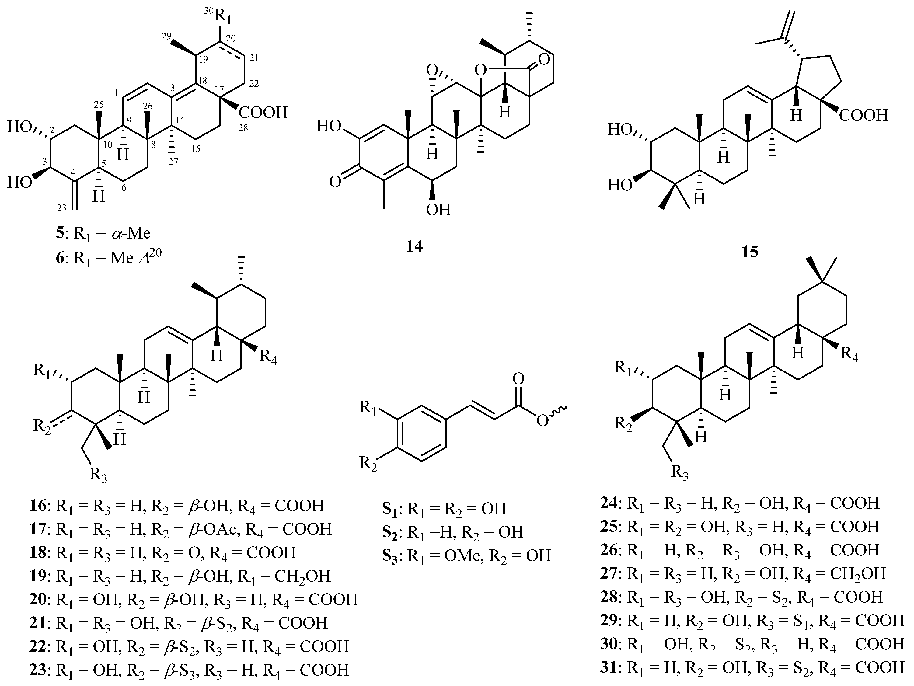
The present phytochemical investigations led to the isolation and characterization of 39 structurally diverse chemical constituents (139). Of these, 35 were obtained from the twigs/leaves, and 7 were isolated from the flower buds, with 3 compounds common to both sources. These constituents include 11 iridoid glycosides (14 and 713, Figure 1), 20 triterpenoids (5, 6, and 1431, Figure 2), and 8 phenylpropanoids (3239, Figure S1).
The 90% MeOH extract of K. amabilis twigs and leaves underwent sequential partitioning and chromatography, resulting in the isolation of compounds 110, 1234, 37, and 38. By comparing observed spectroscopic data and physicochemical properties with reported values, the known compounds from the twigs and leaves were identified as saungmaygaoside C (7) [20], lonicejaposide C (8) [9], saungmaygaoside D (9) [20], kinginoside (10) [21], sweroside (12) [22], 8-epi-loganin (13) [23], 2,6β-dihydroxy-3-oxo-11α,12α-epoxy-24-norursa-1,4-dien-28,13β-olide (14) [24], 2α,3β-dihydroxy-lupa-12,20(29)-dien-28-oic acid (15) [25], ursolic acid (16) [26], 3β-acetyl-ursolic acid (17) [27], 3-oxo-ursolic acid (18), uvaol (19) [28], corosolic acid (20) [29], 2α,23-dihydroxy-3β-(trans-p-coumaroyloxy)-urs-12-en-28-oic aid (21) [30], jacoumaric acid (22) [31], 3β-O-trans-ferulyl-2α-hydroxy-urs-12-en-28-oic acid (23) [32], oleanolic acid (24) [26], maslinic acid (25) [33], hederagenin (26) [34], erythrodiol (27) [35], 3β-O-E-coumaroylarjunolic acid (28) [36], 3β-O-trans-caffeoyl-olean-12-en-28-oic acid (29) [37], 3β-O-trans-caffeoyl-2α-hydroxy-olean-12-en-28-oic acid (30) [38], (23E)-coumaroylhederagenin (31) [39], (E)-isoferulaadehyde (32) [40], 3,4-dimethoxycinnamic acid (33) [41], 4-methoxycinnamic acid (34) [42], 4-O-demethyl rhaphidecursinol A (37) [43], and (−)-pinoresinol (38) [44].
Using similar isolation procedures on the 90% MeOH extract of K. amabilis flower buds, compounds 7, 1113, 35, 36, and 39 were obtained (Figure 1 and Figure S1). By comparing observed spectroscopic data and physicochemical properties with reported values, the known compounds were identified as saungmaygaoside C (7) [20], desferuloylkinginoside (11) [21], sweroside (12) [22], 8-epi-loganin (13) [23], trans-ferulic acid (35) [45], coniferin (36) [46], and (7S,8R)-urolignoside (39) [47].

2.1. Structure Identification of Compounds 16

Amabilioside A (1) was isolated as a colorless oil, and the molecular formula was determined to be C44H58O23 based on the observed ion peak at m/z 953.3315 [M − H] (calcd. for C44H57O23 953.3296) in HRESIMS and 13C NMR (Table 1) data. The 1H NMR (Table 1) spectrum of 1 displayed characteristic resonances for two oxygenated olefinic protons [δH 7.43 (br s, H-3) and 7.23 (br s, H-3′)], a set of terminal double bond [δH 5.74 (m, H-8), 5.26 (br d, J = 17.0 Hz, H-10a), and 5.20 (br d, J = 10.6 Hz, H-10b)], three acetal protons [δH 5.52 (d, J = 5.5 Hz, H-1), 5.47 (d, J = 1.8 Hz, H-1′), and 4.50 (dd, J = 6.9, 4.4 Hz, H-7)], one secondary methyl [δH 1.03 (d, J = 7.0 Hz, Me-10′)], and three methoxy group [δH 3.28, 3.28, and 3.15 (each 3H, s)]. Furthermore, the 1H NMR spectrum showed a pair of distinct trans-olefinic proton signals [δH 7.47 (d, J = 15.9 Hz, H-7″″) and 6.16 (d, J = 15.9 Hz, H-8″″)], as well as three ABX system aromatic proton signals [δH 7.03 (s, H-2″″), 6.94 (d, J = 8.0 Hz, H-6″″), and 6.78 (d, J = 8.0 Hz, H-5″″)]. These signals suggest the presence of an E-caffeoyl group in 1, further confirmed by the absence of NOESY correlations between H-7″″ and H-8″″. Additionally, the characteristic signals of two anomeric protons [δH 4.86 (d, J = 8.1 Hz, H-1‴) and 4.69 (d, J = 7.9 Hz, H-1″)], along with 12 other highly overlapping protons ranging from δH 3.19 to 4.79, suggested the presence of two sugar moieties in 1. Consistent with the above observations, the 13C NMR spectrum along with its HSQC data, displayed a total of 44 carbon signals, including three carbonyls (δC 168.7, 168.3, and 168.0), six aromatic carbons (δC 149.8, 147.0, 127.6, 123.2, 116.5, and 115.3), eight olefinic carbons (δC 153.3, 150.7, 147.0, 135.9, 119.8, 114.7, 114.5, and 112.0), three acetal carbons (δC 104.1, 97.8, and 95.5), and one oxygenated carbon.
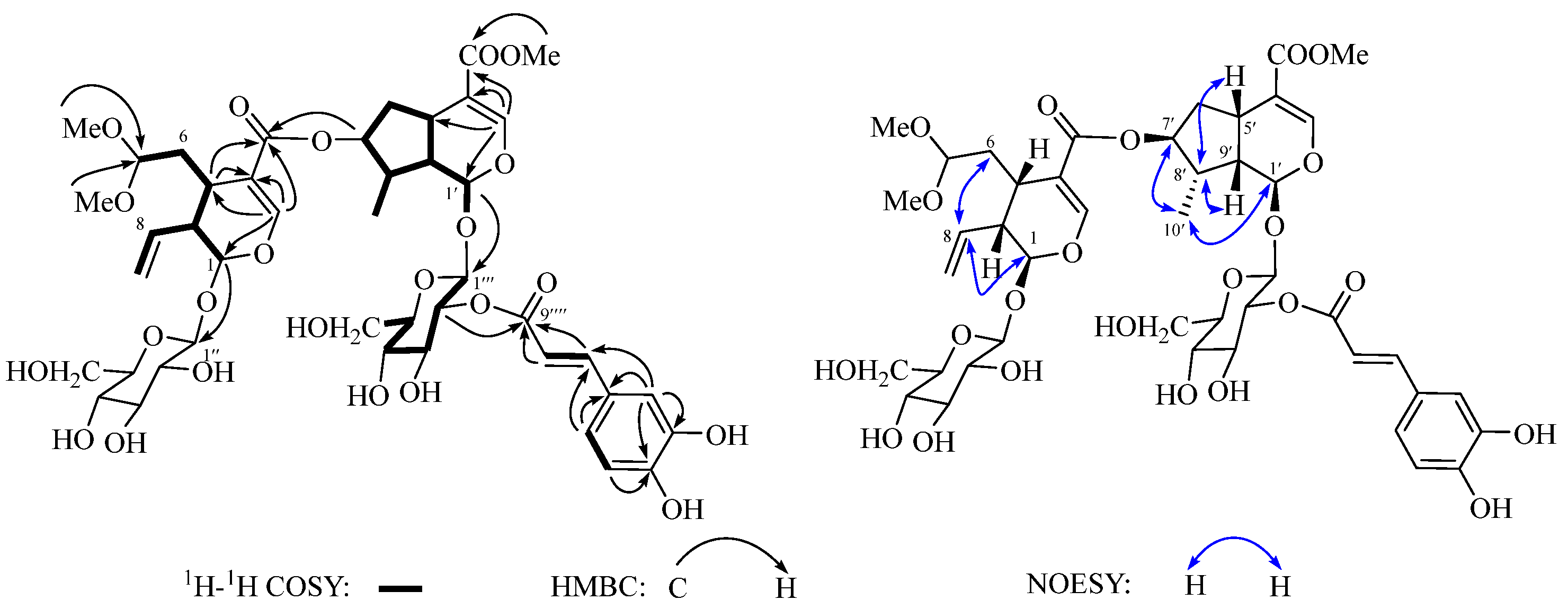
(δC 78.6), and two glucoside units (δC 100.0, 97.6, 78.5, 78.4, 78.0, 75.8, 74.6, 74.5, 71.7, 71.5, 62.8, and 62.7). The above NMR data indicated that 1 is a bis-iridoid glycoside conjugated with an E-caffeoyl group. The 1H and 13C NMR spectroscopic data of the bis-iridoid glycoside moiety were similar to those of saungmaygaoside C (7) [20], a co-occurring bis-iridoid glycosides that was firstly isolated from Picrorhiza kurroa. The main difference is that the chemical shift at C-2‴ (δH 4.79 and δC 74.5 for 1; δH 3.19 and δC 74.6 for 7) in the glycosyl unit is significantly downfield. This suggests that the glycosyl and caffeoyl units are connected through an ester linkage between C-2‴ and C-9″″, which was confirmed by the 1H-1H COSY correlation of H-1‴/H-2‴/H-3‴/H-4‴/H-5‴/H2-6‴ and the HMBC correlation from H-2‴ to C-9″″ (Figure 3). The D-configuration of the sugar moieties in compound 1, isolated in limited quantities, was confirmed by comparing the NMR data with those of the co-occurring bis-iridoid glycosides (saungmaygaoside C, 7) and by considering biogenetic factors. Acid hydrolysis of 7 yielded only a monosaccharide, which was identified as D-glucose based on HPLC and optical rotation analysis. The retention time (tR = 8.2 min, Figure S2) and optical rotation value ( [ α ] D 22 +70.2) of the hydrolysis product matched those of authentic D-glucose. Consequently, based on biogenetic considerations, the glycoside moiety in compound 1 is hypothesized to be analogous to that in 7, specifically D-glucose. The coupling constant of 7.9 and 8.1 Hz between H-1″/H-1‴ and H-2″/H-2‴ indicated a diaxial relationship between these protons and a β-configuration at the anomeric position. The relative configuration of 1 was determined by analyzing the NOESY spectrum. The NOESY correlations (Figure 3) between H-8 with H-1/H2-6, H3-10′ with H-1′/H-7′, and H-5′/H-9′ with H-8′ indicated that the relative configuration of 1 was identical to that of 7. Based on these findings, the structure of 1 was established as shown in Figure 1.
Amabilioside B (2) shares the same molecular formula (C44H58O23) as 1, as determined from the HRESIMS and 13C NMR (Table 1) data. The primary differences are observed in the vicinity of C-2‴ and C-3‴, suggesting that the C-2‴ E-caffeoyl substituent in 1 is located at C-3‴ in 2. This inference was confirmed by the HMBC (Figure S3) correlation from H-3‴ (δH 5.06, dd, J = 9.3, 9.2 Hz) to the carbonyl carbon (δC 169.0) in the E-caffeoyl moiety. The relative configuration of compound 2 was determined to be the same as that of 1 based on coupling constant (J) analysis and NOESY (Figure S3) data. Therefore, the structure of 2 was established as shown in Figure 1.
Amabilioside C (3) was obtained as a colorless oil. Its molecular formula was determined to be C44H56N2O20 based on the HRESIMS ion peak at m/z 933.3505 [M + H]+ (calcd. for C44H57N2O20 933.3499) and 13C NMR data (Table 2), indicating 18 degrees of unsaturation. The IR spectrum showed the presence of hydroxyl (3416 cm−1) and carbonyl (1690 cm−1) functional groups. Interpretation of the 1H (Table 2) and 13C NMR data suggested that compound 3 possesses a typical bis-iridoid glycoside fragment, similar to compounds 1 and 2, conjugated with an aromatic moiety. In addition to the NMR signals corresponding to the bis-iridoid glycoside moiety, the remaining resonances were consistent with a tetrahydro-β-carboline unit, featuring an ortho disubstituted phenyl group [δH 7.48 (d, J = 7.4 Hz, H-9); 7.04 (dd, J = 7.4, 7.0 Hz, H-10); 7.13 (dd, J = 8.0, 7.0 Hz, H-11); 7.31 (d, J = 8.0, H-12)] and a methine-amine [δH 3.89 (m, H-5)]. The NMR data of 3 closely resembled those of dipsaperine, which was previously isolated from the roots of Dipsacus asper [48]. Similar to dipsaperine, compound 3 is a hybrid of bis-iridoid glycoside and tetrahydro-β-carboline-5-carboxylic acid. This was confirmed by the 1H-1H COSY (Figure 4) correlations between H-5/H2-6 and the HMBC (Figure 4) cross-peaks from H-3 to C-2/C-5/C-7 and H-5 to C-3. The relative configuration of 3 was determined by analyzing the coupling constants and NOESY (Figure 4) experiments. The strong key NOESY cross-peaks between H2-14/H-19/H-21 revealed that the CH2-14 group, the terminal double bond, and H-21 were oriented on the same side and were assigned as α-orientation, thereby placing H-15/H-20 in the β-orientation. The NOESY correlation between H-3/H-5, along with the coupling constant of H-5 (J = 11.7 and 4.0 Hz), corresponds to those reported for dipsaperine [48] and 5(S)-5-carboxystrictosidine [49], thereby confirming the α-orientation of H-3 and H-5. Additionally, the secondary methyl group at C-8′ was found to be identical to those in compounds 1 and 2, as confirmed by the NOESY experiment. The stereochemistry of C-3 and C-5 was assigned as S, based on the positive Cotton effect (CE) at 222 nm and negative CE at 236 nm observed in its ECD spectrum (Figure S26) [48], which were consistent with those of dipsaperine. Thus, the structure of 3 was determined as shown in Figure 1.
Amabilioside D (4) was found to possess the same molecular formula (C44H56N2O20) as compound 3, as determined by the HRESIMS ion at m/z 933.3503 [M + H]+ (calcd. for C44H57N2O20 933.3499) and 13C NMR data (Table 2). Detailed analysis of the 1D and 2D NMR (Figure S3) spectroscopic data revealed that 4 shares a similar structure as 3, with a notable difference observed in the tetrahydro-β-carboline-5-carboxylic acid moiety. The two downfield chemical shifts of the methine-amine group in compound 4 [δH 4.84 (dd, J = 3.7, 3.7 Hz, H-3), 4.12 (dd, J = 9.1, 5.7 Hz, H-5); δC 52.7 (C-3), 55.0 (C-5)], were significantly distinct when compared to those in 3 [δH 4.51 (br d, J = 11.3 Hz, H-3), 3.89 (m, H-5); δC 53.2 (C-3), 59.6 (C-5)]. This suggests that the configuration at C-3 in 4 is inverted [50]. This was further supported by the absence of a NOESY correlation (Figure S3) between H-3 and H-5. Similar to compounds 13, the Me-10′ in 4 was assigned an α-orientation, as indicated by the clear NOE cross-peaks between H3-10′ and H-1′/H-7′. Based on these data, the structure of 4 was determined as shown in Figure 1.
Amabiliacid A (5) was obtained as a white powder. Its molecular formula was determined as C29H42O4 based on HRESIMS (m/z 455.3157 [M + H]+, calcd. for 455.3156) and 13C NMR data (Table 3). In the upfield region of the 1H NMR spectrum of 5, three tertiary methyl groups were observed at δH 0.75 (3H, s, Me-25), 0.90 (3H, s, Me-26), and 1.00 (3H, s, Me-27), along with two secondary methyl groups at δH 0.92 (3H, d, J = 7.1 Hz, Me-30) and 1.05 (3H, d, J = 7.2 Hz, Me-29). Additionally, signals at δH 5.16 and 4.73 (each 1H, br s) were observed for an exocylic double bond, along with two olefinic protons at δH 6.56 (1H, dd, J = 11.8, 2.9 Hz, H-11) and 5.72 (1H, d, J = 11.8 Hz, H-11), as well as two oxymethines at δH 3.76 (1H, d, J = 9.1 Hz, H-3) and 3.54 (1H, m, H-2). The 13C NMR spectrum displayed a total of 29 carbon signals, including a carboxyl carbon at δC 180.3 (C-28), six olefinic carbons at δC 105.0 (C-23), 126.5 (C-11), 127.6 (C-12), 136.8 (C-18), 138.9 (C-13), and 151.5 (C-4), as well as two oxygenated carbons at δC 74.2 (C-2) and 79.6 (C-3). The NMR data of 5 closely resembled those of corosolic acid (20) [29], a co-occurring ursane-type triterpenoid. A notable difference between them was the absence of the Me-23 and Me-24 groups in the NMR spectra of 5. Instead, signals typical of two olefinic protons [δH 5.16 and 4.73 (br s)] corresponding to an exocyclic double bond functionality were observed. This suggests that 5 possesses a 24-nor-ursane-type triterpenoid framework. This deduction was confirmed by the 1H-1H COSY (Figure S4) correlations of H-3/H2-23 and H-5/H2-23, as well as the HMBC (Figure S4) correlation from H2-23 to C-3/C-4/C-5. Additionally, the olefinic group in 5 shifted to form an ∆11,13(18) diene, as opposed to the ∆12 configuration in 20. This was further supported by the 1H-1H COSY correlations of H-9/H-11/H-12 and key HMBC cross-peaks from H-9 to C-11/C-12, H-11 to C-13, H-12 to C-13/C-18, H3-27 to C-13, and H3-29 to C-18. The relative configuration of 5 was determined by analyzing the coupling constants (J) and NOESY data. The large J value between H-2 and H-3 (9.1 Hz), along with the NOE (Figure S4) correlations of H-2/H3-25 and H-3/H-5, confirmed the α-orientation of OH-2 and β-orientation of OH-3, respectively. Additionally, the ECD data (Figure S41) of 5 closely matched those of 2α,3β-dihydroxy-24-nor-olean-4(23),11,13(18)-trien-28-oic acid [51] and pterohoonoid D [52], two pentacyclic triterpenoids featuring a unique ∆11,13(18) diene group in rings C and D. This comparison allowed the assignment of the 9R, 14S, and 17S configuration in 5. Consequently, the structure of 5 was defined as (2R,3R,5R,8R,9R,10S,14S,17S,19S,20R)-2α,3β-dihydroxy-24-nor-urs-4(23),11,13(18)-trien-28-oic acid.
The molecular formula of amabiliacid B (6) was determined to be C29H40O4 based on HRESIMS analysis (m/z 453.2996 [M + H]+, calcd. for 453.2999) and 13C NMR data (Table 3), indicating two fewer protons than compound 5. The NMR spectroscopic data of 6 were similar to those of 5, except for the presence of the ∆20(21) group [δH 1.66 (3H, s, H-30), 5.43 (1H, br d, J = 5.0 Hz, H-21); δC 137.6 (C-20), 121.0 (C-21)]. This was further corroborated by the HMBC (Figure S4) cross-peaks, which showed correlations between H3-20 with C-20 and H3-30 with C-20/C-21. The relative configuration of 6 was found to be identical to that of 5, as evidenced by the NOESY (Figure S4) experiment. Consequently, the structure of 6 was determined to be 2α,3β-dihydroxy-24-nor-urs-4(23),11,13(18),20-tetraen-28-oic acid.

2.2. Anti-ACC1 and Anti-acl Bioactivities of the Isolated Compounds

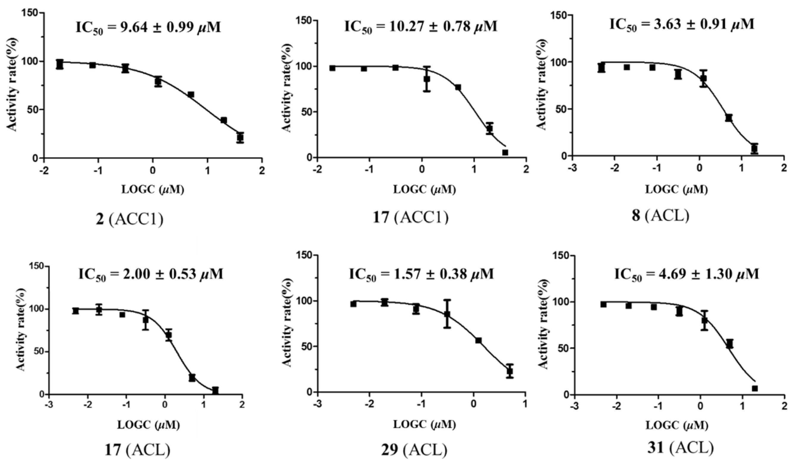
ACC1 is a key enzyme in the de novo fatty acid biosynthesis pathway, catalyzing the conversion of acetyl-CoA to malonyl-CoA [53]. In contrast, ACL plays a critical role in linking carbohydrate and lipid metabolism by producing acetyl-CoA from citrate, which is essential for the biosynthesis of fatty acids and cholesterol [54]. Both enzymes are considered potential therapeutic targets for hyperlipidemia and other metabolic disorders. Firsocostat (formerly named ND-630 or GS-0976) is a liver-targeted allosteric ACC1 inhibitor currently in clinical stages, and it has demonstrated efficacy in reducing hepatic fat content in patients with non-alcoholic steatohepatitis (NASH) [55]. Bempedoic acid, a synthetic dicarboxylic acid-type ACL inhibitor, was approved by FDA in 2020 for the treatment of statin-resistant hypercholesterolemia [56]. As part of our ongoing efforts to discover novel plant-derived compounds for the treatment of ACC1/ACL-related diseases, we evaluated all isolated compounds (139) for their ACC1 and ACL inhibitory activities. Among these, compounds 2 (IC50: 9.6 μM) and 17 (IC50: 10.3 μM) displayed moderate inhibitory effects against ACC1. Compounds 8, 17, 29, and 31 exhibited potent inhibitory effects on ACL, with IC50 values of 3.6, 2.0, 1.6, and 4.7 μM (Table 4 and Figure 5), respectively. The rest of the isolated compounds are inactive (inhibition percentage < 50% at 20 μM). The known inhibitor ND-630 (IC50: 2.0 ± 0.1 nM) [57] and BMS 303141 (IC50: 0.3 ± 0.1 μM) [58] were used as positive controls for ACC1 and ACL, respectively.

2.3. Molecular Docking Simulation of Compounds 2, 17, and 29

Considering the observed ACC1 and ACL inhibitory activities (Table 4), the dual inhibitory compound (17 for ACC1 and ACL) and the most potent individual inhibitor (2 for ACC1; 29 for ACL), were selected for molecular docking studies. These studies aimed to elucidate their potential mechanisms of action as inhibitors of ACC1 (PDB ID: 3TVU) and ACL (PDB ID: 6HXH). The structures of compounds 2, 17, and 29 exhibit favorable binding within the active site pockets of the selected target protein. The binding energies of compound 17 with ACC1 and ACL are −8.8 and −10.4 kcal/mol, respectively. In compound 17, the carbonyl group establishes two hydrogen-bond interactions with Ser138 (3.0 and 2.8 Å) for ACC1 (Figure S5A), and forms hydrogen bonds with Leu266 (2.8 Å) and Thr267 (2.9 Å) for ACL (Figure S5B). The binding mode of compound 2 with ACC1 is shown in Figure 6, demonstrating a binding affinity of −8.8 kcal/mol. The hydroxyl groups in 2 formed four hydrogen bonds with Ile1820 (3.5 Å), Glu2177 (4.0 Å), Gly1579 (2.8 Å), and Arg1578 (3.6 Å) (Figure 6). Additionally, the phenyl ring in 2 formed a π-π stacking interaction with Try1576 (5.2 Å). Compound 29 exhibited a binding energy of −8.8 kcal/mol with ACL, with its hydroxyl and carboxyl groups positioned within the hydrophilic region of the binding site, forming four strong hydrogen-bond interactions with Glu599 (3.2 Å), Gly261 (3.3 Å), Gln505 (3.0, 4.1 Å), and Phe572 (3.0 Å) (Figure S6). Additionally, the carboxylate and carboxyl groups of 29 formed two salt bridge interactions with Lys1017 (4.7 Å) and Lys1018 (5.5 Å). Compounds 2, 17, and 29 exhibited multiple hydrogen bond interactions within the active binding sites of ACC1 and ACL. These interactions are fundamental to protein–ligand binding and likely contribute to the observed inhibitory activity of these compounds.

2.4. Chemotaxonomic Significance

K. amabilis remains rare in the wild but is widely cultivated in China, USA, and several European countries. As a member of the Caprifoliaceae family (commonly known as the honeysuckle family), this shrub represents a monotypic species and is renowned for its abundant blossoms, which display their full floral elegance in late spring or early summer. Comparative studies have been conducted on the morphology, growth, and flowering responses of various Kolkwitzia accessions grown under the same environmental conditions. Characterization of K. amabilis accessions based on flowering patterns and molecular markers was first discussed in 2014 [11]. Its complete chloroplast genome was sequenced, and phylogenetic analysis using a maximum parsimony approach indicated a close relationship between K. amabilis and Lonicera japonica [10], a species in the honeysuckle family. The ability to delimit species accurately is crucial for assessing biodiversity, a necessary step in establishing effective conservation policies [59]. Plant taxonomy has traditionally relied on morphological phenotype traits, it has progressively incorporated additional advanced techniques, including molecular phylogenetic and chemotaxonomy analyses [60,61,62,63,64,65]. The integrative taxonomy approach (ITA), which combines morphological and molecular phylogenetic evidence, has been recognized as one of the most effective methods for resolving complex species boundaries [66,67,68,69]. The bis-iridoids (14 and 79) isolated herein from the twigs/leave and flower buds of K. amabilis exhibit a secoiridoid/iridoid heterodimer structure, composed of secologanic acid derivatives linked to the 7-OH group of loganin. Most secoiridoid/iridoid-subtype dimers have previously been identified within the Caprifoliaceae family [70,71,72,73,74], indicating their potential as valuable chemotaxonomic markers. From a chemotaxonomic perspective, these findings further support the close phylogenetic relationship between Kolkwitzia and Lonicera.

3. Materials and Methods

3.1. General Experimental Procedures

Optical rotations were recorded in MeOH with an SGW-1 digital polarimeter (Shanghai INESA Physico-Optical Instrument Co., Ltd., Shanghai, China). Ultraviolet spectra were measured with a Shimadzu PharmaSpec-1800 UV-visible spectrometer (Shimadzu, Kyoto, Japan). IR spectra were obtained on a Thermo Nicolet iS10 spectrophotometer (Thermo Fisher Scientific, San Jose, CA, USA) in KBr discs. The 1D and 2D NMR spectra were collected on Bruker Avance III 400 MHz NMR instruments (Bruker BioSpin, Rheinstetten, Germany). Chemical shifts were reported in parts per million with the residual solvent signals as an internal standard. HRESIMS data were acquired on a Thermo Fisher Q-Exactive mass spectrometer (Thermo Fisher Scientific) with heated electrospray ionization. Semi-preparative HPLC was performed on a Shimadzu CBMA-20A system equipped (Shimadzu) with an SPD-20A photodiode array detector (Shimadzu) using the Cosmosil Packed Column (10 × 250 mm, 5 μm, 5C18-MS-II or π NAP) with a flow rate of 3 mL/min. Silica gel (Qing Dao Hai Yang Chemical Group Co., Qingdao, China; 200–300 mesh), MCI gel CHP20P (75–150 μm, Mitsubishi Chemical Industries, Tokyo, Japan), and Sephadex LH-20 gel (Amersham Biosciences, Slough, UK; 40–70 μm) were used for column chromatography (CC). Thin-layer chromatography (TLC) was performed on percolated plates (GF254, 0.2–0.25 mm, Qing Dao Hai Yang Chemical Group Co., Qingdao, China). TLC spots were visualized under UV light (254 or 365 nm) and by spaying with 5% H2SO4/vanillin followed by heating to 120 °C.

3.2. Plant Materials

The twigs/leaves of Kolkwitzia amabilis were harvested from Jiashan Lige Ecological Technology Co., Ltd., Jiaxing, Zhejiang province, China, in November 2021, and the flower buds were collected from the same source in April 2023. These plant samples were taxonomically identified by Prof. Ze-Xin Jin (Taizhou University, Taizhou, China). Voucher specimens (twigs/leaves: No. 20211107; flower buds: No. 20230420) have been deposited in the Herbarium of the Institute of Natural Medicine and Health Products, School of Pharmaceutical Science, Taizhou University.

3.3. Extraction and Isolation

The air-dried and milled twigs and leaves of K. amabilis (12.0 kg) were percolated four times with 90% MeOH (20 L) at ambient temperature. The extract was concentrated under reduced pressure, yielding a residue (2.2 kg) that was suspended in H2O and sequentially partitioned with petroleum ether (PE, 3 × 4 L), EtOAc (3 × 4 L), and n-BuOH (3 × 4 L). Following solvent removal, the EtOAc-soluble extract (231 g) was subjected to column chromatography (CC) on a silica gel column and separated into six fractions (Fr.1–Fr.6) using a gradient system of PE/EtOAc (10:1, 5:1, 3:1, 1:1, and 0:1, v/v). Fr.2 was applied to a Sephadex LH-20 column, followed by semi-preparative HPLC (MeCN-H2O, 65:35), to yield compound 17 (8.0 mg, tR = 12.6 min). Fr.3 was subjected to MCI column chromatography using a MeOH-H2O gradient (50:50, 70:30, 85:15, and 100:0, v/v), resulting in four fractions (Fr.3.1–Fr.3.4). Compounds 33 (2.3 mg, tR = 14.3 min) and 34 (1.7 mg, tR = 13.4 min) were obtained from Fr.3.2 via semi-preparative HPLC (MeOH-H2O, 40:60). Fr.3.3 was further fractionated using Sephadex LH-20 column chromatography (MeOH), yielding five subfractions (Fr.3.3.1–Fr.3.3.5) based thin-layer chromatography (TLC) spots analysis. Compounds 15 (0.8 mg, tR = 11.7 min) and 27 (7.4 mg, tR = 13.9 min) were obtained from Fr.3.3.2 via semi-preparative HPLC (MeCN-H2O, 75:25), while compounds 19 (13.5 mg, tR = 14.7 min) and 18 (2.7 mg, tR = 17.1 min) were isolated from Fr.3.3.4 using semi-preparative HPLC (MeCN-H2O, 70:30). Fr.4 was separated by gel permeation chromatography on Sephadex LH-20 (MeOH), followed by semi-preparative HPLC using MeCN-H2O (75:25) to yield compounds 24 (2.4 mg, tR = 11.6 min) and 16 (14.2 mg, tR = 12.1 min). Fr.5 was subjected to MCI column chromatography with MeOH-H2O gradient (50:50, 70:30, 85:15, and 100:0, v/v), yielding seven subfractions (Fr.5.1–Fr.5.7). Purification of Fr.5.2 via semi-preparative HPLC (MeCN-H2O, 30:70) provided compound 32 (1.5 mg, tR = 15.0 min). Further separation of Fr.5.3 over Sephadex LH-20 with MeOH, followed by semi-preparative HPLC (MeOH-H2O, 70:30), afforded compounds 37 (4.2 mg, tR = 18.3 min) and 38 (6.1 mg, tR = 11.6 min). Fr.5.4 was further fractionated using Sephadex LH-20 with MeOH, yielding four fractions (Fr.5.4.1–Fr.5.4.4). Separation of Fr.5.4.1 by semi-preparative HPLC (MeOH-H2O, 90:10) afforded compounds 26 (2.9 mg, tR = 12.0 min) and 25 (6.1 mg, tR = 13.1 min). Subsequently, compounds 5 (3.0 mg, tR = 9.2 min) and 20 (1.0 mg, tR = 15.3 min) were purified from Fr.5.4.2 by semi-preparative HPLC with an isocratic elution (MeCN-H2O, 65:35). Compounds 6 (1.0 mg, tR = 14.8 min), 14 (4.1 mg, tR = 11.0 min), 28 (3.5 mg, tR = 15.0 min), and 21 (2.1 mg, tR = 15.9 min) were obtained from Fr.5.4.3 via semi-preparative HPLC (MeOH-H2O, 85:15), respectively. Fr.5.4.4 was purified by semi-preparative HPLC (MeCN-H2O, 60:40) to furnish compound 29 (2.1 mg, tR = 13.6 min). Fr.5.5 was fractionated using Sephadex LH-20 (MeOH), producing subfractions Fr.5.5.1–Fr.5.5.5. Compounds 31 (0.9 mg, tR = 9.6 min), 30 (1.0 mg, tR = 12.5 min), 22 (3.9 mg, tR = 13.6 min), and 23 (2.9 mg, tR = 15.0 min) were isolated from Fr.5.5.4 by semi-preparative HPLC (MeCN-H2O, 70:30).
Using D-101 macroporous resin column chromatography with a stepwise gradient-elution of MeOH-H2O (30:70, 50:50, 70:30, 90:10, and 100:0, v/v), the n-BuOH-soluble fraction (344 g) was divided into seven fractions (Fr.7–Fr.13). Purification of Fr.9 using Sephadex LH-20 (MeOH), followed by semi-preparative HPLC (MeCN-H2O, 15:85), gave compounds 12 (15.7 mg, tR = 13.4 min) and 13 (10.4 mg, tR = 16.2 min). Fr.11 was further separated using ODS C-18 CC (MeOH-H2O, 50:50, 60:40, 70:30, and 100:0, v/v), yielding eight subfractions (Fr.11.1–Fr.11.8). Fr.11.3 was purified by semi-preparative HPLC (MeCN-H2O, 30:70) to furnish compound 7 (3.2 mg, tR = 15.9 min). Compounds 8 (8.7 mg, tR = 11.9 min), 9 (8.7 mg, tR = 9.4 min), and 4 (5.6 mg, tR = 15.8 min) were obtained from Fr.11.4 by semi-preparative HPLC (MeOH-H2O, 55:45). Further purification of Fr.11.6 by Sephadex LH-20 (MeOH), followed by semi-preparative HPLC (MeCN-H2O, 30:70) gave compounds 10 (14.0 mg, tR = 9.7 min) and 3 (4.5 mg, tR = 14.1 min). Compounds 2 (2.6 mg, tR = 15.1 min) and 1 (4.6 mg, tR = 18.3 min) were obtained from Fr.11.7 using Sephadex LH-20 (MeOH), followed by semi-preparative HPLC (MeCN-H2O, 35:65).
The powdered flower buds (300 g) of K. amabilis were extracted with 90% methanol (4 × 2 L, each extraction for 24 h) at room temperature. The resulting dark green residue (41.0 g) was suspended in H2O (1.0 L) and successively extracted with petroleum ether (PE, 3 × 1.0 L), EtOAc (3 × 1.0 L), and n-BuOH (3 × 1.0 L). The EtOAc-soluble extract (36.3 g) was subjected to silica gel column chromatography and eluted with a PE/EtOAc gradient system (10:1, 5:1, 3:1, 1:1, and 0:1, v/v), yielding four fractions (Fr.14–Fr.17). Purification of Fr.15 using Sephadex LH-20 (MeOH), followed by semi-preparative HPLC (MeOH-H2O, 50:50) afforded compound 35 (2.7 mg, tR = 12.8 min). The n-BuOH-soluble extract (19.3 g) was fractioned on an MCI column with a stepwise gradient elution of MeOH-H2O (30:70, 50:50, 70:30, 85:15, 100:0, v/v), yielding six fractions (Fr.18–Fr.23). Compound 36 (1.4 mg, tR = 11.3 min) was obtained from Fr.19 by semi-preparative HPLC (MeOH-H2O, 25:75). Fr.20 was purified using Sephadex LH-20 (MeOH), followed by semi-preparative HPLC (MeOH-H2O, 30:70) to furnish compounds 12 (11.5 mg, tR = 15.7 min) and 13 (3.5 mg, tR = 16.5 min). Compound 39 (2.6 mg, tR = 17.7 min) was obtained from Fr.21 using Sephadex LH-20 (MeOH), followed by semi-preparative HPLC (MeOH-H2O, 35:65). Fr.23 was further fractionated using Sephadex LH-20 (MeOH), yielding four subfractions (Fr.23.1–Fr.23.5). Separation of Fr.23.3 was performed by semi-preparative HPLC (MeOH-H2O, 50:50), resulting in compound 7 (30.1 mg, tR = 10.8 min), while compound 11 (4.7 mg, tR = 10.1 min) was purified from Fr.23.4 by semi-preparative HPLC with an isocratic elution (MeOH-H2O, 40:60).
Amabilioside A (1): colorless oil; [ α ] D 22 −57 (c 0.1, MeOH); UV (MeOH) λmax (log ε) 226 (1.72), 328 (4.31) nm; IR (KBr) νmax 3460, 1687, 1637, 1555, 1465, 1382, 1310, 1255, 1125, 1082, 1017, and 627 cm−1; 1H and 13C NMR data, see Table 1; HRESIMS m/z 953.3315 [M − H] (calcd. for C44H57O23 953.3296, ∆ = 2.0 ppm).
Amabilioside B (2): colorless oil; [ α ] D 22 −68 (c 0.1, MeOH); UV (MeOH) λmax (log ε) 227 (1.36), 324 (3.71) nm; IR (KBr) νmax 3455, 1628, 1513, 1445, 1383, 1310, 1250, 1118, 1084, 1012, 747, and 635 cm−1; 1H and 13C NMR data, see Table 1; HRESIMS m/z 953.3306 [M − H] (calcd. for C44H57O23 953.3296, ∆ = 1.0 ppm).
Amabilioside C (3): colorless oil; [ α ] D 22 −113 (c 0.1, MeOH); UV (MeOH) λmax (log ε) 224 (3.48); ECD (c 2.11 × 10⁻³ M, MeOH) λmax (log ε) 206 (−7.7), 222 (+9.2), 236 (−31.3) nm; IR (KBr) νmax 3497, 1637, 1557, 1445, 1385, 1310, 1207, 1110, 1080, 987, 747, and 620 cm−1; 1H and 13C NMR data, see Table 2; HRESIMS m/z 933.3505 [M + H]+ (calcd. for C44H57N2O20 933.3499, ∆ = 0.6 ppm).
Amabilioside D (4): colorless oil; [ α ] D 22 −87 (c 0.1, MeOH); UV (MeOH) λmax (log ε) 225 (2.74) nm; ECD (c 1.34 × 10⁻³ M, MeOH) λmax (log ε) 234 (−18.6), 268 (−11.0) nm; IR (KBr) νmax 3456, 1641, 1555, 1445, 1380, 1306, 1214, 1108, 1082, 989, and 630 cm−1; 1H and 13C NMR data, see Table 2; HRESIMS m/z 933.3503 [M + H]+ (calcd. for C44H57N2O20 933.3499, ∆ = 0.4 ppm).
Amabiliacid A (5): white powder; [ α ] D 22 −67 (c 0.1, MeOH); UV (MeOH) λmax (log ε) 250 (3.77) nm; ECD (c 1.52 × 10⁻³ M, MeOH) λmax (log ε) 201 (+23.7), 223 (+9.2), 252 (−32.4) nm; IR (KBr) νmax 3454, 1680, 1637, 1562, 1455, 1382, 1117, 1015, 747, and 620 cm−1; 1H and 13C NMR data, see Table 3; HRESIMS m/z 455.3156 [M + H]+ (calcd. for C29H43O4 455.3156, ∆ = 0 ppm).
Amabiliacid B (6): white powder; [ α ] D 22 −46 (c 0.1, MeOH); UV (MeOH) λmax (log ε) 248 (2.31) nm; IR (KBr) νmax 3417, 1680, 1442, 1361, 1128, 1015, and 620 cm−1; 1H and 13C NMR data, see Table 3; HRESIMS m/z 453.2995 [M + H]+ (calcd. for C29H41O4 453.2996, ∆ = −0.2 ppm).

3.4. Acid Hydrolysis of Saungmaygaoside C (7)

Compound 7 (4.0 mg) was dissolved in 2M aqueous CF3COOH (10 mL), and the solution was heated at 90 °C for 6 h, then allowed to cool to room temperature. After extracting with CHCl3, the aqueous layer was evaporated, yielding a monosaccharide extract (2 mg). The identification of the monosaccharide extract was conducted on an Agilent 1260 Infinity II HPLC system (Agilent, Santa Clara, CA, USA) coupled with an Evaporative Light Scattering Detector (ELSD) (Agilent, Santa Clara, CA, USA) [TSKgel Amide-80 5 μm column (4.6 × 250 mm); mobile phase: MeCN-H2O, 70:30; detection: ELSD; flow rate: 1.0 mL/min]. The peak at tR = 8.2 min matched that of D-glucose [D-(+)-glucose, Batch # 088K00391, Sigma-Aldrich, St. Louis, MO, USA]. The D-configuration was confirmed by its optical rotation value of [ α ] D 22 +70.2 (c 0.2, H2O) {lit [75]: [ α ] D 23 +44.0 (c 0.1, H2O)}.

3.5. ACC1 and ACL Inhibitory Assay

The ACC1 and ACL inhibitory activity assay were conducted using ADP-GloTM luminescence assay reagents (Promega Biotech Co., Ltd, Beijing, China), which measure ACC1 or ACL activity by quantifying the amount of ADP generated in the enzymatic reaction [58]. The known inhibitor ND-630 (ACC1) [57] and BMS 303141 (ACL) [58] were used as positive controls, following previously described procedures [9,15]. The kinase assay was performed in a 384-well plate (ProxiPlateTM-384 Plus, PerkinElmer) with a 5 μL reaction mixture comprising 2.0 μL of ACC1 (or ACL), 2.0 μL of ATP, and 1.0 μL of the tested compounds at varying concentrations. Reactions were conducted in each well for 30 min at room temperature. Following this, 2.0 μL of ATP was added to each well, and the mixture was incubated for 1 h at room temperature. Negative control wells, which lacked the protein kinase, substrate, and ATP, were also included in the kinase assay. After the enzymatic reaction, 2.5 μL of ADP-Glo reagent was added to each well to terminate the kinase reaction and deplete any unconsumed ATP within 1 h at room temperature. Finally, 5.0 μL of kinase detection reagent was added to each well and incubated for 1 h to convert ADP to ATP. The luminescence signal was measured using a PerkinElmer EnVision reader.

3.6. Molecular Docking Simulation

Molecular docking simulations were primarily conducted using the AutoDock software package (http://autodock.scripps.edu/ accessed on 15 May 2024, The Scripps Research Institute, La Jolla, CA, USA). The three-dimensional (3D) structure of ACC1 (PDB ID: 3TVU) and ACL (PDB ID: 6HXH) [76] were retrieved from the RCSB Protein Data Bank (https://www.rcsb.org/ accessed on 15 May 2024). The 3D chemical structures of the ligands (compounds 2, 17, and 29) were subjected to MM2 energy minimization using Chem3D (14.0) software (PerkinElmer, Waltham, MA, USA). AutoDock tools (http://mgltools.scripps.edu/ accessed on 15 May 2024) were used to prepare both ACC1/ACL and the ligands for docking. The protein preparation procedure involved removing water molecules and adding missing polar hydrogen atoms. The docking grid was centered on the ligand position, with the bounding box parameter set to 20 Å. The number of GA (Genetic Algorithm) runs was set to 20, and all other parameters were kept at their default settings. Binding free energy was calculated, and the pose with the lowest binding affinity to the protein was selected as the best conformation. Finally, the docking conformation was visualized using PyMOL (2.5.2) software (https://pymol.org/ accessed on 15 May 2024).

4. Conclusions

In conclusion, this study identifies the twigs/leaves and flower buds of K. amabilis as a rich source of iridoid glycosides (14 and 713), triterpenoids (5, 6, and 1431), and phenylpropanoids (3239), marking the first detailed report of its chemical constituents. Compounds 3 and 4 exhibit a bis-iridoid-monoterpenoid indole alkaloid scaffold featuring a tetrahydro-β-carboline-5-carboxylic acid moiety, while compounds 5 and 6 are 24-nor-ursane-type triterpenoids with a rare ∆11,13(18) transannular double bond configuration. These findings greatly enrich the structural diversity of bis-iridoids and triterpenoids within the Caprifoliaceae family. Together with previous studies, the present results suggest that secoiridoid/iridoid-subtype dimers may serve as valuable chemical markers for this family [8,9,70,71,72,73,74]. Furthermore, several of these structurally intriguing bis-iridoid glycosides and triterpenoids (2, 8, 17, 29, and 31) demonstrate ACC1 or ACL inhibitory activity, indicating their potential as alternative therapeutic agents for treating metabolic diseases. These findings highlight the value of rare horticultural plants as significant natural resources in the discovery of novel bioactive compounds.

Supplementary Materials

The following supporting information can be downloaded at: https://www.mdpi.com/article/10.3390/molecules29245980/s1. Figure S1: Phenylpropanoids from the twigs/leaves and flower buds of K. amabilis; Figure S2: HPLC-ELSD profiles of monosaccharide hydrolysis of compounds 7; Figures S3 and S4: 1H-1H COSY and key HMBC and NOESY correlations of compounds 2 and 46; Figures S5 and S6: Molecular docking model of compounds 17 and 29 bound to ACC1 and ACL; Figures S7–S47: 1D-/2D NMR, HRESIMS, and ECD spectra for the new compounds.

Author Contributions

Conceptualization, J.L. and J.-F.H.; methodology, J.W.; software, Y.-P.T.; data curation, J.W., C.W., C.-X.J., Y.Z., and Y.-M.C.; writing—original draft preparation, J.W.; writing—review and editing, J.W., Z.-Y.Z., and J.-F.H.; supervision, J.L. and J.-F.H.; project administration, J.-F.H.; funding acquisition, J.W. and J.-F.H. All authors have read and agreed to the published version of the manuscript.

Funding

This work was supported by grants from the Zhejiang Provincial Natural Science Foundation (Nos. ZCLQ24H3002 and LY23H300001) and the National Science Foundation of China (Nos. 21937002 and 82404464).

Institutional Review Board Statement

Not applicable.

Informed Consent Statement

Not applicable.

Data Availability Statement

The data presented in this study are available in the article and Supplementary Materials.

Acknowledgments

The authors are grateful to Ze-Xin Jin for the plant identification.

Conflicts of Interest

The authors declare no conflicts of interest.

References

  1. Wang, H.-X.; Liu, H.; Moore, M.J.; Landrein, S.; Liu, B.; Zhu, Z.-X.; Wang, H.-F. Plastid phylogenomic insights into the evolution of the Caprifoliaceae s.l. (Dipsacales). Mol. Phylogenet. Evol. 2020, 142, 106641. [Google Scholar] [CrossRef] [PubMed]
  2. Li, W.J.; Zhang, L.Q.; He, P.; Li, H.Y.; Pan, X.; Zhang, W.L.; Xiao, M.F.; He, F.Y. Traditional uses, botany, phytochemistry, and pharmacology of Lonicerae japonicae flos and Lonicerae flos: A systematic comparative review. J. Ethnopharmcol. 2024, 322, 117278. [Google Scholar] [CrossRef] [PubMed]
  3. Chinese Pharmacopoeia Commission. Pharmacopoeia of the People’s Republic of China Part I; Chinese Medical Science and Technology Press: Beijing, China, 2020. [Google Scholar]
  4. Skala, E.; Szopa, A. Dipsacus and Scabiosa species-The source of specialized metabolites with high biological relevance: A review. Molecules 2023, 28, 3754. [Google Scholar] [CrossRef] [PubMed]
  5. Fang, Z.Y.; Li, J.; Yang, R.; Fang, L.; Zhang, Y.Q. A review: The triterpenoid saponins and biological activities of Lonicera Linn. Molecules 2020, 25, 3773. [Google Scholar] [CrossRef]
  6. Wei, L.L.; Luo, H.; Jin, Y.; Shu, Y.; Wen, C.L.; Qin, T.; Yang, X.R.; Ma, L.Q.; Liu, Y.; You, Y.; et al. Asperosaponin VI protects alcohol-induced hepatic steatosis and injury via regulating metabolism and ER stress. Phytomedicine 2023, 121, 155080. [Google Scholar] [CrossRef]
  7. Liu, Z.H.; Meng, L.J.; Wang, M.K.; Wang, L.; Liu, Y.H.; Hou, G.X.; Li, S.M.; Kang, W.Y. New iridoids from Patrinia scabiosaefolia and their hypoglycemic effects by activating PI3k/Akt signaling pathway. Fitoterapia 2023, 165, 105423. [Google Scholar] [CrossRef]
  8. Zhao, Z.-Y.; Wan, J.; Chen, H.-W.; Sun, Z.-S.; Tao, Y.-T.; Tong, Y.P.; Zang, Y.; Choo, Y.-M.; Wang, P.; Li, Y.-L.; et al. Major specialized natural products from the endangered plant Heptacodium miconioides, potential medicinal uses and insights into its longstanding unresolved systematic classification. Phytochemistry 2024, 228, 114259. [Google Scholar] [CrossRef]
  9. Wan, J.; Jiang, C.-X.; Tang, Y.; Ma, G.-L.; Tong, Y.-P.; Jin, Z.-X.; Zang, Y.; Osman, E.E.A.; Li, J.; Xiong, J.; et al. Structurally diverse glycosides of secoiridoid, bisiridoid, and triterpene-bisiridoid conjugates from the flower buds of two Caprifoliaceae plants and their ATP-citrate lyase inhibitory activities. Bioorg. Chem. 2022, 120, 105630. [Google Scholar] [CrossRef]
  10. Bai, G.-Q.; Zhou, T.; Zhao, J.-X.; Li, W.-M.; Han, G.-J.; Li, S.-F. The complete chloroplast genome of Kolkwitzia amabilis (Caprifoliaceae), an endangered horticultural plant in China. Mitochondrial DNA A 2017, 28, 296–297. [Google Scholar] [CrossRef]
  11. Park, S.H.; Burchi, G.; Roh, M.S.; Joung, Y.H. Characterization of Kolkwitzia amabilis accessions based on flowering and molecular markers. Sci. Hortic. 2014, 165, 190–195. [Google Scholar] [CrossRef]
  12. Naranjo, L.L.; Robacker, C.D. Kolkwitzia × abelia: A new Abelia hybrid with ornamental potential? HortScience 2022, 57, 774–776. [Google Scholar] [CrossRef]
  13. Fu, L.K.; Jin, J.M. China Plant Red Data Book. Rare and Endangered Plants I; Science Press: Beijing, China; New York, NY, USA, 1992; pp. 200–201. [Google Scholar]
  14. Wu, J.; Long, J.Y.; Liu, H.X.; Sun, G.P.; Li, J.; Xu, L.J.; Xu, C.Y. Biogenic volatile organic compounds from 14 landscape woody species: Tree species selection in the construction of urban greenspace with forest healthcare effects. J. Environ. Manag. 2021, 300, 113761. [Google Scholar] [CrossRef] [PubMed]
  15. Wan, J.; Zang, Y.; Xiao, D.-A.; Li, N.; Li, J.M.; Jin, Z.-X.; Chen, D.-L.; Xiong, J.; Li, J.; Hu, J.-F. Stewartiacids A–N, C-23 carboxylated triterpenoids from Chinese stewartia and their inhibitory effects against ATP-citrate lyase and NF-κB. RSC Adv. 2020, 10, 3343–3356. [Google Scholar] [CrossRef] [PubMed]
  16. Zhou, P.-J.; Wu, X.-Y.; Zhao, Z.-Y.; Zang, Y.; Sun, Z.-S.; Li, Y.-L.; Li, N.; Xiong, J.; Choo, Y.-M.; Jin, Z.-X.; et al. Benzofurans and dibenzofurans from galls on twigs of the endangered Chinese endemic tree Parrotia subaequalis and their inhibitory properties against Staphylococcus aureus and ATP-citrate lyase. Phytochemistry 2025, 229, 114309. [Google Scholar] [CrossRef]
  17. Zhao, Z.-Y.; Zang, Y.; Li, J.; Choo, Y.-M.; Xiong, J.; Hu, J.-F. Fortunefuroic acid J from Keteleeria hainanensis and its dual inhibitory effects on ATP-citrate lyase and acetyl-CoA carboxylase. Chem. Biodivers. 2024, 21, e202401520. [Google Scholar] [CrossRef]
  18. Zhou, P.-J.; Zhao, Z.-Y.; Zang, Y.; Xiong, J.; Choo, Y.-M.; Li, J.; Hu, J.-F. Structurally diverse terpenoids from Pseudotsuga brevifolia and their inhibitory effects against ACL and ACC1 enzymes. Chin. J. Nat. Med. 2024, 22, 1–13. Available online: http://www.cjnmcpu.com/article/doi/10.1016/S1875-5364(24)60709-0 (accessed on 10 September 2024).
  19. He, Y.-H.; Li, K.-X.; Wu, Y.-F.; Jin, Z.-X.; Hu, J.-F.; Mao, Y.-C.; Xiong, J. Lirispirolodes A–L, a new class of sesquiterpene-monoterpene heterodimers with anti-neuroinflammatory activity from the rare medicinal plant Liriodendron chinense. Chin. J. Nat. Med. 2024, 22, 1–17. Available online: http://www.cjnmcpu.com/article/doi/10.1016/S1875-5364(24)60702-8 (accessed on 21 August 2024).
  20. Win, N.N.; Kodama, T.; Lae, K.Z.W.; Win, Y.Y.; Ngwe, H.; Abe, I.; Morita, H. Bis-iridoid and iridoid glycosides: Viral protein R inhibitors from Picrorhiza kurroa collected in Myanmar. Fitoterapia 2019, 134, 101–107. [Google Scholar] [CrossRef]
  21. Aimi, N.; Seki, H.; Sakai, S.-I.; Haginiwa, J. Kinginoside, a new acyl group carrying iridoid bioside from Lonicera morrowii. Chem. Pharm. Bull. 1993, 41, 1882–1884. [Google Scholar] [CrossRef]
  22. Cambie, R.C.; Lal, A.R.; Rickard, C.E.F.; Tanaka, N. Chemistry of Fijian plants V.1) Constituents of Fagraea gracilipes A. Gray. Chem. Pharm. Bull. 1990, 38, 1857–1861. [Google Scholar] [CrossRef]
  23. Bianco, A.; Passacantilli, P. 8-Epiloganin, an iridoid glucoside from Odontites verna. Phytochemistry 1981, 20, 1873–1876. [Google Scholar] [CrossRef]
  24. Saito, Y.; Takashima, Y.; Okamoto, Y.; Komiyama, T.; Ohsaki, A.; Gong, X.; Tori, M. Two new norursane-type triterpenoids from Dipsacus chinensis collected in China. Chem. Lett. 2012, 41, 372–373. [Google Scholar] [CrossRef]
  25. Hemmateenejad, B.; Javidnia, K.; Nematollahi, M.; Elyasi, M. QSAR studies on the antiviral compounds of natural origin. J. Iran. Chem. Soc. 2009, 6, 420–435. [Google Scholar] [CrossRef]
  26. Seebacher, W.; Simic, N.; Weis, R.; Saf, R.; Kunert, O. Complete assignment of 1H and 13C NMR resonances of oleanolic acid, 18α-oleanolic acid, ursolic acid and their 11-oxo derivatives. Magn. Reson. Chem. 2003, 41, 636–638. [Google Scholar] [CrossRef]
  27. Liu, Y.-J.; Min, K.; Huang, G.-L.; Chen, Y.-G. Compounds from the acid hydrolysate of glycosides of Boschniakia himalaica. Chem. Nat. Compd. 2019, 55, 105–106. [Google Scholar] [CrossRef]
  28. Min, B.S.; Kim, Y.H.; Lee, S.M.; Jung, H.J.; Lee, J.S.; Na, M.K.; Lee, C.O.; Lee, J.P.; Bae, K.H. Cytotoxic triterpenes from Crataegus pinnatifida. Arch. Pharm. Res. 2000, 23, 155–158. [Google Scholar] [CrossRef]
  29. Acebey-Castellon, I.L.; Voutquenne-Nazabadioko, L.; Mai, H.D.T.; Roseau, N.; Bouthagane, N.; Muhammad, D.; Debar, E.L.M.; Gangloff, S.C.; Litaudon, M.; Sevenet, T.; et al. Triterpenoid saponins from Symplocos lancifolia. J. Nat. Prod. 2011, 74, 163–168. [Google Scholar] [CrossRef]
  30. Sashida, Y.; Ogawa, K.; Mori, N.; Yamanouchi, T. Triterpenoids from the fruit galls of Actinidia polygama. Phytochemistry 1992, 31, 2801–2804. [Google Scholar] [CrossRef]
  31. Numata, A.; Yang, P.M.; Takahashi, C.; Fujiki, R.; Nabae, M.; Fujita, E. Cytotoxic triterpenes from a Chinese medicine, Goreishi. Chem. Pharm. Bull. 1989, 37, 648–651. [Google Scholar] [CrossRef]
  32. Häberlein, H.; Tschiersch, K.-P. Triterpenoids and flavonoids from Leptospermum scoparium. Phytochemistry 1994, 35, 765–768. [Google Scholar] [CrossRef]
  33. Tchivounda, H.P.; Koudogbo, B.; Besace, Y.; Casadevall, E. Triterpene saponins from Cylicodiscus gabunensis. Phytochemistry 1991, 30, 2711–2716. [Google Scholar] [CrossRef] [PubMed]
  34. Chowdhury, M.A.; Ko, H.J.; Lee, H.; Haque, M.A.; Park, I.-S.; Lee, D.-S.; Woo, E.-R. Oleanane triterpenoids from Akebiae caulis exhibitory effects on Aβ42 induced fibrillogenesis. Arch. Pharm. Res. 2017, 40, 318–327. [Google Scholar] [CrossRef] [PubMed]
  35. Kim, D.K.; Nam, I.Y.; Kim, J.W.; Shin, T.Y.; Lim, J.P. Pentacyclic triterpenoids from Ilex macropoda. Arch. Pharm. Res. 2002, 25, 617–620. [Google Scholar] [CrossRef] [PubMed]
  36. Tapondjou, A.L.; Ngounou, N.F.; Lontsi, D.; Sondengam, B.L.; Martin, M.-T.; Bodo, B. Pentacyclic triterpenes from Myrianthus liberecus. Phytochemistry 1995, 40, 1761–1764. [Google Scholar] [CrossRef]
  37. Pan, H.F.; Lundgren, L.N.; Andersson, R. Triterpene caffeates from bark of Betula pubescens. Phytochemistry 1994, 37, 795–799. [Google Scholar] [CrossRef]
  38. Rudiyansyah; Garson, M.J. Secondary metabolites from the wood bark of Durio zibethinus and Durio kutejensis. J. Nat. Prod. 2006, 69, 1218–1221. [Google Scholar] [CrossRef]
  39. Chang, C.-I.; Kuo, C.-C.; Chang, J.-Y.; Kuo, Y.-H. Three new oleanane-type triterpenes from Ludwigia octovalvis with cytotoxic activity against two human cancer cell lines. J. Nat. Prod. 2004, 67, 91–93. [Google Scholar] [CrossRef]
  40. Barakat, H.H.; Nawwar, M.A.M.; Buddrus, J.; Linscheid, M. Niloticol, a phenolic glyceride and two phenolic aldehydes from the roots of Tamarix nilotica. Phytochemistry 1987, 26, 1837–1838. [Google Scholar] [CrossRef]
  41. Xuan, W.-D.; Chen, H.-S.; Yuan, Z.-X.; Zhu, P. Chemical constituents of Nauclea officinalis. Chin. J. Nat. Prod. 2005, 3, 181–183. [Google Scholar]
  42. Zou, J.; Xiao, L.; Zhang, J.J.; Wang, L.; Zhao, C.L.; Ye, J.H.; Zhang, Q.L.; He, K. Chemical constituents of Pteris ensiformis from Guizhou. Biochem. Syst. Ecol. 2023, 110, 104718. [Google Scholar] [CrossRef]
  43. Pel, P.; Kim, Y.-M.; Chin, Y.-W. Chemical constituents with anti-allergic activity from the barks of Cinnamomum cambodianum collected in Cambodia. Bull. Korean Chem. Soc. 2015, 36, 384–387. [Google Scholar] [CrossRef]
  44. Vermes, B.; Seligmann, O.; Wagner, H. Synthesis of biologically active tetrahydro-furofuranlignan-(syringing, pinoresinol)-mono- and bis-glucosides. Phytochemistry 1991, 30, 3087–3089. [Google Scholar] [CrossRef]
  45. Zhu, H.C.; Wu, J.T.; Xu, S.Q.; Zhao, W.; Naseem, A.; Pan, J.; Guan, W.; Kuang, H.X.; Liu, Y.; Yang, B.Y. A new neolignan glycoside from the roots of Eleutherococcus senticosus (Rupr. Et Maxim) Maxim and its chemotaxonomic significance. Biochem. Syst. Ecol. 2024, 113, 104788. [Google Scholar] [CrossRef]
  46. Della Greca, M.; Ferrara, M.; Fiorentino, A.; Monaco, P.; Previtera, L. Antialgal compounds from Zanteddeschia aethiopica. Phytochemistry 1998, 49, 1299–1304. [Google Scholar] [CrossRef]
  47. Kuang, H.-X.; Xia, Y.-G.; Yang, B.-Y.; Wang, Q.-H.; Lü, S.-W. Lignan constituents from Chloranthus japonicus Sieb. Arch. Pharm. Res. 2009, 32, 329–334. [Google Scholar] [CrossRef]
  48. Yu, Z.-P.; Wang, Y.-Y.; Yu, S.-J.; Bao, J.; Yu, J.-H.; Zhang, H. Absolute structure assignment of an iridoid-monoterpenoid indole alkaloid hybrid from Dipsacus asper. Fitoterapia 2019, 135, 99–106. [Google Scholar] [CrossRef]
  49. Aimi, N.; Seki, H.; Sakai, S.-I. Synthesis of lyaloside, a prototypal β-carboline gluco indole alkaloid in Rubiaceous plants. Chem. Pharm. Bull. 1992, 40, 2588–2590. [Google Scholar] [CrossRef]
  50. Saiga, Y.; Iijima, I.; Ishida, A.; Miyagishima, T.; Takamura, N.; Oh-Ishi, T.; Matsumoto, M.; Matsuoka, Y. Synthesis of 1,2,3,4-tetrahydro-β-carboline derivatives as hepatoprotective agents. IV. Positional isomers of 1,2,3,4-tetrahydro-2-methylthiothiocarbonyl-β-carboline-3-carboxylic acid and its 1-alkylated derivatives. Chem. Pharm. Bull. 1987, 35, 3705–3712. [Google Scholar] [CrossRef]
  51. Yu, J.-H.; Yu, Z.-P.; Wang, Y.-Y.; Bao, J.; Zhu, K.-K.; Yuan, T.; Zhang, H. Triterpenoids and triterpenoid saponins from Dipsacus asper and their cytotoxic and antibacterial activities. Phytochemistry 2019, 162, 241–249. [Google Scholar] [CrossRef]
  52. Zhang, J.-S.; Qian, Y.; Xin, Z.-Q.; Cao, X.-X.; Yang, Z.; Zhang, H. Bioactive pentacyclic triterpenoids from the whole plants of Perocephalus hookeri. Phytochemistry 2022, 195, 113040. [Google Scholar] [CrossRef]
  53. Pham, T.; Walden, E.; Huard, S.; Pezacki, J.; Fullerton, M.D.; Baetz, K. Fine-tuning acetyl-CoA carboxylase 1 activity through localization: Functional genomics reveals a role for the lysine acetyltransferase NuA4 and sphingolipid metabolism in regulating ACC1 activity and localization. Genetics 2022, 221, iyac086. [Google Scholar] [CrossRef] [PubMed]
  54. Feng, X.J.; Zhang, L.; Xu, S.W.; Shen, A.-Z. ATP-citrate lyase (ACLY) in lipid metabolism and atherosclerosis: An updated review. Prog. Lipid Res. 2020, 77, 101006. [Google Scholar] [CrossRef] [PubMed]
  55. Bates, J.; Vijayakumar, A.; Ghoshal, S.; Marchand, B.; Yi, S.L.; Kornyeyev, D.; Zagorska, A.; Hollenback, D.; Walker, K.; Liu, K.; et al. Acetyl-CoA carboxylase inhibition disrupts metabolic reprogramming during hepatic stellate cell activation. J. Hepatol. 2020, 73, 896–905. [Google Scholar] [CrossRef] [PubMed]
  56. Cunningham, J.M.; Kiazand, M. Bempedoic acid reduces major adverse cardiovascular events for statin-intolerant patients. J. Gen. Intern. Med. 2024, 39, 1536–1538. [Google Scholar] [CrossRef]
  57. Harriman, G.; Greenwood, J.; Bhat, S.; Huang, X.Y.; Wang, R.Y.; Paul, D.; Tong, L.; Saha, A.K.; Westlin, W.F.; Kapeller, R.; et al. Acetyl-CoA carboxylase inhibition by ND-630 reduces hepatic steatosis, improves insulin sensitivity, and modulates dyslipidemia in rats. Proc. Natl. Acad. USA 2016, 113, E1796–E1805. [Google Scholar] [CrossRef]
  58. Koerner, S.K.; Hanai, J.-I.; Bai, S.; Jernigan, F.E.; Oki, M.; Komaba, C.; Shuto, E.; Sukhatme, V.P.; Sun, L.J. Design and synthesis of emodin derivatives as novel inhibitors of ATP-citrate lyase. Eur. J. Med. Chem. 2017, 126, 920–928. [Google Scholar] [CrossRef]
  59. Quattrini, A.M.; Wu, T.; Soong, K.; Jeng, M.-S.; Benayahu, Y.; McFadden, C.S. A next generation approach to species delimitation reveals the role of hybridization in cryptic species complex of corals. BMC Evol. Biol. 2019, 19, 116. [Google Scholar] [CrossRef]
  60. Eich, E. Solanaceae and Convolvulaceae: Secondary Metabolites: Biosynthesis, Chemotaxonomy, Biological and Economic Significance; Springer: Berlin/Heidelberg, Germany, 2008. [Google Scholar]
  61. Kim, H.K.; Saifullah; Khan, S.; Wilson, E.G.; Kricun, S.D.P.; Meissner, A.; Goraler, S.; Deelder, A.M.; Choi, Y.H.; Verpoorte, R. Metabolic classification of South American Ilex species by NMR-based metabolomics. Phytochemistry 2010, 71, 773–784. [Google Scholar] [CrossRef]
  62. Kim, H.K.; Choi, Y.H.; Verpoorte, R. NMR-based plant metabolomics: Where do we stand, where do we go? Trends Biotechnol. 2011, 29, 267–275. [Google Scholar] [CrossRef]
  63. Vajda, V.; Pucetaite, M.; McLoughlin, S.; Engdahl, A.; Heimdal, J.; Uvdal, P. Molecular signatures of fossil leaves provide unexpected new evidence for extinct plant relationships. Nat. Ecol. Evol. 2017, 1, 1093–1099. [Google Scholar] [CrossRef]
  64. Meier-Kolthoff, J.P.; Göker, M. TYGS is an automated high-throughput platform for state-of-the-art genome-based taxonomy. Nat. Commun. 2019, 10, 2182. [Google Scholar] [CrossRef] [PubMed]
  65. Koltai, H.; Namdar, D. Cannabis phytomolecule ‘Entourage’: From domestication to medical use. Trends Plant Sci. 2020, 25, 976–984. [Google Scholar] [CrossRef] [PubMed]
  66. Dayrat, B. Towards integrative taxonomy. Biol. J. Linn. Soc. 2005, 85, 407–415. [Google Scholar] [CrossRef]
  67. Will, K.W.; Mishler, B.D.; Wheeler, Q.D. The perils of DNA barcoding and the need for integrative taxonomy. Syst. Biol. 2005, 54, 844–851. [Google Scholar] [CrossRef]
  68. Schlick-Steiner, B.C.; Steiner, F.M.; Seifert, B.; Stauffer, C.; Christian, E.; Crozier, R.H. Integrative taxonomy: A multisource approach to exploring biodiversity. Annu. Rev. Entomol. 2010, 55, 421–438. [Google Scholar] [CrossRef]
  69. Meng, S.Y.; Wang, Z.M.; Ye, L. Integrative taxonomy supports two new species of Rhodiola (Crassulaceae) in Xizang. China Divers. 2022, 14, 289. [Google Scholar] [CrossRef]
  70. Bendamene, S.; Boutaghane, N.; Sayagh, C.; Magid, A.A.; Kabouche, Z.; Bensouici, C.; Voutquenne-Nazabadioko, L. Bis-iridoids and other constituents from Scabiosa semipapposa. Phytochem. Lett. 2022, 49, 202–210. [Google Scholar] [CrossRef]
  71. Duan, X.-Y.; Ai, L.-Q.; Qian, C.-Z.; Zhang, M.-D.; Mei, R.-Q. The polymer iridoid glucosides isolated from Dipsacus asper. Phytochem. Lett. 2019, 33, 17–21. [Google Scholar] [CrossRef]
  72. Tomassini, L.; Foddai, S.; Serafini, M.; Cometa, M.F. Bis-iridoid glucosides from Abelia chinensis. J. Nat. Prod. 2000, 63, 998–999. [Google Scholar] [CrossRef]
  73. Podányi, B.; Reid, R.S.; Kocsis, A.; Szabó, L. Laciniatoside V: A new bis-iridoid glucoside, isolation and structure elucidation by 2D NMR spectroscopy. J. Nat. Prod. 1989, 52, 135–142. [Google Scholar] [CrossRef]
  74. Jensen, S.R.; Lyse-Petersen, S.E.; Nielsen, B.J. Novel bis-iridoid glycosides from Dipsacus sylvestris. Phytochemistry 1979, 18, 273–277. [Google Scholar] [CrossRef]
  75. Xiong, J.; Wu, X.-Y.; Wang, P.-P.; Lau, C.; Fan, H.; Ma, G.-L.; Tang, Y.; Li, J.; Hu, J.-F. Acylated iridoid diglycosides from the cultivated endangered ornamental tree Gmelina hainanensis. Phytochem. Lett. 2018, 25, 17–21. [Google Scholar] [CrossRef]
  76. Xiong, J.; Zhou, P.-J.; Jiang, H.-W.; Huang, T.; He, Y.-H.; Zhao, Z.-Y.; Zang, Y.; Choo, Y.-M.; Wang, X.; Chittiboyina, A.G.; et al. Forrestiacids A and B, pentaterpene inhibitors of ACL and lipogenesis: Extending the limits of computational NMR methods in the structure assignments of complex natural products. Angew. Chem. Int. Ed. 2021, 60, 22270–22275. [Google Scholar] [CrossRef] [PubMed]
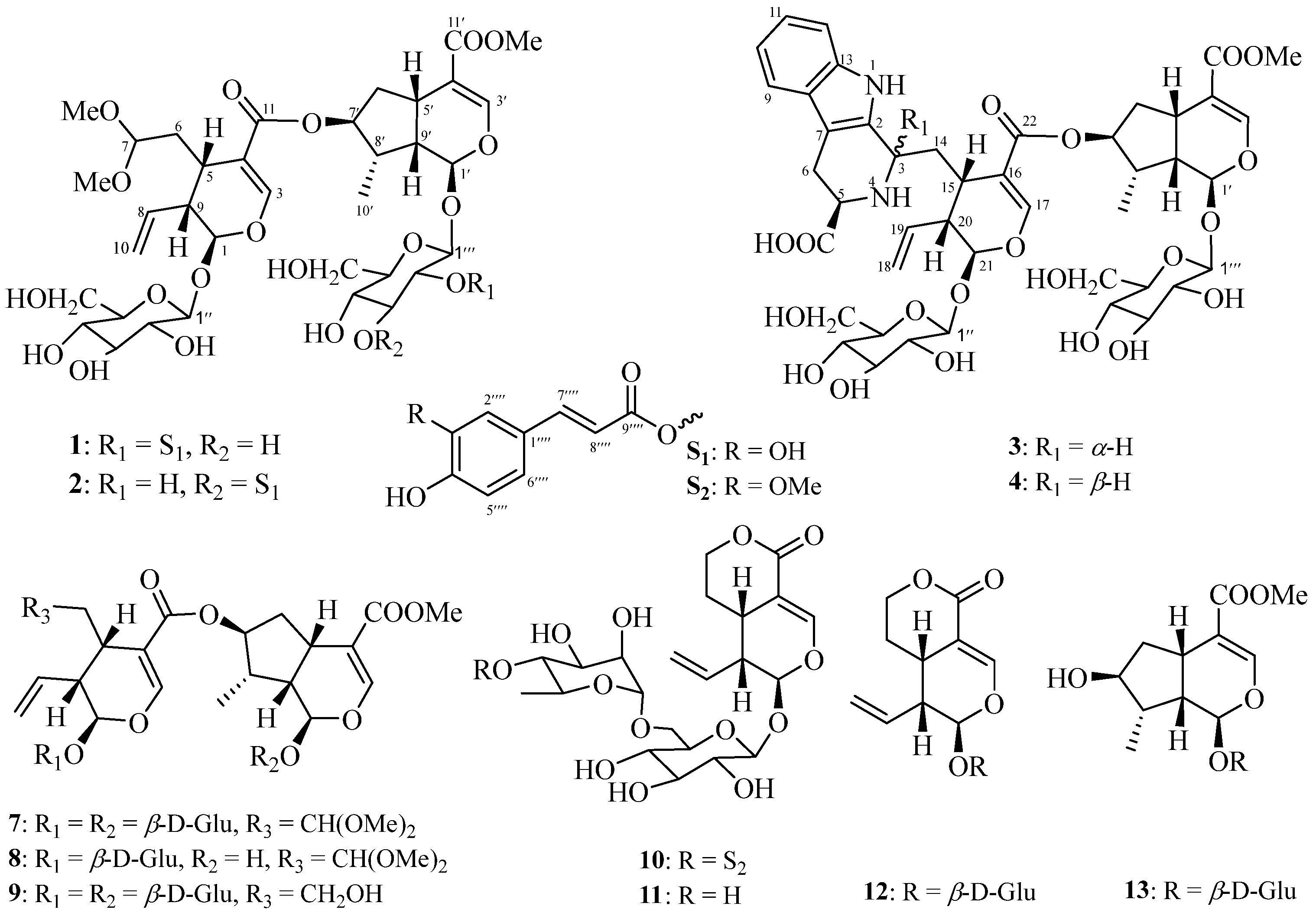
Figure 1. Iridoids (14 and 713) from the twigs/leaves and flower buds of K. amabilis.
Molecules 29 05980 g001
Figure 2. Triterpenoids (5, 6, and 1431) from the twigs and leaves of K. amabilis.
Molecules 29 05980 g002
Figure 3. Key 1H-1H COSY, HMBC, and NOESY correlations of compound 1.
Molecules 29 05980 g003
Figure 4. Key 1H-1H COSY, HMBC, and NOESY correlations of compound 3.
Molecules 29 05980 g004
Figure 5. Acetyl CoA carboxylase 1 (ACC1) inhibitory IC50 values for compounds 2 and 17; ATP-citrate lyase (ACL) inhibitory IC50 values for compounds 8, 17, 29, and 31. Each bar represents the mean ± standard deviation (SD) (n = 3).
Molecules 29 05980 g005
Figure 6. Molecular docking model of compound 2 bound to ACC1 (PDB ID: 3TVU), with various interactions shown as dotted lines (hydrogen bond: red; π-π: grayish green).
Molecules 29 05980 g006
Table 1. 1H (400 MHz) and 13C (100 MHz) NMR data a (δ in ppm) of compounds 1 and 2 in CD3OD.
No.12
δH (J in Hz)δCδH (J in Hz)δC
15.52, d (5.5)97.85.52, d (5.5)97.8
37.43, br s153.37.45, br s153.3
4 112.0 112.0
52.89, m29.52.91, m29.4
62.06, m; 1.62, m33.22.06, m; 1.62, m33.2
74.50, dd (6.9, 4.4)104.14.50, dd (7.3, 4.7)104.1
85.74, m135.95.74, m135.9
92.67, m45.42.70, m45.4
105.26, br d (17.0)119.85.31, br d (17.1)119.8
5.20, br d (10.6)5.27, br d (10.2)
11 168.3 168.3
1′5.47, d (1.8)95.55.34, d (2.0)97.4
3′7.23, br s150.77.43, br s152.5
4′ 114.5 113.4
5′2.93, m30.93.11, m32.4
6′2.17, m; 1.77, m39.62.31, m; 1.75, m40.3
7′5.10, m78.65.20, m78.3
8′1.92, m40.02.12, m41.0
9′2.22, m47.22.12, m47.1
10′1.03, d (7.0)12.81.08, d (5.6)13.7
11′ 168.7 169.3
1″4.69, d (7.9)100.04.68, d (8.0)100.1
2″3.19, m74.63.19, m74.6
3″3.38, m78.03.38, m78.0
4″3.28, m71.53.28, m71.5
5″3.30, m78.43.30, m78.4
6″3.93, dd (12.1, 1.5)62.73.89, dd (12.0, 2.0)62.7
3.69, dd (12.1, 5.8)3.70, dd (12.0, 5.9)
1‴4.86, d (8.1)97.64.79, d (8.0)100.1
2‴4.79, dd (9.0, 8.1)74.53.39, m73.2
3‴3.62, m75.85.06, dd (9.3, 9.2)78.8
4‴3.38, m71.73.52, dd (9.4, 9.3)69.8
5‴3.38, m78.53.27, m78.2
6‴3.92, br d (12.4)62.83.92, dd (12.0, 2.0)62.5
3.68, dd (overlapped)3.66, dd (12.0, 6.3)
1″″ 127.6 127.7
2″″7.03, s115.37.04, s115.1
3″″/4″″ 149.8/147.0 149.8/147.0
5″″6.78, d (8.0)116.56.76, d (8.0)116.5
6″″6.94, d (8.0)123.26.94, d (8.0)123.0
7″″7.47, d (15.9)147.07.59, d (15.9)146.9
8″″6.16, d (15.9)114.76.32, d (15.9)115.3
9″″ 168.0 169.0
7-OMe3.28, s (6H)53.5 or 52.83.29, s (6H)53.6 or 52.7
11′-OMe3.15, s51.73.68, s51.7
a Assignments were made by a combination of 1D and 2D NMR experiments.
Table 2. 1H (400 MHz) and 13C (100 MHz) NMR data a (δ in ppm) of compounds 3 and 4 in CD3OD.
No.34
δH (J in Hz)δCδH (J in Hz)δC
2 130.2 130.0
34.51, br d (11.3) 53.24.84, dd (3.7, 3.7) 52.7
53.89, dd, (11.7, 4.0) 59.64.12, dd (9.1, 5.7) 55.0
63.44, m; 3.01, m 24.03.39, m; 3.13, m 24.1
7/8 108.4/138.5 107.3/138.4
97.48, d (7.4)119.87.47, d (7.8)119.2
107.04, dd (7.4, 7.0)120.77.04, dd (8.0, 7.2)120.5
117.13, dd (8.0, 7.0)123.57.14, dd (7.8, 7.2)123.5
127.31, d (8.0)112.27.36, d (8.0)112.4
13 127.5 127.4
142.45, m; 2.25, m 34.62.36, m; 2.17, m 35.3
153.09, m 32.73.16, m 31.5
16 108.9 110.8
177.87, br s157.37.64, br s154.9
185.40, br d (17.8); 5.26, br d (10.4)119.25.34, br d (17.2); 5.30, br d (10.3)120.8
195.85, m135.25.93, m135.0
202.81, m 45.32.18, m 45.6
215.92, d (8.3) 97.65.63, d (7.1) 97.8
22 171.2 169.3
23 173.5 174.0
1′5.27, d (5.5) 97.55.28, d (4.9) 97.6
3′7.41, br s152.77.44, br s152.7
4′ 113.0 113.1
5′3.12, m 32.83.11, m 32.8
6′2.35, m; 1.75, m 40.52.34, m; 1.76, m 40.4
7′5.29, m 79.85.28, m 79.1
8′2.16, m 41.32.14, m 41.1
9′2.08, m 47.02.07, m 47.1
10′1.09, d (6.7) 14.11.04, d (6.7) 13.9
11′ 169.3 169.4
1″4.84, d (7.7)100.54.75, d (7.8)100.4
2″3.24, m 74.73.21, m 74.7
3″3.40, m 78.03.38, m 78.0
4″3.28, m 71.73.25, m 71.7
5″3.41, m 78.83.25, m 78.6
6″4.02, br d (11.7); 3.66, m 63.13.97, dd (12.0, 2.0); 3.65, m 62.8
1‴4.65, d (7.7)100.24.65, d (7.7)100.2
2‴3.17, m 74.63.17, m 74.7
3‴3.35, m 77.93.38, m 78.0
4‴3.23, m 71.63.25, m 71.6
5‴3.29, m 78.43.34, m 78.4
6‴3.88, br d (11.5); 3.61, m 62.83.89, dd (12.0, 2.0); 3.62, m 62.8
OMe3.68, s 51.83.69, s 51.8
a Assignments were made by a combination of 1D and 2D NMR experiments.
Table 3. 1H (400 MHz) and 13C (100 MHz) NMR data a (δ in ppm) of compounds 5 and 6 in CD3OD.
No.56
δH (J in Hz)δCδH (J in Hz)δC
1α1.13, dd (overlapped) 47.41.13, dd (overlapped) 47.3
2.27, dd (12.4, 5.3)2.27, dd (12.7, 5.3)
23.54, m 74.23.54, m 74.1
33.76, d (9.1) 79.63.77, d (9.0) 79.6
4 151.5 151.5
51.75, br d (overlapped) 51.11.77, br d (12.6) 51.1
61.53, m 22.21.55, m 22.2
71.42, m 32.01.43, m 32.0
8 43.5 43.2
92.15, d (2.9) 53.42.17, d (2.8) 53.4
10 39.2 39.3
116.56, dd (11.8, 2.9)126.56.50, dd (10.6, 2.8)126.7
125.72, d (11.8)127.65.77, d (10.6)127.6
13 138.9 136.3
14 42.0 42.5
151.97, m; 1.07, m 26.11.07, m 26.3
162.23, m; 1.35, m 34.21.90, m; 1.68, m 33.4
17 47.3 47.7
18 136.8 134.9
192.88, m 38.53.22, m 37.5
201.70, m 36.2 137.6
211.96, m; 1.19, m 26.15.43, br d (5.0)121.0
221.73, m 35.32.77, dd (16.0, 6.2) 38.9
1.73, m1.74, br d (16.0)
235.16, 4.73, br s105.05.17, 4.74, br s105.5
250.75, s 16.60.76, s 16.6
260.90, s 17.10.93, s 17.3
271.00, s 20.90.99, s 20.2
28 180.3 182.4
291.05, d (7.2) 21.91.18, d (7.4) 21.4
300.92, d (7.1) 20.21.66, s 21.8
a Assignments were made by a combination of 1D and 2D NMR experiments.
Table 4. ACC1 and ACL inhibitory effects of compounds 2, 8, 17, 29, and 31.
CompoundIC50 a
ACC1ACL
29.6 ± 1.0 μM>20
8>203.6 ± 0.9 μM
1710.3 ± 0.8 μM2.0 ± 0.5 μM
29>201.6 ± 0.4 μM
31>204.7 ± 1.3 μM
ND 630 b2.0 ± 0.1 nMNT d
BMS 303141 cNT d0.3 ± 0.1 μM
a These data are expressed as the mean ± SEM of triplicated experiments. b The positive control for the acetyl CoA carboxylase 1 (ACC1) assay. c The positive control for the ATP-citrate lyase (ACL) assay. d NT: not tested.
Disclaimer/Publisher’s Note: The statements, opinions and data contained in all publications are solely those of the individual author(s) and contributor(s) and not of MDPI and/or the editor(s). MDPI and/or the editor(s) disclaim responsibility for any injury to people or property resulting from any ideas, methods, instructions or products referred to in the content.