Article

Characterization of Bioactive Phenolic Compounds Extracted from Hydro-Distillation By-Products of Spanish Lamiaceae Plants

by
Silvia Pérez-Magariño
*,
Marta Bueno-Herrera
and
M. Carmen Asensio-S.-Manzanera
Agrarian Technological Institute of Castilla and León, Consejería de Agricultura y Ganadería, Ctra Burgos Km 119, Finca Zamadueñas, 47071 Valladolid, Spain
*
Author to whom correspondence should be addressed.
Molecules 2024, 29(22), 5285; https://doi.org/10.3390/molecules29225285
Submission received: 30 September 2024 / Revised: 4 November 2024 / Accepted: 6 November 2024 / Published: 8 November 2024

Abstract

:
Plants of the Lamiaceae family are widely used for the extraction of essential oils, and this industry generates a large number of solid residues as by-products, which contain non-volatile valuable compounds. The aim of this work was to identify and quantify the phenolic compounds present in these solid residues from different important Spanish species of Lamiaceae to characterize and valorize them. Forty-seven phenolic compounds were identified by HPLC-DAD-MS and quantified by HPLC-DAD. Different concentrations and types of phenolic compounds were found between the solid residues. The Rosmarinus officinalis extracts showed the highest total phenolic content due to their high phenolic terpene concentrations. The Thymus mastichina extracts were characterized by kaempferol and flavanones, and some flavones were derived from luteolin and apigenin. Finally, the sample Lavandula and Salvia lavandulifolia extracts presented the lowest content of most phenolic compounds, with the exception of some phenolic acids, such as danshensu, salvianolic acid A, and glucosides of hydroxycinnamic acids. Therefore, this work provides information on the quantification of a large number of phenolic compounds using a simple, sensitive, reproducible, and accurate methodology. In addition, the results indicate that these solid residues still contain important amounts of different polyphenols, which are antioxidants and can be used in different industries.

1. Introduction

Lamiaceae is a cosmopolitan family of great diversity, comprising a large number of species. Species belonging to this family are commonly used as medicinal and/or aromatic plants (MAPs) because of their ability to synthesize volatile compounds and also because they are an important source of polyphenols. Plants of the Lamiaceae family are mainly used as ornamentals for the extraction of essential oils (EOs), for the food industry as nutraceuticals, and for phytotherapy and have increasing value and potential worldwide [1,2,3]. The global market for EOs was valued at USD 7.6 billion in 2018 and is expected to reach USD 15.1 billion by the year 2026, as evidence of the growing use of these products emerges worldwide [4].
EOs are usually extracted from raw plant material (flowers, buds, seeds, leaves, twigs, bark, herbs, wood, fruits and roots) by hydro-distillation or by steam distillation.
The EO yield is 0.5–8 w/w % of the dry plant, so the industrial processing of MAPs generates a large number of solid residues as by-products. Between 2017 and 2021, this industry generated more than 250 thousand tons of distillation residues per year [5]. Their disposal can lead to serious environmental problems, especially if they are incinerated or landfilled. Nowadays, the main challenge for the MAP sector is to add value to the by-products of the distillation and drying process [2]. Some industries attempt to valorize such waste by using them for energy or composting [6]. These recycling systems have some disadvantages. The industry requires a huge investment to recycle the by-products into energy. On the other hand, recycling for composting is not always satisfactory due to the antigermination properties of some MAPs, which can also be transferred from the plant residues. There is an increasing interest in recovering valuable bioactive compounds from the residues of the EO industry for their valorization [7]. These solid distillation residues still contain non-volatile valuable compounds, such as different polyphenols, which can be extracted and used as antioxidants in the food, feed, or cosmetic industries [8,9].
It is known that the distillation process can significantly affect the presence and number of different phenols in the solid residues, but nevertheless, they are an important source of phenolic compounds [6,10,11,12,13,14,15]. These compounds can be extracted using several solvents and their mixtures, such as methanol, ethanol, acetone, ethyl acetate, or water [14]. The nature and content of phenolic compounds varies widely between aromatic plants and species. The most important compounds in the Lamiaceae family are phenolic acids, flavonoids, and phenolic terpenes [16,17,18]. Therefore, the use of organic solvents should be necessary to favor the extraction of more non-polar compounds, mainly flavonoids and phenolic terpenes. Liquid Chromatography with Diode Array Detection and/or Mass Spectrometry is the most commonly used technique to evaluate and characterize the phenolic compounds of different plant materials [17,18,19]. The identification and quantification of phenolic compounds in plant material is a difficult task due to the complexity of polyphenolic structures, the large number of compounds that can be found, and the limited number of commercially available standards.
Therefore, most of the studies focus on the tentative identification of a large number of compounds using complex techniques with MS/MS or Q-TOF-MS detectors, but only some of them are focused on the quantification of these compounds. Moreover, these techniques require a high level of investment, infrastructure, and highly skilled personnel, which are beyond the reach of most industries and laboratories. The aim of this work was to identify and quantify as many as possible of the phenolic compounds present in the solid residue obtained after the EO extraction from different important Spanish species of Lamiaceae in order to characterize and add value to these residues. Six different aromatic plants were studied: Thymus mastichina L. (TM), Salvia lavandulifolia Vahl (SL), Lavandula angustifolia Mill. (LA), Lavandula latifolia Medik (LL), three types of Lavandula x intermedia Emeric ex Loisel. (LI_ Abrial, LI_Grosso and LI_Super), and Rosmarinus officinalis L. (RO). In addition, a simple analytical method using High-Performance Liquid Chromatography with Diode Array Detection (HPLC-DAD) and Mass Spectrometry (MS) was developed and validated. This work provides a simple methodology that allows, for the first time, the complete characterization and simultaneous quantification of 47 phenolic compounds in the by-products obtained from the distillation process of different Spanish MAPs. It has also highlighted the important amounts of bioactive phenolic compounds that can be extracted from these MAPs for use in different industries.

2. Results and Discussion

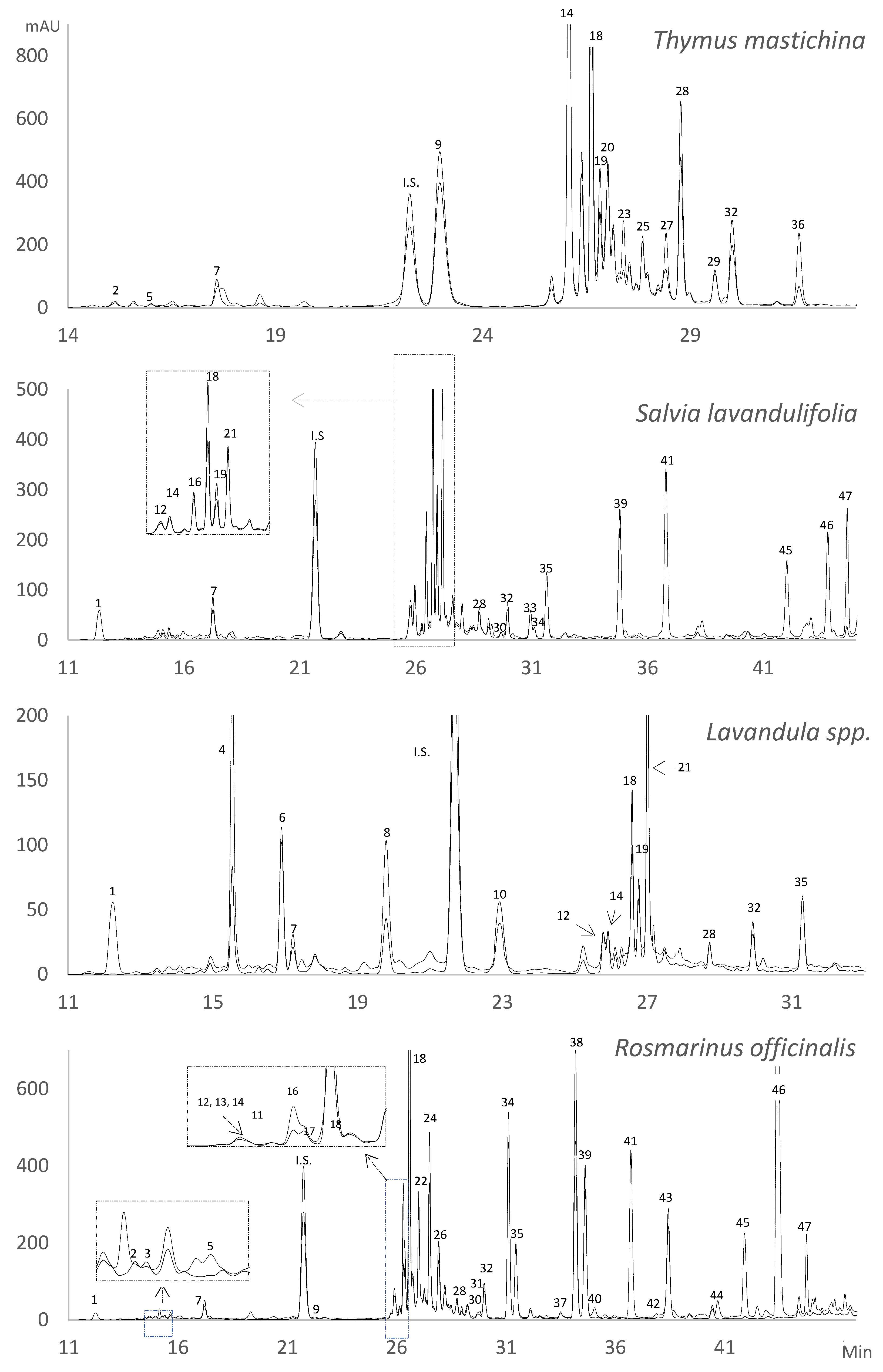
2.1. Identification of Phenolic Compounds in the Solid Residue from Hydro-Distillation of the Aromatic Plants

The extraction technique and the chromatographic method used allowed the characterization of forty-seven phenolic compounds, grouped into phenolic acids, flavonoids (flavones, flavanones, and flavonols), and phenolic terpenes. Table 1 summarizes the spectroscopic and the spectrometric data of all the identified phenolic compounds, numbered according to their elution order. Figure 1 shows the chromatograms of the different phenolic profiles of the Lamiaceae plants studied at two different wavelengths.
Twenty-nine compounds were identified by the standard compounds, and eighteen were tentatively identified from their UV absorption spectra and mass spectral data reported in the bibliography, as discussed below.

2.1.1. Phenolic Acids

Twelve phenolic acids were identified, six by standard compounds (danshensu (3,4-dihydroxyphenyl lactic acid), chlorogenic acid, p-hydroxybenzoic acid, caffeic acid, rosmarinic acid, and salvianolic acid A) and another six were tentatively identified. Peak 3 had the same UV absorption spectrum as chlorogenic acid (UVmax 326 nm), with the same m/z 353 and fragments. It was identified as crytochlorogenic acid, which was also supported by Irakli et al. [18], who identified this compound by its standard and it was detected in RO.
Peak 4 presented the molecular ion m/z 325 and a fragment ion at 163 m/z corresponding to the coumaric acid and a loss of 162 amu that reveals the presence of glucose or galactose. However, no galactoside compounds of hydroxycinnamic acids have been found in the literature, so this glycoside is most probably glucoside [20], and therefore, this compound was tentatively identified as coumaric acid-O-glucoside [8,17]. Similarly, peaks 6 and 10 were identified as glucosides of ferulic acid since they presented the molecular ion at m/z 355 and fragment ions at m/z 193, corresponding to the ferulic acid with a loss of glucose. These two compounds were also identified by Torras-Claveria et al. [8] and Contreras et al. [17] in the genus Lavandula and other Lamiaceae species, respectively. Other glucosides of hydroxycinnamic acids identified were dihydro-p-coumaric acid glucoside or hydroxyhydrocinnamic acid glucoside (peak 8) found in the genus Lavandula and rosmarinic acid-3-O-glucoside (peak 11) in RO. The mass fragmentation data of peak 8 showed the molecular ion at m/z 327 and a fragment ion at m/z 165, indicating the loss of glucose.
These data agree with those found by Contreras et al. [17], who identified this compound in Helleborus niger. Rosmarinic acid-3-O-glucoside presented the molecular ion m/z 521, and it was also identified by Borras-Linares et al. [21] and Sharma et al. [22] in RO.

2.1.2. Flavones and Flavonols

Twenty-one flavones were identified, of which fourteen were standard compounds (luteolin-7-O-rutinoside, luteolin-7-O-glucuronide, luteolin-7-O-glucoside, nepetin-7-O-glucoside (nepitrin), apigenin-7-O-glucoside, scutellarein, luteolin, cirsiliol, apigenin, eupatorine, cirsimaritin, acacetin, genkwanin and salvigenin) and seven were tentatively identified. All of the flavones were derivatives of apigenin and luteolin. Peaks 9, 20, and 26 were luteolin derivatives. Peak 9 was identified as 6-hydroxyluteolin-7-O-glucoside and presented molecular ions at m/z 463 and a fragment ion at m/z 301 due to the loss of a glucose moiety. This compound was found in TM and RO and was also identified by Borras-Linares et al. [21] and Sharma et al. [22] in RO. Peak 20 presented the ion m/z 301 and the same UV absorption spectrum as luteolin, and it was tentatively identified as 6-hydroxyluteolin, being identified only in TM. Peak 26 had a molecular ion at m/z 503 and fragment ions at m/z 399 and 285. This fragmentation pattern agreed with one of the isomers of luteolin-3’-O-(O-acetyl)-β-D-glucuronide identified in RO by Borras-Linares et al. [21], Mena et al. [16], Achour et al. [23], and Sharma et al. [22] and it could be luteolin-3’-O-(2″-O-acetyl)-β-D-glucuronide [21,23].
Peaks 22, 24, 35, and 43 were apigenin derivatives, all of which were identified in RO, and peak 35 was also identified in SL and the genus Lavandula. Peaks 22 and 24 presented the molecular ions at m/z 461 and different fragment ions at m/z 299 and 285, respectively.
The first peak (peak 22) was identified as hispidulin-7-glucoside, with the fragment ion m/z 299 corresponding to hispidulin with a loss of glucose (162 amu), also called homoplantaginin according to the data found in the bibliography [18,22,24].
Peak 24 was identified as scutellarin or scutellarein-7-glucuronide, with the fragment ion m/z 285 corresponding to scutellarein with a loss of glucuronide acid (176 amu), as was previously reported by Sharma et al. [22].
The molecular ion of peak 35 was only found in the positive mode with m/z 315, which may correspond to ladanein, a dimethyl scutellarein derivative, with methylations at 4′ and 7 hydroxyls [25,26]. According to Psarrou et al. [26], this compound showed two maximum absorbances at 286 and 334 nm, which agrees with the data in our study (Table 1). This pattern of the protonated molecular ion [M-H] + was also found in two other flavones, eupatorin and salvigenin as compounds for which a standard was available. Peak 43 also presented an apigenin-type UV spectrum with maximum absorbances at 268 and 332 nm with a molecular ion at m/z 299 in the positive mode. This compound was identified as 4′-methoxytectochrysin or apigenin-4′,7-dimethylether, taking into account the data found in the bibliography and the fact that it was the only flavonoid that elutes between carnosol and carnosic acid [19,25,26,27].
Five flavanones were identified by their respective standards, naringenin, naringenin-7-O-glucoside, eriodictyol, sakuranetin, and hesperidin. The first four compounds were only detected in the TM extracts, and hesperidin was only detected in the SL and RO extracts.
Kaempferol was identified by its standard, and it was the only flavonol detected in the TM extracts.

2.1.3. Phenolic Terpenes

Finally, the phenolic terpenes were detected in the solid residue of RO and SL. Carnosic acid is known to be unstable, and its degradation is favored by temperature, exposure to light, and the presence of oxygen. Under these conditions, carnosic acid can be oxidized to carnosol, rosmanol, epirosmanol, epiisorosmanol, methylepirosmanol, rosmadial, and methylcarnosate, among others [28,29,30]. Bicchi et al. [31] and Troncoso et al. [32] reported that carnosol is the main compound obtained after the degradation of carnosic acid, which can be transformed into other phenolic diterpenes such as rosmanol and its isomers, epirosmanol, and epiisorosmanol. In addition, the use of methanol in the extraction and under acid conditions could break the lactone bond of carnosol, giving rise to other compounds derived from carnosic acid, and carnosol [29].
In this study, eight phenolic terpenes were identified: three by standards (rosmanol, carnosol and carnosic acid), and the other five were tentatively identified. All these compounds showed similar UV spectra to carnosic acid and carnosol with maximum absorbances at 284 nm, so they are derived from carnosic acid.
Peak 40 presented the molecular ion at m/z 359 and a fragment ion at m/z 283, corresponding to the loss of a methoxy (-O-CH3) and an acid (-COOH) group. This compound was identified as rosmanol methyl ether or epirosmanol methyl ether according to Borras-Linares et al. [21], Mena et al. [16], and Sharma et al. [22]. This compound can be formed from carnosol due to the use of methanol for extraction, which agrees with Doolaege et al. [29]. Zhang et al. [30] obtained the corresponding ethyl ether of epirosmanol as a degradation product of carnosol when carnosol was dissolved in ethanol. These degradation products were not formed when an aprotic solvent such as acetonitrile was used.
Peak 42 had a molecular ion at m/z 343 and a fragment ion at m/z 299 due to the loss of an acid group (-COOH), and a maximum absorbance at 288 nm. These data agreed with those found in the bibliography [21,22,30] and it was identified as rosmadial.
Peaks 44 and 47 also presented UV absorption spectra similar to carnosol and carnosic acid and molecular ions at m/z 329 and 345, respectively, which were identified as carnosol isomer and 12-methylcarnosic acid, according to other studies [21,22]. The carnosol isomer showed the same fragmentation pattern as carnosol (with molecular ions at m/z 329 and a fragment ion at m/z 285). On the other hand, 12-methylcarnosic acid had two fragment ions (m/z 301 and 286) corresponding to the loss of the acid group (44 amu) and the subsequent loss of the methyl group (15 amu).
Finally, peak 45 presented a molecular ion at m/z 301 and showed a UV absorption maximum at 280 nm. Different authors have reported a compound with these mass and UV spectral characteristics, but they have assigned different structures. Borras-Linares et al. [33] identified it as salviol, which is an abietatriene compound with a single phenol group isolated from some Salvia species. Doolaege et al. [29] identified and characterized one compound with a molecular weight of 302, which is a fragment ion of m/z 301 and has a UV spectrum similar to carnosic acid. They hypothesized that this compound results from the breakdown of the intramolecular bond of rosmanol and its isomers under acid conditions, followed by the formation of an unsaturated double bound and the loss of a CO-group. The UV spectrum of this compound had an absorbance pattern similar to that of our peak 45. Finally, Zhang et al. [30] proposed a structure that was named 5,6,7,10-tetrahydro-7-hydroxy-rosmariquinone, which was a major degradation product derived from carnosic acid. In all these studies, as in our work, this compound eluted before carnosic acid. With the data available in this study, we could not confirm the structure of this compound, but it was clear that it was derived from carnosic acid. Therefore, this compound has been named a carnosic acid derivative.
Finally, two rosmanol isomers, epiisorosmanol, and epirosmanol, were identified by their MS fragmentation patterns. They presented the same molecular ion at m/z 345 as rosmanol, but they had some differences in the fragmentation pattern and eluted at different retention times. According to Kontogianni et al. [24], Achour et al. [23], and Sharma et al. [22], the first isomer, epiisorosmanol, eluted before rosmanol and showed the major fragment ions at m/z 301 and 283, corresponding to the loss of 44 amu (acid group) and later of 18 amu (water molecule). The second, epirosmanol, eluted after rosmanol and showed only the major fragment at m/z 283. These isomers were clearly identified by MS. However, they were not quantified due to the low signal found in the DAD.

2.2. Results of the Method Validation

Table 2 shows the range, calibration curve, correlation coefficients, LOD, and LOQ for each standard phenolic compound. The linearity in the range indicated that all compounds were satisfactory, with correlation coefficients between 0.9952 and 0.9999. The LOD and LOQ values indicated that the developed method had sufficient sensitivity for the analysis of phenolic compounds in these aromatic plant residues.
The repeatability and reproducibility values were satisfactory, ranging from 0.8 to 7.3% for repeatability and from 2.3 to 9.4% for reproducibility, being less than 10% for all the phenolic compounds (Table 3).
In general, good recoveries were obtained, ranging from 80 to 120% for most of the compounds. Only kaempferol, carnosol, and carnosic acid showed recoveries outside this range. Kaempferol showed recoveries of around 65%, which may be due to the fact that this compound is very reactive and may react with other phenolic compounds [34]. Similarly, carnosol and carnosic acid showed recoveries of around 135% and 67%, respectively. This may be due to the fact that carnosic acid is unstable and can be oxidized to carnosol and other related compounds [28,29,30], as previously commented. Furthermore, carnosol is the main compound obtained after carnosic acid degradation [31,32], which is consistent with the loss of about 33% of carnosic acid and the increase to 33% of carnosol.
Therefore, all these results indicate that this method can be considered precise, reproducible, and accurate.

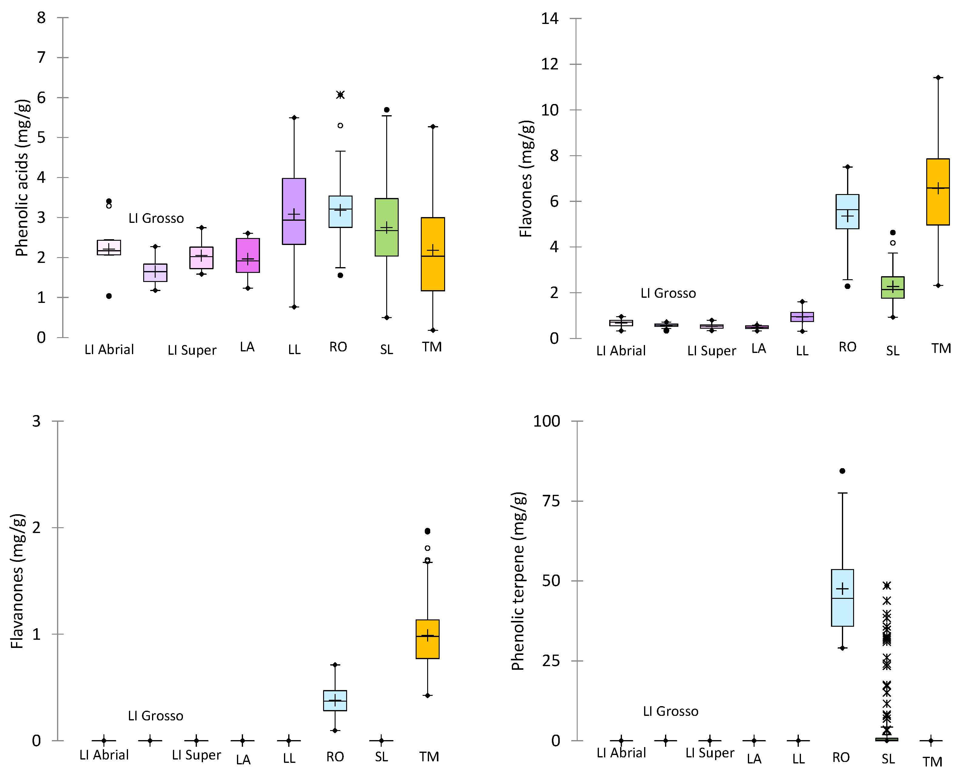
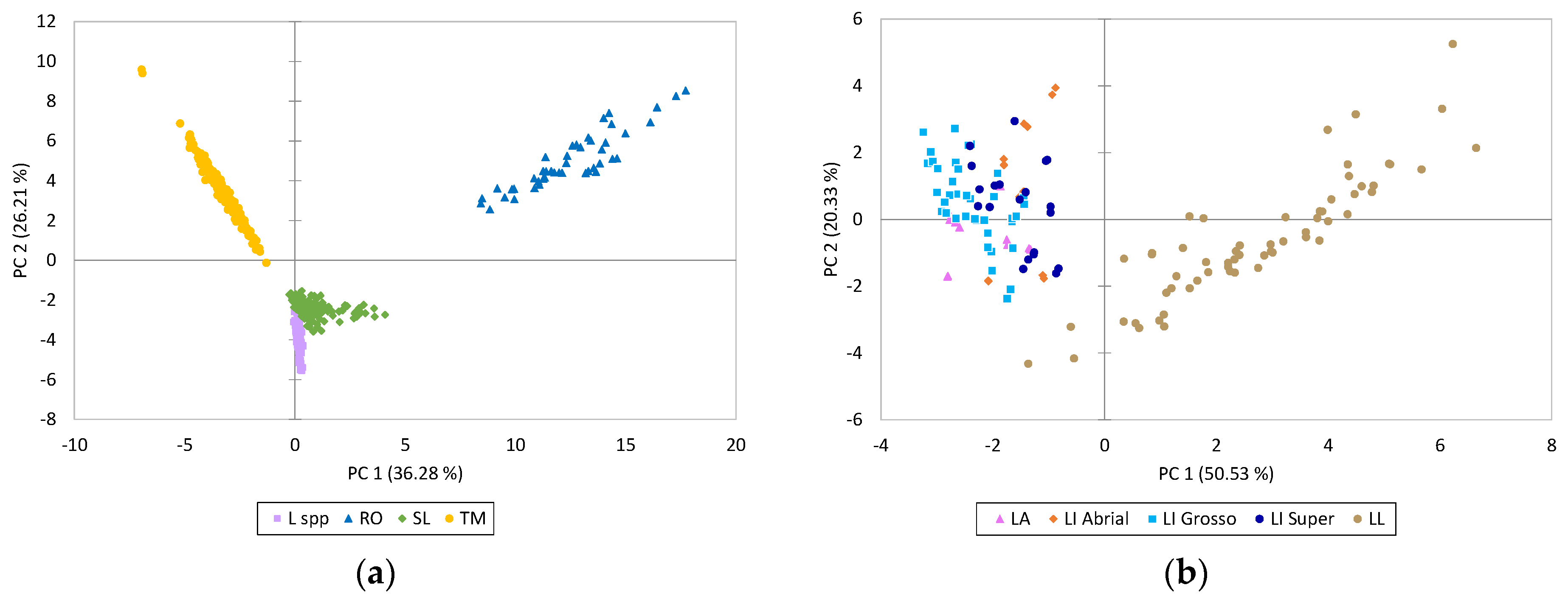
2.3. Quantification of Phenolic Compounds in the Solid Residue from Hydro-Distillation of the Aromatic Plants

Table 4 shows the concentration of the phenolic compounds quantified in the solid residue of the EO by-product of the different Lamiaceae aromatic plants studied. The methodology described above allowed the identification and quantification of forty-seven phenolic compounds. However, not all the compounds were found in the solid residue of the different plants. Only five compounds, caffeic acid, rosmarinic acid, luteolin-7-O-glucoside, luteolin, and apigenin, were found in all the studied by-products.
High differences were found between the solid residues from the Lamiaceae aromatic plants evaluated. The RO extracts showed the highest total phenolic content (56,459 ± 1375 µg/g) due to the high phenolic terpene concentrations. The extracts from TM and SL showed a medium and similar concentration of phenolics (10,002 ± 3193 µg/g and 9301 ± 11,089 µg/g, respectively), although the type of phenolics present in each plant was different. The TM extracts did not contain any phenolic terpenes and were characterized by a higher content of flavones and flavanones in comparison to the SL extracts. The SL extracts were mainly characterized by the presence of phenolic terpenes (46%), followed by phenolic acids (30%) and flavones (24%). Finally, the extracts from Lavandula spp. had the lowest total phenolic content of all the extracts studied (average of 2825 µg/g) and contained only phenolic acids and flavones.
It is noteworthy that differences in the content of phenolic compounds were found between the samples (Figure 2). Although all samples were collected in the same locality, they belonged to several seasons, and different weather conditions can affect the chemical composition. Furthermore, the variability was higher in wild species (TM, SL, LL, and RO) than in those that come from selected clones (Lavandula spp.) (Table 4, Figure 2). Variations in LA and lavandin were exclusively due to the weather conditions between years, while different populations from different locations were used to set the cultivation trials in wild species.
Rosmarinic acid was the major phenolic acid present in the aromatic plants studied as an ester obtained from the esterification product of 3,4-dihydroxyphenyl lactic acid (danshensu) and caffeic acid [35]. The extracts from RO and TM were the richest in rosmarinic acid, followed by the SL extracts. On the other hand, the solid residue of Lavandula spp presented the lowest concentration of rosmarinic acid (between 3 and 10 times lower than the content of the other plant extracts studied) but the highest concentration of danshensu, which is the precursor of rosmarinic acid. Salvianolic acid A was found in SL and Lavandula spp. extracts, which are formed by one molecule of danshensu and two molecules of caffeic acid [36]. Different salvianolic acids were found in high concentrations in Salvia miltiorrhiza (Danshen), which is a plant extensively used in traditional Chinese medicine for the treatment of cardiovascular diseases due to its antioxidant and free radical scavenging activities [37,38]. The rosmarinic acid glucoside was only found in the RO extract and is characteristic of this plant [21]. The hydroxycinnamic acid derivatives of coumaric and ferulic acids were only detected in Lavandula spp. extracts.
The flavonoids were the predominant compounds quantified in TM extracts at 78%. Among the flavonoids, the flavones were the most abundant phenolic group (66%), mainly formed by the aglicones luteolin and apigenin, 6-hydroxyluteolin, 6-hydroxyapigenin, and their O-glycoside derivatives. On the other hand, the RO extracts were characterized by the methyl-ether, methoxy, and glucuronide derivatives of luteolin and apigenin. Among the flavones, the SL extracts stood out for their content of salvigenin, apigenin-7-O-glucoside, and cirsimaritin, and the Lavandula spp. extracts for their content of luteolin-7-O-glucoside, apigenin-7-O-glucoside, and luteolin-7-O-rutinoside. In addition, as previously mentioned, kaempferol was the only flavonol found in TM, and it was not detected in the other solid residues of the MAPs analyzed.
The RO extracts were highlighted for their high phenolic terpene content, accounting for 84% of the total amount of phenolic compounds, with carnosic acid being the most abundant, followed by carnosol, carnosic acid derivative, and 12-methylcarnosic acid. Carnosic acid is a labdane-type diterpene with a strong antioxidant activity, but it is also very unstable in the presence of oxygen [33]. As a result, other phenolic terpenes are formed from the degradation of carnosic acid, including carnosol, methyl carnosate, rosmanol, and rosmadial [29]. Kontogianni et al. [24] attributed the antioxidant activity of fresh RO extracts to the presence of carnosol, rosmarinic acid, and carnosic acid. In addition to the RO extracts, only the SL extracts showed a phenolic terpene content of 46% of the total phenols, but quantitatively, it was 10 times lower than the RO extracts.
A principal component analysis (PCA) was performed to investigate whether the information given by the composition in the phenolic compounds could allow the solid residues obtained from the distillation of the MAPs to be differentiated. The PCA selected four components that explained 75.1% of the total variance (Table S1).
Figure 3a shows the distribution of the samples in the plane defined by the first two dimensions that explained 62.5% of the total variance. As can be observed, all the differences described above in the phenolic profile allowed us to distinguish the solid residues obtained from the distillation of the studied plant species. The RO extracts, cited for the positive side of PC 1 and PC 2, were clearly separated from the other extracts and were positively correlated with the phenolic terpenes; some flavones (mainly glucuronide derivatives of luteolin and apigenin, hispidulin-7-O-glucoside and nepetin-7-O-glucoside); and cryptochlorogenic acid and rosmarinic acid-3-O-glucoside (Table S1). On the other hand, the TM extracts were located on the upper left side of the plane, correlating mainly with kaempferol and flavanones, and some flavones such as aglicones luteolin and apigenin, 6-hydroxyluteolin, 6-hydroxyapigenin, and their O-glycoside derivatives. Finally, the samples belonging to species of the genus Lavandula were grouped together and closer to the S. lavandulifolia samples, indicating that these two species are closer in terms of chemotaxonomy. These samples were located on the lower side of the plane due to the lowest content of most of the phenolic compounds, with the exception of the phenolic acids, danshensu, salvianolic acid A, coumaric acid-O-glucoside, dihydro-p-coumaric acid glucoside, and two ferulic acid-O-glucosides, and the flavone, luteolin-7-O-rutinoside (Table S1).
Another PCA was performed only with the Lavandula spp. samples, and it selected three components that explained 78.3% of the total variance (Table S2). Figure 3b shows the distribution of the Lavandula spp. samples in the plane defined by the first two dimensions that explain 70.9% of the total variance. Among the Lavandula spp. samples, spike lavender (LL), sited on the right side of the plane, was separated from lavender and lavandin, which were grouped together. The LL extracts were positively correlated with the phenolic acids, danshensu, coumaric acid-O-glucoside, dihydro-p-coumaric acid glucoside, rosmarinic acid, and salvianolic acid A, and the flavones, luteolin-7-O-glucoside, and apigenin-7-O-glucoside (Table S2). Although the concentrations of each phenolic compound can be affected by the population origin or the environmental conditions in which the plants grow, the phenolic profile of the species is preserved. Therefore, chemotaxonomy has been widely used in the Lamiaceae family in different genera and has even been used to differentiate taxa at the subspecies level [39,40].

3. Materials and Methods

3.1. Plant Samples

Six different aromatic plants were studied: Thymus mastichina L. (TM), Salvia lavandulifolia Vahl (SL), and Lavandula latifolia Medik (LL) and three types of L. x intermedia Emeric ex Loisel, including (LI_ Abrial, LI_Grosso and LI_Super), L. angustifolia Mill. (LA), and Rosmarinus officinalis L. (RO). All the samples analyzed came from cultivation trials located in the experimental farm of ITACyL in Valladolid. The plant material consisted of several wild populations of TM, SL, LL, and RO (8 to 15 different populations depending on the species) and was previously collected with vegetation multiplied around the Iberian Peninsula. LA, LI_ Abrial, LI_Grosso, and LI_Super plant materials were supplied by a commercial nursery. Samples were collected in different assays from 2013 to 2021. A total of 576 samples from different species and years were analyzed.
The aerial parts of wild and/or cultivated populations of Lamiaceae species were collected during the blossom phase in different years. The plant material was dried at room temperature in the dark for four weeks after collection. When the drying process was finished, leaves and flowers were separated from the stems, and only the mixture of leaves and flowers was used for distillation. The plant material after the distillation process was used for analysis.

3.2. Chemicals

HPLC-grade reagents and analytical-grade reagents were provided by Panreac (Madrid, Spain). The standard phenolic compounds were purchased from Sigma–Aldrich (Steinheim, Germany), Fluka (Buchs, Switzerland), Phytolab (Vestenbergsgreuth, Germany), Extrasynthèse (Lyon, France), and Targetmol (Wellesley Hills, MA, USA), as has been indicated in Table 2. P-coumaric acid was used as the internal standard (IS) and was provided by Sigma–Aldrich (Steinheim, Germany).

3.3. Solid Residue from the Hydro-Distillation of the Aromatic Plants and Extraction of the Phenolic Compounds

In total, 150 g of each dry sample was hydro-distilled (2 L) for 2.5 h using a Clevenger-type apparatus to extract the EOs. The solid residue obtained after the distillation was dried in an oven at 30 °C ± 2 °C for about 48 h. The dry solid residue was ground using a Retsch ZM 1 centrifugal mill (Fisher Scientific S.L., Madrid, Spain) and sieved between 800 and 280 µm.
In total, 0.25 g of the dry solid residue was extracted four times with 10 mL of methanol at room temperature by shaking in an orbital shaker KS-15 A (Bühler, Switzerland) at 150 rpm for 30 min. In the first extraction, 400 µL of p-coumaric acid was added as an internal standard (IS, 400 mg/L). The four extracts were pooled and filtered through nylon membrane filters at 0.45 μm, 47 mm (Filter-Lab, Barcelona, Spain). The extracts of each sample were concentrated under vacuum at 35 °C using a rotary evaporator R-210 (Büchi, Flawil, Switzerland) to a volume of less than 5 mL. This concentrated extract was transferred to a 5 mL volumetric flask and made up to the mark with methanol. The extracts were filtered (PVDF filters, 0.45 μm) and injected into the chromatograph on the day of extraction.

3.4. High-Performance Liquid Chromatography Diode Array Detection (HPLC-DAD) for the Quantification of Phenolic Compounds

The filtered extracts obtained were analyzed with an Agilent Technologies LC series 1200 (Boblingen, Germany) equipped with a quaternary gradient pump, a thermostated column compartment, an automatic injector, and a Diode Array Detection (DAD) system. The chromatographic separation was achieved on a reverse-phase ACE C18 column (250 mm × 4.6 mm i.d., 5 μm particle size) provided by Symta (Madrid, Spain) and thermostated at 25 °C. The mobile phase was solvent A (20 mM formic acid buffer pH = 2.8) and solvent B (methanol). The gradient was linear at a flow rate of 0.8 mL/min: 0 min, 10% B; 5 min, 20% B; 8 min, 38% B; 18 min, 38% B; 20 min, 70% B; 37 min, 90% B; and 38 min, 100% B and then held for 7 min before returning to the initial conditions.
The injection volume of samples and standards was 100 μL, using an injection program mixing 80 μL of solvent A and the 20 μL sample. The UV spectra were recorded in the range of 200–400 nm range, and chromatograms were recorded at 254, 280, 320, and 350 nm.
The quantification was carried out at different wavelengths depending on each compound (Table 1), using the internal standard method. The compounds tentatively identified where commercial standard was available and were quantified using the calibration curve of the available standard with the most similar UV absorption spectrum belonging to the same phenolic class: chlorogenic acid for peak 3, trans-cinnamic acid for peaks 4 and 8, ferulic acid for peaks 6 and 10, rosmarinic acid for peak 11, luteolin for peaks 9, 20, and 26, apigenin for peaks 22, 24, 35, and 43, and rosmanol for peaks 40 and 42 and carnosic acid for peaks 44, 45, and 47. The phenolic compound contents were expressed in micrograms per gram of dry solid residue weight.

3.5. Liquid Chromatography Diode Array Detection and Mass Spectrometer (LC-DAD-MS) for the Identification of Phenolic Compounds

The identification of the phenolic compounds was carried out on an Agilent LC series 1200 (Boblingen, Germany) equipped with a binary pump, a thermostated column compartment, an automatic injector, a 1260 DAD system, and a Mass Spectrometer Series 6410 Triple Quad. The chromatographic conditions were the same as for HPLC-DAD analyses, except that the flow rate was reduced to 0.6 mL/min. An electrospray ionization (ESI) source was used, and the ESI conditions were as follows: capillary voltage, 4 kV; nebulizer pressure, 45 psi; drying gas flow, 8 L/min; and drying gas temperature, 350 °C. Full mass spectra were recorded in both positive and negative ionization modes in the range of m/z 100–1000, using a cone voltage of 150 V.
The compounds without standards were identified tentatively by comparing the UV spectra characteristics and the mass spectra (molecular weight (m/z) and ion fragmentation) with data available in the literature. Most of the compounds were clearly recognized from the negative ESI-MS spectra, but in some cases, they were identified in the positive mode.

3.6. Method Validation

Calibration, precision, accuracy, limits of detection, and quantification were evaluated for method validation.
The internal standard calibration curves were made using the corresponding phenolic standards at twelve different concentrations, and the linearity and range were determined for each phenolic compound.
The precision was calculated by performing three different extractions of each sample on the same day (repeatability, %RSD, n = 3) and on three different days (reproducibility, %RSD, n = 9), and injecting on each day of the extraction.
The accuracy was calculated by spiking two different samples with known amounts of some standards at two different concentrations and analyzing these samples twice a day on three different days for each added concentration to determine the recovery percentage (% RSD, n = 6). Therefore, the matrix effect was considered.
The limit of detection (LOD) is the lowest concentration at which the instrument can detect but not quantify, and the limit of quantification (LOQ) is the lowest concentration at which the instrument can detect and quantify each compound. These limits were calculated from the standard deviation of the y-interceptions of the regression lines (Sy) and the slope of the calibration curve (S) at levels approaching the LOD (LOD = 3.3 × Sy/S and LOQ = 10 × Sy/S).

3.7. Statistical Analyses

The concentrations of each phenolic compound were expressed as the mean ± standard deviation (SD) in the solid residues from each MAP. A Box and Whisker Plot were used to represent the data distribution between the solid residues from different Spanish species of Lamiaceae.
Principal component analysis (PCA) was carried out to determine similarities or differences between the concentration of the phenolic compounds evaluated in the solid residue of the MAPs studied.
All the statistical analyses were performed using the XLSTAT software (Addinsoft, version 2022.1).

4. Conclusions

A simple, sensitive, reproducible, and accurate HPLC-DAD-MS method is proposed that allows the simultaneous identification and quantification of forty-seven phenolic compounds grouped into phenolic acids, flavonoids (flavones, flavanones, and flavonols), and phenolic terpenes in the solid residue obtained after the EO extraction from different important Spanish species of Lamiaceae. The Rosmarinus officinalis extracts showed the highest total phenolic content due to the high concentrations of phenolic terpenes. The Thymus mastichina extracts were characterized by kaempferol and flavanones, and some flavones derived from luteolin and apigenin. Finally, the samples belonging to species of the genus Lavandula were closer to the Salvia lavandulifolia extracts, presenting the lowest content of most of the phenolic compounds, with the exception of some phenolic acids, such as danshensu, salvianolic acid A, and glucosides of hydroxycinnamic acids, and the flavone, luteolin-7-O-rutinoside. These results indicate that these solid residues still contain important amounts of different polyphenols, which are antioxidants and can, therefore, be used in different industries.
Therefore, this work provides information on the quantification of a large number of phenolic compounds using a simple methodology and the characterization and valorization of the solid residues obtained from the distillation process of EOs.

Supplementary Materials

The following supporting information can be downloaded at https://www.mdpi.com/article/10.3390/molecules29225285/s1. Table S1: Factor loadings after varimax rotation of all the samples of solid residues obtained from the distillation of the MAPs; Table S2: Factor loadings after varimax rotation of the Lavandula spp. samples of solid residues obtained from the distillation.

Author Contributions

Conceptualization, S.P.-M. and M.C.A.-S.-M.; methodology, S.P.-M., M.B.-H. and M.C.A.-S.-M.; validation, S.P.-M. and M.B.-H.; formal analysis, S.P.-M.; investigation, M.B.-H.; resources, M.C.A.-S.-M.; writing—original draft preparation, S.P.-M.; writing—review and editing, S.P.-M. and M.C.A.-S.-M.; visualization, S.P.-M.; supervision, S.P.-M.; project administration, M.C.A.-S.-M.; funding acquisition, S.P.-M. and M.C.A.-S.-M. All authors have read and agreed to the published version of the manuscript.

Funding

This research was funded by the Instituto Nacional de Investigación y Tecnología Agraria y Alimentaria (INIA, Spain), grant number RTA2017-00031-C43.

Institutional Review Board Statement

Not applicable.

Informed Consent Statement

Not applicable.

Data Availability Statement

Data are contained within the article and Supplementary Materials.

Conflicts of Interest

The authors declare no conflicts of interest.

References

  1. Brinckmann, J.A.; Kathe, W.; Berkhoudt, K.; Harter, D.E.V.; Schippmann, U. A new global estimation of medicinal and aromatic plant species in commercial cultivation and their conservation status. Econ. Bot. 2022, 76, 319–333. [Google Scholar] [CrossRef]
  2. Taghouti, I.; Cristobal, R.; Brenko, A.; Stara, K.; Markos, N.; Chapelet, B.; Hamrouni, L.; Bursic, D.; Bonet, J.A. The market evolution of medicinal and aromatic plants: A global supply chain analysis and an application of the Delphi method in the Mediterranean area. Forests 2022, 13, 808. [Google Scholar] [CrossRef]
  3. Sharma, A.; Gumber, K.; Gohain, A.; Bhatia, T.; Sohal, H.S.; Mutreja, V.; Bhardwaj, G. Chapter 3—Importance of essential oils and current trends in use of essential oils (aroma therapy, agrofood, and medicinal usage). In Essential Oils. Extraction, Characterization and Applications; Nayik, G.A., Ansari, M.J., Eds.; Academic Press: Cambridge, MA, USA, 2023; pp. 53–83. [Google Scholar] [CrossRef]
  4. Shabankareh, H.G.; Khorasaninejad, S.; Soltanloo, H.; Shariati, V. Physiological response and secondary metabolites of three lavender genotypes under water deficit. Sci. Rep. 2021, 11, 19164. [Google Scholar] [CrossRef] [PubMed]
  5. Stashenko, E.; Martínez, J.R. Essential oils and the circular bioeconomy. In Essential Oils—Recent Advances, New Perspectives and Applications; Viskelis, J., Ed.; IntechOpen: London, UK, 2024; pp. 1–13. [Google Scholar] [CrossRef]
  6. Saha, A.; Basak, B.B. Scope of value addition and utilization of residual biomass from medicinal and aromatic plants. Ind. Crops Prod. 2020, 145, 111979. [Google Scholar] [CrossRef]
  7. Galanakis, C.M. Food Waste Recovery: Processing Technologies and Industrial Techniques, 1st ed.; Academic Press-Elsevier Inc.: London, UK, 2015. [Google Scholar]
  8. Torras-Claveria, L.; Jauregui, O.; Bastida, J. Antioxidant activity and phenolic composition of lavandin (Lavandula x intermedia Emeric ex Loiseleur) waste. J. Agric. Food Chem. 2007, 55, 8436–8443. [Google Scholar] [CrossRef]
  9. Navarrete, A.; Herrero, M.; Martín, A.; Cocero, M.J.; Ibáñez, E. Valorization of solid wastes from essential oil industry. J. Food Eng. 2011, 104, 196–201. [Google Scholar] [CrossRef]
  10. Parejo, I.; Viladomat, F.; Bastida, J.; Rosas-Romero, A.; Flerlage, N.; Burillo, J.; Codina, C. Comparison between the radical scavenging activity and antioxidant activity of six distilled and nondistilled mediterranean herbs and aromatic plants. J. Agric. Food Chem. 2002, 50, 6882–6890. [Google Scholar] [CrossRef]
  11. Asensio-S.-Manzanera, M.C.; Martín, H.; Sanz, M.A.; Herrero, B. Antioxidant activity of Lavandula latifolia, Salvia lavandulifolia and Thymus mastichina collected in Spain. Acta Hortic. 2011, 925, 281–290. [Google Scholar] [CrossRef]
  12. Santana-Méridas, O.; González-Coloma, A.; Sánchez-Vioque, R. Agricultural residues as a source of bioactive natural products. Phytochem. Rev. 2012, 11, 447–466. [Google Scholar] [CrossRef]
  13. Moisa, C.; Copolovici, L.; Bungau, S.; Pop, G.; Imbrea, I.; Lupitu, A.; Nemeth, S.; Copolovici, D. Wastes resulting from aromatic plants distillation. Biosources of antioxidants and phenolic compounds with biological active principles. Farmacia 2018, 66, 289–295. [Google Scholar]
  14. Ortiz de Elguea-Culebras, G.; Melero Bravo, E.; Sánchez-Vioque, R. Potential sources and methodologies for the recovery of phenolic compounds from distillation residues of Mediterranean aromatic plants. An approach to the valuation of by-products of the essential oil market—A review. Ind. Crops Prod. 2022, 175, 114261. [Google Scholar] [CrossRef]
  15. Miljanović, A.; Dent, M.; Grbin, D.; Pedisić, S.; Zorić, Z.; Marijanović, Z.; Jerković, I.; Bielen, A. Sage, Rosemary, and Bay Laurel Hydrodistillation By-Products as a Source of Bioactive Compounds. Plants 2023, 12, 2394. [Google Scholar] [CrossRef] [PubMed]
  16. Mena, P.; Cirlini, M.; Tassotti, M.; Herrlinger, K.A.; Dall’Asta, C.; Del Rio, D. Phytochemical profiling of flavonoids, phenolic acids, terpenoids, and volatile fraction of a rosemary (Rosmarinus officinalis L.) extract. Molecules 2016, 21, 1576. [Google Scholar] [CrossRef] [PubMed]
  17. Contreras, M.M.; Algieri, F.; Rodríguez-Nogales, A.; Gálvez, J.; Segura-Carretero, A. Phytochemical profiling of anti-inflammatory Lavandula extracts via RP–HPLC–DAD–QTOF–MS and –MS/MS: Assessment of their qualitative and quantitative differences. Electrophoresis 2018, 39, 1284–1293. [Google Scholar] [CrossRef]
  18. Irakli, M.; Skendi, A.; Bouloumpasi, E.; Chatzopoulou, P.; Biliaderis, C.G. LC-MS Identification and quantification of phenolic compounds in solid residues from the essential oil industry. Antioxidants 2021, 10, 2016. [Google Scholar] [CrossRef]
  19. Almela, L.; Sánchez-Muñoz, B.; Fernández-López, J.A.; Roca, M.J.; Rabea, V. Liquid chromatograpic–mass spectrometric analysis of phenolics and free radical scavenging activity of rosemary extract from different raw material. J. Chromatogr. A 2006, 1120, 221–229. [Google Scholar] [CrossRef]
  20. Clifford, M.N.; Wu, W.; Kirkpatrick, J.; Kuhnert, N. Profiling the chlorogenic acids and other caffeic acid derivatives of herbal chrysanthemum by LC-MSn. J. Agric. Food Chem. 2007, 55, 929–936. [Google Scholar] [CrossRef]
  21. Borrás-Linares, I.; Stojanović, Z.; Quirantes-Piné, R.; Arráez-Román, D.; Švarc-Gajić, J.; Fernández-Gutiérrez, A.; Segura-Carretero, A. Rosmarinus officinalis leaves as a natural source of bioactive compounds. Int. J. Mol. Sci. 2014, 15, 20585–20606. [Google Scholar] [CrossRef]
  22. Sharma, Y.; Velamuri, R.; Fagan, J.; Schaefer, J. Full-Spectrum analysis of bioactive compounds in Rosemary (Rosmarinus officinalis L.) as influenced by different extraction methods. Molecules 2020, 25, 4599. [Google Scholar] [CrossRef]
  23. Achour, M.; Mateos, R.; Fredj, M.B.; Mtiraoui, A.; Bravo, L.; Saguem, S. A comprehensive characterisation of Rosemary tea obtained from Rosmarinus officinalis L. Collected in a sub-humid area of Tunisia. Phytochem. Anal. 2018, 29, 87–100. [Google Scholar] [CrossRef]
  24. Kontogianni, V.G.; Tomic, G.; Nikolic, I.; Nerantzaki, A.A.; Sayyad, N.; Stosic-Grujicic, S.; Stojanovic, I.; Gerothanassis, I.P.; Tzakos, A.G. Phytochemical profile of Rosmarinus officinalis and Salvia officinalis extracts and correlation to their antioxidant and anti-proliferative activity. Food Chem. 2013, 136, 120–129. [Google Scholar] [CrossRef] [PubMed]
  25. Grayer, R.J.; Eckert, M.R.; Lever, A.; Veitch, N.C.; Kite, G.C.; Paton, A.J. Distribution of exudate flavonoids in the genus Plectranthus. Biochem. Syst. Ecol. 2010, 38, 335–341. [Google Scholar] [CrossRef]
  26. Psarrou, I.; Oreopoulou, A.; Tsimogiannis, D.; Oreopoulou, V. Extraction kinetics of phenolic antioxidants from the hydro distillation residues of rosemary and effect of pretreatment and extraction parameters. Molecules 2020, 25, 4520. [Google Scholar] [CrossRef] [PubMed]
  27. Cuvelier, M.E.; Richard, H.; Berset, C. Antioxidative activity and phenolic composition of pilot-plant and commercial extracts of sage and rosemary. J. Am. Oil Chem. 1996, 73, 645–652. [Google Scholar] [CrossRef]
  28. Schwarz, K.; Ternes, W. Antioxidative constituents of Rosmarinus officinalis and Salvia officinalis. II. Isolation of carnosic acid and formation of other phenolic diterpenes. Z. Lebensm. Unters. Forsch. 1992, 195, 99–103. [Google Scholar] [CrossRef]
  29. Doolaege, E.H.A.; Raes, K.; Smet, K.; Andjelkovic, M.; Van Poucke, C.; De Smet, S.; Verhe, R. Characterization of two unknown compounds in methanol extracts of rosemary oil. J. Agric. Food Chem. 2007, 55, 7283–7287. [Google Scholar] [CrossRef]
  30. Zhang, Y.; Smuts, J.P.; Dodbiba, E.; Rangarajan, R.; Lang, J.C.; Armstrong, D.W. Degradation study of carnosic acid, carnosol, rosmarinic acid, and rosemary extract (Rosmarinus officinalis L.) assessed using HPLC. J. Agric. Food Chem. 2012, 60, 9305–9314. [Google Scholar] [CrossRef]
  31. Bicchi, C.; Binello, A.; Rubiolo, P. Determination of phenolic diterpene antioxidants in rosemary (Rosmarinus officinalis L.) with different methods of extraction and analysis. Phytochem. Anal. 2000, 11, 236–242. [Google Scholar] [CrossRef]
  32. Troncoso, N.; Sierra, H.; Carvajal, L.; Delpiano, P.; Gunther, G. Fast high-performance liquid chromatography and ultraviolet-visible quantification of principal phenolic antioxidants in fresh rosemary. J. Chromatogr. A 2005, 1100, 20–25. [Google Scholar] [CrossRef]
  33. Borrás-Linares, I.; Arráez-Román, D.; Herrero, M.; Ibáñez, E.; Segura-Carretero, A.; Fernández-Gutiérrez, A. Comparison of different extraction procedures for the comprehensive characterization of bioactive phenolic compounds in Rosmarinus officinalis by reversed-phase high-performance liquid chromatography with diode array detection coupled to electrospray time-of-flight mass spectrometry. J. Chromatogr. A 2011, 1218, 7682–7690. [Google Scholar] [CrossRef]
  34. Pérez-Magariño, S.; Ortega-Heras, M.; Cano-Mozo, E. Optimization of a solid-phase extraction method using copolymer sorbents for isolation of phenolic compounds in red wines and quantification by HPLC. J. Agric. Food Chem. 2008, 56, 11560–11570. [Google Scholar] [CrossRef] [PubMed]
  35. Petersen, M.; Simmonds, M.S.J. Rosmarinic acid. Phytochemistry 2003, 62, 121–125. [Google Scholar] [CrossRef] [PubMed]
  36. Hui, X.; Yan-Li, L.; Hong-Cui, T.; Wen-Ting, Z. Preparation and chemical structure identification of methylated metabolites of salvianolic acid A. Chin. J. Anal. Chem. 2014, 42, 65–70. [Google Scholar]
  37. Li, X.; Yu, C.; Lu, Y.; Gu, Y.; Lu, J.; Xu, W.; Xuan, L.; Wang, Y. Pharmacokinetics, tissue distribution, metabolism, and excretion of depside salts from Salvia miltiorrhiza in rats. Drug Metab. Dispos. 2007, 35, 234–239. [Google Scholar] [CrossRef]
  38. Ma, L.; Tang, L.; Yi, Q. Salvianolic Acids: Potential Source of Natural Drugs for the Treatment of Fibrosis Disease and Cancer. Front Pharmacol. 2019, 10, 97. [Google Scholar] [CrossRef]
  39. Koshovyi, O.; Raal, A.; Kovaleva, A.; Myha, M.; Ilina, T.; Borodina, N.; Komissarenko, A. The phytochemical and chemotaxonomic study of Salvia spp. growing in Ukraine. J. Appl. Biol. Biotech. 2020, 8, 29–36. [Google Scholar] [CrossRef]
  40. Staszek, D.; Orłowska, M.; Waksmundzka-Hajnos, M.; Sajewicz, M.; Kowalska, T. Marker fingerprints originating from TLC and HPLC for selected plants from the Lamiaceae family. J. Liq. Chromatogr. R. T. 2013, 36, 2463–2475. [Google Scholar] [CrossRef]
Figure 1. Chromatograms of the phenolic compounds of Thymus mastichina, Salvia lavandulifolia, Lavandula spp., and Rosmarinus officinalis monitored at 280 (dotted line) and 320 nm (continuous line). Peak numbers are the compounds shown in Table 1. IS: internal standard.
Molecules 29 05285 g001
Figure 2. Box and Whisker plot of the different phenolic families of the solid residues from different Spanish species of Lamiaceae. +: mean values; ●, ○ and 🞻: outliers.
Molecules 29 05285 g002
Figure 3. Principal component analysis. (a) Representation of the samples of solid residues of Thymus mastichina L. (TM), Salvia lavandulifolia Vahl (SL), Lavandula spp. (L. spp.), and Rosmarinus officinalis L. (RO); (b) representation of the samples of solid residues of Lavandula latifolia Medik (LL), three types of L. x intermedia Emeric ex Loisel. (LI_ Abrial, LI_Grosso and LI_Super), and L. angustifolia Mill. (LA).
Molecules 29 05285 g003
Table 1. Peak number, spectroscopic and spectrometric data, and quantification wavelength (QW) of the phenolic compounds identified in the solid residue of the EO by-product of Lamiaceae aromatic plants.
PeakCompoundPhenolic Classλ Max (nm)[M-H]Main FragmentsQW (nm)IdC bTM cSL cL cRO c
1Danshensu (3,4-dihydroxyphenyl lactic acid)Phenolic acid282197179, 135280S X eXX
2Chlorogenic acidPhenolic acid300sh a, 326353191, 179320SX X
3Cryptochlorogenic acidPhenolic acid300sh, 326353191, 179320T X
4Coumaric acid-O-glucosidePhenolic acid264, 296sh325163, 119254T X
5p-hydroxybenzoic acidPhenolic acid256137 254SX X
6Ferulic acid-O-glucoside 1Phenolic acid282sh, 304355193, 149320T X
7Caffeic acidPhenolic acid298sh, 320179135320SXXXX
8Dihydro-p-coumaric acid glucosidePhenolic acid276, 314sh327165, 121280T X
96-hydroxyluteolin-7-O-glucosideFlavone282, 344463301350TX X
10Ferulic acid-O-glucoside 2Phenolic acid294, 320355193, 149320T X
11Rosmarinic acid-3-O-glucosidePhenolic acid288sh, 322521359320T X
12Luteolin-7-O-rutinosideFlavone254, 266, 350593285350S XXX
13Luteolin-7-O-glucuronideFlavone258, 266, 350461285350S X
14Luteolin-7-O-glucosideFlavone258, 268, 350447285350SXXXX
15Naringenin-7-O-glucosideFlavanone284, 332sh433271280SX
16Hesperetin-7-rutinoside (hesperidin)Flavanone284, 334sh609301280S X X
17Nepetin-7-O-glucoside (nepitrin)Flavone258, 272, 348477315350S X
18Rosmarinic acidPhenolic acid290sh, 330359197, 179, 161320SXXXX
19Apigenin-7-O-glucosideFlavone268, 336431269320SXXX
206-hydroxyluteolinFlavone252–268, 350301 350TX
21Salvianolic acid APhenolic acid288, 310sh, 336sh493295280S XX
22Hispidulin-7-O-glucosideFlavone276, 334461299350T X
23EriodictyolFlavanone288, 334sh287151, 135280SX
24Scutellarein-7-O-glucuronideFlavone270, 338461285350T X
25ScutellareinFlavone286, 338285 350SX
26Luteolin-3′-acetyl-O-glucuronideFlavone270, 338503399, 285350T X
27NaringeninFlavanone290, 332sh271 280SX
28LuteolinFlavone254, 266, 350285 350SXXXX
29KaempferolFlavonol266, 366285 350SX
30CirsiliolFlavone256–274, 346329 350S X X
31RosmanolPhenolic terpene 290345283280S X
32ApigeninFlavone268, 340269 350SXXXX
33EupatorinFlavone276, 344345 d 350S X
34CirsimaritinFlavone274, 336313298, 283350S X X
35LadaneinFlavone284, 334315 d 350T XXX
36SakuranetinFlavanone288, 330sh285 280SX
37AcacetinFlavone268, 334283 350S X
38GenkwaninFlavone268, 338283 350S X
39SalvigeninFlavone276, 332329 d287 d320S X X
40(Epi)rosmanol methyl etherPhenolic terpene 290359283280T X
41CarnosolPhenolic terpene284329285280S X X
42RosmadialPhenolic terpene284343299, 285280T X
434′-methoxytectochrysinFlavone268, 332299 d 350T X
44Carnosol isomerPhenolic terpene284329285280T X
45Carnosic acid derivativePhenolic terpene280301 280T X
46Carnosic acid Phenolic terpene286331287280S X X
4712-methylcarnosic acid Phenolic terpene284345301, 286280T X
a sh: shoulder. b IdC: identification compound. S: standard compound; T: tentatively identified compound with data reported in the literature. c TM: Thymus mastichina; SL: Salvia lavandulifolia; L: Lavandula spp.; RO: Rosmarinus officinalis. d [M-H]+. e compounds that appear in the different MAPs.
Table 2. Range, linearity, correlation coefficient (R2), sensibility data, and accuracy of the standard phenolic compounds.
CompoundRange (µg/mL)Calibration aR2LOD b (µg/mL)LOQ b (µg/mL)Supplier cAmounts Added (µg/mL)Recovery (% RSD, n = 6)
TMSL
Danshensu2.0–120y = 0.0044x + 0.00120.99841.14.3124/48-97/90
Chlorogenic acid1.0–10y = 0.0130x − 0.00060.99730.371.111/2102/100
p-hydroxybenzoic acid0.94–28y = 0.0427x + 0.01290.99960.070.9221/294/94
Caffeic acid1.0–31y = 0.0223x − 0.00100.99870.702.214/8105/106120/112
Luteolin-7-O-rutinoside1.3–60y = 0.0093x − 0.00090.99970.782.314/8 102/95
Luteolin-7-O-glucuronide2.0–160y = 0.0122x + 0.02240.99910.333.43
Naringenin-7-O-glucoside2.0–40y = 0.0155x + 0.00630.99980.111.34
Rosmarinic acid11–123y = 0.0104x − 0.02680.99983.76.3130/6080/81116/98
Apigenin-7-O-glucoside2.1–83y = 0.0120x + 0.0130.99930.570.954
Salvianolic acid A2.0–80y = 0.0160x + 0.01360.99770.965.216/12 118/102
Eriodictyol2.1–52y = 0.0212x + 0.00880.99960.392.34
Scutellarein0.98–39y = 0.0212x + 0.00090.99860.662.34
Naringenin2.0–40y = 0.0230x + 0.01020.99970.321.84
Luteolin3.1–78y = 0.0200x + 0.00640.99970.382.01
Cirsiliol0.42–2.8y = 0.0285x − 0.00200.99740.140.301
Kaempferol1.0–40y = 0.0130x − 0.00100.99990.260.6827/1460/65
Rosmanol2.2–174y = 0.0018x − 0.00090.99940.894.13
Apigenin1.1–46y = 0.0175x − 0.00600.99970.350.8213/694/96108/103
Cirsimaritin1.3–60y = 0.0072x + 0.00040.99980.451.61
Eupatorin0.96–19y = 0.0226x − 0.00110.99960.230.6544/8 110/116
Sakuranetin2–40y = 0.0201x − 0.00670.99990.181.44
Salvigenin1.0–60y = 0.0147x − 0.02470.99523.77.35
Carnosol2.0–200y = 0.0023x + 0.00380.99850.693.5430/60 140/135
Carnosic acid10–200y = 0.0010x − 0.00620.99741019480/160 64/67
Ferulic acid3.9–90y = 0.0254x − 0.00460.99980.542.22
trans-cinnamic acid5.9–59y = 0.0564x − 0.00940.99970.100.712
a y: relative area; x: concentration (µg/mL). b LOD: limit of detection; LOQ: limit of quantification. c 1: Sigma–Aldrich (Steinheim, Germany); 2: Fluka (Buchs, Switzerland); 3: Phytolab (Vestenbergsgreuth, Germany); 4: Extrasynthèse (Lyon, France); 5: Targetmol (Wellesley Hills, MA, USA).
Table 3. Precision for the phenolic compounds quantified in the solid residue of the EO by-product of Lamiaceae aromatic plants studied.
Repetibility (%RSD, n = 3)Reproducibility (%RSD, n = 9)
Phenolic CompoundsTMSLLLROTMSLLLRO
Phenolic acids
Danshensu-5.0 2.95.3 -6.4 7.4 5.4
Chlorogenic acid2.2--2.96.5--3.5
Cryptochlorogenic acid---3.4---6.5
Coumaric acid-O-glucoside--2.6---8.1 -
p-hydroxybenzoic acid1.2 --6.15.6--7.6
Ferulic acid-O-glucoside 1--2.6---7.6 -
Caffeic acid1.6 2.6 1.01.63.7 4.7 6.4 3.0
Dihydro-p-coumaric acid glucoside--6.4---8.1 -
Ferulic acid-O-glucoside 2--2.2---7.2-
Rosmarinic acid-3-O-glucoside---4.6---5.4
Rosmarinic acid2.4 2.1 3.5 2.16.4 5.1 7.4 2.3
Salvianolic acid A-2.7 2.0 --6.7 6.6 -
Flavones
6-hydroxyluteolin-7-O-glucoside2.2 --4.05.8 --6.2
Luteolin-7-O-rutinoside-2.9 3.8 5.5-5.55.8 6.2
Luteolin-7-O-glucuronide---4.1---7.6
Luteolin-7-O-glucoside1.7 3.5 3.3 1.86.2 7.5 7.3 4.8
Nepetin 7-O-glucoside---2.0---5.0
Apigenin-7-O-glucoside1.6 3.8 3.5 -6.8 6.7 9.3 -
6-hydroxyluteolin1.1 ---6.8 ---
Hispidulin-7-O-glucoside---5.3---6.9
6-hydroxyapigenin-7-O-β-glucoside1.4 ---7.0 ---
Scutellarein-7-O-glucuronide---2.9---5.2
Scutellarein3.6 ---5.9 ---
Luteolin-3′-acetyl-O-glucuronide---2.2---5.2
Luteolin0.8 3.8 3.5 2.64.6 5.1 5.6 3.6
Cirsiliol-5.9 -1.9-6.1 -3.4
Apigenin0.9 5.4 4.3 1.53.7 7.3 8.7 3.1
Eupatorin-2.4 ---4.3 --
Cirsimaritin-2.1-1.5-3.9-2.6
Ladanein-5.1 5.8 1.7-5.2 8.7 3.1
Acacetin---2.6---5.5
Genkwanin---3.0---3.2
Salvigenin-3.5 -2.7-4.7 -3.2
4′-methoxytectochrysin---4.5---4.6
Flavanones
Naringenin-7-O-glucoside<LQ ---<LQ---
Hesperidin-6.2 -6.0-9.3-8.2
Eriodictyol3.2 ---5.8 ---
Naringenin2.6 ---3.8 ---
Sakuranetin4.5 ---5.6 ---
Flavonols
Kaempferol1.3 ---4.0 ---
Phenolic terpene
Rosmanol---7.3---7.6
(Epi)rosmanol methyl ether---5.5---9.4
Rosmadial---4.7---7.1
Carnosol-3.1 -2.7-4.1 -2.8
Carnosol isomer---3.4---4.0
Carnosic acid derivative---5.3---6.7
Carnosic acid-<LQ-3.3-<LQ-5.3
12-methylcarnosic acid ---4.0---5.1
LQ: limit of quantification.
Table 4. Phenolic compounds quantified in the solid residue of the EO by-product of Lamiaceae aromatic plants studied (µg/g of dry weight) a.
Phenolic CompoundsROTMSLLLLALI_AbrialLI_GrossoLI_Super
Phenolic acids
Danshensu307 ± 55-679 ± 2851051 ± 451593 ± 123879 ± 311457 ± 146678 ± 254
Chlorogenic acid44 ± 3437 ± 17------
Cryptochlorogenic acid19 ± 7-------
Coumaric acid-O-glucoside---412 ± 163100 ± 32161 ± 50156 ± 33195 ± 28
p-hydroxybenzoic acid16 ± 1035 ± 18------
Ferulic acid-O-glucoside 1---66 ± 30131 ± 44167 ± 58132 ± 29147 ± 28
Caffeic acid88 ± 14108 ± 32123 ± 3154 ± 1979 ± 1463 ± 18114 ± 4693 ± 54
Dihydro-p-coumaric acid glucoside---244 ± 9769 ± 12116 ± 37107 ± 20104 ± 22
Ferulic acid-O-glucoside 2---57 ± 29 158 ± 54193 ± 75164 ± 32154 ± 37
Rosmarinic acid-3-O-glucoside421 ± 284-------
Rosmarinic acid2283 ± 7201997 ± 11581486 ± 670599 ± 272579 ± 173343 ± 109235 ± 70290 ± 67
Salvianolic acid A--457 ± 273596 ± 304252 ± 98285 ± 135282 ± 98385 ± 113
Flavones
6-hydroxyluteolin-7-O-glucoside40 ± 381600 ± 751------
Luteolin-7-O-rutinoside61 ± 63-298 ± 161158 ± 7591 ± 20150 ± 61122 ± 23146 ± 32
Luteolin-7-O-glucuronide99 ± 91-------
Luteolin-7-O-glucoside79 ± 672235 ± 903195 ± 84385 ± 11814 ± 1352 ± 1945 ± 1362 ± 20
Nepetin 7-O-glucoside352 ± 190-------
Apigenin-7-O-glucoside-793 ± 285426 ± 179273 ± 70153 ± 21255 ± 98162 ± 32133 ± 72
6-hydroxyluteolin-440 ± 242------
Hispidulin-7-O-glucoside302 ± 62-------
6-hydroxyapigenin-7-O-β-glucoside-215 ± 75------
Scutellarein-7-O-glucuronide584 ± 151-------
Scutellarein-201 ± 155------
Luteolin-3′-acetyl-O-glucuronide364 ± 122-------
Luteolin131 ± 43636 ± 323157 ± 5242 ± 2430 ± 1049 ± 2264 ± 1760 ± 22
Cirsiliol56 ± 37-20 ± 14-----
Apigenin195 ± 82665 ± 233145 ± 6129 ± 1095 ± 35100 ± 4481 ± 2164 ± 18
Eupatorin--95 ± 48-----
Cirsimaritin1032 ± 509-382 ± 328-----
Ladanein234 ± 141-74 ± 7146 ± 1493 ± 3372 ± 2093 ± 2570 ± 18
Acacetin127 ± 100-------
Genkwanin606 ± 339-------
Salvigenin398 ± 239-531 ± 343-----
4′-methoxytectochrysin692 ± 308-------
Flavanones
Naringenin-7-O-glucoside-87 ± 85------
Hesperidin380 ± 148-41 ± 86-----
Eriodictyol-311 ± 132------
Naringenin-310 ± 94------
Sakuranetin-279 ± 103------
Flavonols
Kaempferol-268 ± 147------
Phenolic terpene
Rosmanol218 ± 190-------
(Epi)rosmanol methyl ether657 ± 434-------
Rosmadial189 ± 205-------
Carnosol6862 ± 1511-1055 ± 2203-----
Carnosol isomer1809 ± 1605-------
Carnosic acid derivative6937 ± 2416-559 ± 1703-----
Carnosic acid 26,703 ± 9259-867 ± 2687-----
12-Methylcarnosic acid 4174 ± 1789-1769 ± 4541-----
Total phenolic compounds56,459 ± 137510,002 ± 31939301 ± 11,0894015 ± 13432436 ± 4632884 ± 8592213 ± 3412582 ± 431
a means ± standard deviation.
Disclaimer/Publisher’s Note: The statements, opinions and data contained in all publications are solely those of the individual author(s) and contributor(s) and not of MDPI and/or the editor(s). MDPI and/or the editor(s) disclaim responsibility for any injury to people or property resulting from any ideas, methods, instructions or products referred to in the content.

