Article

Structure Characterization of Four New Sesquiterpene Pyridine Alkaloids from Tripterygium wilfordii Hook. f. and Anti-Inflammatory Activity Evaluations

1
School of Pharmaceutical, Hebei University of Chinese Medicine, Shijiazhuang 050091, China
2
National Institutes for Food and Drug Control, Beijing 102629, China
3
School of Traditional Chinese Medicine, Beijing University of Chinese Medicine, Beijing 102488, China
4
Faculty of Functional Food and Wine, Shenyang Pharmaceutical University, Shenyang 110016, China
5
State Key Laboratory of Drug Regulatory Science, Beijing 100050, China
6
Chinese Pharmacopoeia Commission, Beijing 100061, China
*
Authors to whom correspondence should be addressed.
These authors contributed equally to this work.
Molecules 2024, 29(22), 5284; https://doi.org/10.3390/molecules29225284
Submission received: 27 September 2024 / Revised: 26 October 2024 / Accepted: 6 November 2024 / Published: 8 November 2024

Abstract

:
Sesquiterpene pyridine alkaloids (SPAs), as a main class of components in Tripterygium wilfordii Hook. f., possess a variety of bioactivities, such as immunosuppressive, insecticidal, and anti-tumor activities. SPAs can be structurally classed into four subtypes: wilfordate-, evoninate-, iso-wilfordate-, and iso-evoninate types. Our previous study unveiled ten new wilfordate-type SPAs, named wilfordatine A–J, isolated from the roots of Tripterygium wilfordii Hook. f., several of which exhibited significant immunosuppressive activities. As an extension and augmentation of the previous findings, we have now isolated one new iso-wilfordate-type SPA, wilfordatine K (1), alongside three new iso-evoninate-type SPAs, wilfordatines L–N (35), and six known analogs. Their structures were characterized by the extensive use of 1D and 2D NMR spectroscopic analysis, as well as HRMS data. Interestingly, compounds 4 and 6 were found to exhibit potent inhibitory effects on the nuclear factor-kappa B (NF-κB) pathway in lipopolysaccharide (LPS)-induced HEK293/NF-κB-Luc cells, with IC50 values of 1.64 μM and 9.05 μM, respectively. Notably, these two compounds had no influence on the cell viability at a concentration of 100 μM. Consequently, they hold significant promise as potential anti-inflammatory candidates for further exploration and development.

1. Introduction

Tripterygium wilfordii Hook. f., a member of the Celastraceae family [1,2,3], is widely distributed across various regions in China, including Hunan, Zhejiang, and Fujian provinces. It has been clinically used to treat rheumatoid arthritis, systemic lupus erythematosus, and other autoimmune diseases over a long history [4,5,6]. It mainly contains diterpenes, triterpenes, and sesquiterpene pyridine alkaloids (SPAs) [7,8], among which SPAs are the most abundant.
SPAs were characterized by a macrocyclic diacetone skeleton consisting of a polyoxygenated dihydroagarofuran sesquiterpene cone and a pyridine dicarboxylic acid unit, which can be classified into four subtypes: wilfordate (W), evoninate (E), iso-wilfordate (IW), and iso-evoninate (IE) types [9]. Since the discovery of the first SPA by Kuo et al. in 1989 [10], more than 100 SPAs have been reported to date, with the W-type accounting for the most majority, followed by E-type and, to a lesser extent, the IW-type and IE-type [11].
SPAs possess a variety of biological activities, such as anti-inflammatory [12,13], immunosuppressive [14], anti-tumor [15,16], anti-HIV [17,18,19], and insecticidal properties [20,21,22], the first two of which are particularly relevant to the clinical application of T. wilfordii, and several related compounds have been reported. For instance, peritassine A, wilfordinine A, and euonine-suppressed nitric oxide generation at 8.2%, 59.0%, and 54.7%, respectively, in lipopolysaccharide (LPS) induced RAW264.7 cells at a concentration of 5 μM [23]. Moreover, tripterygiumine S and 9′-O-acetyl-7-deacetoxy-7-oxowilfortrine exhibited nitric oxide inhibitory effects with respective IC50 values of 23.80 ± 4.38 μM and 2.59 ± 3.59 μM in the same cells [24]. It is noteworthy that the abovementioned compounds had no influence on the cell viability. Additionally, ebenifoline E-11 (E) and congorinine E-1 (E) demonstrated significant inhibitory effects on the production of cytokines (TNF-α, IL-1β, IL-8, IL-2, IL-4, and IFN-γ) in human peripheral mononuclear cells stimulated by LPS (or phytohemagglutinin) at a concentration of 10 μg/mL, with the inhibitory rates reaching 100% in some cases [25]. It is evident that SPAs represent a new class of potent anti-inflammatory or immunosuppressive agents with low toxicity, which have received extensive attention from scholars.
In a previous study [26], we isolated 20 W-type SPAs from the roots of T. wilfordii, including ten new compounds named wilfordatines A–J. Among them, wilfordatine E, tripfordine A, and wilforine showed significant inhibitory activity on the nuclear factor-kappa B (NF-κB) pathway in LPS-induced HEK293/NF-κB-Luc cells, with IC50 values of 8.75 μM, 0.74 μM, and 15.66 μM, respectively. As an extension and augmentation of these findings, we have now obtained one new IW-type SPA, wilfordatine K (1), alongside three new IE-type SPAs, wilfordatine L–N (35), and six known analogs (Figure 1). The isolation and structural elucidation of these SPAs, as well as the evaluation of their anti-inflammatory activity, will be described in this paper.

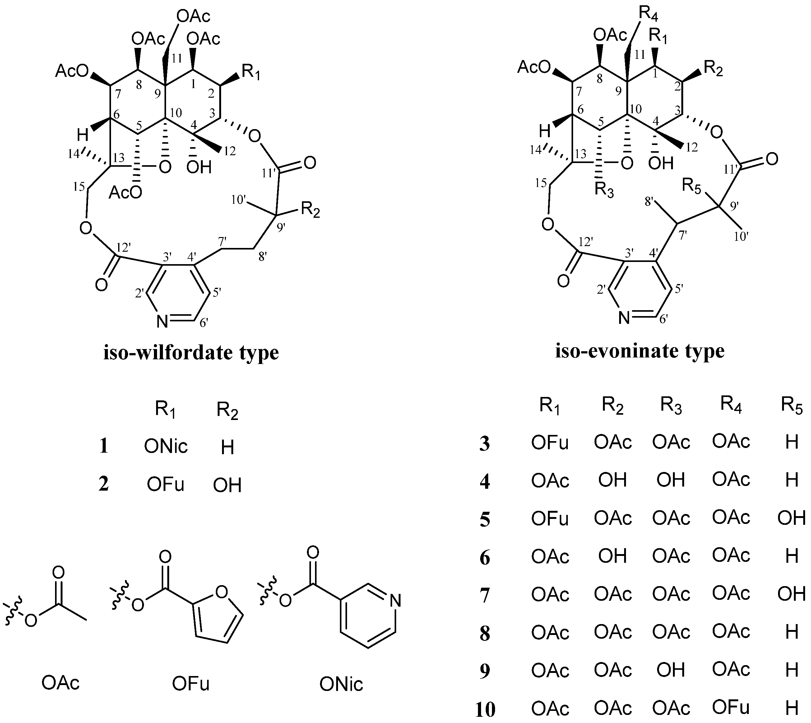
2. Results and Discussion

The CHCl3-soluble fraction of the ethanol extract of the root of T. wilfordii was acid-extracted and alkaline-precipitated to obtain the total alkaloids, which were then subjected to repeated ODS column chromatography and preparative HPLC to yield four new SPAs (1, 35), along with six known analogs (2, 610).
Compound 1 was isolated as a white amorphous powder. The molecular formula C42H48O18N2 was deduced from the quasi-molecular ion [M + H]+ at m/z 869.2991 (calcd 869.2980) in the HRMS data, consistent with 20 degrees of unsaturation (Figure S3). The IR spectrum indicated the presence of hydroxyl (3462 cm−1), methyl (2922 cm−1), carbonyl (1744 cm−1), and ester (1232 cm−1) groups (Figure S2), and the UV spectrum suggested the presence of aromatic rings (223 and 264 nm) (Figure S1). The 1H NMR data (Table 1, Section 3.4, and Figure S4) of compound 1 showed signals for three methyl groups at δH 1.64 (3H, s, H-12), 1.71 (3H, s, H-14), and 1.20 (3H, d, J = 6.6 Hz, H-10′); six oxygenated methines at δH 6.98 (1H, s, H-5), 5.75 (1H, d, J = 3.6 Hz, H-1), 5.55 (1H, t, J = 4.2 Hz, H-7), 5.47 (1H, t, J = 3.0 Hz, H-2), 5.42 (1H, t, J = 4.2 Hz, H-8), and 5.06 (1H, d, J = 3.0 Hz, H-3); two sets of oxygenated methylenes at δH 5.47 (1H, d, J = 13.2 Hz, H-11a), 4.37 (1H, d, J = 13.2 Hz, H-11b), 5.84 (1H, d, J = 12.0 Hz, H-15a), and 3.79 (1H, d, J = 12.0 Hz, H-15b); two aliphatic methines at δH 2.37 (1H, d, J = 3.6 Hz, H-6), and 2.36 (1H, m, H-9′); two sets of aliphatic methylenes at δH 3.90 (1H, m, H-7′a), 2.69 (1H, m, H-7′b), 2.36 (1H, m, H-8′a), and 1.65 (1H, m, H-8′b); 3,4-disubstituted pyridine at δH 9.23 (1H, s, H-2′), 8.69 (1H, d, J = 5.4 Hz, H-6′), and 7.27 (1H, overlapped, H-5′); five acetyloxy groups at δH 2.25(3H, s, 11-OAc), 2.19 (3H, s, 7-OAc), 2.18 (3H, s, 5-OAc), 1.96 (3H, s, 8-OAc), and 1.85 (3H, s, 1-OAc); as well as a nicotinoyloxy group at δH 9.31 (1H, d, J = 1.8 Hz, 2-ONic-2), 8.37 (1H, d, J = 7.8 Hz, 2-ONic-4), 7.48 (1H, dd, J = 7.8, 4.8 Hz, 2-ONic-5), and 8.85 (1H, d, J = 4.8 Hz, 2-ONic-6). The 13C-NMR spectroscopic data (Table 2, Section 3.4, and Figure S5) confirmed the presence of the aforementioned groups, in addition to showing three oxygenated quaternary carbon signals at δC 69.8 (C-4), 93.8 (C-10), and 84.7 (C-13), one aliphatic quaternary carbon signal at δC 52.1 (C-9), and seven ester carbonyl signals at δC 170.4 (11-OAc), 170.1 (7-OAc), 169.9 (5-OAc), 169.3 (1-OAc), 169.0 (8-OAc), 165.8 (2-ONic), 174.6 (C-11′), and 166.7 (C-12′). The 1H-1H COSY cross signals for H-1/H-2/H-3 and H-5/H-6/H-7/H-8 spin systems and the HMBC correlations for H-1/C-9; H-2/C-9; H-3/C-4, C-10; H-5/C-10, C-13; H-6/C-10; H-7/C-5, C-9; H-8/C-9; H-11/C-1, C-8, C-9, C-10; H-12/C-3, C-4, C-10; H-14/C-6, C-13, C-15; and H-15/C-13 (Figure 2A, Figures S6 and S8) suggested the presence of a polyoxygenated dihydroagarofuran sesquiterpene cone unit. In addition, the 1H-1H COSY cross signals for H-7′/H-8′/H-9′/H-10′ and the HMBC correlations for H-5′/C-7′; H-2′/C-12′; and H-10′/C-11′ (Figure 2A, Figures S6 and S8), suggested the presence of a 3-carboxyl-α-methyl-4 -pyridinebutanoic acid unit. The linkage of the above two units was via C-3-O-C-11′ and C-15-O-C-12′, as deduced from the key HMBC correlations for H-3/C-11′ and H-15/C-12′. Therefore, compound 1 was concluded to be an IW-type SPA with a nicotinoyloxy, a hydroxy, and five acetoxy groups attached. The positions of the ester groups were undoubtedly assigned by the HMBC correlations for H-11/δC 170.4 (11-OAc); H-7/δC 170.1 (7-OAc); H-5/δC 169.9 (5-OAc); H-1/δC 169.3 (1-OAc); H-8/δC 169.0 (8-OAc); and H-2/δC 165.8 (2-ONic).
The relative configuration of compound 1 was established by the ROESY experiment and coupling constant analysis. The ROESY correlations for H-1/H-8; H-1/H-14; and H-8/H-14 located these protons on the same face as the dihydroagarofuran skeleton (α-orientation) (Figure 2B and Figure S9). Similarly, the correlations for H-3/H-12; H-5/H-12; and H-11/H-12 were located on the other side of the dihydroagarofuran skeleton (β-orientation) (Figure 2B and Figure S9). The coupling constants between H-1 and H-2 (J1,2 = 3.6 Hz), H-7 and H-8 (J7,8 = 6.0 Hz) indicated that H-2 and H-7 were of the α-orientation.
Therefore, compound 1 was identified as 2β-nicotinoyloxy-1β,7β,8β,11-tetraacetoxy-4α,5α-dihydroxy-3α,15-[2′-methyl-4′-(3″-carboxy-4″-pyridyl) butanoic acid]-dicarbolactone dihydro-β-agarofuran, and was named wilfordatine K.
Compound 3 was obtained as a white amorphous powder. Its molecular formula was determined as C41H47O19N on the basis of the quasi-molecular ion [M + H]+ at m/z 858.2820 (calcd 858.2821) in the HRMS data, consistent with 19 degrees of unsaturation (Figure S12). A comprehensive analysis of compound 3’s IR, UV, and NMR data (Table 1 and Table 2, Section 3.4, and Figures S10, S11, S13 and S14) deduced it to be an SPA. The 1H- and 13C-NMR data of 3 were similar to those of 1, with two major differences. First, there were additional furanoyloxy group signals observed in 3 at δH 7.88 (1H, d, J = 0.6 Hz, 1-OFu-2), 6.58 (1H, t, J = 1.2 Hz, 1-OFu-4), and 7.40 (1H, t, J = 1.8 Hz, 1-OFu-5), replacing the nicotinoyloxy group signals in 1. More significantly, 3 possessed a quite different pyridine dicarboxylic acid moiety from 1, consisting of a 3, 4-disubstituted pyridine at δH 9.00 (1H, s, H-2′), 8.72 (1H, d, H-6′), and 7.36 (1H, d, H-5′); two methyl groups at δH 1.37 (3H, d, J = 7.2 Hz, H-8′), and 1.09 (3H, d, J = 7.2 Hz, H-10′); and two aliphatic methines at δH 4.71 (1H, q, J = 7.2 Hz, H-7′), and 2.46 (1H, q, J = 7.2 Hz, H-9′). It can be seen as a 3-carboxyl-α,β-dimethyl-4-pyridinepropanic acid unit by the 1H-1H COSY cross signals for H-8′/H-7′/H-9′/H-10′ and the HMBC correlations for H-7′/C-4′; H-8′/C-4′; H-5′/C-7′; H-2′/C-12′; and H-9′/C-11′ (Figure 3A and Figures S15 and S17). The macrocyclic diacetone skeleton was also established by the linkage of C-3-O-C-11′ and C-15-O-C-12′, as evidenced by the key HMBC correlations for H-3/C-11′ and H-15/C-12′ (Figure 3A and Figure S17). Therefore, 3 was concluded to be an IE-type SPA with five acetoxy groups, a hydroxy group, and a furanoyloxy group attached. The positions of the ester groups were assigned by the HMBC correlations for H-11/δC 170.3 (11-OAc); H-7/δC 169.9 (7-OAc); H-5/δC 169.9 (5-OAc); H-8/δC 168.9.3 (8-OAc); H-2/δC 168.3 (2-OAc); and H-1/δC 160.8 (1-OFu).
The relative configuration of the dihydroagarofuran sesquiterpene cone in compound 3 was determined to be identical to that in 1 based on the ROESY correlations for H-8/H-1; H-8/H-14; H-12/H-3; H-12/H-5; and H-12/H-11 (Figure 3D and Figure S18). Thus, the structure of compound 3 was identified to be 1β-furanoyloxy-2β,5α,7β,8β,11-pentaacetoxy-4α-hydroxy-3α,15-[2′,3′-dimethyl-3′-(3″-carboxy-4″-pyridyl) propanic acid] dicarbolactone dihydro-β-agarofuran, and was named wilfordatine L.
Compound 4 was obtained as a white amorphous powder with a molecular formula of C34H43O16N determined by HRMS data (m/z 722.2645 [M + H]+, calcd 722.2660), consistent with 14 degrees of unsaturation (Figure S21). The 1H- and 13C-NMR data (Table 1 and Table 2, Section 3.4, and Figures S22 and S23) of compound 4 were comparable to those of 3, except for the absence of an acetyloxy group and a set of 3-furanoyloxy group signals. It is worth noting that compound 4 showed upfield shifts for H-2 (δH 4.10) and H-5 (δH 5.45) in contrast to 3H 5.28 for H-2, δH 7.07 for H-5), suggesting that the ester groups at C-2 and C-5 in compound 3 are replaced by hydroxy groups in compound 4. By analyzing the HMBC spectrum, the four acetoxy groups and the 3-furanoyloxy group were assigned to C-1, C-7, C-8, C-11, and C-2, respectively (Figure 3B and Figure S26). The relative configuration of compound 4 was determined to be the same as that of 3 by the ROESY experiment (Figure 3D and Figure S27). Thus, the structure of compound 4 was identified to be 1β,7β,8β,11-tetraacetoxy-1β,4α,5α-trihydroxy-3α,15-[2′,3′-dimethyl-3′-(3″-carboxy-4″-pyridyl) propanic acid] dicarbolactone dihydro-β-agarofuran, and was named wilfordatine M.
Compound 5 was isolated as a white amorphous powder with a molecular formula of C41H47O20N, as supported by HRMS data (m/z 874.2772 [M + H]+, calcd 874.2770), consistent with 19 degrees of unsaturation (Figure S30). A comparison of the 1H- and 13C-NMR data (Table 1 and Table 2, Section 3.4, and Figures S31 and S32) between compounds 5 and 3 revealed that compound 5 has the same skeleton and substituent groups as 3, with the main difference being the pyridine dicarboxylic acid moiety. In compound 5, the proton signal of H-10′ was observed as a singlet, in contrast to the doublet (J = 7.2 Hz) in 3. Furthermore, compound 5 showed a downfield shift for C-9′ (δC 76.7) relative to 3 (δC 45.7), which, combined with the molecular formula, suggested that C-9′ in compound 5 was substituted by a hydroxy group. The six ester groups in compound 5 were assigned to the same locations as 3 by the HMBC spectrum (Figure 3C and Figure S35). Thus, the structure of compound 5 was identified to be 1β-furanoyloxy-2β,5α,7β,8β,11-pentaacetoxy-4α-hydroxy-3α,15-[2′-hydroxy-2′,3′-dimethyl-3′-(3″-carboxy-4″-pyridyl) propanic acid] dicarbolactone dihydro-β-agarofuran, and was named wilfordatine N.
In addition to the four new SPAs mentioned above, six known analogs were also isolated from T. wilfordii and identified as wilfordinine H (2) [27], wilfordinine A (6) [18], wilfordinine B (7) [18], peritassine A (8) [28], tripfordine C (9) [19], and hypoglaunine D (10) [29] by comparing their NMR and HRMS data (Figures S37–S54) with the literature values.
The inhibitory effects of compounds 2, 4, and 68 on the NF-κB pathway were assessed in LPS-induced HEK293/NF-κB-Luc cells at a concentration of 100 μM. As presented in Table 3, the tested compounds exhibited varying levels of NF-κB inhibition; however, none of them impacted cell viability. Notably, compounds 4 and 6, which inhibited NF-κB by over 45%, were further determined for their respective IC50 values to be 1.64 μM and 9.05 μM, respectively (Figures S55 and S56).

3. Materials and Methods

3.1. General Experimental Procedures

Optical rotations were recorded using a Rudolph Research Analytical Autopol III polarimeter (Hackettstown, NJ, USA). UV spectra were acquired on a Shmadzu UV-2700 UV–visible spectrophotometer (Kyoto, Japan), and IR spectra were measured on a Nicolet iN10 MX spectrometer (Thermo Fisher Scientific, Waltham, MA, USA). NMR experiments were conducted on a Bruker AV-600 spectrometer (Billerica, MA, USA) in CDCl3 with TMS as the internal standard. HRMS data were collected on a Waters Xevo Q-Tof MS spectrometer (Milford, MA, USA). Preparative HPLC separations were performed on a Waters LC Prep 150 System using various columns, such as the Waters XBridge Prep OBD C18 column (30 mm × 150 mm, 10 μm), Waters XSelected CSH Prep C18 column (19 mm × 250 mm, 5 μm), as well as the YMC-Pack Ph column (10 mm × 250 mm, 5 μm). Neutral alumina (100–200 mesh, Sinopharm Chemical Reagent Co., Ltd., Shanghai, China) and ODS (50 μm, YMC, Kyoto, Japan) were used for column chromatography.

3.2. Plant Material

The roots of Tripterygium wilfordii Hook. f. were collected in August 2020 from Shaoyang, China. A voucher specimen was identified by Prof. Shuai Kang from the National Institutes for Food and Drug Control (NIFDC) and deposited at the herbarium of NIFDC (No. 10106900006).

3.3. Extraction and Isolation

The roots of T. wilfordii (50 kg) were pulverized and extracted with 95% ethanol (250 L × 2 h × 3 times) under reflux conditions. The resultant ethanol extract was concentrated under reduced pressure to yield a residue, which was then suspended in water and partitioned using CHCl3 (3 times). A total of 120 g of CHCl3 soluble extract was dissolved in EtOAc and partitioned three times with a 5% HCl aqueous solution. Then, ammonium hydroxide was added to the HCl aqueous layer to adjust the pH to 8~9. After filtration, the residue was redissolved in EtOAc and subjected to chromatography on a neutral alumina column, eluting with EtOAc. After the recovery of EtOAc by evaporation and drying, 21.36 g of the total alkaloids was obtained.
The total alkaloids (12.76 g) were subjected to ODS column chromatography eluting with CH3OH-H2O (35:65 → 100:0) to obtain nine fractions (Fr.1–Fr.9). Fr.2 (587 mg) was isolated by preparative HPLC on a Waters XBridge Prep OBD C18 column using acetonitrile–H2O (30:70, v/v) as the mobile phase (15 mL/min) to afford six subfractions (Fr.2-1–Fr.2-6). Fr.2-3 (173 mg) and Fr.2-4 (58 mg) were repeatedly purified on a Waters XSelected CSH Prep C18 column using an acetonitrile–0.05% trifluoroacetic acid aqueous solution (8 mL/min) to obtain compounds 4 (6.82 mg), and 6 (17.17 mg), respectively. Fr.2-5 (47 mg) was repeatedly purified on a YMC-Pack Ph column using acetonitrile–water (4 mL/min) to afford compound 7 (17.80 mg). Similarly, Fr.4 (2.56 g), Fr.5 (3.40 g), and Fr.6 (2.74 g) were separated by preparative HPLC on a Waters XBridge Prep OBD C18 column using acetonitrile–H2O (35:65 for Fr.4, 40:60 for Fr.5, and 45:55 for Fr.6, v/v) as the mobile phase (15 mL/min) to give several subfractions, which were further purified by semipreparative HPLC on a YMC-Pack Ph column. As a result, compounds 5 (4.78 mg) and 8 (8.71 mg) were obtained from Fr.4; compounds 1 (1.80 mg), 2 (229.80 mg), 3 (4.86mg), and 9 (2.53mg) were obtained from Fr.5; and compound 10 (6.80 mg) was obtained from Fr.6.

3.4. Characterization of New Compounds

Wilfordatine K (1): white amorphous powder; α D 20 +11.11 (c 0.05, MeOH); UV λMeOH max(log ε): 223 (4.18), 264 (3.67) nm; IR (KBr) υmax: 3462, 2922, 2851, 2363, 1744, 1647, 1592, 1371, 1276, 1232, 1188, 1165, 1100, 1048, 1002, 976, 882, 739, 700, 600 cm−1; HRMS m/z 869.2991 [M+H]+ (calcd for C42H49N2O18, 869.2980); 1H-NMR, see Table 1 and 1.85 (3H, s, 1-OAc), 2.18 (3H, s, 5-OAc), 2.19 (3H, s, 7-OAc), 1.96 (3H, s, 8-OAc), 2.25 (3H, s, 11-OAc), 9.31 (1H, d, J = 1.8 Hz, 2-ONic-2), 8.37 (1H, d, J = 7.8 Hz, 2-ONic-4), 7.48 (1H, dd, J = 7.8, 4.8 Hz, 2-ONic-5), 8.85 (1H, d, J = 4.8 Hz, 2-ONic-6); 13C-NMR data, see Table 2 and 169.3/20.5 (1-OAc), 169.9/21.0 (5-OAc), 170.1/21.6 (7-OAc), 169.0/20.5 (8-OAc), 170.4/21.1 (11-OAc), 151.3 (2-ONic-2), 124.8 (2-ONic-3), 137.3 (2-ONic-4), 123.6 (2-ONic-5), 154.3 (2-ONic-6), 165.8 (2-ONic-7).
Wilfordatine L (3): white amorphous powder; α D 20 −38.18 (c 0.06, MeOH); UV λMeOH max(log ε): 227 (4.11), 264 (3.69) nm; IR (KBr) υmax: 3497, 2997, 1745, 1589, 1554, 1508, 1371, 1301, 1254, 1231, 1187, 1160, 1142, 1121, 1096, 1076, 1057, 1005, 960, 874, 835, 790, 760, 604 cm−1; HRMS m/z 858.2820 [M+H]+ (calcd for C41H48NO19, 858.2821); 1H-NMR, see Table 1 and 2.16 (3H, s, 2-OAc), 2.13 (3H, s, 5-OAc), 2.21 (3H, s, 7-OAc), 1.65 (3H, s, 8-OAc), 2.34 (3H, s, 11-OAc), 7.88 (1H, d, J = 0.6 Hz, 1-OFu-2), 6.58 (1H, t, J = 1.2 Hz, 1-OFu-4), 7.40 (1H, t, J = 1.8 Hz, 1-OFu-5); 13C-NMR data, see Table 2 and 168.3/20.9 (2-OAc), 169.9/21.0 (5-OAc), 169.9/21.6 (7-OAc), 168.9/20.1 (8-OAc), 170.3/21.4 (11-OAc), 147.8 (1-OFu-2), 118.4 (1-OFu-3), 109.4 (1-OFu-4), 144.1 (1-OFu-5), 160.8 (1-OFu-6).
Wilfordatine M (4): white amorphous powder; α D 20 −13.95 (c 0.06, MeOH); UV λMeOH max(log ε): 223 (3.83), 264 (3.46) nm; IR (KBr) υmax: 3445, 3408, 2992, 2941, 1748, 1595, 1552, 1459, 1408, 1374, 1239, 1208, 1188, 1156, 1115, 1057, 999, 973, 898, 873, 835, 788, 709, 600 cm−1; HRMS m/z 722.2645 [M + H]+ (calcd for C34H44NO16, 722.2660); 1H-NMR, see Table 1 and 1.93 (3H, s, 1-OAc), 2.13 (3H, s, 7-OAc), 1.97 (3H, s, 8-OAc), 2.20 (3H, s, 11-OAc), 5.88 (1H, d, J = 1.2 Hz, 4-OH); 13C-NMR data, see Table 2 and 169.4/20.8 (1-OAc), 170.0/21.0 (7-OAc), 169.1/20.5 (8-OAc), 169.6/21.5 (11-OAc).
Wilfordatine N (5): white amorphous powder; α D 20 +44.32 (c 0.09, MeOH); UV λMeOH max(log ε): 220 (3.76), 263 (3.45) nm; IR (KBr) υmax: 3566, 3504, 2981, 2939, 1748, 1592, 1372, 1303, 1231, 1158, 1122, 1096, 1077, 1050, 1008, 959, 874, 794, 760, 605 cm−1; HRMS m/z 874.2772 [M + H]+ (calcd for C41H48NO20, 874.2770); 1H-NMR, see Table 1 and 2.19 (3H, s, 2-OAc), 2.15 (3H, s, 5-OAc), 2.33 (3H, s, 7-OAc), 1.67 (3H, s, 8-OAc), 2.41 (3H, s, 11-OAc), 7.90 (1H, d, J = 1.2 Hz, 1-OFu-2), 6.59 (1H, d, J = 1.8 Hz, 1-OFu-4), 7.41 (1H, t, J = 1.8 Hz, 1-OFu-5); 13C-NMR data, see Table 2 and 168.2/20.8 (2-OAc), 169.8/21.0 (5-OAc), 169.5/21.6 (7-OAc), 168.7/20.1 (8-OAc), 170.2/21.4 (11-OAc), 147.9 (1-OFu-2), 118.2 (1-OFu-3), 109.4 (1-OFu-4), 144.2 (1-OFu-5), 160.9 (1-OFu-6).

3.5. Bioassays

The effect of the test compounds on the viability of HEK293 cells was determined by the CCK-8 method [26,30,31]. Briefly, HEK293 cells were inoculated into 96-well plates with 5 × 10³ cells per well and incubated at 37 °C with 5% CO2 for 24 h. The cells were treated with test compounds and incubated for 48 h; then, 20 μL of the CCK-8 reagent was added into each well and incubated for another 2 h. The absorbance at 450 nm was measured with a microplate reader to further calculate the cell viability. All experiments were performed in triplicate.
Anti-inflammation bioassays were carried out using the HEK293/NF-κB-Luc cells, which were produced as the described procedure [26,32,33]. The HEK293/NF-κB-Luc cells were inoculated into 48-well plates and cultured in DMEM supplemented with 10% fetal bovine serum (FBS) for 16 h. Following treatment with the tested compounds, the cells were simulated with 1 μg/mL of LPS for a duration of 24 h. Then, the cells were rinsed twice with phosphate-buffered saline (PBS, pH 7.4) prior to lysing with passive lysis buffer. Then, the inhibitory effects on NF-κB were analyzed using the luciferase assay system according to the manufacturer’s instructions. All the experiments were performed in triplicate.

4. Conclusions

In summary, our continued phytochemical investigation of the total alkaloids of the roots of T. wilfordii led to the identification of ten SPAs belonging to IW- and IE-types, including four new compounds (1, 35). Among them, compounds 4 and 6 exhibited potent NF-κB inhibitory activity in LPS-induced HEK293/NF-κB-Luc cells with no cytotoxicity, which holds significant promise as potential anti-inflammatory candidates for further exploration and development.

Supplementary Materials

The following supporting information can be downloaded at https://www.mdpi.com/article/10.3390/molecules29225284/s1. Figures S1–S36: 1D-, 2D-NMR, HRMS, UV, and IR spectra for the new compounds; Figures S37–S54: HRMS, 1H- and 13C-NMR spectra for the known compounds; and Figures S55 and S56: NF-κB inhibitory effect of compounds 2 and 6.

Author Contributions

Conceptualization, S.-C.M. and Y.-D.W.; methodology, Y.-J.W. and J.-G.Y.; data analysis and structural elucidation, Y.-J.W. and J.-G.Y.; manuscript preparation: Y.-J.W., J.-G.Y., Z.-M.Z. and Q.-F.F.; project administration, S.-C.M.; funding acquisition, Y.-D.W. All authors have read and agreed to the published version of the manuscript.

Funding

The work was financially supported by the NIFDC Fund for Key Technology Research (Open Fund Project No: GJJS-2022-7-2).

Institutional Review Board Statement

Not applicable.

Informed Consent Statement

Not applicable.

Data Availability Statement

The data presented in this study are available in the article and Supplementary Materials.

Acknowledgments

The authors are grateful to Shuai Kang for the plant identification.

Conflicts of Interest

The authors declare no conflicts of interest.

References

  1. Lv, H.W.; Jiang, L.P.; Zhu, M.D.; Li, Y.X.; Luo, M.; Jiang, P.H.; Tong, S.Q.; Zhang, H.J.; Yan, J.Z. The genus Tripterygium: A phytochemistry and pharmacological review. Fitoterapia 2019, 137, 104190. [Google Scholar] [CrossRef] [PubMed]
  2. Law, S.K.Y.; Simmons, M.P.; Techen, N.; Khan, I.A.; He, M.F.; Shaw, P.C.; But, P.P.H. Molecular analyses of the Chinese herb Leigongteng (Tripterygium wilfordii Hook.f.). Phytochemistry 2011, 72, 21–26. [Google Scholar] [CrossRef] [PubMed]
  3. Brinker, A.M.; Ma, J.; Lipsky, P.E.; Raskin, I. Medicinal chemistry and pharmacology of genus Tripterygium (Celastraceae). Phytochemistry 2007, 68, 732–766. [Google Scholar] [CrossRef] [PubMed]
  4. Bao, J.; Dai, S.M. A Chinese herb Tripterygium wilfordii Hook F in the treatment of rheumatoid arthritis: Mechanism, efficacy, and safety. Rheumatol. Int. 2011, 31, 1123–1129. [Google Scholar] [CrossRef] [PubMed]
  5. Tao, X.L.; Lipsky, P.E. The Chinese anti-inflammatory and immunosuppressive herbal remedy Tripterygium wilfordii Hook F. Rheum. Dis. Clin. N. Am. 2000, 26, 29–50. [Google Scholar] [CrossRef]
  6. Song, C.Y.; Xu, Y.G.; Lu, Y.Q. Use of Tripterygium wilfordii Hook F for immune-mediated inflammatory diseases: Progress and future prospects. J. Zhejiang Univ.-Sci. B 2020, 21, 280–290. [Google Scholar] [CrossRef]
  7. Shan, Y.; Zhao, J.; Wei, K.; Jiang, P.; Xu, L.; Chang, C.; Xu, L.; Shi, Y.; Zheng, Y.; Bian, Y.; et al. A comprehensive review of Tripterygium wilfordii hook. f. in the treatment of rheumatic and autoimmune diseases: Bioactive compounds, mechanisms of action, and future directions. Front. Pharmacol. 2023, 14, 1282610. [Google Scholar] [CrossRef]
  8. Sun, X.; Shen, B.; Yu, H.; Wu, W.; Sheng, R.; Fang, Y.; Guo, R. Therapeutic potential of demethylzeylasteral, a triterpenoid of the genus Tripterygium wilfordii. Fitoterapia 2022, 163, 105333. [Google Scholar] [CrossRef]
  9. Chen, Y.W.; Wang, Y.D.; Yan, J.G.; Xu, Y.W.; Liu, Y.Y.; Ma, S.C.; Wu, X.F. Quantification of sesquiterpene pyridine alkaloids from genus Tripterygium by band-selective HSQC NMR. Anal. Chim. Acta 2023, 1274, 341568. [Google Scholar] [CrossRef]
  10. Kuo, Y.H.; Chen, C.H.; Kuo, L.M.Y.; King, M.L.; Wu, T.S.; Lu, S.T.; Chen, I.S.; McPhail, D.R.; McPhail, A.T.; Lee, K.H. Structure and stereochemistry of Emarginatine-A, a novel cytotoxic sesquiterpene pyridine alkaloid from Maytenus emarginata: X-ray crystal structure of Emarginatine-A monohydrate. Heterocycles 1989, 29, 1465–1468. [Google Scholar] [CrossRef]
  11. Yan, J.G.; Wu, X.F.; Chen, M.H.; Dai, Z.; Wang, Y.D.; Ma, S.C. Spectral characteristics of sesquiterpene pyridine alkaloids from Tripterygium plants. Zhongguo Zhong Yao Za Zhi 2022, 47, 4292–4304. [Google Scholar] [CrossRef] [PubMed]
  12. Cao, B.X.; Guan, P.P.; Li, M.L.; Liu, B.; Xu, Y.Y.; Zhang, Z.G.; Han, L.; Huang, X.S. New dihydro-β-agarofuran sesquiterpenoids from Tripterygium wilfordii and their anti-inflammatory activity. Bioorgan. Chem. 2021, 114, 105140. [Google Scholar] [CrossRef] [PubMed]
  13. Fan, D.S.; Zhu, G.Y.; Li, T.; Jiang, Z.H.; Bai, L.P. Dimacrolide Sesquiterpene Pyridine Alkaloids from the Stems of Tripterygium regelii. Molecules 2016, 21, 1146. [Google Scholar] [CrossRef] [PubMed]
  14. Ning, R.N.; Zhao, J.; Chen, L.; Mu, H.Y.; Chen, Z.X.; Yang, K.; Xu, X.; Litifu, D.; Zuo, J.P.; He, S.J.; et al. Macrolide sesquiterpene pyridine alkaloids from Celastrus monospermus and evaluation of their immunosuppressive and anti-osteoclastogenesis activities. Bioorgan. Chem. 2024, 145, 107246. [Google Scholar] [CrossRef]
  15. Kuo, Y.H.; Chen, C.H.; Kuo, L.M.Y.; King, M.L.; Wu, T.S.; Haruna, M.; Lee, K.H. Antitumor Agents, 112. Emarginatine B, a Novel Potent Cytotoxic Sesquiterpene Pyridine Alkaloid from Maytenus emarginata. J. Nat. Prod. 1990, 53, 422–428. [Google Scholar] [CrossRef]
  16. Kuo, Y.H.; Huang, H.C.; Chiou, W.F.; Shi, L.S.; Wu, T.S.; Wu, Y.C. A Novel NO-Production-Inhibiting Triterpene and Cytotoxicity of Known Alkaloids from Euonymus laxiflorus. J. Nat. Prod. 2003, 66, 554–557. [Google Scholar] [CrossRef]
  17. Duan, H.; Takaishi, Y.; Bando, M.; Kido, M.; Imakura, Y.; Lee, K. Novel sesquiterpene esters with alkaloid and monoterpene and related compounds from Tripterygium hypoglaucum: A new class of potent anti-HIV agents. Tetrahedron Lett. 1999, 40, 2969–2972. [Google Scholar] [CrossRef]
  18. Duan, H.Q.; Takaishi, Y.; Imakura, Y.; Jia, Y.F.; Li, D.; Cosentino, L.M.; Lee, K.H. Sesquiterpene Alkaloids from Tripterygium hypoglaucum and Tripterygium wilfordii:  A New Class of Potent Anti-HIV Agents. J. Nat. Prod. 2000, 63, 357–361. [Google Scholar] [CrossRef]
  19. Horiuch, M.; Murakami, C.; Fukamiya, N.; Yu, D.L.; Chen, T.H.; Bastow, K.F.; Zhang, D.C.; Takaishi, Y.; Imakura, Y.; Lee, K.H. Tripfordines A−C, Sesquiterpene Pyridine Alkaloids from Tripterygium wilfordii, and Structure Anti-HIV Activity Relationships of Tripterygium Alkaloids. J. Nat. Prod. 2006, 69, 1271–1274. [Google Scholar] [CrossRef]
  20. Furukawa, M.; Makino, M.; Uchiyama, T.; Ishimi, K.; Ichinohe, Y.; Fujimoto, Y. Sesquiterpene pyridine alkaloids from Hippocratea excelsa. Phytochemistry 2002, 59, 767–777. [Google Scholar] [CrossRef]
  21. Núñez, M.J.; Guadaño, A.; Jiménez, I.A.; Ravelo, A.G.; González Coloma, A.; Bazzocchi, I.L. Insecticidal Sesquiterpene Pyridine Alkaloids from Maytenus chiapensis. J. Nat. Prod. 2004, 67, 14–18. [Google Scholar] [CrossRef] [PubMed]
  22. Ji, Z.Q.; Zhang, Q.Q.; Shi, B.J.; Wei, S.P.; Wang, M.G.; Wu, W.J. Three new insecticidal sesquiterpene pyridine alkaloids from Celastrus angulatus. Nat. Prod. Res. 2009, 23, 470–478. [Google Scholar] [CrossRef]
  23. Wang, C.; Li, C.-J.; Yang, J.-Z.; Ma, J.; Chen, X.-G.; Hou, Q.; Zhang, D.-M. Anti-inflammatory Sesquiterpene Derivatives from the Leaves of Tripterygium wilfordii. J. Nat. Prod. 2013, 76, 85–90. [Google Scholar] [CrossRef]
  24. Gao, C.; Huang, X.X.; Bai, M.; Wu, J.; Li, J.Y.; Liu, Q.B.; Li, L.Z.; Song, S.J. Anti-inflammatory sesquiterpene pyridine alkaloids from Tripterygium wilfordii. Fitoterapia 2015, 105, 49–54. [Google Scholar] [CrossRef]
  25. Duan, H.Q.; Takaishi, Y.; Momota, H.; Ohmoto, Y.; Taki, T.; Jia, Y.F.; Li, D. Immunosuppressive Sesquiterpene Alkaloids from Tripterygium wilfordii. J. Nat. Prod. 2001, 64, 582–587. [Google Scholar] [CrossRef]
  26. Wang, Y.D.; Yan, J.G.; Zhang, Z.M.; Chen, M.H.; Wu, X.F.; Ma, S.C. Immunosuppressive Sesquiterpene Pyridine Alkaloids from Tripterygium wilfordii Hook. f. Molecules 2022, 27, 7274. [Google Scholar] [CrossRef]
  27. Duan, H.Q.; Takaishi, Y. Structures of sesquiterpene polyol alkaloids from Tripterygium hypoglaucum. Phytochemistry 1998, 49, 2185–2189. [Google Scholar] [CrossRef]
  28. Ye, H.Y.; Ignatova, S.; Luo, H.D.; Li, Y.F.; Peng, A.; Chen, L.J.; Sutherland, I. Preparative separation of a terpenoid and alkaloids from Tripterygium wilfordii Hook. f. using high-performance counter-current chromatography: Comparison of various elution and operating strategies. J. Chromatogr. A 2008, 1213, 145–153. [Google Scholar] [CrossRef]
  29. Duan, H.Q.; Takaishi, Y. Sesquiterpene evoninate alkaloids from Tripterygium hypoglaucum. Phytochemistry 1999, 52, 1735–1738. [Google Scholar] [CrossRef]
  30. You, C.X.; Yang, K.; Wang, C.F.; Zhang, W.J.; Wang, Y.; Han, J.; Fan, L.; Du, S.S.; Geng, Z.F.; Deng, Z.W. Cytotoxic Compounds Isolated from Murraya tetramera Huang. Molecules 2014, 19, 13225–13234. [Google Scholar] [CrossRef]
  31. Cai, L.; Qin, X.J.; Xu, Z.H.; Song, Y.Y.; Jiang, H.J.; Wu, Y.; Ruan, H.J.; Chen, J. Comparison of Cytotoxicity Evaluation of Anticancer Drugs between Real-Time Cell Analysis and CCK-8 Method. ACS Omega 2019, 4, 12036–12042. [Google Scholar] [CrossRef] [PubMed]
  32. Fakhrudin, N.; Waltenberger, B.; Cabaravdic, M.; Atanasov, A.G.; Malainer, C.; Schachner, D.; Heiss, E.H.; Liu, R.; Noha, S.M.; Grzywacz, A.M.; et al. Identification of plumericin as a potent new inhibitor of the NF-κB pathway with anti-inflammatory activity in vitro and in vivo. Br. J. Pharmacol. 2013, 171, 1676–1686. [Google Scholar] [CrossRef] [PubMed]
  33. Yang, Y.H.; Huang, J.; Li, X.T.; Lin, R.J.; Wang, X.Y.; Xiao, G.; Zeng, J.N.; Wang, Z.Q. Periplaneta americana extract promotes infectious diabetic ulcers wound healing by downregulation of LINC01133/SLAMF9. Chin. J. Nat. Med. 2024, 22, 608–618. [Google Scholar] [CrossRef] [PubMed]
Figure 1. Chemical structures of the isolated SPAs (110).
Molecules 29 05284 g001
Figure 2. Key 2D NMR correlations of compound 1: (A) The 1H-1H COSY and HMBC correlations; (B) The ROESY correlations.
Molecules 29 05284 g002
Figure 3. Key 2D NMR correlations of compounds 35: (A) The 1H-1H COSY and HMBC correlations of compound 3; (B) The 1H-1H COSY and HMBC correlations of compound 4; (C) The 1H-1H COSY and HMBC correlations of compound 5; (D) The ROESY correlations of compounds 35.
Molecules 29 05284 g003
Table 1. 1H-NMR data of the skeletons for compounds 1, 35 (CDCl3, 600 MHz).
PositionδH (J in Hz) a
1345
15.75 (1H, d, 3.6)5.79 (1H, d, 4.2)5.46 (1H, d, 3.6)5.84 (1H, d, 3.6)
25.47 (1H, t, 3.0)5.28 (1H, dd, 3.6, 2.4)4.10 (1H, t, 3.0)5.39 (1H, dd, 3.6, 2.4)
35.06 (1H, d, 3.0)4.78 (1H, d, 2.4)4.80 (1H, d, 2.4)4.75 (1H, d, 2.4)
56.98 (1H, s)7.07 (1H, s)5.45 (1H, s)7.07 (1H, s)
62.37 (1H, d, 3.6)2.37 (1H, d, 4.2)2.44 (1H, d, 3.0)2.42 (1H, d, 3.6)
75.55 (1H, t, 4.2)5.54 (1H, dd, 6.0, 4.2)5.51 (1H, dd, 6.0, 4.2)5.54 (1H, dd, 6.0, 4.2)
85.42 (1H, d, 6.0)5.43 (1H, d, 6.0)5.34 (1H, d, 6.0)5.41 (1H, d, 6.0)
115.47 (1H, d, 13.2)
4.37 (1H, d, 13.2)
5.25 (1H, d, 13.2)
4.59 (1H, d, 13.2)
5.24 (1H, d, 13.2)
4.64 (1H, d, 13.2)
5.25 (1H, d, 13.2)
4.59 (1H, d, 13.2)
121.64 (3H, s)1.57 (3H, s)1.86 (3H, d, 1.2)1.56 (3H, s)
141.71 (3H, s)1.75 (3H, s)1.67 (3H, s)1.65 (3H, s)
155.84 (1H, d, 12.0)
3.79 (1H, d, 12.0)
6.05 (1H, d, 11.4)
3.70 (1H, d, 11.4)
6.06 (1H, d, 12.0)
3.72 (1H, d, 12.0)
5.11 (1H, d, 11.4)
4.29 (1H, d, 11.4)
2′9.23 (1H, s)9.00 (1H, s)9.04 (1H, br, s)9.00 (1H, s)
5′7.27 (1H, overlapped)7.36 (1H, d, 5.4)7.37 (1H, br, s)7.83 (1H, d, 5.4)
6′8.69 (1H, d, 5.4)8.72 (1H, d, 5.4)8.72 (1H, br, s)8.69 (1H, d, 5.4)
7′3.90 (1H, m)
2.69 (1H, m)
4.71 (1H, q, 7.2)4.79 (1H, q, 7.2)4.25 (1H, q, 7.2)
8′2.36 (1H, m)
1.65 (1H, m)
1.37(3H, d, 7.2)1.34 (3H, d, 7.2)1.19 (3H, d, 7.2)
9′2.36 (1H, m)2.46 (1H, q, 7.2)2.43 (1H, q, 7.2)
10′1.20 (3H, d, 6.6)1.09 (3H, d, 7.2)1.03 (3H, d, 7.2)1.39 (3H, s)
a The 1H-NMR data of the substituent groups for compounds 1, 35 are present in Section 3.4.
Table 2. 13C-NMR data of the skeleton for compounds 1, 35 (CDCl3, 150 MHz).
PositionδC a
1345
173.473.075.472.5
270.369.169.468.3
375.975.877.377.4
469.870.872.570.4
573.673.774.374.2
651.350.551.850.5
769.068.869.168.8
870.771.371.071.2
952.152.351.452.5
1093.894.393.293.2
1160.359.860.759.7
1223.022.723.422.2
1384.784.584.583.4
1417.918.418.618.5
1570.470.271.169.8
2′152.0151.0151.2151.5
3′124.4125.2123.6127.4
4′155.0156.5156.4151.6
5′126.4121.5122.0123.4
6′153.4153.0152.9152.6
7′31.133.333.041.8
8′38.111.311.217.2
9′81.845.745.876.7
10′22.110.19.423.9
11′174.6173.6173.7175.0
12′166.7168.1168.2167.6
a The 13C-NMR data of the substituent groups for compounds 1 and 35 are present in Section 3.4.
Table 3. NF-κB inhibitory effects of 2, 4, and 68 in LPS-induced HEK293/NF-κB-Luc cells.
CompoundsCell Viability (%) (n = 3)NF-κB Inhibitory Rates (%) (n = 3)IC50 (μM)
294.25 ± 5.7533.87 ± 2.06
4100.00 ± 2.5556.41 ± 2.171.64
692.38 ± 7.6248.28 ± 2.849.05
795.01 ± 36.2332.12 ± 5.70
893.90 ± 6.1019.11 ± 3.20
Blank control100.00 ± 3.33
JSH23 a 74.56 ± 1.83
a JSH23 was used as positive control for NF-κB inhibition.
Disclaimer/Publisher’s Note: The statements, opinions and data contained in all publications are solely those of the individual author(s) and contributor(s) and not of MDPI and/or the editor(s). MDPI and/or the editor(s) disclaim responsibility for any injury to people or property resulting from any ideas, methods, instructions or products referred to in the content.

