Article

Erinacenones A–L: Twelve New Isoindolinone Alkaloids from the Edible and Medicinal Mushroom Hericium erinaceus

Anhui Province Key Laboratory of Bioactive Natural Products, School of Pharmaceutical Sciences, Anhui University of Chinese Medicine, Hefei 230012, China
*
Author to whom correspondence should be addressed.
Molecules 2024, 29(20), 4901; https://doi.org/10.3390/molecules29204901
Submission received: 26 September 2024 / Revised: 13 October 2024 / Accepted: 15 October 2024 / Published: 16 October 2024
(This article belongs to the Section Bioorganic Chemistry)

Abstract

:
A total of twelve previously unreported isoindolin-1-one compounds, erinacenones A–L (112), were isolated from liquid cultures of the medicinal fungus Hericium erinaceus. Their structures were elucidated based on spectroscopic data analysis. The absolute configuration of 12 was determined by comparing its optical rotations with values reported in the literature. The most distinctive feature of these compounds is that their nitrogen atoms are connected to different parts of the special structure moieties. Among them, compounds 3 and 4, as well as 10 and 11, are two pairs of isomers differing only by a small change in the position of one double bond. Compounds 4 and 5 were found to show cytotoxic activities, with IC50 values of 24.7 and 18.4 μM, respectively, against MCF-7 cell lines.

1. Introduction

Mushrooms have been used in traditional medicine for a long time. The mushroom Hericium erinaceus, known as ‘Houtou’ in China (and ‘Yamabushitake’ in Japan), is an edible mushroom belonging to the Hericiaceae family of Basidiomycete. It grows on alpine trees. The commercial cultivation of this mushroom is popular worldwide, and it can be cultivated on a large scale using inexpensive substrates such as agricultural wastes. Due to its beneficial health properties, this mushroom is widely used in the diet of East Asian countries [1]. In Chinese and Japanese traditional medicine, this mushroom has been used for centuries as a remedy for gastrointestinal disorders, liver and kidney diseases, and other ailments [1].
Many structurally different and potentially bioactive secondary metabolites of H. erinaceus have been discovered in the last decade [2]. The main constituents of H. erinaceus are cyathane-type diterpenoids (such as erinacines A–I), steroids (such as erinarols A–F), alkaloids (such as hericirine, fumitremorgin, FD-838, 12β-hydroxyverruculogenTR-2, methylthioglioto, and pseurotin), and polysaccharides (such as α-glucans, β-glucans, and glucan-protein complexes) [3].
Bioactive compounds isolated from the fruiting body or mycelium of H. erinaceus have been demonstrated to possess anticancer [4,5], antidiabetic [6], antihyperglycemic [7], hypolipidemic [8], anti-inflammatory [9], antimicrobial [5], and antioxidative properties [10]. Moreover, H. erinaceus has been used to treat Alzheimer’s disease [11], cognitive impairments [12], ischemic stroke [13], and Parkinson’s disease. In recent years, the research on H. erinaceus has been focused on its antidepressant-like effects for the treatment of depressive disorders [14,15,16].
In order to further explore the presence of other potential new secondary metabolites as well as the biological activities of the mushroom H. erinaceus, we carried out a large-scale liquid fermentation. The strains were obtained from Shangri-La, Yunnan, China. A total of twelve previously unreported isoindolin-1-one compounds, erinacenones A–L (112), were isolated from the liquid cultures of the medicinal fungus H. erinaceus. The structure elucidation and bioassay results are reported here.

2. Results and Discussion

Structural Elucidation of the Previously Undescribed Compounds

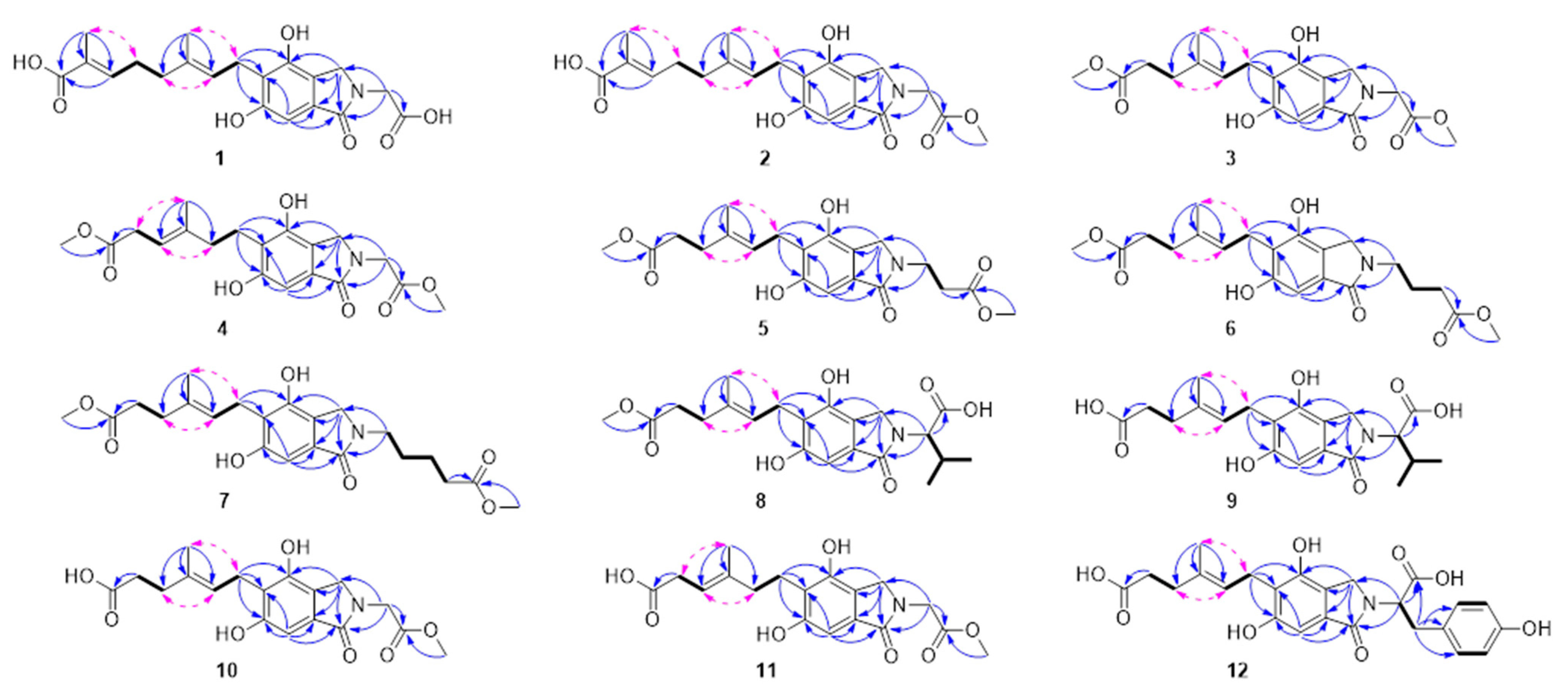
Compound 1 (Figure 1) was obtained as a yellow oil. Based on the molecular ion peak at m/z 412.13668 [M + Na]+ determined by HRESIMS, it corresponds to the molecular formula C20H23NO7. The 1H, 13C, and DEPT NMR spectra (Table 1) showed the presence of two methyl groups (δH 1.76, s; 1.82, s; δC 12.4, 16.3), five methylene groups (δH 2.09, t, J = 7.3 Hz; 2.28, dd, J = 14.9, 7.3 Hz; 3.42, d, J = 7.1 Hz; 4.32, s; 4.39, s; δC 39.4, 28.3, 23.6, 45.0, 50.0), three methines, including two olefinic groups (δH 5.30, t, J = 7.1 Hz; 6.72, t, J = 7.3 Hz; δC 124.5, 143.8), and an aromatic proton (δH 6.76, s; δC 102.0). Additionally, ten quaternary carbons were identified, consisting of five aromatic groups (δC 121.0, 121.7, 131.3, 151.6, 158.0), three carbonyl groups (δC 171.7, 171.8, 173.0), and two olefinic groups (δC 128.8, 134.8). Using the HSQC spectrum, the connectivities of the one-bond 1H–13C were determined. The HMBC correlations from H-7 (δH 6.76) to C-1 (δC 171.8) and from H-3 (δH 4.39) to C-4 (δC 151.6) confirmed the presence of the isoindoline-1-one substructure (Figure 2). Further HMBC correlations, including those from H-1′ (δH 3.42) to C-4, C-5 (δC 121.7), and C-6 (δC 158.0); H-9′ (δH 1.82) to C-2′ (δC 124.5), C-3′ (δC 134.8), and C-4′ (δC 39.4); H-10′ (δH 1.76) to C-6′ (δC 143.8), C-7′ (δC 128.8), and C-8′ (δC 171.7); and H-1″ (δH 4.32) to C-1, C-3 (δC 50.0), and C-2″ (δC 173.0), together with COSY correlations of H-1′/H-2′ and H-4′/H-5′/H-6′, established the gross structure of 1 as depicted in Figure 2. The H-1′/H-9′, H-2′/H-4′, and H-5′/H-10′ ROESY correlations confirmed the E configuration for two double bonds. In light of the comprehensive analysis, the compound was named erinacenone A (see Supplementary Materials).
Compound 2 was obtained as a yellow oil. Based on the molecular ion peak at m/z 404.17023 [M + H]+ determined by HRESIMS, it has the molecular formula C21H25NO7. The 1H and 13C NMR spectra data (Table 1 and Supplementary Materials) of 2 were extremely similar to 1, with the primary distinction being the presence of an additional methoxy group (δC 52.8) in 2. This change was confirmed by the HMBC correlation of H-3″ (δH 3.76, s) to C-2″ (δC 171.2) (Figure 2). The ROESY correlations (H-1′/H-9′, H-2′/H-4′, and H-5′/H-10′) supported the E configuration for two double bonds. Thus, the structure of 2 was designated as erinacenone B.
Compounds 3 and 4 were isolated in the form of a yellow oil and shared an identical molecular formula: C19H23NO7. HRESIMS analysis confirmed this molecular formula. When the NMR data (Table 2 and Table 3) from compounds 3 and 4 were compared to those from compound 2, it was discovered that both compounds had an additional methoxyl group at C-8′ (3: δH 3.55, s; δC 51.9; 4: δH 3.64, s; δC 52.3), while simultaneously losing a trisubstituted double bond and a methyl group, which indicated a structural difference in the side chain at C-5 in the benzene ring. This alteration in the side chain at C-5 was further supported by HMBC correlations (refer to Figure 2). HMBC correlations from H-7′ to C-2′, C-3′, and C-4′, as well as from H-8′ to C-6′, together with COSY correlations of H-4′/H-5′, provided robust evidence for this structural variation. Additionally, the ROESY correlations H-1′/H-7′, H-2′/H-4′, and H-5′/H-7′ confirmed that compounds 3 and 4 are a pair of double-bond positional isomers. Consequently, the structures of 3 and 4 were assigned the names erinacenone C and erinacenone D, respectively.
Compound 5 was isolated as a yellow oil, and its molecular formula was determined to be C20H25NO7 through high-resolution electrospray ionization mass spectrometry (HRESIMS) analysis (m/z 392.17017 [M + H]+, calculated for C20H26NO7, 392.17038). With the exception of an additional methylene group (δH 2.73, t, J = 6.7 Hz; δC 33.9) in 5, the 1H and 13C NMR data of 5 (Table 2 and Table 3) closely resembled that of compound 3. This was confirmed by COSY correlations between H-1″ and H-2″. The H-1′/H-7′ and H-2′/H-4′ ROESY correlations confirmed the E configuration. Consequently, the structure of 5 was assigned the name erinacenone E.
Compound 6 was isolated in the form of a yellow oil. Based on the analysis of the NMR data and HRESIMS at m/z 406.18594 [M + H]+, its molecular formula was determined to be C21H27NO7. In comparison to 5, it is characterized by the presence of an additional methylene group (δH 2.37, overlapped; δC 32.0). This structural distinction was further supported by HRESIMS data and COSY correlations between H-1″, H-2″, and H-3″. The E configuration for the double bond between C-2′ and C-3′ was established by the ROESY correlations of H-1′/H-7′ and H-2′/H-4′. Therefore, the structure of 6 was named erinacenone F.
Compound 7 was isolated in the form of a yellow oil. Based on the analysis of the NMR data and HRESIMS at m/z 442.18347 [M + Na]+, the molecular formula of 7 was determined to be C22H29NO7. The key difference between compounds 7 and 6, according to NMR data, was that compound 7 included an additional methylene group (δH 2.40, overlapped; δC 34.1). This conclusion was confirmed by HRESIMS data and COSY correlations between H-1″, H-2″, H-3″, and H-4″. As previously mentioned, it was determined that the double bond between C-2′ and C-3′ should be configured. Therefore, the structure of 7 was named erinacenone G.
Compound 8 was obtained as a yellow oil. Based on the analysis of the NMR data and HRESIMS at m/z 406.18585 [M + H]+, it was determined that the molecular formula of 8 was C21H27NO7. Comparing the 1D NMR data (Table 4 and Table 5) of 8 with erinacerin E revealed the absence of signals for a trisubstituted double bond and a methyl group, while an additional methyl group signal (δH 3.55, s; δC 51.9) appeared. This change was confirmed by HMBC correlations from H-8′ to C-6′ (δC 175.8) and HRESIMS data. Detailed analysis of the 2D NMR data (Figure 2) revealed the gross structure of 8. The E configuration was confirmed by the ROESY correlation of H-1′/H-7′ and H-2′/H-4′, as described above. Therefore, the structure of 8 was named erinacenone H (Table 4 and Table 5).
Compound 9, isolated as a yellow oil, had a molecular formula of C20H25NO7, determined through the analysis of HRESIMS at m/z 392.17020 [M + H]+, with seven degrees of unsaturation. Structurally similar to 8, the only difference was the absence of a methyl group, confirmed by the 1D NMR spectra (Table 4 and Table 5) and supported by HRESIMS data. Therefore, 9 was named erinacenone I.
Compounds 10 and 11 were obtained as a yellow oil. The results of the HRESIMS analysis revealed that their molecular formulas were the same: C18H21NO7. The NMR spectra (Table 4 and Table 5) of compounds 10 and 11 resembled those of 3. Compound 10 was significantly different from 3 in that it lacked a methyl signal. The HRESIMS analysis confirmed the prediction. The double bond configuration between C-2′ and C-3′ was determined as described in compound 3. Thus, 10 was named erinacenone J. Compounds 10 and 11 were identified as double-bond positional isomers. The ROESY spectrum (H-2′/H-4′ and H-5′/H-7′) established the double-bond configuration between C-3′ and C-4′ for 11, and it was named erinacenone K.
Compound 12, isolated as a yellow oil, had a molecular formula of C24H25NO8 (m/z 456.16529 [M + H]+), determined through HRESIMS analysis, with twelve degrees of unsaturation. The 1D NMR spectra of 12 revealed that it and 11 both included the (E)-6-(4,6-dihydroxy-1-oxoisoindolin-5-yl)-4-methylhex-4-enoate moiety. The key distinction was the substitution of 3-(4-hydroxyphenyl) propionic acid for 3-methylbutyric acid at the nitrogen atom. This result was supported by the COSY correlations of H-1″/H-2″, H-4″/H-5″, and H-7″/H-8″; the HMBC correlations from H-1″ (δH 5.10, dd, J = 11.2, 4.7 Hz) to C-1 (δC 171.8), C-3 (δC 47.0), and C-9″ (δC 174.5) and from H-2″ (δH 3.11, dd, J = 14.6, 11.4 Hz; 2.25, overlapped) to C-3″ (δC 129.3), C-4″ (δC 130.6), and C-8″ (δC 130.6); as well as the HRESIMS data. The double-bond configuration between C-2′ and C-3′ was established based on the ROESY spectrum. Therefore, the structure of 12 was named erinacenone L.
By comparing the optical rotations of compounds 8, 9, and 12 with those of the synthetic phthalimidines, the absolute configuration of C-1″ was determined [17]. Compounds 9 and 12 showed negative specific optical rotations, indicating that they possess the S configuration at C-1″. Compound 8 showed a positive specific rotation, indicating that it possesses the R configuration at C-1″.

3. Materials and Methods

3.1. General Experimental Procedures

The optical rotations were recorded using a Rudolph AutopolIV-T polarimeter (Rudolph, Hackettstown, NJ, USA). UV spectra were obtained using a UH5300 spectrophotometer (Hitachi, Kyoto, Japan). NMR spectra were obtained using a Bruker Avance III 600 MHz or Avance Neo 500 MHz spectrometer (Bruker, Karlsruhe, Germany). HRESIMS data were collected using a Q-Exactive Orbitrap mass spectrometer (Thermo Scientific, Waltham, MA, USA). Sephadex LH-20 (Amersham Biosciences, Uppsala, Sweden) and silica gel (Qingdao Haiyang Chemical Co., Ltd., Qingdao, China) were used for column chromatography (CC). Medium-pressure liquid chromatography (MPLC) was performed on a Interchim PuriFlash 450 instrument (Interchim, Montluçon, France). An Agilent 1260 liquid chromatography system with a DAD detector (Agilent Technologies, Santa Clara, CA, USA) was used for preparative HPLC using a Zorbax SB-C18 column (9.4 × 150 mm, 5 µm, 4 mL·min−1). An Agilent 1260 liquid chromatograph with a Zorbax SB-Aq column (4.6 × 250 mm, 5 µm, 1 mL·min−1) was used for semipreparative HPLC. TLC was performed on GF254 plates (Qingdao Marine Chemical Inc., Qingdao, China).

3.2. Fungal Material

The fungus H. erinaceus (accession No. KU855351.1) was collected in August 2007 in Shangri-La County, Yunnan province, China, and was identified by Prof. Zang (Kunming Institute of Botany, CAS, Kunming, China) at the Kunming Institute of Botany. The strain was deposited at South-Central Minzu University. The strain of H. erinaceus was cultured for 30 days at 24 °C on a rotary shaker at 150 rpm on liquid medium (glucose 5%, yeast extract 0.4%, peptone 0.15%, KH2PO4 0.05%, MgSO4 0.05%, pH 6.5).

3.3. Extraction and Isolation

A 100 L culture broth filtrate was concentrated to 10 L, then extracted with ethyl acetate. Mycelium was extracted with acetone using wall-breaking extraction. The combined extracts were concentrated to yield a crude extract (110.98 g). Silica gel column chromatography (CC) separated the crude extract into fractions A–F. MPLC (MeOH–H2O, 5:1, 20:1, 30:1, 45:1, 65:1, 75:1, 85:1, 100:1; 4 L for each step) was used to separate Fraction B into twelve subfractions (B1–B12). Subfraction B6 was subjected to silica gel CC (v/v 1:0–0:1) to yield subfractions (B6A–B6P). HPLC purified subfraction B6L (MeCN–H2O, 34% isocratic, 4 mL/min) to give compound 4 (tR = 9.93 min, 9.8 mg), 5 (tR = 9.92 min, 1.8 mg), 6 (tR = 10.13 min, 9.0 mg), and 7 (tR = 10.61 min, 2.6 mg). Fraction C was separated by MPLC (MeOH–H2O, 5:1, 20:1, 30:1, 45:1, 65:1, 75:1, 85:1, 100:1; 4 L for each step) into twenty subfractions (C1–C20). Subfraction C6 was further separated by silica gel CC (CHCl3–MeOH from v/v 1:0 to 0:1) to yield subfractions C6A–C6J. HPLC (MeCN–H2O, 32% isocratic, 4 mL/min) purified subfraction C6D to give 8 (tR = 9.90 min, 1.1 mg). Subfraction C10 was separated using silica gel CC (v/v 1:0–0:1) to obtain subfractions C10A–C10K. HPLC purified subfraction C10A (MeCN–H2O, 20% isocratic, 4 mL/min), yielding compound 3 (tR = 9.84 min, 28.3 mg). To obtain subfractions C11A–C11O, silica gel CC (v/v 1:0–0:1) was used to separate subfraction C11. Subfraction C11L was purified using Sephadex LH-20 (MeOH) and HPLC (MeCN–H2O, 25% isocratic, 4 mL/min) to obtain compound 9 (tR = 8.69 min, 9.1 mg). Subfraction C12 was separated by silica gel CC (v/v 1:0–0:1) to obtain subfractions C12A–C12I. Fraction F was separated into twenty-three subfractions (F1–F23) using MPLC (MeOH–H2O, 5:1, 20:1, 30:1, 45:1, 65:1, 75:1, 85:1, 100:1; 4 L for each step). Subfraction F8 was subjected to silica gel CC (v/v 1:0–0:1) to obtain subfractions F6A–F6H. HPLC (MeCN–H2O, 13% isocratic, 4 mL/min) purified subfraction F8B to give compound 10 (tR = 6.43 min, 1.1 mg) and 11 (tR = 6.21 min, 1.0 mg). Subfraction F10 was separated using silica gel CC (v/v 1:0–0:1) to obtain subfractions F10A–F10L. HPLC purified subfraction F10E (MeCN–H2O, 10% isocratic, 4 mL/min), yielding compound 2 (tR = 5.45 min, 16.2 mg). Compound 1 (tR = 7.58 min, 17.3 mg) was obtained by purifying subfraction F10G via HPLC (MeCN–H2O, 20% isocratic, 4 mL/min).

3.4. Characterization Data

3.4.1. Erinacenone A (1)

Yellow oil; UV (MeOH) λmax (log ε) 225 (4.44), 260 (4.25), 300 (3.63) nm; 1H (600 MHz) and 13C NMR (150 MHz) data (CD3OD), see Table 1; HRESIMS m/z 412.13668 (calcd for C20H23NNaO7 [M + Na]+, 412.13667).

3.4.2. Erinacenone B (2)

Yellow oil; UV (MeOH) λmax (log ε) 225 (4.49), 260 (4.32), 300 (3.72) nm; 1H (500 MHz) and 13C NMR (125 MHz) data (CD3OD), see Table 1; HRESIMS m/z 404.17023 (calcd for C21H26O7 [M + H]+, 404.17038).

3.4.3. Erinacenone C (3)

Yellow oil; UV (MeOH) λmax (log ε) 220 (4.72), 265 (4.53), 300 (3.95) nm; 1H (600 MHz) and 13C NMR (150 MHz) data (CD3OD), see Table 2 and Table 3; HRESIMS m/z 378.15475 (calcd for C19H24NO7 [M + H]+, 378.15473).

3.4.4. Erinacenone D (4)

Yellow oil; UV (MeOH) λmax (log ε) 220 (4.33), 260 (4.12), 300 (3.56) nm; 1H (600 MHz) and 13C NMR (150 MHz) data (CD3OD), see Table 2 and Table 3; HRESIMS m/z 378.15463 (calcd for C19H24NO7 [M + H]+, 378.15473).

3.4.5. Erinacenone E (5)

Yellow oil; UV (MeOH) λmax (log ε) 215 (4.50), 260 (4.10), 300 (3.53) nm; 1H (600 MHz) and 13C NMR (150 MHz) data (CD3OD), see Table 2 and Table 3; HRESIMS m/z 392.17017 (calcd for C20H26NO7 [M + H]+, 392.17038).

3.4.6. Erinacenone F (6)

Yellow oil; UV (MeOH) λmax (log ε) 220 (4.48), 260 (4.34), 300 (3.75) nm; 1H (600 MHz) and 13C NMR (150 MHz) data (CD3OD), see Table 2 and Table 3; HRESIMS m/z 406.18594 (calcd for C21H28NO7 [M + H]+, 406.18603).

3.4.7. Erinacenone G (7)

Yellow oil; UV (MeOH) λmax (log ε) 220 (4.57), 260 (4.40), 300 (3.77) nm; 1H (500 MHz) and 13C NMR (125 MHz) data (CD3OD), see Table 2 and Table 3; HRESIMS m/z 442.18347 (calcd for C22H29NNaO7 [M + Na]+, 442.18362).

3.4.8. Erinacenone H (8)

Yellow oil; [α]25D+ 28.8 (c 0.10, MeOH); UV (MeOH) λmax (log ε) 210 (4.46), 265 (3.90), 305 (3.33) nm; 1H (600 MHz) and 13C NMR (150 MHz) data (CD3OD), see Table 4 and Table 5; HRESIMS m/z 406.18585 (calcd for C21H28NO7 [M + H]+, 406.18603).

3.4.9. Erinacenone I (9)

Yellow oil; [α]25D− 10.6 (c 0.25, MeOH); UV (MeOH) λmax (log ε) 220 (4.52), 260 (4.27), 300 (3.68) nm; 1H (600 MHz) and 13C NMR (150 MHz) data (CD3OD), see Table 4 and Table 5; HRESIMS m/z 392.17020 (calcd for C20H26NO7 [M + H]+, 392.17038).

3.4.10. Erinacenone J (10)

Yellow oil; UV (MeOH) λmax (log ε) 215 (4.42), 260 (4.10), 300 (3.51) nm; 1H (600 MHz) and 13C NMR (150 MHz) data (CD3OD), see Table 4 and Table 5; HRESIMS m/z 364.13898 (calcd for C18H22 N O7 [M + H]+, 364.13908).

3.4.11. Erinacenone K (11)

Yellow oil; UV (MeOH) λmax (log ε) 215 (4.32), 260 (3.85), 300 (3.34) nm; 1H (600 MHz) and 13C NMR (150 MHz) data (CD3OD), see Table 4 and Table 5; HRESIMS m/z 386.12082 (calcd for C18H21NNaO7 [M + Na]+, 386.12102).

3.4.12. Erinacenone L (12)

Yellow oil; [α]25D− 69.0 (c 0.05, MeOH); UV (MeOH) λmax (log ε) 225 (4.62), 265 (4.46) nm; 1H (500 MHz) and 13C NMR (125 MHz) data (CD3OD), see Table 4 and Table 5; HRESIMS m/z 456.16529 (calcd for C24H26NO8 [M + H]+, 456.16529).

3.5. Cytotoxicity Assay

Compounds 112 were assessed for cytotoxicity against MCF-7 cell lines (ATCC, Manassas, VA, USA) using the MTT (multiple table tournament) method, as previously reported. Briefly, 1 × 10⁵ cells/mL of adherent cells were seeded in 96-well plates and incubated for 12 h at 37 °C. After this initial period, various concentrations of compounds were added to each well. Following a 48 h incubation, MTT solution was added and incubated for an additional 4 h. Then, MTT was removed and dissolved with MTT lysis solution (20% SDS, 50% DMF). MCF-7 cells were cultured in DMEM medium (Hyclone, Logan, UT, USA) with 10% fetal bovine serum (FBS) supplemented at 37 °C in 5% CO2. Cisplatin served as the positive control. The absorbances were detected at 595 nm on an Envision multilabel plate reader, and the IC50 values were calculated using the Reed-Muench method [18].

4. Conclusions

In the present study, a total of twelve previously unreported isoindolin-1-one compounds, erinacenone A–L (112), were isolated from liquid cultures of the medicinal fungus H. erinaceus. Their structures were elucidated based on spectroscopic data analysis. The absolute configuration of 12 was determined by comparing its optical rotations with values reported in the literature. The most distinctive feature of these compounds is that their nitrogen atoms are connected to different parts of the special structure moieties. Among them, compounds 3 and 4, as well as 10 and 11, are two pairs of isomers differing only by a small change in the position of one double bond. The structures of the twelve isolated compounds are very similar to those reported in the research of Wang et al. [17], Lin et al. [19], and Chen et al., [20], sharing the same core structure. Compounds 4 and 5 were found to show cytotoxic activities with IC50 values of 24.7 and 18.4 μM, respectively, against MCF-7 cell lines (Table 6).
So far, despite the fact that the chemical constituents of the fruiting body and fermentation broth of the mushroom H. erinaceus have been well studied, many new secondary metabolites can still be isolated and obtained. This suggests that strains collected in different regions and seasons can produce a rich variety of secondary metabolites with structural changes under changing fermentation conditions. The appearance of these new secondary metabolites opens up the possibility of discovering new biological activities of these components. It also shows that the mushroom H. erinaceus itself is a treasure trove with great potential that we need to keep exploring. The new alkaloids discovered in the present study have only been screened using simple bioassays so far, and the next step needs to be a systematic screening of different drug targets, with special attention given to their testing on central nervous system models. In addition, the continued search for other types of secondary metabolites from the mushroom H. erinaceus should also be considered.

Supplementary Materials

The following supporting information can be downloaded at: https://www.mdpi.com/article/10.3390/molecules29204901/s1, Figures S1–S84: HRESIMS, HNMR, CNMR, COSY, HSQC, HMBC, ROESY of compounds 112.

Author Contributions

Investigation, data curation, writing—original draft preparation, L.-L.Y.; supervision, project administration, funding acquisition, J.-K.L. All authors have read and agreed to the published version of the manuscript.

Funding

This work was financially supported by the National Natural Science Foundation of China (No. 22177138).

Institutional Review Board Statement

Not applicable.

Informed Consent Statement

Not applicable.

Data Availability Statement

No new data were created or analyzed in this study. Data sharing is not applicable to this article.

Conflicts of Interest

The authors declare that they have no known competing financial interests or personal relationships that could have appeared to influence the work reported in this paper.

References

  1. Szućko-Kociuba, I.; Trzeciak-Ryczek, A.; Kupnicka, P.; Chlubek, D. Neurotrophic and Neuroprotective Effects of Hericium erinaceus. Int. J. Mol. Sci. 2023, 24, 15960. [Google Scholar] [CrossRef] [PubMed]
  2. Friedman, M. Chemistry, Nutrition, and Health-Promoting Properties of Hericium erinaceus (Lion’s Mane) Mushroom Fruiting Bodies and Mycelia and Their Bioactive Compounds. J. Agric. Food Chem. 2015, 63, 7108–7123. [Google Scholar] [CrossRef] [PubMed]
  3. Gravina, A.G.; Pellegrino, R.; Auletta, S.; Palladino, G.; Brandimarte, G.; D’Onofrio, R.; Arboretto, G.; Imperio, G.; Ventura, A.; Cipullo, M.; et al. Hericium erinaceus, a medicinal fungus with a centuries-old history: Evidence in gastrointestinal diseases. World J. Gastroenterol. 2023, 29, 3048–3065. [Google Scholar] [CrossRef] [PubMed]
  4. Wang, J.C.; Hu, S.H.; Su, C.H.; Lee, T.M. Antitumor and immunoenhancing activities of polysaccharide from culture broth of Hericium spp. Kaohsiung J. Med. Sci. 2001, 17, 461–467. [Google Scholar] [PubMed]
  5. Zhang, Z.; Liu, R.N.; Tang, Q.J.; Zhang, J.S.; Yang, Y.; Shang, X.D. A new diterpene from the fungal mycelia of Hericium erinaceus. Phytochem. Lett. 2015, 11, 151–156. [Google Scholar] [CrossRef]
  6. Zhang, Y.; Yang, S.L.; Wang, A.H.; Sun, Z.C.; Zhuo, Y.F.; Xu, Y.T.; He, Y.L. Protective effect of ethanol extracts of Hericium erinaceus on alloxan-induced diabetic neuropathic pain in rats. Evid.-Based Complement. Altern. Med. 2015, 2015, 595480. [Google Scholar] [CrossRef]
  7. Liang, B.; Guo, Z.D.; Xie, F.; Zhao, A.N. Antihyperglycemic and antihyperlipidemic activities of aqueous extract of Hericium erinaceus in experimental diabetic rats. BMC Complement. Altern. Med. 2013, 13, 253. [Google Scholar] [CrossRef] [PubMed]
  8. Yang, B.K.; Park, J.B.; Song, C.H. Hypolipidemic effect of an exo-biopolymer produced from a submerged mycelial culture of Hericium erinaceus. Biosci. Biotechnol. Biochem. 2003, 67, 1292–1298. [Google Scholar] [CrossRef] [PubMed]
  9. Mori, K.; Ouchi, K.; Hirasawa, N. The anti-inflammatory effects of lion’s mane culinary-medicinal mushroom, Hericium erinaceus (higher basidiomycetes) in a coculture system of 3T3-L1 adipocytes and RAW264 macrophages. Int. J. Med. Mushrooms 2015, 17, 609–618. [Google Scholar] [CrossRef] [PubMed]
  10. Rahman, M.A.; Abdullah, N.; Aminudin, N. Inhibitory effect on in vitro LDL oxidation and HMG Co-A reductase activity of the liquid-liquid partitioned fractions of Hericium erinaceus (Bull.) Persoon (Lion’s mane mushroom). BioMed Res. Int. 2014, 828149. [Google Scholar] [CrossRef]
  11. Tzeng, T.T.; Chen, C.C.; Lee, L.Y.; Chen, W.P.; Lu, C.K.; Shen, C.C.; Huang, F.C.Y.; Chen, C.C.; Shiao, Y.J. Erinacine A-enriched Hericium erinaceus mycelium ameliorates Alzheimer’s disease-related pathologies in APPswe/PS1dE9 transgenic mice. J. Biomed. Sci. 2016, 23, 49. [Google Scholar] [CrossRef]
  12. Mori, K.; Inatomi, S.; Ouchi, K.; Azumi, Y.; Tuchida, T. Improving effects of the mushroom Yamabushitake (Hericium erinaceus) on mild cognitive impairment: A double-blind placebo-controlled clinical trial. Phytother. Res. 2009, 23, 367–372. [Google Scholar] [CrossRef] [PubMed]
  13. Lee, K.F.; Chen, J.H.; Teng, C.C.; Shen, C.H.; Hsieh, M.C.; Lu, C.C.; Lee, K.C.; Lee, L.Y.; Chen, W.P.; Chen, C.C.; et al. Protective effects of Hericium erinaceus mycelium and its isolated erinacine A against ischemia-injury-induced neuronal cell death via the inhibition of iNOS/p38 MAPK and nitrotyrosine. Int. J. Mol. Sci. 2014, 15, 15073–15089. [Google Scholar] [CrossRef] [PubMed]
  14. Chiu, C.H.; Chyau, C.C.; Chen, C.C.; Lee, L.Y.; Chen, W.P.; Liu, J.L.; Lin, W.H.; Mong, M.C. Erinacine A-enriched Hericium erinaceus mycelium produces antidepressant-like effects through modulating BDNF/PI3K/Akt/GSK-3β signaling in mice. Int. J. Mol. Sci. 2018, 19, 341. [Google Scholar] [CrossRef]
  15. Ryu, S.; Kim, H.G.; Kim, J.Y.; Kim, S.Y.; Cho, K.O. Hericium erinaceus extract reduces anxiety and depressive behaviors by promoting hippocampal neurogenesis in the adult mouse brain. J. Med. Food 2018, 21, 174–180. [Google Scholar] [CrossRef]
  16. Yao, W.; Zhang, J.C.; Dong, C.; Zhuang, C.; Hirota, S.; Inanaga, K.; Hashimoto, K. Effects of amycenone on serum levels of tumor necrosis factor-α, interleukin-10, and depression-like behavior in mice after lipopolysaccharide administration. Pharmacol. Biochem. Behav. 2015, 136, 7–12. [Google Scholar] [CrossRef] [PubMed]
  17. Wang, K.; Bao, L.; Qi, Q.X.; Zhao, F.; Ma, K.; Pei, Y.F.; Liu, H.W. Erinacerins C–L, Isoindolin-1-ones with α-Glucosidase Inhibitory Activity from Cultures of the Medicinal Mushroom Hericium erinaceus. J. Nat. Prod. 2015, 78, 146–154. [Google Scholar] [CrossRef] [PubMed]
  18. Reed, L.J.; Muench, H. A simple method of estimating fifty percent endpoint. Am. J. Epidemiol. 1938, 27, 493–497. [Google Scholar] [CrossRef]
  19. Lin, J.Y.; Chen, Y.P.; Lin, T.W.; Li, T.J.; Chen, Y.W.; Li, I.C.; Chen, C.C. Discovery of a New Compound, Erinacerin W, from the Mycelia of Hericium erinaceus, with Immunomodulatory and Neuroprotective Effects. Molecules 2024, 29, 812. [Google Scholar] [CrossRef] [PubMed]
  20. Chen, L.; Li, Z.H.; Yao, J.N.; Peng, Y.L.; Huang, R.; Feng, T.; Liu, J.K. Isoindolinone-containing meroterpenoids with α-Glucosidase Inhibitory activity from mushroom Hericium caput-medusae. Fitoterapia 2017, 122, 107–114. [Google Scholar] [CrossRef] [PubMed]
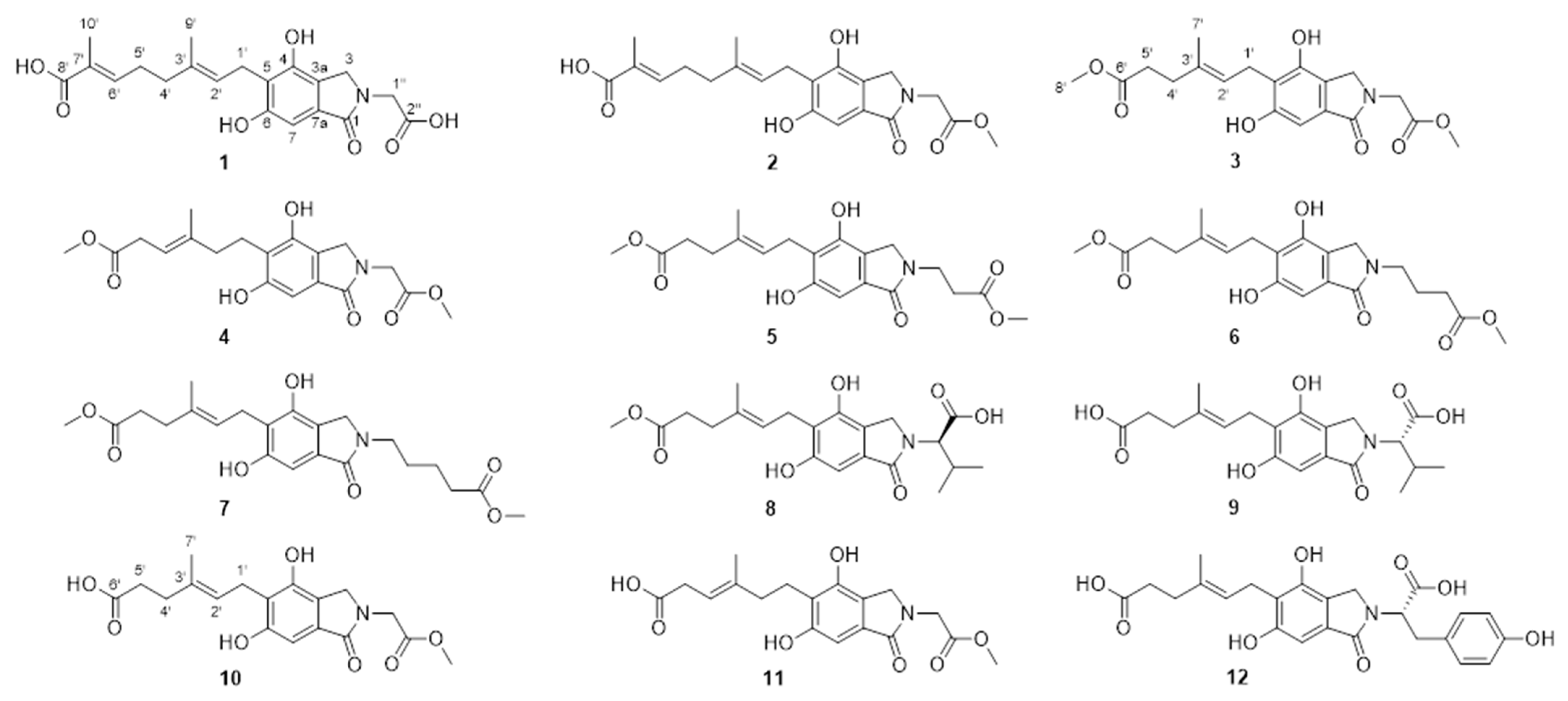
Figure 1. Structures of compounds 112.
Molecules 29 04901 g001
Figure 2. Key 2D NMR correlations for 112. Blue lines (HMBC correlations), purple lines (ROESY correlations).
Molecules 29 04901 g002
Table 1. 1H NMR (600 MHz) and 13C NMR (150 MHz) spectroscopic data for compounds 12.
No.1 2
1171.8 171.9
350.04.39, s44.74.39, s
3a131.3 131.1
4151.6 151.7
5121.7 121.9
6158.0 158.1
7102.06.76, s102.06.76, s
7a121.0 121.0
1′23.63.42, dd (7.1)23.63.43, dd (7.1)
2′124.55.30, t (7.1)124.35.30, t (7.1)
3′134.8 135.0
4′39.42.09, t (7.3)39.52.09, t (7.4)
5′28.32.28, dd (14.9, 7.3)28.42.28, dd (14.9, 7.4)
6′143.86.72, t (7.3)143.36.71, t (7.4)
7′128.8 129.2
8′171.7 172.4
9′16.31.82, s16.31.82, s
10′12.41.76, s12.51.76, s
1″45.04.32, s50.04.39, overlapped
2″173.0 171.2
3″ 52.83.76, s
Measured in methanol-d4.
Table 2. 1H NMR (600 MHz) spectroscopic data for compounds 37.
No.34567
34.38, s4.39, s4.34, s4.30, s4.30, s
76.76, s6.76, s6.72, s6.73, s6.73, s
1′3.40, d (7.1)2.83, m3.39, d (7.1)3.40, d (7.1)3.40, d (7.1)
2′5.28, t (7.1)2.24, t (7.8)5.27, t (7.1)5.27, t (7.1)5.28, t (7.1)
4′2.26, t (7.6)5.26, t (7.1)2.25, t (7.6)2.25, t (7.6)2.25, t (7.5)
5′2.39, m3.04, d (7.1)2.39, t (7.6)2.39, overlapped2.39, overlapped
7′1.80, s1.74, s1.79, s1.79, s1.80, s
8′3.55, s3.64, s3.55, s3.55, s3.55, s
1″4.38, s4.39, s3.85, t (6.7)3.62, t (6.8)3.60, s
2″ 2.73, t (6.7)1.98, m1.71, m
3″ 3.76, s 2.37, overlapped1.63, m
4″3.75, s 3.67, s 2.40, overlapped
5″ 3.58, s
6″ 3.64, s
Measured in methanol-d4.
Table 3. 13C NMR (150 MHz) spectroscopic data for compounds 37.
No.34567
1171.8171.1171.4171.5171.4
349.944.749.748.848.8
3a131.1131.1131.7131.8132.0
4151.7151.8151.6151.6151.6
5121.8122.4121.4121.3121.3
6158.1158.3158.1158.1158.1
7102.0101.9101.7101.8101.8
7a121.0121.0120.6120.4120.4
1′23.623.423.623.623.6
2′124.539.5124.6124.6124.6
3′134.3140.7134.2134.2134.2
4′35.9116.836.036.036.0
5′33.934.333.933.933.9
6′175.8174.8175.8175.8175.8
7′16.116.416.116.116.1
8′51.952.351.951.951.9
1″44.750.039.842.943.0
2″171.1171.833.924.728.7
3″52.852.8173.732.023.1
4″ 52.3175.234.1
5″ 52.1175.6
6″ 52.0
Measured in methanol-d4.
Table 4. 1H NMR (600 MHz) spectroscopic data for compounds 812.
No.89101112
34.60, d (17.0);
4.32, d (17.0)
4.59, overlapped;
4.32, d (17.0)
4.38, s4.39, s4.37, d (16.6);
4.27, d (16.6)
76.75, s6.75, s6.76, s6.75, s6.67, s
1′3.41, d (7.1)3.42, d (7.2)3.42, d (7.1)2.83, t (7.6)3.39, m
2′5.28, t (7.1)5.31, t (7.2)5.30, t (7.1)2.21, t (7.6)5.29, t (7.4)
4′2.26, t (7.6)2.26, t (7.2)2.26, m5.39, m2.25, overlapped
5′2.39, m2.36, overlapped2.32, m2.97, m2.35, m
7′1.80, d1.81, s1.81, s1.74, s1.80, s
8′3.55, s
1″4.56, m4.58, overlapped4.39, s4.39, s5.10, dd (11.2, 4.7)
2″2.34, m2.35, overlapped 3.11, dd (14.6, 11.4);
2.25, overlapped
3″1.09, d (6.6)1.09, d (6.6)3.78, s3.76, s
4″0.89, d (6.6)0.9, d (6.6) 7.04, d (8.5)
5″ 6.65, d (8.5)
7″ 6.65, d (8.5)
8″ 7.04, d (8.5)
Measured in methanol-d4.
Table 5. 13C NMR (150 MHz) spectroscopic data for compounds 812.
No.89101112
1171.8171.9171.8171.9171.8
346.846.850.044.747.0
3a131.2131.1131.1131.0131.4
4151.6151.6151.7151.8151.5
5121.6121.7121.9122.8121.5
6158.1158.1158.2158.3158.0
7101.9102.0102.06.75101.9
7a120.9120.9121.0121.2120.9
1′23.623.623.623.623.6
2′124.6124.2123.939.8124.2
3′134.2134.5135.1138.6134.5
4′36.036.036.6119.535.9
5′33.934.135.437.134.0
6′175.8177.6179.6nd a177.6
7′16.116.216.316.416.2
8′51.9
1″62.862.544.750.057.5
2″30.130.1171.1171.235.9
3″20.120.052.852.8129.3
4″19.619.2 130.6
5″nd a174.5 116.4
6″ 157.2
7″ 116.4
8″ 130.6
9″ 174.5
a Not detected. Measured in methanol-d4.
Table 6. Cytotoxicity inhibitory activity of compounds (IC50, µM).
CompoundsMCF-7
424.7
518.4
Cisplatin a9.12
a Positive control. Other compounds did not show inhibitory activity, or very weak activity. The maximum tested concentration is 100 µM.
Disclaimer/Publisher’s Note: The statements, opinions and data contained in all publications are solely those of the individual author(s) and contributor(s) and not of MDPI and/or the editor(s). MDPI and/or the editor(s) disclaim responsibility for any injury to people or property resulting from any ideas, methods, instructions or products referred to in the content.