Article

Ternary Phenolate-Based Thiosemicarbazone Complexes of Copper(II): Magnetostructural Properties, Spectroscopic Features and Marked Selective Antiproliferative Activity against Cancer Cells

Department of Chemistry, College of Science, Sultan Qaboos University, P.O. Box 36, Al-Khod 123, Muscat, Oman
*
Author to whom correspondence should be addressed.
Molecules 2024, 29(2), 431; https://doi.org/10.3390/molecules29020431
Submission received: 20 December 2023 / Revised: 6 January 2024 / Accepted: 10 January 2024 / Published: 16 January 2024
(This article belongs to the Section Inorganic Chemistry)

Abstract

:
The new diprotic ligand 3,5-di-tert-butylsalicylaldehyde 4-ethyl-3-thiosemicarbazone, abbreviated H2(3,5-t-Bu2)-sal4eT, exists as the thio-keto tautomer and adopts the E-configuration with respect to the imine double bond, as evidenced by single-crystal X-ray analysis and corroborated by spectroscopic characterisation. Upon treatment with Cu(OAc)2·H2O in the presence of either 2,9-dimethyl-1,10-phenanthroline (2,9-Me2-phen) or 1,10-phenanthroline (phen) as a co-ligand in MeOH, this thiosemicarbazone undergoes conformational transformation (relative donor-atom orientations: syn,anti  syn,syn) concomitantly with tautomerisation and double deprotonation to afford the ternary copper(II) complexes [Cu{(3,5-t-Bu2)-sal4eT}(2,9-Me2-phen)] (1) and [Cu2{3,5-t-Bu2)-sal4eT}2(phen)] (2). Crystallographic elucidation has revealed that complex 1 is a centrosymmetric dimer of mononuclear copper(II) complex molecules brought about by intermolecular H-bonding. The coordination geometry at the copper(II) centre is best described as distorted square pyramidal in accordance with the trigonality index (τ = 0.14). The co-ligand adopts an axial–equatorial coordination mode; hence, there is a disparity between its two Cu–N coordinate bonds arising from weakening of the apical one as a consequence of the tetragonal distortion. The axial X-band ESR spectrum of complex 1 is consistent with retention of this structure in solution. Complex 2 is a centrosymmetric dimer of dinuclear copper(II) complex molecules exhibiting intermolecular H-bonding and π-π-stacking interactions. The two copper(II) centres, which are 4.8067(18) Å apart and bridged by the thio-enolate nitrogen of the quadridentate thiosemicarbazonate ligand, display two different coordination geometries, one distorted square planar (τ4 = 0.082) and the other distorted square pyramidal (τ5 = 0.33). Such dinuclear copper(II) thiosemicarbazone complexes, which are crystallographically characterised, are extremely rare. In vitro, complexes 1 and 2 outperform cisplatin as antiproliferative agents in terms of potency and selectivity towards HeLa and MCF-7 cancer cell lines.

1. Introduction

Thiosemicarbazones are multi-purpose hydrazone ligands of considerable interest in coordination chemistry [1]. Their characteristic functionality feature R1R2C=N–N(H)–C(=S)–NR3R4 is derived from the straightforward single-step Schiff-base condensation reaction between an aldehyde or a ketone and a thiosemicarbazide. Amongst their fascinating structural attributes is their coordination versatility, arising from their propensity to undergo concomitant base-/metal-assisted tautomeric transformation and deprotonation as a means to meet charge-neutrality requirements. Moreover, a given thiosemicarbazone is capable of exhibiting different denticities and coordination modes as demanded by the metal centre [2,3,4,5,6,7,8,9]. By strategically employing aldehydes or ketones with moieties bearing donor atoms of interest in appropriate coordination positions, they can be tailor-designed for specially desired hetero-donor environments, coordination spheres [10], physicochemical features and biological properties [3,8,9,11,12,13,14,15,16,17,18,19,20,21,22,23,24,25,26,27]. Substituent groups can impart electronic effects to thiosemicarbazone complexes with interesting impacts on physicochemical and pharmacological properties. The literature is replete with examples of thiosemicarbazone complexes exhibiting a broad spectrum of pharmacological properties including antitumour [3,8,9,11,12,13,14,15,16,17,18,19,20,21,22], antibacterial [20,21,23], antiviral [24,25], antifungal [23,26] and antimalarial properties [22,27].
Thiosemicarbazones stabilise predominantly metal ions from the p-, d- and f-blocks [1]; some of the transition-metal thiosemicarbazone complexes exhibit fascinating magneto-structural [28] and catalytic [29] properties amongst other features. Electrochemically, for copper (Z = 29; [Ar]4s13d10), which is the subject of this paper, Schiff-base complexes shuttle between +1 and +2 oxidation states (CuII/CuI redox couple) [8,15,18,20,30]. Thiosemicarbazone complexes whose cyclic voltammograms exhibit these redox couples with potentials lying within the biologically accessible redox potential window ranging from −0.4 to +0.8 V vs. NHE [30] are of pharmacological importance in that they have the ability to generate intracellular reactive oxygen species (ROS) [8,15,18,20,30] desirable for apoptotic cytotoxicity. Ternary mononuclear complexes of copper(II) of the type [CuL(N,N-donor-L)]n+ abound, where L represents a neutral or anionic polydentate primary ligand and N,N-donor-L stands for a heterocyclic bidentate N,N-donor chelating co-ligand such as 1,10-phenanthroline (phen), 2,2′-bipyridine (bipy), dipyridoquinoxaline (dpq), dipyridophenazine (dppz) or derivatives of these [31,32,33,34,35,36,37,38,39,40,41,42,43,44,45,46,47,48,49,50,51,52,53,54,55,56,57,58]. Such coordination compounds are of considerable interest partly because of their potential to exhibit DNA binding/cleavage [34,39,41,42,43,44,50,51,53,54,55], anticancer [39,43,44,49,53,54] and antimicrobial [35,36,37,40] activities. Some have been explored as models for metallo-enzymes [33,45] while others have been designed to investigate structural and spectroscopic features of interest [4,32,38,46,47,48,52,56,57,58]. There is a paucity of crystallographically characterised ternary copper(II) thiosemicarbazone complexes of this type [4,31,32,33,34,35,36,37,38,39].
In this work, we have synthesised and structurally characterised the ligand 3,5-di-tert-butylsalicylaldehyde 4-ethyl-3-thiosemicarbazone, H2(3,5-t-Bu2)-sal4eT. Reaction of copper(II) ion with this ligand and the co-ligand 2,9-dimethyl-1,10-phenanthroline (2,9-Me2-phen) or 1,10-phenanthroline (phen) in equimolar amounts produced the ternary copper(II) complex [Cu{(3,5-t-Bu2)-sal4eT}(2,9-Me2-phen)] (1) or [Cu2{3,5-t-Bu2)-sal4eT}2(phen)] (2), respectively. The 3D structures have been determined through single-crystal X-ray analyses. Whereas complex 1 is mononuclear, complex 2 is dinuclear. In the crystal lattice, each exists as a dimer arising from two symmetrically related intermolecular H-bonding interactions. To the best of our knowledge, [Cu2{3,5-t-Bu2)-sal4eT}2(phen)] (2) is one of only two examples of crystallographically characterised dinuclear copper(II) thiosemicarbazone complexes of this kind. The other is [Cu2(sal4eT)2(bipy)], previously designated [Cu2(L2)2(bipy)] (bipy = 2,2′-bipyridine) [4]. While complex 2 is a centrosymmetric dimer of dinuclear complexes, the crystallographic asymmetric unit of [Cu2(sal4eT)2(bipy)] consists of two independent intramolecularly hydrogen-bonded dinuclear complex molecules. In both 2 and [Cu2(sal4eT)2(bipy)], the two copper(II) ions are in different coordination spheres. These two complexes differ in the five-coordinate geometry. The antiproliferative activity of complexes 1 and 2 along with H2(3,5-t-Bu2)-sal4eT has been tested against the cancer cell lines MCF-7 (human breast adenocarcinoma) and HeLa (human cervical carcinoma). Whereas the ligand is inactive against HeLa and MCF-7 cancer cells, the complexes are highly potent and selective.

2. Results and Discussion

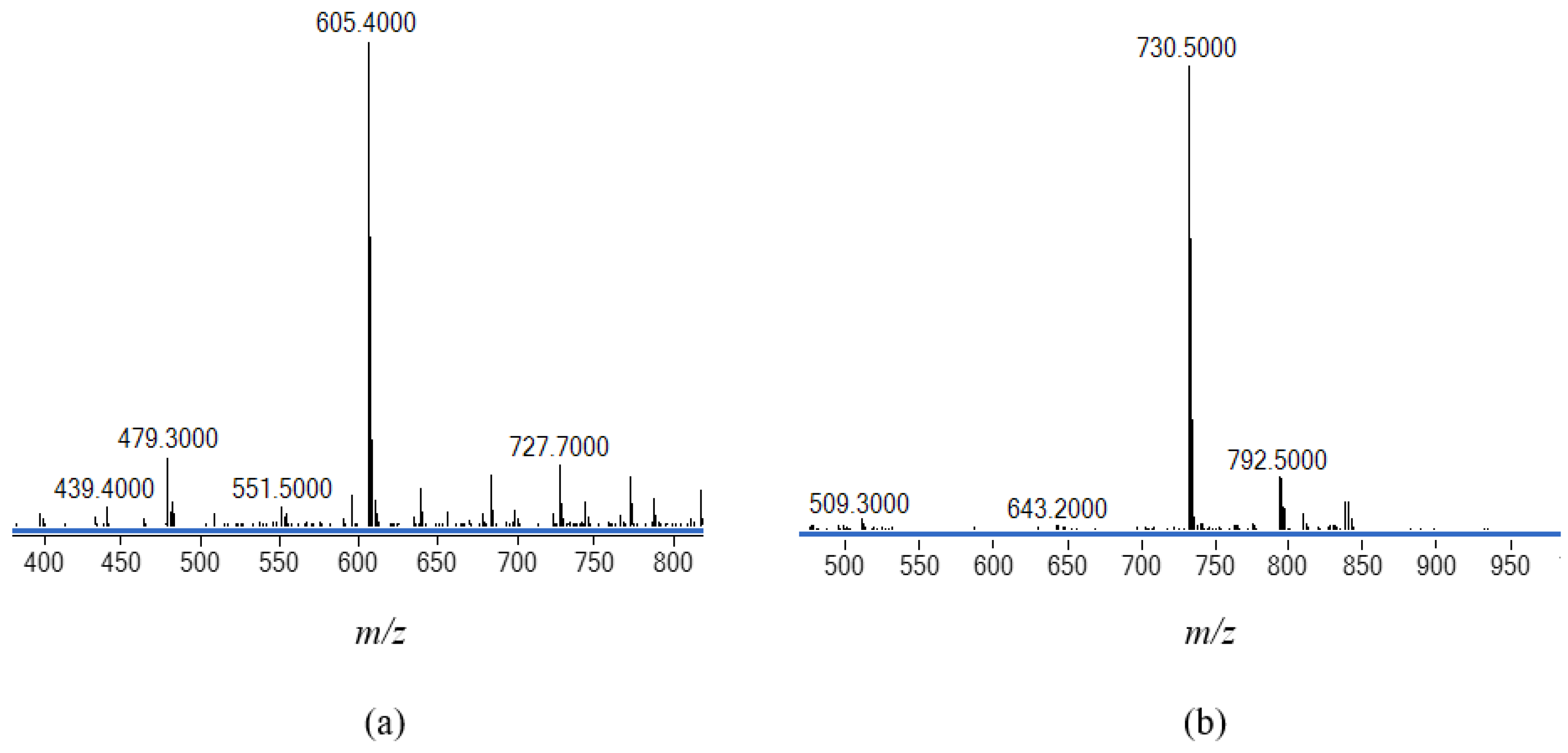
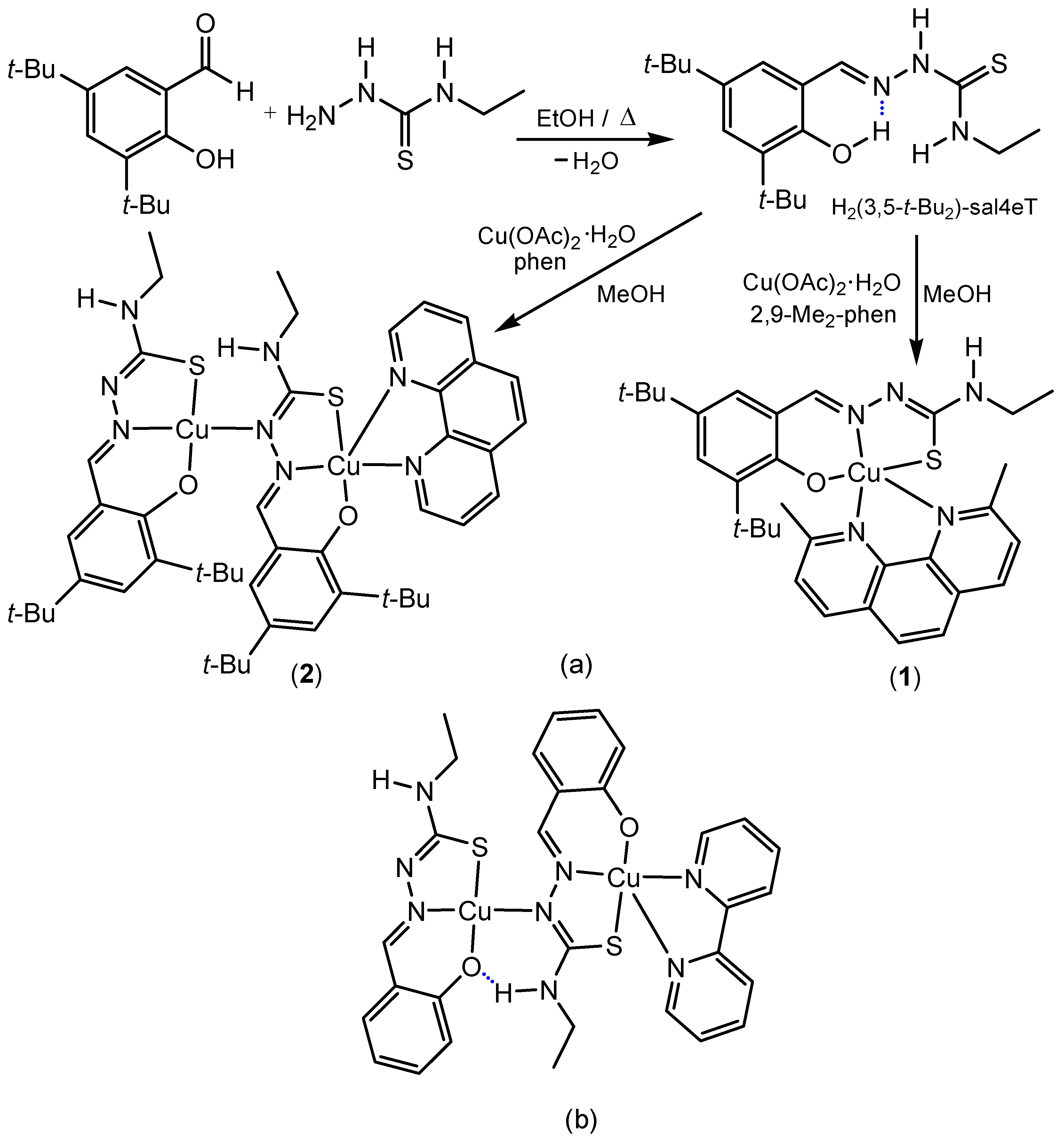
2.1. Synthesis and Chemical Identification of the Thiosemicarbazone Ligand

The thiosemicarbazone H2(3,5-t-Bu2)-sal4eT was synthesised from equimolar amounts of 3,5-di-tert-butylsalicylaldehyde and 4-ethyl-3-thiosemicarbazide through the usual single-step Schiff-base condensation reaction in refluxing absolute ethanol. The resultant light yellow solution afforded long lustrous colourless needles upon slow evaporation of the solvent under ambient conditions over a span of several days. Unlike the synthesis of pyridyl-based thiosemicarbazones [8,13,14,15], this phenolic thiosemicarbazone was produced straightforwardly in high yield without requiring acid-catalysis (Scheme 1). The chemical identity of this ligand was ascertained through microanalysis along with electrospray ionisation (ESI) mass spectrometry. The ESI mass spectrum exhibits a parent peak at m/z = 334.3 [M − H+] in the negative-ion mode (Figure S1) and at m/z = 336.3 [M + H+]+ in the positive-ion mode, consistent with the molecular mass (M = 335.51 amu).

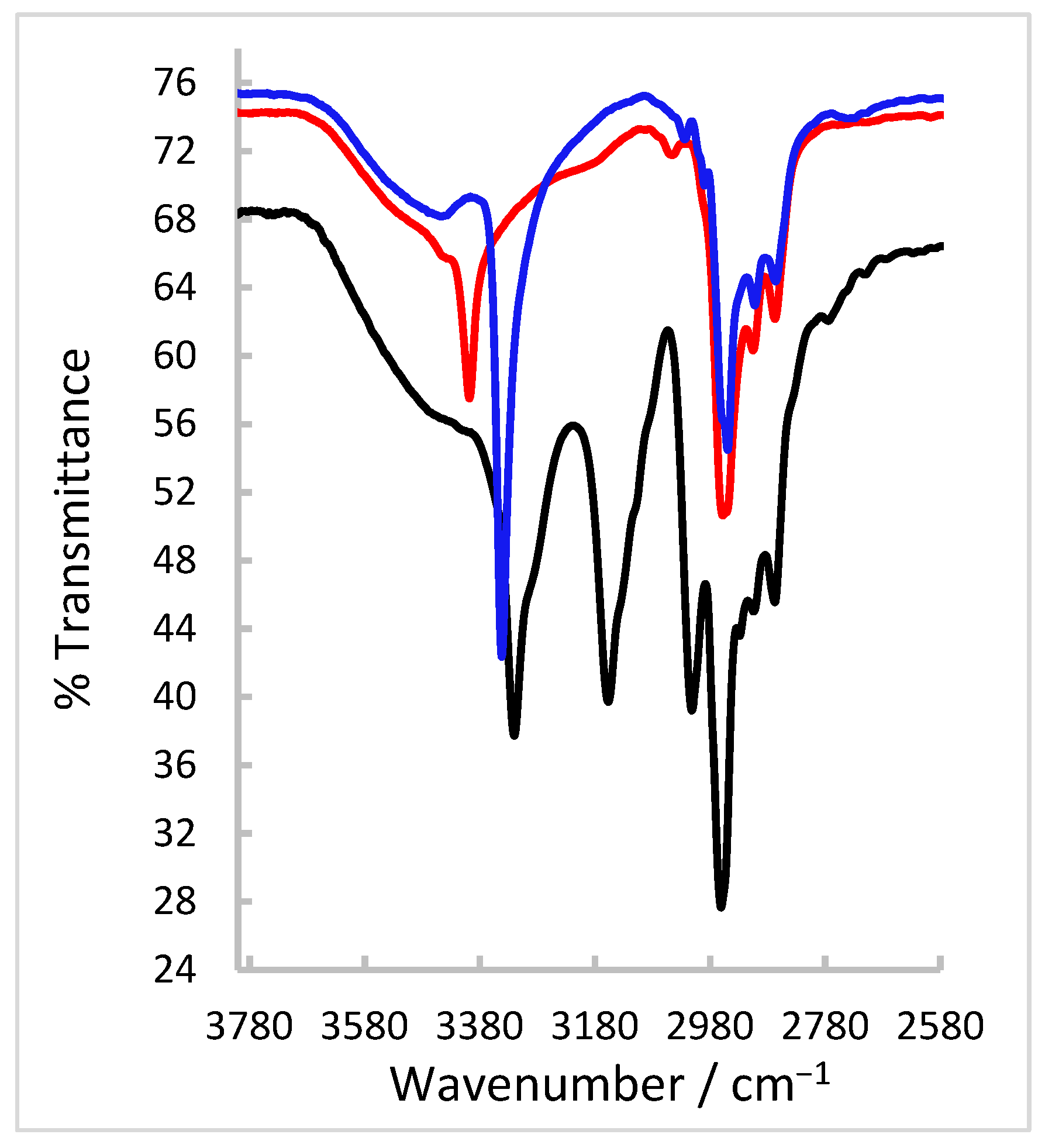
2.2. FT-IR and NMR Spectroscopic Characterisation of the Thiosemicarbazone Ligand

That H2(3,5-t-Bu2)-sal4eT exists as the thio-keto (thione) tautomer in the solid state is demonstrated by the prominent IR absorption band at 3157 cm−1, indicative of the thio-amide ν(N–H) (Figure 1 and Figure S2a). The associated thio-carbonyl bond is characterised by the vibrational band with a stretching frequency of 1032 cm−1. Indeed, the absence of a vibrational band at around 2600 cm−1 due to ν(S–H) [10] excludes the possibility of the occurrence of the thio-enol tautomer. At 3320 cm−1 in the IR spectrum, an absorption band occurs that is ascribable to the N–H stretching of the terminal secondary amino group of the thiosemicarbazone. A characteristic feature of Schiff bases is the imine bond whose presence in H2(3,5-t-Bu2)-sal4eT is evidenced by the absorption at 1609 cm−1 typifying ν(C=N). The tert-buyl C–H stretches are conspicuous given their characteristic pattern of absorption bands in the range of 2867–2960 cm−1 [59]. Contributing to the intensity of these absorptions are the C–H vibrations of the N-ethyl substituent group. The sharp absorption band with a stretching frequency of 3013 cm−1 is attributable to the aromatic ν(C–H). Finally, the broad band at around 3500 cm−1 is typical of phenolic O–H vibrations.
The 1H-NMR spectrum of H2(3,5-t-Bu2)-sal4eT was recorded in DMSO-d6 (Figure S3a) at a radiofrequency of 700 MHz with TMS as an internal reference standard (δ = 0). The broad peak at δ 9.98 assignable to the hydrazinic proton reveals that the thione tautomer of this thiosemicarbazone remains intact in solution. The phenolic proton, which is represented by the singlet at δ 11.27, is the most deshielded on account of its intramolecular interaction with the imine nitrogen atom. The aldimine proton is associated with the sharp singlet at δ 8.28. A broad resonance shaped like an unresolved triplet occurs at δ 8.48 and is attributable to the proton of the amino group between the thio-carbonyl and ethyl groups. The aromatic protons in positions 4 and 6 resonate as doublets at δ 7.13 and 7.29, respectively, with identical coupling constants (J = 2.38 Hz). The N-ethyl group is characterised by partially overlapping quartet signals at δ 3.59 (J = 6.55 Hz) and a triplet resonance at δ 1.16 (J = 7.14 Hz) corresponding to the methylene protons (in non-equivalent environments) and the methyl protons, respectively. Finally, the protons of the 3-tert-butyl and 5-tert-butyl substituent groups have singlet signals with the chemical shifts at δ 1.41 and 1.27, respectively.
Interestingly, when the 1H-NMR spectrum of H2(3,5-t-Bu2)-sal4eT is measured in deuterated methanol (CD3OD), the resonances of the –OH and the two –NH protons disappear (Figure S3b), indicative of rapid exchange of each of these for deuterium on the NMR timescale. The chemical shift at δ 8.14 of the imine –CH=N singlet is the most downfield in this spectrum. The aromatic protons in positions 4 and 6 appear as doublets (δ 7.14, J = 2.38 Hz and δ 7.38, J = 2.44 Hz, respectively). The ethyl –N(H)–CH2CH3 protons are observed as quartet (δ 3.70, J = 7.18 Hz) and triplet (δ 1.24, J = 7.19 Hz) signals, respectively, whereas the 3-tert-butyl and 5-tert-butyl protons occur as singlet peaks at δ 1.44 and 1.30, respectively.
The 13C-NMR spectrum of H2(3,5-t-Bu2)-sal4eT, recorded in DMSO-d6 at 176 MHz (Figure S4), exhibits a resonance for the thio-carbonyl carbon at δ 176.29, confirming the existence of this thiosemicarbazone as the thione tautomer in this solution. At δ 147.54, a signal occurs representing the imine carbon atom. All the aromatic carbon atoms are accounted for in the range of δ 117.75–153.11, with the phenolic carbon being the most deshielded. The signals of the N-ethyl methylene and methyl carbon atoms appear at δ 38.73 and 14.59, respectively. Finally, the tert-butyl carbon atoms have resonances in the range of δ 29.44–34.71.

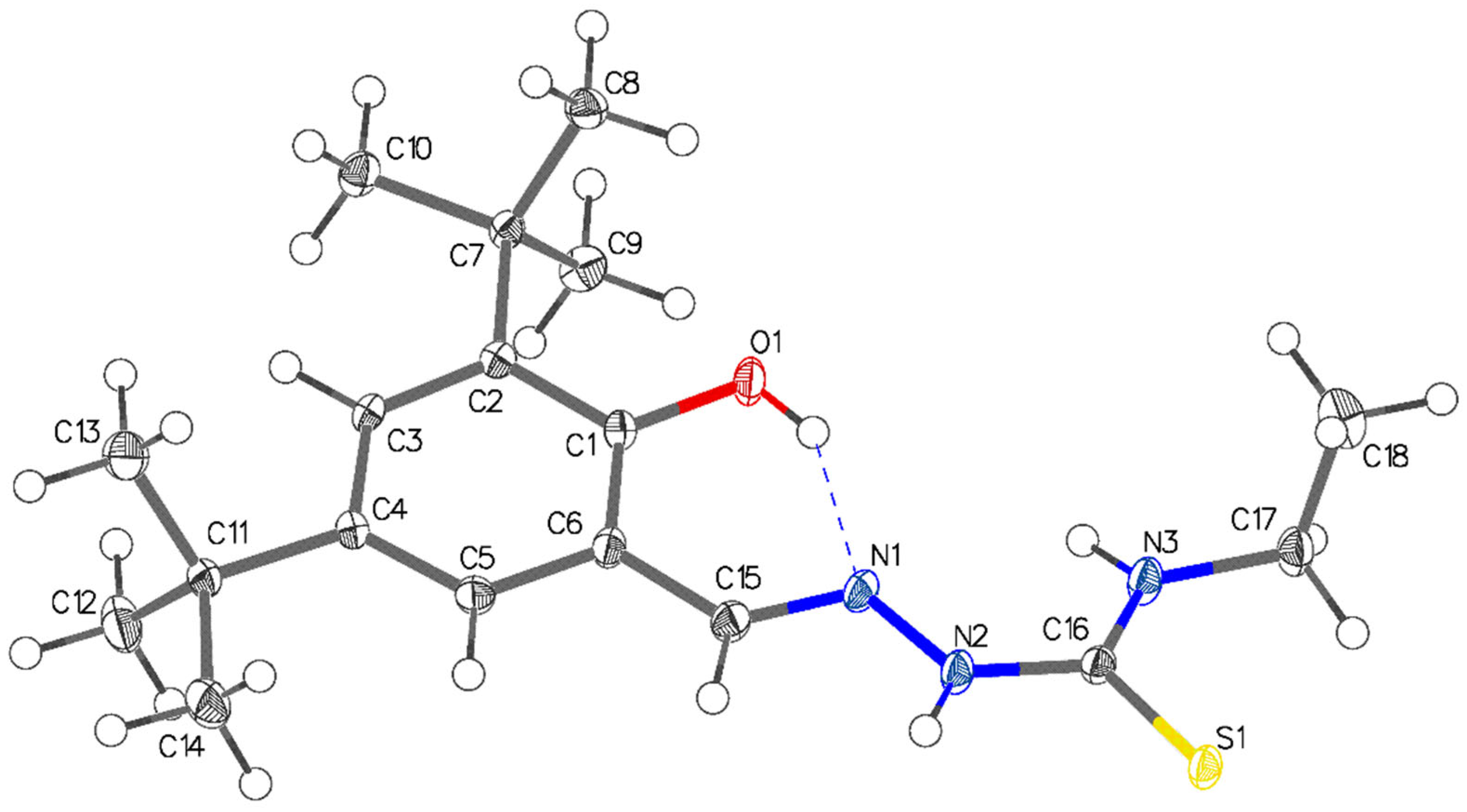
2.3. Single-Crystal X-ray Structural Determination of the Thiosemicarbazone Ligand

Definitive evidence for the solid-state 3D structure of the ligand was obtained through single-crystal X-ray analysis. A colourless needle amenable to X-ray diffraction was grown from a solution of H2(3,5-t-Bu2)-sal4eT in EtOH at room temperature. X-ray data collection was performed at 100 K. Crystal data, details of data collection and parameters for structural solution and refinement are compiled in Table 1. Evidently, this thiosemicarbazone ligand crystallised in the monoclinic space group P21/c with four molecules in the unit cell. The X-ray crystal structure is depicted in Figure 2 while selected bond distances and angles are presented in Table 2. The distance of the Schiff-base bond C=N [C(15)–N(1) = 1.2904(19) Å] lies within the range observed for normal imine bonds [1.26–1.30 Å] [13,14,15,19,20,60,61,62,63,64,65,66,67,68,69] in non-coordinated ligands. Upon reduction in the Schiff base, the imine double bond becomes a single bond (C–N) with a distance of ~1.47 Å [65]. The C(16)–S(1) distance of 1.7029(14) Å verifies the occurrence of the thione tautomer. Literature values for the distance of the thio-carbonyl bond in free thiosemicarbazone ligands range typically from 1.65 to 1.70 Å [13,14,15,19,20,60,61,62,63,64,65,66,67,68,69] (even longer if involved in bifurcated H-bonding) [67]. Both imine nitrogen and thio-carbonyl carbon are sp2-hybridised and the angles around them reflect the angular (with a lone pair) and trigonal planar geometries about these two atoms. The hydrazinic N–N bond [N(1)–N(2) = 1.3854(16) Å] is somewhat longer than most of those reported for other non-coordinated thiosemicarbazones and is consistent with single-bond character.
One of the prominent structural features of interest is the intramolecular H-bonding interaction between the phenolic –O–H group and the imine nitrogen atom [O(1)–H(1)···N(1):O1–H1 = 0.84 Å, H1···N1 = 1.98 Å, O1···N1 = 2.7221(15) Å, O1–H1···N1 = 147.4°]. Indeed, the vast majority of Schiff bases derived from 2-hydroxybenzaldehydes, 2-hydroxyacetophenones, 2-hydroxybenzophenones, 2-hydroxypropiophenones, etc., exhibit this intramolecular electrostatic force. It is well-established that pyridyl-/phenol-based thiosemicarbazones can adopt an E- or Z-configuration with respect to the imine double bond. Moreover, they can also orient themselves in different conformations as a consequence of free rotation about the C(py/phenol)–C(imine) (i.e., C(6)–C(15) in this structure) single bond and the amide N(H)–C(=S) (i.e., N(2)–C(16) in this structure) single bond. Thus the potential donor atoms can be positioned anti or syn relative to each other. Examples of crystallographically observed orientations of phenolic thiosemicarbazones, viz. E(syn,anti) [60,62,63], E(syn,syn) [61,64] and E(ant,anti) [63], are shown in Figure S5. The structure of H2(3,5-t-Bu2)-sal4eT is consistent with the E-configuration; the phenolic –OH group and the imine nitrogen are positioned syn to each other while the thione sulphur points to the opposite side in an anti-orientation relative to the imine nitrogen.

2.4. Synthesis and Chemical Identification of the Copper(II) Thiosemicarbazone Complexes

Reaction of H2(3,5-t-Bu2)-sal4eT with a molar equivalent of Cu(OAc)2·H2O in refluxing MeOH, followed immediately by addition of a stoichiometric amount of 2,9-dimethyl-1,10-phenanthroline (2,9-Me2-phen) or 1,10-phenathroline (phen) with brief heating of the resultant dark olive green solution, afforded the mononuclear copper(II) complex [Cu{(3,5-t-Bu2)-sal4eT}(2,9-Me2-phen)] (1) or the dinuclear copper(II) complex [Cu2{(3,5-t-Bu2)-sal4eT}2(phen)] (2), respectively. The chemical formulations of these two ternary complexes were established through elemental analyses. The mass spectra of the complexes were measured in MeOH. The positive-ion ESI mass spectrum of complex 1 presented in Figure 3a shows a molecular peak at m/z = 605.4 in agreement with the molecular mass of this complex (605.25 amu). As regards the dinuclear complex (2), the parent ion was not detected; however, the ESI spectrum revealed important structural information from the fragmentation pattern. In the negative mode, the spectrum shows a minor peak at m/z = 792.5 consistent with the loss of the phen co-ligand. At m/z = 730.5, a major peak occurs that is ascribable to the fragment [Cu{H(3,5-Bu2)-sal4eT}{(3,5-Bu2)-sal4eT}] (Figure 3b). On the other hand, the positive-ion ESI spectrum exhibits a peak at m/z = 732.6 attributable to the fragment [Cu{(3,5-t-Bu2)-sal4eT}2]+. Further dissociation affords the fragment [Cu{(3,5-t-Bu2)-sal4eT}]+ observed at m/z = 397.2, signifying the loss of one of the thiosemicarbazonate ligands. That complexes 1 and 2 are molecular has been demonstrated through the negligible value of the molar electrical conductivity (ΛM ~3–5 Ω−1 cm2 mol−1) of their nonelectrolyte solutions in MeOH, EtOH, DMF and DMSO at room temperature [70].

2.5. FT-IR Spectroscopy and Magnetic Susceptibility Measurements

A comparison of the IR spectra of the thiosemicarbazone ligand and its copper(II) complexes (1 and 2) in Figure 1 and Figure S2 clearly shows the absence of the vibrational band of the hydrazinic N–H bond from the spectra of the complexes. The disappearance of the hydrazinic proton coupled with the shift in the stretching frequency of the absorption band of the carbon–sulphur bond from 1032 cm−1 for the ligand to 842 and 858 cm−1 for 1 and 2, respectively, is indicative of tautomerisation and deprotonation of the thiosemicarbazone upon coordination to the copper(II) ion, as is indeed necessary for charge-neutrality of the resultant complexes. The presence of the N–H group attached to the terminal ethyl group is proven by the occurrence of sharp absorption bands at 3398 and 3342 cm−1 in the spectra of 1 and 2, respectively. The wavenumbers of the imine bond for 1 and 2 complexes are somewhat lower than that of the free ligand [ν(C=N): 1598 and 1599 cm−1 vs. 1609 cm−1], consistent with coordination of the imine donor atom. Interestingly, the ν(N–N) absorptions for the ligand and complexes 1 and 2 virtually coincide (1172, 1170 and 1169 cm−1, respectively), implying minimal delocalisation of π-electrons, if any, along the ligand backbone in the complexes. Finally, the other ligand IR absorption patterns, especially those of the tert-butyl C–H bonds (2850–2960 cm−1), are retained.
Complexes 1 and 2 are paramagnetic with a single unpaired electron at the metal centre in the ground state. The room-temperature effective magnetic moment [µeff = (8χM)½] of the mononuclear complex (1) is 1.83 µB. It is comparable with the spin-only value [µS = {4S(S + 1)}½, where S = ½] and lies within the range of literature values [45,50,54,56,57,58,59]. In contrast, for the dinuclear complex (2), µeff = 2.38 µB at room temperature, which is close to the spin-only value for two magnetically uncoupled d9 paramagnetic centres [{4S1(S1 + 1) + 4S2(S2 + 1)}½, where S1 = S2 = ½].

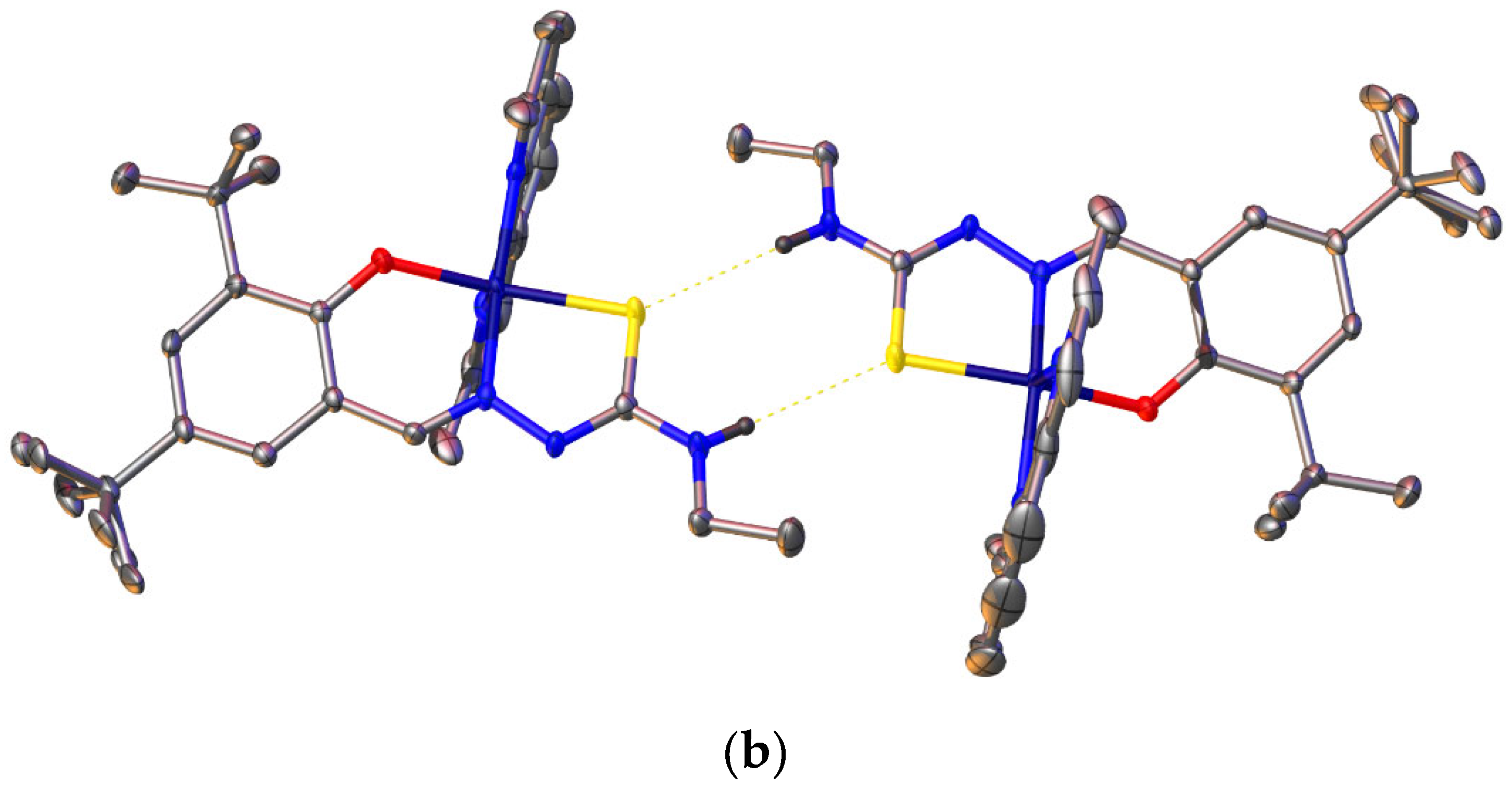
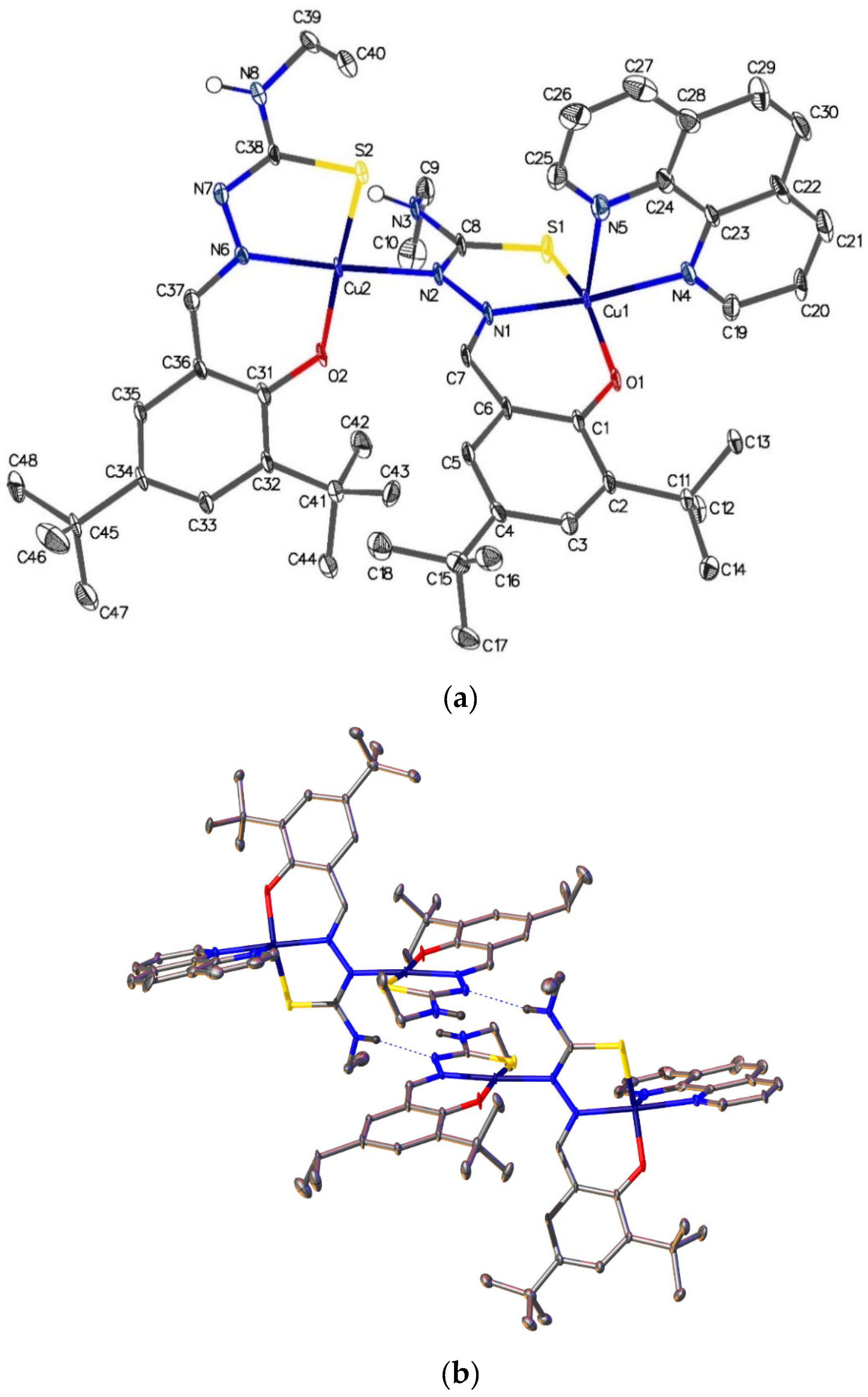
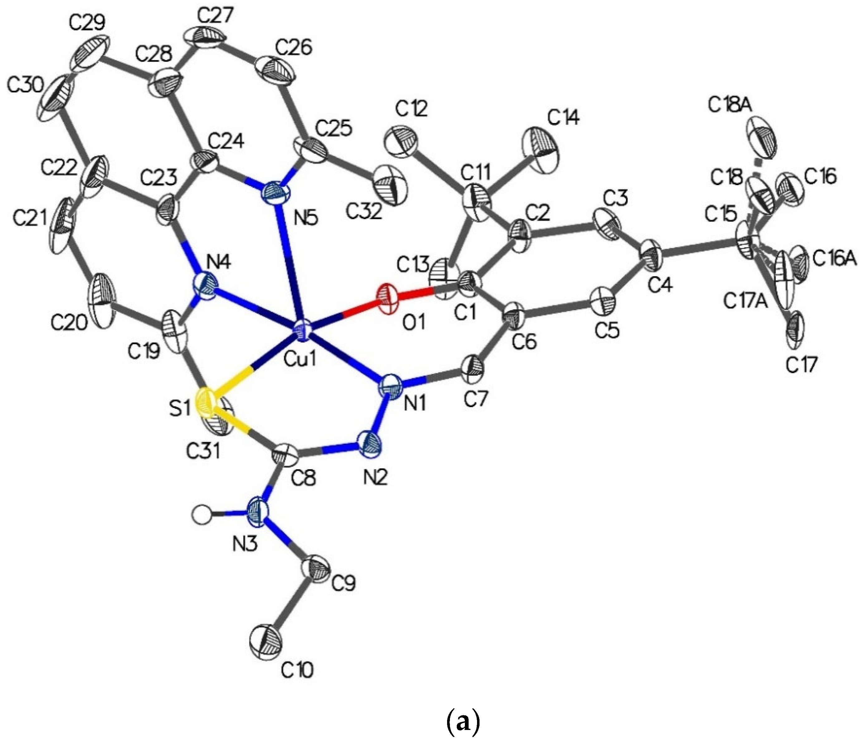
2.6. Single-Crystal X-ray Analyses of the Ternary Copper(II) Complexes

For the complexes [Cu{(3,5-t-Bu2)-sal4eT}(2,9-Me2-phen)] (1) and [Cu2{(3,5-t-Bu2)-sal4eT}2(phen)] (2), X-ray diffraction data were collected on a single crystal at 100 K employing Cu-Kα radiation (λ = 1.54178 Å). Crystal data together with details of data collection and structural refinement are presented in Table 1. Selected bond distances and angles are given in Table 3. Whereas complex 1 crystallised in the monoclinic space group P21/n with Z = 4, complex 2 did so in the triclinic space group P 1 ¯ with two complex molecules in the unit cell. Both complexes 1 and 2 are free of solvent molecules of crystallisation.
The crystal structure of [Cu{(3,5-t-Bu2)-sal4eT}(2,9-Me2-phen)], depicted in Figure 4, reveals that this complex exists as a centrosymmetric dimer of mononuclear molecular ternary complexes of copper(II). The dimerisation occurs via two intermolecular hydrogen-bonding interactions involving the –N4–H group of one complex molecule and the thio-enolate sulphur of the other complex molecule (Figure 4b) [N(3)–H(3)···S(1): N–H = 0.88 Å, H···S = 2.64 Å, N···S = 3.482(3) Å, N–H–S = 159.9° (symmetry code: 1 − x, 1 − y, −z)]. The charge-neutrality of this ternary complex implies that the thiosemicarbazone been doubly deprotonated upon complexation. Indeed, transformation of the ligand from the thio-keto tautomer to the thio-enolate anion is demonstrated through the changes to the lengths of the pertinent bonds of the thio-amide. The thio-amide N–C bond [N(2)–C(16): 1.3499(18) Å in the free ligand] has shortened considerably upon complexation [N(2)–C(8): 1.313(4) Å in complex 1] while the thio-carbonyl (C=S) bond [C(16)–S(1): 1.7029(14) Å in the ligand] has converted to the thio-enolate C–S bond in the complex [C(8)–S(1) = 1.739(3) Å]. Carbon–nitrogen bonds with double-bond character have been reported to have distances in the range 1.27–1.32 Å [66,71,72,73] when the N donor atom is coordinated to a central metal ion. On the other hand, typical lengths of carbon–sulphur bonds with single-bond character in thio-enolate complexes are in the range 1.72–1.77 Å [66,71,72,73]. The distances of the imine C=N [C(7)–N(1) = 1.296(4) Å] and the hydrazinic N–N [N(1)–N(2) = 1.400(4) Å] in 1 are normal with regard to their respective bond orders.
The five-coordinate geometry at the copper(II) centre arises from the tridentate coordination of the thiosemicarbazonate ligand with the donor atoms, namely phenolate oxygen, imine nitrogen and thio-enolate sulphur, arranged meridionally, and the bidentate coordination of the 2,9-Me2-phen co-ligand oriented nearly perpendicularly relative to the primary ligand. The axial–equatorial coordination mode of the pyridyl nitrogen atoms of 2,9-Me2-phen leads to the construction of a coordination sphere best described as distorted square pyramidal in accordance with the trigonality index [τ = (β − α)/60°] [74] of ~0.14, the largest two angles β and α being in the basal plane. The axial Cu–N bond is considerably longer than the one lying equatorially [Cu–Nax (co-ligand) = 2.308(3) Å vs. Cu–Neq (co-ligand) = 2.057(3) Å]. This elongation of the axial coordinate bond is attributable to the tetragonal distortion at the metal centre. Invariably, square pyramidal [31,32,33,34,35,36,37,38,39,40,41,42,43,45,46,47,48,49,50,51,52] and octahedral [44,53] ternary complexes with axial–equatorial coordination of a bidentate co-ligand (bipy, phen or their derivatives) are subject to the Jahn–Teller effect evidenced by this structural feature. In some cases, even binary bis(chelate) copper(II) complexes [75] with two potentially tridentate ligands experience this effect, causing one of the ligands to coordinate bidentately due to considerable weakening of one of the axial Cu–L bonds. The magnitude of the disparity in the Cu–N distances of the asymmetrically coordinated N,N-donor co-ligand is in the range of ~0.22–0.32 Å. Moreover, the complex cation of tris(1,10-phenanthroline)copper(II) perchlorate [76] exhibits Jahn–Teller distortion (Cu–N bond averages: Cu–Nax ~2.33 Å vs. Cu–Neq ~2.04 Å) whereby the axial Cu–Nphen bonds are elongated to the same extent as those in the above-mentioned square pyramidal copper(II) ternary complexes. In contrast, it has been crystallographically proven that the two Cu–N bonds of N,N-donor co-ligands in square pyramidal and octahedral complexes where they lie on the equatorial plane are virtually equivalent as neither is subject to the Jahn–Teller effect [54,55,56,57,58]. In addition, in the complex [Cu{N(CN)2}(phen)2]+ [77] with a distorted trigonal bipyramidal geometry at the metal centre, the phen Cu–N distances are virtually indistinguishable from each other as the Jahn–Teller effect does not apply. The copper(II) ion in [Cu{(3,5-t-Bu2)-sal4eT}(2,9-Me2-phen)] (1) is displaced out of the mean basal plane [N(1), S(1), O(1), N(4)] towards the apical phen N(5) donor atom by 0.1998(12) Å. Finally, the magnetostructural behaviour of this complex is consistent with half occupancy of the dx2y2 orbital in the ground state.
As can be seen from Figure 5, [Cu2{(3,5-t-Bu2)-sal4eT}2(phen)] (2) exists in the crystal lattice as a centrosymmetric dimer of dinuclear molecular ternary complexes of copper(II) stabilised mainly by two types of intermolecular forces. The linkage of two dinuclear complex molecules occurs through two H-bonds between the N4–H group of one dinuclear molecule and the imine nitrogen of another dinuclear molecule [N(3)–H(3)···N(7): N–H = 0.88 Å, H···N = 2.17 Å, N···N = 2.964(11) Å, N–H···N = 150.5° (symmetry code: 2 − x, −y, 1 − z)]. Moreover, this complex exhibits π-π stacking interactions involving the plane N(5), C(24)–C(28) of the phen co-ligand in two complex molecules (symmetry code: 1 − x, 1 − y, 1 − z) (angle of interaction of the two planes = 0.0(7)°, centroid-to-centroid distance = 3.603(8) Å, shift distance = 1.218(17) Å) (Figure S6).
The two copper(II) centres are 4.8067(18) Å apart and display different coordination numbers, one four-coordinate and the other five-coordinate, with the respective coordination geometries being distorted square planar [τ4 = {360° − (α + β)}/141° = 0.082, α = 176.3° and β = 172.1°] [78] and distorted square pyramidal [τ5 = (β − α)/60° = 0.33, β = 172.4° and α = 152.4°] [74]. The two associated thiosemicarbazonate ligands exhibit different denticities: one coordinates in a tridentate fashion to the metal centre (Cu(2) in Figure 5) with coordination number 4, whereas the other adopts the relatively unusual quadridentate coordination mode to bridge the two metal centres with the thio-enolate nitrogen atom, N(2), and coordinate meridionally to the other metal centre (Cu(1) in Figure 5). For the tridentate ligand, coordinated to Cu(2), the distance of the newly formed thio-enolate N=C bond is virtually indistinguishable from that of the imine C=N bond [cf. N(7)–C(38) = 1.316(12) Å vs. C(37)–N(6) = 1.310(12) Å, respectively] and the distance of C(32)–S(2) (1.746(9) Å) lies within the range reported for such thio-enolate bonds. Similarly, for the quadridentate ligand, the distances of the thio-enolate N=C and imine C=N bonds compare favourably [cf. N(2)–C(8) = 1.322(13) Å and C(7)–N(1) = 1.301(13) Å, respectively] [66,71,72,73]. The distance of the thio-enolate C–S bond [C(8)–S(1) = 1.737(9) Å] is normal for complexed thiosemicarbazonate ligands [66,71,72,73]. It is noteworthy that the lengths of the hydrazinic N–N bonds in this dinuclear complex [N(1)–N(2) = 1.381(11) Å and N(6)–N(7) = 1.391(11) Å] are very similar to that observed in the free ligand as a thio-keto (thione) tautomer (1.3854(16) Å), suggesting that there is no delocalisation of electrons involving this chemical bond in the thiosemicarbazonate backbone.
The five-coordinate geometry at Cu(1) is similar to that described for the mononuclear ternary complex (1). The bidentate phen co-ligand adopts the axial–equatorial coordination mode. Consequently, the axial Cu–Nphen bond is tetragonally elongated, even longer than the Cu–S bond [Cu(1)–S(1) = 1.266(3) Å], causing asymmetric coordination of this co-ligand [Cu(1)–N(4)eq = 2.039(8) Å vs. Cu(1)–N(5) = 2.276(9) Å]. The copper(II) ion, Cu(1), resides 0.177(4) Å above the mean basal plane [N(1), S(1), O(1), N(4)] in the direction of the axial N(5)phen atom. Both copper(II) centres [Cu(1) and Cu(2)], regardless of the differences in the coordination geometries, have a dx2y2 ground state. The literature has witnessed a number of examples of crystallographically characterised dinuclear thiosemicarbazone complexes of copper(II), but these tend to have the same coordination geometry at the two metal centres [8,9,10,72]. To the best of our knowledge, the dinuclear complex [Cu2{(3,5-t-Bu2)-sal4eT}2(phen)] (2) is one of only two of its kind. The other structurally characterised dinuclear thiosemicarbazone complex of copper(II) featuring two different coordination spheres is [Cu2(sal4eT)2(bipy)] (Scheme 1), reported as [Cu2(L2)2(bipy)] [4]. Beyond the superficial similarities, there is a sharp distinction between the structures of [Cu2{(3,5-t-Bu2)-sal4eT}2(phen)] (2) and [Cu2(sal4eT)2(bipy)] as regards the orientation of the ligands, intermolecular forces and the five-coordinate geometry at one of the two copper(II) centres. Unlike 2, [Cu2(sal4eT)2(bipy)] exists as two independent complex molecules, which are similar but not identical, and the thiosemicarbazonate ligands are oriented (differently from 2) such that intramolecular hydrogen bonding occurs between the phenolate oxygen atom bonded to the copper(II) ion in the distorted square planar geometry and the N4–H group of the bridging quadridentate ligand. Moreover, the geometries of the five-coordinate copper(II) centres in the two molecules of [Cu2(sal4eT)2(bipy)] were reported as distorted trigonal bipyramidal. We calculated their trigonality indices, τ5 [74], to compare them with that of our dinuclear complex (2). For [Cu2(sal4eT)2(bipy)], the values of τ5 are ~0.50 and ~0.51 (intermediate between square pyramidal and trigonal bipyramidal); in contrast, for 2, τ5 = 0.33, clearly pointing to greater distortion towards square pyramidal.

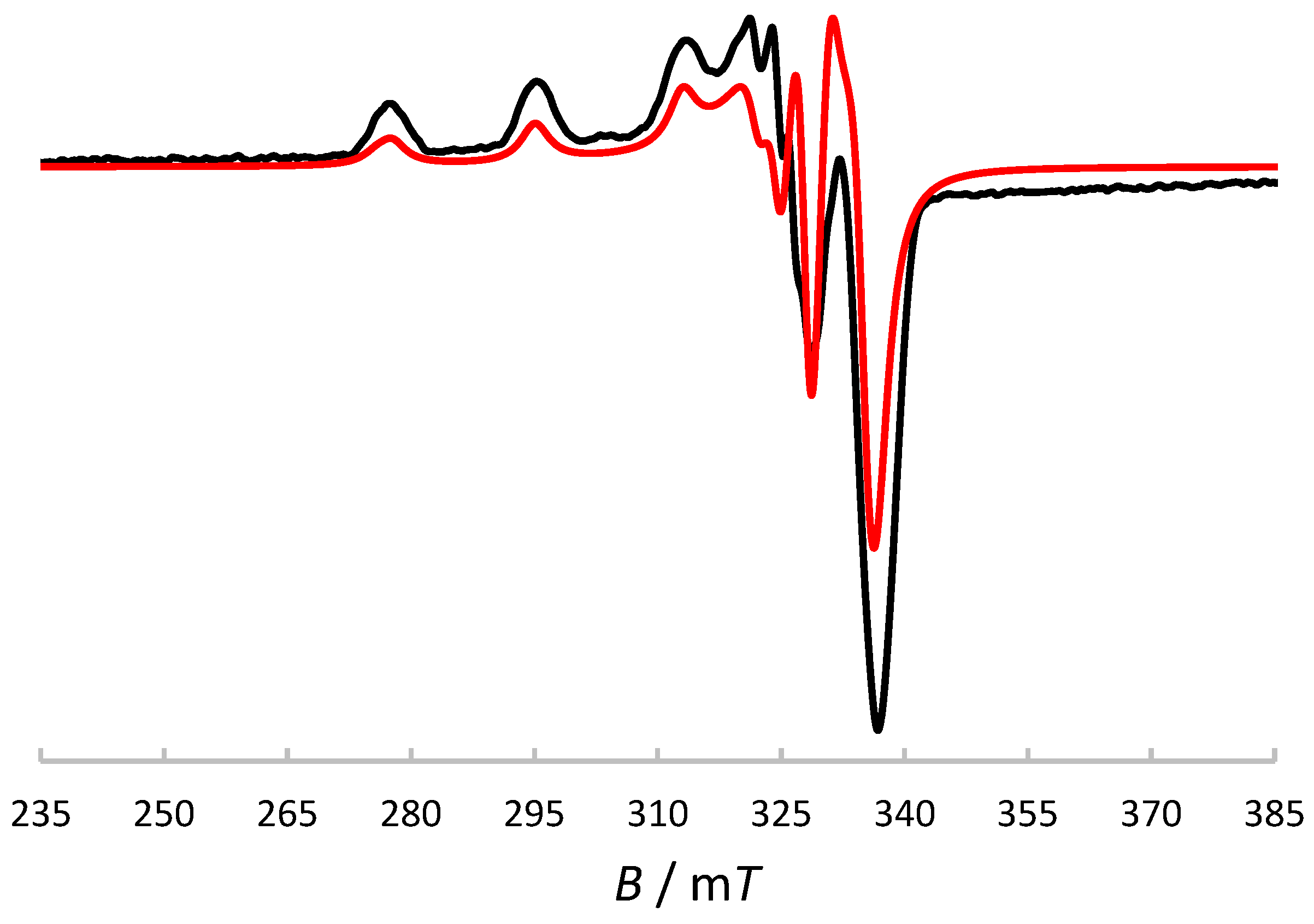
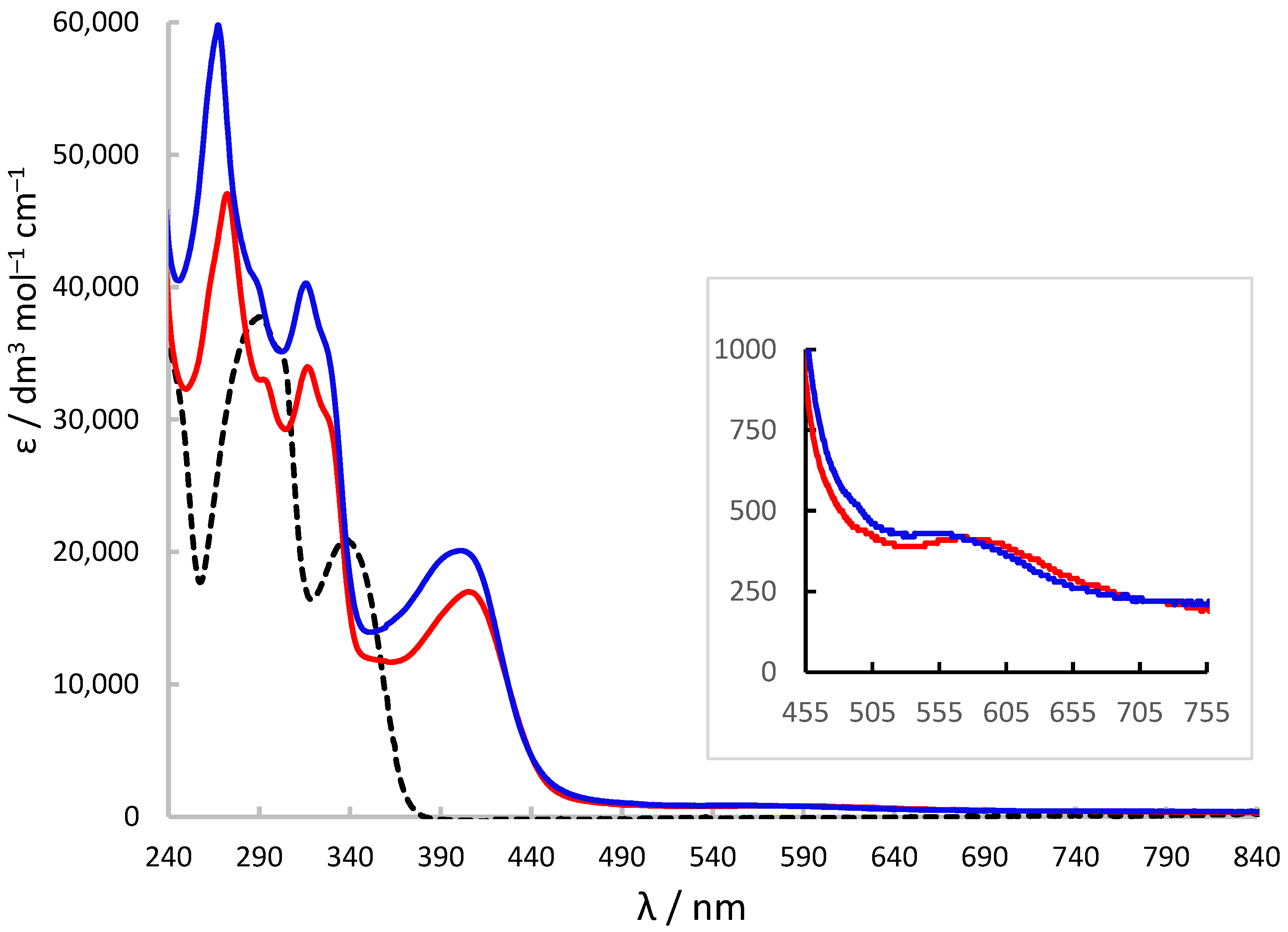
2.7. X-Band ESR and UV-Visible Spectroscopic Characterisation

The X-band ESR spectrum of [Cu{(3,5-t-Bu2)-sal4eT}(2,9-Me2-phen)] (1) in frozen MeOH solution at 77 K, displayed in Figure 6, is axial. The ESR spin Hamiltonian parameters gǁ = 2.20, g = 2.05, Aǁ = 19.3 mT and A = 3 mT (gǁ > g > 2.00; Aǁ > A) are consistent with the dx2y2 ground state [75,79,80,81]. Hence, ESR spectroscopy demonstrated that the crystallographically determined distorted square pyramidal geometry at the metal centre is retained in solution.
Figure 7 shows the electronic absorption spectra of H2(3,5-t-Bu2)-sal4eT, [Cu{(3,5-t-Bu2)-sal4eT}(2,9-Me2-phen)] (1) and [Cu2{(3,5-t-Bu2)-sal4eT}2(phen)] (2). The ligand is colourless; accordingly, its spectrum exhibits UV absorption only. The hugely intense absorption bands are assignable to π π* (280–305 nm) and n  π* (~338 nm) electronic transitions [4]. This pattern of UV absorption is also observed in the spectra of 1 and 2, albeit at somewhat higher energies.
In the visible region of the electronic spectra of 1 and 2, there are intense broad bands centred at 405 nm (εmax ~ 17000 dm3 mol−1 cm−1) and 401 nm (εmax ~ 20100 dm3 mol−1 cm−1), respectively, attributable to phenolate/thio-enolate-to-copper(II) charge-transfer electronic transitions [4]. Spin-allowed, but Laporte-forbidden, ligand-field transitions (Figure 7 inset) were observed as weak broad absorption bands at ~590 (εmax ~ 390 dm3 mol−1 cm−1) and ~570 nm (εmax ~ 420 dm3 mol−1 cm−1) for 1 and 2, respectively. For distorted square-pyramidal copper(II) complexes, such d-d electronic transitions have been assigned previously as dxz,dyzdx2y2 in nature [79].

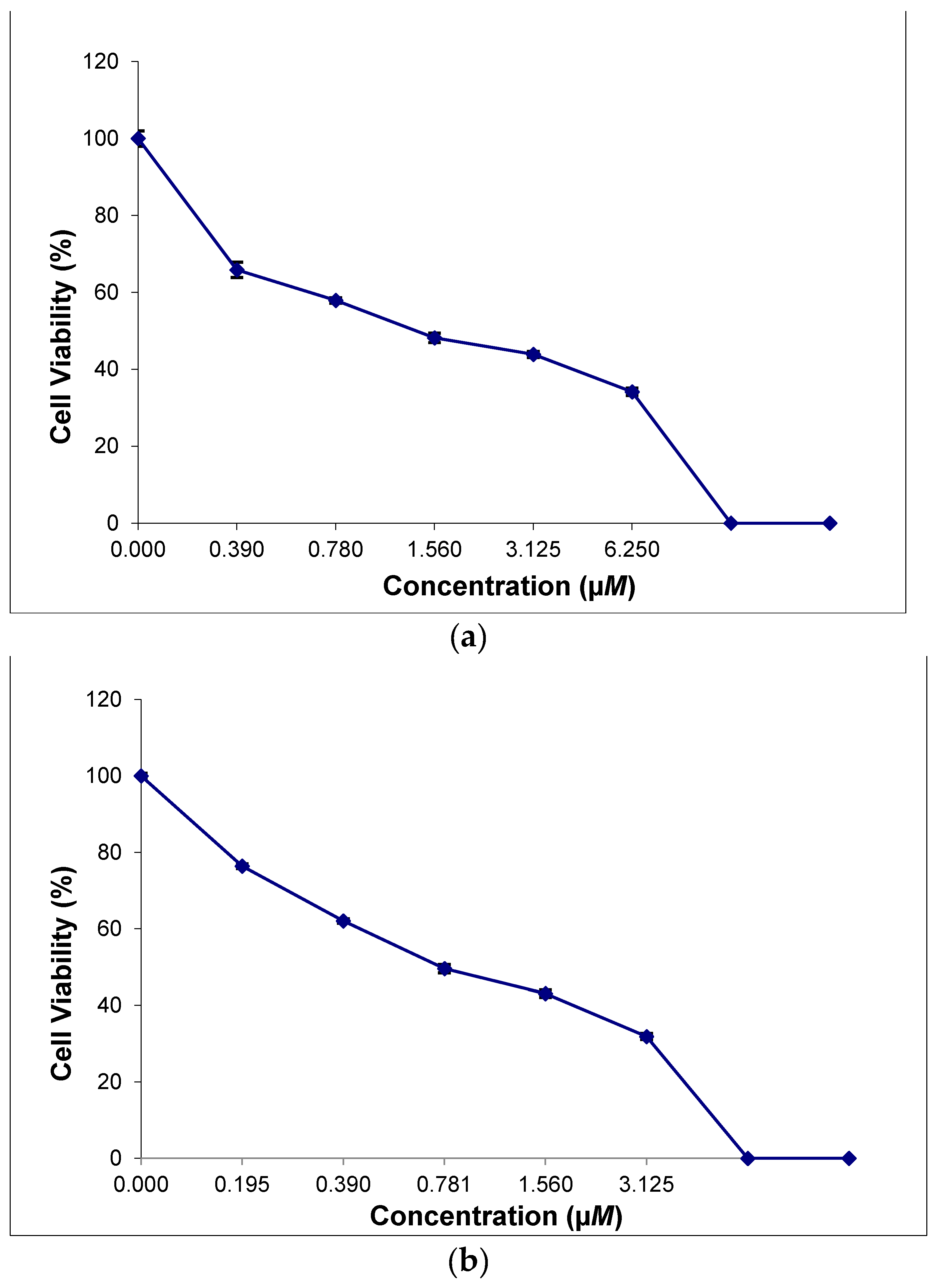
2.8. In Vitro Cytotoxicity of the Thiosemicarbazone Ligand and the Ternary Copper(II) Complexes

The antiproliferative activity of H2(3,5-t-Bu2)-sal4eT, [Cu{(3,5-t-Bu2)-sal4eT}(2,9-Me2-phen)] (1) and [Cu2{(3,5-t-Bu2)-sal4eT}2(phen)] (2) was investigated in two cancer cell lines, namely human cervical carcinoma (HeLa) and human breast adenocarcinoma (MCF-7) using the MTT cell viability assay [MTT = 3-(4,5-dimethylthiazol-2-yl)-2,5-diphenyltetrazolium bromide]. The compounds were dissolved in DMSO (negative control) and it was shown experimentally that the solvent was inactive against the cancer cells (IC50 >> 100 μM). The values of the 50% inhibitory concentrations (IC50) of these substances together with the positive controls (docetaxel and paclitaxel) were determined, as exemplified for complexes 1 and 2 in the HeLa and MCF-7 cancer cells, respectively (Figure 8). The in vitro antiproliferative potential of each substance was tested within the 0.01–100-μM range of concentrations; the results of these cytotoxicity measurements are presented in Table 4. IC50 values for cisplatin were obtained from the literature [82,83]. The details of the MTT cell viability assay are given in the Experiment Section 3.5.
In striking contrast to the marked potent and selective antiproliferative activity of naphthol- and pyridyl-based thiosemicarbazones [11,12], together with their corresponding metal complexes, against tumour cells, the phenolic thiosemicarbazone H2(3,5-t-Bu2)-sal4eT was nontoxic towards both cancer cells in this investigation. However, as is often the case with hydrazones, complexation with metal ions induces pharmacological activity, as can be seen from Table 4. Intriguingly, complex 1 exhibits selective potency towards the Hela cancer cells over the MCF-7 cancer cells. Conversely, the antiproliferative activity of complex 2 is specific towards MCF-7. Although this behaviour has only been observed from tests carried out in vitro, these results show that these copper(II) thiosemicarbazone complexes have potential applications as metallo-drugs in targeted cancer treatment.
Their selectivity over non-cancerous cells such as the human breast epithelial cell line (MCF-10A) has yet to be determined. It is noteworthy that in vitro complexes 1 and 2 are more efficacious as antiproliferative agents than cisplatin. Moreover, cisplatin lacks the cancer-specificity that these complexes possess. They also exhibit higher potent anticancer activity than the standards. Although the study of the mode of action of complexes 1 and 2 as anticancer agents is beyond the scope of this work, it has been amply demonstrated previously for a diverse range of copper(II) complexes, including those of thiosemicarbazones, that the potentials of the CuII/CuI redox couple [8,15,18,20,30] lie within the biologically accessible redox potential window leading to the generation of reactive oxygen species (ROS) that cause apoptotic cell death.
The differences between the anticancer activities of complexes 1 and 2 are fascinating but complicated. Presumably, they arise from the structural differences (mononuclear vs. dinuclear) and the nature of the bidentate N,N-donor co-ligand (phen vs. 2,9-Me2-phen). The cause of cell death is associated with DNA cleavage, ROS generation and the ability of a drug to enter the cancer cell and cause damage. Unfortunately, comparison of this work with previous studies is limited given that [Cu2(sal4eT)2(bipy)] [4], the only closely related dinuclear complex to [Cu2{(3,5-t-Bu2)-sal4eT}2(phen)] (2), was not investigated for anticancer activity. Likewise, the reported crystallographically characterised ternary phenolate-based thiosemicarbazone copper(II) complexes that we are aware of were not tested for cytotoxicity against cancer cells [31,32,33,34,35,36,37,38]. Recently, a paper reported the cytotoxicity of a series of 2-formylpyridyl-based thiosemicarbazone ternary copper(II) complexes with bipy, phen and their derivatives in the HeLa cell line only and observed the effect of the nature of the co-ligands [39]. It is now well-established, as we also observed in this study, that the antiproliferative activity of thiosemicarbazones is enhanced on complexation with bioactive metal ions. As far as we are aware, binary copper(II) complexes of the type [Cu(R1,R2-bipy/R1,R2-phen)n]2+ have not been investigated for anticancer activity. In fact, copper(II) reacts readily with 2,9-Me2-phen to form the red copper(I) complex [Cu(2,9-Me2-phen)2]+. We are not aware of any previous studies that compared anticancer activities of ternary copper(II) complexes with binary copper(II)/bipy or copper(II)/phen complexes. However, it has recently been shown experimentally that free phen, bipy and CuII exhibit much lower cytotoxicity towards MCF-7 cells than the relevant ternary copper(II) complexes with trien as a primary ligand [53]. Finally, carefully designed series of mononuclear and dinuclear ternary phenolate-based thiosemicarbazone complexes similar to 1 and 2, respectively, are required for a systematic study of antiproliferative activity. Previous studies of such phenolate-containing thiosemicarbazone complexes of copper(II) have focused on DNA cleavage, antimicrobial activity and modelling catalytic activity of metallo-enzymes [33,34,35,36,37].

3. Experimental

3.1. Materials and Physical Techniques

All pertinent chemicals, reagents and solvents (HPLC/AR-grade) were purchased from Sigma-Aldrich (Burlington, MA, USA) and used as received. Microanalyses (CHN) were performed on a EuroVector elemental analyser (EuroVector, Pavia, Italy). ESI mass spectra were measured with an Agilent 6460 Triple Quadrupole mass spectrometer (Agilent Technologies, Santa Clara, CA, USA) using MeOH as the matrix. Electrical conductivities of the complexes were determined with a JENWAY 4520 conductivity meter (Cole-Parmer, Vernon Hills, IL, USA) at room temperature using freshly prepared solutions (1 mM) in MeOH, EtO, DMF and DMSO. FT-IR spectra were recorded on a Perkin-Elmer spectrophotometer (4000–400 cm−1) (Perkin-Elmer, Waltham, MA, USA) with the samples compressed as KBr discs using a Specac press (Specac Ltd., Orpington, UK). 1H- and 13C-NMR spectroscopic measurements were carried out at room temperature in DMSO-d6 on a Bruker ASCEN 700 spectrometer (Bruker, Billerica, MA, USA) operating at a radiofrequency of 700 MHz; the chemical shifts are referenced to TMS as an internal standard (δ = 0). X-band ESR spectra of the copper(II) complexes were recorded on a Bruker ELEXSYS E580X FT CW spectrometer (ν ~ 9.4 GHz). Electronic absorption spectra were measured with a Shimadzu 2450 UV-visible spectrophotometer (190–1000 nm) (Shimadzu, Tokyo, Japan) using freshly prepared solutions. Magnetic susceptibility measurements were carried out at room temperature using a Sherwood Scientific magnetic susceptibility balance (Sherwood Scientific, Cambridge, UK). The magnetic data were corrected for diamagnetism using Pascal’s constants in the usual way ( x p a r a = x m e a s x d i a ). Single-crystal X-ray structural determinations were carried out on a Bruker APEX-II CCD area-detector diffractometer or a Bruker D8 Venture CMOS Photon 100 diffractometer. The crystals were mounted in Fomblin oil and cooled in a stream of cold N2. Data were corrected for absorption using empirical methods (SADABS) [84] based upon symmetry equivalent reflections combined with measurements at different azimuthal angles [85]. The crystal structures were solved and refined against F2 values using ShelXT [86] for solution and ShelXL [87] for refinement (using least squares minimisation), accessed via the Olex2 programme [88].

3.2. Synthesis of H2(3,5-t-Bu2)-sal4eT

A sample of 3,5-di-tert-butylsalicylaldehyde (2.3433 g, 10.00 mmol) was dissolved in EtOH (30 mL). Separately, 4-ethyl-3-thiosemicarbazide (1.1919 g, 10.00 mmol) was dissolved in EtOH (30 mL). Then, these two solutions were mixed and the resultant solution heated under reflux over a period of three hours. On standing at room temperature for four days, the solution deposited shiny colourless crystals. This product was filtered off on the Büchner funnel, washed with ice-cold EtOH and dried in air (yield: 2.2419 g, 66.82%). Characterisation: calcd for C18H29N3OS (M = 335.50 g/mol): C, 64.44%; H, 8.71%; N, 12.52%. Found: C, 64.17%; H, 8.55%; N, 12.63%; m.p., 218–219 °C.

3.3. Synthesis of [Cu{(3,5-t-Bu2)-sal4eT}(2,9-Me2-phen)] (1)

To a hot solution of H2(3,5-t-Bu2)-sal4eT (0.1342 g, 0.40 mmol) in MeOH (30 mL), we added Cu(OAc)2·H2O (0.0799 g, 0.40 mmol) and 2,9-dimethyl-1,10-phenanthroline (0.0833 g, 0.40 mmol) consecutively with vigorous swirling. The resultant reaction mixture was heated under reflux for 15 min and then filtered and kept at room temperature. Upon slow solvent evaporation from the solution under ambient conditions over a period of one week, shiny black blocks were formed. This crystalline product was isolated through decantation of the mother liquor, washed with ice-cold EtOH and then left to dry in air (yield: 0.2160 g, 89.23%). Characterisation: calcd for C32H39CuN5OS (M = 605.28 g/mol): C, 63.50%; H, 6.49%; N, 11.57%. Found: C, 63.37%; H, 6.50%; N, 11.58%; m.p., 248−249 °C.

3.4. Synthesis of [Cu2{(3,5-t-Bu2)-sal4eT}2(phen)] (2)

This complex was synthesised as described for complex 1 above except that 1,10-phenanthroline monohydrate (0.0793 g, 0.4000 mmol) was used as a co-ligand instead of 2,9-dimethyl-1,10-phenanthroline. The resultant dark green solution was heated under reflux for 15 min and then filtered. Large shiny black crystals were obtained from the solution after one week of standing at room temperature. After removal of the supernatant, the crystals were washed with ice-cold EtOH and kept in air (yield: 0.1576 g, 80.86%). Characterisation: calcd for C48H62Cu2N8O2S2 (M = 974.25 g/mol): C, 59.17%; H, 6.41%; N, 11.50%. Found: C, 59.53%; H, 6.29%; N, 11.38%; m.p., 222−224 °C.

3.5. Cell Lines, Cell Culture and Anticancer Activity

The cytotoxicity of the thiosemicarbazone ligand H2(3,5-t-Bu2)-sal4eT and its ternary copper(II) complexes [Cu{(3,5-t-Bu2)-sal4eT}(2,9-Me2-phen)] (1) and [Cu2{(3,5-t-Bu2)-sal4eT}2(phen)] (2) was tested against two types of cancer cells, namely MCF-7 (human breast adenocarcinoma) and HeLa (human cervical carcinoma), using the MTT cell viability assay [MTT = 3-(4,5-dimethylthiazol-2-yl)-2,5-diphenyltetrazolium bromide], whereby mitochondrial cells in viable cells cleave the tetrazolium rings of the MTT to produce purple membrane-impermeable formazan crystals that readily dissolve in DMSO, and their quantity is determined through visible spectroscopic measurements. The amount of formazan present is a measure of cell viability. The MCF-7 and HeLa cells were cultured in RPMI-1640 medium containing 10% heat-inactivated foetal bovine serum (FBS), penicillin (100 U/mL) and streptomycin (100 μg/mL).
Three controls were prepared within each 96-well culture plate: DMSO solvent as a negative control and the standards paclitaxel and docetaxel as positive controls. Approximately 10,000 viable cells were seeded per well in 96-well culture and incubated for 24 h at 37 °C and 5% CO2. Solutions of the compounds with different concentrations (Figure 8) were prepared in DMSO and added to each well. The cells were then incubated with the compounds for another 48 h, after which the cytotoxicity was evaluated. Initially, the cells were washed with phosphate-buffered saline (PBS) and then 20 μL of the MTT reagent (5 mg/mL) in PBS was added to each well. After 4 h of incubation at 37 °C, the culture medium was discarded, and then the purple formazan crystals were dissolved in DMSO (100 μL) in each well. The absorbance of each well was measured using a plate reader (Anthous 2020; Austria) at λ = 550 nm against a standard reference solution at 690 nm. Assays were carried out in triplicate in three independent experiments. The concentration required for 50% inhibitory activity (IC50) was determined from a plot of the percentage cytotoxicity versus the concentration on a logarithmic graph.

4. Conclusions

The thiosemicarbazone H2(3,5-t-Bu2)-sal4eT and its ternary copper(II) complexes [Cu{(3,5-t-Bu2)-sal4eT}(2,9-Me2-phen)] (1) and [Cu2{(3,5-t-Bu2)-sal4eT}2(phen)] (2) were synthesised and their chemical identities ascertained through microanalyses, mass spectrometry and vibrational spectroscopy. Definitive evidence for their 3D structures was obtained through single-crystal X-ray analyses. As is commonly the case with phenolic thiosemicarbazones, this ligand was isolated as the thio-keto (thione) tautomer and in the E-configuration with respect to the Schiff-base imine bond. 1H-NMR spectroscopy showed that this isomeric form is maintained in solution. The X-ray structures of complexes [Cu{(3,5-t-Bu2)-sal4eT}(2,9-Me2-phen)] (1) and [Cu2{(3,5-t-Bu2)-sal4eT}2(phen)] (2) showed that the thiosemicarbazone underwent base-/metal-assisted tautomerisation upon coordination to copper(II); moreover, this ligand also demonstrated coordination versatility in that, in complex 2, it employed two different denticities, namely tridentate and quadridentate. The planarity and rigidity of the thiosemicarbazonate ligand and the co-ligand 2,9-dimethyl-1,10-phenanthroline imposed a distorted square-pyramidal geometry at the metal centre of complex 1 (τ = 0.14), whereby the donor atoms of the tridentate primary ligand are arranged meridionally, whereas the bidentate co-ligand adopts an axial–equatorial coordination mode. As always observed in X-ray structures of this type of ternary copper(II) complexes, there is considerable elongation of the axial coordinate bond in conformity with the tetragonal distortion. The ESR spectrum of 1 in frozen solution is axial (gǁ > g > 2.00; Aǁ > A) and indicative of half occupancy of the dx2y2 orbital. The dinuclear complex (2) exhibits different coordination geometries at the copper(II) centres, namely distorted square planar and distorted square pyramidal. Such dinuclear ternary copper(II) complexes are few and far between. Intriguingly, the cytotoxic activities of [Cu{(3,5-t-Bu2)-sal4eT}(2,9-Me2-phen)] and [Cu2{(3,5-t-Bu2)-sal4eT}2(phen)] in the HeLa and MCF-7 cancer cell lines are vastly different. Whereas the former is highly antiproliferative against HeLa cancer cells but non-toxic towards MCF-7 cancer cells, the converse is true for the latter. Such selective cytotoxicity of 1 and 2 towards these cancer cells is not shown with cisplatin. Considering that the primary ligand is the same in complexes 1 and 2, the difference in the pharmacological behaviour probably derives from structural differences and the nature of the co-ligand. A series of complexes of this type are required to draw appropriate conclusions.

Supplementary Materials

The following supporting information can be downloaded at: https://www.mdpi.com/article/10.3390/molecules29020431/s1, CCDC 2323038–2323040 contain the supplementary crystallographic data for H2(3,5-t-Bu2)-sal4eT, 1 and 2, respectively, in this paper. These data can be obtained free of charge at http://www.ccdc.cam.ac.uk/data_request/cif, by emailing [email protected] or by contacting the Cambridge Crystallographic Data Centre, 12 Union Road, Cambridge CB2 1EZ, UK; fax: +44 1223 336033. Figures S1–S6 are available as Supplementary Information.

Author Contributions

Conceptualization, I.K.A.-S. and M.S.S.; methodology, I.K.A.-S. and M.S.S.; software, I.K.A.-S. and M.S.S.; validation, M.S.S.; formal analysis, I.K.A.-S. and M.S.S.; investigation, I.K.A.-S. and M.S.S.; resources, M.S.S.; data curation, M.S.S.; writing—original draft preparation, I.K.A.-S. and M.S.S.; writing—review and editing, M.S.S.; visualization, M.S.S.; supervision, M.S.S.; project administration, M.S.S.; funding acquisition, M.S.S. All authors have read and agreed to the published version of the manuscript.

Funding

Sultan Qaboos University is gratefully acknowledged for its financial support (IKAl-S, PhD Bench Fees; MSS, Dean’s Top-Up Grant, IG/SCI/DEAN/22/01).

Institutional Review Board Statement

Not applicable.

Informed Consent Statement

Not applicable.

Data Availability Statement

Data are contained within the article.

Acknowledgments

We extend our gratitude to the Central Analytical and Applied Research Unit (CAARU) for its NMR spectroscopic measurements (Zahra Al-Mamari and Samuel Premkumar) and mass spectrometric measurements (Sathish B. S. Pandian).

Conflicts of Interest

The authors declare no conflict of interest.

References

  1. Lobana, T.S.; Sharma, R.; Bawa, G.; Khanna, S. Bonding and structure trends of thiosemicarbazone derivatives of metals—An overview. Coord. Chem. Rev. 2009, 253, 977–1055. [Google Scholar] [CrossRef]
  2. Papathanasis, L.; Demertzis, M.A.; Yadav, P.N.; Kovala-Demertzi, D.; Prentjas, C.; Castiñeiras, A.; Skoulika, S.; West, D.X. Palladium(II) and platinum(II) complexes of 2-hydroxyacetophenone N(4)-ethylthiosemicarbazone—Crystal structure and description of bonding properties. Inorg. Chim. Acta 2004, 357, 4113–4120. [Google Scholar] [CrossRef]
  3. Zhang, Z.; Gou, Y.; Wang, J.; Yang, K.; Qi, J.; Zhou, Z.; Liang, S.; Liang, H.; Yang, F. Four copper(II) compounds synthesized by anion regulation: Structure, anticancer function and anticancer mechanism. Eur. J. Med. Chem. 2016, 121, 399–409. [Google Scholar] [CrossRef]
  4. Lobana, T.S.; Kumari, P.; Bitcher, R.; Jasinski, J.P.; Golen, J.A. Metal derivatives of thiosemicarbazones: Crystal and molecular structures of mono- and dinuclear copper(II) complexes with N1-substituted salicylaldehyde rhiosemicarbazones. Z. Anorg. Allg. Chem. 2012, 638, 1861–1867. [Google Scholar] [CrossRef]
  5. Lobana, T.S.; Kumari, P.; Castiñeiras, A.; Butcher, R.J. The effect of C-2 substituents of salicyladehyde-based thiosemicarbazones on the synthesis, spectroscopy, structures, and fluorescence of nickel(II) complexes. Eur. J. Inorg. Chem. 2013, 3557–3566. [Google Scholar] [CrossRef]
  6. Kumari, P.; Lobana, S.T.; Butcher, R.J.; Castiñeiras, A.; Zeller, M. The effect of substituents at C2/N1 atoms of salicylaldehyde and 2-hydroxyacetophenone-based thiosemicarbazones on the nature of nickel(II) complexes with 1,10-phenanthroline and terpyridine as co-ligands. Inorg. Chim. Acta 2018, 482, 268–274. [Google Scholar] [CrossRef]
  7. Pereiras-Gabián, G.; Vázquez-López, M.; Abram, U. Dimeric rhenium(I) carbonyl complexes with thiosemicarbazone backbone. Z. Anorg. Allg. Chem. 2004, 630, 1665–1670. [Google Scholar] [CrossRef]
  8. Qi, J.; Yao, Q.; Tian, L.; Wang, Y. Piperidylthiosemicarbazones Cu(II) complexes with a high anticancer activity by catalysing hydrogen peroxide to degrade DNA and promote apoptosis. Eur. J. Med. Chem. 2018, 158, 853–862. [Google Scholar] [CrossRef]
  9. Qi, J.; Liang, S.; Gou, Y.; Zhang, Z.; Zhou, Z.; Yang, F.; Liang, H. Synthesis of four binuclear copper(II) complexes: Structure, anticancer properties and anticancer mechanism. Eur. J. Med. Chem. 2015, 96, 360–368. [Google Scholar] [CrossRef]
  10. Duan, C.-Y.; Wu, B.-M.; Mak, T.C.W. Synthesis and structural characterization of new quadridentate N3S-compound di-2-pyridyl ketone thiosemicarbazone and its binuclear copper(II) complexes. J. Chem. Soc. Dalton Trans. 1996, 3485–3490. [Google Scholar] [CrossRef]
  11. Lovejoy, D.B.; Richardson, D.R. Novel “hybrid” iron chelators derived from aroylhydrazones and thiosemicarbazones demonstrate selective antiproliferative activity against tumor cells. Blood 2002, 100, 666–676. [Google Scholar] [CrossRef] [PubMed]
  12. Yuan, J.; Lovejoy, D.B.; Richardson, D.R. Novel di-pyridyl-derived iron chelators with marked and selective antitumor activity: In vitro and in vivo assessment. Blood 2004, 104, 1450–1458. [Google Scholar] [CrossRef] [PubMed]
  13. Richardson, D.R.; Sharpe, P.C.; Lovejoy, D.B.; Senaratne, D.; Kalinowski, D.S.; Islam, M.; Bernhardt, P.V. Dipyridyl thiosemicarbazone chelators with potent and selective antitumour activity form iron complexes with redox activity. J. Med. Chem. 2006, 49, 6510–6521. [Google Scholar] [CrossRef] [PubMed]
  14. Kalinowski, D.S.; Yu, Y.; Sharpe, P.C.; Islam, M.; Liao, Y.-T.; Lovejoy, D.B.; Kumar, N.; Bernhardt, P.V.; Richardson, D.R. Design, synthesis, and characterization of novel iron chelators: Structure-activity relationships of the 2-benzoylpyridine thiosemicarbazone series and their 3-nitrobenzoyl analogues as potent antitumor agents. J. Med. Chem. 2007, 50, 3716–3729. [Google Scholar] [CrossRef]
  15. Richardson, D.R.; Kalinowski, D.S.; Richardson, V.; Sharpe, P.C.; Lovejoy, D.B.; Islam, M.; Bernhardt, P.V. 2-Acetylpyridine thiosemicarbazones are potent iron chelators and antiproliferative agents: Redox activity, iron complexation and characterization of their antitumor activity. J. Med. Chem. 2009, 52, 1459–1470. [Google Scholar] [CrossRef]
  16. Jansson, P.J.; Sharpe, P.C.; Bernhardt, P.V.; Richardson, D.R. Novel thiosemicarbazones of the ApT and DpT series and their copper complexes: Identification of pronounced redox activity and characterization of their antitumor activity. J. Med. Chem. 2010, 53, 5759–5769. [Google Scholar] [CrossRef]
  17. Stefani, C.; Punnia-Moorthy, G.; Lovejoy, D.B.; Jansson, P.J.; Kalinowski, D.S.; Sharpe, P.C.; Bernhardt, P.V.; Richardson, D.R. Halogenated 2′-benzoylpyridine thiosemicarbazone (XBpT) chelators with potent and selective anti-neoplastic activity: Relationship to intracellular redox activity. J. Med. Chem. 2011, 54, 6936–6948. [Google Scholar] [CrossRef]
  18. Lovejoy, D.B.; Sharp, D.M.; Seebacher, N.; Obeidy, P.; Prichard, T.; Stefani, C.; Basha, M.T.; Sharpe, P.C.; Jansson, P.J.; Kalinowski, D.S.; et al. Novel second-generation di-2-pyridylketone thiosemicarbazones show synergism with standard chemotherapeutics and demonstrate potent activity against lung cancer xenografts after oral and intravenous administration in vivo. J. Med. Chem. 2012, 55, 7230–7244. [Google Scholar] [CrossRef]
  19. Stefani, C.; Jansson, P.J.; Gutierrez, E.; Bernhardt, P.V.; Richardson, D.R.; Kalinowski, D.S. Alkyl substituted 2′-benzoylpyridine thiosemicarbazone chelators with potent and selective anti-neoplastic activity: Novel ligands that limit methemoglobin formation. J. Med. Chem. 2013, 56, 357–370. [Google Scholar] [CrossRef]
  20. Ohui, K.; Afanasenko, E.; Bacher, F.; Ting, R.L.X.; Zafar, A.; Blanco-Cabra, N.; Torrents, E.; Dömötör, O.; May, N.V.; Darvasiova, D.; et al. New water-soluble copper(II) complexes with morpholine-thiosemicarbazone hybrids: Insights into the anticancer and antibacterial mode of action. J. Med. Chem. 2019, 62, 512–530. [Google Scholar] [CrossRef]
  21. Wang, J.; Zhang, Z.-M.; Li, M.-X. Synthesis, characterization, and biological activity of cadmium(II) and antimony(III) complexes based on 2-acetylpyrazine thiosemicarbazones. Inorg. Chim. Acta 2022, 530, 120671. [Google Scholar] [CrossRef]
  22. Savir, S.; Liew, J.W.K.; Vythilingam, I.; Lim, Y.A.L.; Tan, C.H.; Sim, K.S.; Lee, V.S.; Maah, M.J.; Tan, K.W. Nickel(II) complexes with plyhydroxybenzaldehyde and O,N,S tridentate thiosemicarbazone ligands: Synthesis, cytotoxicity, antimalarial activity, and molecular docking studies. J. Mol. Struct. 2021, 1242, 130815. [Google Scholar] [CrossRef]
  23. Rodríguez-Argüelles, M.C.; López-Silva, E.C.; Sanmartín, J.; Pelagatti, P.; Zani, F. Copper complexes of imidazole-2-, pyrrole-2- and indol-3-carbaldehyde thiosemicarbazones: Inhibitory activity against fungi and bacteria. J. Inorg. Biochem. 2005, 99, 2231–2239. [Google Scholar] [CrossRef] [PubMed]
  24. Genova, P.; Varadinova, T.; Matesanz, A.I.; Marinova, D.; Souza, P. Toxic effects of bis(thiosemicarbazone) compounds and its palladium(II) complexes on herpes simplex virus growth. Toxicol. Appl. Pharmacol. 2004, 197, 107–112. [Google Scholar] [CrossRef] [PubMed]
  25. Karaküҫük-İyidoğan, A.; Taşdemir, D.; Oruҫ-Emre, E.E.; Balzarini, J. Novel platinum(II) and palladium(II) complexes of thiosemicarbazones derived from 5-substitutedthiophene-2-carbaldehydes and their antiviral and cytotoxicity activities. Eur. J. Med. Chem. 2011, 46, 5616–5624. [Google Scholar] [CrossRef]
  26. Bajaj, K.; Buchanan, R.M.; Grapperhaus, C.A. Antifungal activity of thiosemicarbazones, bis(thiosemicarbazones), and their metal complexes. J. Inorg. Biochem. 2021, 225, 111620. [Google Scholar] [CrossRef]
  27. Khanye, S.D.; Smith, G.S.; Lategan, C.; Smith, P.J.; Gut, J.; Rosenthal, P.J.; Chibale, K. Synthesis and in vitro evaluation of gold(I) thiosemicarbazone complexes for antimalarial activity. J. Inorg. Biochem. 2010, 104, 1079–1083. [Google Scholar] [CrossRef]
  28. Kang, S.; Shiota, Y.; Kariyazaki, A.; Kanegawa, S.; Yoshizawa, K.; Sato, O. Heterometallic FeIII/K coordination polymer with a wide thermal hysteretic spin transition at room temperature. Chem. Eur. J. 2016, 22, 532–538. [Google Scholar] [CrossRef]
  29. Kovala-Demertzi, D.; Yadav, P.N.; Demertzis, M.A.; Jasiski, J.P.; Andreadaki, F.J.; Kostas, I.D. First use of a palladium complex with a thiosemicarbazone ligand as catalyst precursor for the Heck reaction. Tetrahedron Lett. 2004, 45, 2923–2926. [Google Scholar] [CrossRef]
  30. Jungwirth, U.; Kowol, C.R.; Keppler, B.K.; Hartinger, C.G.; Berger, W.; Heffeter, P. Anticancer Activity of Metal Complexes: Involvement of Redox Processes. Antioxid. Redox Signal. 2011, 15, 1085–1127. [Google Scholar] [CrossRef]
  31. Mohan, B.; Chaudhary, M. Synthesis, crystal structure, computational study and anti-virus effect of mixed ligand copper(II) complex with ONS donor Schiff base and 1,10-phenanthroline. J. Mol. Struct. 2021, 1246, 131246. [Google Scholar] [CrossRef] [PubMed]
  32. Ambalavanan, P.; Palani, K.; Ponnuswamy, M.N. Crystal structure of [1-(2-phenyloxyN[N-cyclohexylthiouryl)] ethylamine-(1,10-phenanthrolinyl)]copper(II). Cryst. Res. Technol. 2002, 37, 1249–1254. [Google Scholar] [CrossRef]
  33. Naik, A.D.; Reddy, P.A.N.; Nethaji, M.; Chakravarty, A.R. Ternary copper(II) complexes of thiosemicarbazones and heterocyclic bases showing N3OS coordination as models for the type-2 centers of copper monooxygenases. Inorg. Chim. Acta 2003, 349, 149–158. [Google Scholar] [CrossRef]
  34. Thomas, A.M.; Naik, A.D.; Nethaji, M.; Chakravarty, A.R. Synthesis, crystal structure and photo-induced DNA cleavage activity of ternary copper(II)-thiosemicarbazone complexes having heterocyclic bases. Inorg. Chim. Acta 2004, 357, 2315–2323. [Google Scholar] [CrossRef]
  35. Lobana, T.S.; Indoria, S.; Jassal, A.K.; Kaur, H.; Arora, D.S.; Jasinski, J.P. Synthesis, structures, spectroscopy and antimicrobial properties of complexes of copper(II) with salicylaldehyde N-substituted thiosemicarbazones and 2,2′-bipyridine or 1,10-phenanthroline. Eur. J. Med. Chem. 2014, 76, 145–154. [Google Scholar] [CrossRef] [PubMed]
  36. Indoria, S.; Lobana, T.S.; Kauer, H.; Arora, D.S.; Randhawa, B.S.; Jassal, A.K.; Jasinski, J.P. Synthesis and structures of 5-methoxysalicyladehyde thiosemicarbazonates of copper(II): Molecular spectroscopy, ESI-mass studies and antimicrobial activity. Polyhedron 2016, 107, 9–18. [Google Scholar] [CrossRef]
  37. Lobana, T.S.; Indoria, S.; Sood, S.; Arora, D.S.; Randhawa, B.S.; Garcia-Santos, I.; Smolinski, V.A.; Jasinski, J.P. Synthesis of 5-nitrosalicylaldehyde-N-substituted thiosemicarbazonates of copper(II): Molecular structures, spectroscopy, ESI-mass studies and antimicrobial activity. Inorg. Chim. Acta 2017, 46, 248–260. [Google Scholar] [CrossRef]
  38. Ainscough, E.W.; Brodie, A.M.; Ranford, J.D.; Waters, J.M. Reaction of nitrogen and sulphur donor ligands with the antitumour complex [{CuL(MeCO2)}2] (HL = 2-formylpyridine thiosemicarbazone) and the single-crystal X-ray structureof [CuL(bipy)]ClO4 (bipy = 2,2′-bipyridine). J. Chem. Soc. Dalton Trans. 1991, 1737–1742. [Google Scholar] [CrossRef]
  39. Kartikeyan, R.; Murugan, D.; Ajaykamal, T.; Varadhan, M.; Rangasamy, L.; Velusamy, M.; Palaniandavar, M.; Rajendiran, V. Mixed ligand copper(II)-diimine complexes of 2-formylpyridine-N4-phenylthiosemicarbazone: Diimine co-ligands tune the in vitro nanomolar cytotoxicity. Dalton Trans. 2023, 52, 9148–9169. [Google Scholar] [CrossRef]
  40. Sreeja, P.B.; Kurup, M.R.P.; Kishore, A.; Jasmin, C. Spectral characterization, X-ray structure and biological investigations of copper(II) ternary complexes of 2-hydroxyacetophenone 4-hydroxybenzoic acid hydrazone and heterocyclic bases. Polyhedron 2004, 23, 575–581. [Google Scholar] [CrossRef]
  41. Reddy, P.A.N.; Santra, B.K.; Nethji, M.; Chakravarty, A.R. Metal-assisted light-induced DNA cleavage activity of 2-(methylthio)phenylsalicyladimine Shiff base copper(II) complexes having planar heterocyclic bases. J. Inorg. Biochem. 2004, 98, 377–386. [Google Scholar] [CrossRef] [PubMed]
  42. Reddy, P.R.; Shilpa, A.; Raju, N.; Raghavaiah, P. Synthesis, structure, DNA binding and cleavage properties of ternary amino acid Schiff base-phen/bipy Cu(II) complexes. J. Inorg. Biochem. 2011, 105, 1603–1612. [Google Scholar] [CrossRef] [PubMed]
  43. Li, A.; Liu, Y.-H.; Yuan, L.-Z.; Ma, Z.-Y.; Zhao, C.-L.; Xie, C.-Z.; Bao, W.-G.; Xu, J.-Y. Association of structural modifications with bioactivity in three new copper(II) complexes of Schiff base ligands derived from 5-chlorosalicylaldehyde and amino acids. J. Inorg. Biochem. 2015, 146, 52–60. [Google Scholar] [CrossRef] [PubMed]
  44. Rajendiran, V.; Karthik, R.; Palaniandavar, M.; Stoeckli-Evans, H.; Periasamy, V.S.; Akbarsha, M.A.; Srinag, B.S.; Krishnamurthy, H. Mixed-ligand copper(II)-phenolate complexes: Effect of coligand on enhanced DNA and protein binding, DNA cleavage, and anticancer activity. Inorg. Chem. 2007, 46, 8208–8221. [Google Scholar] [CrossRef]
  45. Reddy, P.A.N.; Nethaji, M.; Chakravarty, A.R. Synthesis, crystal structures and properties of ternary copper(II) complexes having 2,2′-bipyridine and amino acid salicylaldiminates as models for the type-2 sites in copper oxidases. Inorg. Chim. Acta 2002, 337, 450–458. [Google Scholar] [CrossRef]
  46. Mathew, N.; Sithambaresan, M.; Kurup, M.R.P. Spectral studies of copper(II) complexes of tridentate acylhdrazone ligands with heterocyclic compounds as coligands: X-ray crystal structure of one acylhydrazone copper(II) complex. Spectrochim. Acta Part A 2011, 79, 1154–1161. [Google Scholar] [CrossRef]
  47. Koh, L.L.; Ranford, J.O.; Robinson, W.T.; Svensson, J.O.; Tan, A.L.C.; Wu, D. Model for the reduced Schiff base intermediate between amino acids and pyridoxal: Copper(II) complexes of N-(2-hdroxybenzyl)amino acids with nonpolar side chains and the crystal structures of [Cu(N-(2-hydroxybenzyl)-D,L-alanine)(phen·H2O and [Cu(N-(2-hydroxybenzyl)-D,L-alanine)(imidazole)]. Inorg. Chem. 1996, 35, 6466–6472. [Google Scholar]
  48. Bernal, M.; García-Vázquez, J.A.; Romero, J.; Gómez, C.; Durán, M.L.; Sousa, A.; Sousa-Pedrares, A.; Rose, D.J.; Maresca, K.P.; Zubieta, J. Electrochemical synthesis of cobalt, nickel, zinc and cadmium complexes with N[(2-hydroxyphenyl)methylidine]-N′-tosylbenzene-1,2-diamine. The crystal structures of {1,10-phenanthroline)[N-2-oxophenyl)methylidine]-N-tosylbenzene-1,2-diaminato}nickel(II) and {1,10-phenanthroline)[N-2-oxophenyl)methylidine]-N′-tosylbenzene-1,2-diaminato}copper(II). Inorg. Chim. Acta 1999, 295, 39–47. [Google Scholar]
  49. Wang, M.-Z.; Meng, Z.-X.; Liu, B.L.; Cai, G.-L.; Zhang, C.-L.; Wang, X.-Y. Novel tumor chemotherapeutic agents and tumor radio-imaging agents: Potential tumor pharmaceuticals of ternary copper(II) complexes. Inorg. Chem. Commun. 2005, 8, 368–371. [Google Scholar] [CrossRef]
  50. Reddy, P.A.N.; Nethaji, M.; Chakravarty, A.R. Hydrolytic cleavage of DNA by ternary amino acid Schiff base copper(II) complexes having planar heterocyclic ligands. Eur. J. Inorg. Chem. 2004, 1440–1446. [Google Scholar] [CrossRef]
  51. Dong, J.; Li, L.; Liu, G.; Xu, T.; Wang, D. Synthesis, crystal structure and DNA-binding properties of a new copper(II) complex with L-valine Schiff base and 1,10-phenanthroline. J. Mol. Struct. 2011, 986, 57–63. [Google Scholar] [CrossRef]
  52. Labisbal, E.; Garcia-Vazquez, J.A.; Romero, J.; Picos, S.; Sousa, A.; Castiñeiras, A.; Maichle-Mössmer, C. Electrochemical synthesis and structural characterization of nickel(II) and copper(II) complexes of tridentate Schiff bases: Molecular structure and the five-coordinated copper(II) complex: 1,10-phenanthroline {2-[2-oxyphenyl)iminomethyl]phenolato}copper(II). Polyhedron 1995, 14, 663–670. [Google Scholar]
  53. Sharma, M.; Ganeshpandian, M.; Majumder, M.; Tamilarasan, A.; Sharma, M.; Mukhopadhyay, R.; Islam, N.S.; Palaniandavar, M. Octahedral copper(II)-diimine complexes of triethylenetetramine: Effect of stereochemical fluxionality and ligand hydrophobicity on CuII/CuI redox, DNA binding and cleavage, cytotoxicity and apoptosis-inducing ability. Dalton Trans. 2020, 49, 8282–8297. [Google Scholar] [CrossRef]
  54. Ng, C.H.; Kong, K.C.; Von, S.T.; Balraj, P.; Jensen, P.; Thirthagiri, E.; Hamada, H.; Chikira, M. Synthesis, characterization, DNA-binding study and anticancer properties of ternary metal(II) complexes of edda and an intercalating ligand. Dalton Trans. 2008, 447–454. [Google Scholar] [CrossRef] [PubMed]
  55. Selvakumar, B.; Rajendiran, V.; Maheswari, P.U.; Stoeckli-Evans, H.; Palaniandavar, M. Structures, spectra, and DNA-binding properties of mixed ligand copper(II) complexes of iminodiacetic acid: The novel role of diamine co-ligands on DNA conformation and hydrolytic and oxidative double strand DNA cleavage. J. Inorg. Biochem. 2006, 100, 316–330. [Google Scholar] [CrossRef]
  56. Yang, C.-T.; Moubaraki, B.; Murray, K.S.; Vittal, J.J. Synthesis, characterization and properties of ternary copper(II) complexes containing reduced Schiff base N-(2-hdroxybenzyl)-amino acids and 1,10-phenanthroline. Dalton Trans. 2003, 880–890. [Google Scholar] [CrossRef]
  57. Yang, C.T.; Moubaraki, B.; Murray, K.S.; Ranford, J.D.; Vittal, J.J. Interconversion of a monomer and two coordination polymers of a copper(II)-reduced Schiff base ligand-1,10-phenanthroline complex based on hydrogen- and coordinative-bonding. Inorg. Chem. 2001, 40, 5934–5941. [Google Scholar] [CrossRef] [PubMed]
  58. Reddy, P.A.N.; Nethaji, M.; Chakravarty, A.R. Ternary mononuclear and ferromagnetically coupled dinuclear copper(II) complexes of 1,10-phenanthroline and N-salicylidine-2-methoxyaniline that show supramolecular self-organization. Eur. J. Inorg. Chem. 2003, 2318–2324. [Google Scholar] [CrossRef]
  59. Matoga, D.; Szklarzewicz, J.; Stadnicka, K.; Shongwe, M.S. Iron(III) complexes with a biologically relevant aroylhydrazone: Crystallographic evidence for coordination versatility. Inorg. Chem. 2007, 46, 9042–9044. [Google Scholar] [CrossRef]
  60. Ali, M.S.; El-Saied, F.A.; Shakdofa, M.M.E.; Karnik, S.; Jaragh-Alhadad, L.A. Synthesis and characterization of thiosemicarbazone metal complexes: Crystal structure, and proliferation activity against breast (MCF7) and lung (A549) cancers. J. Mol. Struct. 2023, 1274, 134485. [Google Scholar] [CrossRef]
  61. Latheef, L.; Manoj, E.; Kurup, M.R.P. Salicylaldehyde 4,4′-(hexane-1,6-diyl)-thiosemicarbazone. Acta Cryst. 2006, C62, o16–o18. [Google Scholar] [CrossRef] [PubMed]
  62. Chattopadhyay, D.; Mazumdar, S.K.; Banerjee, T.; Ghosh, S.; Mak, T.C.W. Structure of salicylaldehyde thiosemicarbazone. Acta Cryst. 1988, C44, 1025–1028. [Google Scholar] [CrossRef]
  63. Vrdoljak, V.; Cindrić, M.; Milić, D.; Matcović-Ćalogović, D.; Novak, P.; Kamenar, B. Synthwsis of five new molybdenum(VI) thiosemicarbazonato complexes. Crystal structures of salicylaldehyde and 3-methoxysalicylaldehyde 4-methylthiosemicarbazones and their molybdenum(VI) complexes. Polyhedron 2005, 24, 1717–1726. [Google Scholar] [CrossRef]
  64. Labisbal, E.; Haslow, K.D.; Sousa-Pedrares, A.; Valdés-Martínez, J.; Hernández-Ortega, S.; West, D.X. Copper(II) and nickel(II) complexes of 5-methyl-2-hydroacetophenone N(4)-substituted thiosemicarbazones. Polyhedron 2003, 22, 2831–2837. [Google Scholar] [CrossRef]
  65. Kowol, C.R.; Reisner, E.; Chiorescu, I.; Arion, V.B.; Galanski, M.; Deubel, D.V.; Keppler, B.K. An electrochemical study of antineoplatic gallium, iron, and ruthenium with redox noninnocent α-N-heterocyclic chalcogensemicarbazones. Inorg. Chem. 2008, 47, 11032–11047. [Google Scholar] [CrossRef] [PubMed]
  66. West, D.X.; Bain, G.A.; Butcher, R.J.; Jasinski, J.P.; Li, Y.; Pozdniakiv, R.Y.; Valdés-Martínez, J.; Toscano, R.A.; Hernández-Ortega, S. Structural studies of three isomeric forms of heterocyclic N(4)-substituted thiosemicarbazones and two nickel(II) complexes. Polyhedron 1996, 15, 665–674. [Google Scholar] [CrossRef]
  67. Kowol, C.R.; Eichinger, R.; Jakupec, M.A.; Galanski, M.S.; Arion, V.B.; Keppler, B.K. Effect of metal ion complexation and chalcogen donor identity on the antiproliferative activity of 2-acetylpyridine N,N-dimethyl(chalcogen)semicarbazones. J. Inorg. Biochem. 2007, 101, 1946–1957. [Google Scholar] [CrossRef]
  68. Usman, A.; Abdul Razak, I.; Chantrapromma, S.; Fun, H.-K.; Philip, V.; Sreekanth, A.; Kurup, M.R.P. Di-2-pyridyl ketone N4,N4-(butane-1,4-diyl)thiosemicarbazone. Acta Cryst. 2002, C58, o652–o654. [Google Scholar]
  69. Philip, V.; Suni, V.; Kurup, M.R.P. Di-2-pyridyl ketone 4-methyl-4-phenylthiosemicarbazone. Acta Cryst. 2004, C60, o856–o858. [Google Scholar] [CrossRef]
  70. Geary, W.J. The use of conductivity measurements in organic solvents for the characterisation of coordination compounds. Coord. Chem. Rev. 1971, 7, 81–122. [Google Scholar] [CrossRef]
  71. Philip, V.; Manoj, E.; Kurup, M.R.P.; Nethaji, M. [Di-2-pyridyl ketone N4,N4-(butane-1,4-diyl)thiosemicarbazonato-κ3N,NS]dioxovanadium(V). Acta Cryst. 2005, C61, m488–m490. [Google Scholar]
  72. Gómez-Saiz, P.; García-Tojal, J.; Mendia, A.; Donnadieu, B.; Lezama, L.; Pizarro, J.L.; Arriortua, M.I.; Rojo, T. Coordination modes in a tridentate NNS (thiosemicarbazonato)copper(II) system containing oxygen-donor coligands—Structures of [{Cu(L)(X)}2] (X = formato, propionato, nitrito). Eur. J. Inorg. Chem. 2003, 518–527. [Google Scholar] [CrossRef]
  73. Shongwe, M.S.; Al-Kharousi, H.N.R.; Adams, H.; Morris, M.J.; Bill, E. Unprecedented [V2O]6+ Core of a centrosymmetric thiosemicarbazonato dimer: Spontaneous deoxygenation of oxovanadium(IV). Inorg. Chem. 2006, 45, 1103–1107. [Google Scholar] [CrossRef] [PubMed]
  74. Addison, A.W.; Rao, T.N.; Reedijk, J.; van Rijn, J.; Verschoor, G.C. Synthesis, structure, and spectroscopic properties of copper(II) compounds containing nitrogen-sulphur donor ligands: The crystal and molecular structure of aqua [1,7-bis(N-methylbenzimidazol-2´-yl)-2,6-dithiaheptane]copper(II) perchlorate. J. Chem. Soc. Dalton Trans. 1984, 1349–1356. [Google Scholar] [CrossRef]
  75. Bernhardt, P.V.; Sharpe, P.C.; Islam, M.; Lovejoy, D.B.; Kalinowski, D.S.; Richardson, D.R. Iron chelators, of the dipyridylketone thiosemicarbazone class: Precomplexation, and transmetalation effects on anticancer activity. J. Med. Chem. 2009, 52, 407–415. [Google Scholar] [CrossRef] [PubMed]
  76. Anderson, O.P. Crystal and molecular structure of tris-(1,10-phenanthroline)copper(II) perchlorate. J. Chem. Soc. Dalton 1973, 1237–1241. [Google Scholar] [CrossRef]
  77. Burčák, M.; Potočňák, I.; Baran, P.; Jäger, L. Low-dimensional compounds containing cyano groups. X. (Dicyanamido-κN1)bis(1,10-phenanthroline-κ2N,N′)copper(II) perchlorate. Acta Cryst. 2004, C60, m601–m604. [Google Scholar] [CrossRef]
  78. Yang, L.; Powell, D.R.; Houser, R.P. Structural variation in copper(I) complexes with pyridylmethylamide ligands: Structural analysis with a new four-coordinate geometry index, τ4. Dalton Trans. 2007, 955–964. [Google Scholar] [CrossRef]
  79. Tei, L.; Blake, A.J.; Lippolis, V.; Wilson, C.; Schröder, M. Methanolysis of nitrile-functionalised pendant arm derivatives of 1,4,7-triazacyclononane upon coordination to CuII. Dalton Trans. 2003, 304–310. [Google Scholar] [CrossRef]
  80. Su, S.-Y.; Liao, S.; Wanner, M.; Fiedler, J.; Zhang, C.; Kang, B.-S.; Kaim, W. The copper(I)/copper(II) transition in complexes with 8-alkylthioquinoline based multidentate ligands. Dalton Trans. 2003, 189–202. [Google Scholar] [CrossRef]
  81. Tubbs, K.J.; Fuller, A.L.; Bennett, B.; Arif, A.M.; Makowska-Grzyska, M.M.; Berreau, L.M. Evaluation of the influence of a thioether substituent on the solid state and solution properties of N3S-ligated copper(II) complexes. Dalton Trans. 2003, 3111–3116. [Google Scholar] [CrossRef]
  82. Liu, Y.H.; Li, A.; Shao, J.; Xie, C.-Z.; Song, X.-Q.; Bao, W.-G.; Xu, J.-Y. Four Cu(II) complexes based on antitumour chelators: Synthesis, structure, DNA binding/damage, HSA interaction and enhanced cytotoxicity. Dalton Trans. 2016, 45, 8036–8049. [Google Scholar] [CrossRef] [PubMed]
  83. Ying, P.; Zeng, P.; Lu, J.; Chen, H.; Liao, X.; Yang, N. New oxidovanadium complexes incorporating thiosemicarbazones and 1,10-phenanthroline dervatives as DNA cleavage, potential anticancer agents, and hydroxyl radical scavenger. Chem. Biol. Drug. Des. 2015, 86, 926–937. [Google Scholar] [CrossRef] [PubMed]
  84. Bruker. SADABS; Bruker Axs Inc.: Madison, WI, USA, 2016. [Google Scholar]
  85. Krause, L.; Herbst-Irmer, R.; Sheldrick, G.M.; Stalke, D. Comparison of silver and molybdenum microfocus X-ray sources for single-crystal structure determination. J. Appl. Crystallogr. 2015, 48, 3–10. [Google Scholar] [CrossRef]
  86. Sheldrick, G.M. SHELXTIntegrated Space-Group and Crystal-Structure Determination. Acta Crystallogr. Sect. A Found. Crystallogr. 2015, 71, 3–8. [Google Scholar] [CrossRef]
  87. Sheldrick, G.M. Crystal Structure Refinement with SHELXL. Acta Crystallogr. Sect. C Struct. Chem. 2015, 71, 3–8. [Google Scholar] [CrossRef]
  88. Dolomanov, O.V.; Bourhis, L.J.; Gildea, R.J.; Howard, J.A.K.K.; Puschmann, H. OLEX2: A Complete Structure Solution, Refinement and Analysis Program. J. Appl. Crystallogr. 2009, 42, 339–341. [Google Scholar] [CrossRef]
Scheme 1. (a) Illustration of synthetic routes to H2(3,5-t-Bu2)-sal4eT and complexes 1 and 2; (b) comparative structural representation of [Cu2(sal4eT)2(bipy)] [4].
Molecules 29 00431 sch001
Figure 1. FT-IR spectra of H2(3,5-t-Bu2)-sal4eT (black line), [Cu{3,5-t-Bu2)-sal4eT}(2,9-Me2-phen)] (1) (blue line) and [Cu2{3,5-t-Bu2)-sal4eT}2(phen)] (2) (red line) in the region 3780–2580 cm−1.
Molecules 29 00431 g001
Figure 2. X-ray crystal structure of H2(3,5-t-Bu2)-sal4eT.
Molecules 29 00431 g002
Figure 3. ESI mass spectra of (a) [Cu{(3,5-t-Bu2)-sal4eT}(2,9-Me2-phen)] (1) in the positive-ion mode and (b) [Cu2{(3,5-t-Bu2)-sal4eT}2(phen)] (2) in the negative-ion mode using MeOH as the matrix.
Molecules 29 00431 g003
Figure 4. (a) Molecular structure of [Cu{(3,5-t-Bu2)-sal4eT}(2,9-Me2-phen)] (1) and (b) illustration of dimerisation of the mononuclear complex molecules.
Molecules 29 00431 g004aMolecules 29 00431 g004b
Figure 5. (a) Molecular structure of [Cu2{(3,5-t-Bu2)-sal4eT}2(phen)] (2) and (b) illustration of dimerisation of the dinuclear complex molecules.
Molecules 29 00431 g005
Figure 6. X-band ESR spectrum of [Cu{(3,5-t-Bu2)-sal4eT}(2,9-Me2-phen)] (1) recorded in frozen MeOH solution at 77 K (ν = 9.3641 GHz) (black line) and simulation with spin Hamiltonian parameters (red line).
Molecules 29 00431 g006
Figure 7. Electronic absorption spectra of H2(3,5-t-Bu2)-sal4eT (dashed line), [Cu{(3,5-t-Bu2)-sal4eT}(2,9-Me2-phen)] (1) (red line) and [Cu2{(3,5-t-Bu2)-sal4eT}2(phen)] (2) (blue line) in MeOH.
Molecules 29 00431 g007
Figure 8. Cytotoxicity of [Cu{(3,5-t-Bu2)-sal4eT}(2,9-Me2-phen)] (1) towards HeLa cells (a) and [Cu2{(3,5-t-Bu2)-sal4eT}2(phen)] (2) towards MCF-7 cells (b).
Molecules 29 00431 g008
Table 1. Selected crystallographic data for H2(3,5-t-Bu2)-sal4eT and complexes 1 and 2.
CompoundH2(3,5-t-Bu2)-sal4eT12
Chemical formulaC18H29N3OSC32H39CuN5OSC48H62Cu2N8O2S2
Molar mass (g mol−1)335.50605.28974.25
T (K)10099.99100.01
Crystal systemmonoclinicmonoclinictriclinic
Space groupP21/cP21/nP 1 ¯
a (Å)18.4754(14)8.5924(3)11.3898(6)
b (Å)9.2040(7)18.5809(6)13.1195(8)
c (Å)11.5714(9)19.4132(6)16.6701(10)
α (°)909078.980(2)
β (°)95.517(2)100.451(2)89.824(2)
γ (°)909083.661(3)
V3)1958.6(3)3047.99(17)2429.7(2)
Z442
ρcalc (g cm−3)1.1381.3191.332
μ (mm−1)0.1731.9042.241
F(000)728.01276.01024.0
Crystal size (mm)0.500 × 0.200 × 0.1500.240 × 0.120 × 0.0800.508 × 0.207 × 0.040
Radiation (λ/Å)MoKα (λ = 0.71073)CuKα (λ = 1.54178)CuKα (λ = 1.54178)
2Θ range (°)4.43–57.2826.638–133.1685.402–133.766
Reflections collected378281960230501
Rint0.06610.09180.0708
GOF on F21.0511.0441.189
R1, wR2 (I ≥ 2σ (I))0.0403, 0.09090.0513, 0.12350.1083, 0.3568
R1, wR2 (all data)0.0607, 0.10100.0749, 0.13780.1180, 0.3692
Table 2. Selected bond distances (Å) and angles (°) for H2(3,5-t-Bu2)-sal4eT.
C(16)–S(1)1.7029(14)
C(1)–O(1)1.3605(16)
N(1)–N(2)1.3854(16)
C(15)–N(1)1.2904(19)
C(16)–N(2)1.3499(18)
C(15)–N(1)–N(2)113.66(12)
N(2)–C(16)–N(3)118.40(13)
N(2)–C(16)–S(1)117.87(11)
N(3)–C(16)–S(1)123.72(11)
Table 3. Selected bond distances (Å) and angles (°) for 1 and 2.
[Cu{(3,5-t-Bu2)-sal4eT}(2,9-Me2-1,10-phen)] (1)
Cu(1)–S(1)2.2823(9)S(1)–C(8)1.739(3)
Cu(1)–O(1)1.934(2)N(2)–C(8)1.313(4)
Cu(1)–N(1)1.959(3)N(1)–N(2)1.400(4)
Cu(1)–N(4)2.057(3)N(1)–C(7)1.296(4)
Cu(1)–N(5)2.308(3)N(3)–C(8)1.357(4)
O(1)–Cu(1)–N(1)91.55(10)N(1)–Cu(1)–N(4)172.64(11)
N(1)–Cu(1)–S(1)84.77(8)N(1)–Cu(1)–N(5)109.81(11)
O(1)–Cu(1)–N(4)90.08(10)N(4)–Cu(1)–S(1)91.76(7)
O(1)–Cu(1)–N(5)98.14(10)N(5)–Cu(1)–S(1)97.38(7)
O(1)–Cu(1)–S(1)164.38(8)N(4)–Cu(1)–N(5)77.04(11)
[Cu2{(3,5-t-Bu2)-sal4eT}2(phen)] (2)
Cu(1)–S(1)2.266(3)Cu(2)–S(2)2.231(3)
Cu(1)–O(1)1.908(7)Cu(2)–O(2)1.888(7)
Cu(1)–N(1)1.952(8)Cu(2)–N(2)2.017(8)
Cu(1)–N(4)2.039(8)Cu(2)–N(6)1.940(8)
Cu(1)–N(5)2.276(9)N(6)–C(37)1.310(12)
S(1)–C(8)1.737(9)S(2)–C(38)1.746(9)
N(2)–C(8)1.322(13)N(7)–C(38)1.316(12)
N(1)–C(7)1.301(13)N(6)–N(7)1.391(11)
N(1)–N(2)1.381(11)N(8)–C(38)1.353(13)
O(1)–Cu(1)–N(1)93.7(3)N(5)–Cu(1)–S(1)102.9(2)
N(1)–Cu(1)–S(1)85.2(2)N(4)–Cu(1)–N(5)77.7(3)
O(1)–Cu(1)–N(4)89.7(3)O(2)–Cu(2)–N(2)88.1(3)
O(1)–Cu(1)–N(5)104.6(3)O(2)–Cu(2)–N(6)94.2(3)
O(1)–Cu(1)–S(1)152.4(2)N(2)–Cu(2)–N(6)176.3(3)
N(1)–Cu(1)–N(4)172.2(4)S(2)–Cu(2)–N(2)91.8(2)
N(1)–Cu(1)–N(5)94.6(3)S(2)–Cu(2)–N(6)86.3(2)
N(4)–Cu(1)–S(1)95.1(2)S(2)–Cu(2)–O(2)172.1(3)
Table 4. Cytotoxicity evaluation of H(3,5-t-Bu2)-sal4eT, 1 and 2 via MTT assay in cancer cell lines over a 24 h incubation period.
IC50/µM
CompoundHeLaMCF-7
H(3,5-t-Bu2)-sal4eT>100>100
[Cu{3,5-t-Bu2)-sal4eT}(2,9-Me2-phen)] (1)1.35 ± 0.11>100
[Cu2{3,5-t-Bu2)-sal4eT}2(phen)] (2)>1000.73 ± 0.06
Docetaxel60.70 ± 5.1392.54 ± 7.99
Paclitaxel15.84 ± 1.28>100
Cisplatin13.28 ± 3.84 [82]13.36 ± 1.25 [83]
Disclaimer/Publisher’s Note: The statements, opinions and data contained in all publications are solely those of the individual author(s) and contributor(s) and not of MDPI and/or the editor(s). MDPI and/or the editor(s) disclaim responsibility for any injury to people or property resulting from any ideas, methods, instructions or products referred to in the content.

