Article

Unveiling the Catalytic Roles of DsBBS1 and DsBBS2 in the Bibenzyl Biosynthesis of Dendrobium sinense

Key Laboratory of Genetics and Germplasm Innovation of Tropical Special Forest Trees and Ornamental Plants—Ministry of Education, School of Tropical Agriculture and Forestry, Hainan University, Haikou 570228, China
*
Authors to whom correspondence should be addressed.
These authors contributed equally to this work.
Molecules 2024, 29(15), 3682; https://doi.org/10.3390/molecules29153682
Submission received: 11 July 2024 / Revised: 29 July 2024 / Accepted: 1 August 2024 / Published: 3 August 2024

Abstract

:
Dendrobium sinense, an endemic medicinal herb in Hainan Island, is rich in bibenzyl compounds. However, few studies have explored the molecular mechanisms of bibenzyl biosynthesis. This study presents a comprehensive analysis of DsBBS1 and DsBBS2 function in D. sinense. A molecular docking simulation revealed high-resolution three-dimensional structural models with minor domain orientation differences. Expression analyses of DsBBS1 and DsBBS2 across various tissues indicated a consistent pattern, with the highest expression being found in the roots, implying that they play a pivotal role in bibenzyl biosynthesis. Protein expression studies identified optimal conditions for DsBBS2-HisTag expression and purification, resulting in a soluble protein with a molecular weight of approximately 45 kDa. Enzyme activity assays confirmed DsBBS2’s capacity to synthesize resveratrol, exhibiting higher Vmax and lower Km values than DsBBS1. Functional analyses in transgenic Arabidopsis demonstrated that both DsBBS1 and DsBBS2 could complement the Atchs mutant phenotype. The total flavonoid content in the DsBBS1 and DsBBS2 transgenic lines was restored to wild-type levels, while the total bibenzyl content increased. DsBBS1 and DsBBS2 are capable of catalyzing both bibenzyl and flavonoid biosynthesis in Arabidopsis. This study provides valuable insights into the molecular mechanisms underlying the biosynthesis of bibenzyl compounds in D. sinense.

1. Introduction

The Dendrobium plants, the second largest within the Orchidaceae family, thrive in warm, humid, and semi-shaded conditions [1]. This genus is widely distributed across the tropical and subtropical regions of Asia–Pacific, extending to Australia and the Americas [1,2]. In China, Dendrobium boasts a rich germplasm resource with approximately 110 species, which are primarily distributed in southwestern regions including Guangdong, Guangxi, Hainan, and Taiwan [3]. The Dendrobium genus possesses significant ecological and cultural importance as well as considerable economic value [4]. Historically, the Dendrobium genus has been highly valued for its diverse pharmacological properties in China and Southeast Asia. Dendrobium plants are abundant in bioactive compounds, such as phenols, polysaccharides, alkaloids, bibenzyls, and flavonoids [2]. Pharmacological studies revealed their anti-aging, anti-microbial, antioxidant, and anti-tumor effects [5].
Dendrobium sinense is indigenous to Hainan Province and is primarily found in the tropical mountain rainforest of the central and western regions of Hainan Island [6]. This plant occupies a significant role in traditional Chinese medicine and has attracted increasing attention because of its diverse chemical constituents and potential pharmacological effects [7]. A previous study identified bibenzyl as a key secondary metabolite in D. sinense, demonstrating substantial inhibitory effects on various human cancer cell lines [8]. Although our prior research identified a key bibenzyl synthase (BBS) enzyme in D. sinense [9], its underlying biosynthetic mechanisms are not yet fully understood.
Bibenzyl compounds, a class of polyketide molecules, have been extensively extracted and identified from the Dendrobium genus, with approximately 89 derivatives identified from 46 species [10]. These small molecules, including erianin, moscatilin, and gigantol, have the capacity to inhibit cell proliferation, migration, and invasion and induce apoptosis, thereby contributing to tumor progression [11,12,13]. Bibenzyl compounds have demonstrated efficacy in alleviating symptoms of diabetes [14]. Additionally, these compounds exhibit neuroprotective properties [15]. Furthermore, bibenzyl compounds have been shown to inhibit the production of pro-inflammatory cytokines and reduce inflammation in cellular models [16]. The antioxidant properties of bibenzyl compounds are also noteworthy, with several derivatives exhibiting superior activity compared to antioxidants such as vitamin C [17]. Bibenzyl compounds from the Dendrobium species represent a rich source of bioactive natural products, yet the biosynthesis of bibenzyl compounds is a complex process. Polyketide synthases (PKSs) catalyze the formation of the polyketide backbone, which is a rate-limiting step in the biosynthesis of diverse polyketide compounds [18].
Type III PKSs are a supergene family of enzymes that play a pivotal role in the biosynthesis of diverse polyketide structures, including but not limited to chalcones, pyrones, acridones, phloroglucinols, stilbenes, and resorcinolic lipids [19]. Currently, chalcone synthase (CHS) is well studied for its role in flavonoid biosynthesis, and it contributes to the production of over 6000 naturally occurring flavonoids [20]. In addition to CHS, plants have also revealed an increasing number of functionally diverse CHS-like type III PKSs, including benzalacetone synthase (BAS), styrylpyrone synthase (SPS), p-coumaroyl triacetic acid synthase (CTAS), and stilbene synthase (STS) [18,19]. Type III PKSs have been engineered to synthesize novel polyketide molecules through precursor-directed and structure-based mutagenesis approaches [21]. Notably, BBS, a member of the type III PKS family, is the rate-limiting enzyme in the accumulation of bibenzyl compounds, but there are currently limited studies on this enzyme [9,22].
The high-throughput sequencing of D. sinense identified a total of ten type III PKSs, including two DsBBS genes [20]. To evaluate the biological activity of DsBBS proteins, a molecular docking analysis was performed to simulate the docking of substrates with the proteins. Subsequently, an RT-qPCR analysis was conducted on D. sinensis roots, pseudobulbs, and leaves to compare the expression patterns of the DsBBS1 and DsBBS2 genes. The enzyme activity of DsBBS1 related to bibenzyl biosynthesis has been previously reported [9], and therefore, this study focused on determining the in vitro activity of DsBBS2. To compare their in vivo activity, Arabidopsis mutants (AT5G13930, SALK_076535C) were selected for a transgenic analysis. A comparative analysis of DsBBS1 and DsBBS2 enzyme activities, both in vitro and in vivo, contribute to clarifying their respective roles and distinguishing their differences. These findings provide a basis for the further elucidation of the molecular mechanisms underlying bibenzyl biosynthesis.

2. Results

2.1. Molecular Docking Simulation of DsBBS1 and DsBBS2

The three-dimensional structural models of DsBBS1 and DsBBS2 were modeled using Phyre2 (Figure 1). A quality evaluation indicated that 99.4% of the amino acids from both the DsBBS1 and DsBBS2 models fall within the allowed regions, implying that they are high-quality prediction models (Figure S1). A comparison of the three-dimensional structures of DsBBS1 and DBBS2 showed a small RMSD value of 0.14. The two structures overlap almost completely except for subtle differences in the domain orientation of one α-helix, two β-sheets, and some random curls (Figure 1).
To mimic the natural interaction of a ligand with the protein, the AutoDock Tools (ADT) program was adopted for molecular docking. As for the p-coumaryl-CoA ligand, although the number of hydrogen bonds in the DsBBS2 protein was higher than that in the DsBBS1 protein, they both contained aspartate at position 136 (ASP-136) (Figure 1). Similarly, the number of hydrogen bonds between the DsBBS2 protein and malonyl-CoA was higher than that in the DsBBS1 protein, but they shared the ASP-136 and leucine at position 137 (LEU-137) (Figure 1). Interestingly, ASP-136 was found in all docking models.

2.2. Expression Analysis of DsBBS1 and DsBBS2 in Different Tissues

To better understand the function of the DsBBS1 and DsBBS2 genes, a comparative analysis of the gene expression levels was conducted in D. sinense roots, pseudobulbs, and leaves. The DsBBS1 and DsBBS2 genes showed a similar expression profile in different tissues, exhibiting the highest expressions in the roots, followed by the pseudobulbs and leaves (Figure 2). This concordance in expression patterns suggested a possible shared or complementary function between the two genes and a potentially high demand for bibenzyl biosynthesis in D. sinense roots.

2.3. Protein Expression of DsBBS2

The activity of the DsBBS1 protein was confirmed in our previous research [9]. Consequently, this study aimed to evaluate the activity of DsBBS2. To optimize the conditions for high-quality DsBBS2 protein expression, a screen of expression and solubility conditions was required. The recombinant DsBBS2-HisTag was induced by 0.1, 0.3, 0.5, 0.8, and 1.0 mmol of isopropyl-β-D-thiogalactopyranoside (IPTG). It was evident that the DsBBS2-HisTag protein was poorly expressed without induction (0 mmol IPTG), yet high yields were achieved upon induction in all tested solubility conditions (Figure 3a). Notably, the highest protein content was achieved with 1.0 mmol/L IPTG induction. Moreover, the expression conditions of the DsBBS2-HisTag protein were optimized at varying temperatures and induction conditions. The DsBBS2-HisTag protein predominantly formed a precipitate at 37 °C, while it was predominantly found in the supernatant at 15 °C (Figure 3b). Therefore, the conditions of 1.0 mmol of IPTG, 15 °C, and 24 h are optimal for the purification of soluble DsBBS2-HisTag protein.
The DsBBS2-HisTag protein was successfully purified using HisTag affinity chromatography. The SDS-PAGE analysis revealed that the DsBBS2-HisTag protein had a molecular weight of approximately 45 kDa (Figure 3c), which aligns with the theoretical value of 42.79 kDa [20]. Protein standards were used with the bicinchoninic acid (BCA) reagent to generate a standard curve. Utilizing the standard curve, the concentrations of the purified DsBBS2-HisTag proteins were determined. The highest concentration achieved for the recombinant DsBBS2 protein was 0.8 μg/μL. These samples are suitable for a subsequent analysis of in vitro enzyme activity.

2.4. Enzyme Activity Analysis of DsBBS2

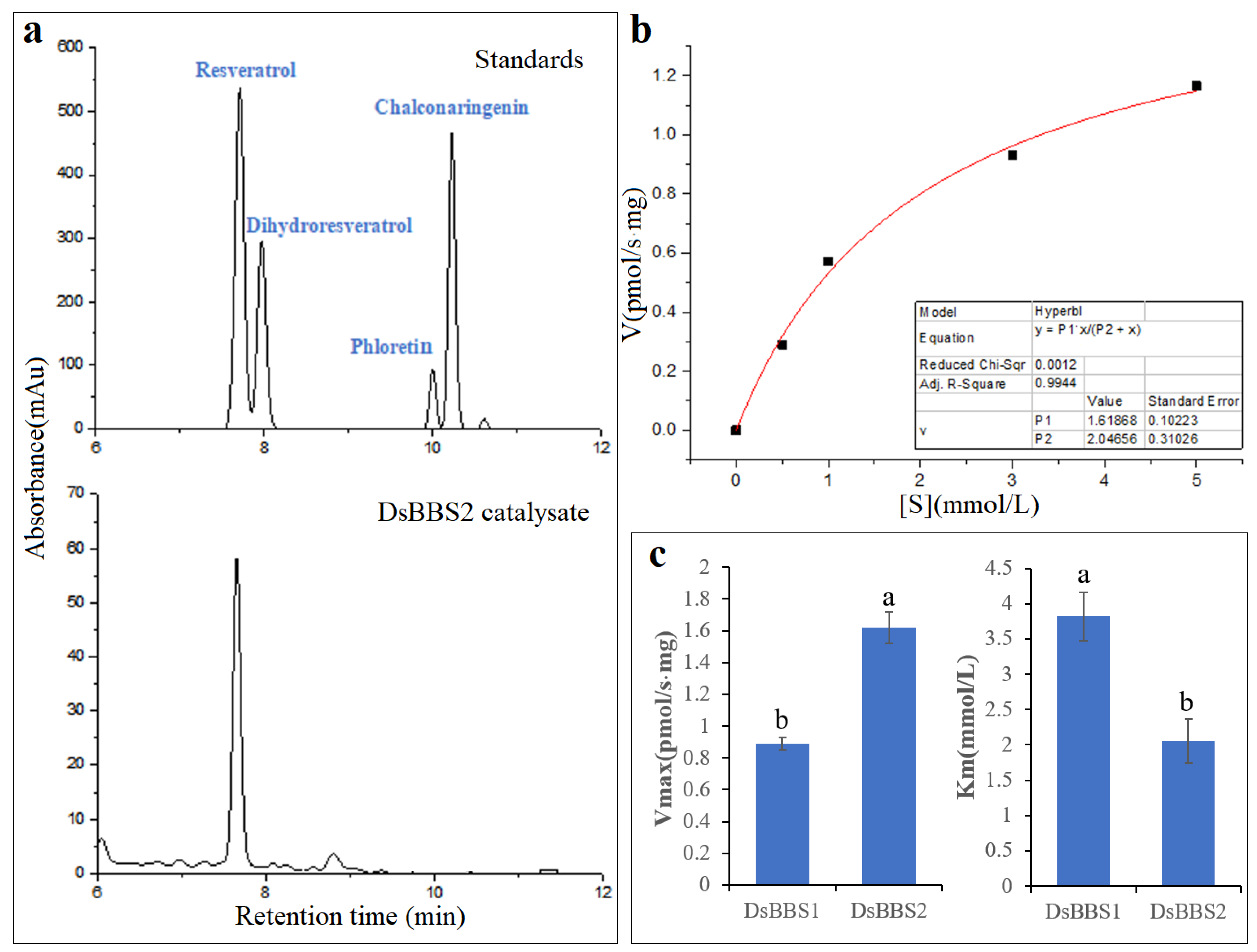
To clearly understand the nature of DsBBS2 and its activity in vitro, the purified recombinant protein was incubated with the substrates. The HPLC results show that one product shared the same retention time with resveratrol (Figure 4a), indicating that DsBBS2 could use the substrate of p-coumaroyl-CoA to produce resveratrol. The kinetic parameters of the purified DsBBS2 were determined by measuring the enzyme activity with p-coumaroyl-CoA at different concentrations. The Vmax of the recombinant DsBBS2-HisTag protein for resveratrol production was 1.62 ± 0.10 pmol/s·mg, and the Km value was 2.05 ± 0.31 mmol/L (Figure 4b). Under the same experimental condition with DsBBS1 [9], the Vmax and Km values of DsBBS2 were significantly higher and lower, respectively, compared with the kinetic parameters for DsBBS1 (Figure 4c).

2.5. Functional Analysis of DsBBS1 and DsBBS2 in Transgenic Arabidopsis

To further verify whether there is a difference in the activity between DsBBS1 and DsBBS2 in vivo, Arabidopsis (Columbia ecotype) was chosen for a transgenic analysis. A homology comparison with Araport11 protein sequences indicated that AtCHS (AT5G13930.1) protein was the one with the highest homology (E-value = 0) to the DsBBS1 and DsBBS2 proteins. Thus, the homozygous Atchs mutant (SALK_076535C) that was identified by the three-primer method was used for further studies. Through resistance screening and PCR validation, the homozygous transgenic lines of DsBBS1 and DsBBS2 were used for functional analysis (Figure S2).
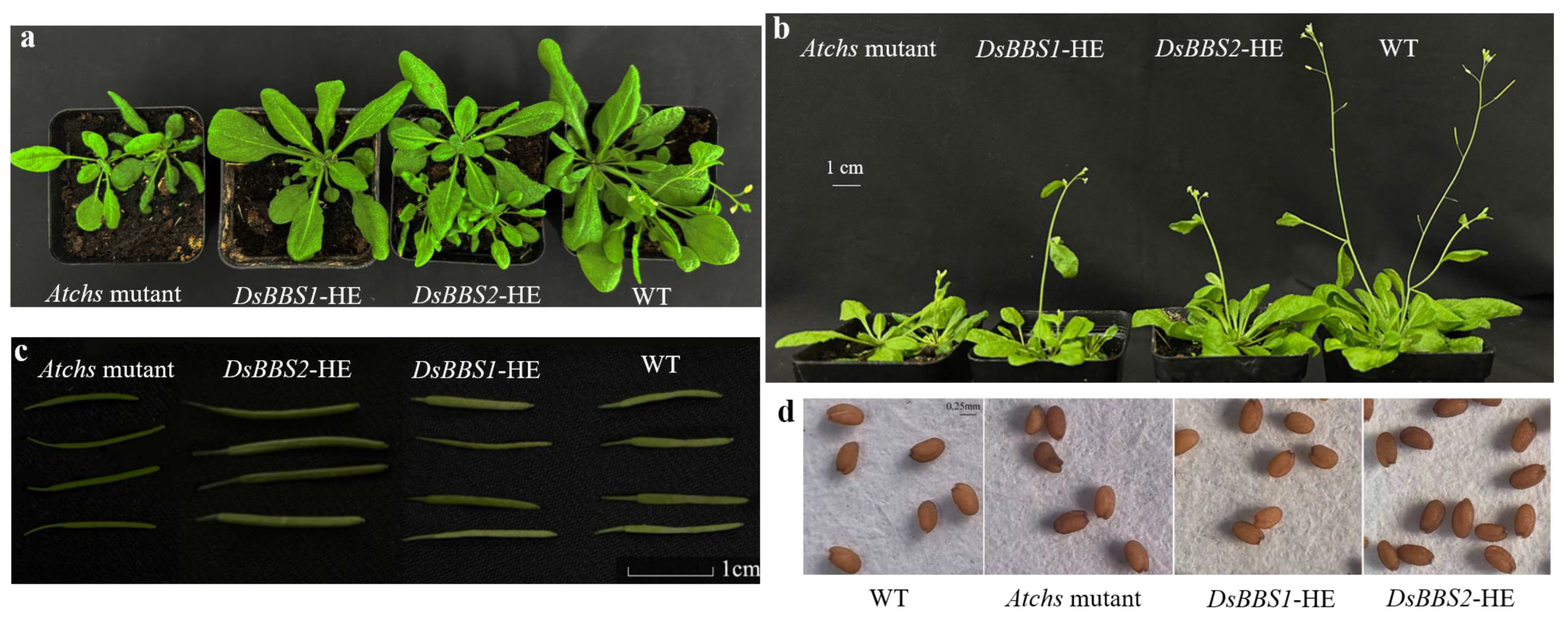
To understand the possible effects of the DsBBS genes on Arabidopsis growth and development, wild-type and Atchs mutant plants were used as controls to observe the differences in developmental phenotypes. During the vegetative growth stage, the growth of Atchs mutants was significantly weaker than that of the wild type (Figure 5a). DsBBS1 and DsBBS2 complementation rescued the Atchs mutant phenotype (Figure 5a). Interestingly, it was observed that the bolting time of the wild type was the shortest, followed by DsBBS1 and DsBBS2 mutant complementation, and the mutant type (Figure 5b). Moreover, the mutant pods were smaller than the wild-type pods and DsBBS1 transgenic lines, whereas the DsBBS2 transgenic lines had the longest pod length (Figure 5c). There was no significant difference in the seed size among the different plants (Figure 5d).
To explore the effects of DsBBS1 and DsBBS2 expression on polyketide biosynthesis, the total bibenzyl and flavonoid contents underwent a comparative analysis. The results show that the total bibenzyl content was 0.76% and 0.72% in the wild type and mutants, repetitively (Figure 6). However, the total flavonoid content was 1.76% in the wild type, which was significantly higher than the 0.27% value found in the Atchs mutants. This finding clearly shows that AtCHS is only involved in the accumulation of flavonoids. Compared with mutant plants, the heterologous expressions of the DsBBS1 and DsBBS2 genes only led to increases in the total bibenzyl content by 66.02% and 32.53%, respectively (Figure 6). Unexpectedly, DsBBS1 and DsBBS2 expressions both led to the recovery of the flavonoid content in the Atchs mutant, exhibiting nearly the same activity (Figure 6). These results strongly suggest that the heterologous expressions of DsBBS1 and DsBBS2 in Arabidopsis successfully compensated for the decrease in the total flavonoid content caused by the loss of Atchs. DsBBS1 and DsBBS2 could catalyze bibenzyl and flavonoid biosynthesis in Arabidopsis.

3. Discussion

Dendrobium species are a significant source of bibenzyls. To date, 89 bibenzyl derivatives have been extracted and identified from 46 Dendrobium species [10]. Bibenzyl compounds have a variety of pharmacological activities, such as anti-tumor activity [23], neuroprotective effects [24], antioxidant effects [25], anti-inflammatory activity [16], and antibacterial and antiviral effects [26]. Considering their multifaceted pharmacological profiles, bibenzyls hold promise for the development of new drugs to treat a variety of conditions. The elucidation of key enzymes involved in bibenzyl biosynthesis are paramount for the enhancement of their biosynthetic efficiency, potentially through metabolic engineering strategies. However, there is little research on the key enzymes involved in the biosynthesis of bibenzyl compounds in Dendrobium species, especially in D. sinense.
Type III PKSs specialize in synthesizing specific types of polyketides, such as stilbenes, flavonoids, and bibenzyls, which possess a wide range of biological activities and are predominantly found in plants [18,27]. Type III PKSs are the first committed enzyme in the biosynthesis of all polyketides. The BBS protein is the rate-limiting enzyme for bibenzyl biosynthesis [22]. Due to the lack of genomic information for D. sinense, the mining of type III PKS genes was performed through transcriptome sequencing. When we performed transcriptome sequencing for the first time using the Illumina platform, only one DsBBS gene was identified, namely DsBBS1 [9]. Subsequently, we combined PacBio third-generation sequencing technology with Illumina sequencing data to reanalyze the type III PKS genes in D. sinense, resulting in the discovery of 10 type III PKS genes, including 7 DsCHS genes, 2 DsBBS genes, and 1 DsPKS gene [20]. This is the most comprehensive analysis of type III PKSs in D. sinense reported so far.
A multiple alignment analysis indicated a 95.10% similarity between the DsBBS1 and DsBBS2 protein sequences [20]. Indeed, the three-dimensional structural models of DsBBS1 and DsBBS2 were almost identical (Figure 1). The similarity in protein structure between DsBBS1 and DsBBS2 implies that they likely have similar functions, catalyzing the formation of bibenzyl compounds [19,28]. Nevertheless, no matter which substrate (malonyl-CoA or p-coumaryl-CoA) is applied, the number of hydrogen bonds in the molecular docking results for the DsBBS2 protein was higher than that for the DsBBS1 protein. This is mainly due to differences in the non-conserved amino acid residues near the catalytic center. There is a growing body of evidence that optimal hydrogen bonding in enzyme active sites may offer a significant contribution to catalytic accelerations [29]. Thus, DsBBS1 and DsBBS2 have the same catalytic activity, but their catalytic efficiency may vary.
To further explore the functional differences between DsBBS1 and DsBBS2, the expression levels of DsBBS1 and DsBBS2 were detected in different tissues. The resulting expression profiles of the DsBBS1 and DsBBS2 genes were same, with a significantly higher expression in the roots (Figure 2). A previous study showed that D. sinense roots had a higher bibenzyl content than the pseudobulbs and leaves [9]. The positive relationship between the gene expression levels and bibenzyl content suggests that DsBBS1 and DsBBS2 are key genes for bibenzyl biosynthesis and accumulation in roots. It is difficult to tell from these expression results whether there are functional differences between the two DsBBS genes.
Therefore, to further explore the differences between their enzyme activities, the recombinant protein was obtained through prokaryotic protein expression and chromatography purification. The kinetics of in vitro DsBBS1 enzyme activity has been carried out [9], and thus, we carried out the present study on the DsBBS2 enzyme activity. Interestingly, the Vmax and Km values of the recombinant DsBBS2 protein were higher and lower than those of DsBBS1 protein, respectively [9]. These findings imply that when using malonyl-CoA and p-coumaryl-CoA as substrates, the recombinant DsBBS2 protein showed higher catalytic efficiency than DsBBS1 [30]. It was reported that enzyme–substrate interactions were driven by the stereospecific binding of the substrate to the active site of the enzyme [31]. One limitation of this in vitro enzyme activity measurement is that only one common substrate (p-coumaryl-CoA) of type III PKSs was studied. The preferred physiological substrate for DsBBS1 and DsBBS2 needs further verification.
To confirm the DsBBS1 and DsBBS2 functions in vivo, we generated their transgenic plants for functional studies. A total of four type III PKS genes have been identified in Arabidopsis [32], but no BBS gene was found. A homology comparison showed that the AtCHS (AT5G13930.1) protein was the one with the highest homology both to DsBBS1 and DsBBS2. Thus, the homozygous Atchs mutant was selected for transgenic studies. The AtCHS has been identified as the only gene participating in flavonoid biosynthesis [33]. Indeed, the total flavonoid content clearly dropped in the Atchs mutant, but there was no difference in the bibenzyl content between the mutant-type and wild-type plants (Figure 6). Despite AtCHS having a high homology with DsBBS1 and DsBBS2, the loss of AtCHS function only affects flavonoid accumulation.
It is apparent that a significant increase in the total bibenzyl content was observed both in the DsBBS1 and DsBBS2 transgenic lines compared with the wild type (Figure 6). The findings indicate that heterologous expressions of DsBBS1 and DsBBS2 can increase the bibenzyl content, which is consistent with the findings of a previous report [34]. Interestingly, the expressions of heterologous DsBBS1 and DsBBS2 genes both can compensate for the loss of Atchs and restore the total flavonoid content (Figure 6). This result may be explained by the fact that the biosynthesis of bibenzyls and flavonoids uses the same substrates, and all type III PKSs share a common three-dimensional overall fold [28]. These characteristics can lead to complementary functions between AtCHS and DsBBS. It is also worth noting that there was no significant difference in the total flavonoid content or bibenzyl content between the DsBBS1 and DsBBS2 transgenic lines (Figure 6). In contradiction to the results of the in vitro enzyme activity assay, there was no significant difference in the enzyme activity between the heterologous expressions of DsBBS1 and DsBBS2 in Arabidopsis. The inconsistency may stem from differences between in vitro and in vivo conditions; for instance, plant cells might offer an optimal physiological environment and substrate for the activity of DsBBS1 and DsBBS2. Additionally, variations in protein processing, modification, and folding across distinct expression systems can affect enzyme activity [35].

4. Materials and Methods

4.1. DsBBS Homology Modeling and Molecular Docking

The protein sequences of DsBBS1 (WHE45950.1) and DsBBS2 (WHE45951.1) were downloaded from the NCIB database [20]. The three-dimensional structures of the proteins were predicted using the Phyre2 server (default parameters). Visualization of the three-dimensional structures was performed using PyMOL 2.52.4, with the alignment module being employed to compare structural differences between the two proteins. The SAVES online server (https://saves.mbi.ucla.edu/, accessed on 10 December 2023) was utilized to assess the reliability of the predicted model. The three-dimensional structures of p-coumaroyl-CoA and malonyl-CoA were obtained from the Pubchem database (pubchem.ncbi.nlm.nih.gov/, accessed on 10 December 2023). Using ADT 4.2.6, semi-flexible docking of the two DsBBS proteins with p-coumaroyl-CoA and malonyl-CoA was conducted. The ligand was set to 17 twistable keys, and the binding pocket size parameters were set to X:126, Y:126, and Z:106. The coordinates of the search space center were X center: 26.535, Y center: 54.225, and Z center: 54.353, and simulation docking was repeated 50 times using a genetic algorithm. The docking results were visualized in PyMOL, and hydrogen bonds were plotted in residue mode.

4.2. RT-qPCR Analysis

Total RNA was isolated from the roots, pseudobulbs, and leaves of D. sinense using the RNA Easy Plant Tissue Kit (Taingen, Beijing, China). Subsequently, cDNA was synthesized from the total RNA using the High-Capacity cDNA Archive Kit (Thermo Fisher Scientific, Shanghai, China). RT-qPCR was conducted using the MonAmpTM ChemoHS qPCR Mix Kit (Monad, Guangzhou, China) on a LightCycler 96 System (Roche). Based on our previous report, ADF11 and ACBP2 were used as the reference genes across various tissues of D. sinense [36]. Details of the RT-qPCR primers are provided in Supplemental Table S1. The RT-qPCR primers are shown in Supplemental Table S1. Expression levels were calculated using the 2−ΔΔCt method. Reactions were performed in triplicate.

4.3. DsBBS2 Cloning and Protein Purification

The cloning of DsBBS2 genes was achieved using the primers DsBBS2-F and DsBBS2-R (Supplementary Table S1). The resulting PCR products were then cloned into PCloneEZ-TOPO (Solarbio, Beijing, China), and sequencing was carried out by Sangon, Guangzhou, China.
The pET28a vector underwent digestion with EcoRI and SacI (NEB, Beijing, China). Specially designed primers of DsBBS2-HisTag-F and DsBBS2-HisTag-R included the terminal homologous sequence of the pET28a vector (15 bp), restriction enzyme cutting sequence, and the gene amplification primer sequence (Supplementary Table S1). Utilizing the ClonExpress® II One Step Cloning Kit (Vazyme, Nanjing, China), the DsBBS2 gene was seamlessly integrated into the linearized pET28a vector to produce pET28a-DsBBS2. Subsequently, the pET28a-DsBBS2 vector was introduced into the Escherichia coli strain BL21(DE3) for the purpose of protein expression. Protein expression and purification were carried out according to previous methods [9,20]. The purity of the DsBBS2-HisTag protein was assessed through SDS-PAGE, and the protein bands were visualized after staining with Coomassie brilliant blue.

4.4. Enzymatic Assay

To assess the enzymatic activity of the DsBBS2 protein, an in vitro enzyme activity assay was carried out. The enzymatic reaction mixture, including 10 μg DsBBS-HisTag protein, 50 mM Hepes buffer (Bioshorp, Beijing, China), and 0.5 mM Malonyl-CoA lithium salt (Yuanye, Shanghai, China), was prepared. During kinetic studies, the enzymatic reaction rate of DsBBS2 was measured for different concentrations of p-coumaroyl CoA (Yuanye, Shanghai, China), including 0.5, 1.0, 3.0, and 5.0 mM. This mixture was incubated at 37 °C for 30 min to allow for enzymatic reaction. Subsequently, 50 μL of methanol was added to halt the reaction. After centrifugation at 12,000 rpm for 10 min, a 20 μL aliquot of the supernatant was analyzed using high-performance liquid chromatography (HPLC) to determine the production content.

4.5. HPLC Analysis

The samples were analyzed using an LC-100 PUMP system (Wufeng, Shanghai, China) paired with an HC-C18 column (18 μm, 4.6 × 250 mm, supplied by Agilent, Santa Clara, CA, USA). The chromatographic conditions were set as follows: a column temperature of 30 °C, an injection volume of 20 μL, solution A consisting of 0.1% phosphate water, and solvent B being 100% acetonitrile. The flow rate was maintained at 1 mL/min. The gradient elution program for the solvents was as follows: from 0 to 6 min, 70% solution A and 30% solvent B were used; from 6 to 40 min, there was a gradual shift to 55% solution A and 45% solvent B. The HPLC data were estimated based on the calibration curve established with resveratrol standard (Yuanye, Shanghai, China).

4.6. Genetic Transformation in Arabidopsis

The T-DNA insertion mutants of Atchs (AT5G13930 and SALK_076535C) were purchased from AraShare (www.arashare.cn/index/, accessed on 13 September 2021). Three-primer method was used to screen for homozygous mutations (Supplementary Table S1), which were used for genetic transformation. The linearization of the pBI121 vector was conducted by BamHI and SmaI (Tolobio, Beijing, China). Using specially designed primers (Supplementary Table S1), the DsBBS1 and DsBBS2 genes were seamlessly integrated into pBI121 to produce pBI121-DsBBS1 and pBI121-DsBBS2 using the ClonExpress® II One Step Cloning Kit (Vazyme, Nanjing, China), respectively. The pBI121-DsBBS1 and pBI121-DsBBS2 vectors were transformed into Agrobacterium tumefaciens GV3101. The floral dip method was used for genetic transformation in Arabidopsis. The T3 homozygous transgenic lines were screened as previously described [20].

4.7. Determination of Total Bibenzyl and Flavonoid Contents

Three-month-old Arabidopsis seedlings were used as the material to collect leaves for the analysis of the total bibenzyl and flavonoid contents according to a previous method [20,22]. Briefly, the freeze-dried sample was refluxed with anhydrous ethanol at 90 °C for 2 h, followed by centrifugation at 12,000 rpm for 10 min. The supernatant was then concentrated and mixed with 10 mL methanol for the analysis of the total bibenzyl content. As for the determination of total flavonoid content, the concentrated supernatant was transferred into a volumetric flask (25 mL), and anhydrous ethanol was added to obtain a final volume of 25 mL. The assay involved the addition of 30% ethanol and reagents such as 5% sodium nitrite and 10% aluminum nitrate, with subsequent reactions induced by 1 mol/L NaOH. Gigantol (Yuanye, Shanghai, China) was used for generating standard curves to analyze the total bibenzyl content, while rutin (Yuanye, Shanghai, China) was used for the total flavonoid content. Absorbance of the bibenzyl and flavonoid compounds was measured at 280 and 510 nm, respectively. The assay was performed in biological triplicate with three technical replicates each time. The ANOVA analysis was conducted using GraphPad Prism 8.0.2 (p < 0.05).

5. Conclusions

This study has elucidated the functional roles of the DsBBS1 and DsBBS2 genes in the biosynthesis of bibenzyl compounds in D. sinense. The molecular docking analysis and gene expression profiles indicated that both genes play a significant role in the production of bibenzyls. The optimization of DsBBS2-HisTag expression conditions and the subsequent enzyme activity analysis revealed that DsBBS2 was able to catalyze the formation of bibenzyl compounds. A functional analysis in Arabidopsis demonstrated that both DsBBS1 and DsBBS2 can compensate for the loss of Atchs, restoring the total flavonoid content to wild-type levels. Moreover, the heterologous expressions of the DsBBS1 and DsBBS2 genes led to an increase in the total bibenzyl content. These findings enhance our understanding of the molecular mechanisms underlying bibenzyl biosynthesis in D. sinense.

Supplementary Materials

The following supporting information can be downloaded at https://www.mdpi.com/article/10.3390/molecules29153682/s1, Supplementary Table S1: The primers for gene cloning, vector construction, and RT-qPCR. Supplementary Figure S1: The reliability of the predicted model was evaluated. If the proportion of amino acid residues falling within the allowed region of the protein model is higher than 90%, the conformation of the model can be considered to conform to the principles of stereochemistry. Supplementary Figure S2: Identification of transgenic plants was performed by PCR. NC, negative control; PC, positive control.

Author Contributions

Conceptualization, J.W. and J.N.; methodology, L.L. and H.Y.; formal analysis, L.L. and H.Y.; validation, L.Y. and Q.O.; data curation, Y.Z. and L.Y.; writing—review and editing, J.N.; funding acquisition, Y.Z., J.W. and J.N.; project administration, J.W. and J.N. All authors have read and agreed to the published version of the manuscript.

Funding

This research was funded by the National Natural Science Foundation of China, grant number 32160400; the Collaborative Innovation Center project of Hainan University, grant number 320RC469; and the Hainan University Research Project, grant number KYQD(ZR)-22056.

Institutional Review Board Statement

Not applicable.

Informed Consent Statement

Not applicable.

Data Availability Statement

All data generated or analyzed during this study are included in this published article.

Conflicts of Interest

The authors declare no conflicts of interest.

References

  1. Chase, M.W.; Cameron, K.M.; Freudenstein, J.V.; Pridgeon, A.M.; Salazar, G.; Van Den Berg, C.; Schuiteman, A. An Updated Classification of Orchidaceae. Bot. J. Linn. Soc. 2015, 177, 151–174. [Google Scholar] [CrossRef]
  2. Li, P.-Y.; Li, L.; Wang, Y.-Z. Traditional Uses, Chemical Compositions and Pharmacological Activities of Dendrobium: A Review. J. Ethnopharmacol. 2023, 310, 116382. [Google Scholar] [CrossRef] [PubMed]
  3. Cheng, J.; Dang, P.-P.; Zhao, Z.; Yuan, L.-C.; Zhou, Z.-H.; Wolf, D.; Luo, Y.-B. An Assessment of the Chinese Medicinal Dendrobium Industry: Supply, Demand and Sustainability. J. Ethnopharmacol. 2019, 229, 81–88. [Google Scholar] [CrossRef] [PubMed]
  4. Tang, X.; Yuan, Y.; Zhang, J. How Climate Change Will Alter the Distribution of Suitable Dendrobium Habitats. Front. Ecol. Evol. 2020, 8, 536339. [Google Scholar] [CrossRef]
  5. Teixeira da Silva, J.A.; Ng, T.B. The Medicinal and Pharmaceutical Importance of Dendrobium Species. Appl. Microbiol. Biot. 2017, 101, 2227–2239. [Google Scholar] [CrossRef] [PubMed]
  6. Zhang, C.; Chen, J.; Huang, W.; Song, X.; Niu, J. Transcriptomics and Metabolomics Reveal Purine and Phenylpropanoid Metabolism Response to Drought Stress in Dendrobium sinense, an Endemic Orchid Species in Hainan Island. Front. Genet. 2021, 12, 692702. [Google Scholar] [CrossRef] [PubMed]
  7. Chen, X.-J.; Mei, W.-L.; Zuo, W.-J.; Zeng, Y.-B.; Guo, Z.-K.; Song, X.-Q.; Dai, H.-F. A New Antibacterial Phenanthrenequinone from Dendrobium sinense. J. Asian Nat. Prod. Res. 2013, 15, 67–70. [Google Scholar] [CrossRef] [PubMed]
  8. Chen, X.-J.; Mei, W.-L.; Cai, C.-H.; Guo, Z.-K.; Song, X.-Q.; Dai, H.-F. Four New Bibenzyl Derivatives from Dendrobium sinense. Phytochem. Lett. 2014, 9, 107–112. [Google Scholar] [CrossRef]
  9. Chen, Y.; Wang, Y.; Liang, C.; Liu, L.; Song, X.; Zhao, Y.; Wang, J.; Niu, J. Characterization of the Key Bibenzyl Synthase in Dendrobium sinense. Int. J. Mol. Sci. 2022, 23, 6780. [Google Scholar] [CrossRef]
  10. He, L.; Su, Q.; Bai, L.; Li, M.; Liu, J.; Liu, X.; Zhang, C.; Jiang, Z.; He, J.; Shi, J.; et al. Recent Research Progress on Natural Small Molecule Bibenzyls and Its Derivatives in Dendrobium Species. Eur. J. Med. Chem. 2020, 204, 112530. [Google Scholar] [CrossRef]
  11. Chanvorachote, P.; Kowitdamrong, A.; Ruanghirun, T.; Sritularak, B.; Mungmee, C.; Likhitwitayawuid, K. Anti-Metastatic Activities of Bibenzyls from Dendrobium pulchellum. Nat. Prod. Commun. 2013, 8, 1934578X1300800127. [Google Scholar] [CrossRef]
  12. Zhu, Q.; Sheng, Y.; Li, W.; Wang, J.; Ma, Y.; Du, B.; Tang, Y. Erianin, a Novel Dibenzyl Compound in Dendrobium Extract, Inhibits Bladder Cancer Cell Growth via the Mitochondrial Apoptosis and JNK Pathways. Toxicol. Appl. Pharm. 2019, 371, 41–54. [Google Scholar] [CrossRef] [PubMed]
  13. Zhao, G.Y.; Deng, B.W.; Zhang, C.Y.; Cui, Y.D.; Bi, J.Y.; Zhang, G.G. New Phenanthrene and 9, 10-Dihydrophenanthrene Derivatives from the Stems of Dendrobium officinale with Their Cytotoxic Activities. J. Nat. Med. 2018, 72, 246–251. [Google Scholar] [CrossRef] [PubMed]
  14. Sarakulwattana, C.; Mekboonsonglarp, W.; Likhitwitayawuid, K.; Rojsitthisak, P.; Sritularak, B. New Bisbibenzyl and Phenanthrene Derivatives from Dendrobium scabrilingue and Their α-Glucosidase Inhibitory Activity. Nat. Prod. Res. 2020, 34, 1694–1701. [Google Scholar] [CrossRef]
  15. Huang, J.-M.; Huang, F.-I.; Yang, C.-R. Moscatilin Ameliorates Tau Phosphorylation and Cognitive Deficits in Alzheimer’s Disease Models. J. Nat. Prod. 2019, 82, 1979–1988. [Google Scholar] [CrossRef]
  16. Kongkatitham, V.; Muangnoi, C.; Kyokong, N.; Thaweesest, W.; Likhitwitayawuid, K.; Rojsitthisak, P.; Sritularak, B. Anti-Oxidant and Anti-Inflammatory Effects of New Bibenzyl Derivatives from Dendrobium parishii in Hydrogen Peroxide and Lipopolysaccharide Treated RAW264. 7 Cells. Phytochem. Lett. 2018, 24, 31–38. [Google Scholar] [CrossRef]
  17. Li, Y.; Wang, C.L.; Wang, Y.J.; Wang, F.F.; Guo, S.X.; Yang, J.S.; Xiao, P.G. Four New Bibenzyl Derivatives from Dendrobium candidum. Chem. Pharm. Bull. 2009, 57, 997–999. [Google Scholar] [CrossRef] [PubMed]
  18. Guo, D.; Wang, H.; Zhang, S.; Lan, T. The Type III Polyketide Synthase Supergene Family in Plants: Complex Evolutionary History and Functional Divergence. Plant J. 2022, 112, 414–428. [Google Scholar] [CrossRef] [PubMed]
  19. Yu, D.; Xu, F.; Zeng, J.; Zhan, J. Type III Polyketide Synthases in Natural Product Biosynthesis. IUBMB Life 2012, 64, 285–295. [Google Scholar] [CrossRef]
  20. Wang, Y.; Liu, L.; Ou, Q.; You, H.; Wang, J.; Niu, J. Alternative First Exons Drive Enzymatic Activity Variation in Chalcone synthase 3 of Dendrobium sinense. Forests 2023, 14, 1702. [Google Scholar] [CrossRef]
  21. Morita, H.; Yamashita, M.; Shi, S.-P.; Wakimoto, T.; Kondo, S.; Kato, R.; Sugio, S.; Kohno, T.; Abe, I. Synthesis of Unnatural Alkaloid Scaffolds by Exploiting Plant Polyketide Synthase. Proc. Natl. Acad. Sci. USA 2011, 108, 13504–13509. [Google Scholar] [CrossRef]
  22. Boddington, K.F.; Soubeyrand, E.; Van Gelder, K.; Casaretto, J.A.; Perrin, C.; Forrester, T.J.; Parry, C.; Al-Abdul-Wahid, M.S.; Jentsch, N.G.; Magolan, J.; et al. Bibenzyl Synthesis in Cannabis sativa L. Plant J. 2022, 109, 693–707. [Google Scholar] [CrossRef]
  23. Nam, B.; Ryu, S.M.; Lee, D.; Jung, C.-H.; Jin, C.H.; Kim, J.-B.; Lee, I.-S.; Han, A.-R. Identification of Two New Phenanthrenes from Dendrobii Herba and Their Cytotoxicity towards Human Hypopharynx Squamous Carcinoma Cell (FaDu). Molecules 2019, 24, 2339. [Google Scholar] [CrossRef]
  24. Yang, L.; Liu, S.-J.; Luo, H.-R.; Cui, J.; Zhou, J.; Wang, X.-J.; Sheng, J.; Hu, J.-M. Two New Dendrocandins with Neurite Outgrowth-Promoting Activity from Dendrobium officinale. J. Asian Nat. Prod. Res. 2015, 17, 125–131. [Google Scholar] [CrossRef]
  25. Choonong, R.; Sermpradit, W.; Kitisripanya, T.; Sritularak, B.; Putalun, W. The Contents of Bibenzyl Derivatives, Flavonoids and a Phenanthrene in Selected Dendrobium Spp. and the Correlation with Their Antioxidant Activity. Scienceasia 2019, 45, 245–252. [Google Scholar] [CrossRef]
  26. Sukphan, P.; Sritularak, B.; Mekboonsonglarp, W.; Lipipun, V.; Likhitwitayawuid, K. Chemical Constituents of Dendrobium venustum and Their Antimalarial and Anti-Herpetic Properties. Nat. Prod. Commun. 2014, 9, 1934578X1400900625. [Google Scholar] [CrossRef]
  27. Abe, I. Biosynthesis of Medicinally Important Plant Metabolites by Unusual Type III Polyketide Synthases. J. Nat. Med. 2020, 74, 639–646. [Google Scholar] [CrossRef]
  28. Lin, Z.; Qu, X. Emerging Diversity in Polyketide Synthase. Tetrahedron Lett. 2022, 110, 154183. [Google Scholar] [CrossRef]
  29. Spies, M.A.; Reese, J.G.; Dodd, D.; Pankow, K.L.; Blanke, S.R.; Baudry, J. Determinants of Catalytic Power and Ligand Binding in Glutamate Racemase. J. Am. Chem. Soc. 2009, 131, 5274–5284. [Google Scholar] [CrossRef]
  30. Halwachs, W. KM and Vmax from Only One Experiment. Biotechnol. Bioeng. 1978, 20, 281–285. [Google Scholar] [CrossRef]
  31. Strickland, M.; Kale, S.; Strub, M.-P.; Schwieters, C.D.; Liu, J.; Peterkofsky, A.; Tjandra, N. Potential Regulatory Role of Competitive Encounter Complexes in Paralogous Phosphotransferase Systems. J. Mol. Biol. 2019, 431, 2331–2342. [Google Scholar] [CrossRef] [PubMed]
  32. Naake, T.; Maeda, H.A.; Proost, S.; Tohge, T.; Fernie, A.R. Kingdom-Wide Analysis of the Evolution of the Plant Type III Polyketide Synthase Superfamily. Plant Physiol. 2021, 185, 857–875. [Google Scholar] [CrossRef] [PubMed]
  33. Saito, K.; Yonekura-Sakakibara, K.; Nakabayashi, R.; Higashi, Y.; Yamazaki, M.; Tohge, T.; Fernie, A.R. The Flavonoid Biosynthetic Pathway in Arabidopsis: Structural and Genetic Diversity. Plant Physiol. Biochem. 2013, 72, 21–34. [Google Scholar] [CrossRef] [PubMed]
  34. Serazetdinova, L.; Oldach, K.H.; Lörz, H. Expression of Transgenic Stilbene Synthases in Wheat Causes the Accumulation of Unknown Stilbene Derivatives with Antifungal Activity. J. Plant Physiol. 2005, 162, 985–1002. [Google Scholar] [CrossRef] [PubMed]
  35. Nasreen, K.; Parray, Z.A.; Ahamad, S.; Ahmad, F.; Ahmed, A.; Freeh Alamery, S.; Hussain, T.; Hassan, M.I.; Islam, A. Interactions under Crowding Milieu: Chemical-Induced Denaturation of Myoglobin Is Determined by the Extent of Heme Dissociation on Interaction with Crowders. Biomolecules 2020, 10, 490. [Google Scholar] [CrossRef]
  36. Wang, Y.; Pei, X.; Fu, Y.; Huo, K.; Liang, C.; Liu, L.; Wang, J.; Niu, J. Genome-Wide Identification of Superior Reference Genes in Dendrobium sinense. Chin. J. Trop. Crops 2023, 44, 1365. [Google Scholar] [CrossRef]
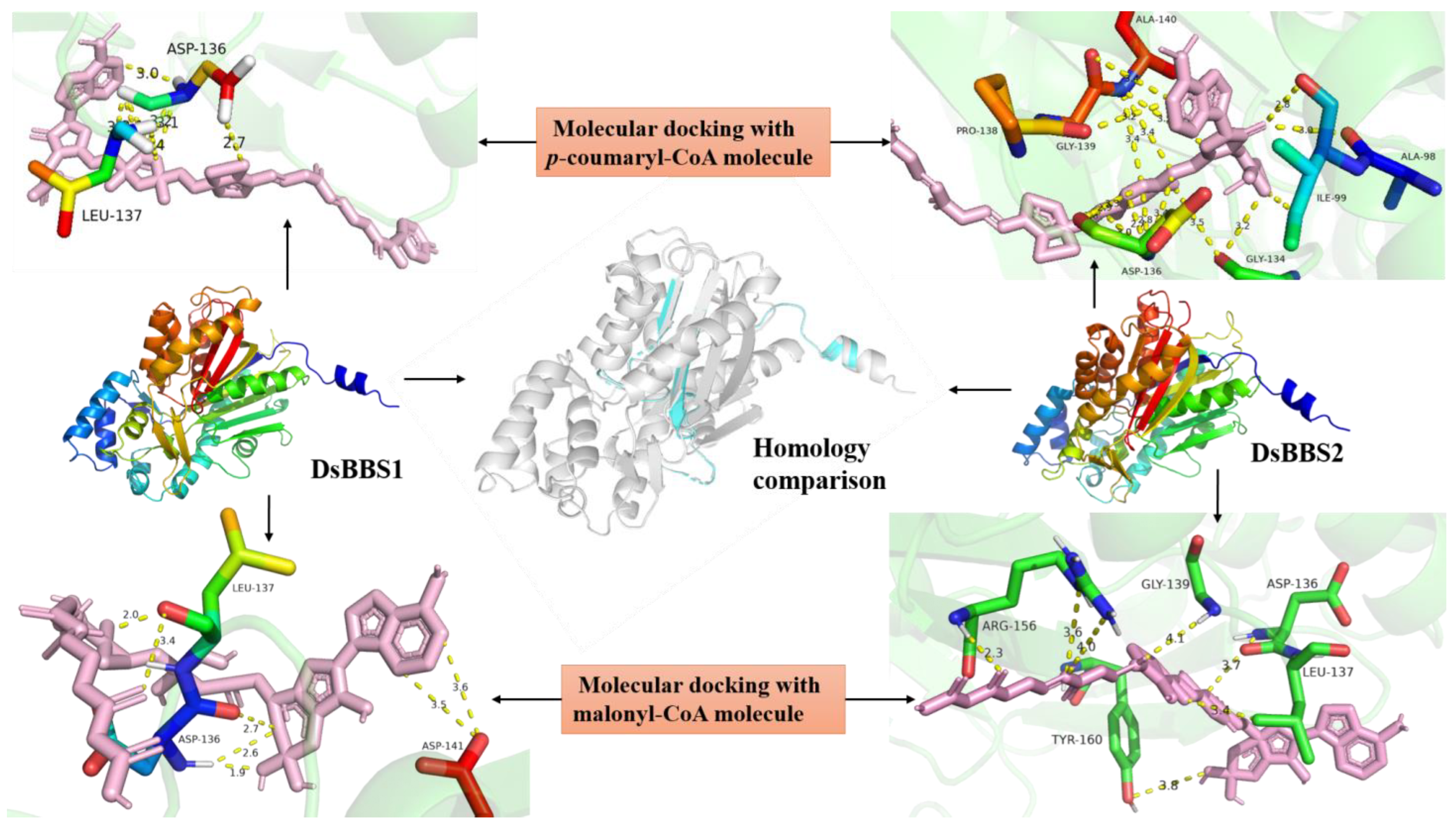
Figure 1. Mimetic molecular docking of DsBBS proteins. Three-dimensional structural models of the DsBBS1 and DsBBS2 proteins were created using the Phyre2 server. The three-dimensional structures of DsBBS1 and DBBS2 were compared using PyMOL. The overlapping parts of the two three-dimensional structures are marked in gray, and the differences are marked in blue. The RMSD between the two structures is 0.14. The molecular docking of DsBBS1 and DBBS2 with p-coumaryl-CoA and malonyl-CoA was performed using ADT 4.2.6. Dotted yellow lines represent hydrogen bonds. The specific number on the bond indicates bond length, and the unit is Å.
Molecules 29 03682 g001
Figure 2. The relative expression levels of DsBBS1 and DsBBS2 in different D. sinense tissues. ADF11 and ACBP2 were used as the reference genes. The expression levels were calculated using the 2−ΔΔCt method. Different letters indicate significant difference (p < 0.05; ANOVA).
Molecules 29 03682 g002
Figure 3. The protein purification of DsBBS2-HisTag protein. (a) The optimum inducement concentration of IPTG. (b) The optimum inducement time and temperature. (c) An electrophoretic diagram of purified DsBBS protein. Abbreviations: CF, cleaning fluid; CK, control check; IPTG, isopropyl-β-D-thiogalactopyranoside; M, marker; Pre, precipitate; Sup, supernatant.
Molecules 29 03682 g003
Figure 4. The in vitro enzyme assays of DsBBS2. (a) HPLC chromatograms of the reaction productions. The above picture shows the standards, and the below shows the productions by DsBBS2. (b) The nonlinear curve fitting of DsBBS2 activity. (c) A comparative analysis of the Vmax and Km values between DsBBS1 and DsBBS2. Different letters indicate significant difference (p < 0.05; t-test).
Molecules 29 03682 g004
Figure 5. Comparative analysis of phenotypes among different Arabidopsis plants. (a,b) show healthy plants after 25 and 30 days of sowing, respectively. (c) Comparative analysis of pod. Plants were grown for about 60 days after sowing. Largest pod was taken from strongest inflorescence. (d) Comparative analysis of mature seeds. DsBBS1-HE and DsBBS2-HE, respectively, represent heterologous expressions of DsBBS1 and DsBBS2 in Atchs mutant.
Molecules 29 03682 g005
Figure 6. Total bibenzyl and flavonoid contents in Arabidopsis leaves. Sample was freeze-dried. Different letters indicate significant difference (p < 0.05; ANOVA). DsBBS1-HE and DsBBS2-HE, respectively, represent heterologous expressions of DsBBS1 and DsBBS2 in Atchs mutant.
Molecules 29 03682 g006
Disclaimer/Publisher’s Note: The statements, opinions and data contained in all publications are solely those of the individual author(s) and contributor(s) and not of MDPI and/or the editor(s). MDPI and/or the editor(s) disclaim responsibility for any injury to people or property resulting from any ideas, methods, instructions or products referred to in the content.