Article

Efficient Synthesis, Structural Characterization, Antibacterial Assessment, ADME-Tox Analysis, Molecular Docking and Molecular Dynamics Simulations of New Functionalized Isoxazoles

1
Engineering Laboratory of Organometallic, Molecular Materials and Environment, Faculty of Sciences Dhar EL Mahraz, Sidi Mohamed Ben Abdellah University, P.O. Box 1796, Atlas, Fez 30000, Morocco
2
LIMAS, Faculty of Sciences Dhar El Mahraz, Sidi Mohamed Ben Abdellah University, P.O. Box 1796, Atlas, Fez 30000, Morocco
3
Faculty of Health and Life Sciences, INTI International University, Persiaran Perdana BBN, Putra Nilai, Nilai 71800, Malaysia
4
Faculty of Sciences and Technologies Mohammedia, University Hassan II, B.P. 146, Mohammedia 28800, Morocco
5
Laboratory of Biotechnology Conservation and Valorisation of Natural Resources, Faculty of Sciences Dhar El Mahraz, Sidi Mohamed Ben Abdallah University, Fez 30000, Morocco
6
Laboratory of Materials Engineering for the Environment and Natural Ressources, Faculty of Sciences and Techniques, University of Moulay Ismail of Meknès, B.P 509, Boutalamine, Errachidia 52000, Morocco
7
Laboratory of Heterocyclic Organic Chemistry URAC 21, Pharmacochemistry Competence Center, Av. Ibn Battouta, BP 1014, Faculty of Sciences, Mohammed V University in Rabat, Rabat 10010, Morocco
8
Department of Chemistry, Tulane University, New Orleans, LA 70118, USA
9
Laboratory of Carbohydrate and Nucleoside Chemistry (LCNC), Department of Chemistry, Faculty of Science, University of Chittagong, Chittagong 4331, Bangladesh
10
Department of Pharmaceutical Chemistry, College of Pharmacy, King Saud University, Riyadh 11451, Saudi Arabia
*
Author to whom correspondence should be addressed.
Molecules 2024, 29(14), 3366; https://doi.org/10.3390/molecules29143366
Submission received: 11 May 2024 / Revised: 28 June 2024 / Accepted: 11 July 2024 / Published: 17 July 2024
(This article belongs to the Special Issue Application of Natural or Synthetic Products in Computer-Aided Drugs)

Abstract

:
This work describes the synthesis, characterization, and in vitro and in silico evaluation of the biological activity of new functionalized isoxazole derivatives. The structures of all new compounds were analyzed by IR and NMR spectroscopy. The structures of 4c and 4f were further confirmed by single crystal X-ray and their compositions unambiguously determined by mass spectrometry (MS). The antibacterial effect of the isoxazoles was assessed in vitro against Escherichia coli, Bacillus subtilis, and Staphylococcus aureus bacterial strains. Isoxazole 4a showed significant activity against E. coli and B. subtilis compared to the reference antibiotic drugs while 4d and 4f also exhibited some antibacterial effects. The molecular docking results indicate that the synthesized compounds exhibit strong interactions with the target proteins. Specifically, 4a displayed a better affinity for E. coli, S. aureus, and B. subtilis in comparison to the reference drugs. The molecular dynamics simulations performed on 4a strongly support the stability of the ligand–receptor complex when interacting with the active sites of proteins from E. coli, S. aureus, and B. subtilis. Lastly, the results of the Absorption, Distribution, Metabolism, Excretion and Toxicity Analysis (ADME-Tox) reveal that the molecules have promising pharmacokinetic properties, suggesting favorable druglike properties and potential therapeutic agents.

Graphical Abstract

1. Introduction

Pathogenic microorganisms like bacteria are responsible for many serious infections, threatening well-being and even leading to large-scale health crises [1,2]. Combating these bacterial infections has become a serious and persistent concern for the global scientific community, due to the increased resistance of microorganisms caused by random genetic mutations [3,4]. Moreover, the widespread and inappropriate use of existing antibacterial drugs contributes to the proliferation of microorganisms that are resistant to antibacterial treatments [5,6]. This multi-drug resistance poses a major risk, leading to high morbidity and mortality rates [7,8]. Therefore, there is an urgent need to discover more effective therapies and develop new powerful antibacterial drugs, capable of effectively targeting a wide range of microorganisms.
Heterocyclic compounds indeed offer a rich reservoir of molecular structures with diverse biological activities, making them valuable in drug discovery to combat infectious diseases [9]. Medicinal chemists continue to focus on the modification of heterocyclic molecules to develop new therapeutic agents with improved efficacy and safety profiles [10,11]. Additionally, heterocyclic chemistry in recent years has undergone considerable development due to the diverse biological activities exhibited by many heterocyclic compounds [12,13]. The distinctive features of heterocyclic motifs lie in their structures in saturated, partially saturated, or aromatic heterocyclic compounds, as well as in functional groups [14,15]. They are the active ingredient of many families of both natural and synthetic products effective in therapeutic, agrochemical [16,17], industrial [18], and other fields.
Isoxazoles are an attractive type of heterocyclic compound for medicinal research due to their broad spectrum of biological activity. These include ones having antihistaminic [19], antifungal [20,21], antimicrobial, [22,23], antiviral [24,25], anti-inflammatory [26,27], antioxidant [22,28], and anticancer [29,30] activities as well as ones used as herbicides [31], fungicides [32], insecticides [33], and anticorrosive [34,35,36] coatings, among others. Indeed, there are several drugs currently on the market which incorporate the isoxazole moiety as the main pharmacophore (Figure 1). Also, isoxazoles are precursors to many bioactive molecules, including amino acids, amino-alcohols, and amino esters [23,24].
Chemoinformatics has indeed emerged as a crucial tool in various scientific disciplines, including chemistry, biology, and materials science [35,36]. These techniques have been widely used to discover new candidates because they offer a powerful alternative for predicting and interpreting complex experimental data. In particular, in silico molecular docking studies are of great importance in drug design, as they can predict the likely interactions between potential drug molecules (ligands) and their target proteins (enzymes or receptors) [37,38]. Moreover, molecular dynamics simulation analysis allows researchers to track the behavior of complexes formed between ligands and target proteins under in silico physiological conditions, exploring and evaluating their stability over time [39,40]. These computational simulations provide valuable insights that complement experimental methods and enhance the efficiency of the drug development process.
Building upon the observed beneficial effects demonstrated by the above-mentioned type of isoxazole compounds and as a continuation of our research on the synthesis and biological evaluation of new heterocyclic systems [41,42,43], we report here the functionalization of new isoxazole systems synthesized from (Z)-2-benzylidenebenzofuran-3(2H)-one (aurone) through the benzoyloxy and acetoxy groups, as well as an assessment of their antibacterial activity using both in vitro and in silico approaches.

2. Results and Discussion

2.1. Syntheses of Functionalized Isoxazoles 4 and 5

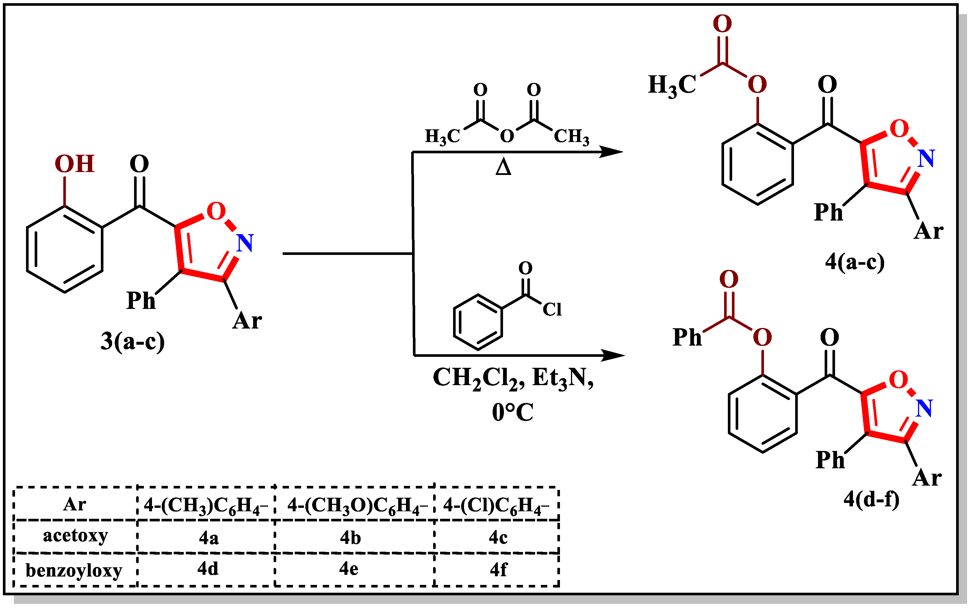
Initially, the precursors 3(ac) were synthesized by a 1,3-dipolar cycloaddition (1,3-DC) reaction between the dipolarophile (Z)-2-benzylidenebenzofuran-3(2H)-one 1 and 1,3-dipoles of the arylnitriloxide type (Scheme 1). These were generated in situ from chlorinated aldoximes 2(ac) and triethylamine in chloroform at ambient temperature in the presence of 1 to prevent the dimerization of the dipole into furoxane. This reaction results in the formation of isoxazoles 3(ac) directly instead of spiroisoxazolines [44,45].
The synthesis of the functionalized isoxazoles 4(af) relied on the reactivity of the hydroxy group of 3(ac) towards benzoyl chloride and acetic anhydride, respectively, forming the new isoxazole compounds with ester functional group in satisfactory yields (Scheme 2).
The structures of the functionalized isoxazole products were determined by IR, 1H-NMR, and 13C-NMR spectroscopies plus MS and confirmed using single-crystal X-ray diffraction for 4c and 4f. The physical and spectroscopic features of all isoxazole products are outlined in Table 1. indicating the successful formation of pure products in good yields. Specifically, the IR spectra of 4(af) show the disappearance of the absorption band of the alcohol function (OH) of 3(ac) and the appearance of an absorption band around 1750 cm−1 corresponding to the C=O stretching vibration of the ester group. The 1H-NMR spectra all indicate the disappearance of the singlet signal at 11.63 ppm assigned to the hydroxyl proton for 3(ac). The methyl group of the ester functional group, where present, appears as a singlet at around 2.2 ppm while the 13C-NMR spectra show, besides the peaks assigned to the aromatic carbons, a peak around 168 ppm attributable to the carbonyl C=O of the ester functional group. High-resolution mass spectroscopy showed molecular ion peaks exactly corresponding to the molecular masses of the proposed products.

2.2. X-ray Diffraction Data and Structural Determination of 4c and 4f

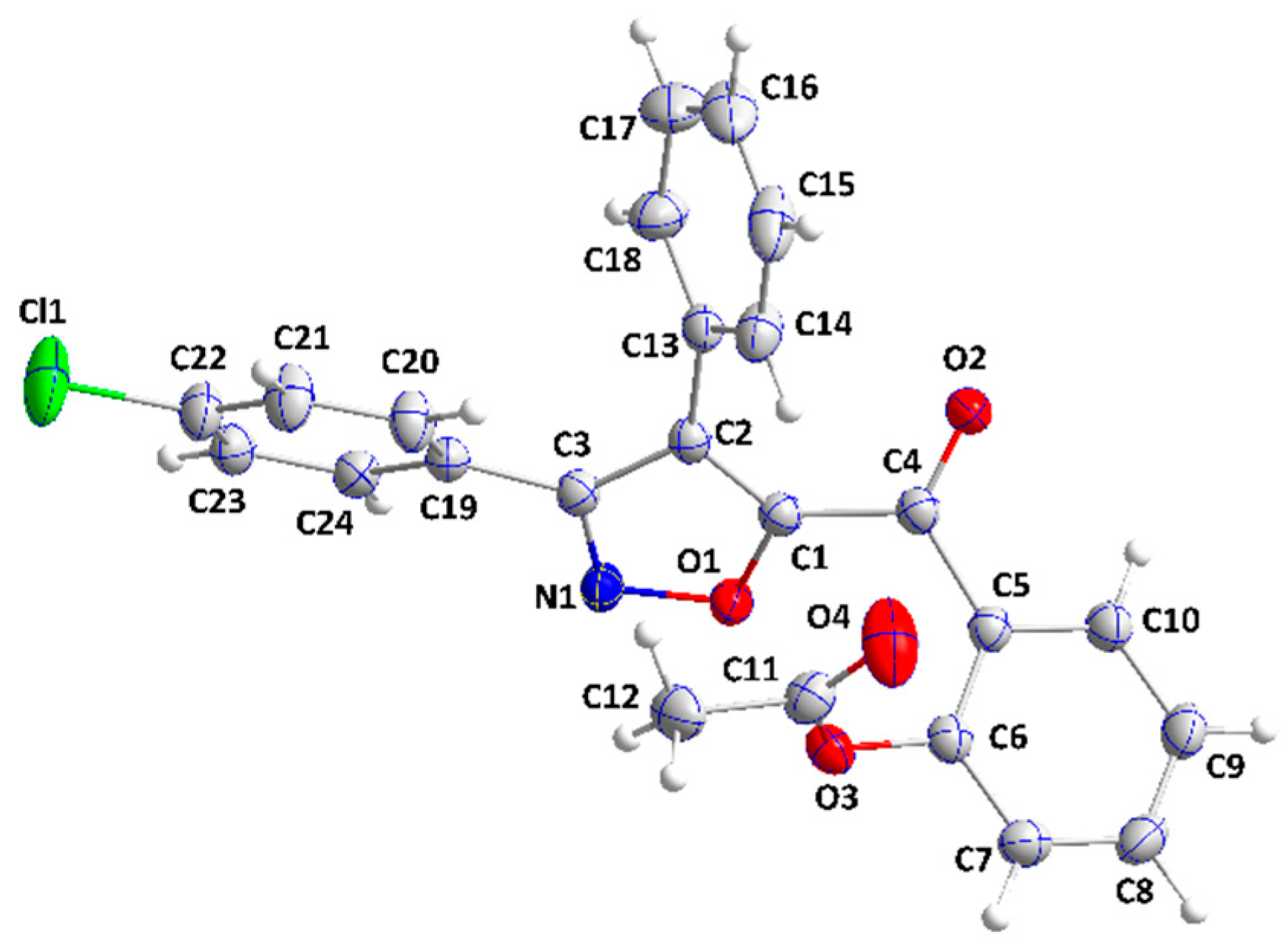
In 4c, both substituents on the C5···C10 ring extend in the same direction, giving the molecule a “pincer-like” conformation (Figure 2).
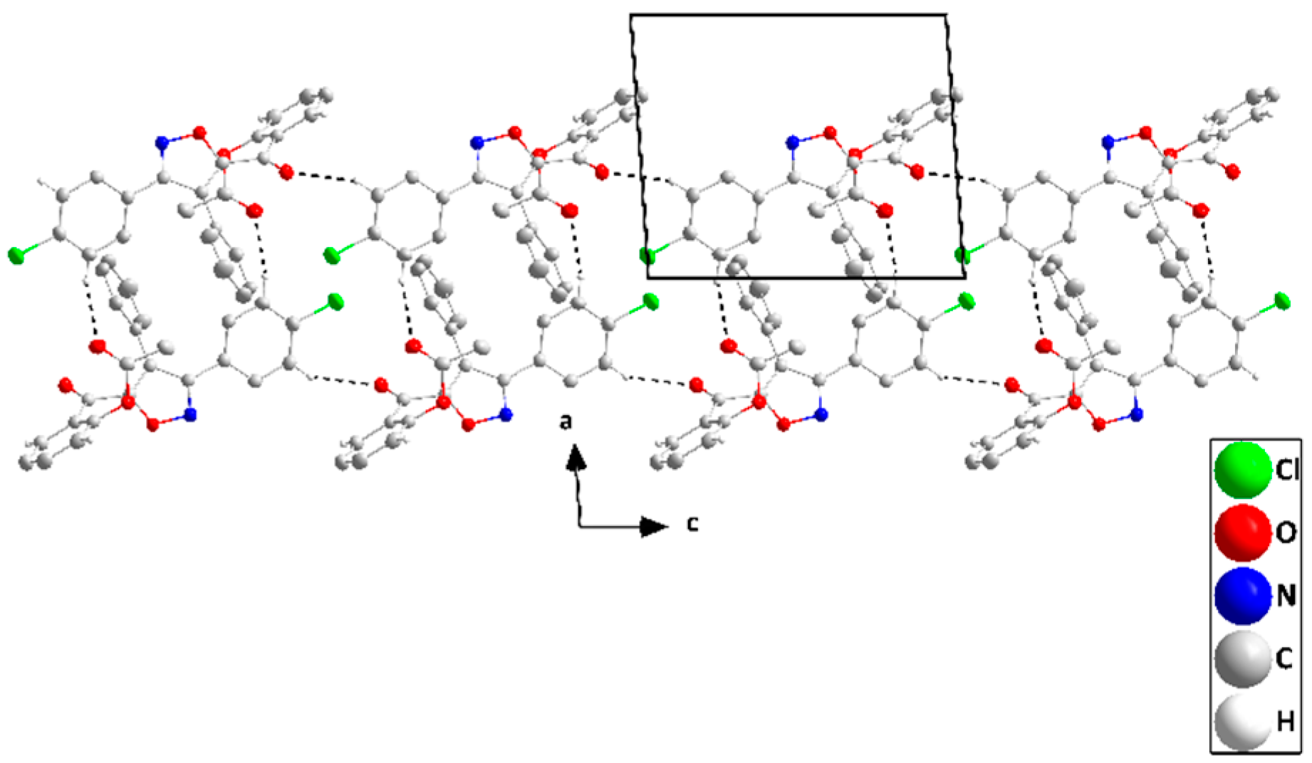
The isoxazole ring is planar to within 0.0080(8)Å and the mean planes of the C5···C10 and the C13···C18 rings are inclined to the above plane by 62.39(5) and 59.44(6)°, respectively. The dihedral angle between the mean planes of the C19···C24 and the isoxazole rings is 35.61(4)° while that between the mean planes of the C13···C18 and the C19···C24 rings is 67.20(6)°. All the bond distances and interbond angles appear as expected for the formulation given. In the crystal, the C23—H23···O2 hydrogen bonds (Table 2) form chains of molecules that extend along the c-axis direction.
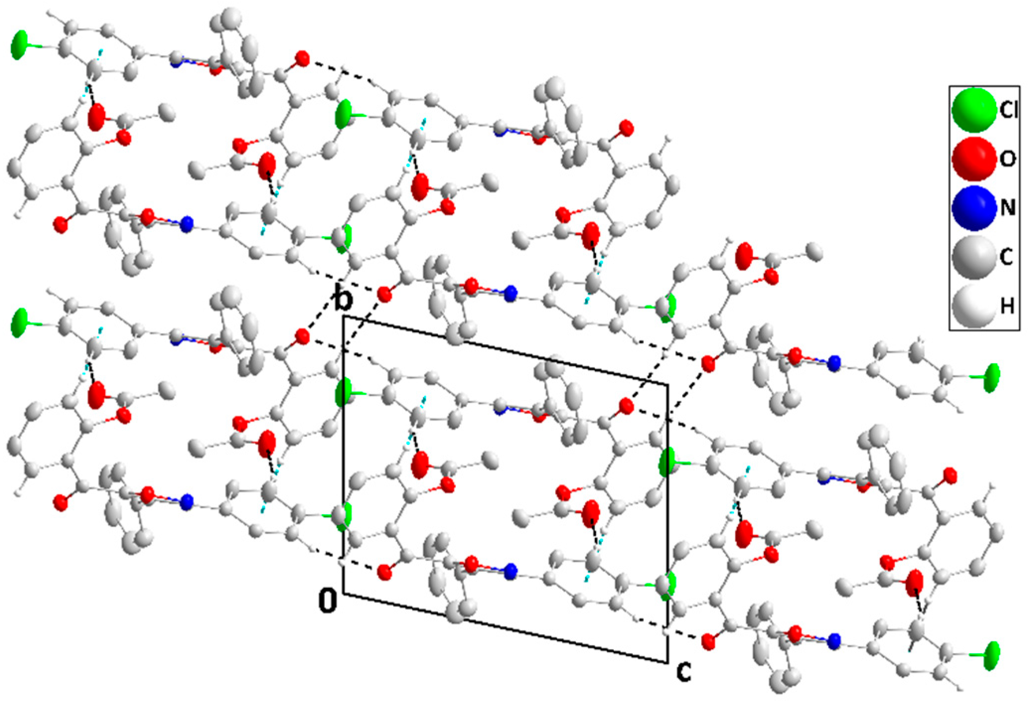
These are connected in pairs across inversion centers by C21—H21···O4 hydrogen bonds (Table 2) to form ribbons (Figure 3), which are then linked by C10—H10···O2 hydrogen bonds (Table 2) into stepped layers parallel to 1 1 ¯ 0 . The layers are joined by C7—H7···Cg4 interactions (Table 2) to form the full 3-D structure (Figure 4).
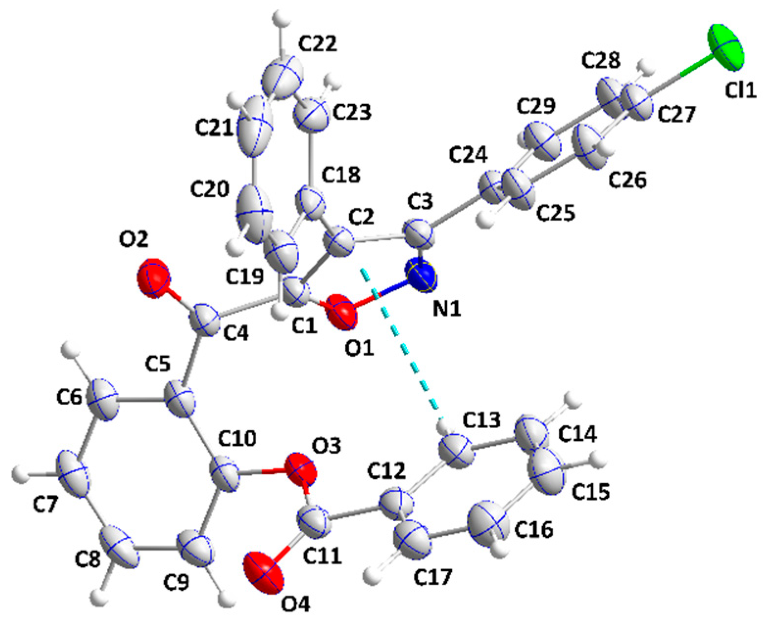
Compound 4f adopts a “pincer”-like conformation aided by a C13—H13···Cg1 interaction (Table 3) and with the C5···C10 ring at the vertex (Figure 5).
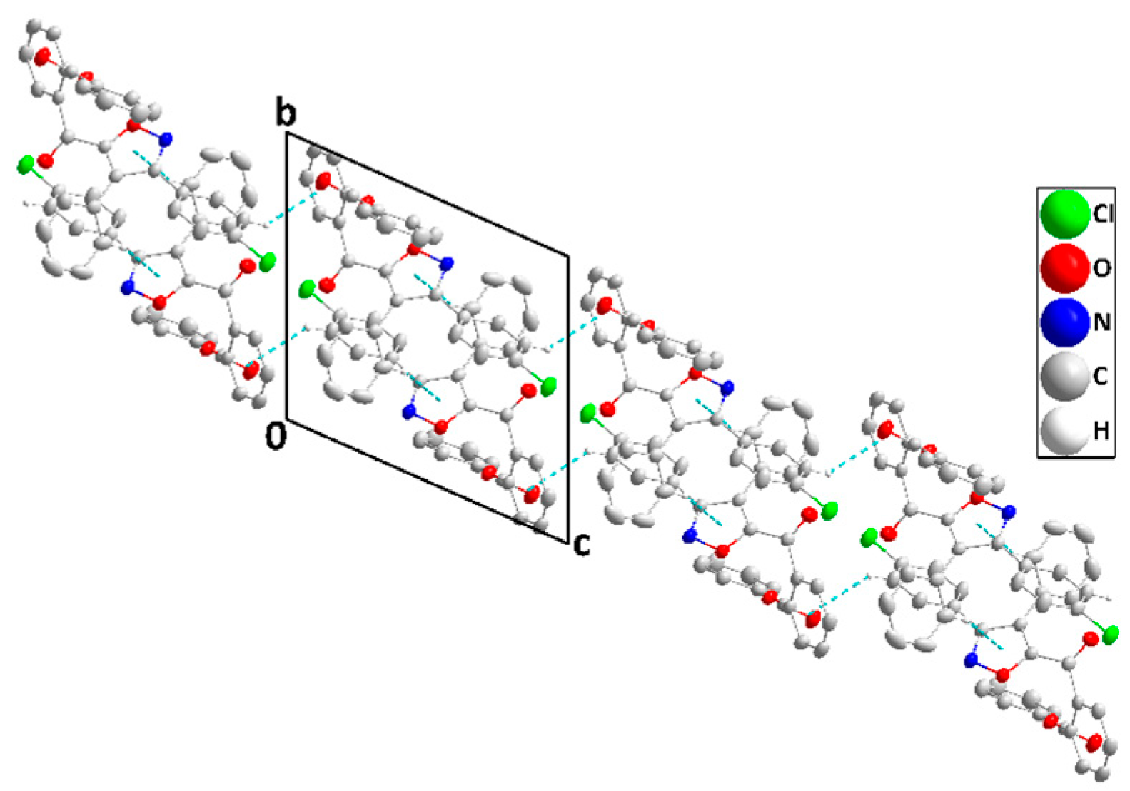
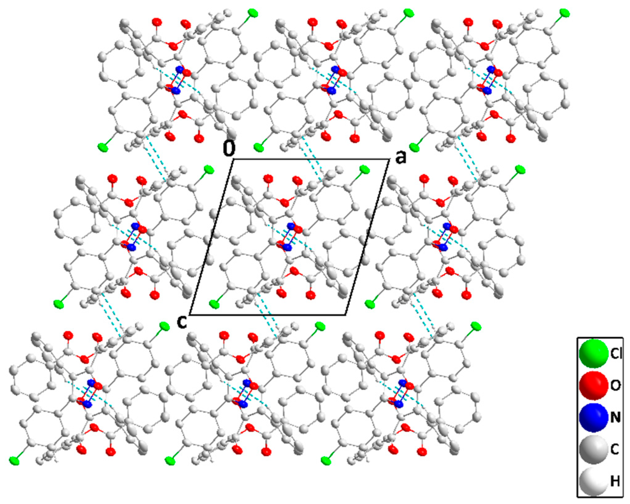
The isoxazole ring appears planar although the slight elongation of the displacement ellipsoids for O1 and N1 normal to the plane could suggest the presence of two opposite non-planar conformers. The mean planes of the C18···C23 and the C24···C29 rings are inclined to that of the isoxazole ring by 59.33(6) and 40.92(8)°, respectively, while the dihedral angles between the mean planes of the C5···C10 and the C12···C17 rings and that of the isoxazole ring are 64.80(7) and 55.80(7)°, respectively. All the bond distances and interbond angles appear as expected for the formulation given. In the crystal, the C28···H28···Cg2 interactions (Table 3) form chains of molecules extending along the c-axis direction, which are paired up by inversion-related C23—H23···Cg1 interactions (Table 3) to form ribbons (Figure 6). The ribbons pack with normal van der Waals contacts (Figure 7).

2.3. Antibacterial Screening of Isoxazoles 4(af)

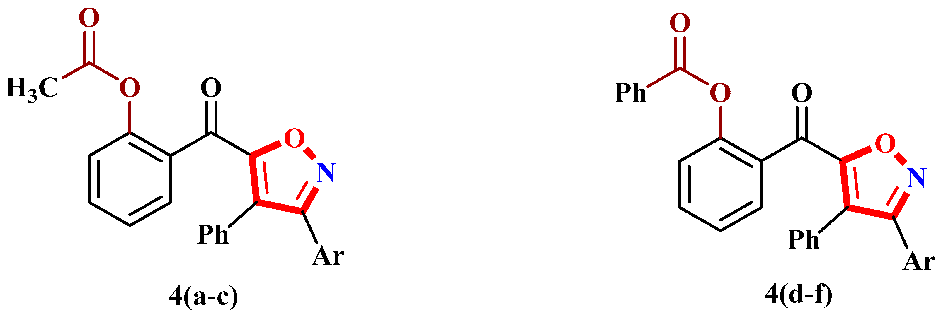
Functionalized isoxazoles play a significant role in medicinal chemistry and biological sciences and have shown many applications in drug development for the management of a variety of life-threatening pathologies [46,47]. The acetoxy 4(ac) and the benzoyloxy 4(df) isoxazole derivatives were synthesized (Figure 8) and subsequently tested in vitro for their antibacterial action against Gram-positive and Gram-negative bacterial species such as Staphylococcus aureus (CECT 976), Bacillus subtilis (DSM 6633), and Escherichia coli (K12). The disk diffusion technique was used to assess bacterial susceptibility to the different isoxazoles and the results for the new isoxazoles and reference drugs are given in Table 4.
The preliminary screening results show that 4(af) possess variable antibacterial properties against the tested bacteria when compared to standard drugs. The most promising results against the Gram-negative bacterium Escherichia coli were shown by 4a, which carries the methyl substituent, with a zone of inhibition of 17.5 ± 1.20 mm, compared to the known antibiotic streptomycin (ZI = 24 ± 1.60). Compound 4e showed robust efficacy against Escherichia coli with an inhibition zone of 14.5 ± 0.90 mm, while 4c and 4f, both with a chlorine substituent, showed inhibition zones of 13.5 ± 1.50 and 13.25 ± 0.40 mm, respectively. The Gram-positive bacterium Bacillus subtilis was found to be less sensitive to 4(af). In particular, 4a exhibited good antibacterial action against Bacillus subtilis, with an inhibition zone of 14.5 ± 0.80 mm, comparable to ampicillin (ZI = 16 ± 1.30 mm) while 4c, 4d, and 4f also displayed antibacterial action against Bacillus subtilis with inhibition zones of 11 ± 1.20, 11.5 ± 1.15, and 11.25 ± 0.75 mm, respectively. For the Gram-positive bacterium strain Staphylococcus aureus, the best antibacterial activity was shown by 4f, which carries the chlorine substituent, with an inhibition zone of 15.5 ± 1.55 mm, compared to ampicillin (ZI = 23 ± 2.60 mm). Compounds 4d and 4e also exhibited excellent action against Staphylococcus aureus, with inhibition zones of 12 ± 1.05 and 13.75 ± 1.25 mm, respectively. To validate the outcomes of the experimentally determined biological properties, in silico studies has been conducted.

2.4. Molecular Docking Studies

The molecular docking technique is used to explore how drugs bind to the active sites of target proteins, and to identify the precise interactions involved. In this context, we carried out molecular docking simulations of 4(af) and proteins from different bacteria: Escherichia coli (6kzv), Staphylococcus aureus (5tw8) and Bacillus subtilis (1of0). Using these simulations, we were able to detect how these molecules bind and interact with bacterial proteins. The binding affinity results are presented in Table 5. The binding affinity observed for E. coli ranges from −9.38 to −12.84 kcal/mol, with streptomycin exhibiting the highest affinity. These high binding affinity values suggest that these molecules have considerable potential to act as potent inhibitors of the Escherichia coli protein receptor. In the case of Staphylococcus aureus, the binding affinity values range from −8.16 to −10.45 kcal/mol. Compound 4d exhibits the highest binding affinity, which surpasses that of the standard drug Ampicillin, suggesting its good potential as an antibacterial agent against Staphylococcus aureus. For Bacillus subtilis, the range of binding affinities is −8.58 to −11.17 kcal/mol, with 4f displaying the highest affinity, showing that 4f outperforms Ampicillin and indicating its promising inhibitory activity against this pathogen, thereby offering a promising alternative for addressing antibacterial resistance issues associated with this strain. Furthermore, 4b exhibits the lowest binding affinity among the tested compounds for both Staphylococcus aureus and Bacillus subtilis, which is consistent with the in vitro results. This correlation between the in silico and in vitro results reinforces the predictive value of molecular docking studies in the identification and optimization of new antibacterial agents.
The energetics of the interaction of 4(a–f) associated with docking revealed a similar complexation energy for the majority of them, indicating that the molecular interactions are quite similar in all cases. To elucidate these interactions, a detailed study was carried out on 4a with the aim of identifying the essential interactions between 4a and the target proteins of different bacteria and to assess its potential as a multi-target inhibitor. The results show that 4a forms specific interactions with proteins from Escherichia coli, Staphylococcus aureus and Bacillus subtilis (shown in Figure 9) which are similar to those of the reference drugs.
For Escherichia coli (6kzv), 4a formed two hydrogen bonds with the residues ILE78 (2.76 Å) and THR165 (2.83 Å), two Pi-Cation electrostatic interactions with ARG76 (3.96 Å and 3.79 Å), and two Pi-Anion interactions with GLU50 (3.36 Å and 4.076 Å). On the other hand, streptomycin forms hydrogen bonds with ASN46, GLU50, ASP73, ARG76, and THR165, at distances of 2.81 Å, 1.86 Å, 1.72 Å, 2.75 Å, and 2.25 Å, respectively. For Staphylococcus aureus, 4a formed four significant hydrogen bonds with SER75, SER116, SER262, and TYR291, at distances of 2.48 Å, 2.59 Å, 2.83 Å, and 2.89 Å, respectively, showing similar interactions compared to ampicillin, which interacts through several hydrogen bonds with SER75 (2.81 Å and 2.15 Å), GLU114 (2.21 Å and 2.06 Å), SER262 (2.23 Å), and TYR291 (2.86 Å and 2.44 Å). Finally, for Bacillus subtilis, 4a formed a significant hydrogen bond-type interaction with CYS229 (2.19 Å). Conversely, ampicillin established several hydrogen bonds with CYS229 (2.39 Å), CYS322 (1.85 Å and 2.20 Å), and ASP331 (1.88 Å and 1.75 Å), illustrating increased complexity in its interaction with the bacterial target.

2.5. Molecular Dynamics Simulation Analysis

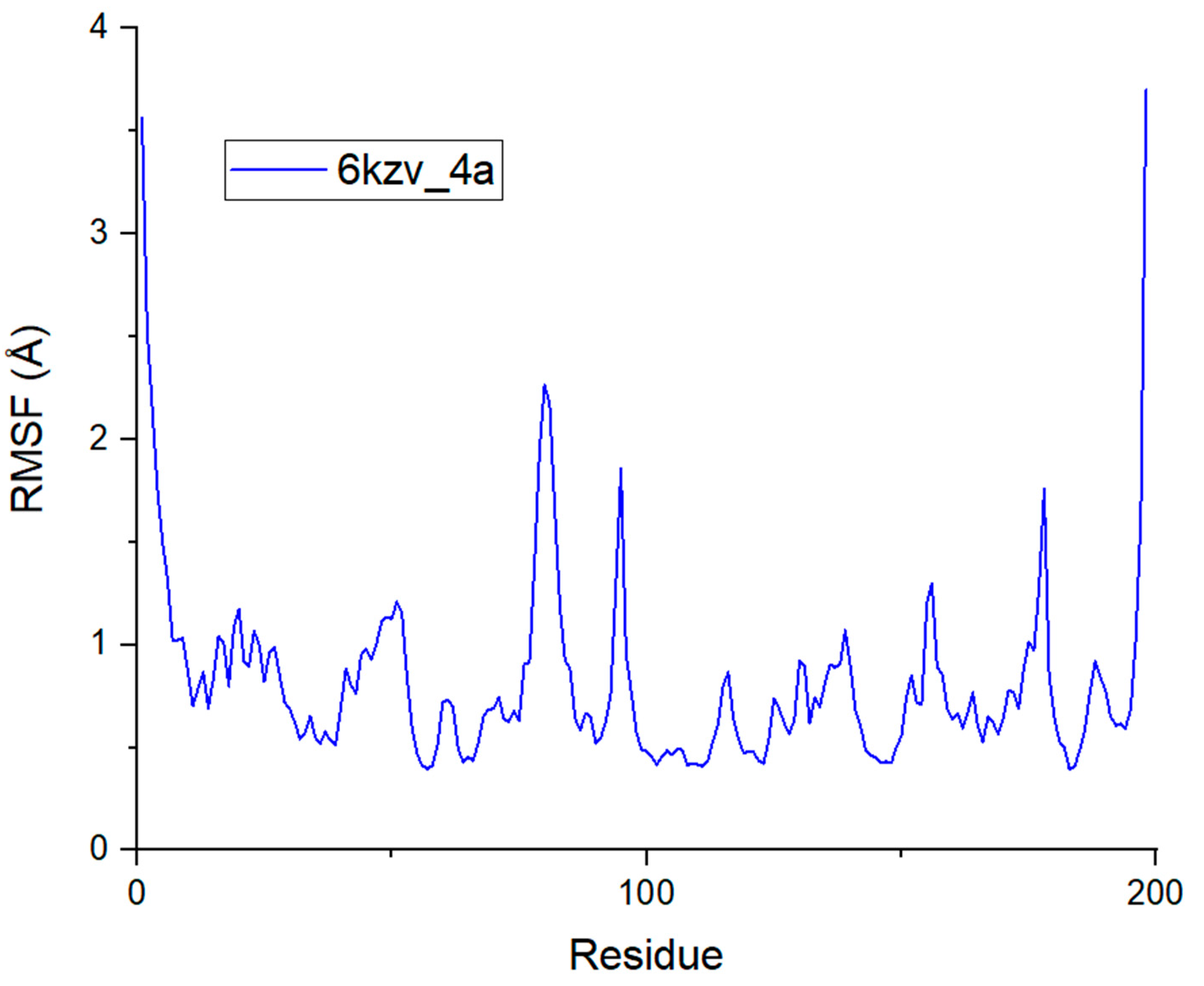
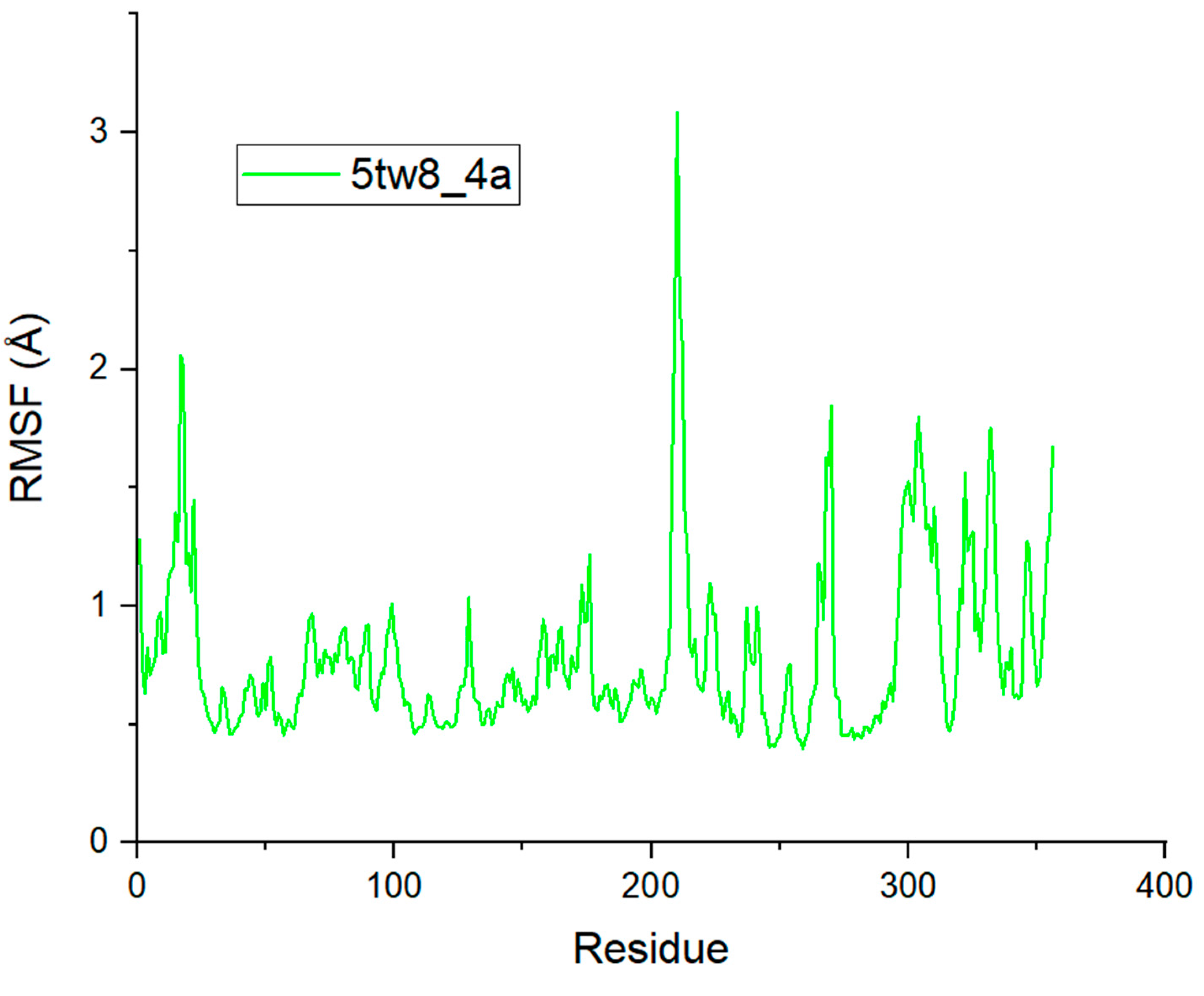
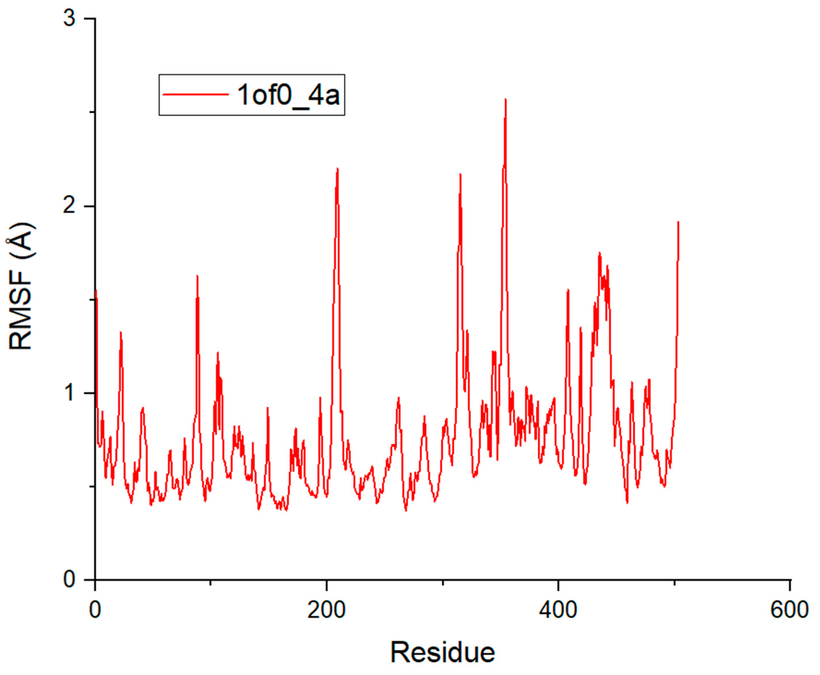
Since 4a was identified to have the best antibacterial efficacy, the molecular docking results were supplemented by molecular dynamics (MD) simulations. Variations in the conformation and dynamics of the complexes formed between Escherichia coli, Bacillus subtilis, Staphylococcus aureus, and 4a were quantified using the RMSD (root mean square deviation) and RMSF (root mean square fluctuation) indicators, illustrated in Figure 10 and Figure 11, respectively.
In the case of the Escherichia coli (6kzv) protein (Figure 10), the RMSD changes reveal a significant increase in the oscillations which rise from 0.8 to 1.6 Å in just 10 nanoseconds. This increase seems to be mainly due to the initial kinetic effect that the complexes display during the transition phase. After this phase, stabilization and equilibrium were observed for all the complexes. On the other hand, for Staphylococcus aureus (5tw8) and Bacillus subtilis (1of0), the RMSD evolutions indicate almost identical variations, with an oscillation between 1 and 1.5 Å throughout the molecular dynamics (MD) simulation. The average values (RMSD) observed for the complexes formed between 4a and the proteins of Escherichia coli, Staphylococcus aureus, and Bacillus subtilis were 1.82 Å, 1.56 Å, and 1.70 Å, respectively. The fact that these RMSD values are below 2 Å means that 4a has remarkable stability within the active sites of Escherichia coli, Staphylococcus aureus, and Bacillus subtilis proteins.
The analysis of the RMSF trajectories provides essential information on the stability of the ligand–receptor interaction, enabling us to assess the stability, rigidity, and compactness of the protein being analyzed. A high RMSF value indicates high flexibility, meaning that the residues are less stable, while a low value indicates greater rigidity and stability. For the protein from Escherichia coli (6kzv) (Figure 11), most residues display similar RMSF values, although some show notable deviations, notably at the ASP6 (3.56 Å), THR85 (2.26 Å), ALA100 (1.85 Å), GLY200 (1.76 Å), and GLY220 (3.69 Å) positions. These variations are mainly found in regions of the protein that are not directly involved in its activity, making their impact relatively low. In contrast, essential residues within the active site, such as GLU50, ASP73, ARG76, and THR165, show much more restricted movements, with RMSFs of no more than 0.9 Å, indicating the importance of the hydrogen bonds formed by these residues in stabilizing the interactions between the Escherichia coli (6kzv) protein and the ligands, thereby ensuring the greater stability of the complex. For the Staphylococcus aureus (5tw8) and Bacillus subtilis (1of0) proteins (Figure 3), the RMSF fluctuations indicate significant variations for certain residues; for example, ALA41 (2.05 Å), HIS234 (3.08 Å), GLY296 (1.84 Å), GLY330 (1.97 Å), and ASP358 (1.75 Å) in Staphylococcus aureus, and PRO217 (2.01 Å), GLY323 (2.17 Å), GLN362 (2.57 Å), and PRO511 (1.91 Å) in Bacillus subtilis. These residues located outside the active sites have a limited impact on the protein’s ability to bind ligands. On the other hand, residues fundamental to the function of these proteins showed low RMSF values, suggesting the existence of hydrogen bonds for stabilizing the protein–ligand complexes in Staphylococcus aureus and Bacillus subtilis. These observations confirm the results of the RMSD analysis, highlighting good structural stability in the ligand–protein complexes, mainly due to strong hydrogen bonding interactions.

2.6. ADME-Tox Analyses

The evaluation of 4a, 4b, 4c, 4d, 4e, and 4f as potential pharmaceutical agents for the treatment of bacterial infections was based on the predictions of their ADMET pharmacokinetic parameters. The results, presented in Table 6, show promising pharmacokinetic properties, suggesting their potential efficacy as therapeutic agents.
The intestinal absorption rates of the compounds ranged from 97.961% to 100%, indicating the excellent absorption capacity in the human intestine, which is crucial for their oral efficacy. With regard to distribution, the predictions concerning the permeability of the blood–brain barrier (BBB) and access to the central nervous system (CNS) indicate that all the compounds analyzed show negative values for LogBB and LogPS [48]. These results imply a limited distribution within these zones. This limitation is potentially beneficial in the treatment of certain bacterial infections, as it reduces the likelihood of neurological side effects by preventing drugs from reaching the CNS in large quantities. The metabolism studies showed that all the compounds acted as potential substrates and/or inhibitors of the CYP3A4 enzyme, which played a crucial role in this study [49,50]. The total clearance values suggest moderate to low excretion for all compounds, suggesting that these molecules could persist in the body for a prolonged period. Finally, the predicted non-toxicity of all the compounds studied is a positive aspect that reinforces the safety profile of the ligands for further development. In summary, the ADMET prediction results offer valuable insights into the potential of the synthesized compounds as future therapeutic agents.

3. Summary

To conclude, this work reports the synthesis, crystallographic analysis, biological evaluation, and in silico studies of new ester-functionalized isoxazole compounds. The compounds were obtained through a series of reactions using an efficient and robust procedure starting from 2-benzylidenebenzofuran-3(2H)-one (aurone). Structural determination was achieved using IR, 1H-NMR, and 13C-NMR spectroscopy and mass spectrometry with further confirmation via X-ray diffraction for compounds 4c and 4f. The antibacterial screening of the synthesized ester-functionalized isoxazole compounds against three bacterial strains—Escherichia coli, Staphylococcus aureus, and Bacillus subtilis—showed that the compounds possess good antibacterial properties. Furthermore, the molecular docking studies revealed significant binding interactions between the isoxazole compounds and the bacterial target proteins of Escherichia coli (6kzv), Staphylococcus aureus (5tw8), and Bacillus subtilis (1of0). The MD simulation results indicate the remarkable stability of compound 4a within the active sites of Escherichia coli, Staphylococcus aureus, and Bacillus subtilis proteins. Lastly, the results of the ADMET profiles of the isoxazole compounds indicate that they have good bioavailability. Taken together, the findings of this research could provide important insights to design and synthesize new ester-functionalized isoxazole compounds that are likely to be promising antibacterial agents.

4. Materials and Methods

4.1. Chemistry

Complete insights into the reagents and solvents required for compound synthesis and the equipment used in spectroscopic characterization and X-ray crystallographic analysis as well as the relevant synthesis procedures are clearly described in the Supplementary Material file.

4.2. Antibacterial Screening Protocol

The technique and bacterial strains used to screen the antibacterial properties of 4(af) are included in the Supplementary Material file.

4.3. Molecular Docking

We conducted molecular docking simulations with compounds 4(af) using specific target proteins from different bacteria. The proteins selected for these simulations are as follows:
Escherichia coli (PDB ID: 6kzv) [51]:
  • Target protein: Gyrase A (DNA gyrase subunit A).
  • Biological role: Gyrase A is essential for bacterial survival as it catalyzes the negative supercoiling of DNA, which is crucial for DNA replication and transcription. Inhibiting this enzyme can prevent DNA replication, leading to cell death.
Staphylococcus aureus (PDB ID: 5tw8) [52]:
  • Target protein: PBP2a (Penicillin-binding protein 2a).
  • Biological role: PBP2a is involved in bacterial cell wall synthesis. This protein confers resistance to β-lactam antibiotics by preventing these antibiotics from binding to penicillin-binding proteins, allowing the bacteria to continue synthesizing its cell wall despite the presence of the antibiotic.
Bacillus subtilis (PDB ID: 1of0) [53]:
  • Target protein: endospore coat protein.
  • Biological role: This protein is involved in the formation of the endospore coat, a resistant structure that protects bacterial spores under extreme environmental conditions. Inhibiting this protein can disrupt spore formation, reducing bacterial survival in adverse conditions.
Three-dimensional crystal structures of target proteins were extracted from the Protein Data Bank (PDB). These structures were prepared by removing water molecules, ligands, and non-protein elements. The software tools Discovery Studio, Autodock 4 and Autodock Tools were used to further investigate compound–protein interactions and evaluate interaction energies using a three-dimensional grid [54]. The dimensions of the central analysis grid were carefully adjusted for each protein to ensure accurate placement of ligands in the complex [44,53]. The results of the study were visualized in two dimensions to better understand the binding interactions.

4.4. Molecular Dynamics (MD)

Molecular dynamics simulations used NAMD software [55] associated with CHARMM36 force field [56]. The simulation setup consisted of enclosing the system in a cubic box, measuring 10 Å on each side, filled with TIP3P water molecules. The system was neutralized by adding NaCl ions at a concentration of 0.154 M, and applying the Monte Carlo technique [57]. The initial phase involved minimizing the energy of each system using a 10,000-step gradient descent method. This was followed by a 100 ns stabilization phase under the NVT ensemble (number of particles, volume, temperature), keeping the system at a constant temperature of 310 K. This was followed by an additional 100 ns of unrestricted molecular dynamics simulation under the NPT (number of atoms, pressure, temperature) ensemble for each system [48]. The stability of the systems was evaluated by analyzing the molecular dynamics trajectories through the Visual Molecular Dynamics (VMD) software [58].

4.5. In Silico Pharmacokinetics ADMET

The integration of computer technology has transformed the way we discover new drugs, making the process both faster and more accurate [58]. Computational studies give us an improved understanding of a compound’s behavior with regard to absorption, distribution, metabolism, excretion, and toxicity (ADMET). These analyses exploit information on how a drug acts to anticipate its effects from the earliest stages of development. The pkCSM online platform is particularly useful for assessing a drug’s capacity to be absorbed by the human intestine, its distribution in the body, its biological transformation and elimination, as well as its toxicity profile [56]. Computer simulation has thus become indispensable in the evaluation of pharmacokinetic parameters linked to ADMET.

Supplementary Materials

The following supporting information can be downloaded at https://www.mdpi.com/article/10.3390/molecules29143366/s1, Spectroscopic data and copies of IR, NMR, and MS spectra of the newly synthesized compounds, and all additional details can be found in the electronic Supplementary Material. CCDC 2348110 and 2348111 contain the supplementary crystallographic data for the compounds 4c and 4f, and can be obtained free of charge from the Cambridge Crystallographic Data Centre via www.ccdc.cam.ac.uk/data_request/cif. References [46,59,60,61,62,63,64,65,66,67] are cited in the supplementary materials.

Author Contributions

Conceptualization, A.A., N.B. and Y.R.; methodology, A.A.; software, H.H.; validation, A.A., K.B. and K.C. (Khalid Chebbac); formal analysis, K.C. (Karim Chkirate); investigation, A.N.; resources, J.T.M.; data curation, S.M.A.K.; writing—original draft preparation, A.A.; writing—review and editing, M.C. and M.M.A.; visualization, G.A.H.; supervision, M.E.Y.; project administration, M.E.Y.; funding acquisition, M.M.A. All authors have read and agreed to the published version of the manuscript.

Funding

This research was supported by the Researchers Supporting Project number (RSPD2024R628), King Saud University, Riyadh, Saudi Arabia.

Institutional Review Board Statement

Not applicable.

Informed Consent Statement

Not applicable.

Data Availability Statement

All the data generated by this research is included in the article and Supplementary Materials.

Acknowledgments

The authors extend their appreciation to the Researchers Supporting Project number (RSPD2024R628), King Saud University, Riyadh, Saudi Arabia, for supporting this research. The support of the NSF-MRI Grant #1228232 for the purchase of the diffractometer and Tulane University for the support of the Tulane Crystallography Laboratory are gratefully acknowledged.

Conflicts of Interest

The authors have no conflicts of interest to declare that are relevant to the content of this article.

References

  1. Morens, D.M.; Fauci, A.S. Emerging Infectious Diseases: Threats to Human Health and Global Stability. PLoS Pathog. 2013, 9, e1003467. [Google Scholar] [CrossRef] [PubMed]
  2. Bloom, D.E.; Cadarette, D. Infectious Disease Threats in the Twenty-First Century: Strengthening the Global Response. Front. Immunol. 2019, 10, 549. [Google Scholar] [CrossRef] [PubMed]
  3. Davies, J. Origins and Evolution of Antibiotic Resistance. Microbiologia 1996, 12, 9–16. [Google Scholar] [CrossRef] [PubMed]
  4. Chang, H.-H.; Cohen, T.; Grad, Y.H.; Hanage, W.P.; O’Brien, T.F.; Lipsitch, M. Origin and Proliferation of Multiple-Drug Resistance in Bacterial Pathogens. Microbiol. Mol. Biol. Rev. 2015, 79, 101–116. [Google Scholar] [CrossRef] [PubMed]
  5. Vivas, R.; Barbosa, A.A.T.; Dolabela, S.S.; Jain, S. Multidrug-Resistant Bacteria and Alternative Methods to Control Them: An Overview. Microb. Drug Resist. 2019, 25, 890–908. [Google Scholar] [CrossRef] [PubMed]
  6. Dadgostar, P. Antimicrobial Resistance: Implications and Costs. Infect. Drug Resist. 2019, 12, 3903–3910. [Google Scholar] [CrossRef] [PubMed]
  7. Molalign, S.; Wencheko, E. Risk Factors of Mortality in Patients with Multi-Drug Resistant TB. Ethiop. J. Health Dev. 2015, 29, 82–88. [Google Scholar]
  8. Assefa, M. Multi-Drug Resistant Gram-Negative Bacterial Pneumonia: Etiology, Risk Factors, and Drug Resistance Patterns. Pneumonia 2022, 14, 4. [Google Scholar] [CrossRef] [PubMed]
  9. Verma, R.; Verma, S.K.; Rakesh, K.P.; Girish, Y.R.; Ashrafizadeh, M.; Sharath Kumar, K.S.; Rangappa, K.S. Pyrazole-Based Analogs as Potential Antibacterial Agents against Methicillin-Resistance Staphylococcus Aureus (MRSA) and Its SAR Elucidation. Eur. J. Med. Chem. 2021, 212, 113134. [Google Scholar] [CrossRef]
  10. Wu, G.; Zhao, T.; Kang, D.; Zhang, J.; Song, Y.; Namasivayam, V.; Kongsted, J.; Pannecouque, C.; De Clercq, E.; Poongavanam, V.; et al. Overview of Recent Strategic Advances in Medicinal Chemistry. J. Med. Chem. 2019, 62, 9375–9414. [Google Scholar] [CrossRef]
  11. Kidwai, M.; Venktaramanan, R.; Mohan, R.; Sapra, P. Cancer Chemotherapy and Heterocyclic Compounds. Curr. Med. Chem. 2012, 9, 1209–1228. [Google Scholar] [CrossRef] [PubMed]
  12. Sadek, K.U.; Mekheimer, R.A.; Abd-Elmonem, M.; Abo-Elsoud, F.A.; Hayallah, A.M.; Mostafa, S.M.; Abdellattif, M.H.; Abourehab, M.A.S.; Farghaly, T.A.; Elkamhawy, A. Recent Developments in the Synthesis of Hybrid Heterocycles, a Promising Approach to Develop Multi-Target Antibacterial Agents. J. Mol. Struct. 2023, 1286, 135616. [Google Scholar] [CrossRef]
  13. Javahershenas, R.; Nikzat, S. Recent Developments Using Malononitrile in Ultrasound-Assisted Multicomponent Synthesis of Heterocycles. Ultrason. Sonochem. 2023, 102, 106741. [Google Scholar] [CrossRef] [PubMed]
  14. Dadiboyena, S.; Nefzi, A. Synthesis of Functionalized Tetrasubstituted Pyrazolyl Heterocycles—A Review. Eur. J. Med. Chem. 2011, 46, 5258–5275. [Google Scholar] [CrossRef] [PubMed]
  15. Kaur, N.; Grewal, P.; Bhardwaj, P.; Verma, Y.; Ahlawat, N. Synthesis of Higher-Membered Heterocycles. Synth. Commun. 2023, 53, 443–475. [Google Scholar] [CrossRef]
  16. Lamberth, C. Oxazole and Isoxazole Chemistry in Crop Protection. J. Heterocycl. Chem. 2018, 55, 2035–2045. [Google Scholar] [CrossRef]
  17. Guo, K.L.; Zhao, L.X.; Wang, Z.W.; Gao, Y.C.; Li, J.J.; Gao, S.; Fu, Y.; Ye, F. Design, Synthesis, and Bioevaluation of Substituted Phenyl Isoxazole Analogues as Herbicide Safeners. J. Agric. Food Chem. 2020, 68, 10550–10559. [Google Scholar] [CrossRef]
  18. Bumagin, N.A.; Potkin, V.I. Functionalized Isoxazole and Isothiazole Ligands: Design, Synthesis, Palladium Complexes, Homogeneous and Heterogeneous Catalysis in Aqueous Media. Russ. Chem. Bull. 2016, 65, 321–332. [Google Scholar] [CrossRef]
  19. Díaz-Trelles, R.; Novelli, A.; Fernández-Sánchez, M.T. RNA Synthesis-Dependent Potentiation of α-Amino-3-Hydroxy-5-Methyl-4-Isoxazole Propionate Receptor-Mediated Toxicity by Antihistamine Terfenadine in Cultured Rat Cerebellar Neurons. Neurosci. Lett. 2003, 345, 136–140. [Google Scholar] [CrossRef]
  20. Trefzger, O.S.; Barbosa, N.V.; Scapolatempo, R.L.; das Neves, A.R.; Ortale, M.L.F.S.; Carvalho, D.B.; Honorato, A.M.; Fragoso, M.R.; Shuiguemoto, C.Y.K.; Perdomo, R.T.; et al. Design, Synthesis, Antileishmanial, and Antifungal Biological Evaluation of Novel 3,5-Disubstituted Isoxazole Compounds Based on 5-Nitrofuran Scaffolds. Arch. Pharm. 2019, 353, 1900241. [Google Scholar] [CrossRef]
  21. Ni, T.; Chi, X.; Xie, F.; Li, L.; Wu, H.; Hao, Y.; Wang, X.; Zhang, D.; Jiang, Y. Design, Synthesis, and Evaluation of Novel Tetrazoles Featuring Isoxazole Moiety as Highly Selective Antifungal Agents. Eur. J. Med. Chem. 2023, 246, 115007. [Google Scholar] [CrossRef]
  22. Pradeep Kumar, M.; Ayodhya, D.; Rambabu, A. Shivaraj Synthesis, Characterization, X-ray Crystal Structure, Antioxidant, Antimicrobial, and DNA Binding Interaction Studies of Novel Copper (II)-Isoxazole Binary Complexes. Results Chem. 2023, 5, 100846. [Google Scholar] [CrossRef]
  23. Khanage, S.; Mohite, P.; Pandhare, R.; Raju, A. 1,2,4-Triazol Içeren Izoksazol Türevlerinin Sentezi Ve Farmakolojik Etkisi. Marmara Pharm. J. 2012, 16, 134–140. [Google Scholar] [CrossRef]
  24. Sysak, A.; Obmińska-Mrukowicz, B. Isoxazole Ring as a Useful Scaffold in a Search for New Therapeutic Agents. Eur. J. Med. Chem. 2017, 137, 292–309. [Google Scholar] [CrossRef]
  25. Liu, T.; Huang, B.; Zhan, P.; De Clercq, E.; Liu, X. Discovery of Small Molecular Inhibitors Targeting HIV-1 Gp120-CD4 Interaction Drived from BMS-378806. Eur. J. Med. Chem. 2014, 86, 481–490. [Google Scholar] [CrossRef] [PubMed]
  26. Agrawal, N.; Mishra, P. The Synthetic and Therapeutic Expedition of Isoxazole and Its Analogs. Med. Chem. Res. 2018, 27, 1309–1344. [Google Scholar] [CrossRef] [PubMed]
  27. Mączyński, M.; Artym, J.; Kociȩba, M.; Kochanowska, I.; Ryng, S.; Zimecki, M. Anti-Inflammatory Properties of an Isoxazole Derivative—MZO-2. Pharmacol. Rep. 2016, 68, 894–902. [Google Scholar] [CrossRef] [PubMed]
  28. Bhatia, R.; Vyas, A.; El-Bahy, S.M.; Hessien, M.M.; Mersal, G.A.M.; Ibrahim, M.M.; Dogra, R.; Kumar, B. Rationale Design, Synthesis, Pharmacological and In-Silico Investigation of Indole-Functionalized Isoxazoles as Anti-Inflammatory Agents. ChemistrySelect 2022, 7, e202200800. [Google Scholar] [CrossRef]
  29. Arya, G.C.; Kaur, K.; Jaitak, V. Isoxazole Derivatives as Anticancer Agent: A Review on Synthetic Strategies, Mechanism of Action and SAR Studies. Eur. J. Med. Chem. 2021, 221, 113511. [Google Scholar] [CrossRef]
  30. Hawash, M.; Jaradat, N.; Eid, A.M.; Abubaker, A.; Mufleh, O.; Al-Hroub, Q.; Sobuh, S. Synthesis of Novel Isoxazole–Carboxamide Derivatives as Promising Agents for Melanoma and Targeted Nano-Emulgel Conjugate for Improved Cellular Permeability. BMC Chem. 2022, 16, 47. [Google Scholar] [CrossRef]
  31. Ye, F.; Zhai, Y.; Kang, T.; Wu, S.L.; Li, J.J.; Gao, S.; Zhao, L.X.; Fu, Y. Rational Design, Synthesis and Structure-Activity Relationship of Novel Substituted Oxazole Isoxazole Carboxamides as Herbicide Safener. Pestic. Biochem. Physiol. 2019, 157, 60–68. [Google Scholar] [CrossRef]
  32. Lin, X.; Li, Y.; Zhong, W.; Hong, T.; Li, L.; Song, S.; He, D. Synthesis, Bioactivity, and QSAR Study of 3,4-Dichlorophenyl Isoxazole-Substituted Stilbene Derivatives against the Phytopathogenic Fungus Botrytis Cinerea. J. Agric. Food Chem. 2021, 69, 9520–9528. [Google Scholar] [CrossRef] [PubMed]
  33. Yang, Z.; Zhao, Y.; Li, P.; He, Y. Design, Synthesis, and Insecticidal Activity of Novel Isoxazole Derivatives Containing Bisamide Moiety. J. Heterocycl. Chem. 2019, 56, 3042–3047. [Google Scholar] [CrossRef]
  34. Lasri, M.; Bimoussa, A.; Ait-karra, A.; Laamari, Y.; Zakir, O.; Idouhli, R.; Maatallah, M.; Eddine, K.M.; Auhmani, A.; Itto, M.Y.A.; et al. Synthesis and Evaluation of Benzo[1,2,3]selenadiazole-Isoxazoles as Corrosion Inhibitors for Copper in NaCl: An Integrated Experimental and Theoretical Approach. Colloids Surf. A Physicochem. Eng. Asp. 2024, 695, 134227. [Google Scholar] [CrossRef]
  35. Hu, F.; Szostak, M. Recent Developments in the Synthesis and Reactivity of Isoxazoles: Metal Catalysis and Beyond. Adv. Synth. Catal. 2015, 357, 2583–2614. [Google Scholar] [CrossRef]
  36. Kohler, E.P.; Davis, A.R. Isoxazoline Oxides. X. Reduction. J. Am. Chem. Soc. 1930, 52, 4520–4528. [Google Scholar] [CrossRef]
  37. Akella, L.B.; DeCaprio, D. Cheminformatics Approaches to Analyze Diversity in Compound Screening Libraries. Curr. Opin. Chem. Biol. 2010, 14, 325–330. [Google Scholar] [CrossRef] [PubMed]
  38. Begam, B.F.; Kumar, J.S. A Study on Cheminformatics and Its Applications on Modern Drug Discovery. Procedia Eng. 2012, 38, 1264–1275. [Google Scholar] [CrossRef]
  39. Altındağ, F.D.; Sağlık, B.N.; Acar Çevik, U.; Işıkdağ, İ.; Özkay, Y.; Karaca Gençer, H. Novel Imidazole Derivatives as Antifungal Agents: Synthesis, Biological Evaluation, ADME Prediction and Molecular Docking Studies. Phosphorus Sulfur Silicon Relat. Elem. 2019, 194, 887–894. [Google Scholar] [CrossRef]
  40. Patil, M.; Poyil, A.N.; Joshi, S.D.; Patil, S.A.; Patil, S.A.; Bugarin, A. Synthesis, Molecular Docking Studies, and Antimicrobial Evaluation of New Structurally Diverse Ureas. Bioorg. Chem. 2019, 87, 302–311. [Google Scholar] [CrossRef]
  41. Morris, C.J.; Corte, D. Della Using Molecular Docking and Molecular Dynamics to Investigate Protein-Ligand Interactions. Mod. Phys. Lett. B 2021, 35, 2130002. [Google Scholar] [CrossRef]
  42. Chalkha, M.; Chebbac, K.; Nour, H.; Asmae, N.; El Moussaoui, A.; Tüzün, B.; Bourhia, M.; Chtita, S.; Bakhouch, M.; Laaroussi, H.; et al. In Vitro and In Silico Evaluation of the Antimicrobial and Antioxidant Activities of Spiropyrazoline Oxindole Congeners. Arab. J. Chem. 2023, 17, 105465. [Google Scholar] [CrossRef]
  43. Kanzouai, Y.; Chalkha, M.; Hadni, H.; Laghmari, M.; Bouzammit, R.; Nakkabi, A.; Benali, T.; Tüzün, B.; Akhazzane, M.; El Yazidi, M.; et al. Design, Synthesis, in-Vitro and in-Silico Studies of Chromone-isoxazoline Conjugates as Anti-bacterial Agents. J. Mol. Struct. 2023, 129, 136205. [Google Scholar] [CrossRef]
  44. Rhazi, Y.; Chalkha, M.; Nakkabi, A.; Hammoudan, I.; Akhazzane, M.; Bakhouch, M.; Chtita, S.; El Yazidi, M. Novel Quinazolinone–Isoxazoline Hybrids: Synthesis, Spectroscopic Characterization, and DFT Mechanistic Study. Chemistry 2022, 4, 969–982. [Google Scholar] [CrossRef]
  45. Chalkha, M.; Moussaoui, A.E.; Hadda, T.B.; Berredjem, M.; Bouzina, A.; Almalki, F.A.; Saghrouchni, H.; Bakhouch, M.; Saadi, M.; El Ammari, L.; et al. Crystallographic Study, Biological Evaluation and DFT/POM/Docking Analyses of Pyrazole Linked Amide Conjugates: Identification of Antimicrobial and Antitumor Pharmacophore Sites. J. Mol. Struct. 2022, 1252, 131818. [Google Scholar] [CrossRef]
  46. Arzine, A.; Abchir, O.; Chalkha, M.; Chebbac, K.; Rhazi, Y.; Barghady, N.; Yamari, I.; Moussaoui, A.E.L.; Nakkabi, A.; Akhazzane, M.; et al. Design, Synthesis, In-Vitro, In-Silico and DFT Studies of Novel Functionalized Isoxazoles as Antibacterial and Antioxidant Agents. Comput. Biol. Chem. 2023, 108, 107993. [Google Scholar] [CrossRef] [PubMed]
  47. Elyazidi, M.; Bougrin, K.; Daou, B.; Doua, H.; Soufiaoui, M. Microwave-Assisted Synthesis of New Spiro-isoxazolino-indol-3-ones and 5-Aroylisoxazoles on KF-Al2O3 under Solvent-Free Conditions. J. Soc. Chim. Tunis. 2003, 6, 239–245. [Google Scholar]
  48. Chalkha, M.; Ameziane el Hassani, A.; Nakkabi, A.; Tüzün, B.; Bakhouch, M.; Benjelloun, A.T.; Sfaira, M.; Saadi, M.; Ammari, L.E.; Yazidi, M. El Crystal Structure, Hirshfeld Surface and DFT Computations, along with Molecular Docking Investigations of a New Pyrazole as a Tyrosine Kinase Inhibitor. J. Mol. Struct. 2023, 1273, 134255. [Google Scholar] [CrossRef]
  49. Madhavan, S.; Keshri, S.K.; Kapur, M. Transition Metal-Mediated Functionalization of Isoxazoles: A Review. Asian J. Org. Chem. 2021, 10, 3127–3165. [Google Scholar] [CrossRef]
  50. Hadni, H.; Elhallaoui, M. Discovery of Anti-Colon Cancer Agents Targeting Wild-Type and Mutant P53 Using Computer-Aided Drug Design. J. Biomol. Struct. Dyn. 2023, 41, 10171–10189. [Google Scholar] [CrossRef]
  51. Islam, A.U.; Hadni, H.; Ali, F.; Abuzreda, A.; Kawsar, S.M.A. Synthesis, Antimicrobial Activity, Molecular Docking, Molecular Dynamics Simulation, and ADMET Properties of the Mannopyranoside Derivatives as Antimicrobial Agents. J. Taibah Univ. Sci. 2024, 18, 2327101. [Google Scholar] [CrossRef]
  52. Yamada, M.; Hatsuta, K.; Niikawa, M.; Imaishi, H. Detoxification of Aflatoxin B1 Contaminated Maize Using Human CYP3A4. J. Microbiol. Biotechnol. 2020, 30, 1207–1213. [Google Scholar] [CrossRef]
  53. Ushiyama, F.; Amada, H.; Takeuchi, T.; Tanaka-Yamamoto, N.; Kanazawa, H.; Nakano, K.; Mima, M.; Masuko, A.; Takata, I.; Hitaka, K.; et al. Lead Identification of 8-(Methylamino)-2-oxo-1,2-dihydroquinoline Derivatives as DNA Gyrase Inhibitors: Hit-to-Lead Generation Involving Thermodynamic Evaluation. ACS Omega 2020, 5, 10145–10159. [Google Scholar] [CrossRef] [PubMed]
  54. Alexander, J.A.N.; Chatterjee, S.S.; Hamilton, S.M.; Eltis, L.D.; Chambers, H.F.; Strynadka, N.C.J. Structural and Kinetic Analyses of Penicillin-Binding Protein 4 (PBP4)-Mediated Antibiotic Resistance in Staphylococcus aureus. J. Biol. Chem. 2018, 293, 19854–19865. [Google Scholar] [CrossRef] [PubMed]
  55. Enguita, F.J.; Marçal, D.; Martins, L.O.; Grenha, R.; Henriques, A.O.; Lindley, P.F.; Carrondo, M.A. Substrate and Dioxygen Binding to the Endospore Coat Laccase from Bacillus Subtilis. J. Biol. Chem. 2004, 279, 23472–23476. [Google Scholar] [CrossRef]
  56. El Rhabori, S.; El Aissouq, A.; Daoui, O.; Elkhattabi, S.; Chtita, S.; Khalil, F. Design of New Molecules against Cervical Cancer Using DFT, Theoretical Spectroscopy, 2D/3D-QSAR, Molecular Docking, Pharmacophore and ADMET Investigations. Heliyon 2024, 10, e24551. [Google Scholar] [CrossRef]
  57. Phillips, J.C.; Braun, R.; Wang, W.; Gumbart, J.; Tajkhorshid, E.; Villa, E.; Chipot, C.; Skeel, R.D.; Kalé, L.; Schulten, K. Scalable Molecular Dynamics with NAMD. J. Comput. Chem. 2005, 26, 1781–1802. [Google Scholar] [CrossRef] [PubMed]
  58. Hadni, H.; Fitri, A.; Touimi Benjelloun, A.; Benzakour, M.; Mcharfi, M.; Benbrahim, M. Identification of Terpenoids as Potential Inhibitors of SARS-CoV-2 (Main Protease) and Spike (RBD) via Computer-Aided Drug Design. J. Biomol. Struct. Dyn. 2023, 1–14. [Google Scholar] [CrossRef]
  59. Beney, C.; Mariotte, A.; Boumendjel, A. An Efficient Synthesis of 4,6-Dimethoxyaurones. Heterocycles 2001, 55, 967–972. [Google Scholar] [CrossRef]
  60. Masoomi, S.; Alipour, E.; Ali, M.; Reza, A.; Shafiee, A. Synthesis of Novel 2-Benzylidenebenzofuran-3(2H)-one Derivatives. Iran. J. Org. Chem. 2011, 3, 733–736. [Google Scholar]
  61. Bruker APEX4, SAINT & SHELXTL; Bruker AXS LLC: Madison, WI, USA, 2021.
  62. Krause, L.; Herbst-Irmer, R.; Sheldrick, G.M.; Stalke, D. Comparison of Silver and Molybdenum Microfocus X-ray Sources for Single-Crystal Structure Determination. J. Appl. Crystallogr. 2015, 48, 3–10. [Google Scholar] [CrossRef] [PubMed]
  63. Sheldrick, G.M. SHELXT—Integrated Space-Group and Crystal-Structure Determination. Acta Crystallogr. Sect. A Found. Crystallogr. 2015, 71, 3–8. [Google Scholar] [CrossRef] [PubMed]
  64. Sheldrick, G.M. Crystal Structure Refinement with SHELXL. Acta Crystallogr. Sect. C Struct. Chem. 2015, 71, 3–8. [Google Scholar] [CrossRef] [PubMed]
  65. Brandenburg, K.; Putz, H. DIAMOND; Crystal Impact GbR: Bonn, Germany, 2012. [Google Scholar]
  66. Chalkha, M.; Nour, H.; Chebbac, K.; Nakkabi, A.; Bahsis, L.; Bakhouch, M.; Akhazzane, M.; Bourass, M.; Chtita, S.; Bin Jardan, Y.A.; et al. Synthesis, Characterization, DFT Mechanistic Study, Antimicrobial Activity, Molecular Modeling, and ADMET Properties of Novel Pyrazole-Isoxazoline Hybrids. ACS Omega 2022, 7, 46731–46744. [Google Scholar] [CrossRef]
  67. Chebbac, K.; Benziane Ouaritini, Z.; El Moussaoui, A.; Chalkha, M.; Lafraxo, S.; Bin Jardan, Y.A.; Nafidi, H.A.; Bourhia, M.; Guemmouh, R. Antimicrobial and Antioxidant Properties of Chemically Analyzed Essential Oil of Artemisia annua L. (Asteraceae) Native to Mediterranean Area. Life 2023, 13, 807. [Google Scholar] [CrossRef]
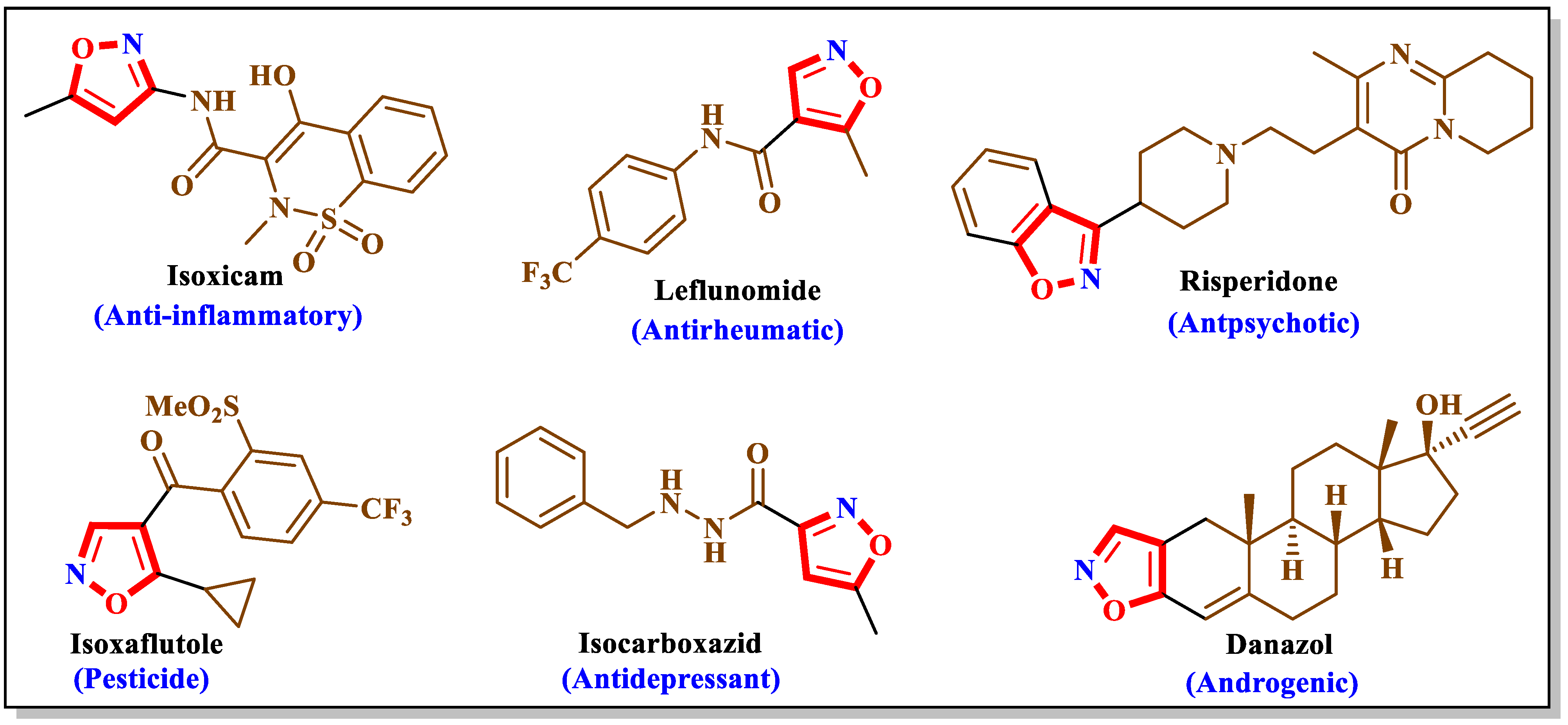
Figure 1. Examples of isoxazole-containing drugs.
Molecules 29 03366 g001
Scheme 1. Synthetic route of the compounds 3(ac).
Molecules 29 03366 sch001
Scheme 2. Synthetic route of functionalized isoxazole compounds.
Molecules 29 03366 sch002
Figure 2. Perspective view of isoxazole 4c with labeling scheme and 50% probability ellipsoids.
Molecules 29 03366 g002
Figure 3. A portion of one ribbon viewed along the b-axis direction with C—H···O hydrogen bonds depicted by dashed lines. Non-interacting hydrogen atoms are omitted for clarity (isoxazole 4c).
Molecules 29 03366 g003
Figure 4. Packing viewed along the a-axis direction with C—H···O hydrogen bonds and C—H···π(ring) interactions shown, respectively, by black and light blue dashed lines. Non-interacting hydrogen atoms are omitted for clarity (isoxazole 4c).
Molecules 29 03366 g004
Figure 5. Perspective view of isoxazole 4f with labeling scheme and 50% probability ellipsoids. The intramolecular C—H···π(ring) interaction is depicted by a dashed line.
Molecules 29 03366 g005
Figure 6. A portion of one ribbon viewed along the a-axis direction with C—H···π(ring) interactions depicted by dashed lines. Non-interacting hydrogen atoms and the intramolecular C—H···π(ring) interactions are omitted for clarity (isoxazole 4f).
Molecules 29 03366 g006
Figure 7. Packing viewed along the b-axis direction with C—H···π(ring) interactions depicted by dashed lines. Non-interacting hydrogen atoms and the intramolecular C—H···π(ring) interactions are omitted for clarity (isoxazole 4f).
Molecules 29 03366 g007
Figure 8. Structures of the synthesized isoxazoles 4(af).
Molecules 29 03366 g008
Figure 9. Two-dimensional visualization of the interactions between 4a and antibacterial proteins versus reference drugs.
Molecules 29 03366 g009
Figure 10. The RMSD values of Escherichia coli (6kzv_4a), Staphylococcus aureus (5tw8_4a), and Bacillus subtilis (1of0_4a) in complex with 4a.
Molecules 29 03366 g010
Figure 11. The RMSF values of Escherichia coli (6kzv_4a), Staphylococcus aureus (5tw8_4a), and Bacillus subtilis (1of0_4a) in complex with 4a.
Molecules 29 03366 g011aMolecules 29 03366 g011bMolecules 29 03366 g011c
Table 1. Physicochemical characteristics and characterization data of 4(af).
ArFormula
(M. g/mol)
M.p
(°C)
Yield a (%)1H-NMR (ppm)13C-NMR (ppm)IR (cm−1)MS (m/z)
[M+H]+
CH3C=O(ketone)C=O(ketone)
OCH3C=O(ester)C=O(ester)
CH3(ester)C=NC=N
4a4-(CH3)C6H4C25H19NO4
(397.43)
142–144953.38181.781667398.41
---168.911753
2.20162.301603
4b4-(CH3O)C6H4C25H19NO5
(413.42)
140–14290---181.791666414.36
3.83168.921756
2.20160.921604
4c4-(Cl)C6H4C24H16ClNO4
(417.84)
128–13085---181.551677417.34
---168.951764
2.21161.391609
4d4-(CH3)C6H4C30H21NO4
(459.50)
118–120922.23182.071667461.42
----164.361754
----161.891600
4e4-(CH3O)C6H4C30H21NO5
(475.50)
124–12689----182.081670476.43
3.9164.351741
----161.991597
4f4-(Cl)C6H4C29H18ClNO4
(479.91)
126–13091-----181.881677481.36
-----164.301760
-----161.391609
a Isolated yield of products after purification.
Table 2. Hydrogen bond geometry (Å, °). Cg4 is the centroid of the C19···C24 benzene ring.
D—H···AD—HH···AD···AD—H···A
C7—H7···Cg4 i0.952.773.5994 (17)146
C10—H10···O2 ii0.952.433.3645 (18)170
C21—H21···O4 iii0.952.363.289 (2)164
C23—H23···O2 iv0.952.583.4615 (17)154
Symmetry codes: (i) −x + 1, −y + 1, −z + 1; (ii) −x + 1, −y + 2, −z + 2; (iii) −x, −y + 1, −z + 1; (iv) x, y, z − 1.
Table 3. Cg1 and Cg2 are, respectively, the centroids of the C1/C2/C3/O1/N1 and the C5···C10 rings.
D—H···AD—HH···AD···AD—H···A
C13—H13···Cg10.952.873.534 (2)128
C23—H23···Cg1 i0.952.873.744 (2)154
C28—H28···Cg2 ii0.952.763.532 (2)140
Symmetry codes: (i) −x + 1, −y + 1, −z + 1; (ii) x, y, z − 1.
Table 4. In vitro antibacterial activity of 4(af).
Zone of Inhibition (ZI) in mm a
CompoundsTested Bacteria
ArGroupEscherichia coliBacillus subtilisStaphylococcus aureus
4a4-(CH3)C6H4acetoxy17.5 ± 1.2014.5 ± 0.809.5 ± 1.40
4b4-(CH3O)C6H4acetoxy12.5 ± 1.40--
4c4-(Cl)C6H4acetoxy13.5 ± 1.5011 ± 1.2010.5 ± 1.50
4d4-(CH3)C6H4benzoyloxy11 ± 0.5011.5 ± 1.1512 ± 1.05
4e4-(CH3O)C6H4benzoyloxy14.5 ± 0.9009 ± 0.6013.75 ± 1.25
4f4-(Cl)C6H4benzoyloxy13.25 ± 0.4011.25 ± 0.7515.5 ± 1.55
Ampicillin NT16 ± 1.3023 ± 2.60
Streptomycin 24 ± 1.60NTNT
a Results are expressed as means ± standard deviations of duplicate experiments; NT: not tested. -: no activity.
Table 5. Binding affinity between 4(af) and bacterial proteins (kcal/mol).
CompoundsEscherichia coli (6kzv) (kcal/mol)Staphylococcus aureus (5tw8) (kcal/mol)Bacillus subtilis (1of0) (kcal/mol)
4a−10.82−9.01−9.21
4b−10.18−8.16−8.58
4c−9.38−10.02−8.75
4d−10.24−10.45−10.47
4e−10.07−10.43−10.32
4f−10.85−10.34−11.17
Ampicillin −9.58−8.82
Streptomycin−12.84
Table 6. In silico ADMET predictions for 4a, 4b, 4c, 4d, 4e, and 4f.
CompoundsAbsorptionDistributionMetabolismExcretionToxicity
Intestinal
Absorption
(Human)
BBB PermeabilityCNS PermeabilitySubstrateInhibitorTotal
Clearance
AMES
Toxicity
CYP
2D63A41A22C192C92D63A4
Numeric
(%
Absorbed)
Numeric (LogBB)Numeric (LogPS)Categorical (Yes/No)Numeric (Log
mL/min/kg)
Categorical (Yes/No)
4a99.42−0.626−1.646NoYesYesYesYesNoYes0.283No
4b100−0.873−1.921NoYesYesYesYesNoYes0.314No
4c97.961−0.826−1.606NoYesYesYesYesYesYes0.15No
4d99.926−0.637−1.364NoYesNoYesYesNoNo0.502No
4e100−0.884−1.639NoYesNoYesYesNoYes0.486No
4f98.467−0.837−1.324NoYesNoYesYesNoNo0.11No
Disclaimer/Publisher’s Note: The statements, opinions and data contained in all publications are solely those of the individual author(s) and contributor(s) and not of MDPI and/or the editor(s). MDPI and/or the editor(s) disclaim responsibility for any injury to people or property resulting from any ideas, methods, instructions or products referred to in the content.