
Essential Oil from Vietnamese Peperomia leptostachya Hook. & Arn. (Piperaceae): Chemical Composition, Antioxidant, Anti-Inflammatory, Cytotoxic Activities, and In Silico Analysis

 , ,
, ,
Abstract
1. Introduction
2. Results
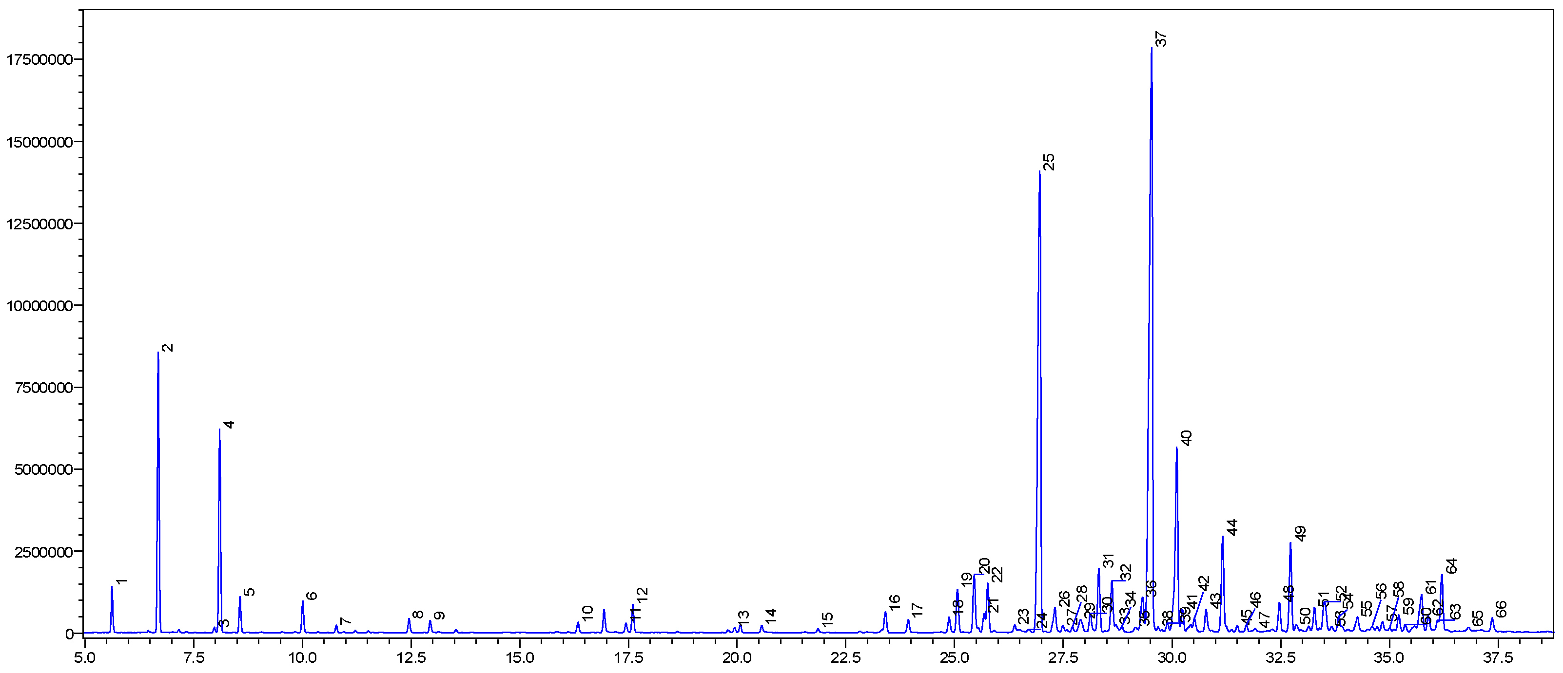
2.1. GC-MS Profiles
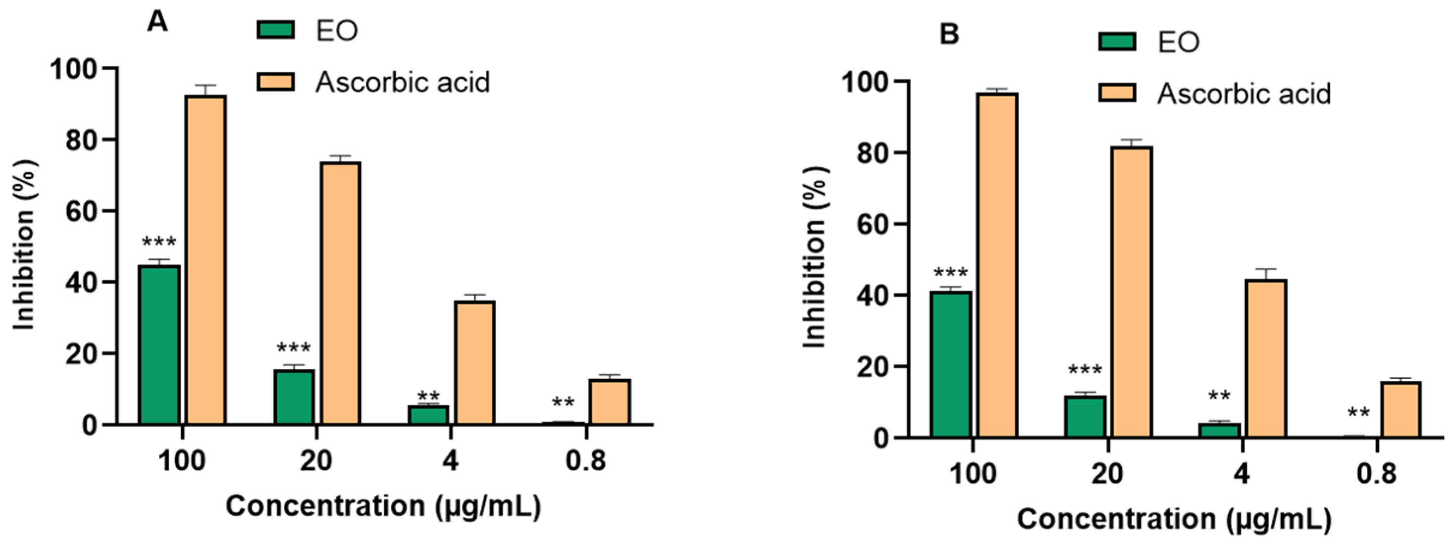
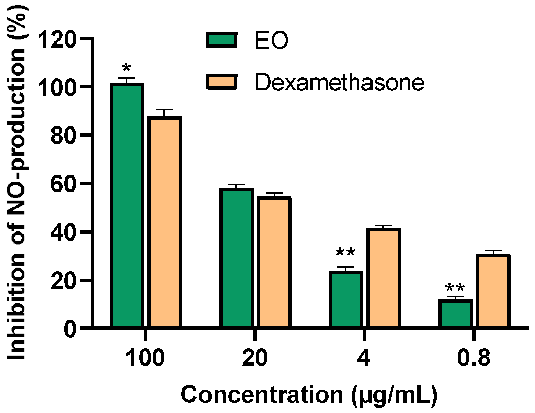
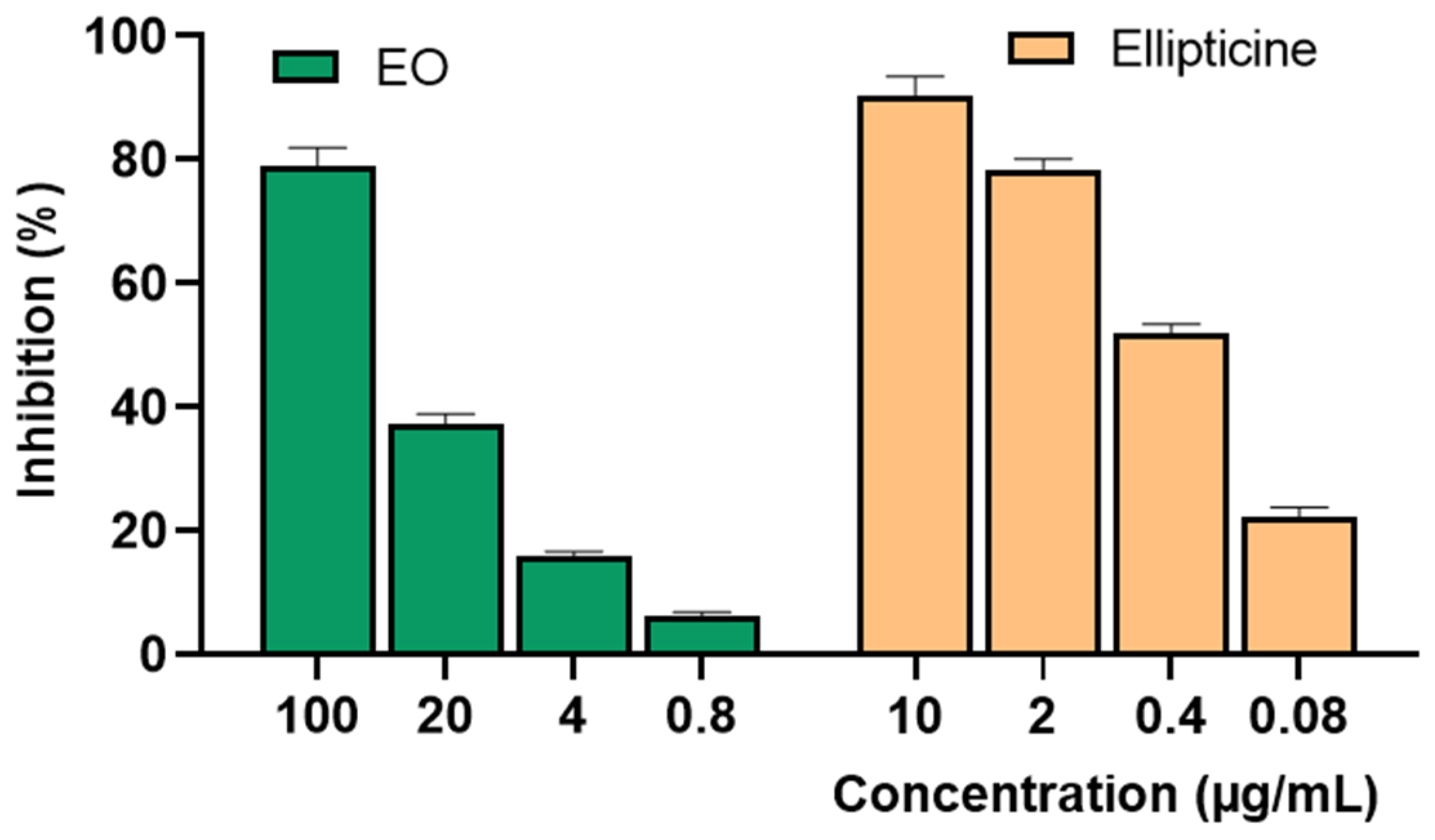
2.2. Biological Activities of Essential Oil
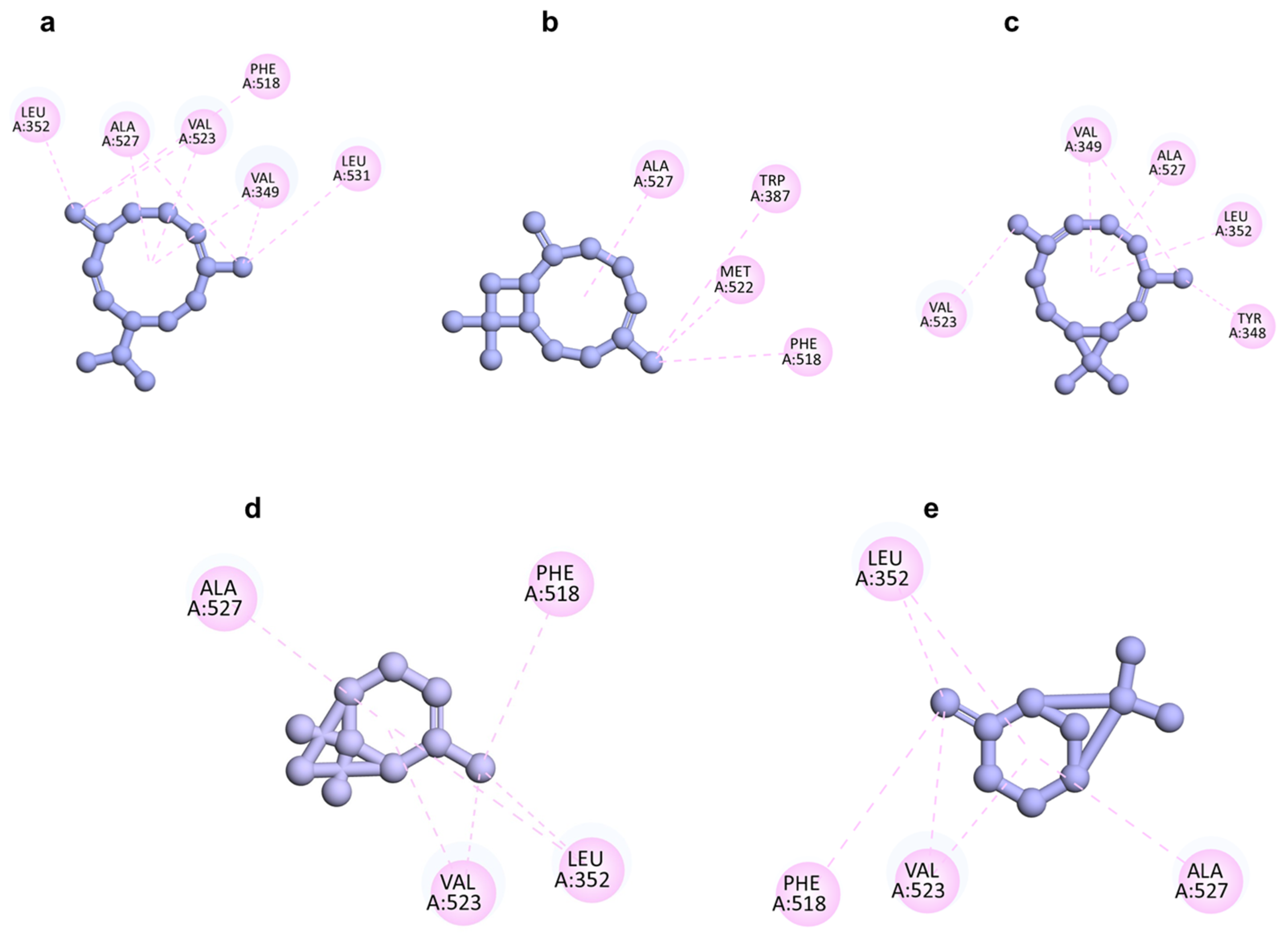
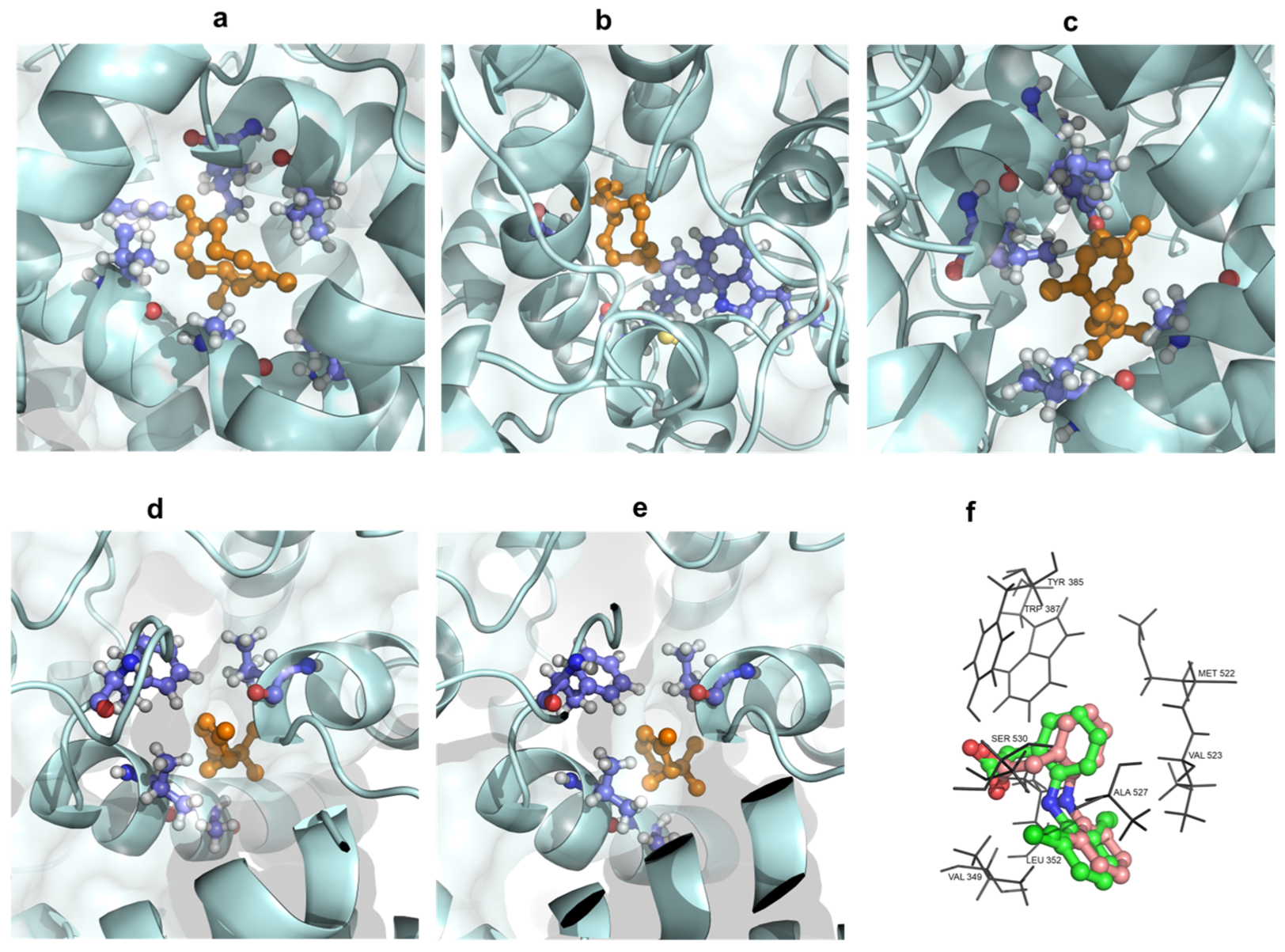
2.3. Molecular Docking Analysis
3. Discussion
4. Materials and Methods
4.1. General Procedures
4.2. Plant Material and Extraction
4.3. Distillation
4.4. GC-MS Analysis
4.5. DPPH and ABTS Radical Scavenging Activities
4.6. Anti-Inflammatory Assay
4.7. Cytotoxicity Assays
4.8. In Silico Analysis
5. Conclusions
Author Contributions
Funding
Institutional Review Board Statement
Informed Consent Statement
Data Availability Statement
Conflicts of Interest
References
- de Sousa, D.P.; Damasceno, R.O.S.; Amorati, R.; Elshabrawy, H.A.; de Castro, R.D.; Bezerra, D.P.; Nunes, V.R.V.; Gomes, R.C.; Lima, T.C. Essential oils: Chemistry and pharmacological activities. Biomolecules 2023, 13, 1144. [Google Scholar] [CrossRef]
- Pizzino, G.; Irrera, N.; Cucinotta, M.; Pallio, G.; Mannino, F.; Arcoraci, V.; Squadrito, F.; Altavilla, D.; Bitto, A. Oxidative stress: Harms and benefits for human health. Oxid. Med. Cell Longev. 2017, 2017, 8416763. [Google Scholar] [CrossRef] [PubMed]
- Masyita, A.; Sari, R.M.; Astuti, A.D.; Yasir, B.; Rumata, N.R.; Emran, T.B.; Nainu, F.; Simal-Gandara, J. Terpenes and terpenoids as main bioactive compounds of essential oils, their roles in human health and potential application as natural food preservatives. Food Chem. X 2022, 13, 100217. [Google Scholar] [CrossRef]
- Neganova, M.; Liu, J.; Aleksandrova, Y.; Klochkov, S.; Fan, R. Therapeutic influence on important targets associated with chronic inflammation and oxidative stress in cancer treatment. Cancers 2021, 13, 6062. [Google Scholar] [CrossRef]
- Salleh, W.M.N.H.W.; Ghani, N.A. Mini review on botany, traditional uses, phytochemistry and biological activities of Piper amalago (Piperaceae). Malays. J. Chem. 2022, 24, 201–208. [Google Scholar] [CrossRef]
- Alves, N.S.F.; Inoue, S.G.K.; Carneiro, A.R.; Albino, U.B.; Setzer, W.N.; Maia, J.G.; Andrade, E.H.; da Silva, J.K.R. Variation in Peperomia pellucida growth and secondary metabolism after rhizobacteria inoculation. Pandey AK. PLoS ONE 2022, 17, e0262794. [Google Scholar] [CrossRef]
- Ho, K.L.; Yong, P.H.; Wang, C.W.; Kuppusamy, U.R.; Ngo, C.T.; Massawe, F.; Ng, Z.X. Peperomia pellucida (L.) Kunth and eye diseases: A review on phytochemistry, pharmacology and toxicology. J. Integr. Med. 2022, 20, 292–304. [Google Scholar] [CrossRef]
- Gutierrez, Y.V.; Yamaguchi, L.F.; de Moraes, M.M.; Jeffrey, C.S.; Kato, M.J. Natural products from Peperomia: Occurrence, biogenesis and bioactivity. Phytochem. Rev. 2016, 15, 1009–1033. [Google Scholar] [CrossRef]
- Alves, N.S.F.; Setzer, W.N.; Da Silva, J.K.R. The chemistry and biological activities of Peperomia pellucida (Piperaceae): A critical review. J. Ethnopharmacol. 2019, 232, 90–102. [Google Scholar] [CrossRef]
- Valarezo, E.; Herrera-García, M.; Astudillo-Dávila, P.; Rosales-Demera, I.; Jaramillo-Fierro, X.; Cartuche, L.; Meneses, M.A.; Morocho, V. Study of the chemical composition and biological activity of the essential oil from Congona (Peperomia inaequalifolia Ruiz and Pav.). Plants 2023, 12, 1504. [Google Scholar] [CrossRef]
- de Araujo Morandim-Giannetti, A.; Pin, A.R.; Santo Pietro, N.A.; de Oliveira, H.C.; Mendes-Giannini, M.J.S.; Alecio, A.C. Composition and antifungal activity against Candida albicans, Candida parapsilosis, Candida krusei and Cryptococcus neoformans of essential oils from leaves of Piper and Peperomia species. J. Med. Plants Res. 2010, 4, 1810–1814. [Google Scholar]
- Okoh, S.; Iweriebor, B.C.; Okoh, O.; Okoh, A. Bioactive constituents, radical scavenging, and antibacterial properties of the leaves and stem essential oils from Peperomia pellucida (L.) Kunth. Pharmacogn Mag. 2017, 13 (Suppl. S3), S392–S400. [Google Scholar] [CrossRef] [PubMed]
- Usman, L.A.; Ismaeel, R.O. Chemical composition of root essential oil of Peperomia pellucida (L.) Kunth. grown in Nigeria. J. Essent. Oil-Bear. Plants 2020, 23, 628–632. [Google Scholar] [CrossRef]
- de Lira, P.N.B.; da Silva, J.K.R.; Andrade, E.H.A.; Sousa, P.J.C.; Silva, N.N.S.; Maia, J.G.S. Essential oil composition of three Peperomia species from the Amazon, Brazil. Nat. Prod. Commun. 2009, 4, 427–430. [Google Scholar] [CrossRef] [PubMed]
- Mora-Vivas, F.D.; Velasco, J.; Díaz, T.; Rojas-Fermín, L.; de Torres, L.D.; Ríos-Tesch, N.; Carmona, J. Composición química y actividad antibacteriana del aceite esencial de Peperomia acuminata de los Andes venezolanos. Rev. Peru. Biol. 2016, 23, 301–304. [Google Scholar] [CrossRef]
- Dorla, E.; Gauvin-Bialecki, A.; Deuscher, Z.; Allibert, A.; Grondin, I.; Deguine, J.; Laurent, P. Insecticidal activity of the leaf essential oil of Peperomia borbonensis MIQ. (Piperaceae) and its major components against the melon fly Bactrocera cucurbitae (Diptera: Tephritidae). Chem. Biodiv. 2017, 14, e1600493. [Google Scholar] [CrossRef] [PubMed]
- Paola, L.L.; Yoni, F.; Catalina, M.B.; Bandoni, A.; Jorge, D.C.; Pastor, D. Composition of the essential oil of two Peperomia from Peru: P. galioides and P. chalhuapuquiana. Rev. Latinoamer. Quim. 2007, 35, 7–12. [Google Scholar]
- Mathieu, G. Peperomia leptostachya (Piperaceae) revived. Candollea 2020, 75, 45. [Google Scholar] [CrossRef]
- de Lange, P.J. Peperomia leptostachya (Piperaceae) on Raoul Island, Kermadec Islands—A name reinstated. Trilepidea 2020, 197, 3–8. [Google Scholar]
- Adams, R.P. Identification of Essential Oil Components by Gas Chromatography Mass Spectroscopy; Allured Publishing Corporation: Carol Stream, IL, USA, 2007. [Google Scholar]
- Castro-Alvarez, A.; Costa, A.M.; Vilarrasa, J. The performance of several docking programs at reproducing protein-macrolide-like crystal structures. Molecules 2017, 22, 136. [Google Scholar] [CrossRef]
- da Fonseca, A.M.; Caluaco, B.J.; Madureira, J.M.C.; Cabongo, S.Q.; Gaieta, E.M.; Djata, F.; Colares, R.P.; Neto, M.M.; Fernandes, C.F.C.; Marinho, G.S.; et al. Screening of potential inhibitors targeting the main protease structure of SARS-CoV-2 via molecular docking, and approach with molecular dynamics, RMSD, RMSF, H-Bond, SASA and MMGBSA. Mol. Biotechnol 2023. [Google Scholar] [CrossRef] [PubMed]
- Rivera, P.N.; Mosquera, T.; Baldisserotto, A.; Abad, J.; Aillon, C.; Cabezas, D. Chemical composition and in-vitro biological activities of the essential oil from leaves of Peperomia inaequalifolia Ruiz & Pav. Am. J. Essent. Oil Nat. Prod. 2015, 2, 29–31. [Google Scholar]
- Ilyas, S.; Naz, S.; Aslam, F.; Parveen, Z.; Ali, A. Chemical composition of essential oil from in vitro grown Peperomia obtusifolia through GC-MS. Pak. J. Bot. 2014, 46, 667–672. [Google Scholar]
- De Feo, V.; Belaunde, A.J.; Sandoval, J.G.; Senatore, F.; Formisano, C. Antibacterial activity and composition of the essential oil of Peperomia galioides HBK (piperaceae) from Peru. Nat. Prod. Commun. 2008, 3, 933–936. [Google Scholar] [CrossRef]
- Zoghbi, M.; Andrade, E.; Lobato, R.; Tavares, A.; Souza, A.; Conceição, C.; Guimarães, E. Peperomia circinnata Link and Peperomia rotundifolia (L.) Kunth growing on different host-trees in Amazon: Volatiles and relationship with bryophytes. Biochem. Syst. Ecol. 2005, 33, 269–274. [Google Scholar] [CrossRef]
- Mesquita, K.D.S.M.; Feitosa, B.d.S.; Cruz, J.N.; Ferreira, O.O.; Franco, C.d.J.P.; Cascaes, M.M.; de Oliveira, M.S.; Andrade, E.H.d.A. Chemical composition and preliminary toxicity evaluation of the essential oil from Peperomia circinnata Link var. circinnata. (Piperaceae) in Artemia salina leach. Molecules 2021, 26, 7359. [Google Scholar] [CrossRef] [PubMed]
- Pinheiro, B.; Silva, A.; Souza, G.; Figueiredo, J.; Cunha, F.; Lahlou, S.; da Silva, J.; Maia, J.; Sousa, P. Chemical composition, antinociceptive and anti-inflammatory effects in rodents of the essential oil of Peperomia serpens (Sw.) Loud. J. Ethnopharmacol. 2011, 138, 479–486. [Google Scholar] [CrossRef] [PubMed]
- Horváth, G.; Ács, K. Essential oils in the treatment of respiratory tract diseases highlighting their role in bacterial infections and their anti-inflammatory action: A review. Flavour Fragr. 2015, 30, 331–341. [Google Scholar] [CrossRef] [PubMed]
- Barta, J.A.; Powell, C.A.; Wisnivesky, J.P. Global epidemiology of lung cancer. Ann. Glob. Health 2019, 85, 8. [Google Scholar] [CrossRef]
- Turini, M.E.; DuBois, R.N. Cyclooxygenase-2: A therapeutic target. Annu. Rev. Med. 2002, 53, 35–57. [Google Scholar] [CrossRef]
- Walduck, A.K.; Weber, M.; Wunder, C.; Juettner, S.; Stolte, M.; Vieth, M.; Wiedenmann, B.; Meyer, T.F.; Naumann, M.; Hoecker, M. Identification of novel Cyclooxygenase-2-dependent genes in Helicobacter pylori infection in vivo. Mol. Cancer 2009, 8, 22. [Google Scholar] [CrossRef] [PubMed]
- Marrelli, M.; De Luca, M.; Toma, C.-C.; Grande, F.; Occhiuzzi, M.A.; Caruso, R.; Conforti, F.; Statti, G. Enhancing the nitric oxide inhibitory activity using a combination of plant essential oils and mixture design approach. Heliyon 2024, 10, e31080. [Google Scholar] [CrossRef] [PubMed]
- Theodosis-Nobelos, P.; Kourti, M.; Tziona, P.; Kourounakis, P.N.; Rekka, E.A. Esters of some non-steroidal anti-inflammatory drugs with cinnamyl alcohol are potent lipoxygenase inhibitors with enhanced anti-inflammatory activity. Bioorg. Med. Chem. Lett. 2015, 25, 5028–5031. [Google Scholar] [CrossRef] [PubMed]
- Kim, M.J.; Yang, K.W.; Kim, S.S.; Park, S.M.; Park, K.J.; Kim, K.S.; Choi, Y.H.; Cho, K.K.; Lee, N.H.; Hyun, C.G. Chemical composition and anti-inflammatory effects of essential oil from Hallabong flower. EXCLI J. 2013, 12, 933. [Google Scholar] [PubMed]
- Kim, M.G.; Kim, S.M.; Min, J.H.; Kwon, O.K.; Park, M.H.; Park, J.W.; Ahn, H.I.; Hwang, J.Y.; Oh, S.R.; Lee, J.W.; et al. Anti-inflammatory effects of linalool on ovalbumin-induced pulmonary inflammation. Int. Immunopharmacol. 2019, 74, 105706. [Google Scholar] [CrossRef] [PubMed]
- Pham, T.V.; Truong, V.; Dang, N.T.T.; Vo, H.Q.; Ho, D.V. Chemical composition of the essential oils of Distichochlamys citrea leaves collected from central Vietnam. Vietnam J. Chem. 2017, 55, 358–362. [Google Scholar]
- Pham, T.V.; Ngo, H.P.T.; Dang, N.T.T.; Nguyen, H.K.; Hoang, H.T.N.; Pham, T. Volatile constituents and anti-osteoporotic activity of the n-hexane extract from Homalomena gigantea rhizome. Nat. Prod. Commun. 2022, 17, 1934578X221125433. [Google Scholar] [CrossRef]
- Dai, D.N.; Pham, T.V.; Luyen, N.D.; Dung, V.T.; Huong, L.T.; Son, N.T. Essential oils of two Vietnamese plants Piper betle f. densum (Piperaceae) and Disepalum plagioneurum (Annonaceae): Chemical composition, antimicrobial, and cytotoxic activities. Nat. Prod. Commun. 2023, 18, 1934578X231190689. [Google Scholar] [CrossRef]
- Pham, T.V.; Ha, N.X.; Luyen, N.D.; Xuan, T.H.; Le Quoc, T.; Hung, N.H.; The, S.N. Chemical composition, mosquito larvicidal and antimicrobial activities, and molecular docking study of essential oils of Cinnamomum melastomaceum, Neolitsea buisanensis and Uvaria microcarpa from Vietnam. Chem. Biodiv. 2023, 20, e202300652. [Google Scholar] [CrossRef]
- Huong, L.T.; Sam, L.N.; Giang, C.N.; Dai, D.N.; Pham, T.V.; Luyen, N.D.; Setzer, W.N.; Son, N.T. Essential oils of two Zingiber plants Zingiber eberhardtii Gagnep. and Zingiber skornickovae N.S. Lý: Chemical profiles and antimicrobial effects. J. Essent. Oil-Bear. Plants 2023, 26, 761–768. [Google Scholar] [CrossRef]
- Ho, D.V.; Hoang, H.N.T.; Nguyen, N.H.; Do, H.B.; Vo, H.Q.; Le, A.T.; Le, T.Q.; Pham, T.V. GC-MS characterization, in vitro antioxidant and anti-inflammatory activities of essential oil from the leaves of Litsea balansae Lecomte. Nat. Prod. Commun. 2023, 18, 1934578X231214159. [Google Scholar] [CrossRef]
- Buranasudja, V.; Vimolmangkang, S.; Sanookpan, K.; Binalee, A.; Bao Nguyen, A.; Dong Thach, U.; Hang Do, B.; Son Dang, V.; Minh Do, K.; Minh Nguyen, H. Antioxidant, anti-skin-aging, anti-Inflammatory, and anti-acetylcholinesterase activities of Rourea oligophlebia extracts. J Chem. Biodiver. 2023, 20, e202201096. [Google Scholar] [CrossRef] [PubMed]
- Pham, T.V.; Ho, D.V.; Le, A.T.; Ngo, Y.D.; Dang, N.T.T.; Le, T.Q.; Nguyen, B.C. Chemical composition and biological activities of essential oil from Grewia bulot leaves. Nat. Prod. Res. 2023, 1–7. [Google Scholar] [CrossRef] [PubMed]
- Khan, M.T. Predictions of the ADMET properties of candidate drug molecules utilizing different QSAR/QSPR modelling approaches. Curr. Drug. Metab. 2010, 11, 285–295. [Google Scholar] [CrossRef]

| No | RT | Compound | RI Exp | RI Lit | Concentration (%) | Classify Compds |
|---|---|---|---|---|---|---|
| 1 | 5.62 | n-Nonane | 900 | 900 | 0.9 | - |
| 2 | 6.69 | α-Pinene | 933 | 932 | 6.2 | MH |
| 3 | 7.97 | Sabinene | 972 | 969 | 0.1 | MH |
| 4 | 8.10 | β-Pinene | 976 | 974 | 4.7 | MH |
| 5 | 8.57 | β-Myrcene | 991 | 988 | 0.8 | MH |
| 6 | 10.01 | Limonene | 1028 | 1024 | 0.8 | MH |
| 7 | 10.78 | (E)-β-Ocimene | 1047 | 1044 | 0.2 | MH |
| 8 | 12.45 | Terpinolene | 1088 | 1086 | 0.4 | MH |
| 9 | 12.94 | Linalool | 1100 | 1095 | 0.3 | OM |
| 10 | 16.34 | Terpinen-4-ol | 1177 | 1174 | 0.3 | OM |
| 11 | 16.94 | α-Terpineol | 1191 | 1186 | 0.7 | OM |
| 12 | 17.60 | n-Decanal | 1206 | 1201 | 0.8 | - |
| 13 | 19.94 | (Z)-Cinnamyl alcohol | 1259 | 1259 | 0.1 | - |
| 14 | 20.56 | n-Decanol | 1273 | 1272 | 0.2 | - |
| 15 | 21.86 | Carvacrol | 1302 | 1298 | 0.1 | OM |
| 16 | 23.41 | δ-Elemene | 1338 | 1335 | 0.7 | SH |
| 17 | 23.93 | α-Cubebene | 1350 | 1348 | 0.4 | SH |
| 18 | 24.87 | α-Ylangene | 1372 | 1373 | 0.5 | SH |
| 19 | 25.07 | α-Copaene | 1377 | 1374 | 1.3 | SH |
| 20 | 25.45 | β-Bourbonene | 1386 | 1387 | 1.8 | SH |
| 21 | 25.68 | iso-Longifolene | 1391 | 1389 | 0.6 | SH |
| 22 | 25.76 | β-Elemene | 1393 | 1389 | 1.5 | SH |
| 23 | 26.38 | (Z)-Caryophyllene | 1408 | 1408 | 0.2 | SH |
| 24 | 26.71 | cis-Nerone | 1416 | 1412 | 0.2 | OM |
| 25 | 26.96 | (E)-Caryophyllene | 1422 | 1417 | 17.4 | SH |
| 26 | 27.31 | β-Copaene | 1430 | 1430 | 0.9 | SH |
| 27 | 27.49 | γ-Elemene | 1435 | 1434 | 0.2 | SH |
| 28 | 27.71 | Aromadendrene | 1440 | 1439 | 0.1 | SH |
| 29 | 27.89 | 6,9-Guaiadiene | 1444 | 1442 | 0.5 | SH |
| 30 | 28.12 | Spirolepechinene | 1450 | 1449 | 0.6 | SH |
| 31 | 28.32 | α-Humulene | 1455 | 1452 | 1.9 | SH |
| 32 | 28.62 | allo-Aromadendrene | 1462 | 1458 | 1.6 | SH |
| 33 | 28.71 | cis-Cadina-1(6),4-diene | 1464 | 1461 | 0.2 | SH |
| 34 | 28.85 | cis-Muurola-4(14),5-diene | 1468 | 1465 | 0.2 | SH |
| 35 | 29.16 | Dauca-5,8-diene | 1475 | 1471 | 0.1 | SH |
| 36 | 29.32 | trans-Cadina-1(6),4-diene | 1479 | 1475 | 1.3 | SH |
| 37 | 29.53 | Germacrene D | 1484 | 1480 | 25.1 | SH |
| 38 | 29.68 | β-Selinene | 1488 | 1489 | 0.1 | SH |
| 39 | 29.89 | cis-β-Guaiene | 1493 | 1492 | 0.2 | SH |
| 40 | 30.11 | Bicyclogermacrene | 1498 | 1500 | 6.6 | SH |
| 41 | 30.24 | α-Muurolene | 1501 | 1500 | 0.7 | SH |
| 42 | 30.52 | δ-Amorphene | 1509 | 1511 | 0.4 | SH |
| 43 | 30.78 | γ-Cadinene | 1515 | 1513 | 0.7 | SH |
| 44 | 31.16 | δ-Cadinene | 1525 | 1522 | 3.1 | SH |
| 45 | 31.50 | γ-Cuprenene | 1534 | 1532 | 0.2 | SH |
| 46 | 31.71 | α-Cadinene | 1539 | 1537 | 0.2 | SH |
| 47 | 31.91 | α-Calacorene | 1544 | 1544 | 0.2 | SH |
| 48 | 32.47 | Germacrene B | 1558 | 1559 | 0.9 | SH |
| 49 | 32.73 | (E)-Nerolidol | 1565 | 1561 | 2.6 | OS |
| 50 | 32.86 | Maaliol | 1568 | 1566 | 0.2 | OS |
| 51 | 33.28 | Spathulenol | 1579 | 1577 | 0.7 | OS |
| 52 | 33.50 | Caryophyllene oxide | 1585 | 1582 | 1.4 | OS |
| 53 | 33.67 | cis-β-Elemenone | 1589 | 1589 | 0.1 | OS |
| 54 | 33.83 | Viridiflorol | 1593 | 1592 | 0.5 | OS |
| 55 | 34.27 | Ledol | 1605 | 1602 | 0.5 | OS |
| 56 | 34.60 | β-Oplopenone | 1614 | 1607 | 0.1 | OS |
| 57 | 34.84 | Humulane-1,6-dien-3-ol | 1620 | 1619 | 0.3 | OS |
| 58 | 34.84 | trans-Isolongifolanone | 1625 | 1625 | 0.3 | OS |
| 59 | 35.22 | 1-epi-Cubenol | 1630 | 1627 | 0.5 | OS |
| 60 | 35.36 | cis-Cadin-4-en-7-ol | 1634 | 1635 | 0.2 | OS |
| 61 | 35.74 | epi-α-Cadinol | 1644 | 1638 | 1.6 | OS |
| 62 | 35.89 | α-Muurolol | 1648 | 1644 | 0.5 | OS |
| 63 | 36.11 | α-Cadinol | 1654 | 1652 | 0.4 | OS |
| 64 | 36.21 | Valerianol | 1657 | 1656 | 1.8 | OS |
| 65 | 36.82 | Bulnesol | 1673 | 1670 | 0.2 | OS |
| 66 | 37.36 | Eudesma-4(15),7-dien-1β-ol | 1688 | 1687 | 0.5 | OS |
| Total | 99.6 | |||||
| Monoterpenes (MH) | 13.2 | |||||
| Monoterpenoids (OM) | 1.6 | |||||
| Sesquiterpenes (SH) | 70.4 | |||||
| Sesquiterpenoids (OS) | 12.4 | |||||
| Other compounds (-) | 2.0 |
| The Half-Maximal Inhibitory Concentration: IC50 Values (µg/mL) | ||||
|---|---|---|---|---|
| Sample | Antioxidant Activity | Anti-Inflammatory Activity | Cytotoxic Activity | |
| DPPH | ABTS | |||
| EOs | >100 | >100 | 15.15 ± 0.68 | 37.45 ± 2.43 |
| Positive controls | 7.37 ± 0.27 a | 4.58 ± 0.23 a | 12.61 ± 0.98 b | 0.35 ± 0.04 c |
| Compound | Docking Score (kcal/mol) | The Concentration in the EO (%) |
|---|---|---|
| a. Germacrene D | −8.6 | 25.1 |
| b. (E)-caryophyllene | −6.6 | 17.4 |
| c. Bicyclogermacrene | −7.7 | 6.6 |
| d. α-pinene | −6.1 | 6.2 |
| e. β-pinene | −6.0 | 4.7 |
| Positive control: Diclofenac | −8.4 |
Disclaimer/Publisher’s Note: The statements, opinions and data contained in all publications are solely those of the individual author(s) and contributor(s) and not of MDPI and/or the editor(s). MDPI and/or the editor(s) disclaim responsibility for any injury to people or property resulting from any ideas, methods, instructions or products referred to in the content. |
© 2024 by the authors. Licensee MDPI, Basel, Switzerland. This article is an open access article distributed under the terms and conditions of the Creative Commons Attribution (CC BY) license (https://creativecommons.org/licenses/by/4.0/).
Share and Cite
Nguyen, H.M.; Pham, T.V.; Vo, H.Q.; Nguyen, H.T.; Nguyen, L.T.K.; Nguyen, B.C.; Chung, K.L.; Ho, D.V. Essential Oil from Vietnamese Peperomia leptostachya Hook. & Arn. (Piperaceae): Chemical Composition, Antioxidant, Anti-Inflammatory, Cytotoxic Activities, and In Silico Analysis. Molecules 2024, 29, 2808. https://doi.org/10.3390/molecules29122808
Nguyen HM, Pham TV, Vo HQ, Nguyen HT, Nguyen LTK, Nguyen BC, Chung KL, Ho DV. Essential Oil from Vietnamese Peperomia leptostachya Hook. & Arn. (Piperaceae): Chemical Composition, Antioxidant, Anti-Inflammatory, Cytotoxic Activities, and In Silico Analysis. Molecules. 2024; 29(12):2808. https://doi.org/10.3390/molecules29122808
Chicago/Turabian StyleNguyen, Hien Minh, Ty Viet Pham, Hung Quoc Vo, Hoai Thi Nguyen, Linh Thuy Khanh Nguyen, Bao Chi Nguyen, Khanh Linh Chung, and Duc Viet Ho. 2024. "Essential Oil from Vietnamese Peperomia leptostachya Hook. & Arn. (Piperaceae): Chemical Composition, Antioxidant, Anti-Inflammatory, Cytotoxic Activities, and In Silico Analysis" Molecules 29, no. 12: 2808. https://doi.org/10.3390/molecules29122808
APA StyleNguyen, H. M., Pham, T. V., Vo, H. Q., Nguyen, H. T., Nguyen, L. T. K., Nguyen, B. C., Chung, K. L., & Ho, D. V. (2024). Essential Oil from Vietnamese Peperomia leptostachya Hook. & Arn. (Piperaceae): Chemical Composition, Antioxidant, Anti-Inflammatory, Cytotoxic Activities, and In Silico Analysis. Molecules, 29(12), 2808. https://doi.org/10.3390/molecules29122808