Next Article in Journal
Chalcogen-Bond-Assisted Formation of the N→C Dative Bonds in the Complexes between Chalcogenadiazoles/Chalcogenatriazoles and Fullerene C60
Next Article in Special Issue
Recent Advances in Polysaccharides from Chaenomeles speciosa (Sweet) Nakai.: Extraction, Purification, Structural Characteristics, Health Benefits, and Applications
Previous Article in Journal
Synthesis of Unsymmetrically Condensed Benzo- and Thienotriazologermoles
 
 
Correction published on 22 October 2024, see Molecules 2024, 29(21), 4987.
Article

The Isolation, Structural Characterization and Anti-Inflammatory Potentials of Neutral Polysaccharides from the Roots of Isatis indigotica Fort.

1
Heilongjiang Provincial Key Laboratory of New Drug Development and Pharmacotoxicological Evaluation, College of Pharmacy, Jiamusi University, Jiamusi 154007, China
2
College of Rehabilitation Medicine, Jiamusi University, Jiamusi 154007, China
*
Authors to whom correspondence should be addressed.
Molecules 2024, 29(11), 2683; https://doi.org/10.3390/molecules29112683
Submission received: 28 April 2024 / Revised: 30 May 2024 / Accepted: 1 June 2024 / Published: 5 June 2024 / Corrected: 22 October 2024

Abstract

:
Polysaccharides have been assessed as a potential natural active component in Chinese herbal medicine with anti-inflammatory properties. However, the complex and indefinite structures of polysaccharides limit their applications. This study explains the structures and anti-inflammatory potentials of three neutral polysaccharides, RIP-A1 (Mw 1.8 × 10⁴ Da), RIP-B1 (Mw 7.4 × 10⁴ Da) and RIP-B2 (Mw 9.3 × 10⁴ Da), which were isolated from the roots of Isatis indigotica Fort. with sequenced ultrafiltration membrane columns, DEAE-52 and Sephadex G-100. The planar structures and microstructures of RIP-A1, RIP-B1 and RIP-B2 were further determined by HPGPC, GC–MS, methylation analysis, FT-IR, SEM and AFM, in which the structure of RIP-A1 was elucidated in detail using 1D/2D NMR. The Raw 264.7 cells were used for the anti-inflammatory activity in vitro. The results showed that RIP-A1, RIP-B1 and RIP-B2 are all neutral polysaccharides, with RIP-A1 having the smallest Mw and the simplest monosaccharide composition of the three. RIP-A1 is mainly composed of Ara and Gal, except for a small quantity of Rha. Its main structure is covered with glycosidic linkages of T-α-Araf, 1,2-α-Rhap, 1,5-α-Araf, T-β-Galp, 1,2,4-α-Rhap, 1,3,5-α-Araf and 1,6-β-Galp with 0.33:0.12:1.02:0.09:0.45:11.41:10.23. RIP-A1 significantly inhibited pro-inflammatory cytokines (NO, TNF-α, IL-6 and IL-1β) and increased anti-inflammatory cytokines (IL-4) in LPS-stimulated RAW 264.7 cells. Moreover, RIP-A1 could significantly inhibit the mRNA expression of TNF-α, IL-6 and L-1β. It could also activate IKK, p65 and IκBα (the components of the NF-κB signaling pathway). In conclusion, the above results show the structural characterization and anti-inflammatory potentials of RIP-A1 as an effective natural anti-inflammatory drug.

1. Introduction

Inflammation is an intrinsic immune response triggered by infection or tissue injury [1]. The local symptoms of inflammation include redness, swelling, heat, pain and loss of function, sometimes accompanied by fever. Increasing evidence indicates a strong correlation between excessive inflammation and several disorders, such as pulmonitis, carditis, gastritis, arthritis, enteritis and cancer [2,3,4,5]. Inflammatory stimuli such as lipopolysaccharide (LPS) could prompt macrophages to secrete pro-inflammatory mediators and cytokines, which cause tissue responses in different phases of inflammation [6]. Toll-like receptor 4 (TLR4) is activated and transmits signals through LPS, resulting in the activation of nuclear factor κappaβ (NF-κB) and its subsequent translocation into the nucleus [7]. After that, NF-κB can bind to the promoter regions of genes that produce inflammatory mediators. The NF-κB signaling system plays a crucial role in inflammation and innate immune responses, serving as a critical anti-inflammatory pathway [8]. Research into efficacious natural anti-inflammatory drugs has garnered significant attention in recent years as a novel approach to some diseases.
Polysaccharides are a group of beneficial macromolecules of natural products that have been reported to perform various activities, like anti-oxidant, anti-inflammatory, anti-tumor, anti-microbial and immunomodulatory activities [9,10,11,12]. Fortunately, polysaccharides are widely distributed in plants, animals, microorganisms and fungi. Recently, it has been shown that polysaccharides have abundant resources, low toxicity and significant activity, which have made them an appealing subject for investigation [13,14,15,16]. Understanding the structures of polysaccharides is crucial for investigating their uses with more effectiveness. Although the structure–activity relationship of polysaccharides is not clear, molecular weight (Mw), monosaccharide composition, glycosidic linkages and configuration are believed to influence the activity of polysaccharides [17,18]. Some research utilizes in vitro investigations using RAW 264.7 cells to ascertain the anti-inflammatory effects of polysaccharides [19,20,21]. Research has indicated that polysaccharides with low molecular weights and small numbers of monosaccharide types can inhibit the synthesis of pro-inflammatory mediators when exposed to LPS, leading to an anti-inflammatory effect. Strong anti-inflammatory effects and wound healing are provided by the hydrolyzed low-molecular weight polysaccharide from Enteromorpha prolifera (LPEP). The anti-inflammatory effectiveness of the Lycium barbarum polysaccharide (LBP) fraction is significantly improved by its high levels of arabinose and galactose. Moreover, the anti-inflammatory effects of polysaccharides differ depending on their specific glycosidic bond configurations or three-dimensional structures. The anti-inflammatory effects of fungal polysaccharides are primarily due to their glycosidic linkages. The differences in the molecular weight distributions and three-dimensional structures of crude polysaccharides (CP) and water-soluble nondigestible polysaccharides (NDP) may contribute to their anti-inflammatory properties [22,23,24,25,26].
Isatidis Radix is the dried root of Isatis indigotica Fort. (also known in Chinese as Ban-Lan-gen), which is mainly produced in northern China, India and Southeast Asia. Traditionally, it is believed that Isatidis Radix has been applied in clearing heat and detoxifying for centuries [27]. It is used in clinical practice for the therapy of influenza, sore throats, inflammation and viral pneumonia [28,29,30,31]. Isatidis Radix has a diverse range of beneficial compounds, including alkaloids, organic acids, flavonoids, polysaccharides and glycosides [32,33,34]. In the last few decades, alkaloids have become generally recognized as one of the main active constituents of this herb [35]. Furthermore, the advancement of polysaccharide pharmacology and structural characterization has revealed promising therapeutic advantages in the field of medicine [36]. Isatidis Radix polysaccharides (RIP) are macromolecules that exhibit a range of biological activities, including combating the hepatitis B virus [37], improving obesity and acting as anti-oxidants and anti-inflammatory agents [38,39]. Despite the acquisition of various RIPs in recent years, the structural characteristics of neutral polysaccharides, their anti-inflammatory efficacy and the association between these traits remain unknown [40,41]. Thus, it was postulated that the neutral polysaccharide obtained and refined from the roots of Isatis indigotica Fort. may possess anti-inflammatory properties due to its polysaccharide structure.
This work employed a range of separation techniques and structural identification methods to isolate and structurally characterize neutral polysaccharides from the roots of Isatis indigotica Fort. Various analyses were carried out to investigate the structural properties of these active polysaccharides, offering empirical support for the future advancement and utilization of Isatis indigotica Fort. Moreover, this work investigated the structure–function relationships of polysaccharides by examining their anti-inflammatory characteristics, which is crucial for assessing the potential of these molecules for novel anti-inflammatory drugs. In summary, this research provides a thorough comprehension of RIP, hence facilitating their practical implementation in the medical sector.

2. Results and Discussion

2.1. Extraction, Isolation and Purification of Polysaccharide

Polysaccharides from the roots of Isatis indigotica Fort. were isolated and purified, as schematically depicted in Figure 1. The crude polysaccharide RIP (3.92%, wt%) was obtained from Isatis indigotica Fort. via a series of processes including deracination, hot water extraction, ethanol precipitation and freeze-drying. Then, the RIP was sequentially graded by 150 kDa and 20 kDa ultrafiltration membranes to collect three sub-fractions, RIP-A (54.32%, wt%; <20 kDa), RIP-B (35.70%, wt%; 20–150 kDa) and RIP-C (9.98%, wt%; >150 kDa). The sugar contents of RIP-A–C were 67.04%, 78.49% and 59.32%, respectively. Among the three fractions, it was found that RIP-A had the largest proportion, while RIP-B had the highest sugar content. Therefore, RIP-A and RIP-B were primarily considered for isolation and purification. As shown in Figure S1A, RIP-A was purified by the DEAE-52 column to get five sub-fractions (RIP-A1–5). RIP-B was chromatographed on the Sephadex G-100 column (GE Healthcare Life Science, Uppsala, Sweden) to obtain two sub-fractions named RIP-B1 and -B2 (Figure S1B). Moreover, three homogeneous polysaccharides—RIP-A1 (33.1%, wt%), RIP-B1 (33.3%, wt%) and RIP-B2 (19.6%, wt%)—were acquired, which were all observed as a single and symmetrical peak by a high-performance gel permeation chromatography (HPGPC) chromatogram (Figure S2A–C).

2.2. Molecular Weight and Monosaccharide Composition

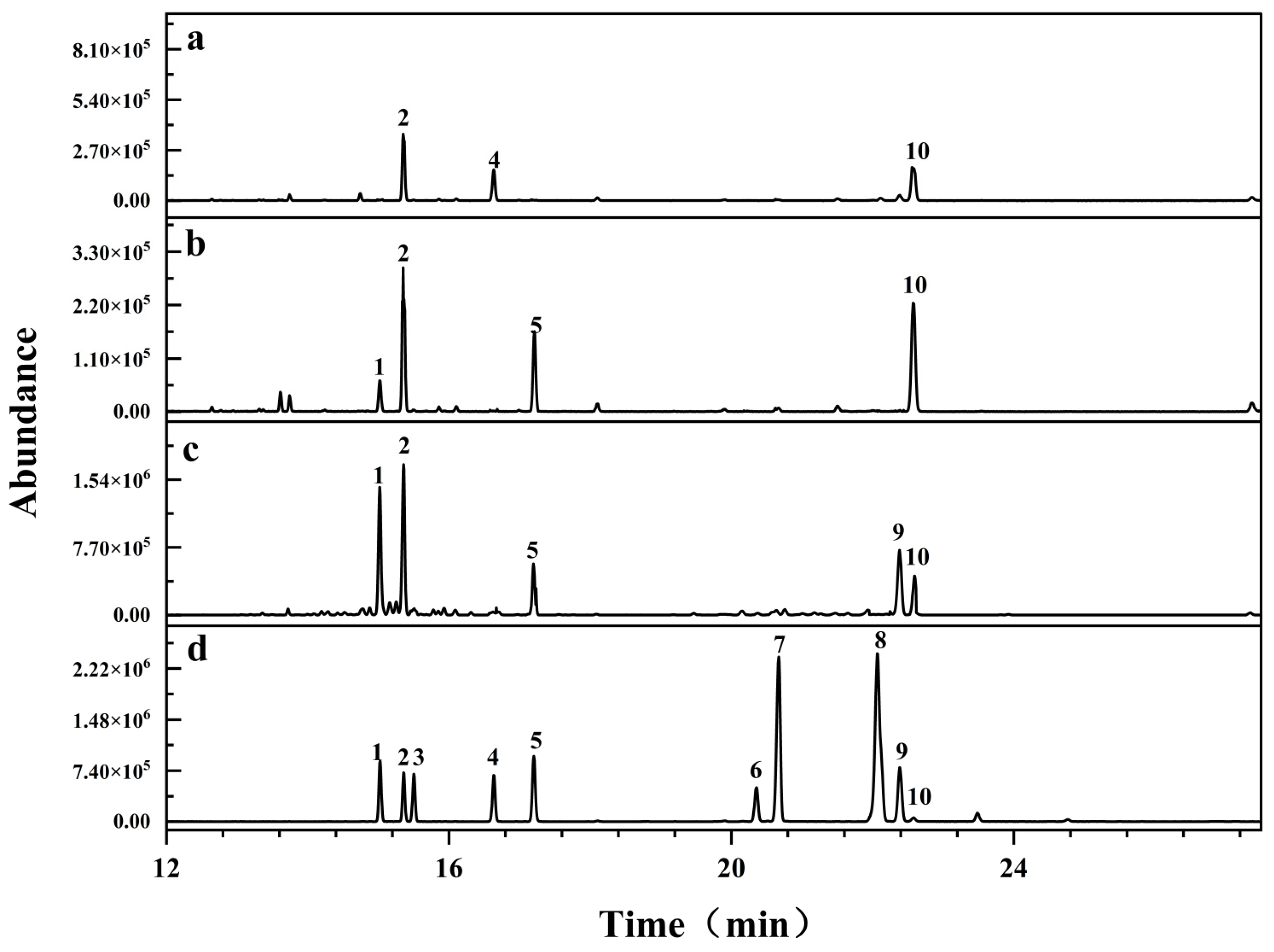
RIP-A1, RIP-B1 and RIP-B2 exhibited elution times of around 15.583, 12.535 and 12.038 min, respectively, in the HPGPC chromatogram. According to the calibration curve of standard dextran (log Mw = −0.2027x + 7.4201; R2 = 0.9908), the average molecule weights of RIP-A1, RIP-B1 and RIP-B2 were calculated as 1.8 × 10⁴ Da, 7.4 × 10⁴ Da and 9.3 × 10⁴ Da, respectively. Low Mw polysaccharides were found to have higher physiological activity among a variety of polysaccharide components. Polysaccharides with a lower Mw exhibited a higher tendency to traverse cell membranes and induce biological responses [42,43]. Hence, RIP-A1 might potentially be the most active one among them. In this study, the monosaccharide compositions of RIP-A1, RIP-B1 and RIP-B2 were examined via gas chromatography–mass spectrometry (GC–MS)-based trimethylsilyl-alditol (TMSA) derivatives [44]. As shown in Figure 2, it was found that RIP-A1 consisted of Ara, Rha and Gal in a molar proportion of 2.19:1.00:1.70. RIP-B1 consisted of Xyl, Ara, Fuc and Gal in a molar proportion of 1.00:5.00:2.83:5.58. RIP-B2 consisted of Xyl, Ara, Rib, Fuc, Glc and Gal in a molar proportion of 2.67:3.30:3.54:1.17:2.11:1.00. The monosaccharide contents of RIP-A1, RIP-B1 and RIP-B2 exhibited variations and none of them included GalA or GluA. Thus, it is possible that RIP-A1, RIP-B1 and RIP-B2 are neutral homopolysaccharides. Previous studies have reported that a neutral polysaccharide (RIWP) with a small Mw of 5.7 × 10⁴ Da, from Isatis indigotica Fort., was primarily made up of Glu, Gal and Ara [39]. Two fractions of pectin (WRIP-A-A and WRIP-A-B) and one neutral polysaccharide (WRIP-N) were obtained from the roots of Isatis indigotica Fort. Nevertheless, the compositions of WRIP-A-A and WRIP-A-B were elucidated, but WRIP-N remained unexplained [40]. Notably, a crude polysaccharide has been extracted from Isatis indigotica Fort., consisting of Gal, Glu, Fuc, Xyl, Ara, Man, GalA and GluA [45]. A relevant previous study suggests that RIP-A1, RIP-B1 and RIP-B2 are novel neutral polysaccharides from the roots of Isatis indigotica Fort. In addition, RIP-A1 exhibits the smallest Mw and has the simplest monosaccharide composition compared to RIP-B1 and RIP-B2.

2.3. Methylation Analysis

The glycosidic bonds and proportional molar ratios of RIP-A1, RIP-B1 and RIP-B2 were elucidated by methylation analysis (Table 1). The primary glycosidic linkages of RIP-A1 and RIP-B1 were identified as the 1,3,5-Araf and 1,6-Galp residues. This suggests that their glycan chains are linear and the branching point is at the O-3 position of the 1,3,5-Araf residue. In addition, small quantities of T-Arap and 1,5-Araf were found in RIP-A1, suggesting that T-Arap and 1,5-Araf are linked to arabinan side chains. T-Xylp and T-Arap were observed in RIP-B1, possibly as termini of arabinan side chains. However, RIP-B2 is mainly composed of 1,4-Glcp and 1,6-Galp residues, indicating a linear glycan chain. Moreover, small amounts of T-Xylp, T-Arap and 1,3,5-Araf were reflected in RIP-B2. These findings provide additional evidence that the structural characterization of RIP-A1, RIP-B1 and RIP-B2 shows variation during methylation analysis. Overall, the methylation results were consistent with the monosaccharide analyses. RIP-A1, RIP-B1 and RIP-B2 have similar glycosidic bond species. However, the specific arrangement of glycosidic bonds in RIP-A1 may be associated with the anti-inflammatory properties of neutral polysaccharides [46,47].

2.4. FT-IR Spectra

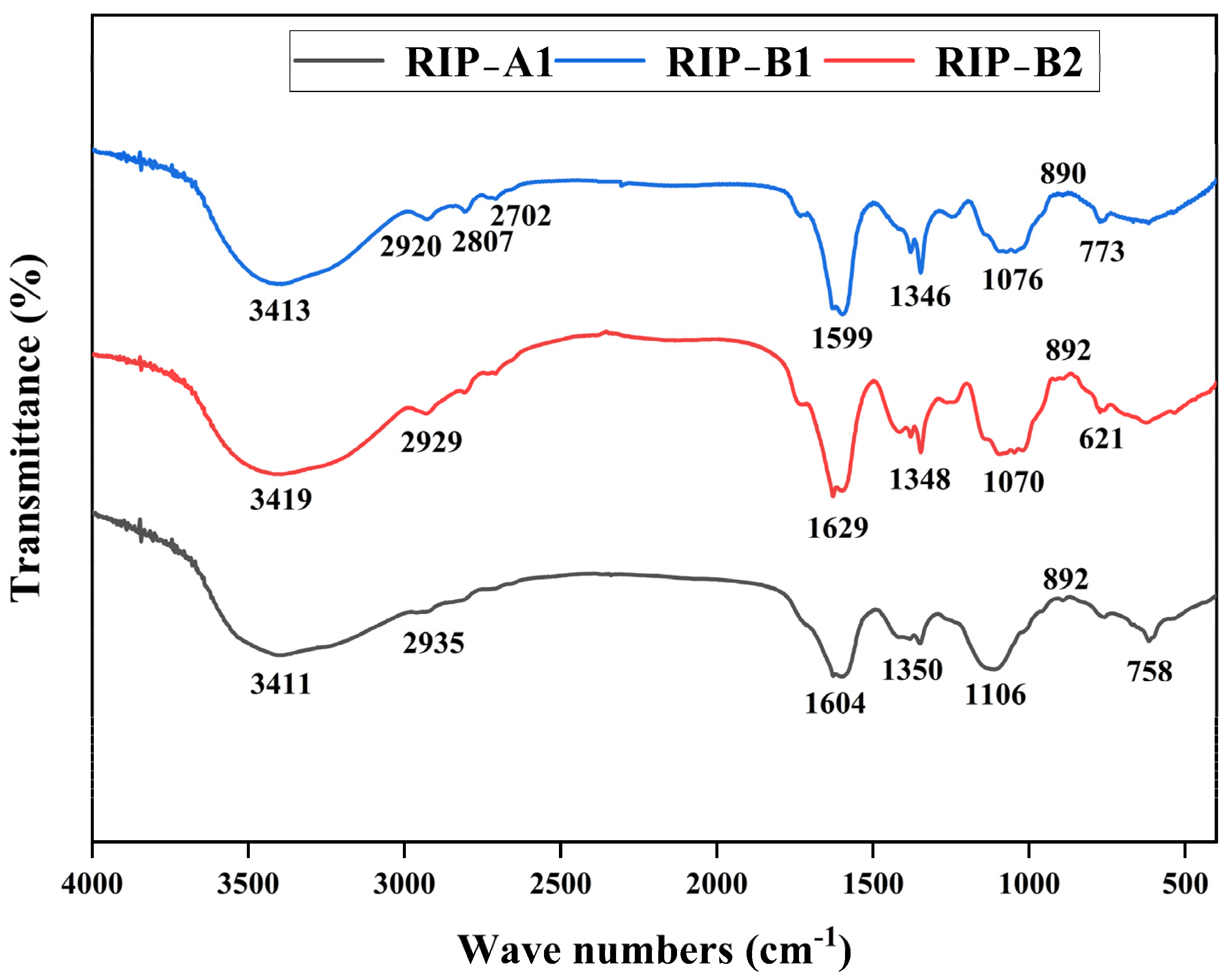
As shown in Figure 3, RIP-A1, RIP-B1 and RIP-B2 each possess a prominent and wide absorption peak that results from the -OH stretching vibration in the polysaccharide structure (3000~3500 cm−1) [48]. The resonance of the C-H bond in the methyl group within the sugar ring (2800~3000 cm−1) produces the signal shown in the spectrum [49]. The absorption peaks are located at 1000~1200 cm−1, and they were found to originate from the stretching vibration of the C-O-H pendant group, the C-O-C glycosidic band vibration of the pyranose ring and the stretching vibration of the asymmetric ring, respectively [50]. The presence of these absorption peaks indicates that polysaccharides are present. The results suggest that the absorption peaks of RIP-A1, RIP-B1 and RIP-B2 exhibit high similarity. The carboxyl group of these polysaccharides does not exhibit C=O stretching, so they should be neutral polysaccharides [46]. In general, the results obtained from the FT-IR spectra are in agreement with the monosaccharide analysis.

2.5. NMR Analysis of RIP-A1

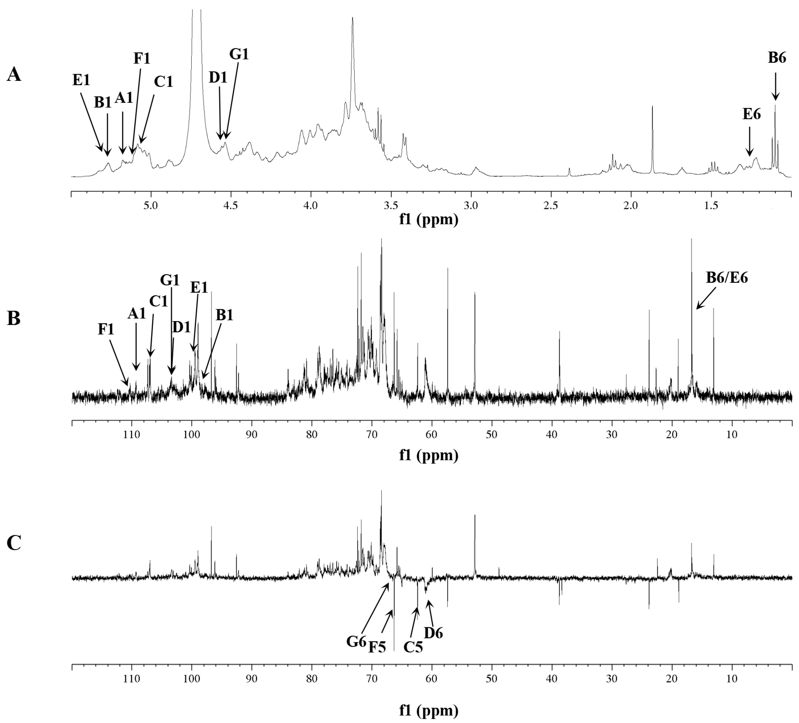
RIP-A1 was subjected to further structural elucidation by 1H, 13C and DEPT-135 in conjunction with 2D NMR spectroscopy. The hydrogen and carbon signals are shown in Table 2. Figure 4A displays the 1H spectrum, which reveals the presence of anomeric protons at δ 5.16, 5.26, 5.07, 4.56, 5.01, 5.27 and 4.55. These protons indicate the existence of seven potential glycosidic residues (A–G) in RIP-A1. Combining the results of methylation analysis and other relevant data, T-Araf (A), 1,2-Rhap (B), 1,5-Araf (C), T-Galp (D), 1,2,4-Rhap (E), 1,3,5-Araf (F) and 1,6-Galp (G) were tentatively confirmed [46,51,52]. Anomeric signals with δ > 5.0 ppm were generally recognized as α-configurations, whereas those with δ < 5.0 ppm were recognized as β-configurations [53]. The 1H spectrum revealed that A (δ 5.16), B (δ 5.26), C (δ 5.07), E-F (δ 5.01) and F (δ 5.27) were in α-configurations, but D (δ 4.56) and G (δ 4.55) were in β-configurations based on the anomeric signals.
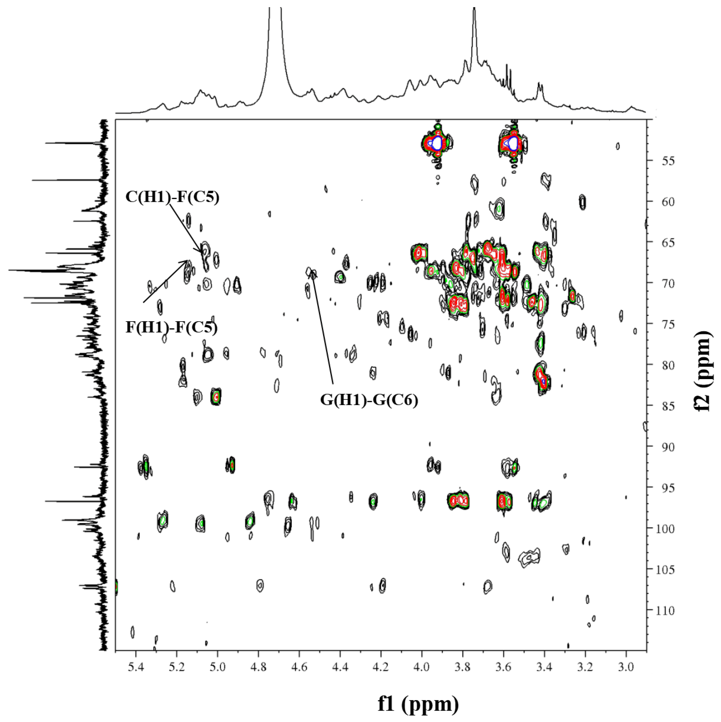
The anomeric signals (δH/C) at 5.16/109.4, 5.26/98.4, 5.07/106.9, 4.56/103.4, 5.27/99.7, 5.13/110.5 and 4.55/103.5 corresponded to T-α-Araf-(1→(A), →2)-α-Rhap-(1→(B), →5)-α-Araf-(1→(C), T-β-Galp-(1→(D), →2,4)-α-Rhap-(1→(E), →3,5)-α-Araf-(1→(F) and →6)-β-Galp-(1→(G) through the 1H, 13C (Figure 4A,B) and HSQC (Figure 5) spectra. In addition, the DEPT-135 spectrum (Figure 4C) was used for the assignment of some specific carbon signals of residues A, C, D, F and G. The inverted signals at δC 64.4, 65.1 and 66.5 corresponded to the C-5 of residues A, C and F. Then, the inverted signals at δC 61.0 and 66.4 were found to correlate with the C6 of residues D and G, respectively.
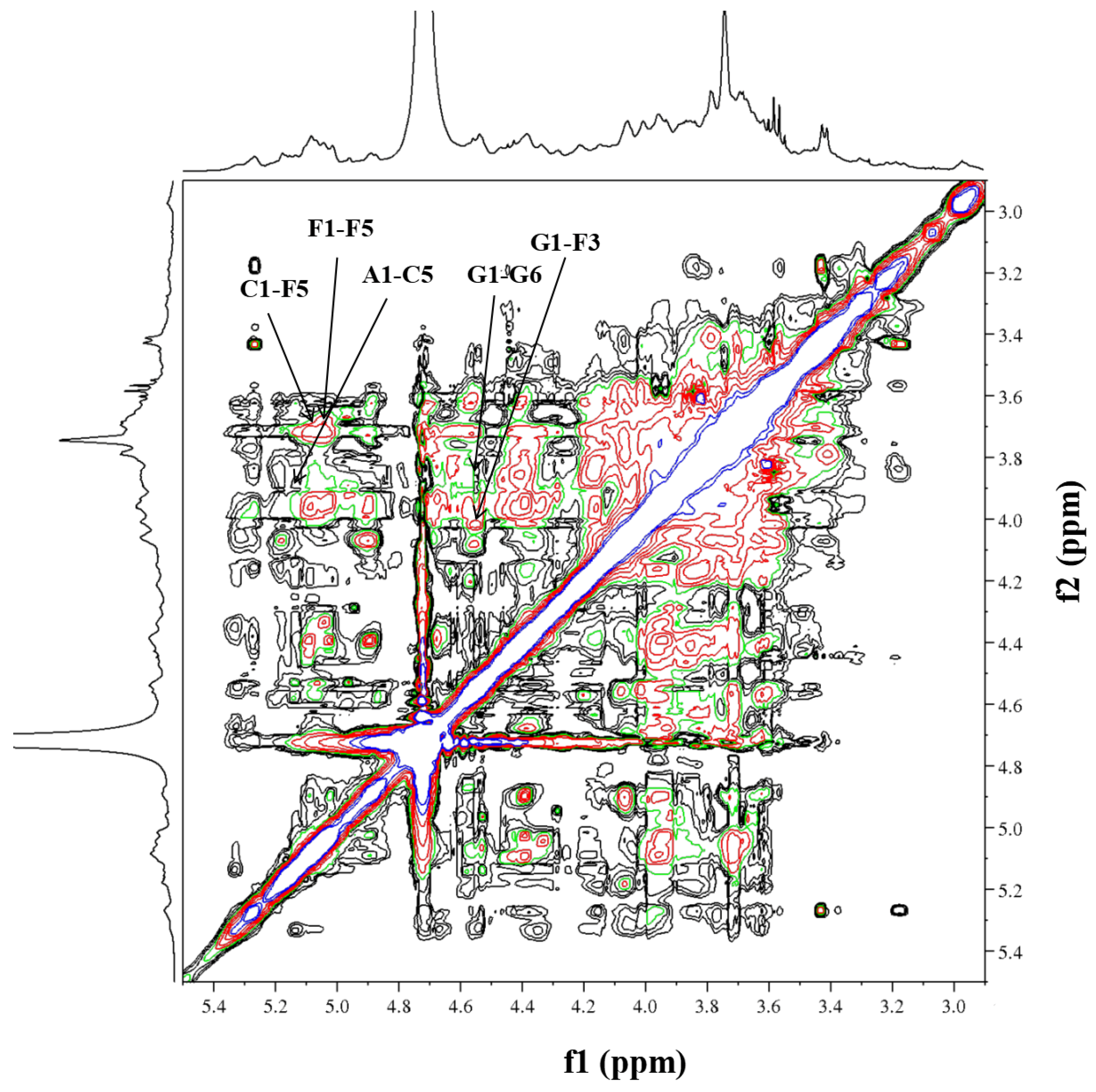
Initially, residue A exhibited an anomeric proton signal at δH 5.16. Further analysis of the 1H-1H COSY data (Figure 6) showed that the positions of its H2–H5 signals were at δH 4.15, 3.65, 3.75 and 3.66. Afterwards, the cross-peaks of C2–C6 were identified at δH 81.3, 80.4, 75.8 and 64.4 in the HSQC spectrum. The residue C displayed an anomeric proton signal at δH 5.07, and the chemical shift positions of H2–H5 at δH 4.53, 3.92, 4.24 and 3.86 were assigned by cross-peaks in the 1H-1H COSY spectrum. The 13C NMR chemical shifts of C2–C5 at δC 78.1, 76.7, 78.0 and 65.1 were assigned in the HSQC spectrum. The residue F, which had an anomeric proton signal at δH 5.13, showed cross-peaks between H2–H5 at δH 4.05, 3.99, 3.83 and 3.70 in the 1H-1H COSY spectrum. Next, the information on the main chain could be obtained from the HMBC (δH/C) (Figure 7) and NOESY (δH/H) (Figure 8) spectra. The correlated peaks A (H1)/C (C5) at δH/C 5.16/65.1, C (H1)/F (C5) at δH/C 5.07/66.5 and F (H1)/F (C5) at δH/C 5.13/66.5 indicate that a linear main chain is present as T-α-Araf-(1→5)-α-Araf-(1→5)-α-Araf-(1→ and the branching point is arabinosyl at C-3, which concurs with the methylation analysis. Prior studies revealed comparable structural components in arabinan from Ephedra sinica, Ligusticum chuanxiong Hort. and Rehmannia glutinosa Libosch. However, differences were observed at the sites of the branching points [54,55,56].
Regarding residue D, the anomeric proton (δH 4.56) may be attributed to the H-1 of Galp, which is related to the terminal position. According to the 1H-1H COSY spectrum, δH 3.63, 3.81, 4.11, 3.80, 3.67 and 3.78 were the chemical shift positions for H2 to H6. The 13C chemical shift locations were attributable to C-2 (δC 72.7), C-3 (δC 73.0), C-4 (δC 74.3), C-5 (δC 75.7) and C-6 (δC 61.0) in the HSQC spectrum. Moreover, the same data analysis method was used for the 1H and 13C of residue G, wherein the 1H and 13C NMR chemical shifts were δH-C 4.55/103.5, 3.63/72.9, 3.81/72.0, 4.16/66.7, 3.80/75.7 and 3.85, 3.78/68.4, respectively. Additionally, the chain of galactosan could be readily deduced by HMBC (δH/C) and NOESY (δH/H) correlations, e.g., D(H1)/G(C6) at δH/C 4.56/68.4, G(H1)/G(C6) at δH/C 4.55/68.4, D(H1)/G(H6) at δH/H 4.56/3.78 and D(H1)/G(H6) at δH/H 4.55/3.85. Thus, the inter-residual sequences of galactan were conjectured to be the occurrence of T-β-Galp-(1→6)-β-Galp-(1→6)-β-Galp-(1→.
It is noteworthy that some cross-peaks were investigated between G (H1) and F (H3) at δH/H 4.55/3.99, in the NOESY spectrum, suggesting that the galactan chains might be directly linked to the C-3 position of residue F. Previous research on traditional Chinese medicine has identified comparable structural fragments of neutral polysaccharides [57]. However, the presence of cross-peaks between residues B and E could not be verified due to their limited and unclear appearances in the HMBC and NOESY spectra. This outcome conforms with the methylation analysis. According to the aforementioned data, the primary composition of RIP-A1 consists of chains made up of arabinan (1,3,5-α-Araf) and galactan (1,6-β-Galp), with branching occurring at the C-3 position of the arabinosyl unit.
Previous studies have revealed comparable structures in arabinogalactans obtained from plant polysaccharides, including HH1-1 (Carthamus tinctorius L.), LAG-W (Larix kaempferi) and LBGP70-OL (Lycium barbarum). For instance, the backbone of HH1-1 was found to be composed primarily of 1,6-linked Galp branches at C-3. These branches predominantly contain 1,5-linked and 1,3,5-linked terminal arabinose and terminal galactose, as shown by the methylation and NMR analyses [58]. The arabinogalactan LAG-W was shown to possess a homogenous structure with a backbone composed of repetitive →3)-β-Galp-(1→ residues. Additionally, it contains minor quantities of T-α-Araf-(1→3)-α-Araf-(1→6)-β-Galp-(1→ residues [59]. The structural features of arabinogalactan from Lycium barbarum revealed that the backbone consists solely of →6)-β-Galp-(1→ residues, which are replaced at the C3 position. The side chains, on the other hand, contain α-Araf-(1→3)-β-Araf-(1→3)-β-Araf-(1→. However, several pectin structures were previously identified in the roots of Isatis indigotica Fort. This study primarily elucidates the structural characterization of arabinogalactan.

2.6. SEM and AFM Analysis

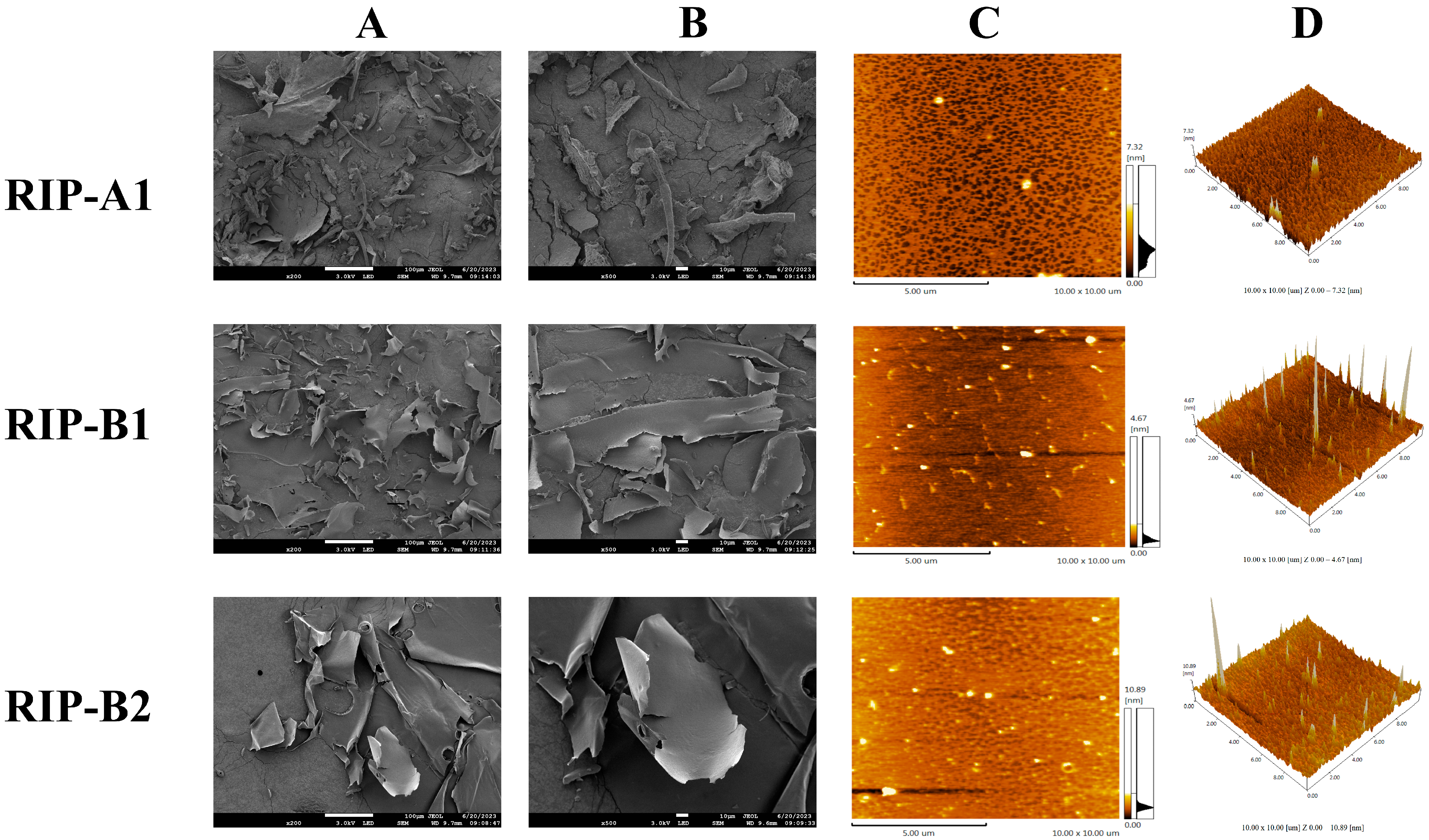
RIP-A1, RIP-B1 and RIP-B2’s microstructures were investigated using a scanning electron microscope (SEM). The SEM images obtained with the three polysaccharides, of magnification (200- and 500-fold), are shown in Figure 9A,B. RIP-A1 exhibits a rough, wrinkled and uneven surface, with small voids and a honeycomb shape. RIP-B1 has a loosely arranged large lamellar structure, with slight folds on the surface and relatively flat edges. RIP-B2 has a smooth surface and flat edges, with a lamellar shape. Furthermore, the varying outcomes of RIP-A1, RIP-B1 and RIP-B2 may be linked to their differences in monosaccharide composition and strong intermolecular interactions [57,60,61].
Atomic force microscopy (AFM) has gained popularity recently as a tool for studying the chain structures of polysaccharide macromolecules. According to the data shown in Figure 9C,D, AFM analysis—conducted on a 2D floor plan—indicates that RIP-A1 displays a uniformly even polymer particle structure. RIP-B1 exhibits chain and irregular spherical conformations. RIP-B2 reveals a uniformly sized mesh conformation. The formation of polymer particles and sugar chains can be observed, which suggests that entangled states may exist in RIP-A1, RIP-B1 and RIP-B2 molecules [50,62]. Upon the examination of a 3D floor plan, it was seen that the surface of the polysaccharide is characterized by sharp peaks and a loose distribution. The molecular heights of RIP-A1 (2.08 nm), RIP-B1 (10.83 nm) and RIP-B2 (11.63 nm) were revealed by AFM measurements, while single-chain polysaccharides typically present heights of 0.1 to 1.0 nm. The intermolecular interactions, such as Van der Waals forces, acting between the individual polysaccharide chains might have led to their entanglement and formation of polymers. This entanglement may have caused the measured value to be larger than the actual value [63]. This might account for the discrepancy. The findings of RIP-A1, RIP-B1 and RIP-B2 exhibit variability, which may be related to differences in molecular weights. Previous research on the SEM and AFM of polysaccharides from the roots of Isatis indigotica Fort. is limited. In this study, the variations observed in the results of the three polysaccharides, when subjected to the same freeze-drying conditions, could potentially impact the isolation and purification process, as well as the polysaccharides themselves.

2.7. Anti-Inflammatory Activity Based on Raw 264.7 Cell Model

2.7.1. Determination of Cytotoxicity

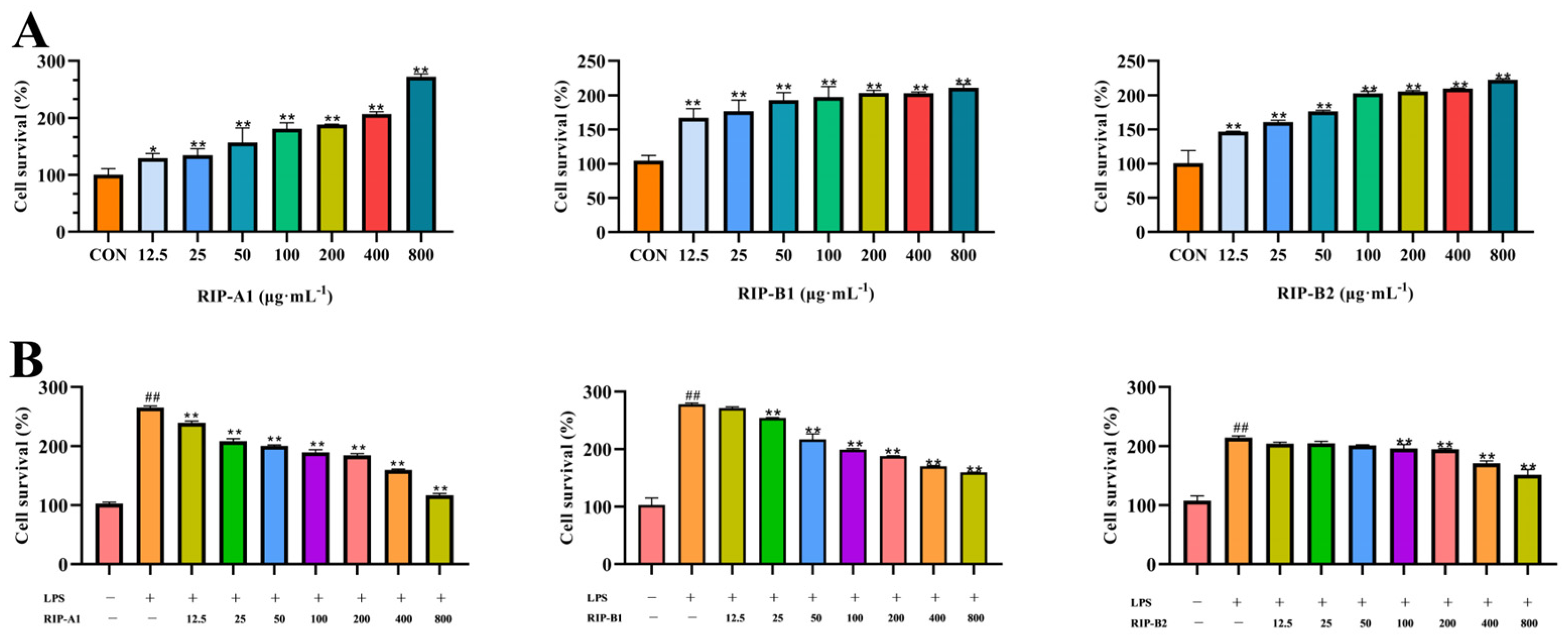
The influence of polysaccharides on the proliferation of RAW 264.7 cells was assessed by observing alterations in cell viability. According to Figure 10A, when cells were incubated for 24 h, RIP-A1, RIP-B1 and RIP-B2 did not induce cytotoxicity in RAW 264.7 cells. However, the cell viability was significantly increased (p < 0.05) in the concentration range of 12.5–800 μg·mL−1 compared to the control. This is because the presence of polysaccharides can induce the activation of protein kinase, which is important for cell proliferation, specifically in response to mitogens [64]. These data indicate that RIP-A1, RIP-B1 and RIP-B2 treatment does not cause cytotoxicity in RAW 264.7 cells.

2.7.2. Screening of Anti-Inflammatory Activity of RIP-A1, RIP-B1 and RIP-B2

Effects of RIP-A1, RIP-B1 and RIP-B2 on RAW 264.7 Cell Activity Stimulated by LPS

LPS is an established immunomodulator that participates in several inflammatory reactions by causing the excessive production of specific inflammatory cytokines [65]. Initially, LPS was utilized to stimulate the cells for 24 h. After that, different concentrations of RIP-A1, RIP-B1 and RIP-B2 (50, 100, 200, 400 and 800 μg·mL−1) were introduced to the cells for another 24 h. The decreasing cell viability exhibited a positive correlation with the increasing concentrations of RIP-A1, RIP-B1 and RIP-B2 compared to the control. Figure 10B demonstrates that RIP-A1, RIP-B1 and RIP-B2 (at concentrations of 100, 200, 400 and 800 μg·mL−1) effectively inhibited the proliferation of LPS-activated RAW 264.7 cells. This inhibition was statistically significant (p < 0.01) and exhibited a dose-dependent relationship. These observations align with the overall trend observed in previously documented polysaccharides from the roots of Isatis indigotica Fort., which have demonstrated anti-inflammatory properties [39].

IL-4, IL- 1β, NO, TNF-α and IL-6 Determination

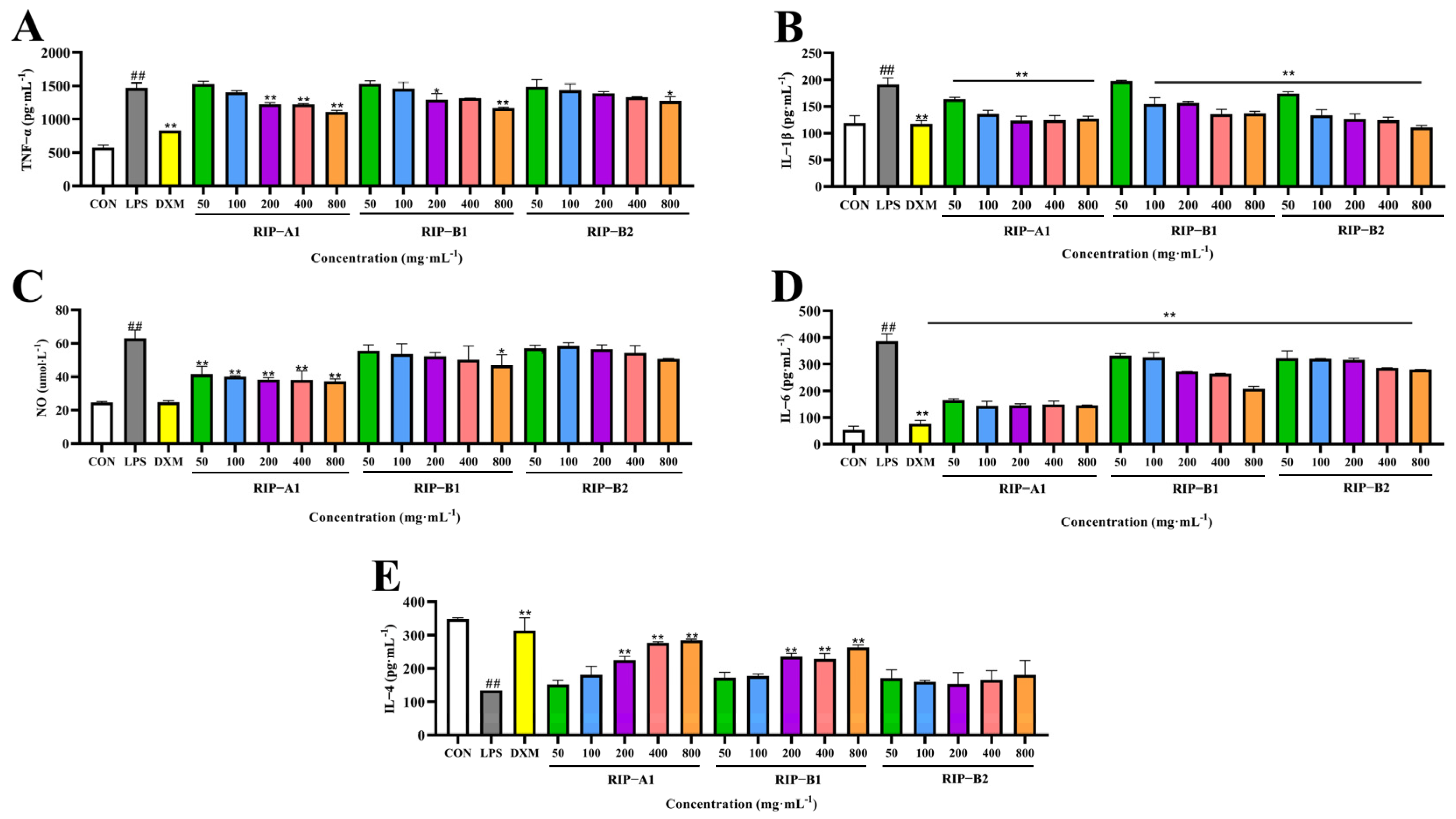
Immune and non-immune cells co-secrete intercellular signaling proteins known as cytokines, which regulate the body’s inflammatory response and immunity [46]. In contrast to the control group, the LPS group exhibited a significant increase in the IL-1β, IL-6, TNF-α and NO levels, accompanied by a significant decline in IL-4 levels (for all groups, p < 0.01), demonstrating that the LPS activation of the cells was successful in inducing an inflammatory response. As shown in Figure 11, the results reveal that RIP-A1, RIP-B1 and RIP-B2 suppressed the synthesis of IL-1β and IL-6 in a dose-dependent manner (range of 50 to 800 μg·mL−1). However, except for RIP-B2, all of them had a dose-dependent effect on the generation of IL-4. The NO content assay showed a significant decrease following treatment with RIP-A1 (p < 0.01) and RIP-B2 dosages of 400 and 800 ug·mL−1. Significant decreases in the TNF-α levels were observed at RIP-A1 doses of 200, 400 and 800 ug·mL−1 (p < 0.01). This research demonstrates that the molecular weights, mono-saccharide compositions, glycosidic linkages and conformations all impact the binding of receptors and the recognition of polysaccharide sites in inflammatory cells [66,67]. Polysaccharides with lower Mw are more likely to cross cell membranes and have biological effects [43]. Thus, RIP-A1, RIP-B1 and RIP-B2 may exhibit anti-inflammatory properties, varying based on their structural dissimilarities. The results demonstrate that RIP-A1 has the highest efficacy in terms of anti-inflammatory activities.

2.7.3. Effect of RIP-A1 on RAW 264.7 Cells’ mRNA Expression of IL-1β, IL-6 and TNF-α Caused by LPS

Cytokine synthesis is closely associated with mRNA expression and plays a vital role in intercellular communication in vivo. It is also essential for anti-inflammatory responses [46]. To investigate RIP-A1’s anti-inflammatory mechanism in RAW 264.7 cells, quantitative reverse-transcription PCR (qRT-PCR) was used to quantify the mRNA expression levels for IL-1β, IL-6 and TNF-α. As demonstrated in Figure 12, RIP-A1 exhibited dose-dependent inhibition of IL-1β, IL-6 and TNF-α mRNA expression. The LPS control group exhibited significantly elevated mRNA expression levels of IL-1β, IL-6 and TNF-α compared to the control group (p < 0.01), indicating the successful establishment of the inflammation model. In both DXM and RAW 264.7 cells, RIP-A1 reduced the mRNA levels of IL-6, IL-1β and TNF-α (p < 0.01) compared to the LPS control group. According to these results, LPS-induced RAW 264.7 cells’ expressions and secretions of IL-1β, IL-6 and TNF-α may be inhibited by RIP-A1.

2.7.4. Effect of RIP-A1 on LPS-induced NF-κB Signaling Pathway in RAW 264.7 Cells

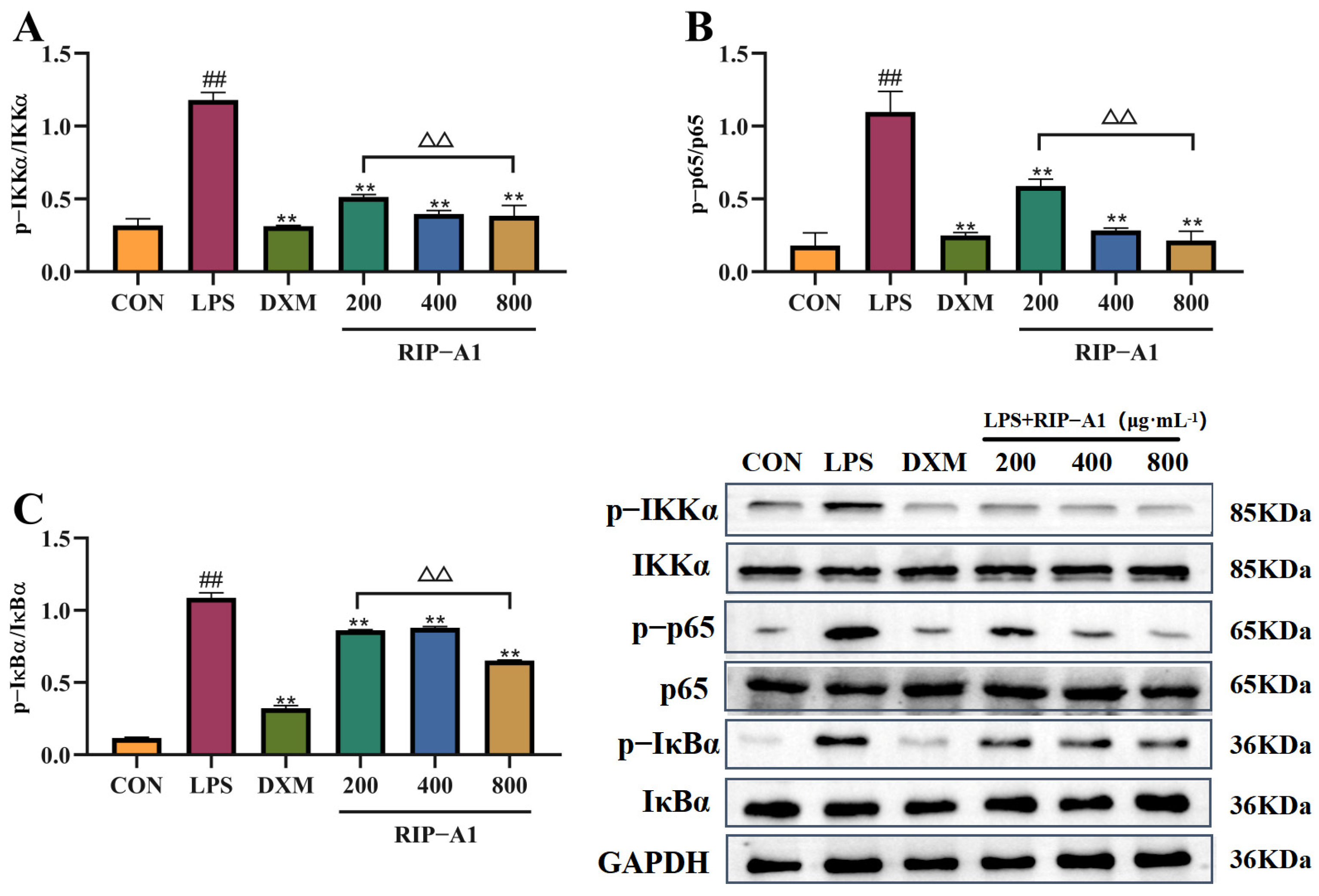
Evidence has shown that LPS generated by Gram-negative (G) bacteria can reach the bloodstream via penetrating compromised mucosal barriers, potentially activating the NF-κB signaling pathway [68]. Phosphorylating IKKα, p65 and IκBα may activate transcription factors through the activation of the NF-κB signaling pathway. Figure 13 illustrates the expression of the NF-kB signaling pathway, showing increased phosphorylation levels of IKKα, p65 and IκBα in response to LPS compared to the control group. The RIP-A1 treatment decreased the phosphorylation of IKKα, p65 and IκBα in a concentration-dependent manner. According to these results, the inhibition of phosphorylation by the receptor proteins mentioned earlier prevents the activation of the NF-κB signaling pathway, as well as the production of cytokines and NO. As shown in Figure 14, this may explain the anti-inflammatory properties of RIP-A1 on RAW 264.7 cells stimulated by LPS.
Previous studies have identified polysaccharides with comparable structures derived from several plants, many of which have anti-inflammatory properties. Examples include Lycium barbarum polysaccharide (LBP) and LAG-W (Larix kaempferi). For instance, the presence of large amounts of arabinose and galactose has a significant (p < 0.001) positive impact on the anti-inflammatory properties of the Lycium barbarum polysaccharide fraction (LBGP-I-3). This is achieved by increasing NO levels, enhancing phagocytosis and promoting acid phosphatase activity in RAW 264.7 cells. Additionally, the improved phagocytosis effect of LBGP-I-3 may be attributed to its ability to stimulate TLR4 receptors on the surfaces of macrophages. The addition of LAG-W significantly enhances the capacity of macrophages to engulf foreign particles while also triggering the release of NO and the cytokines TNF-α, IL-1β and IL-6. To summarize, the structural properties of arabinogalactan are associated with its significant anti-inflammatory effect [24,59,69].

3. Materials and Methods

3.1. Materials and Reagents

Radix isatidis plants were cultivated in Bajingzi Township, Datong District, Daqing City, Heilongjiang Province. D-Xylose (XYl), L-Rhamnose (Rha), L-Fucose (Fuc), D-Galactose (Gal), D-Ribose (Rib) and D-Arabinose (Ara) were acquired from Biotopped (Beijing, China). D-Mannose (Man) and D-Glucose (Glc) were acquired from Shanghai Yuanye Bio-Technology Co., Ltd. (Shanghai, China). Hexamethyl disilyamine (HMDS) and Chlorotrimethylsilane (TMCS) were purchased from Aladdin (Shanghai, China). ELISA kits were procured from Jiangsu Meimian Industrial Co., Ltd. (Jiangsu, China). Antibodies against p65, p-p65, IκBα, p-IκBα, IKKα and p-IKKα were obtained from ABclonal Technology Co., Ltd. (Wuhan, China). A BeyoRT™ III First Strand cDNA Synthesis Kit was procured from Beyotim (Shanghai, China). Unless otherwise noted, all chemicals and reagents were analytical grade. Distilled water was used in all experiments.

3.2. Exaction and Isolation of Polysaccharides

The Radix isatidis were ground and defatted with 70% ethanol. Following that, deionized water was used to extract the residues. The water-based extracts were collected, filtrated, concentrated, precipitated, centrifugated and lyophilized. After being deproteinized using the Sevag-enzymatic process, the crude polysaccharides were given the designation RIP (n = 3) [70,71]. According to previous methods, the RIP (300 g) was dissolved in 5 L of dH2O and isolated by 20 KDa and 150 KDa ultrafiltration membranes (BONA-GM-20, BONA GROUP, Jinan, China) in sequence, and was designated as RIP-A–C. The DEAE-52 cellulose column (5 cm × 60 cm) was used to purify RIP-A1 in the amount of 1 mL·min−1 using a gradient elution NaCl solution (0–0.5 M) [47]. RIP-B was dissolved in dH2O and purified using a Sephadex G-100 column (2.6 cm × 100 cm), eluting with 0.1 M NaCl in the amount of 0.6 mL·min−1 [72]. Then these main fractions were concentrated and lyophilized to get purified polysaccharides (RIP-A1, RIP-B1 and RIP-B2). The content of sugar was measured using the phenol-sulfuric acid method [73].

3.3. Structural Analysis of Polysaccharides

3.3.1. Molecular Weight Determination

The Mw determination was performed using a modified version of a previously reported protocol. The Mw of RIP was determined using a HPGPC system fitted with a column of UltrahydrogelTM Linear (7.8 mm × 300 mm, Waters, Milford, MA, USA) on an HPLC-evaporative light scattering detector (ELSD, Agilent 1260II HPLC, c.01.08.210, Agilent Technologies, Santa Clara, CA, USA). Distilled water was used as the mobile phase, with an injection volume of 10 μL and a flow rate of 0.8 mL·min−1. Standard molecular markers, T-series dextran (Mw = 270, 80, 50, 20 and 15 kDa), were employed (n = 3).

3.3.2. Monosaccharide Composition Analysis

The monosaccharide compositions of polysaccharides were analyzed using methods based on previously reported work [44]. The mixed monosaccharide standards were added with NH4OH and kept at room temperature for 15 min. The procedure was completed by adding NaBH4, followed by securing the reaction vessel with a screw cover and letting the residue in the vial stand for 6 h at 25 °C. After that, the residue was dried by evaporating the NaBH4 and neutralized with acetic acid. The 5 mg of polysaccharides was dissolved in 2 M TFA and allowed to hydrolyze for 4 h at 110 °C in glass vials with caps. Following this, the hydrolysates were washed with methanol and repeatedly evaporated until completely dry to eliminate any remaining TFA. The hydrolyzed production and mixed monosaccharide standards were subjected to the same processes. The final solution was gently stirred and placed in a water bath at 70 °C for 30 min. Before GC–MS analysis, the solution was filtered via a 0.22 μm membrane. The following temperature conditions were applied to the TMSA derivatization process: 0 min at 180 °C, 4 °C·min−1 at 180–190 °C, 1 °C·min−1 at 190–200 °C, 2 °C·min−1 at 200–230 °C and 230–300 °C, followed by 5 min of holding.

3.3.3. Infrared Spectra (IR) Analysis

To prepare the samples for infrared spectrum analysis, the freeze-dried RIP-A1, RIP-B1 and RIP-B2 were crushed with dry KBr powder and pressed into pellets. The FT-IR spectrum of polysaccharides was acquired by a Fourier-transform infrared spectrograph (VERTEX-70, FTR 5.4.9, Bruker, Rheinstetten, Germany) at frequencies between 500 and 4000 cm−1 to obtain structural data.

3.3.4. Methylation Analysis

According to the earlier approach, the freeze-dried RIP-A1, RIP-B1 and RIP-B2 (2 mg) were completely methylated and were hydrolyzed, reduced, acetylated and analyzed using a GC–MS system (Agilent 7890A-5975C, 10.0, Agilent Technologies, Santa Clara, CA, USA) that was fitted with a DB-5 silica capillary column (60 m × 0.25 mm × 0.25 μm) [74]. The following temperature conditions were applied: 80 °C for 1 min, 80–200 °C at 5 °C·min−1, 200–220 °C at 2 °C·min−1 and 220–270 °C at 10 °C·min−1 and a 5 min hold time.

3.3.5. NMR Analysis

The RIP-A1 was dissolved in D2O for NMR analysis. 1D/2D-NMR spectra were registered on an NMR spectrometer (Bruker AM-400, 11.1, Bruker, Fällanden, Switzerland).

3.3.6. SEM and AFM Analysis

The surface characterization of RIP-A1, RIP-B1 and RIP-B2 was performed by SEM (JSM-7800F, B17.0.07, JEOL Ltd., Tokyo, Japan). Dried RIP-A1, RIP-B1 and RIP-B2 were secured with electrical tapes and coated using a vacuum spraying device. An acceleration voltage was applied while the constructed sample was operated under a vacuum.
Following the dissolution of RIP-A1, RIP-B1 and RIP-B2 in distilled water with a concentration of 5 µg·mL−1, the resulting mixture was passed through a cellulose membrane (0.22 μm). After applying the sample solution over a freshly cleaved mica, drying was performed at room temperature. The AFM analysis was conducted using SPM-9700 apparatus (SPM-9700, 2.61, Shimadzu, Kyoto, Japan). Working conditions were set to a scanning range of 1.000 × 1.000 μm with a scanning frequency of 1.00 Hz and tapping mode was selected.

3.4. Anti-Inflammatory Activity on RAW 264.7 Cells

3.4.1. Determination of Cytotoxicity

Briefly, 105 RAW 264.7 cells were cultivated into each well of a 96-well plate and incubated [38]. The cells were allowed to adhere for 24 h. After that, the cells were treated with different concentrations of RIP-A1, RIP-B1 and RIP-B2 (formulated as 12.5, 25, 50, 100, 200, 400 and 800 μg·mL−1) for 24 h. After adding 10% CCK-8 solution, the cells were incubated for 2 h. Using a microplate reader, the cell viability was determined at 450 nm (SpectraMax ABS Plus, 7.1.0, Molecular Devices, San Jose, CA, USA).

3.4.2. Screening of Anti-Inflammatory Activity of RIP-A1, RIP-B1 and RIP-B2

Effects of RIP-A1, RIP-B1 and RIP-B2 on LPS-Induced Activity of RAW 264.7 Cells

RAW 264.7 cells were cultured and treated with LPS (1 μg·mL−1) for 24 h to induce inflammation [39,75]. The cells were cultured with various doses of RIP-A1, RIP-B1 and RIP-B2 (25, 50, 100, 200, 400 and 800 μg·mL−1). The LPS group consisted of cells only treated with LPS, while the control group consisted of RAW 264.7 cells with complete medium only. A microplate reader (SpectraMax ABS Plus, 7.1.0, Molecular Devices, San Jose, CA, USA) was then used to measure the absorbance at 450 nm.

Effects of RIP-A1, RIP-B1 and RIP-B2 on LPS-induced IL-4, IL-1β, NO, TNF-α and IL-6 Production in RAW 264.7 Cells

The experiment was set up with a blank control group, an LPS control group, a positive control group (10 μg·mL−1 of dexamethasone, DXM) and the polysaccharide group (final concentrations of 50, 100, 200, 400 and 800 μg·mL−1). The 48-well plates containing RAW 264.7 cells (105 cells/well) were incubated for 24 h at 37 °C in a humidified incubator with 5% CO2. Cells were treated with LPS (except for the normal group), then the supernatant was collected and IL-1β, IL-6, TNF-α, NO and IL-4 levels were assessed using an ELISA kit according to the provided instructions [61].

3.4.3. qRT-PCR Analysis

The extraction of total RNA from RAW 264.7 cells, including differentiated cells with and without RIP-A1, was performed using TRIzol reagent following the directions provided by the manufacturer. A cDNA reverse transcription kit was used to get cDNA from 1 μg RNA from each sample. The mRNA expression was measured by qPCR under the following reaction conditions: 40 cycles of PCR amplification, pre-denaturation at 95 °C for 2 min, denaturation at 95 °C for 15 s and annealing and extension at 60 °C for 30 s. By normalizing the Ct data to GAPDH expression, the comparative CT (2−ΔΔCt) approach was used to assess the relative gene expression. Table 3 contains a list of the primer set sequences employed for amplification [76].

3.4.4. Western Blotting Analysis

The Western blotting technique was employed, following the methods used in a previously reported experiment with minor modifications [38]. Radioimmunoprecipitation assay (RIPA) buffer was utilized to extract the whole protein content from RAW 264.7 cells. Subsequently, a bicinchoninic acid (BCA) kit was employed to measure the protein concentrations in these lysates. After separating proteins using sodium dodecyl-sulfate polyacrylamide gel electrophoresis (SDS-PAGE), the proteins were transferred onto polyvinylidene fluoride (PVDF) membranes and incubated overnight with antibodies specific to p65, p-p65, IκBα, p-IκBα, IKKα and p-IKKα at 4 °C. The membranes were then washed with tris-buffered saline with Tween-20 (TBST). An ODYSSEY CLx Gel Imaging System (Tanon-4200, 2.6.0.0, Tanon, Shanghai, China) was used to image the resultant blots.

3.5. Data Analysis

Multiple comparison tests and one-way analysis of variance (ANOVA) were used to evaluate the data. The data were presented as the means ± standard errors of the means ( x ¯ ± s). Differences were considered significant when p < 0.05 and highly significant when p < 0.01. GraphPad Prism 8.0 (GraphPad Software, Boston, MA, USA) and Origin 2022 were used for graphic data processing.

4. Conclusions

To summarize, this study extracted and isolated three novel polysaccharides (RIP-A1, RIP-B1 and RIP-B2) from the roots of Isatis indigotica Fort. The primary structures of RIP-A1, RIP-B1 and RIP-B2 were identified by chemical and spectral methods, indicating that they are neutral polysaccharides with different Mw and monosaccharide compositions. In addition, RIP-A1 (Mw = 1.8 × 10⁴ Da) was determined to be a smaller Mw arabinogalactan with glycosidic linkages of T-α-Araf, 1,2-α-Rhap, 1,5-α-Araf, T-β-Galp, 1,2,4-α-Rhap, 1,3,5-α-Araf and 1,6-β-Galp. However, RIP-B1 (Mw = 7.4 × 10⁴ Da) and RIP-B2 (Mw = 9.3 × 10⁴ Da) are more complex neutral polysaccharides consisting of Xyl or Glc. The structural features and differences between RIP-A1, RIP-B1 and RIP-B2 could affect their anti-inflammation activity. The anti-inflammation activities of RIP-A1, RIP-B1 and RIP-B2 in RAW 264.7 cells were evaluated. In contrast to RIP-B1 and RIP-B2, RIP-A1 exhibited the best anti-inflammatory activity in vitro. The results indicate that RIP-A1 has a considerable inhibitory effect on the production of pro-inflammatory cytokines within specific concentration ranges. It also reduces the mRNA levels of IL-1β, IL-6 and TNF-α and downregulates the expressions of p-IKKα, p-p65 and p-IκBα. Therefore, it may be speculated that RIP-A1 demonstrates an anti-inflammatory impact by stimulating the NF-κB signaling pathway. In future investigations, the focus should be on investigating the connection between RIP-A1 and cellular receptors. This work presents a rigorous theoretical basis for the structure–activity relationship of RIP-A1 and reveals its potential as a natural anti-inflammatory compound with therapeutic properties.

Supplementary Materials

The following supporting information can be downloaded at: https://www.mdpi.com/article/10.3390/molecules29112683/s1, Figure S1. Elution profiles. (A) Elution curves of RIP-A1-5 components. (B) Elution curves of RIP-B1-2 components. Figure S2. Molecular weight of RIP-A1, RIP-B1 and RIP-B2. (A) HPLC chromatogram of RIP-A1. (B) HPLC chromatogram of RIP-B1. (C) HPLC chromatogram of RIP-B2.

Author Contributions

Writing—original draft preparation, Y.S. and S.W.; writing—review and editing, M.S. and H.Z. (Huiming Zhang); visualization, H.Z. (Hong Zhao) and L.W.; data curation, H.Z. (Hongbo Zhao); supervision, H.Q. and Y.Z. All authors have read and agreed to the published version of the manuscript.

Funding

This research was funded by the Postdoctoral Funded Project of Heilongjiang Province (no. LBH-Z22292); the Excellent Youth Project of Heilongjiang Natural Science Foundation (no. YQ2023H001); the North Medicine and Functional Food Characteristic Subject Project in Heilongjiang Province (no. HLJTSXK-2022-03); the Doctoral Special Research Fund Launch Project of Jiamusi University (no. JMSUBZ2021-10); the Basic Research Project of the Fundamental Research Business Expenses of Education Department in Heilongjiang Province (no. 2021-KYYWF-0586); the Jiamusi University National Fund Cultivation Program (no. JMSUGPZR2023-001); and the Key Laboratory of New Drug Development and Drug Toxicology Evaluation in Heilongjiang Province (no. kfkt2022-14).

Institutional Review Board Statement

The study was not involving humans or animals.

Informed Consent Statement

Not applicable.

Data Availability Statement

Data are contained within the article.

Conflicts of Interest

The authors declare no conflicts of interest.

Abbreviations

RIP, Radix Isatidis polysaccharides; HPGPC, high-performance gel permeation chromatography; HPLC, high-performance liquid chromatography; GC–MS, gas chromatography–mass spectrometry; TMSA, trimethylsilyl-alditol; Ara, arabinose; Rha, rhamnose; Gal, galactose; Xyl, xylose; Fuc, fucose; Rib, ribose; Glc, glucose; GalA, galacturonic acid; Man, mannose; GluA, glucuronic acid; SEM, scanning electron microscope; AFM, atomic force microscopy; qRT-PCR, quantitative reverse-transcription PCR; HMDS, hexamethyl disilyamine; ELSD, evaporative light scattering detector; DXMS, dexamethasone; RIPA buffer, radioimmunoprecipitation assay buffer; BCA, bicinchoninic acid; SDS-PAGE, sodium dodecyl-sulfate polyacrylamide gel electrophoresis; PVDF, polyvinylidene fluoride; TBST, tris-buffered saline with Tween-20; LPS, lipopolysaccharide; TLR4, toll-like receptor 4; NF-κB, nuclear factor κappaβ; Mw, molecular weight; FT-IR, Fourier-transform infrared; TMCS, chlorotrimethylsilane.

References

  1. Jin, M.Y.; Li, M.Y.; Huang, R.M.; Wu, X.Y.; Sun, Y.M.; Xu, Z.L. Structural features and anti-inflammatory properties of pectic polysaccharides: A review. Trends Food Sci. Technol. 2021, 107, 284–298. [Google Scholar] [CrossRef]
  2. Vos, T.; Lim, S.S. Global burden of 369 diseases and injuries in 204 countries and territories, 1990–2019: A systematic analysis for the Global Burden of Disease Study 2019. Lancet 2020, 396, 1204–1222. [Google Scholar] [CrossRef] [PubMed]
  3. Torres, A.; Cilloniz, C.; Niederman, M.S.; Menéndez, R.; Chalmers, J.D.; Wunderink, R.G.; van der Poll, T. Pneumonia. Nat. Rev. Dis. Primers 2021, 7, 25. [Google Scholar] [CrossRef] [PubMed]
  4. Kowalski, S.; Karska, J.; Tota, M.; Skinderowicz, K.; Kulbacka, J.; Drąg-Zalesińska, M. Natural Compounds in Non-Melanoma Skin Cancer: Prevention and Treatment. Molecules 2024, 29, 728. [Google Scholar] [CrossRef]
  5. Liu, Z.; Du, J.; Wang, Y.; Song, H.; Lu, L.; Wu, R.; Jin, C. The NLRP3 molecule is responsible for mediating the pyroptosis of intestinal mucosa cells and plays a crucial role in salmonellosis enteritis in chicks. Mol. Immunol. 2024, 168, 47–50. [Google Scholar] [CrossRef] [PubMed]
  6. Chen, S.; Zeng, J.; Li, R.; Zhang, Y.; Tao, Y.; Hou, Y.; Yang, L.; Zhang, Y.; Wu, J.; Meng, X. Traditional Chinese medicine in regulating macrophage polarization in immune response of inflammatory diseases. J. Ethnopharmacol. 2024, 325, 117838. [Google Scholar] [CrossRef]
  7. Liu, Z.; Liu, M.; Wang, H.; Qin, P.; Di, Y.; Jiang, S.; Li, Y.; Huang, L.; Jiao, N.; Yang, W. Glutamine attenuates bisphenol A-induced intestinal inflammation by regulating gut microbiota and TLR4-p38/MAPK-NF-κB pathway in piglets. Ecotoxicol. Environ. Saf. 2024, 270, 115836. [Google Scholar] [CrossRef]
  8. Zhang, B.; Liang, J.; Fan, H.; Lei, K.; Li, H.; Liu, D.; Zheng, F.; He, M.; Chen, Y. Study on anti-inflammatory effect of Shangkehuangshui in vitro and in vivo based on TLR4/TLR2-NF-κB signaling pathway. J. Ethnopharmacol. 2024, 323, 117709. [Google Scholar] [CrossRef]
  9. Fu, Y.P.; Li, C.Y.; Peng, X.; Zou, Y.F.; Rise, F.; Paulsen, B.S.; Wangensteen, H.; Inngjerdingen, K.T. Polysaccharides from Aconitum carmichaelii leaves: Structure, immunomodulatory and anti-inflammatory activities. Carbohydr. Polym. 2022, 291, 119655. [Google Scholar] [CrossRef]
  10. Yu, Q.; Li, W.; Liang, M.; Li, G.; Wu, Z.; Long, J.; Yuan, C.; Mei, W.; Xia, X. Preparation, Characterization, and Antioxidant Activities of Extracts from Amygdalus persica L. Flowers. Molecules 2024, 29, 633. [Google Scholar] [CrossRef]
  11. Song, J.; Chen, Y.; Lv, Z.; Taoerdahong, H.; Li, G.; Li, J.; Zhao, X.; Jin, X.; Chang, J. Structural characterization of a polysaccharide from Alhagi honey and its protective effect against inflammatory bowel disease by modulating gut microbiota dysbiosis. Int. J. Biol. Macromol. 2024, 259, 128957. [Google Scholar] [CrossRef]
  12. Zhang, Y.; Cui, Y.; Feng, Y.; Jiao, F.; Jia, L. Lentinus edodes Polysaccharides Alleviate Acute Lung Injury by Inhibiting Oxidative Stress and Inflammation. Molecules 2022, 27, 7328. [Google Scholar] [CrossRef] [PubMed]
  13. Yang, X.; Li, A.; Li, X.; Sun, L.; Guo, Y. An overview of classifications, properties of food polysaccharides and their links to applications in improving food textures. Trends Food Sci. Technol. 2020, 102, 1–15. [Google Scholar] [CrossRef]
  14. Barbosa, J.R.; de Carvalho Junior, R.N. Polysaccharides obtained from natural edible sources and their role in modulating the immune system: Biologically active potential that can be exploited against COVID-19. Trends Food Sci. Technol. 2021, 108, 223–235. [Google Scholar] [CrossRef] [PubMed]
  15. Yang, W.; Zhao, P.; Li, X.; Guo, L.; Gao, W. The potential roles of natural plant polysaccharides in inflammatory bowel disease: A review. Carbohydr. Polym. 2022, 277, 118821. [Google Scholar] [CrossRef]
  16. Xue, H.K.; Hao, Z.T.; Gao, Y.C.; Cai, X.; Tang, J.T.; Liao, X.J.; Tan, J.Q. Research progress on the hypoglycemic activity and mechanisms of natural polysaccharides. Int. J. Biol. Macromol. 2023, 252, 126199. [Google Scholar] [CrossRef] [PubMed]
  17. Zou, Y.F.; Zhang, Y.Y.; Paulsen, B.S.; Fu, Y.P.; Huang, C.; Feng, B.; Li, L.X.; Chen, X.F.; Jia, R.Y.; Song, X.; et al. Prospects of Codonopsis pilosula polysaccharides: Structural features and bioactivities diversity. Trends Food Sci. Technol. 2020, 103, 1–11. [Google Scholar] [CrossRef]
  18. Liang, X.; Liu, M.; Wei, Y.; Tong, L.; Guo, S.; Kang, H.; Zhang, W.; Yu, Z.; Zhang, F.; Duan, J.A. Structural characteristics and structure-activity relationship of four polysaccharides from Lycii fructus. Int. J. Biol. Macromol. 2023, 253, 127256. [Google Scholar] [CrossRef] [PubMed]
  19. Bhardwaj, M.; Padmavathy, T.K.; Mani, S.; Malarvizhi, R.; Sali, V.K.; Vasanthi, H.R. Sulfated polysaccharide from Turbinaria ornata suppress lipopolysaccharide-induced inflammatory response in RAW 264.7 macrophages. Int. J. Biol. Macromol. 2020, 164, 4299–4305. [Google Scholar] [CrossRef] [PubMed]
  20. Bezerra, I.d.L.; Caillot, A.R.C.; Palhares, L.C.G.F.; Santana-Filho, A.P.; Chavante, S.F.; Sassaki, G.L. Structural characterization of polysaccharides from Cabernet Franc, Cabernet Sauvignon and Sauvignon Blanc wines: Anti-inflammatory activity in LPS stimulated RAW 264.7 cells. Carbohydr. Polym. 2018, 186, 91–99. [Google Scholar] [CrossRef] [PubMed]
  21. Wei, S.; Wang, L.; Chen, X.; Wang, Y.; Tong, L.; Han, Q.; Ren, B.; Guo, D. Anti-inflammatory activity of Boletus aereus polysaccharides: Involvement of digestion and gut microbiota fermentation. Food Chem. X 2024, 21, 101052. [Google Scholar] [CrossRef] [PubMed]
  22. Jiang, F.; Ding, Y.; Tian, Y.; Yang, R.; Quan, M.; Tong, Z.; Zhang, X.; Luo, D.; Chi, Z.; Liu, C. Hydrolyzed low-molecular-weight polysaccharide from Enteromorpha prolifera exhibits high anti-inflammatory activity and promotes wound healing. Biomater. Adv. 2022, 133, 112637. [Google Scholar] [CrossRef]
  23. Wang, Z.; Zheng, Y.; Lai, Z.; Hu, X.; Wang, L.; Wang, X.; Li, Z.; Gao, M.; Yang, Y.; Wang, Q.; et al. Effect of monosaccharide composition and proportion on the bioactivity of polysaccharides: A review. Int. J. Biol. Macromol. 2024, 254, 127955. [Google Scholar] [CrossRef]
  24. Gong, G.; Dang, T.; Deng, Y.; Han, J.; Zou, Z.; Jing, S.; Zhang, Y.; Liu, Q.; Huang, L.; Wang, Z. Physicochemical properties and biological activities of polysaccharides from Lycium barbarum prepared by fractional precipitation. Int. J. Biol. Macromol. 2018, 109, 611–618. [Google Scholar] [CrossRef]
  25. Yang, C.H.; Su, C.H.; Liu, S.C.; Ng, L.T. Isolation, Anti-Inflammatory Activity and Physicochemical Properties of Bioactive Polysaccharides from Fruiting Bodies of Cultivated Cordyceps cicadae (Ascomycetes). Int. J. Med. Mushrooms 2019, 21, 995–1006. [Google Scholar] [CrossRef] [PubMed]
  26. Yuan, Q.; Liu, W.; Hao, W.; Chen, Y.; Xiao, Y.; Li, H.; Shui, M.; Wu, D.T.; Wang, S. Glycosidic linkages of fungus polysaccharides influence the anti-inflammatory activity in mice. J. Adv. Res. 2024, 1232, 50. [Google Scholar] [CrossRef]
  27. Chen, J.; Zhu, Z.; Gao, T.; Chen, Y.; Yang, Q.; Fu, C.; Zhu, Y.; Wang, F.; Liao, W. Isatidis Radix and Isatidis Folium: A systematic review on ethnopharmacology, phytochemistry and pharmacology. J. Ethnopharmacol. 2022, 283, 114648. [Google Scholar] [CrossRef]
  28. Shi, C.; Zhou, L.; Li, H.; Shi, X.; Zhang, Y.; Lu, Y.; Zhu, H.; Chen, D. Intestinal microbiota metabolizing Houttuynia cordata polysaccharides in H1N1 induced pneumonia mice contributed to Th17/Treg rebalance in gut-lung axis. Int. J. Biol. Macromol. 2022, 221, 288–302. [Google Scholar] [CrossRef] [PubMed]
  29. Yoo, D.G.; Kim, M.C.; Park, M.; Park, K.M.; Quan, F.S.; Song, J.M.; Wee, J.J.; Wang, B.Z.; Cho, Y.K.; Compans, R.W.; et al. Protective Effect of Ginseng Polysaccharides on Influenza Viral Infection. PLoS ONE 2012, 7, 33678. [Google Scholar] [CrossRef] [PubMed]
  30. Zhang, X.; Wu, D.; Tian, Y.; Chen, X.; Lan, J.; Wei, F.; Li, Y.; Luo, Y.; Sun, X. Ganoderma lucidum polysaccharides ameliorate lipopolysaccharide-induced acute pneumonia via inhibiting NRP1-mediated inflammation. Pharm. Biol. 2022, 60, 2201–2209. [Google Scholar] [CrossRef]
  31. Chen, L.; Lai, Y.; Dong, L.; Kang, S.; Chen, X. Polysaccharides from Citrus grandis L. Osbeck suppress inflammation and relieve chronic pharyngitis. Microb. Pathog. 2017, 113, 365–371. [Google Scholar] [CrossRef] [PubMed]
  32. Shi, Y.; Zheng, C.; Li, J.; Yang, L.; Wang, Z.; Wang, R. Separation and Quantification of Four Main Chiral Glucosinolates in Radix Isatidis and Its Granules Using High-Performance Liquid Chromatography/Diode Array Detector Coupled with Circular Dichroism Detection. Molecules 2018, 23, 1305. [Google Scholar] [CrossRef] [PubMed]
  33. Nie, L.X.; Wu, Y.L.; Dai, Z.; Ma, S.C. Antiviral activity of Isatidis Radix derived glucosinolate isomers and their breakdown products against influenza A in vitro/ovo and mechanism of action. J. Ethnopharmacol. 2020, 251, 112550. [Google Scholar] [CrossRef] [PubMed]
  34. Shi, P.; Yan, Z.; Chen, M.; Li, P.; Wang, D.; Zhou, J.; Wang, Z.; Yang, S.; Zhang, Z.; Li, C.; et al. Effects of dietary supplementation with Radix Isatidis polysaccharide on egg quality, immune function, and intestinal health in hens. Res. Vet. Sci. 2024, 166, 105080. [Google Scholar] [CrossRef]
  35. Li, R.L.; Zhang, Q.; Liu, J.; He, L.Y.; Huang, Q.W.; Peng, W.; Wu, C.J. Processing methods and mechanisms for alkaloid-rich Chinese herbal medicines: A review. J. Integr. Med. 2021, 19, 89–103. [Google Scholar] [CrossRef] [PubMed]
  36. Li, Z.; Li, L.; Zhou, H.; Zeng, L.; Chen, T.; Chen, Q.; Zhou, B.; Wang, Y.; Chen, Q.; Hu, P.; et al. Radix isatidis Polysaccharides Inhibit Influenza a Virus and Influenza A Virus-Induced Inflammation via Suppression of Host TLR3 Signaling In Vitro. Molecules 2017, 22, 116. [Google Scholar] [CrossRef]
  37. Wang, T.; Wang, X.; Zhuo, Y.; Si, C.; Yang, L.; Meng, L.; Zhu, B. Antiviral activity of a polysaccharide from Radix Isatidis (Isatis indigotica Fortune) against hepatitis B virus (HBV) in vitro via activation of JAK/STAT signal pathway. J. Ethnopharmacol. 2020, 257, 112782. [Google Scholar] [CrossRef]
  38. Yuan, Y.; Wang, D.N.; Zhang, W.Y.; Sun, T.T.; Feng, Y.T.; Liu, M.X.; Li, J.P. Protective effects of Radix Isatidis polysaccharide ameliorates obesity via promotion AMPK pathway in high-fat-diet-induced obese rats and 3T3-L1 adipocyte cells. J. Pharm. Pharmacol. 2022, 74, 1170–1179. [Google Scholar] [CrossRef] [PubMed]
  39. Du, Z.; Liu, H.; Zhang, Z.; Li, P. Antioxidant and anti-inflammatory activities of Radix Isatidis polysaccharide in murine alveolar macrophages. Int. J. Biol. Macromol. 2013, 58, 329–335. [Google Scholar] [CrossRef]
  40. Pak, U.; Yu, Y.; Ning, X.; Ho, C.; Ji, L.; Mayo, K.H.; Zhou, Y.; Sun, L. Comparative study of water-soluble polysaccharides isolated from leaves and roots of Isatis indigotica Fort. Int. J. Biol. Macromol. 2022, 206, 642–652. [Google Scholar] [CrossRef]
  41. Wang, C.; Ruan, S.; Gu, X.; Zhu, B. Antiviral activities of Radix Isatidis polysaccharide against type II herpes simplex virus in vitro. Food Sci. Technol. 2018, 38, 180–183. [Google Scholar] [CrossRef]
  42. Liu, W.; Wang, H.; Pang, X.; Yao, W.; Gao, X. Characterization and antioxidant activity of two low-molecular-weight polysaccharides purified from the fruiting bodies of Ganoderma lucidum. Int. J. Biol. Macromol. 2010, 46, 451–457. [Google Scholar] [CrossRef]
  43. He, L.; Yan, X.; Liang, J.; Li, S.; He, H.; Xiong, Q.; Lai, X.; Hou, S.; Huang, S. Comparison of different extraction methods for polysaccharides from Dendrobium officinale stem. Carbohydr. Polym. 2018, 198, 101–108. [Google Scholar] [CrossRef]
  44. Xia, Y.G.; Wang, T.L.; Sun, H.M.; Liang, J.; Kuang, H.X. Gas chromatography–mass spectrometry-based trimethylsilyl-alditol derivatives for quantitation and fingerprint analysis of Anemarrhena asphodeloides Bunge polysaccharides. Carbohydr. Polym. 2018, 198, 155–163. [Google Scholar] [CrossRef]
  45. Li, J.P.; Yuan, Y.; Zhang, W.Y.; Jiang, Z.; Hu, T.J.; Fenga, Y.T.; Liu, M.X. Effect of Radix isatidis polysaccharide on alleviating insulin resistance in type 2 diabetes mellitus cells and rats. J. Pharm. Pharmacol. 2018, 71, 220–229. [Google Scholar] [CrossRef] [PubMed]
  46. Peng, Y.; Zhu, X.; Yang, G.; Zhang, J.; Wang, R.; Shen, Y.; Li, H.; Gatasheh, M.K.; Abbasi, A.M.; Yang, X. Ultrasonic extraction of Moringa oleifera seeds polysaccharides: Optimization, purification, and anti-inflammatory activities. Int. J. Biol. Macromol. 2024, 258, 128833. [Google Scholar] [CrossRef]
  47. Yang, B.; Luo, Y.; Sang, Y.; Kan, J. Isolation, purification, structural characterization, and hypoglycemic activity assessment of polysaccharides from Hovenia dulcis (Guai Zao). Int. J. Biol. Macromol. 2022, 208, 1106–1115. [Google Scholar] [CrossRef]
  48. Yang, Y.; Lei, Z.; Zhao, M.; Wu, C.; Wang, L.; Xu, Y. Microwave-assisted extraction of an acidic polysaccharide from Ribes nigrum L.: Structural characteristics and biological activities. Ind. Crop. Prod. 2020, 147, 112249. [Google Scholar] [CrossRef]
  49. Ebada, D.; Hefnawy, H.T.; Gomaa, A.; Alghamdi, A.M.; Alharbi, A.A.; Almuhayawi, M.S.; Alharbi, M.T.; Awad, A.; Al Jaouni, S.K.; Selim, S.; et al. Characterization of Delonix regia Flowers’ Pigment and Polysaccharides: Evaluating Their Antibacterial, Anticancer, and Antioxidant Activities and Their Application as a Natural Colorant and Sweetener in Beverages. Molecules 2023, 28, 3243. [Google Scholar] [CrossRef]
  50. Cheng, Z.; Zheng, Q.; Duan, Y.; Cai, M.; Zhang, H. Effect of subcritical water temperature on the structure, antioxidant activity and immune activity of polysaccharides from Glycyrrhiza inflata Batalin. Int. J. Biol. Macromol. 2024, 261, 129591. [Google Scholar] [CrossRef] [PubMed]
  51. Zeng, F.; Chen, W.; He, P.; Zhan, Q.; Wang, Q.; Wu, H.; Zhang, M. Structural characterization of polysaccharides with potential antioxidant and immunomodulatory activities from Chinese water chestnut peels. Carbohydr. Polym. 2020, 246, 116551. [Google Scholar] [CrossRef] [PubMed]
  52. Shen, Y.; Guo, Y.L.; Zhang, Y.; Li, Y.; Liang, J.; Kuang, H.X.; Xia, Y.G. Structure and immunological activity of an arabinan-rich acidic polysaccharide from Atractylodes lancea (Thunb.) DC. Int. J. Biol. Macromol. 2022, 199, 24–35. [Google Scholar] [CrossRef]
  53. Shi, Z.; An, L.; Zhang, S.; Li, Z.; Li, Y.; Cui, J.; Zhang, J.; Jin, D.-Q.; Tuerhong, M.; Abudukeremu, M.; et al. A heteropolysaccharide purified from leaves of Ilex latifolia displaying immunomodulatory activity in vitro and in vivo. Carbohydr. Polym. 2020, 245, 116469. [Google Scholar] [CrossRef]
  54. Brecker, L.; Wicklein, D.; Moll, H.; Fuchs, E.C.; Becker, W.-M.; Petersen, A. Structural and immunological properties of arabinogalactan polysaccharides from pollen of timothy grass (Phleum pratense L.). Carbohydr. Res. 2005, 340, 657–663. [Google Scholar] [CrossRef]
  55. Zhang, S.; An, L.; Li, Z.; Wang, X.; Wang, H.; Shi, L.; Bao, J.; Lan, X.; Zhang, E.; Lall, N.; et al. Structural elucidation of an immunological arabinan from the rhizomes of Ligusticum chuanxiong, a traditional Chinese medicine. Int. J. Biol. Macromol. 2021, 170, 42–52. [Google Scholar] [CrossRef]
  56. Zhang, H.; Yue, Y.; Zhang, Q.; Liang, L.; Li, C.; Chen, Y.; Li, W.; Peng, M.; Yang, M.; Zhao, M.; et al. Structural characterization and anti-inflammatory effects of an arabinan isolated from Rehmannia glutinosa Libosch. Carbohydr. Polym. 2023, 303, 120441. [Google Scholar] [CrossRef] [PubMed]
  57. Zhang, W.; Xiang, Q.; Zhao, J.; Mao, G.; Feng, W.; Chen, Y.; Li, Q.; Wu, X.; Yang, L.; Zhao, T. Purification, structural elucidation and physicochemical properties of a polysaccharide from Abelmoschus esculentus L (okra) flowers. Int. J. Biol. Macromol. 2020, 155, 740–750. [Google Scholar] [CrossRef] [PubMed]
  58. Yao, Y.; Yao, J.; Du, Z.; Wang, P.; Ding, K. Structural elucidation and immune-enhancing activity of an arabinogalactan from flowers of Carthamus tinctorius L. Carbohydr. Polym. 2018, 202, 134–142. [Google Scholar] [CrossRef] [PubMed]
  59. Huang, W.; Xie, Y.; Guo, T.; Dai, W.; Nan, L.; Wang, Q.; Liu, Y.; Lan, W.; Wang, Z.; Huang, L.; et al. A new perspective on structural characterisation and immunomodulatory activity of arabinogalactan in Larix kaempferi from Qinling Mountains. Int. J. Biol. Macromol. 2024, 265, 130859. [Google Scholar] [CrossRef]
  60. Wang, L.; Zhang, P.-z.; Shen, J.-w.; Qian, Y.-y.; Liu, M.; Ruan, Y.; Wang, X.-g.; Zhang, S.-n.; Ma, B.-j. Physicochemical properties and bioactivities of original and Se-enriched polysaccharides with different molecular weights extracted from Pleurotus ostreatus. Int. J. Biol. Macromol. 2019, 141, 150–160. [Google Scholar] [CrossRef]
  61. Zhang, J.; Wang, C.; Li, Q.; Liang, W. Polysaccharides from Radix Peucedani: Extraction, Structural Characterization and Antioxidant Activity. Molecules 2023, 28, 7845. [Google Scholar] [CrossRef]
  62. Jiang, G.; Wang, B.; Wang, Y.; Kong, H.; Wang, Y.; Gao, P.; Guo, M.; Li, W.; Zhang, J.; Wang, Z.; et al. Structural characteristics of a novel Bletilla striata polysaccharide and its activities for the alleviation of liver fibrosis. Carbohydr. Polym. 2023, 313, 120781. [Google Scholar] [CrossRef] [PubMed]
  63. Posé, S.; Kirby, A.R.; Mercado, J.A.; Morris, V.J.; Quesada, M.A. Structural characterization of cell wall pectin fractions in ripe strawberry fruits using AFM. Carbohydr. Polym. 2012, 88, 882–890. [Google Scholar] [CrossRef]
  64. Ren, Y.; Zheng, G.; You, L.; Wen, L.; Li, C.; Fu, X.; Zhou, L. Structural characterization and macrophage immunomodulatory activity of a polysaccharide isolated from Gracilaria lemaneiformis. J. Funct. Foods 2017, 33, 286–296. [Google Scholar] [CrossRef]
  65. Tang, C.; Sun, J.; Zhou, B.; Jin, C.; Liu, J.; Gou, Y.; Chen, H.; Kan, J.; Qian, C.; Zhang, N. Immunomodulatory effects of polysaccharides from purple sweet potato on lipopolysaccharide treated RAW 264.7 macrophages. J. Food Biochem. 2018, 42, 3. [Google Scholar] [CrossRef]
  66. Zou, Y.-F.; Fu, Y.-P.; Chen, X.-F.; Austarheim, I.; Inngjerdingen, K.T.; Huang, C.; Lei, F.-Y.; Song, X.; Li, L.; Ye, G.; et al. Polysaccharides with immunomodulating activity from roots of Gentiana crassicaulis. Carbohydr. Polym. 2017, 172, 306–314. [Google Scholar] [CrossRef]
  67. Zhu, Y.; Wang, X.; Pan, W.; Shen, X.; He, Y.; Yin, H.; Zhou, K.; Zou, L.; Chen, S.; Liu, S. Exopolysaccharides produced by yogurt-texture improving Lactobacillus plantarum RS20D and the immunoregulatory activity. Int. J. Biol. Macromol. 2019, 121, 342–349. [Google Scholar] [CrossRef] [PubMed]
  68. Tang, J.; Xu, L.; Zeng, Y.; Gong, F. Effect of gut microbiota on LPS-induced acute lung injury by regulating the TLR4/NF-kB signaling pathway. Int. Immunopharmacol. 2021, 91, 107272. [Google Scholar] [CrossRef]
  69. Zhang, X.R.; Qi, C.H.; Cheng, J.P.; Liu, G.; Huang, L.J.; Wang, Z.F.; Zhou, W.X.; Zhang, Y.X. Lycium barbarum polysaccharide LBPF4-OL may be a new Toll-like receptor 4/MD2-MAPK signaling pathway activator and inducer. Int. Immunopharmacol. 2014, 19, 132–141. [Google Scholar] [CrossRef]
  70. Zhang, S.J.; Hu, T.T.; Chen, Y.Y.; Wang, S.; Kang, Y.F. Analysis of the polysaccharide fractions isolated from pea (Pisum sativum L.) at different levels of purification. J. Food Biochem. 2020, 44, 13248. [Google Scholar] [CrossRef]
  71. Shen, Y.; Liang, J.; Guo, Y.-L.; Li, Y.; Kuang, H.-X.; Xia, Y.-G. Ultrafiltration isolation, structures and anti-tumor potentials of two arabinose- and galactose-rich pectins from leaves of Aralia elata. Carbohydr. Polym. 2021, 255, 117326. [Google Scholar] [CrossRef] [PubMed]
  72. Peng, Y.; Li, Y.; Pi, Y.; Yue, X. Effects of almond (Armeniaca Sibirica L. Lam) polysaccharides on gut microbiota and anti-inflammatory effects on LPS-induced RAW264.7 cells. Int. J. Biol. Macromol. 2024, 263, 130098. [Google Scholar] [CrossRef]
  73. DuBois, M.; Gilles, K.A.; Hamilton, J.K.; Rebers, P.A.; Smith, F. Colorimetric method for determination of sugars and related substances. Anal. Chem. 1956, 28, 350–356. [Google Scholar] [CrossRef]
  74. Xia, Y.G.; Wang, T.L.; Yu, S.M.; Liang, J.; Kuang, H.X. Structural characteristics and hepatoprotective potential of Aralia elata root bark polysaccharides and their effects on SCFAs produced by intestinal flora metabolism. Carbohydr. Polym. 2019, 207, 256–265. [Google Scholar] [CrossRef]
  75. Fan, J.; Huang, X.; Dou, M.; Tang, S.; Wang, G.; Fan, Y.; Luo, A.; Wang, G.; Wang, Y. Structural Characterization and Immunoenhancing Properties of Polysaccharide CPTM-P1 from Taxus media. Molecules 2024, 29, 1370. [Google Scholar] [CrossRef] [PubMed]
  76. Yang, Y.; Yin, X.; Zhang, D.; Zhang, B.; Lu, J.; Wang, X. Structural Characteristics, Antioxidant, and Immunostimulatory Activities of an Acidic Polysaccharide from Raspberry Pulp. Molecules 2022, 27, 4385. [Google Scholar] [CrossRef]
Figure 1. Workflow of isolation and purification from the roots of Isatis indigotica Fort.
Molecules 29 02683 g001
Figure 2. Monosaccharide compositions of RIP-A1, RIP-B1 and RIP-B2. GC–MS diagram of RIP-A1, RIP-B1 and RIP-B2 monosaccharide compositions. (a): RIP-A1, (b): RIP-B1, (c): RIP-B2, (d): mixed monosaccharide standards, 1, Xyl; 2, Ara; 3, Rib; 4, Rha; 5, Fuc; 6, GlcUA; 7, GalUA; 8, Man; 9, Glc; 10, Gal.
Molecules 29 02683 g002
Figure 3. The FT-IR spectra of RIP-A1, RIP-B1 and RIP-B2.
Molecules 29 02683 g003
Figure 4. 1D-NMR of RIP-A1. (A) 1H NMR spectra (400 MHz). (B) 13C NMR spectra (100 MHz). (C) DEPT-135 spectra (100 MHz).
Molecules 29 02683 g004
Figure 5. HSQC spectra of RIP-A1.
Molecules 29 02683 g005
Figure 6. 1H-1H COSY spectra of RIP-A1.
Molecules 29 02683 g006
Figure 7. HMBC spectra of RIP-A1.
Molecules 29 02683 g007
Figure 8. NOESY spectra of RIP-A1.
Molecules 29 02683 g008
Figure 9. Surface morphology of RIP-A1, RIP-B1 and RIP-B2. (A) SEM image, 200× magnification. (B) SEM image, 500× magnification. (C) AFM image. (D) 3D topography.
Molecules 29 02683 g009
Figure 10. Effect of RIP-A1, RIP-B1 and RIP-B2 on cytotoxicity and activity in Raw 264.7 cells caused by LPS. (A) The effects of RIP-A1, RIP-B1 and RIP-B2 on the proliferation of RAW 264.7 cells ( x ¯ ± s; n = 3) * p < 0.05, ** p < 0.01 vs. control. (B) The impacts of RIP-A1, RIP-B1 and RIP-B2 on activity in RAW 264.7 cells caused by LPS ( x ¯ ± s; n = 3), ** p < 0.01 vs. the LPS-stimulated cells, ## p < 0.01 vs. control.
Molecules 29 02683 g010
Figure 11. The effects of RIP-A1, RIP-B1 and RIP-B2 on the secretion of TNF-α, IL-1β, NO, IL-6 and IL-4 in RAW 264.7 cells. (A) TNF-α. (B) IL-1β. (C) NO. (D) IL-6. (E) IL-4. ( x ¯ ± s; n = 3) * p < 0.05, ** p < 0.01 vs. the LPS-stimulated cells, ## p < 0.01 vs. control.
Molecules 29 02683 g011
Figure 12. Expressions of IL-6, TNF-α and IL-1β mRNA in RAW 264.7 cells. (A) IL-6. (B) TNF-α (C) IL-1β. ( x ¯ ± s; n = 3) * p < 0.05, ** p < 0.01 vs. the LPS-stimulated cells, ## p < 0.01 vs. control, ΔΔ p < 0.01 vs. comparison with 200 control group.
Molecules 29 02683 g012
Figure 13. The p-IKKα, IKKα, p-p65, p65, p-IκBα and IκBα protein expression levels in RAW 264.7 cells. (A) p-IKKα and IKKα. (B) p-p65 and p65. (C) p-IκBα and IκBα. ( x ¯ ± s; n = 3), ** p < 0.01 vs. the LPS-stimulated cells, ## p < 0.01 vs. control, ΔΔ p < 0.01 vs. comparison with 200 control group.
Molecules 29 02683 g013
Figure 14. Diagram of the anti-inflammatory mechanism of RIP-A1.
Molecules 29 02683 g014
Table 1. The glycosidic linkage types and molar percentages of RIP-A1, RIP-B1 and RIP-B2 based on the methylation and GC–MS analyses.
Type of Glycosidic BondPMAAMass Fragments (m/z)Mol (%)
RIP-A1RIP-B1RIP-B2
T-Arap1,5-di-O-Ac-2,3,4-tri-O-Me Ara59, 71, 87, 102, 118, 129, 1450.333.20.30
1,5-Araf1,4,5-tri-O-Ac-2,3-di-O-Me Ara59, 71, 87, 102, 118, 129, 142, 1891.020.360.01
1,3,5-Araf1,3,4,5-tet-O-Ac-2-O-Me Ara74, 85, 99, 118, 127, 141, 159, 17311.418.90.80
1,2-Rhap1,2,5-tri-O-Ac-3,4-di-O-Me Man57, 85, 115, 131, 190, 281, 3040.12--
1,2,4-Rhap1,2,4,5-tet-O-Ac-3-O-Me Man59, 74, 88, 101, 130, 143, 190, 2030.45--
T-Galp1,5-di-O-Ac-2,3,4,6-tet-O-Me Gal59, 74, 88, 101, 130, 143, 190, 2030.09--
1,6-Galp1,5,6-tri-O-Ac-2,3,4-tri-O-Me Gal59, 71, 88, 99, 117, 129, 15710.239.64.60
T-Xylp1,5-di-O-Ac-2,3,4-tri-O-Me Xyl59, 73, 87, 101, 115, 129-2.80.77
1,4-Fucp1,4,5-tri-O-Ac-2,3-di-O-Me Gal59, 72, 87, 101, 118, 129, 143-0.010.01
1,4-Glcp1,4,5-tri-O-Ac-2,3,6-tri-O-Me Glc59, 71, 99, 117, 129, 142--7.51
Ac: acetyl; Me: methyl.
Table 2. 1H/13C chemical shifts of RIP-A1 from HSQC data.
Sugar ResiduesChemical Shifts, δ (ppm)
H-1/C-1H-2/C-2H-3/C-3H-4/C-4H-5/C-5H-6/C-6
(A) T-α-Araf-(1→5.16/109.44.15/81.33.65/80.43.75/75.83.66/64.4
(B) →2)-α-Rhap-(1→5.26/98.44.06/76.73.56/69.73.18/75.83.53/75.71.10/16.7
(C) →5)-α-Araf-(1→5.07/106.94.53/78.13.92/76.74.24/78.03.86/65.1
(D) T-β-Galp-(1→4.56/103.43.63/72.73.81/73.04.11/74.33.80/75.73.67, 3.78/61.0
(E) →2,4)-α-Rhap-(1→5.27/99.74.04/74.13.88/76.63.50/72.73.52/71.81.25/16.7
(F) →3,5)-α-Araf-(1→5.13/110.54.05/81.13.99/84.13.83/81.13.70/66.5
(G) →6)-β-Galp-(1→4.55/103.53.63/72.93.81/72.04.16/66.73.8075.73.85, 3.78/68.4
Table 3. Real-time quantitative PCR primer sequences.
GenePrimary Sequence
IL-6Forward:5′-CCAAGAGGTGAGTGCTTCCC-3′
Reverse:5′-CTGTTGTTCAGACTCTCTCCCT-3′
IL-1βForward:5′-GACGTGGAACTGGCAGAAGAG-3
Reverse:5′-TTGGTGGTTTGTGAGTGTGAG-3
TNF-αForward:5′-GCAACTGTTCCTGAACTCAACT-3
Reverse:5′-ATCTTTTGGGGTCCGTCAACT-3
β-actionForward:5′-GTGCTATGTTGCTCTAGACTTCG-3
Reverse:5′-ATGCCACAGGATTCCA-TACC-3
Disclaimer/Publisher’s Note: The statements, opinions and data contained in all publications are solely those of the individual author(s) and contributor(s) and not of MDPI and/or the editor(s). MDPI and/or the editor(s) disclaim responsibility for any injury to people or property resulting from any ideas, methods, instructions or products referred to in the content.