


New Triazole-Isoxazole Hybrids as Antibacterial Agents: Design, Synthesis, Characterization, In Vitro, and In Silico Studies

 ,
,  , , , ,
  , , , and
Abstract
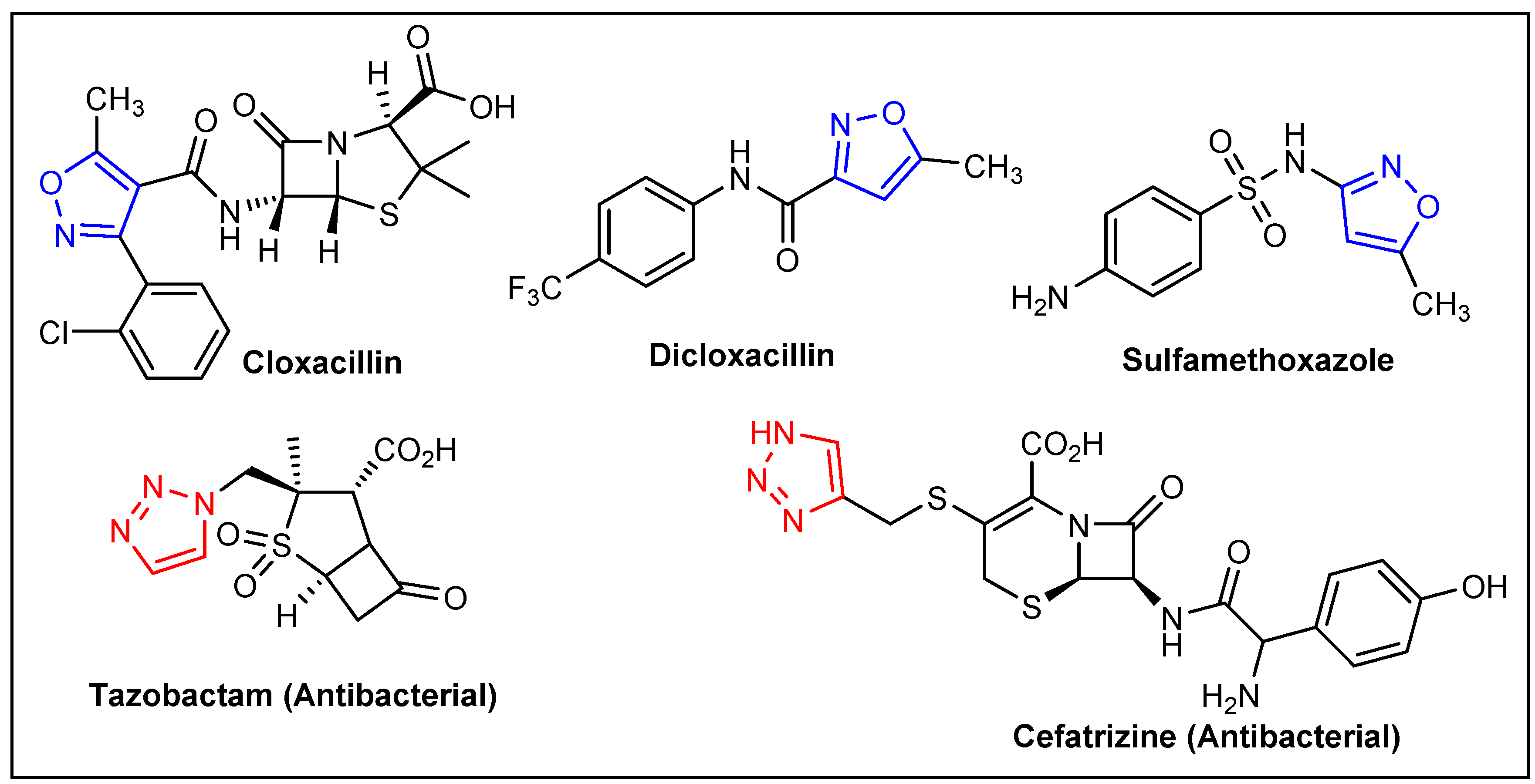
1. Introduction
2. Results and Discussion
2.1. Synthesis and Characterization
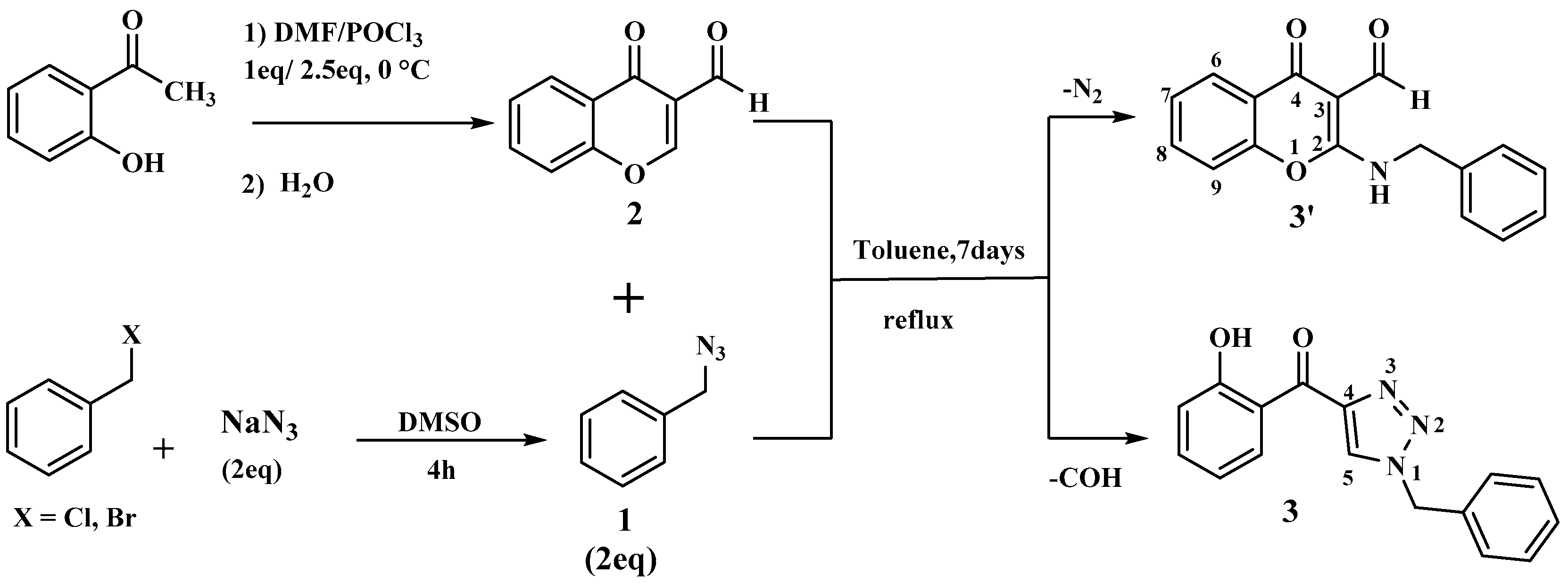
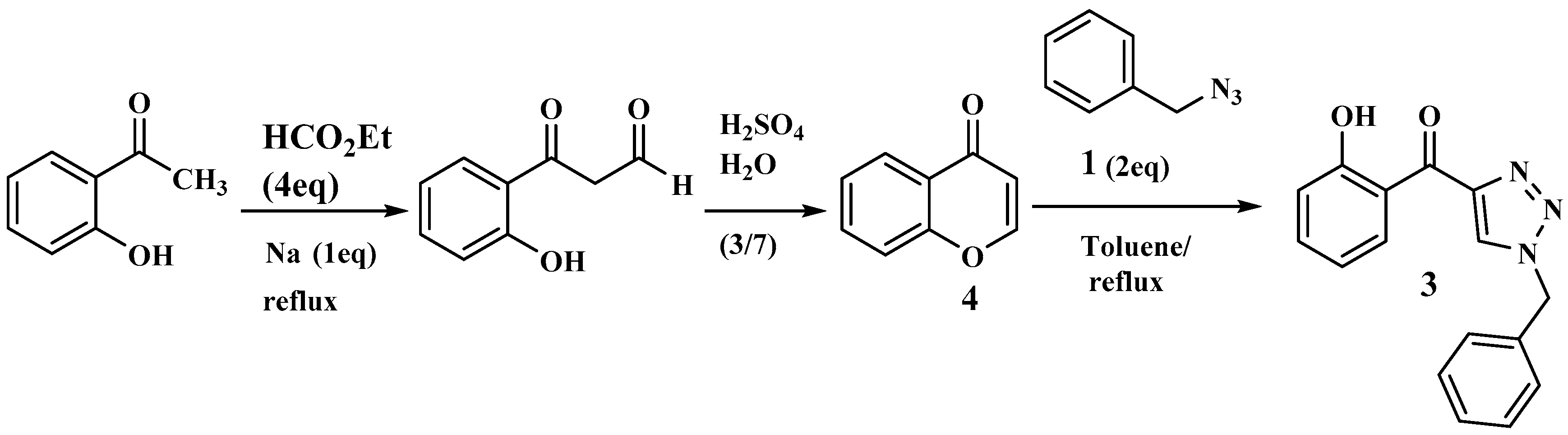
2.1.1. Reaction of Benzyl Azide and 3-Formylchromone
2.1.2. Synthesis of Molecules Hybrids
- a.
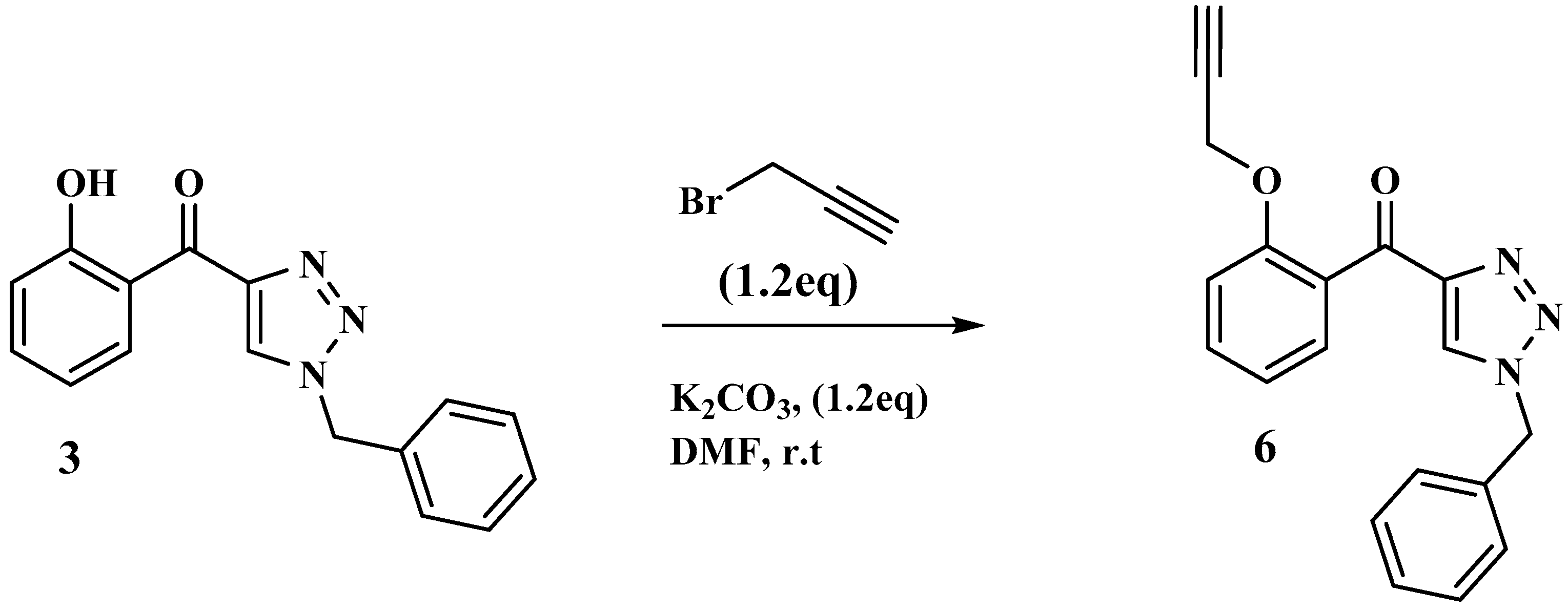
- Synthesis of precursor 6
- b.
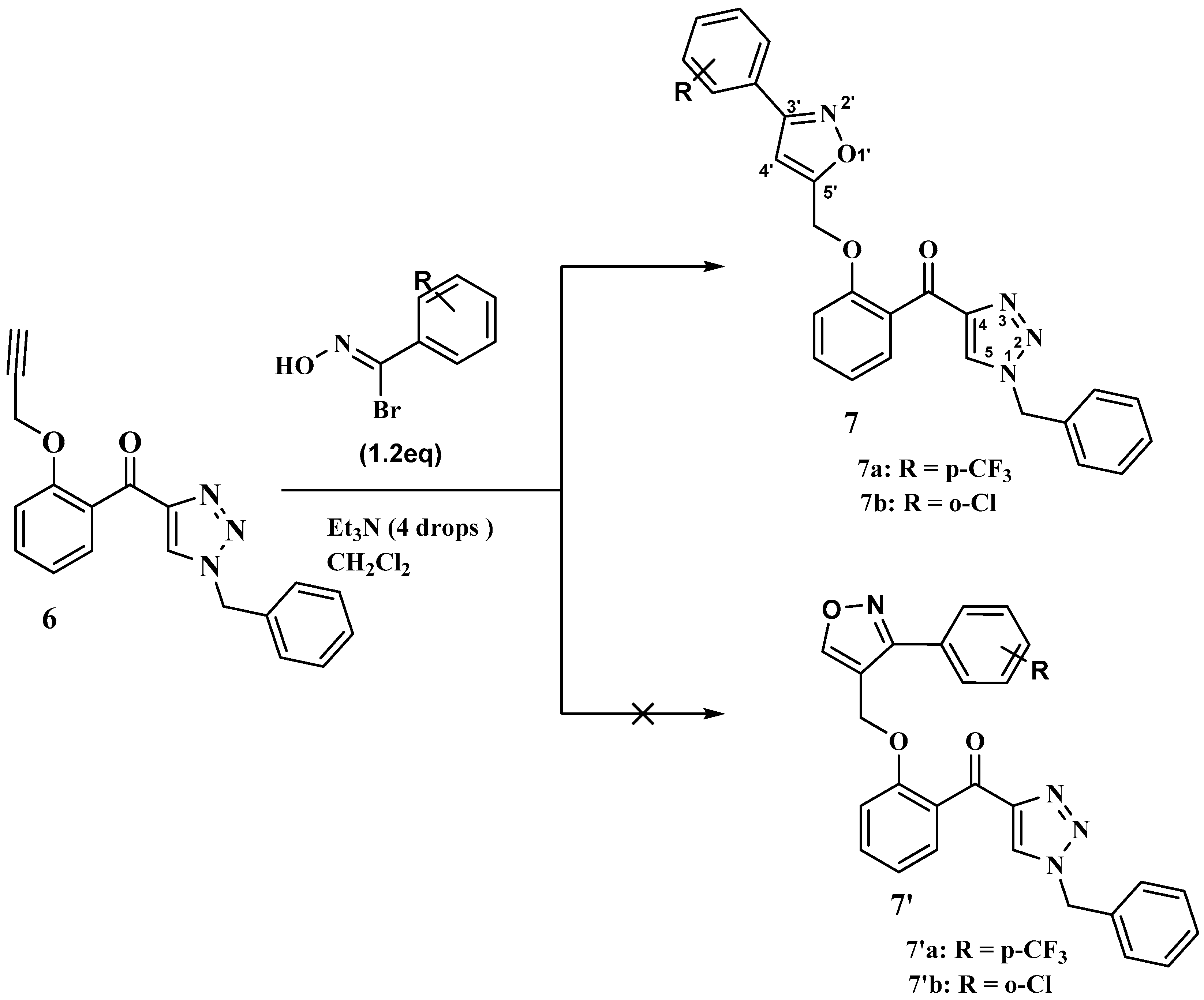
- Synthesis of triazole-isoxazole conjugates from precursor 6
2.2. Antibacterial Assessment
2.2.1. Antibiotic Susceptibility Test
2.2.2. Disk Diffusion Test
2.2.3. Minimum Inhibitory Concentration (MIC) and Minimum Bactericidal Concentration (MBC) Determinations
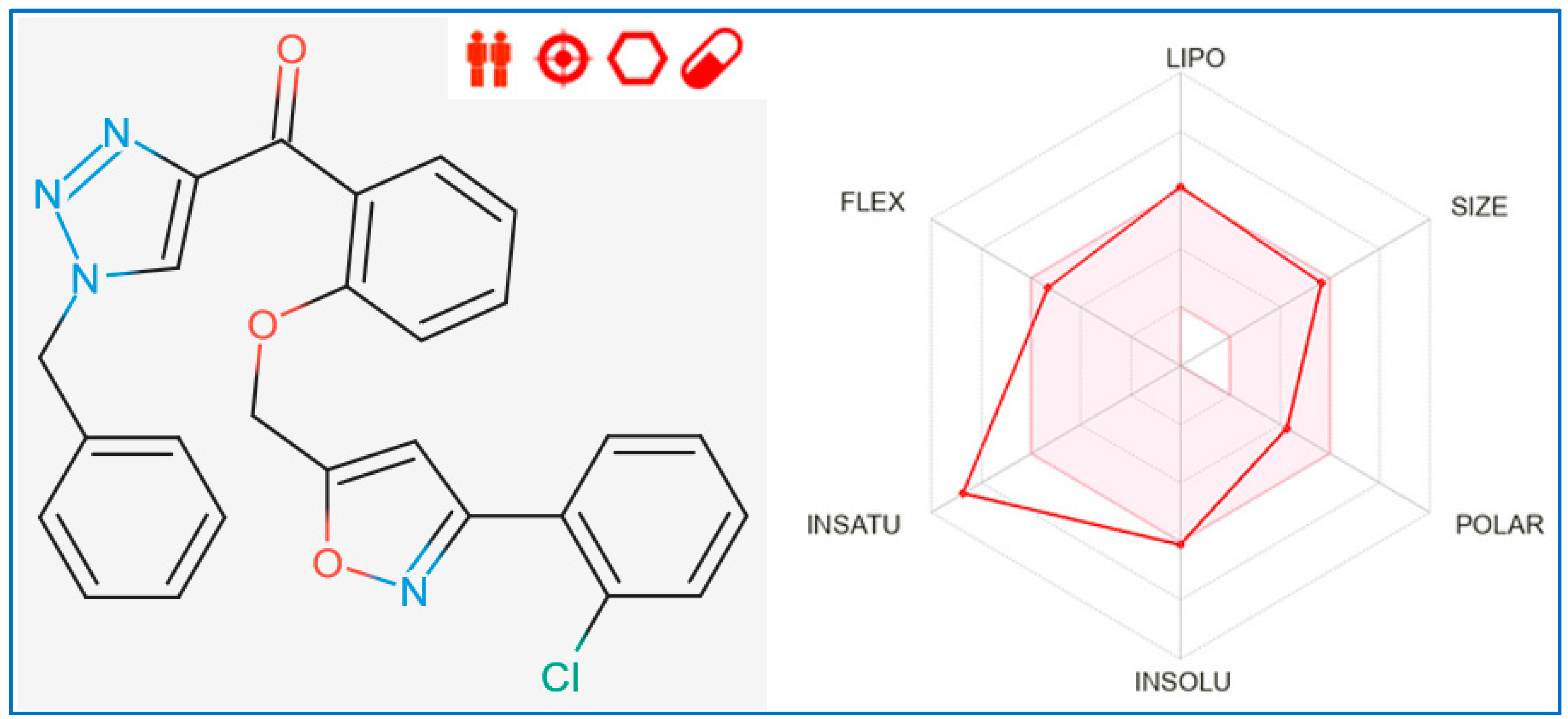
2.3. Drug Similarity and ADME-Tox Predictions
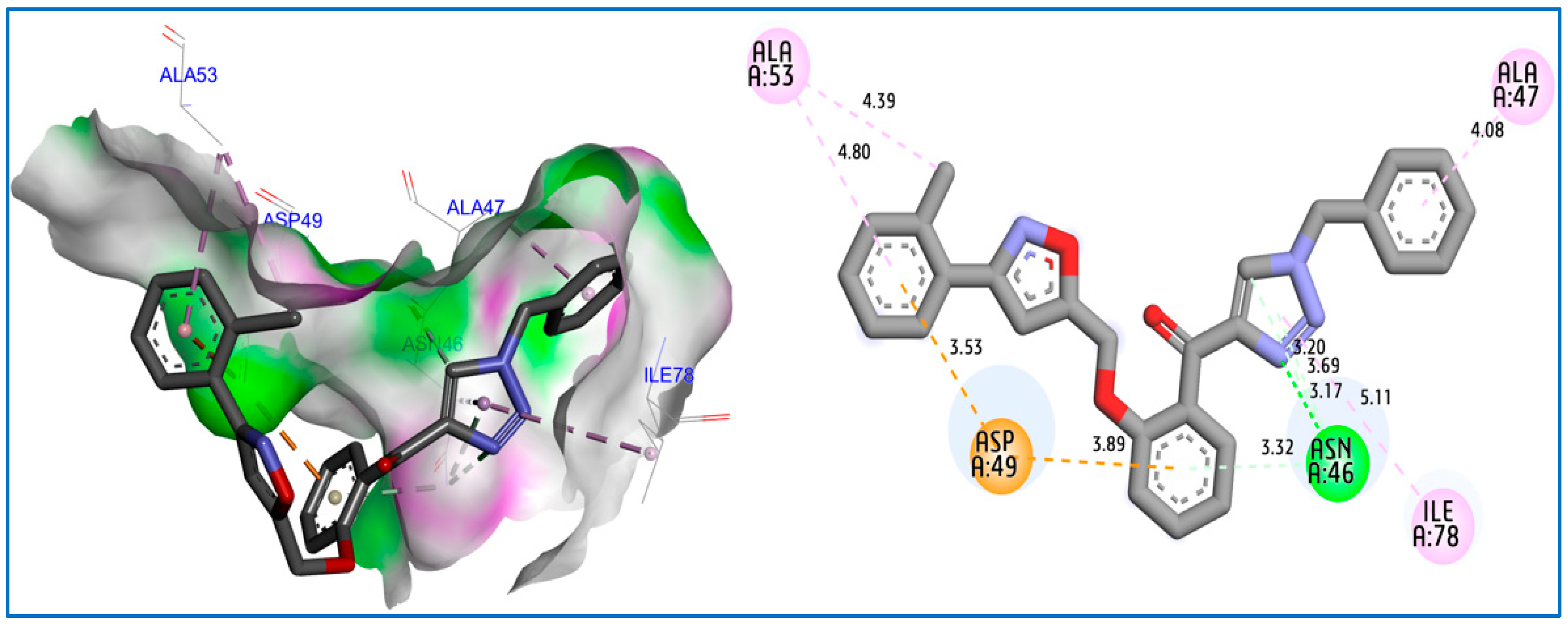
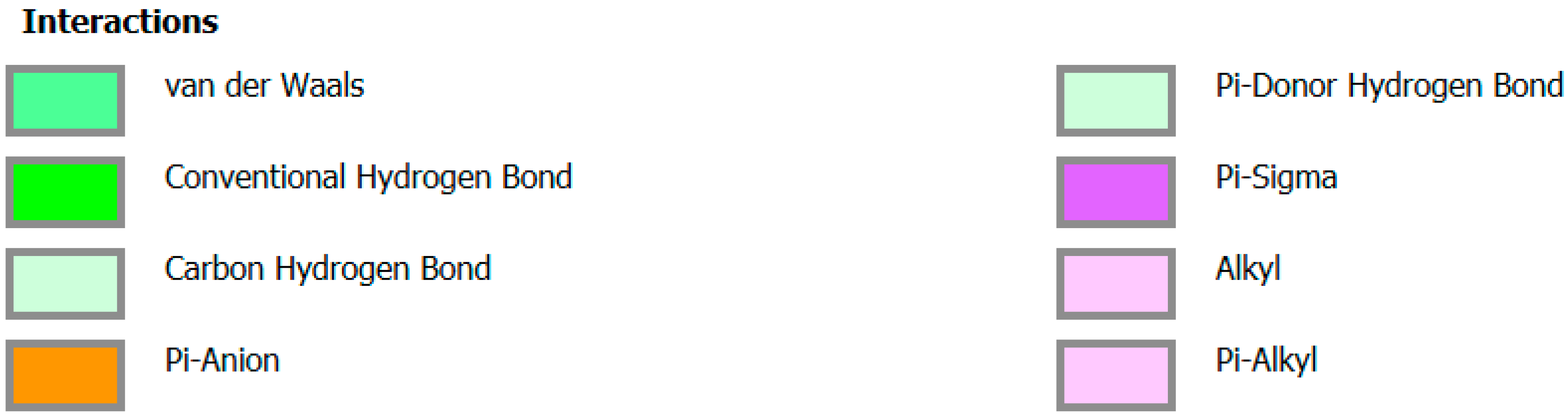
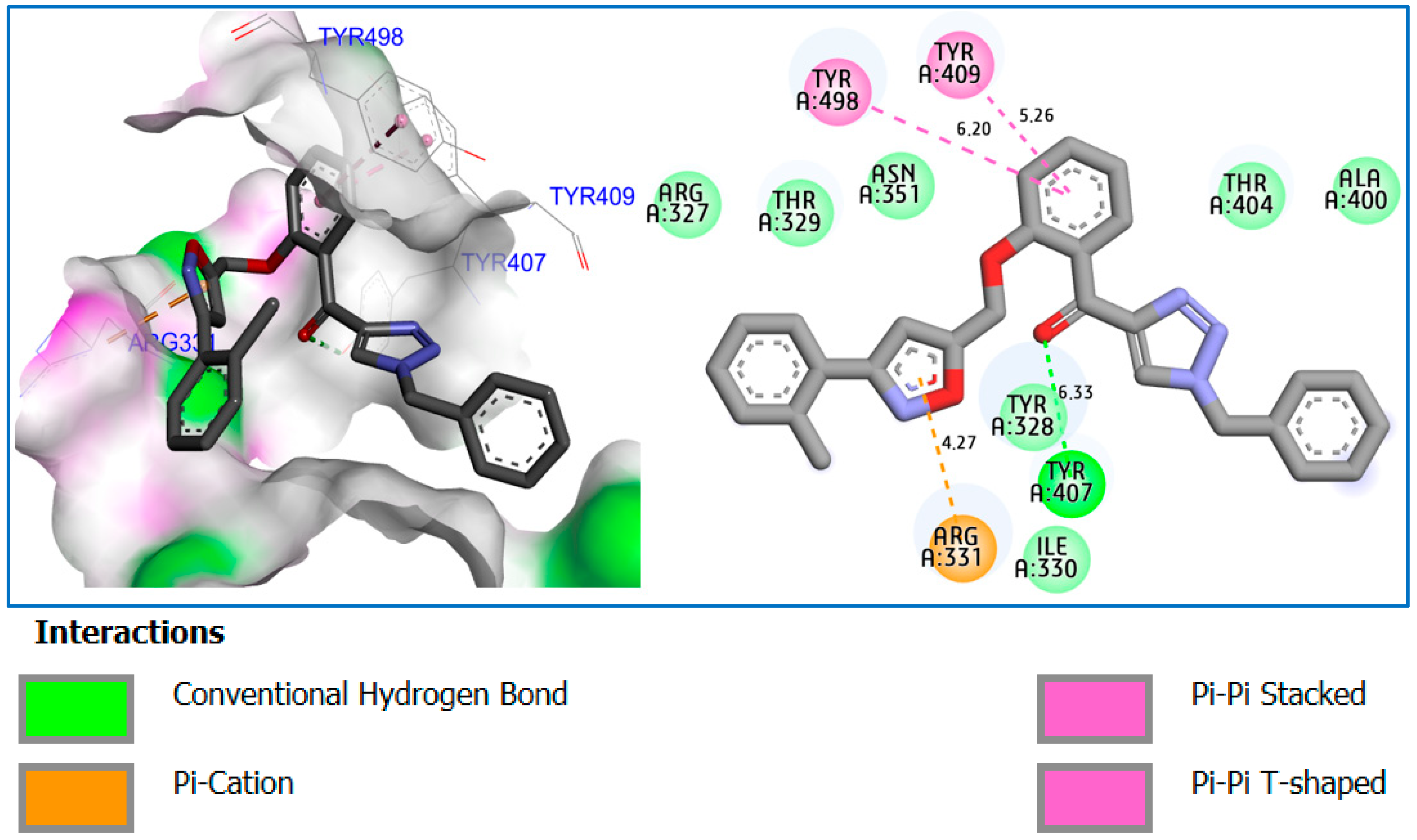
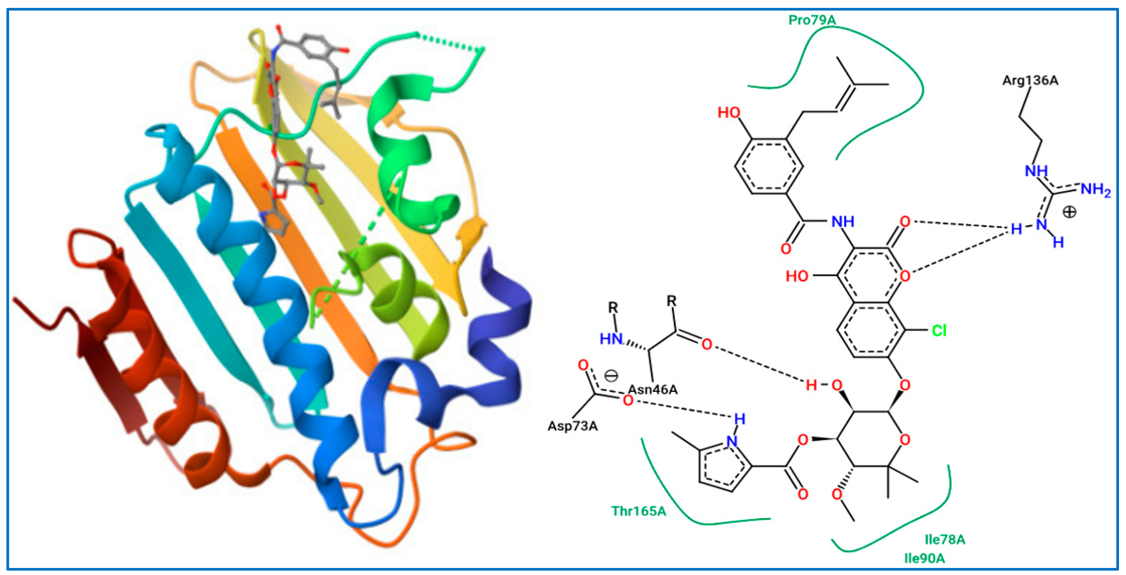
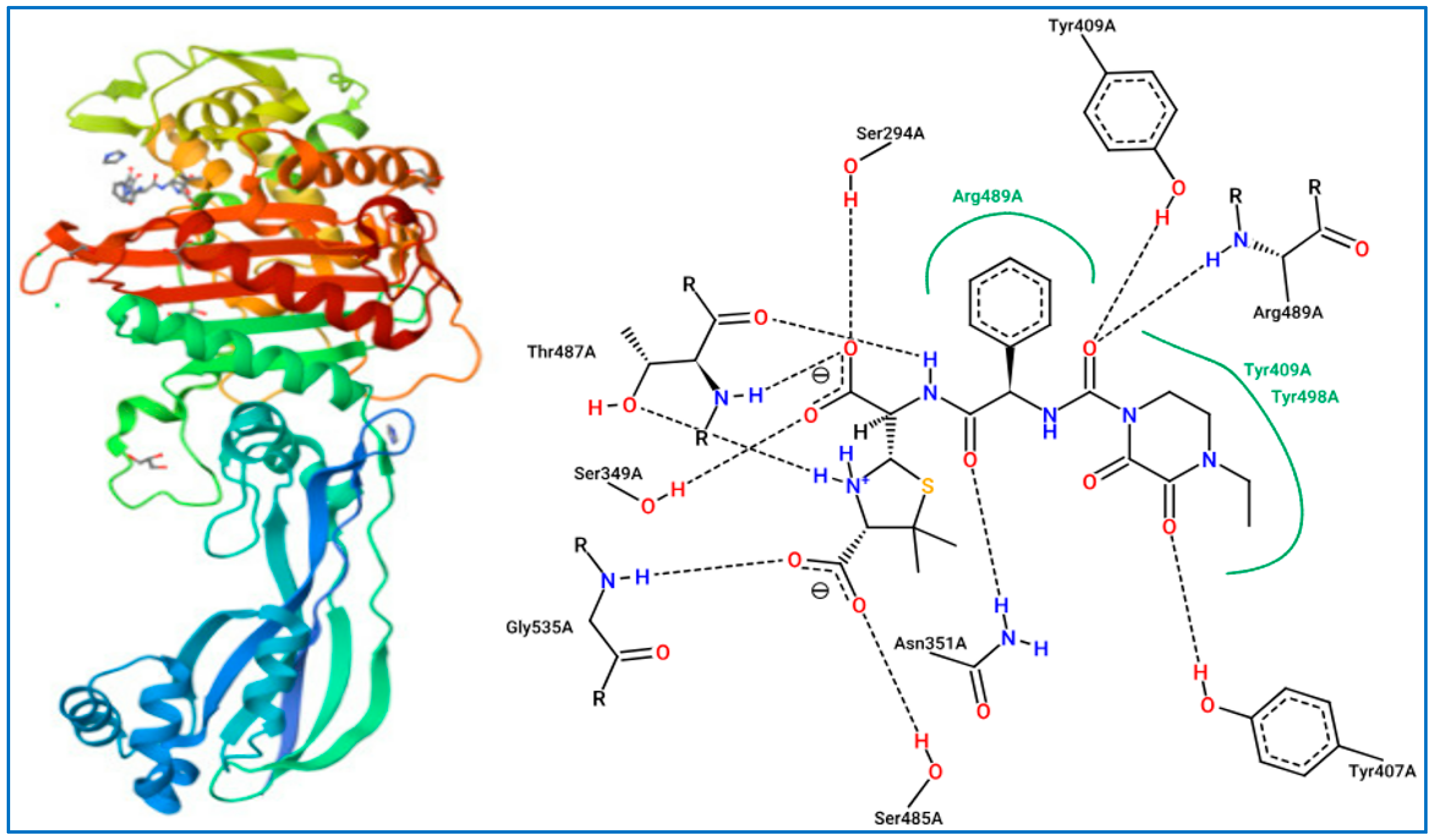
2.4. Docking Molecular Study
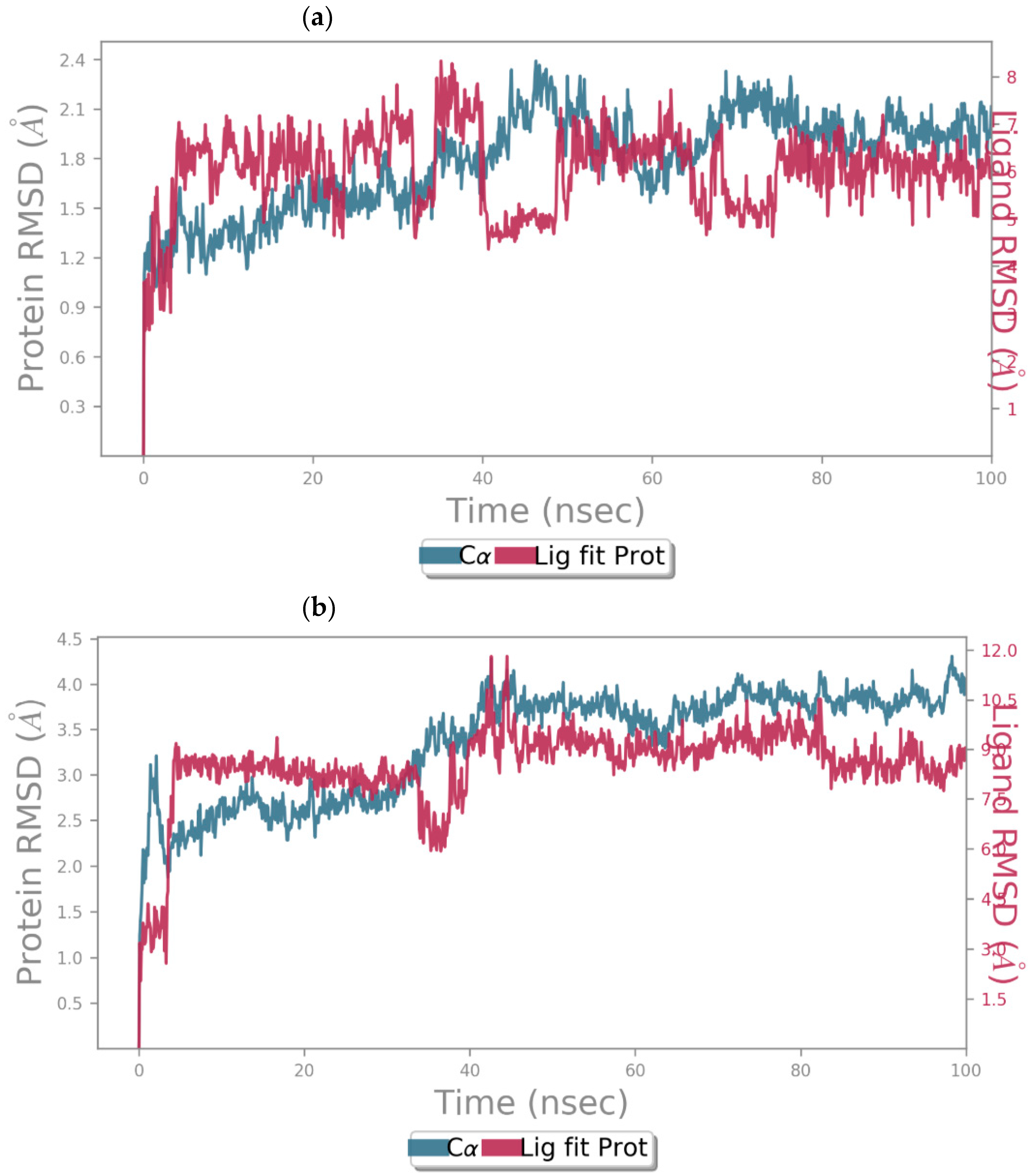
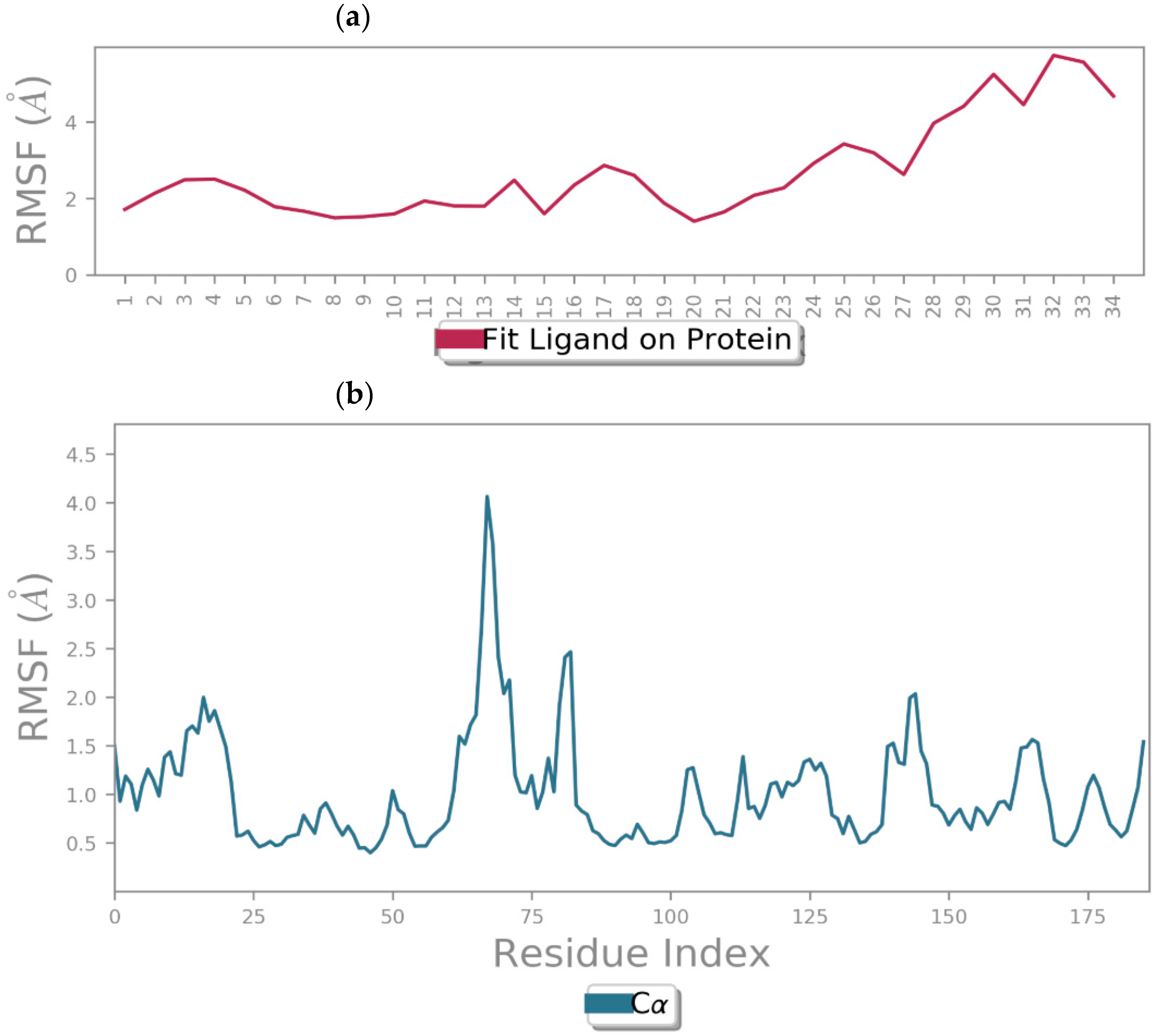
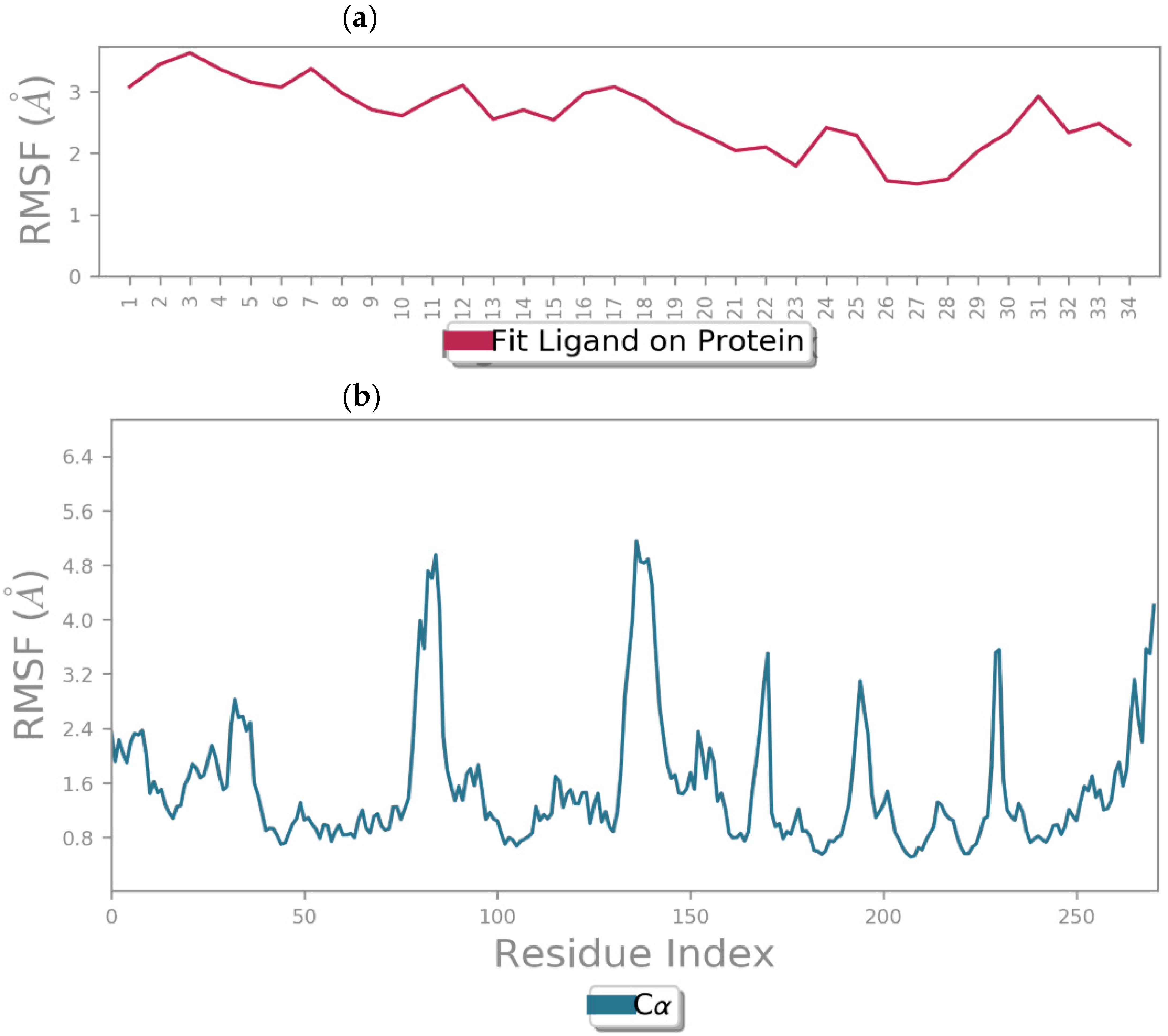
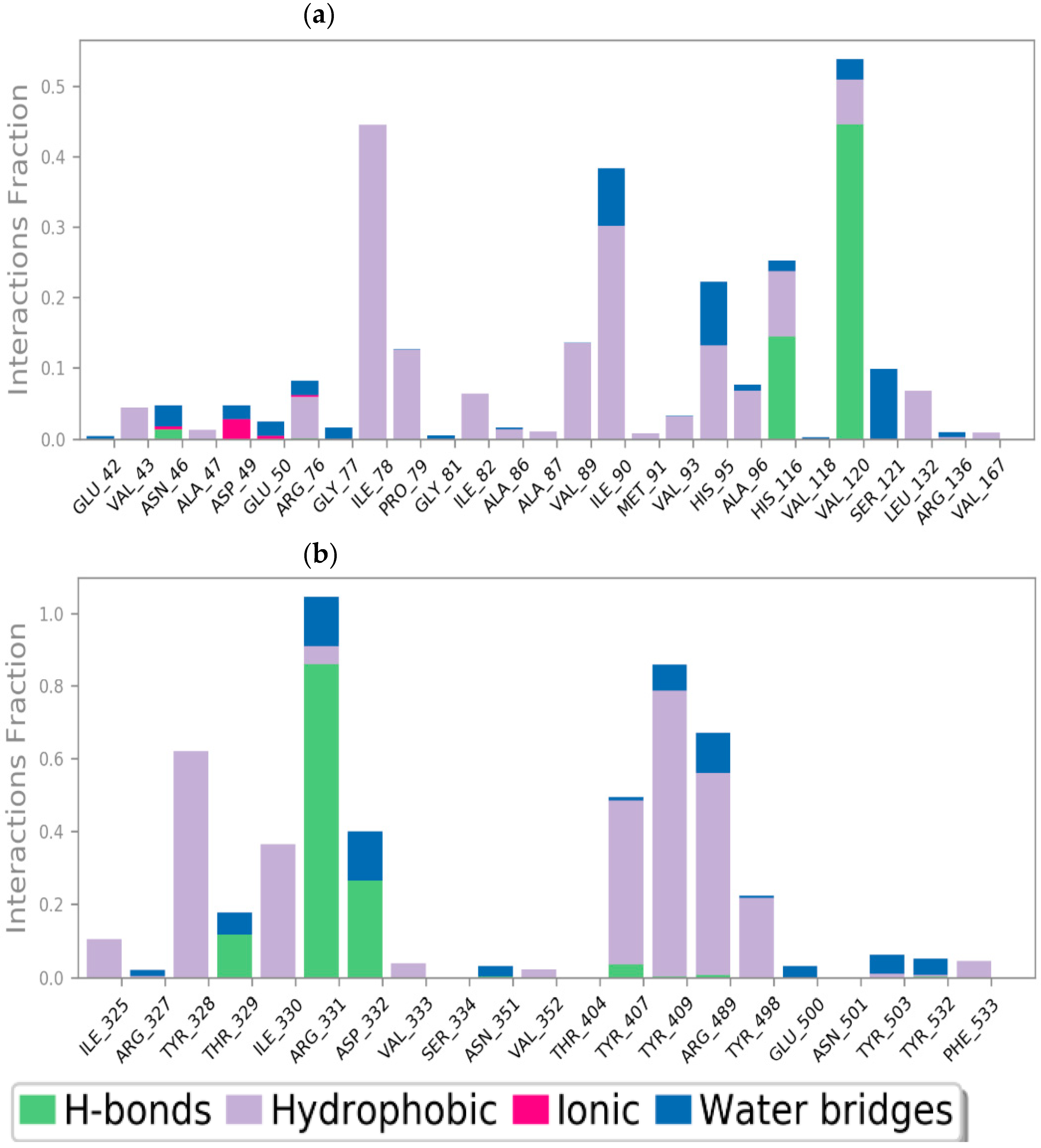
2.5. Dynamics Molecular Simulations
3. Materials and Methods
3.1. General Information
3.2. Evaluation of Antibacterial Activity against Pathogenic Strains
3.2.1. Solution Preparation
3.2.2. Tested Bacterial Strains
3.2.3. Bacterial Pre-Culture Preparation
3.2.4. Antibiotic Susceptibility Test
3.2.5. Qualitative and Quantitative Evaluation
- (a)
- Disk diffusion test
- (b)
- MIC determination
- (c)
- MBC determination
3.3. In Silico Studies
4. Conclusions
Supplementary Materials
Author Contributions
Funding
Institutional Review Board Statement
Informed Consent Statement
Data Availability Statement
Conflicts of Interest
References
- Dua, R.; Shrivastava, S.; Sonwane, S.K.; Srivastava, S.K. Pharmacological Significance of Synthetic Heterocycles Scaffold: A Review. Adv. Biol. Res. 2011, 5, 120–144. [Google Scholar]
- Essien, K.E.; Odiongenyi, A.O.; BoEkom, E.J.; Abai, E.J. Corrosion Inhibition Potential of Two Isoxazole Derivatives: Experimental and Theoretical Analyses. J. Mater. Environ. Sci. 2022, 13, 928–944. Available online: http://www.jmaterenvironsci.com (accessed on 22 May 2024).
- Guo, K.L.; Zhao, L.X.; Wang, Z.W.; Gao, Y.C.; Li, J.J.; Gao, S.; Fu, Y.; Ye, F. Design, synthesis, and bioevaluation of substituted phenyl isoxazole analogues as herbicide safeners. J. Agric. Food Chem. 2020, 68, 10550–10559. [Google Scholar] [CrossRef] [PubMed]
- Yang, Z.; Zhao, Y.; Li, P.; He, Y. Design, synthesis, and insecticidal activity of novel isoxazole derivatives containing bisamide moiety. J. Heterocycl. Chem. 2019, 56, 3042–3047. [Google Scholar] [CrossRef]
- Ratcliffe, P.; Abernethy, L.; Ansari, N.; Cameron, K.; Clarkson, T.; Dempster, M.; Dunn, D.; Easson, A.M.; Edwards, D.; Everett, K.; et al. Discovery of potent, soluble and orally active TRPV1 antagonists. Structure–activity relationships of a series of isoxazoles. Bioorg. Med. Chem. Lett. 2011, 21, 4652–4657. [Google Scholar] [CrossRef] [PubMed]
- Dheer, D.; Singh, V.; Shankar, R. Medicinal attributes of 1,2,3-triazoles: Current developments. Bioorg. Chem. 2017, 71, 30–54. [Google Scholar] [CrossRef] [PubMed]
- Rao, Y.J.; Srinivas, A. Synthesis of New Hybrid Heterocyclic Compounds Having 1,2,3-Triazole and Isoxazole via Click Chemistry. J. Heterocycl. Chem. 2014, 51, 1675–1678. [Google Scholar] [CrossRef]
- Suman, P.; Murthy, T.R.; Rajkumar, K.; Srikanth, D.; Dayakar, C.; Kishor, C.; Addlagatta, A.; Kalivendi, S.V.; Raju, B.C. Synthesis and structure–activity relationships of pyridinyl-1H-1,2,3-triazolyldihydroisoxazoles as potent inhibitors of tubulin polymerization. Eur. J. Med. Chem. 2015, 90, 603–619. [Google Scholar] [CrossRef]
- Ashok, D.; Gundu, S.; Aamate, V.K.; Devulapally, M.G.; Bathini, R.; Manga, V. Dimers of coumarin-1,2,3-triazole hybrids bearing alkyl spacer: Design, microwave-assisted synthesis, molecular docking and evaluation as antimycobacterial and antimicrobial agents. J. Mol. Struct. 2018, 1157, 312–321. [Google Scholar] [CrossRef]
- Cao, X.; Wang, W.; Wang, S.; Bao, L. Asymmetric synthesis of novel triazole derivatives and their in vitro antiviral activity and mechanism of action. Eur. J. Med. Chem. 2017, 139, 718–725. [Google Scholar] [CrossRef]
- Keivanloo, A.; Bakherad, M.; Abbasi, F.; Besharati-Seidani, T.; Amin, A.H. Efficient synthesis of novel 1,2,3-triazole-linked quinoxaline scaffold via copper-catalyzed click reactions. RSC Adv. 2016, 6, 105433–105441. [Google Scholar] [CrossRef]
- Niu, T.F.; Lv, M.F.; Wang, L.; Yi, W.B.; Cai, C. Chemoselective preparation of 1,2,3-triazole–isoxazole bisfunctional derivatives and their application in peptidomimetic synthesis. Org. Biomol. Chem. 2013, 11, 1040–1048. [Google Scholar] [CrossRef] [PubMed]
- Bacheley, L.; Guillamot, G.; Phansavath, P.; Ratovelomanana-Vidal, V. Direct ring fluorination of 3-substituted 5-(1,3-dioxane) acetal isoxazoles: Application to the formal synthesis of a bioactive fluorinated isoxazole. C. R. Chim. 2024, 27, 1–6. [Google Scholar] [CrossRef]
- Gaied, L.B.; Zantour, H. Action des hydrazines et de l’hydroxylamine sur quelques phosphonates β, β-bifonctionnalises: Synthese de phosphonoaminopyrazoles et isoxazoles. Phosphorus Sulfur. Silicon Relat. Elem. 2000, 157, 153–164. [Google Scholar] [CrossRef]
- Hu, F.; Szostak, M. Recent Developments in the Synthesis and Reactivity of Isoxazoles: Metal Catalysis and beyond. Adv. Synth. Catal. 2015, 357, 2583–2614. [Google Scholar] [CrossRef]
- Yang, Z.; Li, P.; Gan, X. Novel pyrazole-hydrazone derivatives containing an isoxazole moiety: Design, synthesis, and antiviral activity. Molecules 2018, 23, 1798. [Google Scholar] [CrossRef] [PubMed]
- Maksimenko, A.S.; Kislyi, V.P.; Chernysheva, N.B.; Strelenko, Y.A.; Zubavichus, Y.V.; Khrustalev, V.N.; Semenova, M.N.; Semenov, V.V. Effective Synthesis of 3,4-Diaryl-isoxazole-5-carboxamides and their Antiproliferative Properties. Eur. J. Org. Chem. 2019, 2019, 4260–4270. [Google Scholar] [CrossRef]
- Canada, Q. Synthesis of thiazole-containing pantothenamide analogues as potential antimicrobial agents. In Annica Chu; McGill University (Canada): Montreal, QC, Canada, 2021; pp. 1–200. [Google Scholar]
- Muhammed, M.T.; ER, M.; Akkoc, S. Molecular modeling and in vitro antiproliferative activity studies of some imidazole and isoxazole derivatives. J. Mol. Struct. 2023, 1282, 135066. [Google Scholar] [CrossRef]
- Bhatia, R.; Vyas, A.; El-Bahy, S.M.; Hessien, M.M.; Mersal, G.A.M.; Ibrahim, M.M.; Dogra, R.; Kumar, B. Rationale Design, Synthesis, Pharmacological and In-silico Investigation of Indole-Functionalized Isoxazoles as Anti-inflammatory Agents. ChemistrySelect 2022, 7, e202200800. [Google Scholar] [CrossRef]
- Filali, I.; Romdhane, A.; Znati, M.; Jannet, H.B.; Bouajila, J. Synthesis of New Harmine Isoxazoles and Evaluation of their Potential Anti-Alzheimer, Anti-inflammatory, and Anticancer Activities. Med. Chem. 2016, 12, 184–190. [Google Scholar] [CrossRef]
- Çalışkan, B.; Sinoplu, E.; İbiş, K.; Güzelcan, E.A.; Atalay, R.Ç.; Banoglu, E. Synthesis and cellular bioactivities of novel isoxazole derivatives incorporating an arylpiperazine moiety as anticancer agents. J. Enzym. Inhib. Med. Chem. 2018, 33, 1352–1361. [Google Scholar] [CrossRef] [PubMed]
- Hawash, M.; Jaradat, N.; Abualhasan, M.; Thaher, M.; Sawalhi, R.; Younes, N.; Shanaa, A.; Nuseirat, M.; Mousa, A. In vitro and in vivo assessment of the antioxidant potential of isoxazole derivatives. Sci. Rep. 2022, 12, 18223. [Google Scholar] [CrossRef] [PubMed]
- Azzali, E.; Machado, D.; Kaushik, A.; Vacondio, F.; Flisi, S.; Cabassi, C.S.; Lamichhane, G.; Viveiros, M.; Costantino, G.; Pieroni, M. Substituted N-Phenyl-5-(2-(phenylamino)thiazol-4-yl)isoxazole-3-carboxamides Are Valuable Antitubercular Candidates that Evade Innate Efflux Machinery. J. Med. Chem. 2017, 60, 7108–7122. [Google Scholar] [CrossRef] [PubMed]
- Song, D.; Bi, F.; Zhang, N.; Qin, Y.; Liu, X.; Teng, Y.; Ma, S. Design, synthesis of novel 4,5-dihydroisoxazole-containing benzamide derivatives as highly potent FtsZ inhibitors capable of killing a variety of MDR Staphylococcus aureus. Bioorg. Med. Chem. 2020, 28, 115729. [Google Scholar] [CrossRef] [PubMed]
- Güell, I.; Micaló, L.; Cano, L.; Badosa, E.; Ferre, R.; Montesinos, E.; Bardají, E.; Feliu, L.; Planas, M. Peptidotriazoles with antimicrobial activity against bacterial and fungal plant pathogens. Peptides 2012, 33, 9–17. [Google Scholar] [CrossRef] [PubMed]
- Derivatives, T.; Abdelkafi-koubaa, Z.; Aissa, I.; Jannet, H.B.; Srairi-abid, N.; Marrakchi, N.; Menif, S. Tyrosol Derivatives, Bearing 3,5-Disubstituted Isoxazole and 1,4-Disubstituted Triazole, as Potential Antileukemia Agents by Promoting Apoptosis. Molecules 2022, 27, 5086. [Google Scholar] [CrossRef] [PubMed]
- Taia, A.; Essaber, M.; Oubella, A.; Aatif, A.; Bodiguel, J.; Jamart-Grégoire, B.; Itto, M.Y.A.; Morjani, H. Synthesis, characterization, and biological evaluation of new heterocyclic systems 1,2,3-triazole-isoxazoline from eugenol by the mixed condensation reactions. Synth. Commun. 2020, 50, 2052–2065. [Google Scholar] [CrossRef]
- Bumagin, N.A.; Kletskov, A.V.; Petkevich, S.K.; Kolesnik, I.A.; Lyakhov, A.S.; Ivashkevich, L.S.; Baranovsky, A.V.; Kurman, P.V.; Potkin, V.I. Substituted 1-(isoxazol-3-yl)methyl-1H-1,2,3-triazoles: Synthesis, palladium(II) complexes, and high-turnover catalysis in aqueous media. Tetrahedron 2018, 74, 3578–3588. [Google Scholar] [CrossRef]
- Machado, N.F.L.; Marques, M.P.M. Bioactive Chromone Derivatives—Structural Diversity. Curr. Bioact. Compd. 2010, 6, 76–89. [Google Scholar] [CrossRef]
- Sharma, S.K.; Kumar, S.; Chand, K.; Kathuria, A.; Gupta, A.; Jain, R. An update on natural occurrence and biological activity of chromones. Curr. Med. Chem. 2011, 18, 3825–3852. Available online: https://www.ingentaconnect.com/content/ben/cmc/2011/00000018/00000025/art00003 (accessed on 27 January 2024). [CrossRef]
- Jiao, R.; Xu, F.; Huang, X.; Li, H.; Liu, W.; Cao, H.; Zang, L.; Li, Z.; Hua, H.; Li, D. Antiproliferative chromone derivatives induce K562 cell death through endogenous and exogenous pathways. J. Enzym. Inhib. Med. Chem. 2020, 35, 759–772. [Google Scholar] [CrossRef] [PubMed]
- Ali, T.E.S.; Ibrahim, M.A. Synthesis and antimicrobial activity of chromone-linked 2-pyridone fused with 1,2,4-triazoles, 1,2,4-triazines and 1,2,4-triazepines ring systems. J. Braz. Chem. Soc. 2010, 21, 1007–1016. [Google Scholar] [CrossRef]
- Dofe, V.S.; Sarkate, A.P.; Lokwani, D.K.; Shinde, D.B.; Kathwate, S.H.; Gill, C.H. Novel O-Alkylated Chromones as Antimicrobial Agents: Ultrasound Mediated Synthesis, Molecular Docking and ADME Prediction. J. Heterocycl. Chem. 2017, 54, 2678–2685. [Google Scholar] [CrossRef]
- Kaushik, P.; Shakil, N.A.; Rana, V.S. Synthesis, Biological Evaluation, and QSAR Studies of 3-Iodochromone Derivatives as Potential Fungicides. Front. Chem. 2021, 9, 636882. [Google Scholar] [CrossRef] [PubMed]
- Cano, P.A.; Islas-Jácome, A.; Rangel-Serrano, Á.; Anaya-Velázquez, F.; Padilla-Vaca, F.; Trujillo-Esquivel, E.; Ponce-Noyola, P.; Martínez-Richa, A.; Gámez-Montaño, R. In vitro studies of chromone-tetrazoles against pathogenic protozoa, bacteria, and fungi. Molecules 2015, 20, 12436–12449. [Google Scholar] [CrossRef] [PubMed]
- Hussein, M.A.; Zyaan, O.H.; Monsef, A.H.A.; Rizk, S.A.; Farag, S.M.; Hafez, S.E.; Khaled, A.S.; Helmy, O.M. Synthesis, molecular docking and insecticidal activity evaluation of chromones of date palm pits extract against Culex pipiens (Diptera: Culicidae) Homology modeling and virtual screening studies View project. Int. J. Mosq. Res. 2018, 5, 22–32. [Google Scholar]
- Zhao, P.-L.; Li, J.; Yang, G.-F. Synthesis and insecticidal activity of chromanone and chromone analogues of diacylhydrazines. Bioorg. Med. Chem. 2007, 15, 1888–1895. [Google Scholar] [CrossRef] [PubMed]
- Hu, Q.F.; Zhou, B.; Gao, X.M.; Yang, L.Y.; Shu, L.D.; Shen, Y.; Li, G.P.; Che, C.T.; Yang, G.Y. Antiviral chromones from the stem of Cassia siamea. J. Nat. Prod. 2012, 75, 1909–1914. [Google Scholar] [CrossRef] [PubMed]
- Kanzouai, Y.; Laghmari, M.; Yamari, I.; Bouzammit, R.; Bahsis, L.; Benali, T.; Chtita, S.; Bakhouch, M.; Akhazzane, M.; El Kouali, M.; et al. Chromone-isoxazole hybrids molecules: Synthesis, spectroscopic, MEDT, ELF, antibacterial, ADME-Tox, molecular docking and MD simulation investigations. J. Biomol. Struct. Dyn. 2023. [Google Scholar] [CrossRef]
- Kanzouai, Y.; Chalkha, M.; Hadni, H.; Laghmari, M.; Bouzammit, R.; Nakkabi, A.; Benali, T.; Tüzün, B.; Akhazzane, M.; El Yazidi, M.; et al. Design, synthesis, in-vitro and in-silico studies of chromone-isoxazoline conjugates as anti-bacterial agents. J. Mol. Struct. 2023, 1293, 136205. [Google Scholar] [CrossRef]
- Chalkha, M.; Chebbac, K.; Nour, H.; Nakkabi, A.; El Moussaoui, A.; Tüzün, B.; Bourhia, M.; Chtita, S.; Bakhouch, M.; Laaroussi, H.; et al. In vitro and in silico evaluation of the antimicrobial and antioxidant activities of spiropyrazoline oxindole congeners. Arab. J. Chem. 2024, 17, 105465. [Google Scholar] [CrossRef]
- Harnisch, H. Chromon-3-carbaldehyde. Justus Liebigs Ann. Chem. 1973, 765, 8–14. [Google Scholar] [CrossRef]
- Lieber, E.; Chao, T.S.; Rao, C.N.R. Improved Method for the Synthesis of Alkyl Azides. J. Org. Chem. 1957, 22, 238–240. [Google Scholar] [CrossRef]
- Schönberg, A.; Sina, A. On Visnagin and Khellin and Related Compounds. A Simple Synthesis of Chromone. J. Am. Chem. Soc. 1950, 72, 3396–3399. [Google Scholar] [CrossRef]
- Xu, S.; Zhuang, X.; Pan, X.; Zhang, Z.; Duan, L.; Liu, Y.; Zhang, L.; Ren, X.; Ding, K. 1-Phenyl-4-benzoyl-1H-1,2,3-triazoles as orally bioavailable transcriptional function suppressors of Estrogen-related receptor α. J. Med. Chem. 2013, 56, 4631–4640. [Google Scholar] [CrossRef] [PubMed]
- Kant, R.; Kumar, D.; Agarwal, D.; Gupta, R.D.; Tilak, R.; Awasthi, S.K.; Agarwal, A. Synthesis of newer 1,2,3-triazole linked chalcone and flavone hybrid compounds and evaluation of their antimicrobial and cytotoxic activities. Eur. J. Med. Chem. 2016, 113, 34–49. [Google Scholar] [CrossRef] [PubMed]
- Hawash, M.; Kahraman, D.C.; Ergun, S.G.; Cetin-Atalay, R.; Baytas, S.N. Synthesis of novel indole-isoxazole hybrids and evaluation of their cytotoxic activities on hepatocellular carcinoma cell lines. BMC Chem. 2021, 15, 66. [Google Scholar] [CrossRef] [PubMed]
- Singh, I.; Zarafshani, Z.; Lutz, J.-F.-O.; Heaney, F. Metal-free “click” chemistry: Efficient polymer modification via 1, 3-dipolar cycloaddition of nitrile oxides and alkynes. Macromolecules 2009, 42, 5411–5413. [Google Scholar] [CrossRef]
- Jeddi, M.; El Hachlafi, N.; El Fadili, M.; Benkhaira, N.; Al-Mijalli, S.H.; Kandsi, F.; Abdallah, E.M.; Ouaritini, Z.B.; Bouyahya, A.; Lee, L.-H.; et al. Antimicrobial, antioxidant, α-amylase and α-glucosidase inhibitory activities of a chemically characterized essential oil from Lavandula angustifolia Mill.: In vitro and in silico investigations. Biochem. Syst. Ecol. 2023, 111, 104731. [Google Scholar] [CrossRef]
- Benkhaira, N.; El Hachlafi, N.; El Fadili, M.; Jeddi, M.; Abdnim, R.; Bnouham, M.; Koraichi, S.I.; Fikri-Benbrahim, K. Unveiling the phytochemical profile, in vitro bioactivities evaluation, in silico molecular docking and ADMET study of essential oil from Clinopodium nepeta grown in Middle Atlas of Morocco. Biocatal. Agric. Biotechnol. 2023, 54, 102923. [Google Scholar] [CrossRef]
- Assaggaf, H.; El Hachlafi, N.; El Fadili, M.; Elbouzidi, A.; Ouassou, H.; Jeddi, M.; Alnasser, S.M.; Qasem, A.; Attar, A.; AL-Farga, A.; et al. GC/MS Profiling, In Vitro Antidiabetic Efficacy of Origanum compactum Benth. Essential Oil and In Silico Molecular Docking of Its Major Bioactive Compounds. Catalysts 2023, 13, 1429. [Google Scholar] [CrossRef]
- El Fadili, M.; Er-rajy, M.; Eltayb, W.A.; Kara, M.; Assouguem, A.; Saleh, A.; Al Kamaly, O.; Zarougui, S.; Elhallaoui, M. In-silico screening based on molecular simulations of 3,4-disubstituted pyrrolidine sulfonamides as selective and competitive GlyT1 inhibitors. Arab. J. Chem. 2023, 16, 105105. [Google Scholar] [CrossRef]
- El Fadili, M.; Er-rajy, M.; Imtara, H.; Kara, M.; Zarougui, S.; Altwaijry, N.; Al Kamaly, O.; Al Sfouk, A.; Elhallaoui, M. 3D-QSAR, ADME-Tox In Silico Prediction and Molecular Docking Studies for Modeling the Analgesic Activity against Neuropathic Pain of Novel NR2B-Selective NMDA Receptor Antagonists. Processes 2022, 10, 1462. [Google Scholar] [CrossRef]
- El Fadili, M.; Er-rajy, M.; Abdalla, M.; Abuelizz, H.A.; Zarougui, S.; Alkhulaifi, F.M.; Alahmady, N.F.; Shami, A.; Elhallaoui, M. In-silico investigations of novel tacrine derivatives potency against Alzheimer’s disease. Sci. Afr. 2024, 23, e02048. [Google Scholar] [CrossRef]
- El Fadili, M.; Er-rajy, M.; Eltayb, W.A.; Kara, M.; Imtara, H.; Zarougui, S.; Al-Hoshani, N.; Hamadi, A.; Elhallaoui, M. An in-silico investigation based on molecular simulations of novel and potential brain-penetrant GluN2B NMDA receptor antagonists as anti-stroke therapeutic agents. J. Biomol. Struct. Dyn. 2023. [Google Scholar] [CrossRef]
- Barhouchi, B.; Menacer, R.; Bouchkioua, S.; Mansour, A.; Belattar, N. Compounds from myrtle flowers as antibacterial agents and SARS-CoV-2 inhibitors: In-vitro and molecular docking studies. Arab. J. Chem. 2023, 16, 104939. [Google Scholar] [CrossRef]
- Skariyachan, S.; Ravishankar, R.; Gopal, D.; Muddebihalkar, A.G.; Uttarkar, A.; Praveen, P.K.U.; Niranjan, V. Response regulator GacA and transcriptional activator RhlR proteins involved in biofilm formation of Pseudomonas aeruginosa are prospective targets for natural lead molecules: Computational modelling, molecular docking and dynamic simulation studies. Infect. Genet. Evol. 2020, 85, 104448. [Google Scholar] [CrossRef]
- Antioxidant, A.P.; Alzahrani, A.J. Promising Antioxidant and Antimicrobial Potencies of Chemically-Profiled Extract from Withania aristata (Aiton) Pauquy against Clinically-Pathogenic Microbial Strains. Molecules 2022, 27, 3614. [Google Scholar] [CrossRef] [PubMed]
- Kowalska-Krochmal, B.; Dudek-Wicher, R. The minimum inhibitory concentration of antibiotics: Methods, interpretation, clinical relevance. Pathogens 2021, 10, 165. [Google Scholar] [CrossRef]
- Mammate, N.; El Oumari, F.E.; Imtara, H.; Belchkar, S.; Touimi, G.B.; Al-Zharani, M.; Rudayni, H.A.; Qurtam, A.A.; Aleissa, M.S.; Nasr, F.A.; et al. Anti-Struvite, Antimicrobial, and Anti-Inflammatory Activities of Aqueous and Ethanolic Extracts of Saussurea costus (Falc) Lipsch Asteraceae. Molecules 2023, 28, 667. [Google Scholar] [CrossRef]
- Parvekar, P.; Palaskar, J.; Metgud, S.; Maria, R.; Dutta, S. The minimum inhibitory concentration (MIC) and minimum bactericidal concentration (MBC) of silver nanoparticles against Staphylococcus aureus. Biomater. Investig. Dent. 2020, 7, 105–109. [Google Scholar] [CrossRef] [PubMed]
- Ed-Dahmani, I.; El Fadili, M.; Kandsi, F.; Conte, R.; El Atki, Y.; Kara, M.; Assouguem, A.; Touijer, H.; Lfitat, A.; Nouioura, G.; et al. Phytochemical, Antioxidant Activity, and Toxicity of Wild Medicinal Plant of Melitotus albus Extracts, In Vitro and In Silico Approaches. ACS Omega 2024, 9, 9236–9246. [Google Scholar] [CrossRef] [PubMed]
- Nouioura, G.; El Fadili, M.; Ghneim, H.K.; Zbadi, L.; Maache, S.; Zouirech, O.; Danouche, M.; Aboul-Soud, M.A.M.; Giesy, J.P.; Lyoussi, B.; et al. Exploring the essence of celery seeds (Apium graveolens L.): Innovations in microwave-assisted hydrodistillation for essential oil extraction using in vitro, in vivo and in silico studies. Arab. J. Chem. 2024, 17, 105726. [Google Scholar] [CrossRef]
- El Fadili, M.; Er-Rajy, M.; Kara, M.; Assouguem, A.; Belhassan, A.; Alotaibi, A.; Mrabti, N.N.; Fidan, H.; Ullah, R.; Ercisli, S.; et al. QSAR, ADMET In Silico Pharmacokinetics, Molecular Docking and Molecular Dynamics Studies of Novel Bicyclo (Aryl Methyl) Benzamides as Potent GlyT1 Inhibitors for the Treatment of Schizophrenia. Pharmaceuticals 2022, 15, 670. [Google Scholar] [CrossRef] [PubMed]


| Strains | ||||
|---|---|---|---|---|
| Antibiotic | Staphylococcus aureus ATCC 25923 | Staphylococcus aureus ATCC BAA-44 | Escherichia coli ATCC 25922 | Pseudomonas aeruginosa |
| Ampicillin (AMP) | R (10) * | R (10) | R (0) | R (0) |
| Norfloxacin (NOR) | S (26) | R (10) | - | - |
| Cefoxitin (FOX) | S (30) | R (9) | - | R (0) |
| Cefotaxime (CTX) | - | - | S (36) | R (16) |
| Imipenem (IMP) | - | - | S (36) | I (30) |
| Amikacin (AK) | S (21) | R (13) | S (34) | S (24) |
| Fosfomycin (FOS) | - | - | S (40) | S (20) |
| Molecules | Staphylococcus aureus ATCC 25923 | Staphylococcus aureus ATCC BAA-44 | Escherichia coli ATCC 25922 | Pseudomonas aeruginosa |
|---|---|---|---|---|
| 3′ | - | - | - | - |
| 3 | - | - | - | - |
| 6 | - | - | - | - |
| 7a | - | - | - | - |
| 7b | - | - | 36.4 ± 1.07 * | 11.25 ± 1.02 |
| Compound | Test | Staphylococcus aureus ATCC 25923 | Staphylococcus aureus ATCC BAA-44 | Escherichia coli ATCC 25922 | Pseudomonas aeruginosa |
|---|---|---|---|---|---|
| 7b | MIC | - | - | 15 | 30 |
| MBC | - | - | 30 | - |
| Studied Compound | Physicochemical Properties | Lipinski Rules | ||||
|---|---|---|---|---|---|---|
| Molecular Weight (g/mol) | Molar Refractive Index | Log P (Octanol/Water) | Hydrogen Bonds Acceptors | Hydrogen Bonds Donors | Categorical (Yes/No) | |
| Rule | ≤500 | 40 ≤ MR ≤ 130 | <5 | ≤10 | <5 | |
| 7b | 470.91 | 127.13 | 3.67 | 6 | 0 | YES |
| Studied Compound | A | D | M | E | T | |||||||||
|---|---|---|---|---|---|---|---|---|---|---|---|---|---|---|
| Human Intestinal Absorption | Blood–Brain Barrier Permeability | Central Nervous System Permeability | Substrate | Inhibitor | Total Clearance | AMES Test of Toxicity | Hepatotoxicity | Skin Sensitization | ||||||
| Cytochromes | ||||||||||||||
| 2D-6 | 3A-4 | 1A-2 | 2C-19 | 2C-9 | 2D-6 | 3A-4 | ||||||||
| (% Absorbed) | (Log BB) | (Log PS) | (No/Yes) | Numeric (Log mL/min/kg) | Categorical (No/Yes) | |||||||||
| 7a | 94.946 | −1.465 | −2.06 | No | Yes | No | Yes | Yes | No | Yes | 0.039 | No | Yes | No |
| 7b | 96.39 | −1.199 | −2.097 | No | Yes | No | Yes | Yes | No | Yes | 0.34 | No | Yes | No |
| Pathogen Type | Pathogen Species | Strain |
|---|---|---|
| Gram-positive | Staphylococcus aureus | ATCC 25923 |
| ATCC BAA-44 | ||
| Gram-negative | Escherichia coli | ATCC 25922 |
| Pseudomonas aeruginosa | Clinical isolate |
| Antibiotic Class | Antibiotic | Concentration (µg/disk) |
|---|---|---|
| Penicillin | Ampicillin | 25 |
| Fluoroquinolones | Norfloxacin | 10 |
| Cephalosporines | Cefoxitin | 30 |
| Cefotaxime | 30 | |
| Carbapenem | Imipenem | 30 |
| Aminosides | Amikacin | 30 |
| Fosfomycin | Fosfomycin | 200 |
Disclaimer/Publisher’s Note: The statements, opinions and data contained in all publications are solely those of the individual author(s) and contributor(s) and not of MDPI and/or the editor(s). MDPI and/or the editor(s) disclaim responsibility for any injury to people or property resulting from any ideas, methods, instructions or products referred to in the content. |
© 2024 by the authors. Licensee MDPI, Basel, Switzerland. This article is an open access article distributed under the terms and conditions of the Creative Commons Attribution (CC BY) license (https://creativecommons.org/licenses/by/4.0/).
Share and Cite
Bouzammit, R.; Belchkar, S.; El fadili, M.; Kanzouai, Y.; Mujwar, S.; Alanazi, M.M.; Chalkha, M.; Nakkabi, A.; Bakhouch, M.; Gal, E.; et al. New Triazole-Isoxazole Hybrids as Antibacterial Agents: Design, Synthesis, Characterization, In Vitro, and In Silico Studies. Molecules 2024, 29, 2510. https://doi.org/10.3390/molecules29112510
Bouzammit R, Belchkar S, El fadili M, Kanzouai Y, Mujwar S, Alanazi MM, Chalkha M, Nakkabi A, Bakhouch M, Gal E, et al. New Triazole-Isoxazole Hybrids as Antibacterial Agents: Design, Synthesis, Characterization, In Vitro, and In Silico Studies. Molecules. 2024; 29(11):2510. https://doi.org/10.3390/molecules29112510
Chicago/Turabian StyleBouzammit, Rachid, Salim Belchkar, Mohamed El fadili, Youssra Kanzouai, Somdutt Mujwar, Mohammed M. Alanazi, Mohammed Chalkha, Asmae Nakkabi, Mohamed Bakhouch, Emese Gal, and et al. 2024. "New Triazole-Isoxazole Hybrids as Antibacterial Agents: Design, Synthesis, Characterization, In Vitro, and In Silico Studies" Molecules 29, no. 11: 2510. https://doi.org/10.3390/molecules29112510
APA StyleBouzammit, R., Belchkar, S., El fadili, M., Kanzouai, Y., Mujwar, S., Alanazi, M. M., Chalkha, M., Nakkabi, A., Bakhouch, M., Gal, E., Gaina, L. I., & al houari, G. (2024). New Triazole-Isoxazole Hybrids as Antibacterial Agents: Design, Synthesis, Characterization, In Vitro, and In Silico Studies. Molecules, 29(11), 2510. https://doi.org/10.3390/molecules29112510