Article

Three New Ionone Glycosides from Rhododendron capitatum Maxim

1
Shaanxi Key Laboratory of Natural Products & Chemical Biology, College of Chemistry & Pharmacy, Northwest A&F University, Yangling 712100, China
2
Shaanxi Jiahe Phytochemistry Company, Xi’an 710077, China
*
Authors to whom correspondence should be addressed.
These authors have contributed equally to this work.
Molecules 2024, 29(11), 2462; https://doi.org/10.3390/molecules29112462
Submission received: 20 April 2024 / Revised: 16 May 2024 / Accepted: 20 May 2024 / Published: 23 May 2024
(This article belongs to the Special Issue Discovery, Isolation, and Mechanisms of Bioactive Natural Products)

Abstract

:
Six ionone glycosides (13 and 57), including three new ones, named capitsesqsides A−C (13), together with an eudesmane sesquiterpenoid glycoside (4) and three known triterpenoid saponins (810) were isolated from Rhododendron capitatum. The structures of these compounds were determined by extensive spectroscopic techniques (MS, UV, 1D-NMR, and 2D-NMR) and comparison with data reported in the literature. The absolute configurations were determined by comparison of the experimental and theoretically calculated ECD curves and LC-MS analyses after acid hydrolysis and derivatization. The anti-inflammatory activities of these compounds were evaluated in the LPS-induced RAW264.7 cells. Molecular docking demonstrated that 2 has a favorable affinity for NLRP3 and iNOS.

1. Introduction

Ionones are decarbonated sesquiterpenes comprising cyclised isoprene units. They are widely distributed in plants of the Scrophulariaceae [1], Solanaceae [2], and Simaroubaceae [3] families, with a varied range of pharmacological activities including α-glucosidase inhibition [1] and anti-inflammatory [3,4,5], anti-tumour [5], and anti-platelet aggregation properties [6].
Rhododendron capitatum Maxim belongs to the Ericaceae family and is a small deciduous shrub, mainly distributed in the Shaanxi and Qinghai Provinces of China [7]. R. capitatum has a high horticultural value due to its bright colours and beautiful flowers and is often grown as an ornamental. As a Tibetan medicine for the treatment of gastric cold, abdominal pain, pharyngalgia, cough, and inflammation [8], previous phytochemical research has shown it contains a variety of structurally diverse meroterpenoids, grayanane diterpenoids, flavonoids, and coumarins [9]. These compounds exhibit a variety of biological and pharmacological activities, including anti-inflammatory, antiviral, cytotoxic, and hypoglycaemic activities [7,8,9,10]. As part of our continuing studies on chemical components with novel structures and significant pharmacological activities from folk medicinal plants found in the Qinling region [11,12], chemical investigations of the aerial parts of R. capitatum were undertaken, leading to the isolation and identification of three new ionone glycosides (13) and seven known compounds (410). This is the first report on the sesquiterpenoid compounds from R. capitatum (Figure 1), wherein compound 1 was a novel ionone with a 6/7 bicyclic skeleton. All the isolated compounds were evaluated for anti-inflammatory activities in the LPS-induced RAW264.7 cells model.

2. Results

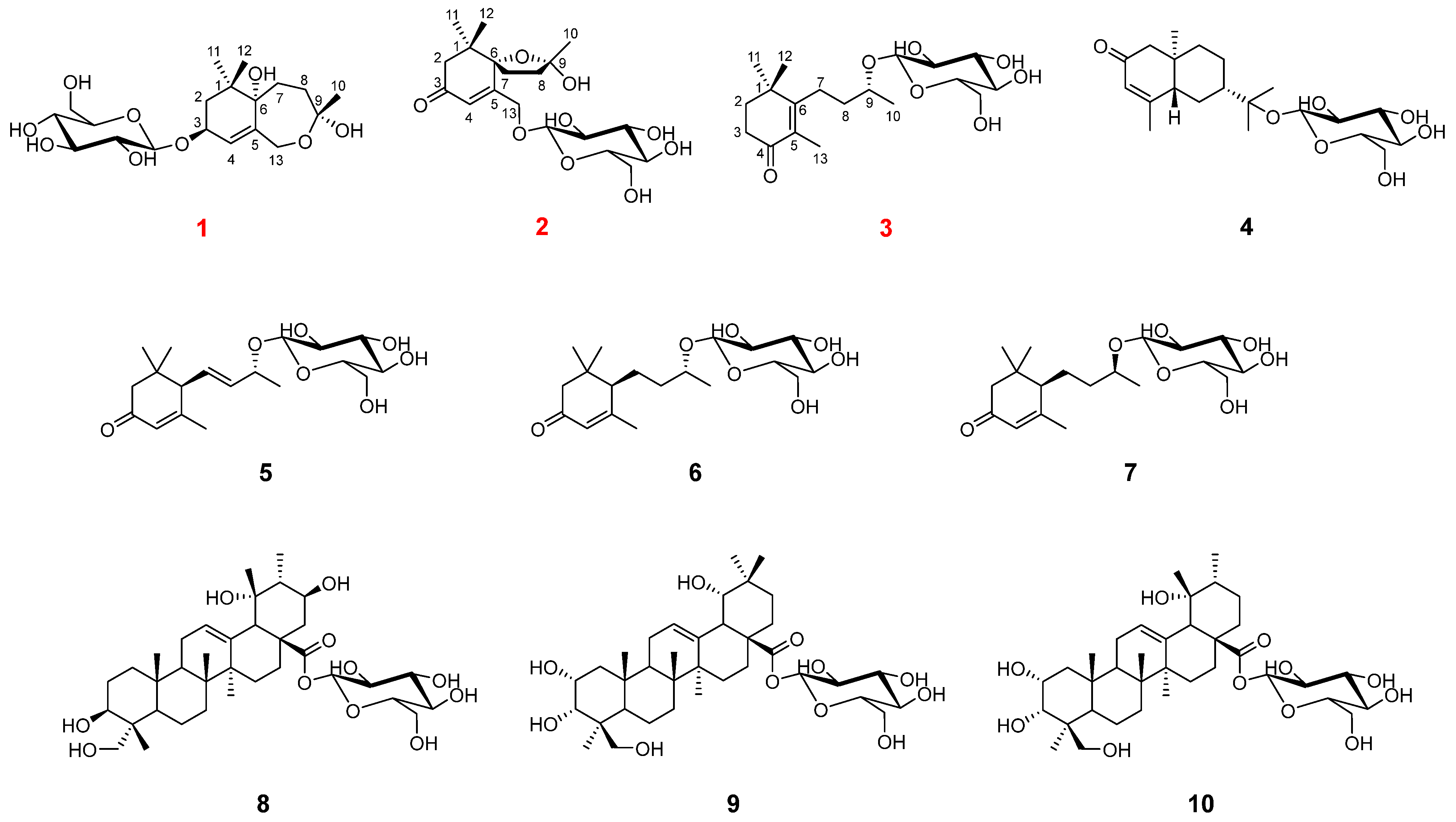
2.1. Structure Elucidation
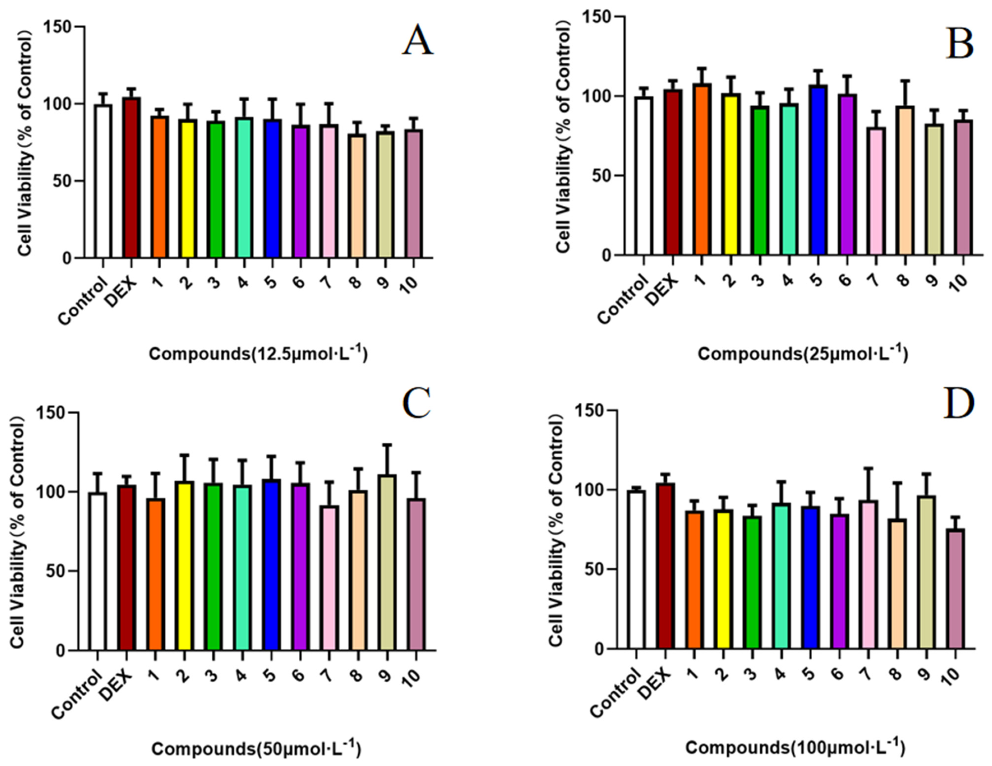
Compound 1 was obtained as colourless gum, and its molecular formula was established as C19H32O9 from its HRESIMS data (m/z 373.1937 [M + Na]+, calcd for C19H32O9Na, 427.1939). The 1H NMR spectrum of 1 (Table 1) displayed the signals of an olefinic proton at δH 5.77 (1H, br s, H-4), two oxygenated methylene protons at δH 4.48 (1H, d, J = 12.3 Hz, H-13a) and 4.02 (1H, d, J = 12.3 Hz, H-13b), an oxygenated methine proton at δH 4.36 (1H, dd, J = 6.4, 4.7 Hz, H-3), and three methyl groups at δH 1.39 (3H, s, H3-10), 0.99 (3H, s, H3-11), and 0.96 (3H, s, H3-12). The corresponding 13C NMR data (Table 1) showed 19 carbon signals, including one double bond pair at δC 124.8 (C-4) and 137.3 (C-5); four oxygenated carbons at δC 73.9 (C-3), 85.4 (C-6), 108.3 (C-9), and 66.7 (C-13); one quaternary carbon at δC 35.8 (C-1); three methylenes at δC 41.8 (C-2), 29.8 (C-7), and 35.7 (C-8); three methyls at δC 24.3 (C-10), 23.5 (C-11), and 24.1 (C-12); along with a β-glucose at δC 103.5 (C-1′), 75.1 (C-2′), 78.0 (C-3′), 71.6 (C-4′), 77.9 (C-5′), and 62.8 (C-6′). The sugar moiety was determined as D-glucose by LC-MS analyses after acid hydrolysis and derivatization. The 1H-1H COSY correlations (Figure 2) including H2-2/H-3/H-4 and H2-7/H2-8 led to the establishment of two coupled proton groups. The HMBC correlations (Figure 2) from H-4 to C-2/C-6/C-13, from H2-13 to C-4/C-6/C-9, from H2-7 to C-9, from H2-8 to C-6, and from H3-10 to C-8/C-13, combined with the degree of unsaturation, indicated that 1 was a bicyclic ionone glycoside and similar to 7,8-dihydro-3β,6α-dihydroxy-α-ionone 9-O-β-D-glucopyranoside [13], except the sugar was located at C-3 instead of C-9, and a hemiacetal structure was formed between C-9 and C-13. Those changes were verified via HMBC relationships (Figure 2) of H-1′/C-3, H2-13/C-9, and H3-10/C-13, respectively. Meanwhile, 1 was a novel ionone with a 6/7 bicyclic skeleton. In the NOESY spectrum (Figure 3), the correlations between H3-11 and H-3 indicated that OH-3 and H3-12 are located at the same side and regarded as β-oriented; the correlations between H3-12 and H2-7/H3-10 indicated that H2-7 and H3-10 are also β-oriented. So, the most probable configuration of 1 is either 3S, 6S, 9R or 3R, 6R, 9S. The absolute configurations at the stereogenic centres of 1 were determined to be 3S, 6S, 9R by comparison of the experimental ECD curve and calculated electronic circular dichroism (ECD) data at the B3LYP/6-311G(d,p) level in MeOH (Figure 4). Therefore, compound 1 was determined as shown in Figure 1, and named as capitsesqside A.
Compound 2 possessed the molecular formula C19H30O9, as determined by a quasi-molecular ion peak [M + Na]+ at m/z 425.1782 (calcd for C19H30O9Na, 425.1782) in its HRESIMS spectrum. The characteristic signals of an olefinic proton at δH 6.19 (1H, s, H-4); two oxygenated methylene protons at δH 4.73 (1H, dd, J = 18.4, 2.0 Hz, H-13a) and 4.54 (1H, dd, J = 18.4, 2.0 Hz, H-13b); and three methyl groups at δH 1.52 (3H, s, H3-10), 1.03 (3H, s, H3-12), and 1.01 (3H, s, H3-11) were presented in the 1H NMR spectrum. The 13C NMR and HSQC spectra (Table 1) showed that 2 contains a keto carbonyl group, one olefinic bond, three quaternary carbons, four methylenes, three methyls, and a glucose moiety. Furthermore, a β-glucose moiety was observed by the signals [δH 4.33 (1H, d, J = 7.7 Hz, H-1′), 3.26 (1H, m, H-2′), 3.36 (1H, m, H-3′), 3.36 (1H, m, H-4′), 3.26 (1H, m, H-5′), 3.86 (1H, dd, J = 12.0, 2.0 Hz, H-6′a), 3.68 (1H, dd, J = 12.0, 5.3 Hz, H-6′b)], and [δC 104.0 (C-1′), 75.0 (C-2′), 78.1 (C-3′), 71.6 (C-4′), 78.1 (C-5′), 62.7 (C-6′)]. The D-configuration of glucose was deduced based on LC-MS analyses after acid hydrolysis and derivatization. Combination with the 1H-1H COSY correlation (Figure 2) of H2-7/H2-8 as well as the HMBC correlations (Figure 2) from H3-11/H3-12 to C-1, H-4 to C-3/C-5/C-6, H2-7 to C-9, and H2-8/H3-10 to C-6 suggested that 2 was also a bicyclic ionone glycoside, and the aglycone of 2 was similar to (2S,5S)-2-hydroxy-2,6,10,10-tetramethyl-1-oxaspiro[4.5]dec-6-en-8-one [14]. The difference was that the CH3-13 was oxidised to CH2OH-13 in 2, which can be proved by the chemical shifts of CH2OH-13 (δH 4.73/4.54, δC 67.9). The HMBC correlations of H-1′ with C-13 suggested that the sugar moiety linked at the C-13 (Figure 2). Relative configurations of the stereogenic centres in 2 were determined by the NOESY correlations (Figure 3) of H3-10 with H3-11/H3-12 [15]. Finally, the absolute configuration of compound 2 was defined as 6S,9S by comparison of the experimental and calculated ECD spectra (Figure 4). Consequently, the structure of 2 was finally determined as shown in Figure 1 and named as capitsesqside B.
Compound 3 was a colourless gum. The molecular formula of 3 was deduced by a quasi-molecular ion peak [M + Na]+ at m/z 395.2037 (calcd for C19H32O7Na, 395.2040) in the HRESIMS spectrum. The 1H NMR data (Table 1) showed the characteristic signals: an oxygenated methine proton at δH 3.97 (1H, m, H-9) and four methyl groups at δH 1.76 (3H, s, H3-13), 1.23 (3H, d, J = 6.2 Hz, H3-10), 1.20 (3H, s, H3-11), and 1.20 (3H, s, H3-12). The 13C NMR spectrum showed a total of 19 carbons, including four methyls, four methylenes, one methine, four quaternary carbons, and a glucose signal. The sugar moiety in 3 was also identified as D-glucose by LC-MS analyses after acid hydrolysis and derivatization. A comparison of the NMR data of 3 with those of phoebenoside A [16] indicated that they possess the same planar structures as hydroxymegastigman-5-en-4-one 9-O-β-D-glucopyranoside. The absolute configuration of C-9 in phoebenoside A was S. The chemical shifts of C-9 (δC 75.7), C-10 (δC 19.8), and C-1′ (δC 102.2) in 3 confirmed the 9R configuration by comparison with the corresponding data of (3R,9S)-megastigman-5-en-3,9-diol 9-O-β-D-glucopyranoside [C-9 (δC 77.9), C-10 (δC 21.8), and C-1′ (δC 103.9)] and (3R,9R)-megastigman-5-en-3,9-diol 9-O-β-D-glucopyranoside [C-9 (δC 76.1), C-10 (δC 19.8), and C-1′ (δC 102.2)] in the same solvent CD3OD [17]. Therefore, the structure of 3 was defined as (9R)-hydroxymegastigman-5-en-4-one 9-O-β-D-glucopyranoside and named as capitsesqside C.
The seven known compounds (410) isolated from R. capitatum were shown to be (5R,7R,10S)-isopterocarpolone β-D-glucopyranoside (4) [18], (6R,9R)-3-oxo-α-ionol β-D-glucopyranoside (5) [19], (6R,9R)-3-oxo-α-ionol glucosides (6) [19], (6R,9S)-3-oxo-α-ionol glucosides (7) [20], incarvilloside A (8) [21], incarvilloside B (9) [21], and 2α,3α,19α,24-tetrahydroxyurs-12-en-28-oic acid β-D-glucopyranosyl ester (10) [22].

2.2. Effect of Compounds 110 on the LPS-Induced Production of NO

The NOD-like receptor family pyrin domain containing 3 (NLRP3) protein is a member of the inflammatory vesicle protein family, and aberrant activation of this protein has been implicated in the pathogenesis of several inflammatory diseases. Inducible nitric oxide synthase (iNOS) and its product NO play an important role in the inflammatory response and oxidative stress, which is one of the major molecular mechanisms of inflammation. The MTT assay was used to examine the cytotoxicity of compounds 110 in RAW 264.7 cells. The results showed no significant cytotoxicity of any compounds at the testing concentrations (Figure 5). Compounds 110 were examined for inhibition of NO production in LPS (lipopolysaccharide)-induced RAW 264.7 cells by using the Griess method (Figure 6). In comparison with the positive control, the compounds 2 and 5 exhibited stronger NO inhibitory activity, and other compounds showed a slight inhibitory effect on NO production (Figure 6). The results of the molecular docking analysis indicated that compound 2 exhibited a high affinity for NLRP3 and iNOS, with binding energies of −6.52 and −6.64 kcal/mol, respectively (Figure 7). The carbonyl group at C-3 and hydroxy group at C-9 interact by hydrogen bonding in the NLRP3 pocket with amino acid residues PHE-575 and ALA-228, respectively. Moreover, the hydroxy group at C-9 also forms a hydrogen bonding interaction with PHE-363 in the iNOS pocket. However, compound 5 interacts more weakly than compound 2 with NLRP3 and iNOS.

3. Materials and Methods

3.1. General Experimental Procedures

UV and CD spectra were performed on an Applied Photophysics Chirascan spectrometer (Applied Photophysics Ltd., Surrey, UK). Optical rotations were determined using an Auton Paar MCP300 automatic polarimeter (Anton Paar GmbH, Graz, Austria). A Bruker AM-400 spectrometer with tetramethylsilane (TMS) as internal standard was used to acquire 1D and 2D NMR spectra. HRESIMS data were performed on a QTOF 6600 mass spectrometer (AB-SCIEX, Foster City, CA, USA). The TripleTOF 6600 UPLC/MS spectrometer (AB-SCIEX, Foster City, CA, USA) with a Kinetex C18 100Å column (2.6 μm, 2.1 × 50 mm) was used to analyse the test sample and standard sugar, coupled to an electrospray ionisation (ESI) interface. Sephadex LH-20 (GE Healthcare, Boston, MA, USA), MCI gel CHP20, and silica gel (100~200 mesh, 200~300 mesh, Qingdao Marine Chemical Co., Ltd., Qingdao, China) were employed for column chromatography (CC). Semipreparative HPLC was achieved on an Agilent 1100 series system using a 9.4 mm × 250 mm, 5 μm, YMC C18 column. Thin-layer chromatography (TLC) was performed on silica gel 60F254 and RP-18 F254S plates (Merck KGaA, Darmstadt, Germany). Reagents for the glucose discrimination (L-cysteine methyl ester hydrochloride and o-tolyl isothiocyanate) were purchased from Shanghai Macklin Biochemical Co., Ltd. (Shanghai, China), and D-glucuronic acid was obtained from Shanghai Yuanye Biotechnology Co., Ltd. (Shanghai, China). All other chemicals utilised in this study were of analytical grade.

3.2. Plant Material

The aerial parts of R. capitatum were collected in Mount Taibai, Shaanxi Province, in June 2019 (GPS coordinates: 107°25′–107°52′ E, 33°55′–33°35′ N) and identified by Dr. Zhen-Hai Wu, College of Life Sciences, Northwest A&F University. A voucher specimen (No. WUK 0480711) could be found in the Herbarium of the College of Life Sciences, Northwest A&F University.

3.3. Extraction and Isolation

The dried and crushed aerial parts of R. capitatum (8.7 kg) were dried and crushed and extracted four times with MeOH (30 L) for 3 h each time, resulting in a crude extract (1.2 kg). The crude extract was then dissolved in a triple amount of H2O water and partitioned sequentially with equal volumes of petroleum ether, EtOAc, and n-butanol to obtain three partitions. The n-butanol fraction (450.0 g) was separated into six fractions (Fr.A1~Fr.A6) using a MCI gel CHP 20P column and eluted with EtOH (0%, 20%, 40%, 60%, 80%, and 95%). Silica gel flash column chromatography (FCC) with dichloromethane/MeOH (40:1 to 1:1) was used to purify Fr.A4 (130.0 g), resulting in four fractions: Fr.A4-1~Fr.A4-4. Fr.A4-2 (5.0 g) was then divided into five subfractions (Fr.A4-2-1~Fr.A4-2-5) using silica gel FCC with dichloromethane/MeOH (40:1 to 2:1). The major fraction, Fr.A4-2-3 (1.4 g), was further purified by Sephadex LH-20 column eluting with MeOH, resulting in four subfractions: Fr.A4-2-3-1~Fr.A4-2-3-4. The Fr.A4-2-3-1 (322.0 mg) fraction was separated by semipreparative HPLC (MeOH/H2O, 49%:51%; flow rate: 2 mL/min) to obtain compound 1 (8.0 mg; tR = 18.5 min), compound 2 (4.0 mg; tR = 22.8 min), compound 3 (23.0 mg; tR = 25.6 min), compound 4 (7.0 mg; tR = 28.9 min), compound 5 (5.1 mg; tR = 15.7 min), compound 6 (3.1 mg; tR = 20.2 min), and compound 7 (3.2 mg; tR = 21.2 min). Fr.A5 (60.0 g) was separated on a silica gel column using dichloromethane/MeOH (20:1 to 2:1) elution to yield six subfractions (Fr.A5-1~Fr.A5-6). Fr.A5-5 (1.7 g) was subjected to Sephadex LH-20 column elution with MeOH to obtain subfractions Fr.A5-5-1~Fr.A5-5-5. Fr.A5-5-3 (640.0 mg) was then purified by semipreparative HPLC (MeOH/H2O, 50%:50%; flow rate: 2 mL/min) to yield compounds 8 (136.0 mg; tR = 27.2 min), 9 (12.0 mg; tR = 31.4 min), and 10 (28.0 mg; tR = 33.8 min).
Capitsesqside A (1): Colourless gum; [α ] D 20 + 25.4 (c 0.1, MeOH); UV (MeOH) λmax (log ε): 200.0 (2.30) nm; CD (MeOH) λmaxε) 206 (– 24.86); 1H and 13C NMR data see Table 1; HRESIMS [M + Na]+ m/z 427.1937 (calcd for C19H32O9Na, 427.1939).
Capitsesqside B (2): Colourless gum; [α ] D 20 + 19.1 (c 0.1, MeOH); UV (MeOH) λmax (log ε): 238.0 (2.39); CD (MeOH) λmaxε) 220 (+ 18.10), 247 (– 22.34); 1H and 13C NMR data see Table 1; HRESIMS [M + Na]+ m/z 425.1782 (calcd for C19H30O9Na, 425.1782).
Capitsesqside C (3): Colourless gum; [α ] D 20 + 22.0 (c 0.1, MeOH); UV (MeOH) λmax (log ε): 200.0 (2.65), 245.0 (2.64) nm; 1H and 13C NMR data see Table 1; HRESIMS [M + Na]+ m/z 395.2037 (calcd for C19H32O7Na, 395.2040).

3.4. Acid Hydrolysis of Compounds 13

Compounds 13 (1 mg) were dissolved in 2 N HCl (3 mL) and stirred in water at 90 °C for 2.5 h. The resulting acid aqueous solutions were then concentrated under reduced pressure. To each residue, 1 mL of water was added, and the resulting solutions were extracted with 3 × 1 mL of EtOAc. The residue was dissolved in pyridine (1 mL) containing L-cysteine methyl ester hydrochloride (1 mg, Macklin, Shanghai, China) and stirred at 60 °C for 1.5 h. To each mixture, o-tolyl isothiocyanate (20.0 μL, Macklin, Shanghai, China) was added, and the resulting mixture was stirred at 60 °C for an additional 1.5 h. Finally, the compounds were analysed directly by LC-MS [23].

3.5. ECD Calculations

Gaussian 16 software was used to perform ECD calculations. Conformation optimization was carried out in the gas phase using density functional theory (DFT) at the B3LYP/6-31G(d) level. Time-dependent density functional theory was also employed. (TDDFT) ECD calculations were carried out in MeOH (PCM) using the B3LYP/6-311G(d,p) level, and ECD spectra were obtained with SpecDis 1.7 [24].

3.6. Cell Culture

The murine macrophage RAW 264.7 cells were obtained from the Institute of Materia Medica, Chinese Academy of Medical Sciences and Peking Union Medical College. They were regularly maintained at 37 °C in DMEM (Cbico, New York, NY, USA) containing 10% FBS (ABW, Frickenhausen, German) and 100 U/mL penicillin (Solarbio, Beijing, China) in a humidified 5% CO2 atmosphere.

3.7. MTT Assay for Cytotoxicity

The MTT assay was used to determine cytotoxicity after pretreatment with compounds 110. RAW 264.7 cells were plated at a density of 8 × 10³ cells/well in a 96-well plate and incubated for 24 h before sample treatment. The cells were then pretreated with various concentrations of compounds (12.5, 25, 50, and 100 μM) for 24 h. Next, 10 μL of 5 mg/mL MTT was added to each well, followed by an additional 4 h incubation. The cultured medium was removed, and the formazan crystals were dissolved in 150 μL of DMSO. The absorbance was measured at 490 nm using a Multidetection microplate reader (BioTek Instruments, Inc., Winooski, VT, USA).

3.8. Measurement of NO

The concentration of nitrite was measured to indicate NO production using the Griess reaction. Briefly, RAW 264.7 cells were seeded into 96-well tissue culture plates at a density of 2 × 10⁴ cells/mL and stimulated with 1 μg/mL of LPS in the presence or absence of compounds. After incubation at 37 °C for 24 h, 50 μL of cell-free supernatant was mixed with 100 μL of Griess reagent. The mixture was then reacted at 37 °C for 10 min while avoiding light. Absorbance was measured at 550 nm against a calibration curve with sodium nitrite standards.

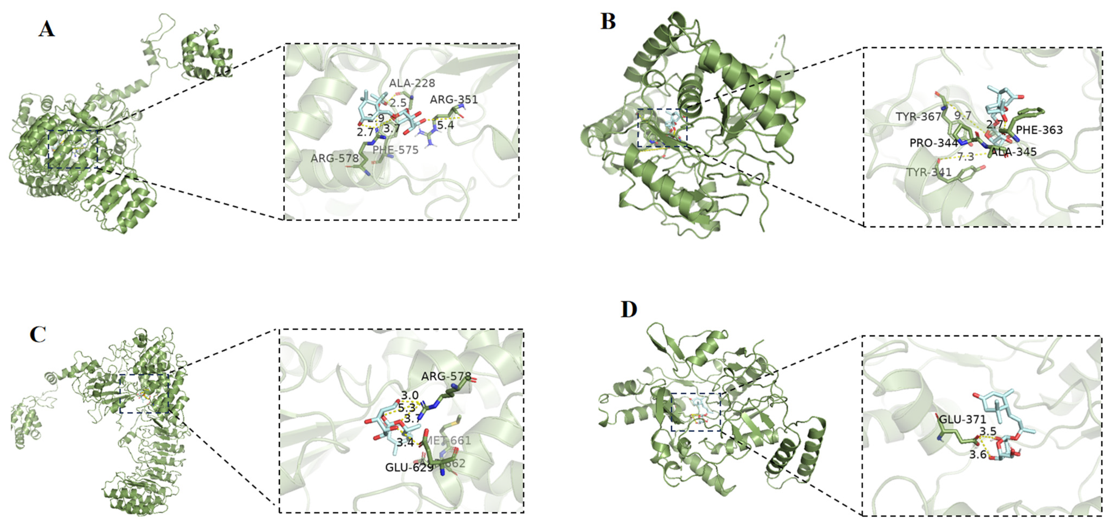
3.9. Molecular Docking

Molecular docking studies were performed to predict the binding interaction of compounds 2 and 5 to NLRP3 (PDB ID: 7PZC) and iNOS (PDB ID: 3E7G) using the software Autodock 4.2 Vina along with AutoDock Tools (ADT 1.5.6) according to the previously described method [25].

4. Conclusions

In conclusion, three novel ionone glycosides, capitsesqsides A−C (13), together with seven known compounds were isolated from R. capitatum. Among them, compound 1 was a rare 6/7 bicyclic skeleton ionone. Compounds 2 and 5 showed potent anti-inflammatory activity in LPS-induced NO production in RAW 264.7 cells. Compared to compounds 2, 3, and 57, compound 1 showed reduced activity, suggesting that an α,β-unsaturated ketone group may be an active unit, while compound 4 and three triterpenoids (810) showed weak NO inhibitory activity. Molecular docking results suggested that NLRP3 and iNOS may be potential target proteins for the anti-inflammatory activity of compound 2.

Supplementary Materials

The following supporting information can be downloaded at: https://www.mdpi.com/article/10.3390/molecules29112462/s1, Figures S1–S6: The 1D and 2D NMR spectra of compound 1 in CD3OD, Figure S7: The HRESIMS spectrum of compound 1, Figures S8 and S9: The UV and ECD spectra of compound 1, Figure S10: The LC-MS spectrum of the glucose moiety of compound 1, Figures S11–S16: The 1D and 2D NMR spectra of compound 2 in CD3OD, Figure S17: The HRESIMS spectrum of compound 2, Figures S18 and S19: The UV and ECD spectra of compound 2, Figure S20: The LC-MS spectrum of the glucose moiety of compound 2, Figures S21–S25: The 1D and 2D NMR spectra of compound 3 in CD3OD, Figure S26: The HRESIMS spectrum of compound 3, Figure S27: The UV spectrum of compound 3, Figure S28: The LC-MS spectrum of the glucose moiety of compound 3, Figure S29: The LC-MS spectrum of the D-glucose.

Author Contributions

J.-R.Y.: writing original draft. Y.-T.Z.: isolation and identification of compounds. Y.-Q.Z.: HRESIMS test. H.-Q.L.: cell test. C.-H.L.: language polishing and writing revision, corresponding author. J.-M.G.: designing experiment and funding acquisition, corresponding author. All authors have read and agreed to the published version of the manuscript.

Funding

This research was funded by National Natural Science Foundation of China (No. 32070388).

Institutional Review Board Statement

Not applicable.

Informed Consent Statement

Not applicable.

Data Availability Statement

The data presented in this study are available in the Supplementary Materials.

Conflicts of Interest

Author Jun-Ren Yang was employed by the company, Shaanxi Jiahe Phytochem Co., Ltd. The funder was not involved in the study design, collection, analysis, interpretation of data, the writing of this article or the decision to submit it for publication.

References

  1. Chen, X.; Cao, Y.G.; Ren, Y.J.; Liu, Y.L.; Fan, X.L.; He, C.; Li, X.D.; Ma, X.Y.; Zheng, X.K.; Feng, W.S. Ionones and lignans from the fresh roots of Rehmannia glutinosa. Phytochemistry 2022, 203, 113423. [Google Scholar] [CrossRef] [PubMed]
  2. Bendjedou, H.; Maggi, F.; Bennaceur, M.; Mancinelli, M.; Benamar, H.; Barboni, L. A new ionone derivative from Lycium intricatum Boiss. (Solanaceae). Nat. Prod. Res. 2022, 36, 687–694. [Google Scholar] [CrossRef] [PubMed]
  3. Liu, C.; Cheng, R.R.; Han, Z.Z.; Yang, Y.B.; Zhou, Y.; Yang, L.; Wang, Z.T. A new ionone derivative from the leaves of Picrasma quassioides. J. Asian Nat. Prod. Res. 2019, 21, 652–658. [Google Scholar] [CrossRef]
  4. Liang, X.X.; Li, Q.; Li, H.J.; Ning, Y.M.; Zhang, R.H.; Zhang, X.J.; Li, X.L.; Xiao, W.L. Centrantheroside F, a new ionone glycoside from Centranthera grandiflora. J. Asian Nat. Prod. Res. 2022, 24, 777–783. [Google Scholar] [CrossRef] [PubMed]
  5. Zhou, J.; Shi, G.R.; Zhang, W.Q.; Chen, R.Y.; Liu, Y.F.; Yu, D.Q. Four ionones and ionone glycosides from the whole plant of Rehmannia piasezkii. J. Asian Nat. Prod. Res. 2022, 24, 955–962. [Google Scholar] [CrossRef]
  6. Hu, B.; Hu, H.; Pu, C.; Peng, D.; Wei, Z.; Kuang, H.; Wang, Q. New Ionone glycosides from the aerial parts of Allium sativum and their anti-platelet aggregation activity. Planta Med. 2023, 89, 729–734. [Google Scholar] [CrossRef]
  7. Liao, H.B.; Huang, G.H.; Yu, M.H.; Lei, C.; Hou, A.J. Five pairs of meroterpenoid enantiomers from Rhododendron capitatum. J. Org. Chem. 2017, 82, 1632–1637. [Google Scholar] [CrossRef] [PubMed]
  8. Liao, H.B.; Lei, C.; Gao, L.X.; Li, J.Y.; Li, J.; Hou, A.J. Two enantiomeric pairs of meroterpenoids from Rhododendron capitatum. Org. Lett. 2015, 17, 5040–5043. [Google Scholar] [CrossRef] [PubMed]
  9. Liang, C.; Kjaerulff, L.; Hansen, P.R.; Kongstad, K.T.; Staerk, D. Dual high-resolution α-glucosidase and PTP1B inhibition profiling combined with HPLC-PDA-HRMS-SPE-NMR analysis for the identification of potentially antidiabetic chromene meroterpenoids from Rhododendron capitatum. J. Nat. Prod. 2021, 84, 2454–2467. [Google Scholar] [CrossRef]
  10. He, J.; Shang, X.; Dai, L.; Yang, X.; Li, B.; Wei, Y.; Zhang, J.; Pan, H. Chemical constituents, antibacterial, acaricidal and anti-inflammatory activities of the essential oils from four Rhododendron species. Front. Vet. Sci. 2022, 9, 882060. [Google Scholar] [CrossRef]
  11. Xie, J.Y.; Li, P.; Yan, X.T.; Gao, J.M. Discovery from Hypericum elatoides and synthesis of hyperelanitriles as α-aminopropionitrile-containing polycyclic polyprenylated acylphloroglucinols. Commun. Chem. 2024, 7, 1. [Google Scholar] [CrossRef] [PubMed]
  12. Yan, X.T.; Chen, J.X.; Wang, Z.X.; Zhang, R.Q.; Xie, J.Y.; Kou, R.W.; Zhou, H.F.; Zhang, A.L.; Wang, M.C.; Ding, Y.X.; et al. Hyperhubeins A–I, Bioactive Sesquiterpenes with Diverse Skeletons from Hypericum hubeiense. J. Nat. Prod. 2023, 86, 119–130. [Google Scholar] [CrossRef] [PubMed]
  13. De Tommasi, N.; Piacente, S.; De Simone, F.; Pizza, C. Constituents of Cydonia vulgaris: Isolation and structure elucidation of four new flavonol glycosides and nine new α-ionol-derived glycosides. J. Agric. Food Chem. 1996, 44, 1676–1681. [Google Scholar] [CrossRef]
  14. Xiong, L.; Zhou, Q.M.; Peng, C.; Xie, X.F.; Guo, L.; Li, X.H.; Liu, J.; Liu, Z.H.; Dai, O. Sesquiterpenoids from the herb of Leonurus japonicus. Molecules 2013, 18, 5051–5058. [Google Scholar] [CrossRef] [PubMed]
  15. Knapp, H.; Weigand, C.; Gloser, J.; Winterhalter, P. 2-Hydroxy-2,6,10,10-tetramethyl-1-oxaspiro[4.5]dec-6-en-8-one: Precursor of 8,9-dehydrotheaspirone in white-fleshed nectarines. J. Agric. Food Chem. 1997, 45, 1309–1313. [Google Scholar] [CrossRef]
  16. Viet Thanh, N.T.; Minh, T.T.; Linh, N.T.; Phuong Ly, G.T.; Trang, D.T.; Tai, B.H.; Kiem, P.V. Megastigmane glycosides from Phoebe tavoyana. Nat. Prod. Commun. 2019, 14, 1934578X1985243. [Google Scholar] [CrossRef]
  17. Yamano, Y.; Shimizu, Y.; Ito, M. Stereoselective synthesis of optically active 3-hydroxy-7,8-dihydro-β-ionol-glucosides. Chem. Pharm. Bull. 2003, 51, 878–882. [Google Scholar] [CrossRef] [PubMed]
  18. Kitajima, J.; Kamoshita, A.; Ishikawa, T.; Takano, A.; Fukuda, T.; Isoda, S.; Ida, Y. Glycosides of Atractylodes iancea. Chem. Pharm. Bull. 2003, 51, 673–678. [Google Scholar] [CrossRef] [PubMed]
  19. Pabst, A.; Barron, D.; Sémon, E.; Schreier, P. Two diastereomeric 3-oxo-α-ionol β-D-glucosides from raspberry fruit. Phytochemistry 1992, 31, 1649–1652. [Google Scholar] [CrossRef]
  20. Takeda, Y.; Zhang, H.; Masuda, T.; Honda, G.; Otsuka, H.; Sezik, E.; Yesilada, E.; Sun, H. Megastigmane glucosides from Stachys byzantina. Phytochemistry 1997, 44, 1335–1337. [Google Scholar] [CrossRef]
  21. Ge, X.; Lin, D.C.; Zhang, W.D.; Zhang, X.R. Triterpenoid saponins and monoterpenoid glycosides from Incarvillea delavayi. J. Asian Nat. Prod. Res. 2009, 11, 838–844. [Google Scholar] [CrossRef]
  22. Ono, M.; Chikuba, T.; Mishima, K.; Yamasaki, T.; Ikeda, T.; Yoshimitsu, H.; Nohara, T. A new diterpenoid and a new triterpenoid glucosyl ester from the leaves of Callicarpa japonica Thunb. var. luxurians Rehd. J. Nat. Med. 2009, 63, 318–322. [Google Scholar] [CrossRef]
  23. Zhu, Y.T.; Fang, H.B.; Liu, X.N.; Yan, Y.M.; Feng, W.S.; Cheng, Y.X.; Wang, Y.Z. Unusual acetylated flavonol glucuronides, oxyphyllvonides A-H with renoprotective activities from the fruits of Alpinae oxyphylla. Phytochemistry 2023, 215, 113849. [Google Scholar] [CrossRef]
  24. Bruhn, T.; Schaumlöffel, A.; Hemberger, Y.; Bringmann, G. SpecDis: Quantifying the comparison of calculated and experimental electronic circular dichroism spectra. Chirality 2013, 25, 243–249. [Google Scholar] [CrossRef]
  25. Kou, R.W.; Han, R.; Gao, Y.Q.; Li, D.; Yin, X.; Gao, J.M. Anti-neuroinflammatory polyoxygenated lanostanoids from Chaga mushroom Inonotus obliquus. Phytochemistry 2021, 184, 112647. [Google Scholar] [CrossRef]
Figure 1. Structures of compounds 110 (13 were new compounds).
Molecules 29 02462 g001
Figure 2. Key HMBC (H→C,Molecules 29 02462 i001) and 1H-1H COSY (Molecules 29 02462 i002) correlations of compounds 13.
Molecules 29 02462 g002
Figure 3. Key NOESY (Molecules 29 02462 i003) correlations of compounds 1 and 2.
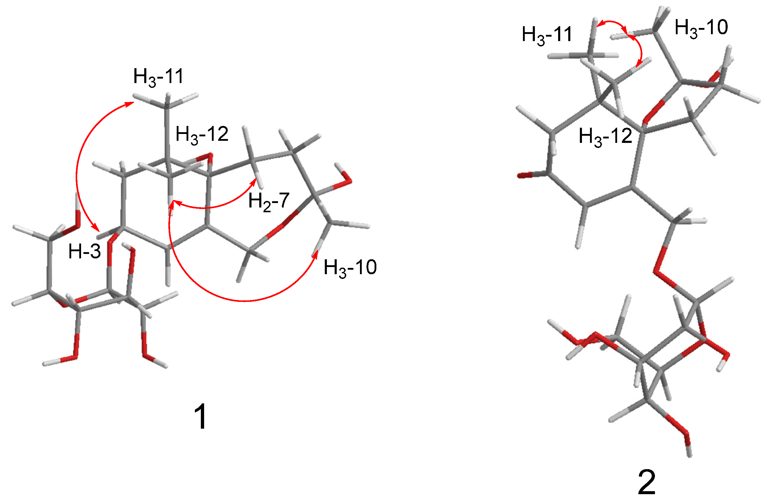
Molecules 29 02462 g003
Figure 4. Experimental and calculated ECD curves of compounds 1 and 2.
Molecules 29 02462 g004
Figure 5. Effects of compounds 110 on cell viability ((A): 12.5 μM, (B): 25 μM, (C): 50 μM, and (D): 100 μM). The concentrations of these compounds ranged from 12.5 to 100 μM. Analysis of cell viability was by GraphPad Prism (8.0.2), and data are expressed as the mean ± SD. Three independent experiments were performed. Dexamethasone (DEX) was used as a positive control.
Molecules 29 02462 g005
Figure 6. Effects of compounds 2, 3 and 510 on LPS-induced production of NO in RAW 264.7 cells. The concentrations of these compounds ranged from 12.5 to 100 μM ((A): compounds 2 and 3, (B): compounds 5 and 6, (C): compounds 7 and 8, and (D): compounds 9 and 10). Analysis of cell viability was by GraphPad Prism, and data are expressed as the mean ± SD. Compare with control, ## p < 0.01; compare with LPS, ns > 0.05, * p < 0.05, ** p < 0.01, *** p < 0.001. Three independent experiments were performed. Dexamethasone (DEX, 10 μM) was used as a positive control.
Molecules 29 02462 g006
Figure 7. (A): The molecular interactions of compound 2 with NLRP3 by molecular docking simulation. (B): The molecular interactions of compound 2 with iNOS by molecular docking simulation. (C): The molecular interactions of compound 5 with NLRP3 by molecular docking simulation. (D): The molecular interactions of compound 5 with iNOS by molecular docking simulation.
Molecules 29 02462 g007
Table 1. 13C NMR (100 MHz) and 1H NMR (400 MHz) data of 13 in CD3OD.
No.123
δCδHδCδHδCδH
135.8 43.0 37.6
241.81.75 (m)50.72.58 (d, 17.4)
1.20 (d, 17.4)
38.41.82 (t, 7.2)
373.94.36 (dd, 6.4, 4.7)200.7 35.12.45 (t, 7.2)
4124.85.77 (br s)122.66.19 (s)201.5
5137.3 168.1 131.6
685.4 91.41.50 (m)168.7
729.82.10 (dd, 12.8, 4.6)
1.75 (m)
31.72.44 (m)
2.10 (dd, 14.5, 5.9)
27.92.53 (m)
2.31 (m)
835.72.19 (m)
1.96 (dd, 12.8, 4.6)
40.02.10 (dd, 14.5, 5.9)
2.01 (dd, 14.5, 9.6)
37.21.67 (m)
9108.3 111.1 75.73.97 (m)
1024.31.39 (s)21.41.52 (s)19.81.23 (d, 6.2)
1123.50.99 (s)23.31.01 (s)27.21.20 (s)
1224.10.96 (s)24.91.03 (s)27.21.20 (s)
1366.74.48 (d, 12.3)
4.02 (d, 12.3)
67.94.73 (dd, 18.4, 2.0)
4.54 (dd, 18.4, 2.0)
11.71.76 (s)
1′-Gly103.54.40 (d, 7.8)104.04.33 (d, 7.7)102.24.35 (d, 7.8)
2′-Gly75.13.17 (m)75.03.26 (m)75.23.17 (dd, 9.0, 7.8)
3′-Gly78.03.35 (m)78.13.36 (m)77.93.37 (m)
4′-Gly71.63.28 (m)71.63.36 (m)71.83.37 (m)
5′-Gly77.93.28 (m)78.13.26 (m)78.23.28 (m)
6′-Gly62.83.86 (dd, 12.0, 2.2)
3.67 (dd, 12.0, 5.4)
62.73.86 (dd, 12.0, 2.0)
3.68 (dd, 12.0, 5.3)
62.93.88 (dd, 12.0, 2.0)
3.66 (dd, 12.0, 5.0)
Disclaimer/Publisher’s Note: The statements, opinions and data contained in all publications are solely those of the individual author(s) and contributor(s) and not of MDPI and/or the editor(s). MDPI and/or the editor(s) disclaim responsibility for any injury to people or property resulting from any ideas, methods, instructions or products referred to in the content.

