Article

Co-Doping Effect of Mn2+ and Eu3+ on Luminescence in Strontiowhitlockite Phosphors

by
Ivan V. Nikiforov
1,
Dmitry A. Spassky
2,3,*,
Nataliya R. Krutyak
2,3,
Roman Yu. Shendrik
4,
Evgenia S. Zhukovskaya
1,
Sergey M. Aksenov
5,6 and
Dina V. Deyneko
1,5
1
Department of Chemistry, Lomonosov Moscow State University, 119991 Moscow, Russia
2
Skobeltsyn Institute of Nuclear Physics, Lomonosov Moscow State University, 119991 Moscow, Russia
3
Institute of Physics, University of Tartu, 50411 Tartu, Estonia
4
Vinogradov Institute of Geochemistry, Siberian Branch of the Russian Academy of Sciences, 664033 Irkutsk, Russia
5
Laboratory of Arctic Mineralogy and Material Sciences, Kola Science Centre, Russian Academy of Sciences, 184209 Apatity, Russia
6
Geological Institute, Kola Science Centre, Russian Academy of Sciences, 184209 Apatity, Russia
*
Author to whom correspondence should be addressed.
Molecules 2024, 29(1), 124; https://doi.org/10.3390/molecules29010124
Submission received: 31 October 2023 / Revised: 14 December 2023 / Accepted: 16 December 2023 / Published: 24 December 2023

Abstract

:
A new series of Sr-based phosphates, Sr9−xMnxEu(PO4)7, were synthesized using the high-temperature solid-state method in air. It was found that these compounds have the same structure as strontiowhitlockite, which is a β-Ca3(PO4)2 (or β-TCP) structure. The concentration of Mn2+ ions required to form a pure strontiowhitlockite phase was determined. An unusual partial reduction of Eu3+ to Eu2+ in air was observed and confirmed by photoluminescence (PL) and electron spin resonance (ESR) spectra measurements. The PL spectra recorded under 370 nm excitation showed transitions of both 4f5d–4f Eu2+ and 4f–4f Eu3+. The total integral intensity of the PL spectra, monitored at 395 nm, decreased with increasing Mn2+ concentration due to quenching effect of Eu3+ by the Mn2+ levels. The temperature dependence of Eu2+ photoluminescence in a Sr9−xMnxEu(PO4)7 host was investigated. The conditions for the reduction of Eu3+ to Eu2+ in air were discussed.

1. Introduction

One of the primary objectives in addressing light-emitting diode (LED) issues is to identify an optimal host for harnessing the photoluminescence properties of cation activators. Another objective is to regulate emissions through chemical deposition. Research has clearly shown that certain phosphates [1,2], aluminates [3,4], silicates [5,6], glasses [7,8], and so on serve as excellent hosts for rare-earth elements and transition metals with photoluminescence properties in the visible region. However, each of these hosts has its drawbacks, such as a high synthesis temperature with reduced atmosphere [9], non-green chemistry preparation techniques [10], low isomorphic capacity, and variation in the substitution types [11]. In this context, β-Ca3(PO4)2-type (or β-TCP) phosphors are considered suitable materials due to their wide capacity for substitutions of Ca2+ ions with either homovalent or heterovalent ions, resulting in excellent properties [12].
Calcium-based phosphates with a β-TCP structure are still of great interest. However, it has been shown that Sr-based phosphates with a β-Ca3(PO4)2 structure exhibit photoluminescence properties that are several times higher [13,14]. The related mineral—strontiowhitlockite [15], or Sr9Mg(PO4)6(PO3OH)—is the strontium analogue of whitlockite or β-Ca3(PO4)2 and also belongs to the cerite supergroup. The replacement of Ca2+ with Sr2+ ions leads to an increase in the photoluminescence properties [16] or their modification [17,18] due to the formation of more distorted luminescence center environments. It is worth noting that pure Sr3(PO4)2 differs from the β-Ca3(PO4)2 structure [19] and is known as a mineral palmierite family member. Furthermore, a comprehensive substitution of Ca2+ → Sr2+ was investigated in detail in [20]. It was shown that Sr-based phosphates form a β-Ca3(PO4)2 structure only with the stoichiometric formula Sr8M2+R3+(PO4)7 [21] or Sr9R´3+(PO4)7 (where R3 is a rare earth element, and R´3+ is a tripositive ion, such as Sc3+ or Fe3+, [2,22]). Despite numerous articles on the photoluminescence properties of strontiowhitlockite-type phosphors, the full concentration series with Sr2+M2+ or Sr2+R3+ has not been discussed. However, a similar series on Ca-based phosphates, such as Ca9−xM2+xEu(PO4)7, was previously described in detail for M2+ = Zn2+ [23], Mg2+ [24], and Mn2+ [25].
The control of luminescence properties is achieved by using different pairs of ions in the host material, such as Tb3+/Eu3+ [26], Sm3+/Eu3+ [27], Ce3+/Mn2+ [28], Ce3+/Tb3+ [29], Eu2+/ Mn2+ [30], and others. The choice of these pairs is based on the existence of energy transfer processes between them, resulting in unique combinations of emitting colors. Another method to modify photoluminescence is by changing the oxidation state [31,32] of the ion-activator.
In this study, we investigate Sr-based phosphors with the β-TCP structure and common formula Sr9−xMnxEu(PO4)7. The influence of Mn2+ and Eu3+ co-doping on the photoluminescence properties as well as the impact of homovalent Ca2+ → Mn2+ substitution on the structure formation are also investigated. The abnormal self-reduction process of Eu3+ ions in the strontiowhitlockite host in air was observed.

2. Results and Discussion

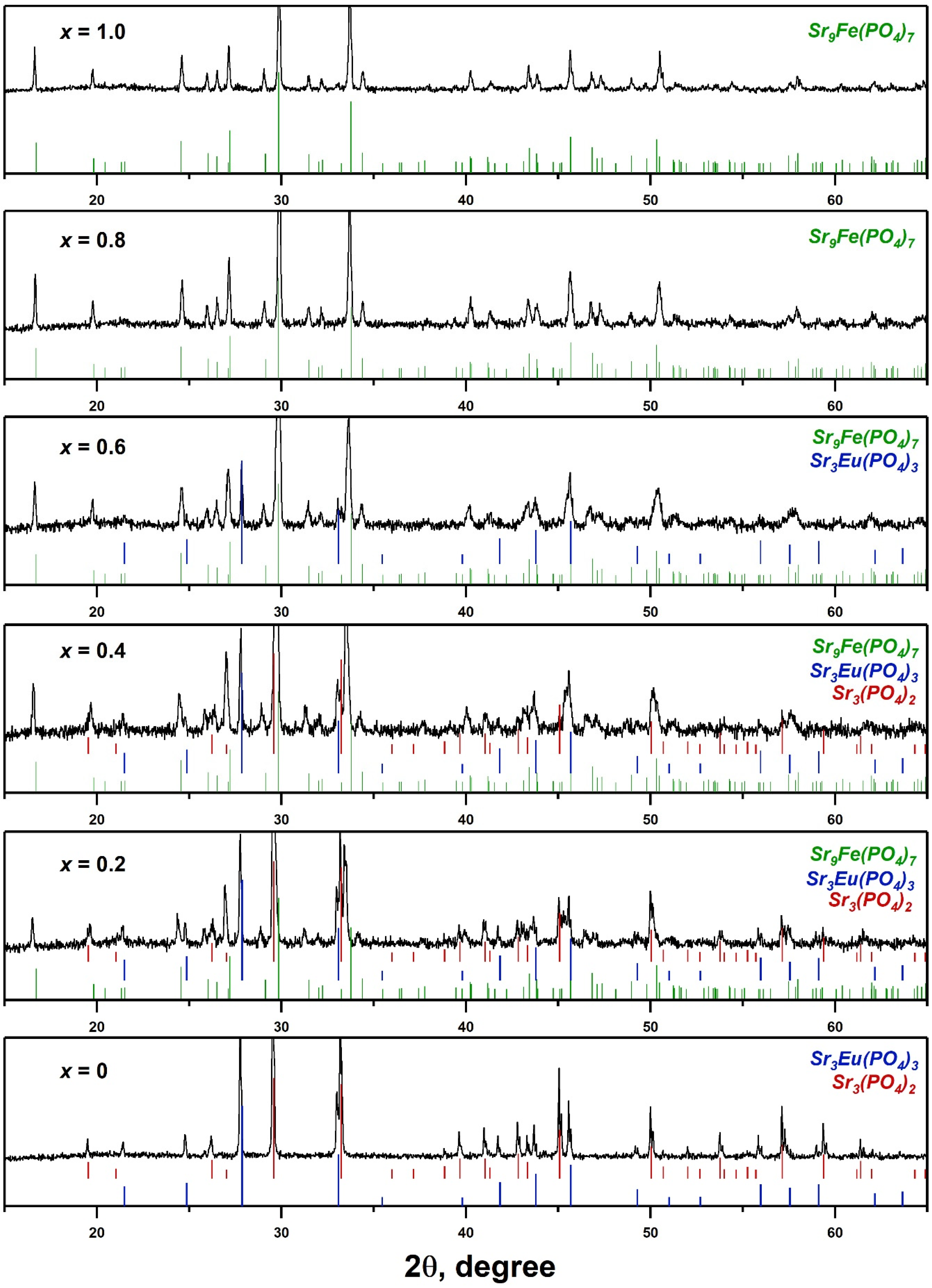
2.1. PXRD and SHG Study
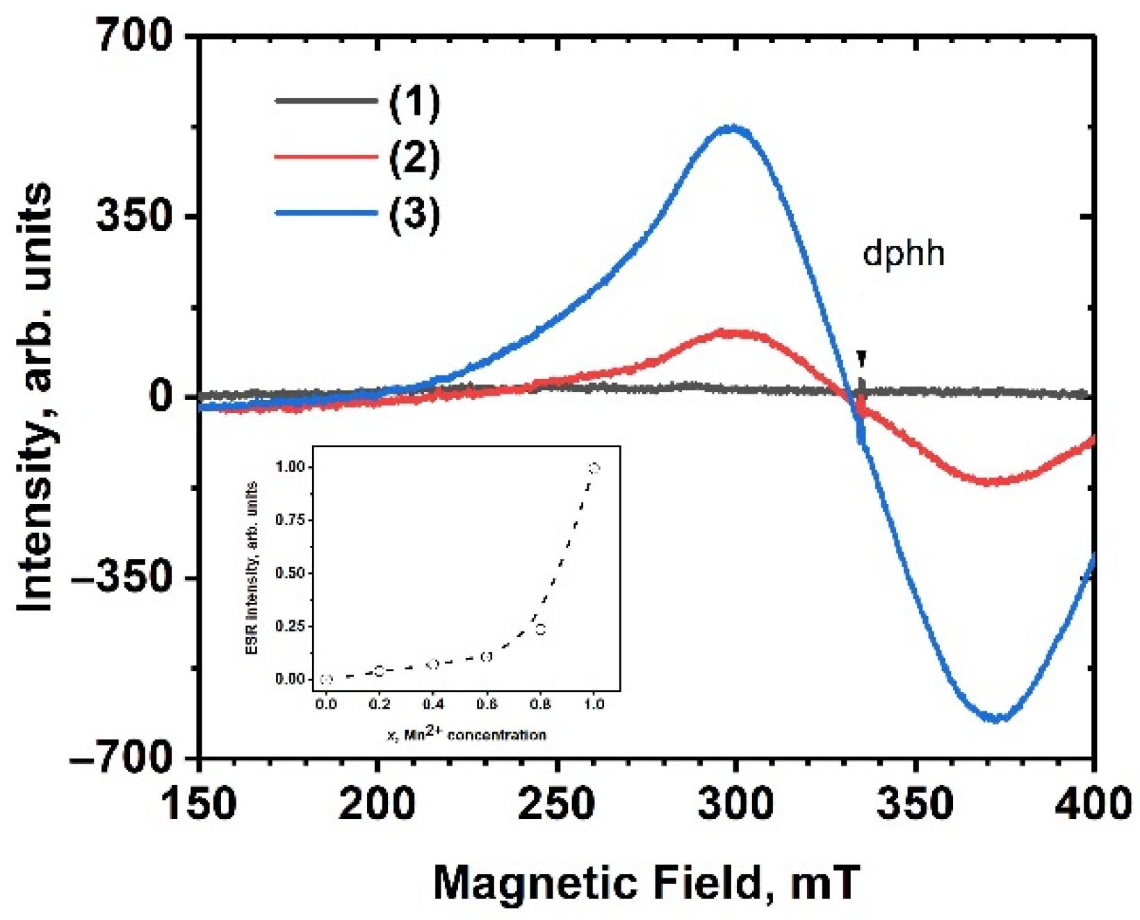
The PXRD patterns of SrMnxEu are shown in Figure 1. Unlike the similar Ca-based solid solution Ca9−xMnxEu(PO4)7 [25], an unbroken series of solid solutions with the strontiowhitlockite structure was not formed. However, the Sr8MnEu(PO4)7 sample crystallized in the trigonal Sr9Fe1.5(PO4)7-type, or strontiowhitlockite-type, structure (space group (sp.gr.) R 3 ¯ m, PDF-2 Card 51–427) (Figure 1). It appears that Mn2+ ions in the octahedra site played a critical role in the structure stabilization, similar to the Mg2+ ions in strontiowhitlockite. A similar structure formation was found in some related works, for example for Sr9MnK(PO4)7:Eu2+,Ce3+ [33] or Sr8MgCe(PO4)7:Eu2+,Mn2+ [34] on Sr-based phosphors.
Among the synthesized compounds, only the SrMn0.8Eu and SrMn1.0Eu samples were single-phased. Therefore, the limit content of Mn2+ ions required to form the strontiowhitlockite structure is x = 0.8. The observed reflections on the PXRD patterns for SrMn0Eu corresponded to the superposition of eulytite-typeSr3Eu(PO4)3 (sp.gr. I 4 ¯ 3d, PDF-2 Card 48–410) and palmierite-type Sr3(PO4)2 (sp.gr. R 3 ¯ m, PDF-2 Card 85–502) structures. The samples with x = 0.2–0.4 were characterized by mixtures of phases with Sr9Fe1.5(PO4)7, Sr3(PO4)2, and Sr3Eu(PO4)3 structures (Figure 1). The quantitative analysis of the phase content calculated using the Jana2006 software is shown in Table 1.
The formation of the β–Ca3(PO4)2-type structure has been described in previous studies [35,36]. These studies found that that samples with the general stoichiometric formula Sr9R(PO4)7 (where R = La–Sm) did not crystallize in the strontiowhitlockite structure. However, the Ca-based phosphate series with the general formula Ca9R3+(PO4)7 is known to crystallize in the β-Ca3(PO4)2 structure [37].
The ionic radius of Sr2+ is significantly larger than that of Ca2+, resulting in structural defects that distort the β-Ca3(PO4)2 structure. This distortion can lead to the formation of eulytite-type Sr3R(PO4)3 phosphate, which contains an excess of Sr2+ ions. To stabilize the β-Ca3(PO4)2 structure in Sr-based phosphates, small ions such as Mn2+, Zn2+, Mg2+ can be added [13,38]. Figure 2 shows the different structural sites. In the case of Sr2+ with Eu3+, they occupy the largest sites as SrO10 and SrO9. The smallest Mn2+ ion prefers to occupy the octahedral SrO6 site in Sr9Fe1.5(PO4).
The presence of the non-centrosymmetric eulytite-type Sr3Eu(PO4)3 phase was confirmed through an SHG study. The SHG signal was dependent on the phase composition, which was determined using PXRD data. Consequently, the highest SHG signal was observed for SrMn0Eu, indicating a significant amount of the non-centrosymmetric eulytite-type phase. Increasing the concentration of Mn2+ in the SrMnxEu solid solution resulted in a reduction in the eulytite-type phase, which was also evident in the decrease in the SHG signal. For the SrMn0.8Eu and SrMn1.0Eu samples with the strontiowhitlockite structure, the SHG signals were comparable to the systematic errors in the measurement method.

2.2. ESR Analyze

No ESR signal was detected in the sample without Mn, i.e., SrMn0Eu, while the Mn2+-containing samples showed a wide, structureless signal with a g-factor of 1.997 (Figure 3). The shape of the ESR spectra remained unchanged as the temperature cooled down to 77 K. Furthermore, the signal intensity displayed non-linear behavior in relation to the Mn concentration (Figure 3, inset).
On one hand, the observed ESR signal was attributed to the presence of manganese ions. This is supported by the fact that the ESR of Mn2+ exhibited a signal in a region with a g-factor close to 2 [39,40,41,42]. However, the characteristic sextet pattern of Mn2+ was not observed in the studied samples.
Similar ESR spectra are often observed in Eu2+-doped compounds [43,44,45]. This observation is further supported by the non-linear increase in the ESR signal intensity with the increase in the Mn2+ concentration, particularly in the samples with x = 0.8 and 1.0 (Figure 3 inset). Simultaneously, the luminescence of Eu3+ was quenched (see below). These finding suggest the presence of Eu2+ ions in the samples. The broadening of the ESR signal may be attributed to the exchange interaction between manganese and europium ions, similar to what has been observed in silicates [45].

2.3. Diffuse Absorption

The diffuse absorption spectra of SrMnxEu are shown in Figure 4. The spectra for the samples with x = 0 and 0.6 exhibited a similar structure. However, the absorption spectrum of the SrMn1.0Eu showed a prominent edge starting at 400 nm and extending towards the shorter-wavelength region of the spectrum (Figure 4). This behavior can be explained by the formation of a single-phase sample for SrMn1.0Eu. The absorption bands of Mn2+ are typically found in the 370–440 nm spectral range and are attributed to d-d transitions. These absorption bands often have a low oscillator strength and can appear broadened when Mn2+ ions occupy multiple non-equivalent positions within the lattice [46,47]. Additionally, a prominent absorption edge is observed in this region. The combination of these factors can mask the absorption bands of Mn2+.
It is possible that manganese ions can exist in a different oxidation state. One hypothetical substitution scheme could be Mn2+ + Eu3+ ↔ Mn3+ + Eu2+. An indicator of the presence of Mn3+ ions in the lattice is the occurrence of a broad absorption band in the visible spectral region, typically peaking around 450–700 nm [48]. The broadening of this band can be attributed to the occupancy of different positions by Mn3+ ions. A wide plateau of low intensity can be observed in the visible region of the presented spectra (Figure 4). However, it is difficult to confidently conclude the presence of manganese 3+ solely based on the absorption spectra.

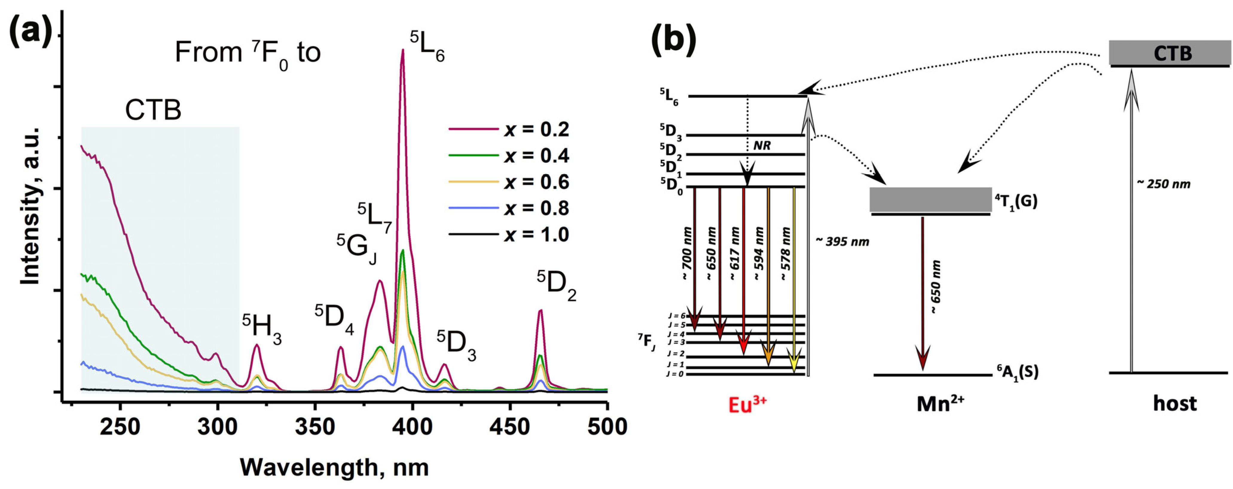
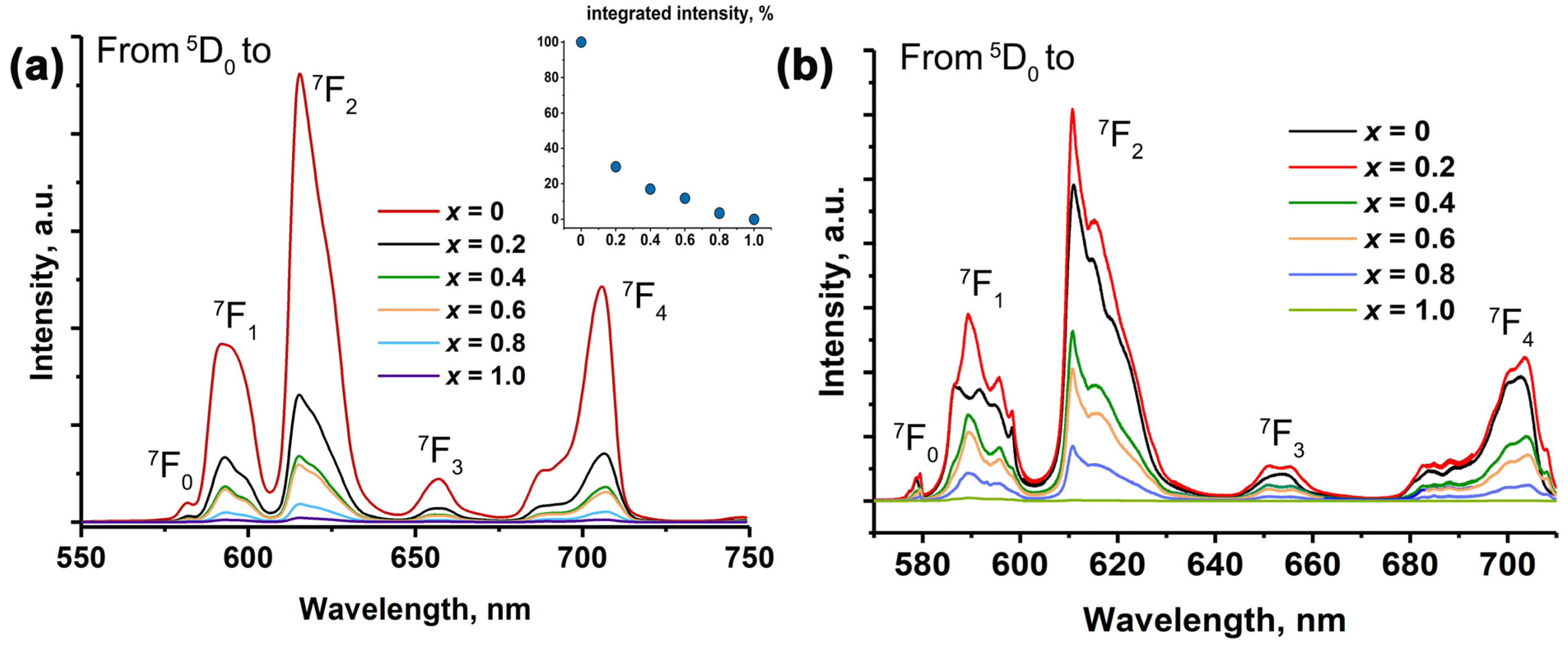
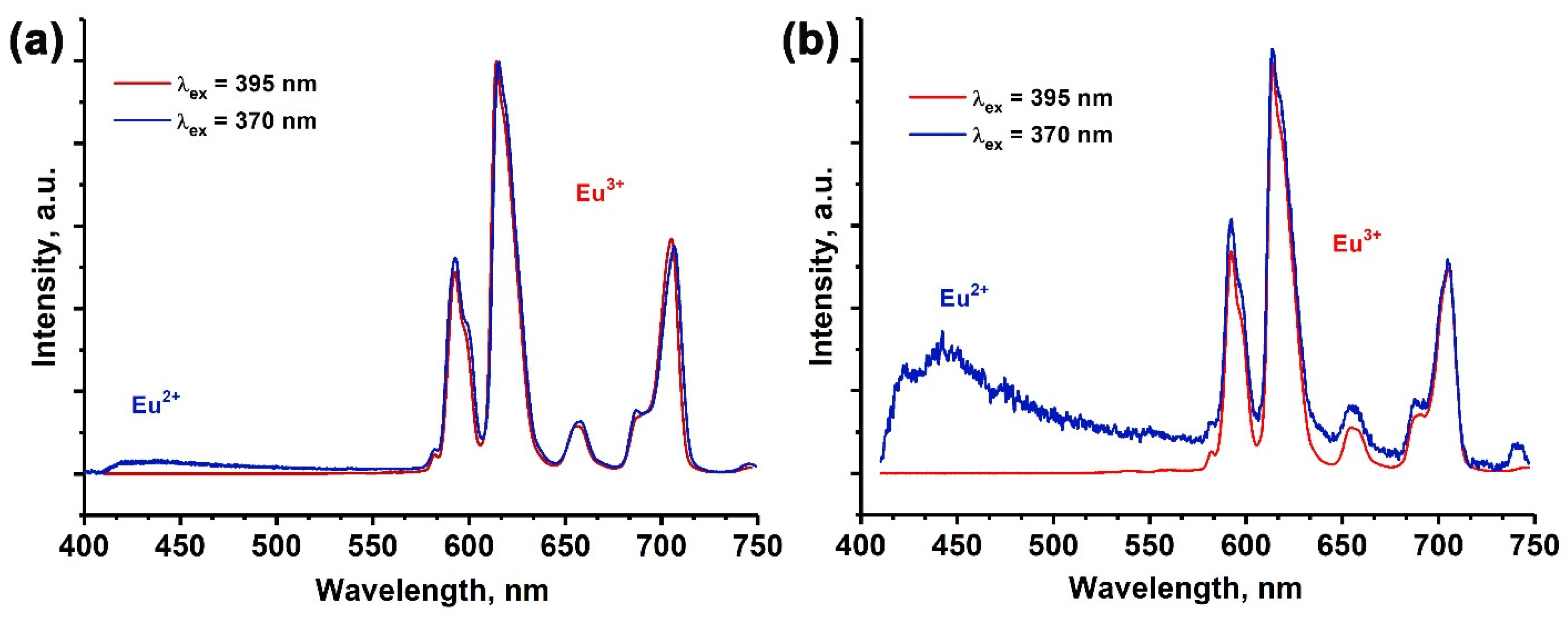
2.4. Photoluminescence Properties

The VUV excitation spectra of the Eu3+ 4f–4f luminescence, measured at the 5D07F2 band, are shown in Figure 5. In the SrMn0Eu sample undoped by Mn2+ ions, a broad band centered around 250 nm and a relatively sharp band around 150 nm were observed. The broad band at 250 nm was attributed to the charge transfer band from (CTB) O2− to Eu3+, while the sharp band at 150 nm corresponded to host excitation. In the SrMnxEu solid solutions, the position of the band at 250 nm shifted to a shorter wavelength as the concentration of Mn2+ ions increased. Additionally, the intensity of the band at 150 nm significantly decreased. At 120–160 nm, Mn2+ ions typically exhibit intra-ionic 3d-4s transitions [49,50], which have a high oscillator strength. This indicates strong absorption bands in this wavelength range. Consequently, the strong absorption leads to non-radiative relaxation of excitations, resulting in the quenching of luminescence when excited in this specific region.
The photoluminescence properties of SrMnxEu solid solutions are sensitive to phase purity and chemical composition. Figure 6a shows the PLE spectra. The number and position of the observed bands, corresponding to 4f–4f transitions of the Eu3+ ions, remained unchanged for the samples with x = 0.2–0.8. The broad band ranging from 250 to 300 nm was attributed to the CTB, while the sharp peaks in the range of 300–500 nm arose from the f–f transitions of Eu3+. Specifically, the peaks located at 320, 361, 376, 382, 395, 416 and 465 nm corresponded to the 7F05H3, 5D4, 5GJ, 5L7, 5L6, 5D3, and 5D2 transitions of Eu3+ ions [5,51,52,53]. All the spectral lines in the SrMnxEu host appeared to be wider when compared to those in other hosts that have been described. This could potentially be attributed to the presence of Eu3+ ions in the different environments. The presence of Mn2+ ions caused a notable reduction in the intensity of both the CTB and the standard transitions of Eu3+. The observed decrease in intensity was attributed to the increase in the Mn2+ concentration in the SrMnxEu solid solutions. This decrease could be attributed to the quenching effect by Mn2+ and the abnormal reduction of Eu3+ during synthesis. The proposed energy transfer schema is shown in Figure 6b, with the most intensive line observed at 395 nm.
The PL spectra for SrMnxEu, excited at 395 nm (Figure 7), exhibited characteristic lines corresponding to Eu3+ transitions. The sharp lines at 578, 594, 617, 650, and 700 nm corresponded to the transitions 5D07FJ (J = 0, 1, 2, 3, 4), with the main band at 615 nm. The resulting emission was observed in the red region of the visible spectrum [54,55]. Previous studies have shown that the total integral intensity is higher for the host Sr8MEu3+(PO4)7 (where M = Mg, Zn) compared to Ca-based phosphates [13], regardless of the synthesis method. In this work, an increase in the Mn2+ concentration led to a decrease in the total integral intensity. Additionally, a gap was observed for the samples with x = 0 and 0.2 (Figure 7a, insert).
A decrease in the total integral intensity of Eu3+ transitions was also observed for the single-phased SrMn0.8Eu and SrMn1.0Eu with a β-Ca3(PO4)2-type (or strontiowhitlockite) structure. This can be explained by the energy transfer process from the Eu3+ to Mn2+ levels through nonradiative transitions to the excited 4T1(G) state and emission to the ground 6A1(S) state. It is possible that the Mn2+ emission overlapped with the 5D07F0,1,3 Eu3+ transition [56,57], which can be observed in the high-spectral-resolution PL spectra. Furthermore, the emission of Mn2+ could be decreased through a concentration-quenching process. A proposed schema of this process is shown in Figure 7b. Similar behavior in the quenching of Eu3+ photoluminescence by Mn2+ ions has been previously observed in Ca9−xMnxEu(PO4)7 [25] and in isostructural Ca3(VO4)2:Eu3+, Mn2+ [58].
It is important to note that the spectral profile of the SrMn0Eu was significantly different compared to the others, as clearly seen in the high-spectral-resolution spectra (Figure 7b). The profiles for SrMn0.2Eu, SrMn0.4Eu, and SrMn0.6Eu were similar, indicating a similar oxygen environment. These samples consisted of two phases with β-TCP and eulytite types. This suggests that with the Mn2+ concentration at x = 0.2, Eu3+ primarily occupied sites in the whitlockite Sr9Fe1.5(PO4)7 and eulytite Sr3Eu(PO4)3 structures with an excess of Sr2+ ions forming the Sr3(PO4)2 phase.
Additional information about the phase composition and oxygen environment of the emission centers can be obtained thought consideration the forbidden electric dipole 5D07F0 transition of Eu3+ (Figure 8) [59]. The non-degenerate energy levels indicated the number of nonequivalent sites for Eu3+. For the sample with x = 0, this transition can be represented by two Gaussian components, indicating two non-equal oxygen environments for Eu3+. It should be noted that the Sr3(PO4)2 structure exhibited two non-equal sites for Eu3+ occupation, but one of them was a ч. Therefore, the observed transition reflects the influence of the larger site. The average Eu–O distance in the polyhedral structure of Sr3(PO4)2 was 2.7476 Å. In the Sr3Eu(PO4)3, only one site was observed, with an average Eu–O distance of approximately 2.6754 Å. This point was clearly demonstrated in previous studies [23,60], where it was shown that for the 5D07F0 transition, the Eu–O distance has a direct correlation with the wavelength of the transition. Hence, line A corresponds to the Eu3+ environment in the Sr3Eu(PO4)3, while line B corresponds to the Eu3+ in Sr3(PO4)2 (Figure 8b).
Regarding the SrMn0.2Eu sample, the 5D07F0 transition can be described by four Gaussian components (Figure 8c). The increase in the number of components was attributed to the formation of the β-Ca3(PO4)2-type structure (Figure 8c). Fitting line A remains unchanged in terms of the maximum and position, indicating the Eu3+ oxygen environment in the Sr3Eu(PO4)3. Line B corresponds to the Eu3+ in the Sr3(PO4)2 host. Additional lines C and D correspond to the Eu3+ in the strontiowhitlockite [13]. Therefore, some polyhedra in the strontiowhitlockite and Sr3(PO4)2 hosts were approximately the same, which is why lines B and C have very closely centered maximum values.
The observed 5D07F0 transitions for SrMn0.4Eu and SrMn0.6Eu can be accurately fitted by three Gaussian components, indicating that Eu3+ ions were mainly involved in the Sr3Eu(PO4)3- and strontiowhitlockite-type hosts. For the single-phased samples SrMn0.8Eu and SrMn1.0Eu, the 5D07F0 transition can be fitted by two Gaussian components. This suggests that there are two different environments for Eu3+ in the host [13], as is shown in Figure 2 for strontiowhitlockite with the presence of SrO10 and SrO9 sites.
According to the ESR data analysis, it was confirmed that Eu2+ ions were detected in the samples. As a result, the PL spectra were monitored for all the samples, employing an excitation wavelength of 370 nm (Figure 9). Notably, for the samples with x ≥ 0.2, distinct 4f5d–4f Eu2+ transitions were registered. The observed band appeared to be asymmetrical in shape and was predominantly centered around the wavelength of approximately 445 nm.
The intensity of the Eu2+ emission band at ~445 nm was higher for the x = 0.8 sample (Figure 9b) compared to the x = 0.2 sample (Figure 9a). Consequently, it can be inferred that the addition of Mn2+ to the samples resulted in an overall increase in the total integral intensity of the Eu2+ transitions. The presence of Eu2+ emissions in the PL spectra indicates the abnormal reduction of Eu3+ ions in the strontiowhitlockite host. Moreover, the increase in the Mn2+ doping in the SrMnxEu solid solutions led to an increase in the Eu2+ ion concentration and a more efficient reduction process.

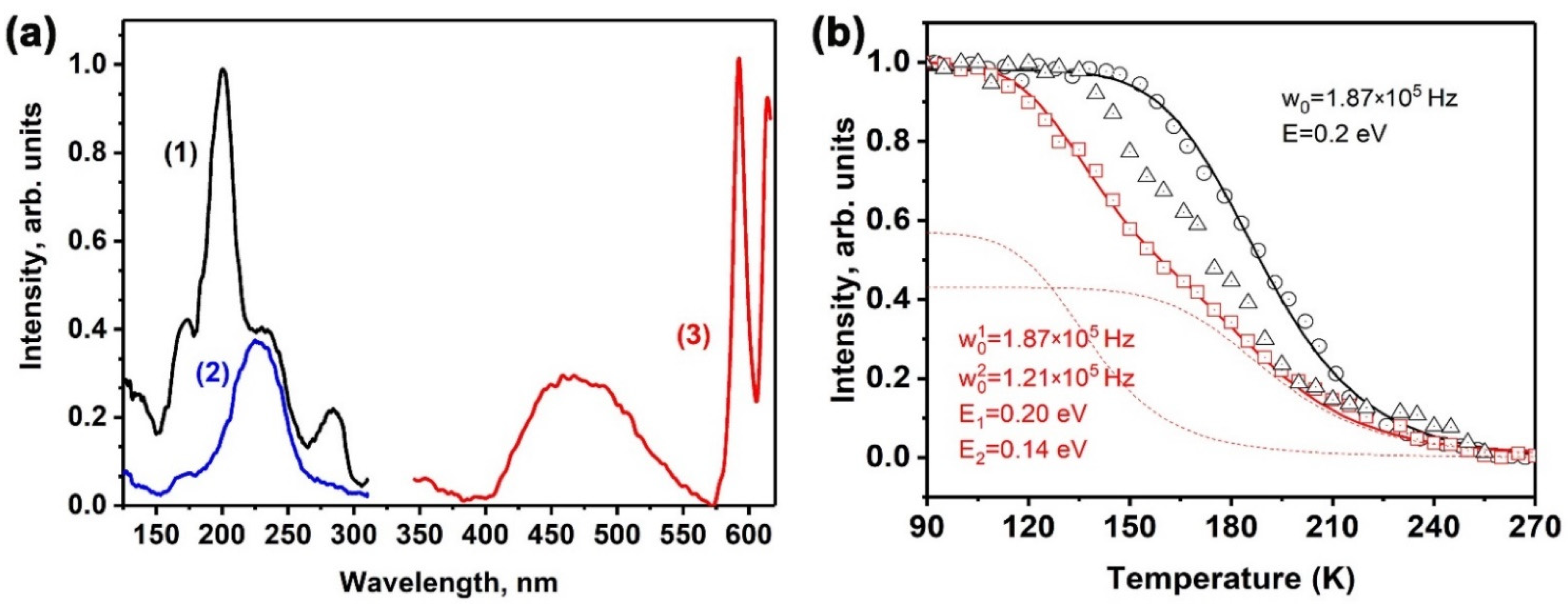
2.5. Temperature Dependence of Photoluminescence

Upon cooling to 80 K, a broad band appeared in the PL spectra centered at 470 nm (Figure 10a line 3). The PLE spectra of this band are shown in Figure 10a (Figure 10a lines 1 and 2). Under monitoring at 470 nm, the PLE spectrum consisted of several bands at 285, 230, 200, and 170 nm (Figure 10a line 1), with the most intense band being observed at 200 nm. When monitoring at 595 nm, the PLE spectrum showed only one band peaked at 230 nm, corresponding to charge transfer effects.
Figure 10b demonstrates the temperature dependence of the luminescence intensity of an emission band centered at 470 nm under 230 nm excitation. The observed behavior of the dependence follows Mott’s law, with an activation energy (E) of 0.2 eV and a frequency factor (w0) of 1.87∙105 Hz. This luminescence band is potentially associated with 5d–4f transitions in the Eu2+ ions. Therefore, the activation energy in the temperature quenching curve of the luminescence could correspond to the energy difference between the high-energy excited 5d states of Eu2+ and the bottom of the conduction band.
With an increasing concentration of Mn2+ ions, the temperature dependence of luminescence underwent changes. At lower temperatures, the luminescence intensity of Eu2+ was observed to be quenched (Figure 10b). This phenomenon can be explained by the interaction between the Mn2+ ions and the surrounding environment. In the case of SrMn1.0Eu, the temperature dependence of the luminescence can be well described by the sum of two Mott’s functions, which provides valuable insights into the underlying mechanisms of luminescence:
I T = 0.43 1 + w 0 1 exp E 1 k B T 1 + 0.57 1 + w 0 2 exp E 2 k B T 1
where w 0 1 = 1.87 × 10 5   H z , w 0 2 = 1.21 × 10 5   H z , E 1 = 0.20 eV, and E 2 = 0.14 eV. It was observed that the samples with a high Mn2+ content exhibited the presence of two distinct Eu2+ centers, which provides evidence for the existence of multiple Eu2+ centers within the SrMn1.0Eu sample.

2.6. The Decay Curves

The decay curves were collected for the single-phased samples of SrMn0.8Eu and SrMn1.0Eu (Figure 11). The curves were well fitted by the double exponent function:
I(t) = Aexp(−t1) + Aexp(−t2)
where I(t) is the intensity at time t, τ1 and τ2 are the decay times for the exponential components, and A1 and A2 are fitting constants. The average lifetimes were calculated using the following equation [61]:
τ = A 1 τ 1 2 + A 2 τ 2 2   A 1 τ 1 + A 2 τ 2
The calculated average lifetime for the SrMn0.8Eu was 1.97 ms (A1 = 0.3, τ1 = 2.35, A2 = 0.3, τ2 = 0.69), while for the SrMn1.0Eu, it was 1.19 ms (A1 = 0.11, τ1 = 2.1, A2 = 0.6, τ2 = 0.59). The values were lower compared to other Eu3+-doped strontiowhitlockite samples [62]. The low decay time of the Eu3+ emission may indicate charge transfer processes from the Eu3+ levels.

2.7. The Abnormal Reduction Process

The presence of Eu2+ ions in the host, as indicated by the ESR and photoluminescence data, suggests that the Eu3+ reduced to Eu2+ during the synthesis in air. This reduction process has been observed previously in several works [25,32,63,64,65,66]. The authors propose that this abnormal reduction of Eu3+ to Eu2+ in air occurs through a charge compensation mechanism. In [67], the conditions for the reduction process in solid-state compounds were proposed. A detailed analysis of previously obtained data on Eu3+ spectra in different phosphate hosts reveals this abnormal reduction, which follows Pei’s rules with one additional modification (Table 2).
Due to the ionic radii mismatch between Sr2+ and Eu3+ ions, Sr-based phosphates with a β-TCP-type structure are suitable for reducing Eu3+ to Eu2+ in air. Additionally, the synthesis products (NH3, see Section 2.1, reaction) create a weak reducing atmosphere, further promoting the reduction of Eu3+ to Eu2+. One possible reduction scheme, based on diffuse absorption, is Mn2+ + Eu3+ ↔ Mn3+ + Eu2+, where Mn2+ acts as the reducing agent and Eu3+ as the oxidant. The reduction process occurs due to the susceptibility of the structure to the reducing agent NH3 and the presence of Mn2+ ions.

2.8. Color Characteristics

One of the important characteristics of phosphors is their CIE coordinates. These coordinates can be determined from the emission spectral data of the samples. The calculated results are shown in Figure 12. For the Sr8.2Mn0.8Eu(PO4)7 sample monitored at 395 nm, the color coordinates (0.647; 0.351) fell in the red-orange region on the CIE diagram (Figure 12, point 1). When excited at 370 nm, the color coordinates (0.399; 0.270) were within the pink region (Figure 12, point 2).

3. Materials and Methods

3.1. Synthesis

The series of Sr9−xMnxEu(PO4)7 (named SrMnxEu, x = 0, 0.2, 0.4, 0.6, 0.8, 1) was synthesized using a high-temperature solid-state method. Stoichiometric mixtures of NH4H2PO4 (99.9%), SrCO3 (99.9%), MnCO3 (99.99%), and Eu2O3 (99.9%) were used as the starting materials. The amounts of reactants were calculated based on the following reaction:
(18 − 2x) SrCO3 + 2x MnCO3 + 14 NH4H2PO4 + Eu2O3 → 2 Sr9−xMnxEu(PO4)7 + 18 CO2 + 14 NH3 +21 H2O
The required amounts were mixed in an agate mortar with acetone for better homogenization. The resulting mixture was then transferred to an alundum crucible for stepwise heating:
  • Slow heating up to 200 °C for 8 h, followed by annealing for 8 h in air.
  • Heating to 1100 °C for 12 h, followed by annealing for 24 h in air.
This slow heating method was chosen to guarantee the uniform removal of volatile by-products from the reaction. The powder X-ray diffraction (PXRD) patterns of the precursors were checked using the JCPDS PDF-2 database, which did not show any reflections of impurity phases.

3.2. Characterization

Powder X-ray diffraction (PXRD) patterns of SrMnxEu were collected on a Thermo ARL X’TRA powder diffractometer (CuKα radiation, λ = 1.5418 Å, Bragg–Brentano geometry, Peltier-cooled CCD detector). The PXRD data were collected over the 5°–70° 2θ range with steps of 0.02°. The Le Bail decomposition [68] was applied for the PXRD analysis using the JANA2006 software [69].
The second harmonic generation (SHG) signal was measured with a Q-switched YAG: Nd laser at λω =1064 nm in the reflection mode.
For collecting the electron spin resonance (ESR) spectra, the powder samples were placed in a quartz tube and measured using an RE–1306 X–band ESR spectrometer (KBST, Smolensk, Russia) operating at a frequency of 9.3841 GHz at room temperature.
The VUV excitation luminescence was recorded using an MDR-2 monochromator (LOMO, Saint Petersburg, Russia) equipped with a grate of 1200 lines per mm. A Hamamatsu photomodule operating in the photon counting mode was used for the detection. Excitation was carried out using a Hamamatsu L7293-50 deuterium lamp with a magnesium fluoride window coupled with a VMR-2 vacuum monochromator. The excitation spectra were corrected using sodium salicylate. The registration of the temperature was performed using a type K thermocouple.
The diffuse absorption spectra were registered with a Lambda 950 spectrophotometer equipped with integrated sphere in the transmittance regime (Perkin-Elmer, New-York city, NY, USA).
The photoluminescence emission (PL) and excitation (PLE) spectra were recorded under excitation in the UV-Vis spectral region using a specialized laboratory set-up. An ARC 150 W Xe lamp was used as an excitation source. The primary monochromator MDR-206 was used for the selection of the excitation wavelength. The PLE spectra were measured with a spectral resolution of 5 nm. The luminescence spectra were detected using an Oriel MS257 spectrograph using a 300 gr/mm or 2400 gr/mm grating with a spectral resolution of 1.5 nm and 0.32 nm, respectively (“low” and “high” resolution). A Marconi 30-11 CCD detector was used for the registration. The luminescence spectra were corrected for the spectral sensitivity of the registration channel. The measured excitation spectra were normalized on the excitation spectrum of yellow lumogen. All measurements were performed at room temperature.
The luminescence decay time was registered using a Perkin-Elmer LS-55 spectofluorimeter (Perkin-Elmer, New-York city, NY, USA) equipped with a Xe lamp with a 10 mks pulse duration. All measurements were performed at room temperature and corrected for the sensitivity of the spectrometer.

4. Conclusions

New solid solutions of Sr9−xMnxEu(PO4)7 with a strontiowhitlockite structure were synthesized using a high-temperature solid-state method in air. The concentration limit of Mn2+ ions in the host for the formation of the strontiowhitlockite (or β-TCP) phase was determined at x ≥ 0.8. The composition of the multi-phased samples was confirmed by X-ray and SHG analyses. The quenching of the Eu3+ emission was observed under a 395 nm excitation with the Mn2+ concentration. It was proposed that Eu3+ excitation was quenched through the Mn2+ levels, following potential reaction scheme Mn2+ + Eu3+ ↔ Mn3+ + Eu2+. The ESR and PL spectra measurements confirmed the abnormal partial reduction of Eu3+ → Eu2+ in air and the presence of Eu2+ ions in the host. Both 4f5d–4f Eu2+ and 4f–4f Eu3+ transitions were observed in the PL spectra under a 370 nm excitation. The intensity of the Eu2+ emission decreased with heating from 80 K to 270 K in Sr9−xMnxEu(PO4)7. The detailed analysis of the 5D07F0 transition showed the presence of several non-equal Eu3+ environments. The decay curves were measured, and it was found that the decay times for the Eu3+ levels were lower compared to other strontiowhitlockite-based phosphors. The conditions for the occurrence of Eu3+ → Eu2+ reduction in air were discussed.

Author Contributions

Conceptualization, E.S.Z. and D.V.D.; methodology, I.V.N.; software, E.S.Z., N.R.K. and S.M.A.; validation, I.V.N., D.A.S. and D.V.D.; formal analysis, I.V.N., N.R.K. and R.Y.S.; investigation, E.S.Z. and D.A.S.; resources, R.Y.S. and S.M.A.; data curation, E.S.Z., N.R.K., R.Y.S. and D.A.S.; writing—original draft preparation, I.V.N. and S.M.A.; writing—review and editing, E.S.Z. and I.V.N.; visualization, I.V.N. and N.R.K.; supervision, D.A.S., S.M.A. and D.V.D.; project administration, D.V.D. and R.Y.S.; funding acquisition, D.V.D. and R.Y.S. All authors have read and agreed to the published version of the manuscript.

Funding

This research was funded by the Russian Science Foundation (grant 23-73-10007). Diffuse absorption spectra were measured using the facilities of the Centers for Collective Use: “Center for isotopic-geochemical investigations” at the Vinogradov Institute of Geochemistry SB RAS. The VUV measurements were supported by the Project 0284-2021-0004 (Materials and Technologies for the Development of Radiation Detectors, Luminophores, and Optical Glasses).

Institutional Review Board Statement

Not applicable.

Informed Consent Statement

Not applicable.

Data Availability Statement

The data presented in this study are available on request from the corresponding author.

Conflicts of Interest

The authors declare no conflicts of interest.

References

  1. Zhang, J.; Wang, Y.; Wen, Y.; Zhang, F.; Liu, B. Luminescence properties of Ca10K(PO4)7:RE3+ (RE=Ce, Tb, Dy, Tm and Sm) under vacuum ultraviolet excitation. J. Alloys Compd. 2011, 509, 4649–4652. [Google Scholar] [CrossRef]
  2. Dong, X.; Zhang, J.; Zhang, X.; Hao, Z.; Luo, Y. New orange–red phosphor Sr9Sc(PO4)7:Eu3+ for NUV-LEDs application. J. Alloys Compd. 2014, 587, 493–496. [Google Scholar] [CrossRef]
  3. Kim, K.-B.; Kim, Y.-I.; Chun, H.-G.; Cho, T.-Y.; Jung, J.-S.; Kang, J.-G. Structural and Optical Properties of BaMgAl10O17:Eu2+ Phosphor. Chem. Mater. 2002, 14, 5045–5052. [Google Scholar] [CrossRef]
  4. Chen, J.; Yang, C.; Chen, Y.; He, J.; Liu, Z.-Q.; Wang, J.; Zhang, J. Local Structure Modulation Induced Highly Efficient Far-Red Luminescence of La1−xLuxAlO3:Mn4+ for Plant Cultivation. Inorg. Chem. 2019, 58, 8379–8387. [Google Scholar] [CrossRef] [PubMed]
  5. Guo, H.; Zhang, H.; Wei, R.; Zheng, M.; Zhang, L. Preparation, structural and luminescent properties of Ba2Gd2Si4O13:Eu3+ for white LEDs. Opt. Express 2011, 19, A201–A206. [Google Scholar] [CrossRef]
  6. Stefańska, D.; Dereń, P.J. Luminescence investigation and thermal stability of blue-greenish emission generated from Ca3MgSi2O8: Eu2+ phosphor. Opt. Mater. 2018, 80, 62–64. [Google Scholar] [CrossRef]
  7. Thulasiramudu, A.; Buddhudu, S. Optical characterization of Eu3+ and Tb3+ ions doped zinc lead borate glasses. Spectrochim. Acta Part A 2007, 66, 323–328. [Google Scholar] [CrossRef]
  8. Bungala Chinna, J.; Jakka, S.K.; Mohan Babu, A.; Sasikala, T.; Rama Moorthy, L. Green fluorescence of Tb3+- doped LBTAF glasses. Phys. B 2009, 404, 2020. [Google Scholar]
  9. Lü, W.; Jiao, M.; Shao, B.; Zhao, L.; You, H. Enhancing Photoluminescence Performance of SrSi2O2N2:Eu2+ Phosphors by Re (Re = La, Gd, Y, Dy, Lu, Sc) Substitution and Its Thermal Quenching Behavior Investigation. Inorg. Chem. 2015, 54, 9060–9065. [Google Scholar] [CrossRef]
  10. Osborne, R.A.; Cherepy, N.J.; Seeley, Z.M.; Payne, S.A.; Drobshoff, A.D.; Srivastava, A.M.; Beers, W.W.; Cohen, W.W.; Schlagel, D.L. New red phosphor ceramic K2SiF6:Mn4+. Opt. Mater. 2020, 107, 110140. [Google Scholar] [CrossRef]
  11. Xu, S.; Li, P.; Wang, Z.; Li, T.; Bai, Q.; Sun, J.; Yang, Z. Luminescence and energy transfer of Eu2+/Tb3+/Eu3+ in LiBaBO3 phosphors with tunable-color emission. J. Mater. Chem. C 2015, 3, 9112–9121. [Google Scholar] [CrossRef]
  12. Yuan, Y.; Lin, H.; Cao, J.; Guo, Q.; Xu, F.; Liao, L.; Mei, L. A novel blue-purple Ce3+ doped whitlockite phosphor: Synthesis, crystal structure, and photoluminescence properties. J. Rare Earths 2020, 39, 621–626. [Google Scholar] [CrossRef]
  13. Deyneko, D.V.; Nikiforov, I.V.; Spassky, D.A.; Berdonosov, P.S.; Dzhevakov, P.B.; Lazoryak, B.I. Sr8MSm1−xEux(PO4)7 phosphors derived by different synthesis routes: Solid state, sol-gel and hydrothermal, the comparison of properties. J. Alloys Compd. 2021, 887, 161340. [Google Scholar] [CrossRef]
  14. Shuang, D.; Wanjun, T.; Guangyong, X.; Yuhong, Y.; Zhengxi, H. Synthesis and photoluminescence tuning of (Sr,Ln)9Mg1.5(PO4)7 phosphors by Ln (Ln = Y, La, Gd, Lu) substitution. J. Lumin. 2020, 224, 117331. [Google Scholar] [CrossRef]
  15. Britvin, S.N.; Pakhomovskii, Y.A.; Bogdanova, A.N.; Skiba, V.I. Strontiowhitlockite, Sr9Mg(PO3OH)(PO4)6, a new mineral species from the Kovdor Deposit, Kola Peninsula, U.S.S.R. Can. Mineral. 1991, 29, 87–93. [Google Scholar]
  16. Han, J.; Pan, F.; Zhou, W.; Qiu, Z.; Tang, M.; Wang, J.; Lian, S. Dual energy transfer controlled photoluminescence evolution in Eu and Mn co-activated β-Ca2.7Sr0.3(PO4)2 phosphors for solid-state lighting. RSC Adv. 2015, 5, 98026–98032. [Google Scholar] [CrossRef]
  17. Tang, W.; Zhang, Z. Realization of color tuning via solid-solution and energy transfer in Ca3−xSrx(PO4)2:Eu2+,Mn2+ phosphors. J. Mater. Chem. C 2015, 3, 5339–5346. [Google Scholar] [CrossRef]
  18. Ji, H.; Huang, Z.; Xia, Z.; Molokeev, M.S.; Atuchin, V.V.; Huang, S. Cation Substitution Dependent Bimodal Photoluminescence in Whitlockite Structural Ca3−xSrx(PO4)2:Eu2+ (0 ≤ x ≤ 2) Solid Solution Phosphors. Inorg. Chem. 2014, 53, 11119–11124. [Google Scholar] [CrossRef]
  19. Szyszka, K.; Nowak, N.; Kowalski, R.M.; Zukrowski, J.; Wiglusz, R.J. Anomalous luminescence properties and cytotoxicity assessment of Sr3(PO4)2 co-doped with Eu2+/3+ ions for luminescence temperature sensing. J. Mater. Chem. C 2022, 10, 9092–9105. [Google Scholar] [CrossRef]
  20. Belik, A.A.; Izumi, F.; Stefanovich, S.Y.; Malakho, A.P.; Lazoryak, B.I.; Leonidov, I.A.; Leonidova, O.N.; Davydov, S.A. Polar and Centrosymmetric Phases in Solid Solutions Ca3−xSrx(PO4)2 (0 ≤ x ≤ 16/7). Chem. Mater. 2002, 14, 3197–3205. [Google Scholar] [CrossRef]
  21. Zhou, N.; Gao, P.; Yang, Y.; Zhong, Y.; Xia, M.; Zhang, Y.; Tian, Y.; Lu, X.; Zhou, Z. Novel orange–red emitting phosphor Sr8ZnY(PO4)7:Sm3+ with enhanced emission based on Mg2+ and Al3+ incorporation for plant growth LED lighting. J. Taiwan Inst. Chem. Engin. 2019, 104, 360–368. [Google Scholar] [CrossRef]
  22. Ding, X.; Li, Z.; Xia, D. New whitlockite-type structure material Sr9Y(PO4)7 and its Eu2+ doped green emission properties under NUV light. J. Lumin. 2020, 221, 117114. [Google Scholar] [CrossRef]
  23. Deyneko, D.V.; Aksenov, S.M.; Nikiforov, I.V.; Stefanovich, S.Y.; Lazoryak, B.I. Symmetry Inhomogeneity of Ca9−xZnxEu(PO4)7 Phosphor Determined by Second-Harmonic Generation and Dielectric and Photoluminescence Spectroscopy. Cryst. Growth Des. 2020, 20, 6461–6468. [Google Scholar] [CrossRef]
  24. Deyneko, D.V.; Nikiforov, I.V.; Spassky, D.A.; Dikhtyar, Y.Y.; Aksenov, S.M.; Stefanovich, S.Y.; Lazoryak, B.I. Luminescence of Eu3+ as a probe for the determination of the local site symmetry in β-Ca3(PO4)2-related structures. CrystEngComm 2019, 21, 5235–5242. [Google Scholar] [CrossRef]
  25. Sipina, E.V.; Spassky, D.A.; Krutyak, N.R.; Morozov, V.A.; Zhukovskaya, E.S.; Belik, A.A.; Manylov, M.S.; Lazoryak, B.I.; Deyneko, D.V. Abnormal Eu3+ → Eu2+ Reduction in Ca9−xMnxEu(PO4)7 Phosphors: Structure and Luminescent Properties. Materials 2023, 16, 1383. [Google Scholar] [CrossRef] [PubMed]
  26. Liu, C.; Hou, D.; Yan, J.; Zhou, L.; Kuang, X.; Liang, H.; Huang, Y.; Zhang, B.; Tao, Y. Energy Transfer and Tunable Luminescence of NaLa(PO3)4:Tb3+/Eu3+ under VUV and Low-Voltage Electron Beam Excitation. J. Phys. Chem. 2014, 118, 3220–3229. [Google Scholar] [CrossRef]
  27. Wang, Z.; Lou, S.; Li, P. Luminescent properties and energy transfer of Sr3La(PO4)3:Sm3+, Eu3+ for white LEDs. J. Alloys Compd. 2014, 586, 536–541. [Google Scholar] [CrossRef]
  28. Zhang, X.; Xu, J.; Gong, M. Site-occupancy, luminescent properties and energy transfer of a violet-to-red color-tunable phosphor Ca10Li(PO4)7: Ce3+, Mn2+. J. Lumin. 2017, 183, 348–354. [Google Scholar] [CrossRef]
  29. Guo, H.; Devakumar, B.; Li, B.; Huang, X. Novel Na3Sc2(PO4)3:Ce3+,Tb3+ phosphors for white LEDs: Tunable blue-green color emission, high quantum efficiency and excellent thermal stability. Dyes Pigm. 2018, 151, 81–88. [Google Scholar] [CrossRef]
  30. Li, P.; Wang, Z.; Yang, Z.; Guo, Q. A novel, warm, white light-emitting phosphor Ca2PO4Cl:Eu2+, Mn2+ for white LEDs. J. Mater. Chem. 2014, 2, 7823–7829. [Google Scholar] [CrossRef]
  31. Szyszka, K.; Watras, A.; Wiglusz, R.J. Strontium Phosphate Composite Designed to Red-Emission at Different Temperatures. Materials 2020, 13, 4468. [Google Scholar] [CrossRef] [PubMed]
  32. Liu, J.; Liang, K.; Wu, Z.-C.; Mei, Y.-M.; Kuang, S.-P.; Li, D.-X. The reduction of Eu3+ to Eu2+ in a new orange–red emission Sr3P4O13: Eu phosphor prepared in air and its photoluminescence properties. Ceram. Int. 2014, 40, 8827–8831. [Google Scholar] [CrossRef]
  33. Xie, G.; Wu, M.; Li, T.; You, Q.; Tang, W. Luminescence Enhancement of Red-Emitting Sr9MnK(PO4)7 Phosphor via Energy Transfer and Charge Compensation. Phys. Status Solidi B 2022, 259, 2200259. [Google Scholar] [CrossRef]
  34. Tang, W.; Ding, C. Luminescence Tuning of Sr8MgCe(PO4)7: Eu2+, Mn2+ Phosphors: Structure Refinement, Site Occupancy, and Energy Transfer. Z. Anorg. Allg. Chem. 2018, 644, 893–900. [Google Scholar] [CrossRef]
  35. Belik, A.A.; Izumi, F.; Ikeda, T.; Lazoryak, B.I.; Morozov, V.A.; Malakho, A.P.; Stefanovich, S.Y.; Grebenev, V.V.; Shelmenkova, O.V.; Kamiyama, T.; et al. Structural changes and phase transitions in whitlockite-like phosphates. Phosphorus Sulfur Silicon Relat. Elem. 2002, 177, 1899–1902. [Google Scholar] [CrossRef]
  36. Belik, A.A.; Izumi, F.; Ikeda, T.; Okui, M.; Malakho, A.P.; Morozov, V.A.; Lazoryak, B.I. Whitlockite-Related Phosphates Sr9A(PO4)7 (A=Sc, Cr, Fe, Ga, and In): Structure Refinement of Sr9In(PO4)7 with Synchrotron X-ray Powder Diffraction Data. J. Solid State Chem. 2002, 168, 237–244. [Google Scholar] [CrossRef]
  37. Bessière, A.; Benhamou, R.A.; Wallez, G.; Lecointre, A.; Viana, B. Site occupancy and mechanisms of thermally stimulated luminescence in Ca9Ln(PO4)7 (Ln=lanthanide). Acta Mater. 2012, 60, 6641–6649. [Google Scholar] [CrossRef]
  38. Luo, J.; Zhou, W.; Fan, J.; Sun, Z.; Zhang, X. Composition modification for tuning the luminescent property in Sr19(Mg,Mn)2(PO4)14: Eu2+ phosphors. J. Lumin. 2021, 239, 118369. [Google Scholar] [CrossRef]
  39. Miura, M.; Hasegawa, A.; Watanabe, M. The Electron Spin Resonance of Mn2+ Ion in Polyphosphate. Bull. Chem. Soc. Jpn. 1968, 41, 1035–1038. [Google Scholar] [CrossRef]
  40. Naga Bhaskararao, Y.; Satyavathi, K.; Subba Rao, M.; Cole, S. Synthesis and characterization of Mn2+ doped CdOZn3(PO4)2 nanocomposites. J. Mol. Struct. 2017, 1130, 585–591. [Google Scholar] [CrossRef]
  41. Jerroudi, M.; Bih, L.; Haily, E.; Bejjit, L.; Haddad, M.; Manoun, B.; Lazor, P. ESR, physical and structural studies on Mn2+ doped in mixed alkali phosphate glasses. Mater. Today Proc. 2021, 37, 3876–3881. [Google Scholar] [CrossRef]
  42. Kaneva, E.; Shendrik, R.; Mesto, E.; Bogdanov, A.; Vladykin, N. Spectroscopy and crystal chemical properties of NaCa2[Si4O10]F natural agrellite with tubular structure. Chem. Phys. Lett. 2020, 738, 136868. [Google Scholar] [CrossRef]
  43. Singh, V.; Gundu Rao, T.K.; Zhu, J.-J. Preparation, luminescence and defect studies of Eu2+-activated strontium hexa-aluminate phosphor prepared via combustion method. J. Solid State Chem. 2006, 179, 2589–2594. [Google Scholar] [CrossRef]
  44. Dhoble, S.J.; Moharil, S.V.; Gundu Rao, T.K. Correlated ESR, PL and TL studies on Sr5(PO4)3Cl:Eu thermoluminescence dosimetry phosphor. J. Lumin. 2007, 126, 383–386. [Google Scholar] [CrossRef]
  45. Choi, N.-S.; Park, K.-W.; Park, B.-W.; Zhang, X.-M.; Kim, J.-S.; Kung, P.; Margaret Kim, S. Eu2+–Mn2+ energy transfer in white-light-emitting T-phase (Ba,Ca)2SiO4:Eu2+, Mn2+ phosphor. J. Lumin. 2010, 130, 560–566. [Google Scholar] [CrossRef]
  46. Guo, N.; You, H.; Jia, C.; Ouyang, R.; Wu, D. A Eu2+ and Mn2+-coactivated fluoro-apatite-structure Ca6Y2Na2(PO4)6F2 as a standard white-emitting phosphor via energy transfer. Dalton Trans. 2014, 43, 12373–12379. [Google Scholar] [CrossRef] [PubMed]
  47. Kaneva, E.; Shendrik, R.; Pankrushina, E.; Dokuchits, E.; Radomskaya, T.; Pechurin, M.; Ushakov, A. Frankamenite: Relationship between the Crystal–Chemical and Vibrational Properties. Minerals 2023, 13, 1017. [Google Scholar] [CrossRef]
  48. Fridrichová, J.; Bačík, P.; Ertl, A.; Wildner, M.; Dekan, J.; Miglierini, M. Jahn-Teller distortion of Mn3+-occupied octahedra in red beryl from Utah indicated by optical spectroscopy. J. Mol. Struct. 2018, 1152, 79–86. [Google Scholar] [CrossRef]
  49. Sabatini, J.F.; Salwin, A.E.; McClure, D.S. High-energy optical-absorption bands of transition-metal ions in fluoride host crystals. Phys. Rev. B 1975, 11, 3832–3841. [Google Scholar] [CrossRef]
  50. True, M.; Kirm, M.; Negodine, E.; Vielhauer, S.; Zimmerer, G. VUV spectroscopy of Tm3+ and Mn2+ doped LiSrAlF6. J. Alloys Compd. 2004, 374, 36–39. [Google Scholar] [CrossRef]
  51. Garcia, C.R.; Oliva, J.; Garcia-Lobato, M.A.; Martínez, A.I.; Ochoa-Valiente, R.; Hirata, G.A. Red-emitting SrGe4O9:Eu3+ phosphors obtained by combustion synthesis. Ceram. Int. 2017, 43, 12876–12881. [Google Scholar] [CrossRef]
  52. Liang, C.-H.; Chang, Y.-C.; Chang, Y.-S. Synthesis and Photoluminescence Characteristics of Color-Tunable BaY2ZnO5:Eu3+ Phosphors. Appl. Phys. Lett. 2008, 93, 211902. [Google Scholar] [CrossRef]
  53. Ferhi, M.; Horchani-Naifer, K.; Férid, M. Spectroscopic properties of Eu3+-doped KLa(PO3)4 and LiLa(PO3)4 powders. Opt. Mater. 2011, 34, 12–18. [Google Scholar] [CrossRef]
  54. Su, B.; Xie, H.; Tan, Y.; Zhao, Y.; Yang, Q.; Zhang, S. Luminescent properties, energy transfer, and thermal stability of double perovskites La2MgTiO6:Sm3+, Eu3+. J. Lumin. 2018, 204, 457–463. [Google Scholar] [CrossRef]
  55. Ye, M.; Zhou, G.; Zhou, L.; Lu, D.; Li, Y.; Xiong, X.; Yang, K.; Chen, M.; Pan, Y.; Wu, P.; et al. Luminescent properties and energy transfer process of Sm3+-Eu3+ co-doped MY2(MoO4)4 (M=Ca, Sr and Ba) red-emitting phosphors. Solid State Sci. 2016, 59, 44–51. [Google Scholar] [CrossRef]
  56. Félix-Quintero, H.; Falcony, C.; Hernández, A.J.; Camarillo, G.E.; Flores, J.C.; Murrieta, S.H. Mn2+ to Eu3+ energy transfer in zinc phosphate glass. J. Lumin. 2020, 225, 117337. [Google Scholar] [CrossRef]
  57. Li, Y.; Li, W.; Yu, Y.; Zheng, C. Influences of Mn2+/Eu3+ dopants on the microstructures and optical properties of glass-embedded CsPbBr3 quantum dots. Opt. Mater. Express 2023, 13, 1488–1496. [Google Scholar] [CrossRef]
  58. Zhang, H.; Lü, M.; Xiu, Z.; Wang, S.; Zhou, G.; Zhou, Y.; Wang, S.; Qiu, Z.; Zhang, A. Synthesis and photoluminescence properties of a new red emitting phosphor: Ca3(VO4)2:Eu3+; Mn2+. Mater. Res. Bull. 2007, 42, 1145–1152. [Google Scholar] [CrossRef]
  59. Zhang, J.; Cai, G.; Wang, W.; Ma, L.; Wang, X.; Jin, Z. Tuning of Emission by Eu3+ Concentration in a Pyrophosphate: The Effect of Local Symmetry. Inorg. Chem. 2020, 59, 2241–2247. [Google Scholar] [CrossRef]
  60. Deyneko, D.V.; Spassky, D.A.; Morozov, V.A.; Aksenov, S.M.; Kubrin, S.P.; Molokeev, M.S.; Lazoryak, B.I. Role of the Eu3+ Distribution on the Properties of β-Ca3(PO4)2 Phosphors: Structural, Luminescent, and 151Eu Mössbauer Spectroscopy Study of Ca9.5−1.5xMgEux(PO4)7. Inorg. Chem. 2021, 60, 3961–3971. [Google Scholar] [CrossRef]
  61. Guo, Q.; Wang, Q.; Jiang, L.; Liao, L.; Liu, H.; Mei, L. A novel apatite, Lu5(SiO4)3N:(Ce,Tb), phosphor material: Synthesis, structure and applications for NUV-LEDs. Phys. Chem. Chem. Phys. 2016, 18, 15545–15554. [Google Scholar] [CrossRef] [PubMed]
  62. Sun, W.; Li, H.; Li, B.; Du, J.; Hao, J.; Hu, C.; Wang, Y.; Yi, X.; Pang, R.; Li, C. Energy transfer and luminescence properties of a green-to-red color tunable phosphor Sr8MgY(PO4)7:Tb3+,Eu3+. J. Mater. Sci.-Mater. Electron. 2019, 30, 9421–9428. [Google Scholar] [CrossRef]
  63. Peng, M.; Pei, Z.; Hong, G.; Su, Q. The reduction of Eu3+ to Eu2+ in BaMgSiO4:Eu prepared in air and the luminescence of BaMgSiO4:Eu2+ phosphor. J. Mater. Chem. 2003, 13, 1202–1205. [Google Scholar] [CrossRef]
  64. Grandhe, B.K.; Bandi, V.R.; Jang, K.; Kim, S.-S.; Shin, D.-S.; Lee, Y.-I.; Lim, J.-M.; Song, T. Reduction of Eu3+ to Eu2+ in NaCaPO4:Eu phosphors prepared in a non-reducing atmosphere. J. Alloys Compd. 2011, 509, 7937–7942. [Google Scholar] [CrossRef]
  65. Chen, J.; Liang, Y.; Zhu, Y.; Liu, S.; Li, H.; Lei, W. Abnormal reduction of Eu3+ to Eu2+ in Sr5(PO4)3Cl:Eu phosphor and its enhanced red emission by the charge compensation. J. Lumin. 2019, 214, 116569. [Google Scholar] [CrossRef]
  66. Deyneko, D.V.; Spassky, D.A.; Antropov, A.V.; Ril’, A.I.; Baryshnikova, O.V.; Pavlova, E.T.; Lazoryak, B.I. Anomalous oxidation state of europium in the Sr3(PO4)2-type phosphors doped with alkaline cations. Mater. Res. Bull. 2023, 165, 112296. [Google Scholar] [CrossRef]
  67. Pei, Z.; Su, Q.; Zhang, J. The valence change from RE3+ to RE2+ (RE = Eu, Sm, Yb) in SrB4O7: RE prepared in air and the spectral properties of RE2+. J. Alloys Compd. 1993, 198, 51–53. [Google Scholar] [CrossRef]
  68. Le Bail, A.; Duroy, H.; Fourquet, J.L. Ab-initio structure determination of LiSbWO6 by X-ray powder diffraction. Mater. Res. Bull. 1988, 23, 447–452. [Google Scholar] [CrossRef]
  69. Petříček, V.; Dušek, M.; Palatinus, L. Crystallographic Computing System JANA2006: General features. Z. Kristallogr. Cryst. Mater. 2014, 229, 345–352. [Google Scholar] [CrossRef]
Figure 1. The PXRD patterns of Sr9−xMnxEu(PO4)7 and the Bragg reflections of Sr9Fe1.5(PO4)7 (PDF-2 Card 51–427), Sr3Eu(PO4)3 (PDF–2 Card 48–410), and Sr3(PO4)2 (PDF–2 Card 85–502).
Molecules 29 00124 g001
Figure 2. Oxygen environment of Sr2+ ions oriented along the abc axes and ab projection of structures, respectively: Sr9Fe1.5(PO4)7 (a,d), Sr3Eu(PO4)3 (b,e), and Sr3(PO4)2 (c,f).
Molecules 29 00124 g002
Figure 3. ESR spectra of Sr9−xMnxEu(PO4)7 x = 0 (1), x = 0.8 (2), and x = 1.0 (3) samples. The inset shows concentration dependence of integral intensity of ESR signal.
Molecules 29 00124 g003
Figure 4. The diffuse absorption spectra of Sr9−xMnxEu(PO4)7: x = 0 (1), x = 0.6 (2), and x = 1.0 (3).
Molecules 29 00124 g004
Figure 5. The excitation spectra of Eu3+ luminescence monitored at 5D07F2 emission band for Sr9−xMnxEu(PO4)7 x = 0 (1), x = 0.2 (2), x = 0.6 (3), and x = 0.8 (4).
Molecules 29 00124 g005
Figure 6. (a) The PLE spectra for Sr9−xMnxEu(PO4)7, monitored at 620 nm; (b) the proposed schema of energy transfer processes in host.
Molecules 29 00124 g006
Figure 7. (a) The low-resolution and (b) high-resolution PL of Sr9−xMnxEu(PO4)7exc = 395 nm) (the insert shows the integral intensity of the PL spectra on Mn2+ ion concentration).
Molecules 29 00124 g007
Figure 8. (a) The 5D07F0 transition for Sr9−xMnxEu(PO4)7, (bg) fitting by Gauss components for x = 0, 0.2, 0.4, 0.6, 0.8, and 1.0, respectively, λexc = 395 nm.
Molecules 29 00124 g008
Figure 9. The PL spectra for Sr9−xMnxEu(PO4)7 with x = 0.2 (a) and 0.8 (b) (λexc = 395 and 370 nm).
Molecules 29 00124 g009
Figure 10. (a) The PLE spectra of (1) Eu2+em = 470 nm) and (2) Eu3+em = 595 nm), and (3) the PL spectrum (λexc = 230 nm for Sr9.2Mn0.8Eu(PO4)7 measured at 80 K); (b) the temperature dependence of Eu2+ luminescence in Sr9−xMnxEu(PO4)7 (x = 0.6 (black circles), x = 0.8 (black triangles), and x = 1.0 (red squares) with the two separate Mott–Seitz curves (dash red lines)).
Molecules 29 00124 g010
Figure 11. The decay curves for Sr9−xMnxEu(PO4)7 (x = 0.8, 1.0) samples monitored at 395 nm excitation and 615 nm emission.
Molecules 29 00124 g011
Figure 12. The CIE coordinates for Sr9.2Mn0.8Eu(PO4)7 at λexc = 395 (1) and 370 (2) nm.
Molecules 29 00124 g012
Table 1. The phase composition and the SHG signals for Sr9−xMnxEu(PO4)7 samples.
Whitlockite-TypePalmierite-TypeEulytite-TypeSHG
Sr9Fe1.5(PO4)7Sr3(PO4)2Sr3Eu(PO4)3
sp.gr. R 3 ¯ msp.gr. R 3 ¯ msp.gr. I 4 ¯ 3d
CentrosymmetricCentrosymmetricNon-Centrosymmetric
x = 0045%55%1.1
x = 0.249%23%28%0.7 ± 0.1
x = 0.467%13%20%0.5 ± 0.1
x = 0.683%017%0.3 ± 0.1
x = 0.8100%000.1 ± 0.1
x = 1.0100%000.1 ± 0.1
Table 2. The conditions for the reduction of Eu3+ to Eu2+ in air.
ConditionsPresent WorkRemarks
(1) No oxidizing ions should be present in the host.There were no oxidizing ions in the SrMnxEu host.
(2) The dopant R3+ ions must replace host cations with a different oxidation state.Eu3+ replaced Sr2+ ions in SrMnxEu host.In the β-Ca3(PO4)2 structure, Eu3+ can also replace Ca2+ ions in the host. Eu2+ emission was found in some hosts.
(3) The host cations must have similar radii to the divalent R2+ ions.rVIII(Eu2+) = 1.25 Å was close to rVIII(Sr2+) = 1.26 Å.The similarity of the ionic radii explains the more common abnormal reduction in air in the Sr-based host compared to the Ca-based one.
Disclaimer/Publisher’s Note: The statements, opinions and data contained in all publications are solely those of the individual author(s) and contributor(s) and not of MDPI and/or the editor(s). MDPI and/or the editor(s) disclaim responsibility for any injury to people or property resulting from any ideas, methods, instructions or products referred to in the content.

