Article

Novel Derivatives of Eugenol as a New Class of PPARγ Agonists in Treating Inflammation: Design, Synthesis, SAR Analysis and In Vitro Anti-Inflammatory Activity

1
Department of Pharmaceutical Chemistry, Farooqia College of Pharmacy, Mysuru 570 015, India
2
Department of Pharmaceutical Chemistry, JSS College of Pharmacy, JSS Academy of Higher Education & Research, Mysuru 570 015, India
*
Author to whom correspondence should be addressed.
Molecules 2023, 28(9), 3899; https://doi.org/10.3390/molecules28093899
Submission received: 13 February 2023 / Revised: 5 March 2023 / Accepted: 9 March 2023 / Published: 5 May 2023
(This article belongs to the Section Medicinal Chemistry)

Abstract

:
The main objective of this research was to develop novel compounds from readily accessed natural products especially eugenol with potential biological activity. Eugenol, the principal chemical constituent of clove (Eugenia caryophyllata) from the family Myrtaceae is renowned for its pharmacological properties, which include analgesic, antidiabetic, antioxidant, anticancer, and anti-inflammatory effects. According to reports, PPARγ regulates inflammatory reactions. The synthesized compounds were structurally analyzed using FT-IR, 1HNMR, 13CNMR, and mass spectroscopy techniques. Molecular docking was performed to analyze binding free energy and important amino acids involved in the interaction between synthesized derivatives and the target protein. The development of the structure–activity relationship is based on computational studies. Additionally, the stability of the best-docked protein–ligand complexes was assessed using molecular dynamic modeling. The in-vitro PPARγ competitive binding Lanthascreen TR-FRET assay was used to confirm the affinity of compounds to the target protein. All the synthesized derivatives were evaluated for an in vitro anti-inflammatory activity using an albumin denaturation assay and HRBC membrane stabilization at varying concentrations from 6.25 to 400 µM. In this background, with the aid of computational research, we were able to design six novel derivatives of eugenol synthesized, analyzed, and utilized TR-FRET competitive binding assay to screen them for their ability to bind PPARγ. Anti-inflammatory activity evaluation through in vitro albumin denaturation and HRBC method revealed that 1f exhibits maximum inhibition of heat-induced albumin denaturation at 50% and 85% protection against HRBC lysis at 200 and 400 µM, respectively. Overall, we found novel derivatives of eugenol that could potentially reduce inflammation by PPARγ agonism.

1. Introduction

Nuclear hormone receptor is also referred to as peroxisome proliferator-activated receptors (PPARs). PPARα, PPARβ (also called PPARδ), and PPARγ are three different subtypes [1]. They are crucial for the metabolism of glucose and lipids, as well as for the proliferation and differentiation of cells. The liver, kidney, and intestinal mucosa possess high levels of PPARα which is involved in the catabolism of fatty acids. PPARβ is ubiquitously expressed and it plays a role in cell differentiation and metabolism of lipids [2]. The target protein PPARγ is said to regulate inflammatory responses in the physiological systems of the body. The PPARγ has been shown to be essential for controlling inflammatory and immunological responses. PPARγ is abundantly expressed in dendritic cells, macrophages, adipose tissue, and microglia. Numerous inflammatory factors, including Tumor Necrosis factor alpha (TNF-α), interleukin-6 (IL-6), interleukin-8 (IL-8), cyclooxygenase (COX2), and inducible nitric oxide synthase, can be activated by nuclear factor-kappa B (NF-κB). Reactive oxygen species (ROS) produced by cellular changes, encourage the development of inflammatory factors [3]. PPARγ activation plays an important role and is crucial for controlling proliferation, differentiation, metabolism, development, and inflammatory reactions [4]. PPARγ has the ability to primarily prevent NF-κB activation [5,6] and also mediates the down-regulation of pro-inflammatory cytokines and interleukins IL-2, IL-6, TNF-α, and iNOS as depicted in Figure 1.
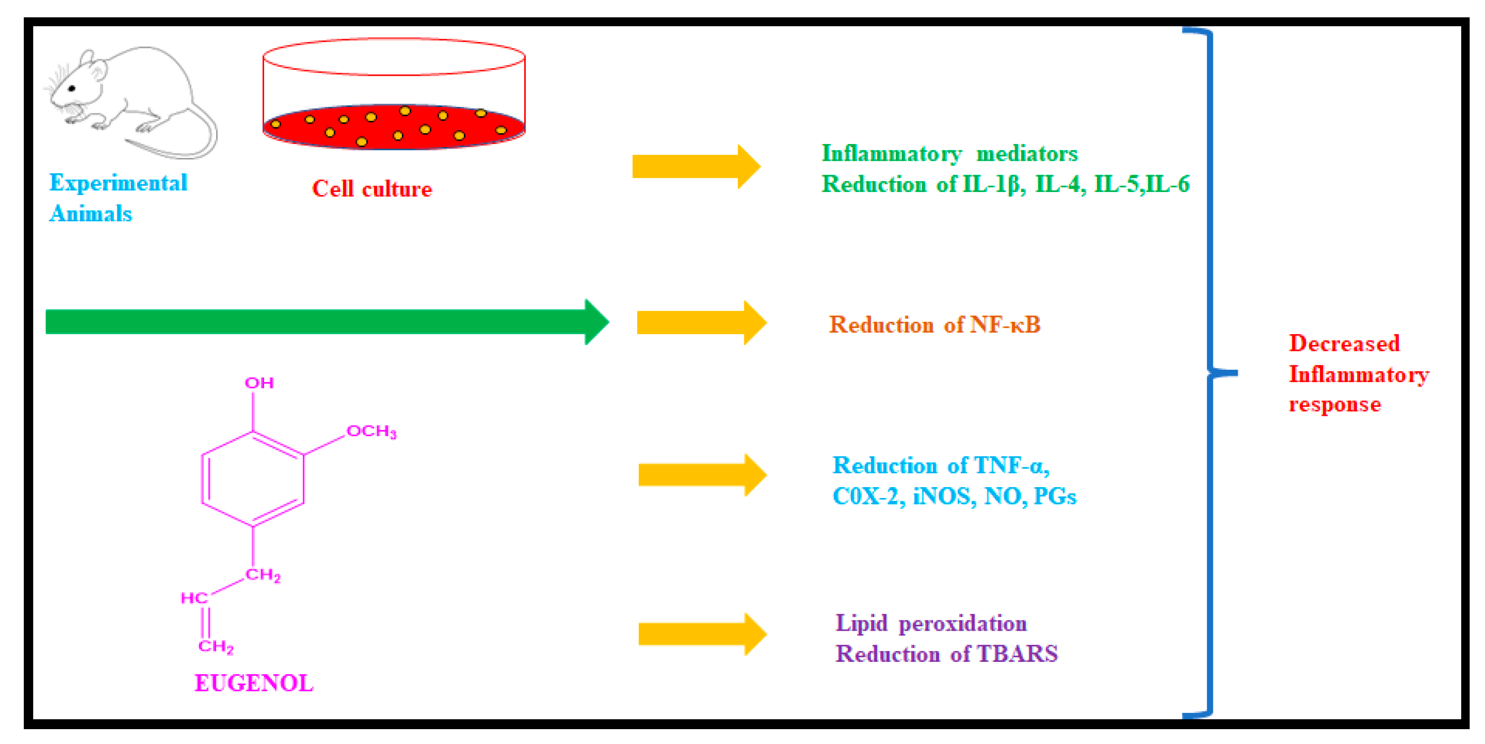
Numerous illnesses are influenced by inflammation [1], which is connected to the release of cytokines and oxygen-free radicals from activated inflammatory cells like eosinophils, neutrophils, macrophages, and monocytes. Proinflammatory chemokines and cytokines are upregulated during inflammation, and this activation and interaction with a variety of immunological factors result in inflammation [7,8]. Eugenol (4-allyl-2-methoxyphenol) is a phenolic substance belonging to the phenylpropanoid family. It is the principal constituent of clove (Eugenia caryophyllata) and belongs to the family Myrtacea [9]. The pharmacologically active compound eugenol is present in essential oils of plants [10]. It is also found in nutmeg (Myristic fragrans), basil (Ocimum basilicum L.), Melissa officinalis (Lemon Balm), cinnamon (Cinnamomum zeylanicum), Ocium tenuiflorum (Tulsi), and Illicium anisatum (Star Anise). Eugenol has proven to have many biological activities, including antibacterial [11], antioxidant [12,13], antitubercular [14], anticancer [15], antifungal [16], anti-inflammatory [17], and anesthetic activity [18]. The MEK phox pathway is inhibited by eugenol [19,20]. Several proinflammatory mediators, such as interleukins IL-5, interleukins IL-6, PGE2, Cox-2, and iNOS as well as activated NF-κB have all been reported to be inhibited by eugenol as shown in Figure 2.
Considering the importance of eugenol as a bioactive substance, we aimed to obtain novel derivatives of eugenol and to study possible anti-inflammatory activity. As a result, we synthesized eugenol-based PPARγ agonists analyzed, and screened for PPARγ protein binding, and then assessed their in vitro anti-inflammatory activity using an albumin denaturation assay and HRBC membrane stabilization method.

2. Results

2.1. Design of Eugenol Derivatives as PPARγ Agonists

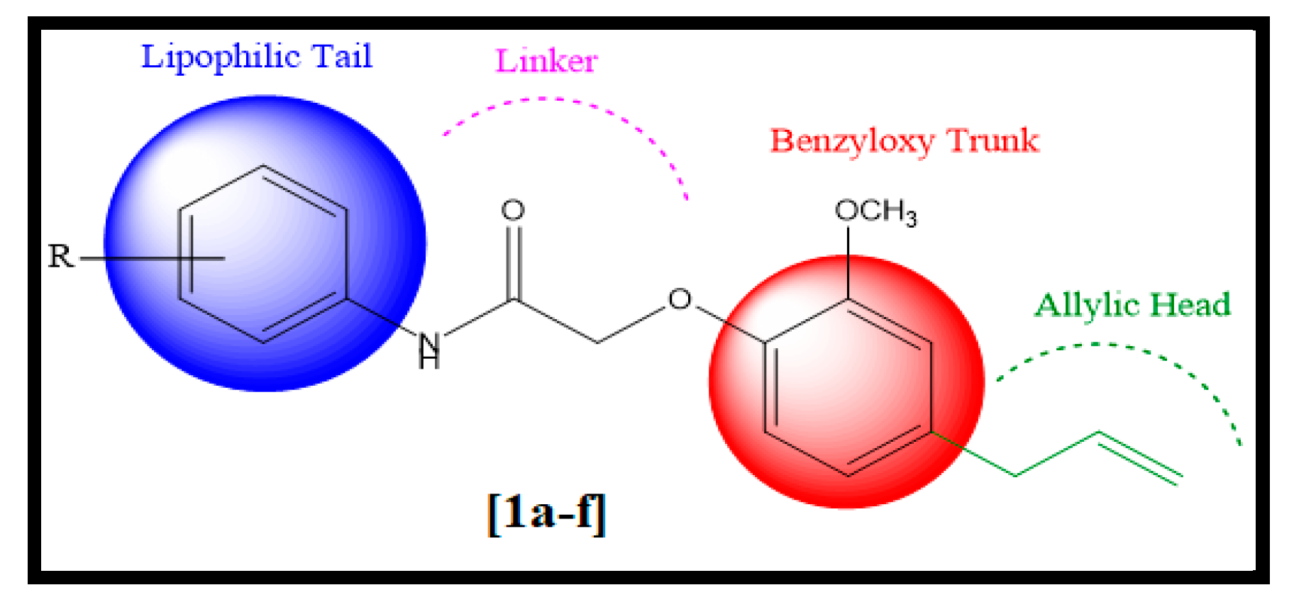
The structural characteristics of PPARγ agonists include a heterocyclic head, typically thiazolidinedione, followed by a benzyloxy trunk, a two-carbon linker, and a lipophilic tail. Based on these structural characteristics, we designed novel eugenol derivatives with a benzyloxy trunk, two carbon linkers, and a lipophilic tail Figure 3. However, the allylic head is used in place of the heterocyclic head.

2.2. Synthesis

The rationally designed eugenol derivatives involved the formation of substituted 2-chloro-N-phenylacetamide 2 by treating various aromatic amines 1 with chloroacetyl chloride using dichloromethane as a solvent, following this the substituted 2-chloro-N-phenylacetamide 2 were then linked to eugenol in the presence of dry acetone and anhydrous potassium carbonate to produce novel eugenol derivatives (1a1f) in good yields (70–87%) according to (Scheme 1) [21]. By using the IR, 1HNMR, 13CNMR, and mass spectral techniques, the structures of synthesized compounds were confirmed and evaluated. The absence of the -OH group at 3350 cm−1 in IR spectra confirmed the formation of 2. C=O and N-H groups were detected in the IR bands at 1690 cm−1 and 3460 cm−1, respectively. DMSO-d6 and CDCl3 were employed as the solvent for 1H-NMR and 13C-NMR. The chemical shifts are expressed as a number of parts per million (ppm), where s stands for singlet, d for doublet, t for triplet, and m for multiplet. All the synthesized compounds have a singlet in their 1H-NMR spectra that is attributed to the characteristic of the NH group and is present at about 8.73–9.97 ppm in addition to aromatic protons. one proton of CH= generates a multiplet at around δ 5.1–6.1 ppm and a doublet is produced by two protons of =CH2 at about δ 5.15 ppm. Similar to this, all the 13CNMR spectra show distinct signals attributed to the ketonic carbon at δ 167.6 ppm in addition to other typical carbon signals. ESI-MSMS was used to calculate the molecular mass of all the synthesized derivatives. All these facts support and confirm the formation of synthesized compounds (1a1f).

2.3. ADMET and TOPKAT Profile

For the purposes of determining the efficacies or dangers of small molecules, ADMET and toxic prediction of the compounds are key characteristics that must be evaluated [22]. The utilization of small molecular tools to predict the pharmacokinetics and dynamic profiles of synthesized compounds (Table 1). The ability of a drug to cross the blood–brain barrier and penetration is an important drug-related parameter. Nearly all compounds, including standard, have high blood–brain barrier penetration (level = 1), good human intestinal absorption (level = 0), low (level =2) to good (level = 3) solubility, and do not inhibit the activity of the CYP450 metabolizing enzyme. Furthermore, all the synthesized compounds obey Pfizer’s rule of 5 which is a key descriptor for oral bioavailability [23,24] ll compounds are free from mutagenicity and carcinogenicity as depicted in Table 1 and Table 2.

2.4. Structure-Based Drug Designing

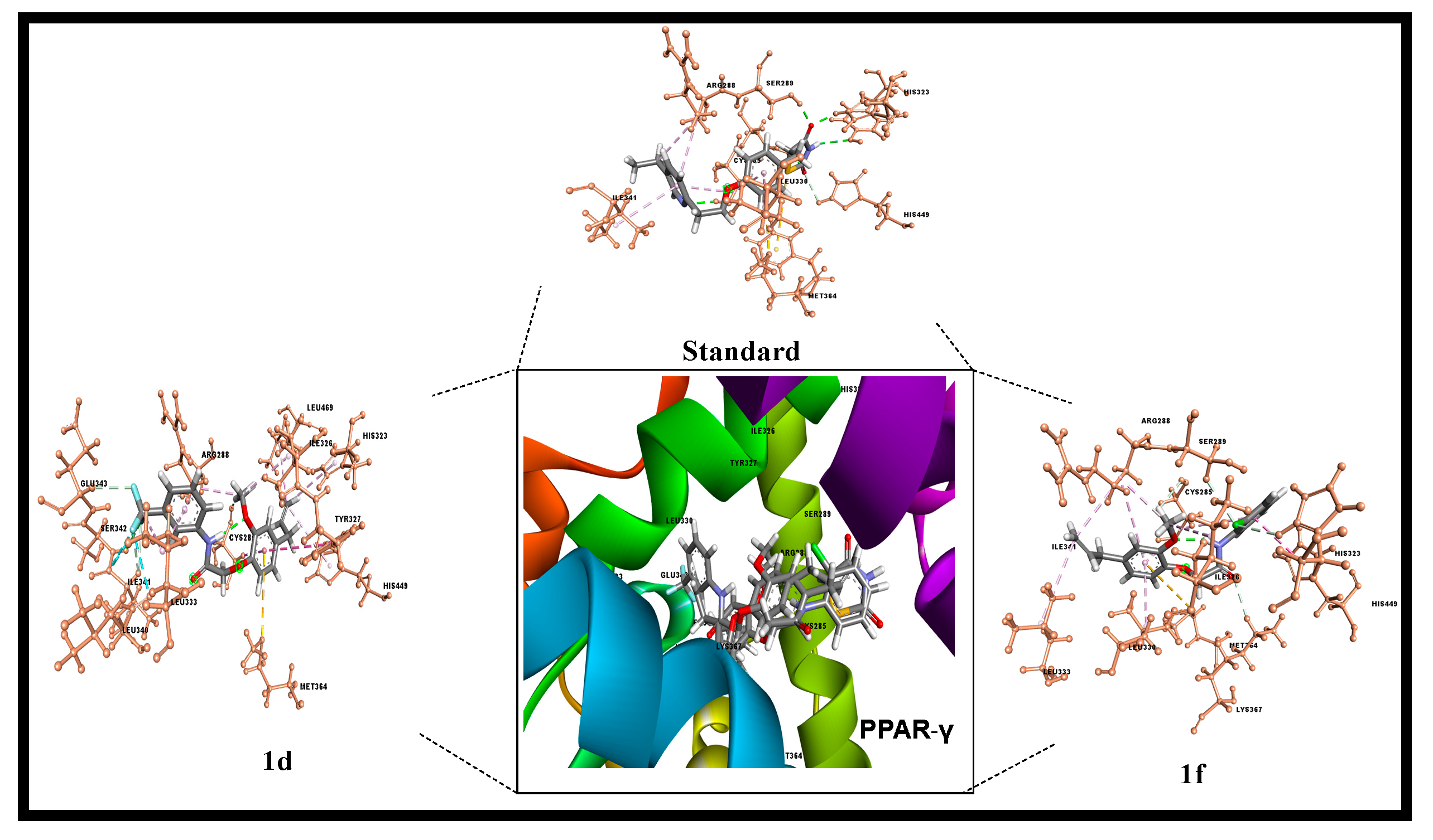
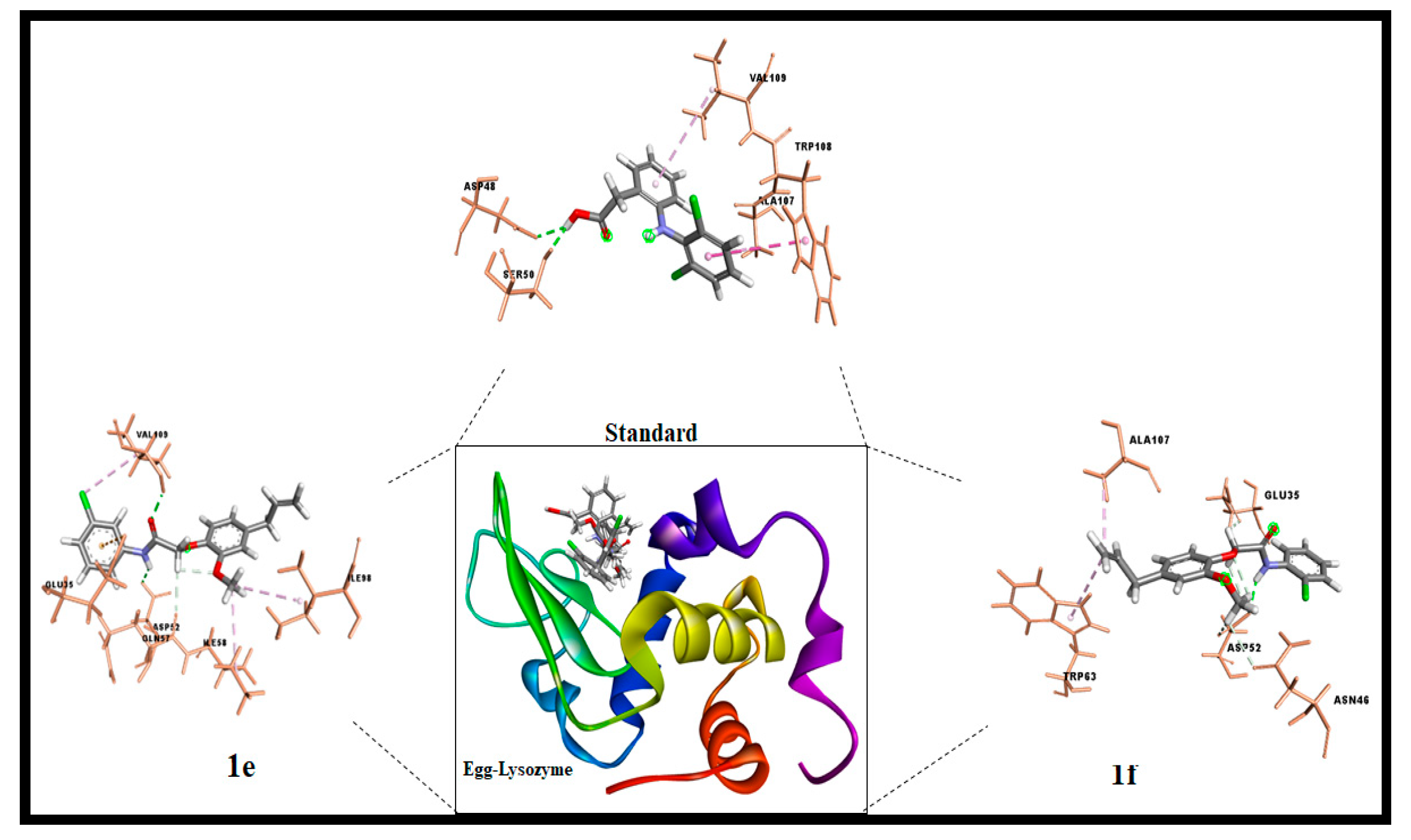
The concept of structure-based drug designing was implemented for known compounds and drug-targeting proteins [25]. The ligand binding domain region of Peroxisome Proliferator-activated Receptor PPAR gamma amino acids such as Phe282, Cys285, Gln286, Arg288, His323, Tyr327, Leu330, Gly338, Met364, His449, and Leu469. Receptor–ligand interactions analysis shows that all compounds have the potential to bind at the ligand binding site in varying docking energy scores Table 3 [26]. The Cdocker energy is significantly high 1d, 1f, and Standard. The 1f shows two hydrogen bonding interactions (Cys285, His323) with active site amino acid while 1d interactions are shared with active site neighboring announced residues. Moreover, it is common for both compounds to show hydrophobic interactions with Cys285, Arg288, Met364, and His449. Similarly, 1e binding energies are significant and high for 1f compared with standard (Diclofenac). Further, both hydrophobic and hydrogen bond interactions are observed at the binding site of the egg lysozyme anti-inflammatory drug target Figure 4 and Figure 5.

2.5. Structure–Activity Relationships (SAR)

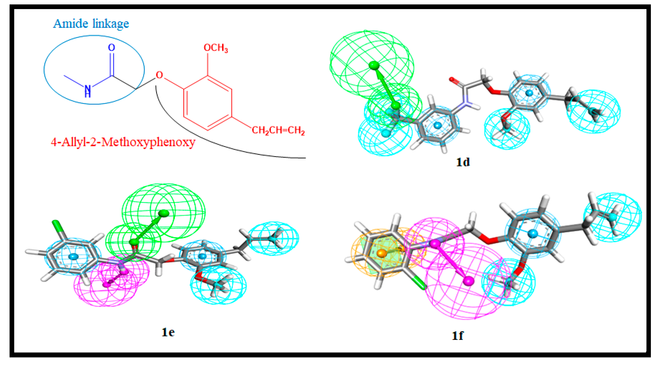
In order to synthesize multiple compounds for the better and more potent acquisition of molecules for various pharmacological targets, a structure–activity relationship (SAR) is an important tool and information source in organic chemistry [27]. The functional moieties connected to the compounds play a crucial role in predicting the compound’s biological activity from their chemical structure. In this study, interaction pharmacophores were employed to analyze the pharmacophore interactions in derivatives of receptor–ligand complexes (1a1f). Interestingly, 4-Allyl-2-Methoxyphenoxy is common in all the compounds and shares two hydrophobic and one aromatic hydrophobic Figure 6. In contrast, in highly active compounds benzene ring shares ring aromatic, and the location of each functional group induces the biological activity. Benzene ring with chloride (Cl) substitution in ortho position (1f) is a highly active compound among all compounds in this library. In the meta position, the benzene ring’s chloride (Cl) substitution shows less biological activity. The halogen (F) replacement at the meta position of the bone ring shows a slight deviation in the activity, while the sole benzene and (NO2) functional group attached aromatic ring shows less activity. Hence the position of the ortho and meta-functional group is considered a crucial pharmacophore and directly proportional to the biological activity. The halogen group (F, Cl) interacts with active site residues of the drug targets and enhanced the anti-inflammatory effects in ppar gamma binding and egg lysozyme assay. Therefore, SAR relation is a powerful technique utilized in combinatorial chemistry to predict the functional and biological activities of the molecules [28].
In the Pharmacophore model: The orange color symbolizes the ring aromatic (RA), the green sphere is the hydrogen bond acceptor (HBA), the magenta sphere is the hydrogen bond donor (HBD), and the cyan sphere is the hydrophobic region.

2.6. Time-Dependent Parameter Conformational Analysis of the Complex

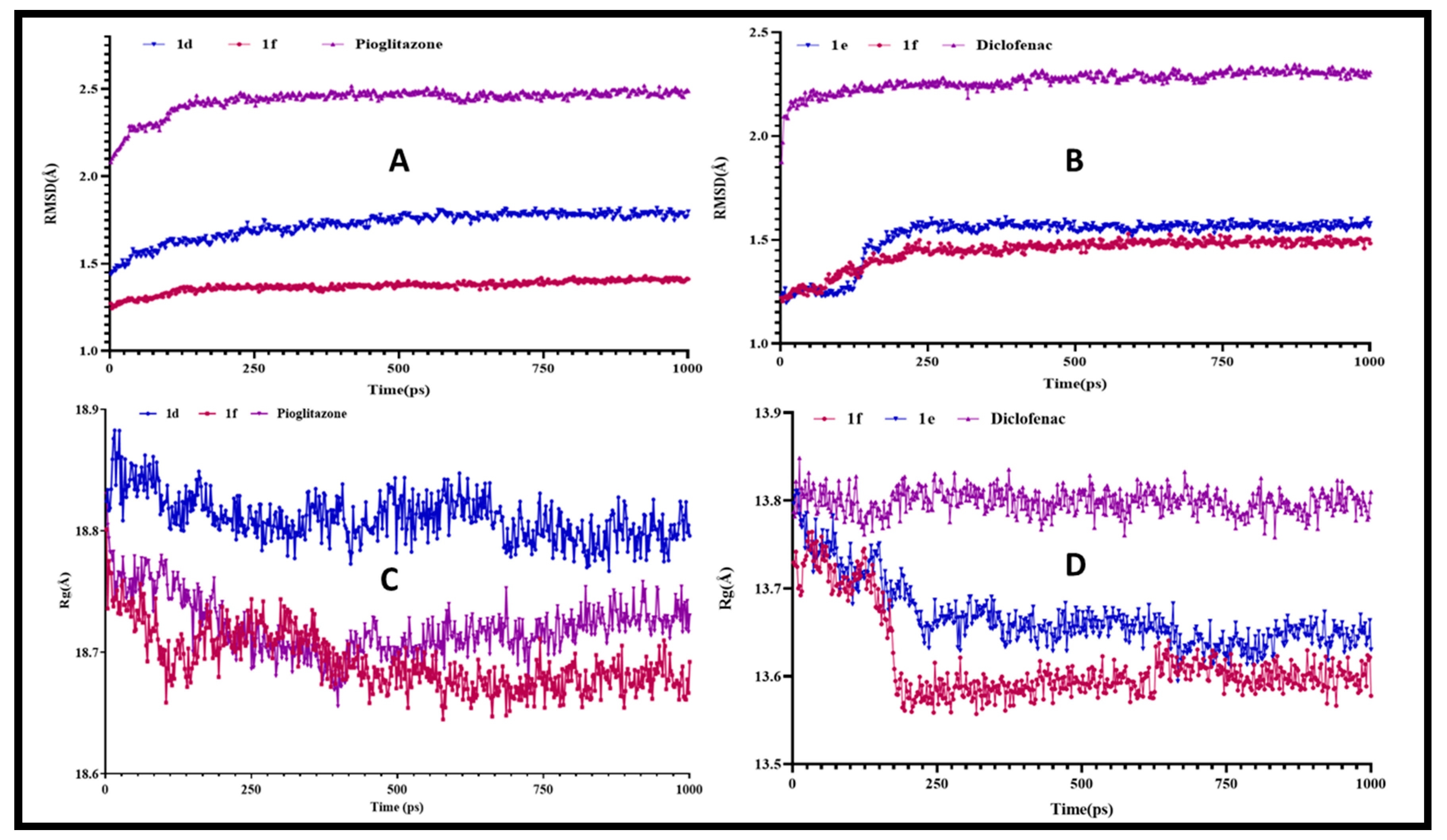
The binding modes of best-docked molecules of receptor-ligand were studied using a molecular dynamics simulation study for a simulation time of 1000ps. In order to assess the stability of the system, the geometric parameters of the protein–ligand complexes, such as the radius of gyration (Rg) and root mean square deviation (RMSD), were calculated [29]. The RMSD is used to measure the root mean square deviation of atomic positions of each conformation. The average distance between the atoms of various structural conformations of protein and ligand over a period of time. The average RMSD of the Cα atoms of Peroxisome Proliferator-activated Receptor (PPAR) gamma and heavy atoms of 1d and 1f was found to be 1.722 ± 0.07966 and 1.373 ± 0.03305 Å, respectively (Figure 7A). In contrast, the standard drug Pioglitazone with Peroxisome Proliferator-activated Receptor (PPAR) gamma complex had an average RMSD of 2.440 ± 0.07039 Å. Among the three complexes, 1f complex deviations are lower than standard and 1d which agrees with biological activity. Similarly, analyses of lysozyme bound complex average root mean square deviations are 1.518 ± 0.1071Å, 1.447 ± 0.07017 Å, and 2.268 ± 0.04858 Å (Figure 7B) of 1e, 1f, and standard drug (Diclofenac), respectively. Hence, a conformational change in the compounds has a direct influence on biological activity.
Rg can be explained as the root mean square distance from each atom of the system to its center of mass [30]. The Rg values for protein-ligand complexes: Peroxisome Proliferator-activated Receptor (PPAR) gamma 1f and Pioglitazone show fluctuations between 18.65 Å to 18.8 Å (Figure 7C). However, deviations are slightly less for 1d ranging from 18.8 Å to 18.9 Å. Nevertheless, anti-inflammatory drug target lysozyme bound complexes Rg values are between 13.6 Å to 13.9 Å (Figure 7D) among 1f complex 3D compactness shows least as compared with other complexes. The energy parameters analysis of all the complexes shows fewer variations in Table 4 that indicates energetically all three complexes are good Overall, the Peroxisome Proliferator-activated Receptor PPAR gamma-1f complex shows fewer deviations with time-dependent parameter analysis and 1f Lysozyme complex shows best in inflammatory activity.

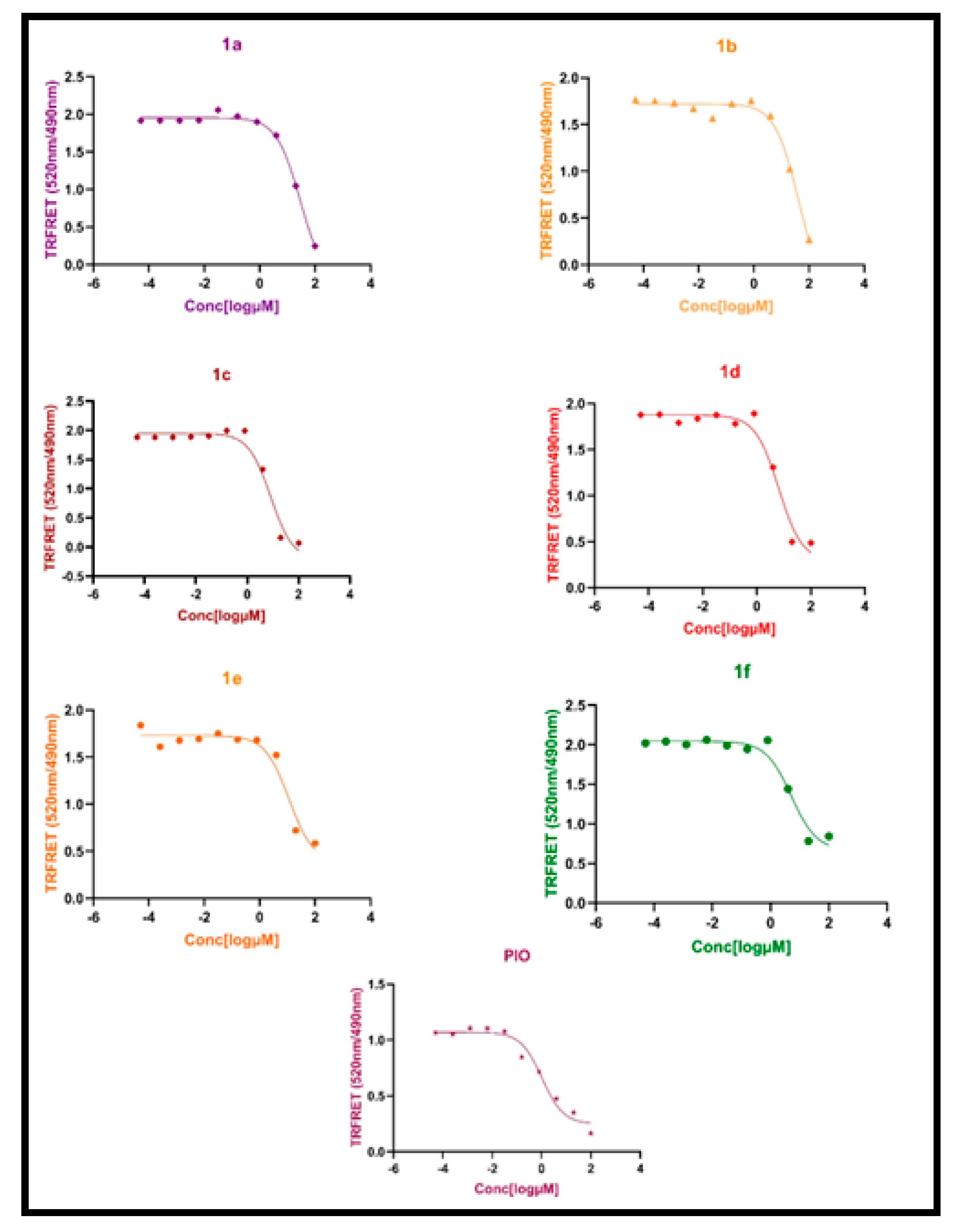
2.7. TR-FRET Assay

Using the Lanthasreen TR-FRET assay, the synthesized compounds were first preliminary tested for the PPARγ binding affinity [31]. A high 520/495 ratio is identified when the antibody is bound to the receptor because energy is transferred from the antibody to the fluorescent ligand (tracer) during this process [32]. A chemical under test displaces the tracer from PPAR-LBD, which lowers the TR-FRET ratio and decreases the FRET signal Figure 8 [33]. The compounds 1d and 1f showed a higher binding affinity for PPARγ among other synthesized compounds in comparison with standard pioglitazone. The assay results indicate that compounds 1d and 1f show an IC50 value of 6.47 µM and 5.15 µM in comparison with standard 1.77 µM, respectively. The IC50 values of synthesized compounds and standard drug pioglitazone are tabulated in Table 5.

2.8. In Vitro Anti-Inflammatory Activity

2.8.1. Albumin Denaturation Assay

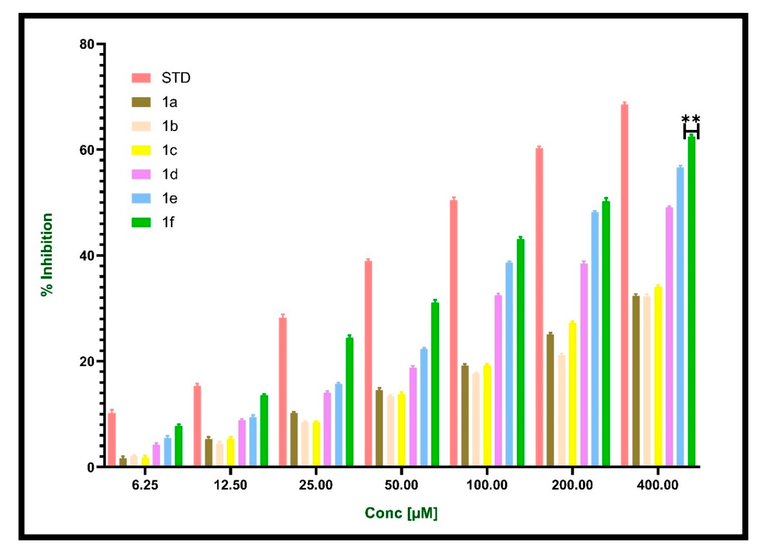
Inflammation is accompanied by protein denaturation. Tissue protein denaturation is a well-established cause of Inflammatory illnesses. Inflammatory disorders including rheumatoid arthritis, diabetes, and cancer have been linked to the denaturation of proteins. The ability of a substance to stop proteins from becoming denaturized has a role in the prevention of inflammatory disorders [34,35]. The possible anti-inflammatory activity of newly synthesized compounds and standard diclofenac was evaluated by the egg albumin denaturation assay method. Denaturation of proteins is accomplished by keeping the reaction mixture in a water bath at 70 °C for 10 min. As indicated in Table 6, demonstrated the effectiveness of synthesized compounds at different concentrations in preventing heat-induced albumin denaturation and maximum inhibition was observed for compound 1f 50.221 ± 0.60 at 200µM when compared with standard diclofenac 49.469 ± 0.51 at 100 µM. Albumin denaturation was significantly inhibited by standard Diclofenac and synthesized compounds at their respective IC50 values (Table 7 and Figure 9).
When a protein is exposed to external stress such as a strong acid or basic, an organic solvent, or heat, albumin denaturation occurs, which results in the loss of the protein’s secondary and tertiary structure. Biological proteins lose their biological function when they are denatured. Therefore, a ligand’s capacity to prevent protein denaturation denotes the possibility of anti-inflammatory activity [36,37]. Albumin denaturation assay clearly showed anti-inflammatory properties. According to the current study, newly synthesized compounds can stabilize lysosomal membranes in vivo and inhibit the production of autoantigens caused by the denaturation of protein. From the results, it was observed that compounds 1d, 1e, and 1f exhibit high activity when compared to other compounds. The active compound 1f containing chloro group at the ortho position is the highly potent compound with an IC50 value of 67.02 µM when compared to 1e containing chloro group and 1d containing trifluoro group at the meta position with an IC50 values of 96.23 µM and 106.8 µM, respectively. The nitro group at para position 1b is less active with an IC50 value of 294.3 µM than the methyl group at the ortho position 1c with an IC50 value of 134.0 µM.

2.8.2. HRBC Method

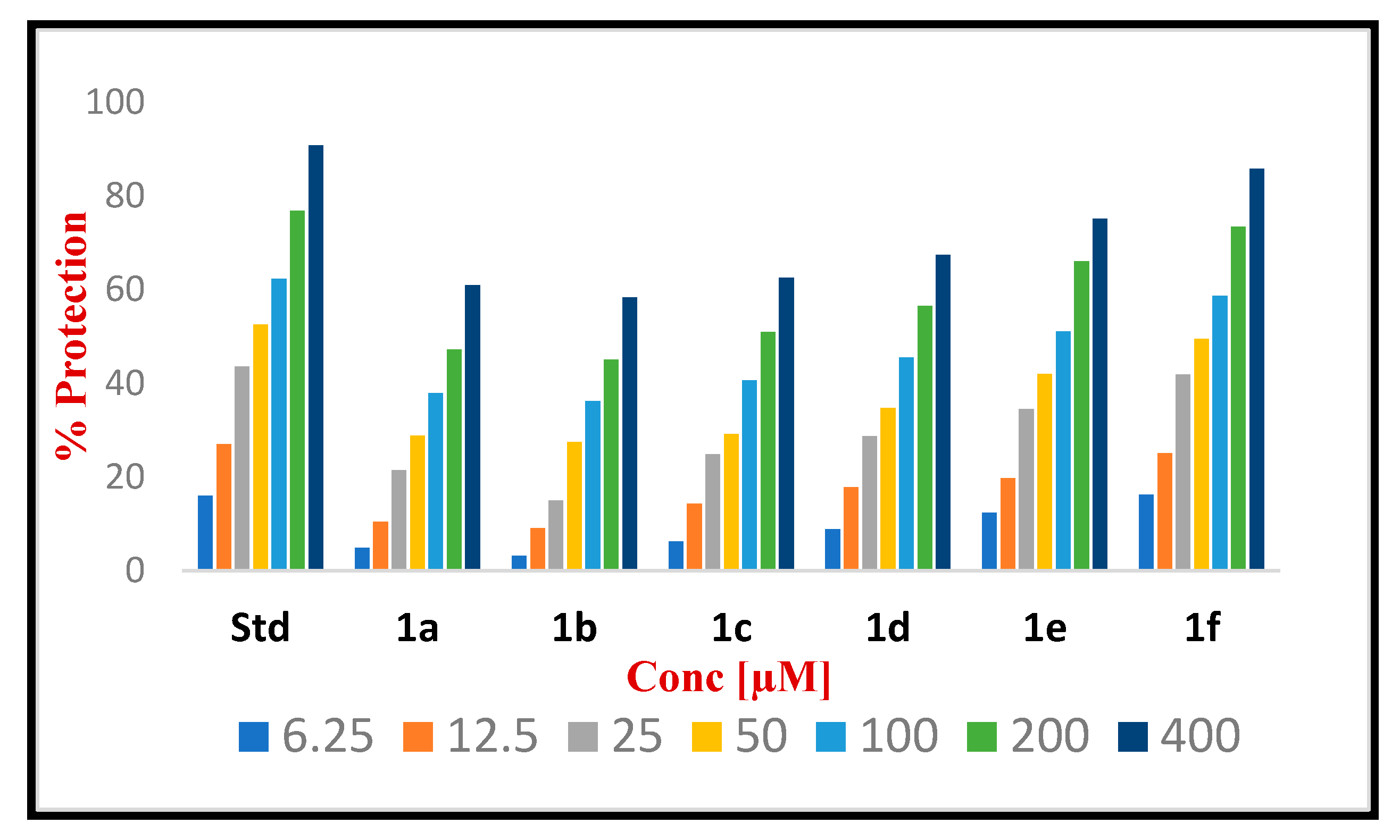
The principle of this procedure is to stabilize the membrane of human red blood cells by hypotonic membrane lysis. The release of lysosomal enzymes is a sign of inflammation. Lysosomal enzymes are released during the lysis of human red blood cells (HRBCs), and hence it is taken as a measure to evaluate the anti-inflammatory activity of synthesized compounds to reduce inflammation [38]. Lysosomal enzyme levels typically decrease as the HRBC membrane is stabilized. Therefore, the compounds that can cause HRBC membrane stabilization are expected to be anti-inflammatory [39]. In the current study, the activity of each synthesized compound was assessed as a percentage prevention of HRBC lysis in the concentration range of 6.25–400 µM. Diclofenac sodium was used as a standard drug. By analyzing the results, it can be concluded that all the synthesized compounds significantly reduced inflammation using the HRBC membrane stabilization approach. All compounds were found to have concentration-dependent membrane-stabilizing action. Compounds possessing chloro group at the ortho position 1f and chloro group at the meta position 1e as well as the trifluoro substitution in the meta position 1d have generally stabilized the HRBC in the range of 67–85% as shown in Figure 10. Additionally, compounds 1a with a single benzene ring and 1c with a methyl substitution in the ortho position showed 60.88 and 62.54% prevention against HRBC lysis, respectively. The nitro substitution of compound 1b has the least stabilizing effect. Compounds 1f was found to exhibit the highest level of membrane stabilization with 85.79% protection against HRBC lysis in comparison to standard diclofenac sodium which showed 90.77% protection, respectively, at 400 µM Table 8. IC50 values of synthesized derivatives of eugenol and standard diclofenac are depicted in Table 9.

3. Discussion

We have developed a simple method to synthesize new eugenol derivatives (1a1f) as PPARγ agonists based on the prediction of in silico results which we then analyzed with spectroscopic methods (FTIR, 1HNMR, 13CNMR, and Mass spectrometry). The novel derivatives obey the Pfizer rule and potentially all compounds are free from mutagenicity and carcinogenicity. Molecular docking was utilized to assess the designed compound’s ability to bind to the target protein. The docked structures at the binding sites were demonstrated by molecular dynamics simulations to be stable. All the newly synthesized compounds were found to be potential PPARγ agonists by molecular analysis and confirmed by in vitro TR-FRET protein binding assay. Thus, the synthesized compounds have proven to be a potential PPARγ agonist by molecular studies and in vitro assays. According to in vitro anti-inflammatory results, the synthesized compounds at concentrations ranging from 6.25 to 400 µM effectively reduced heat-induced albumin denaturation and exhibited maximum membrane stabilization with 85% protection against HRBC lysis in comparison to standard diclofenac sodium which showed 90% protection, respectively.

4. Materials and Methods

4.1. In Silico Studies and Spectral Data Analysis

The novel derivatives of eugenol were designed based on pharmacodynamics and pharmacokinetics requirements. BIOVIA, Discovery Studio 2019 (Dassault systems Biovia corp) was used to perform in silico prediction in order to comprehend the molecular behavior. Bayesian and regression models were also used to examine the compound’s established dosage range, mutagenicity, and carcinogenicity. All of the chemicals and solvents were purchased and procured from spectrochem (Lucknow, India) and Merck Ltd.(St. Louis, MI, USA) The melting points of the novel derivatives were established in open capillaries using a melting point instrument and are uncorrected. Aluminum plates that have been precoated with silica gel G are used to carry out TLC using n-hexane and ethyl acetate as the mobile phase (9:1) used to monitor the reactions and chemical purity and were made visible by UV rays. IR spectra are presented in cm−1 and were acquired using a shimadzu infrared FTIR spectrophotometer (FTIR-8400S, Shimadzu, Kyoto, Japan) utilizing KBr pellet technique. The Agilent VNMRS 400 equipment (Agilent 400 Hz, Agilent, Santa Clara, CA, USA) was used to record the 1H-NMR & 13C-NMR spectra with a chemical shift are given in ppm ranging from 0 to 10. Tetramethylsilane (TMS) is used as an internal standard. Coupling constants were recorded in hertz (Hz). Mass spectra were recorded using ESI-MSMS (Make-Waters USS, Model-Xevo G2-XS Q TOF, Make-Waters, Illinois, IL, USA).

4.2. General Procedure for the Synthesis of Eugenol Derivatives

  • Step-1: Synthesis of substituted 2-chloro-N-phenylacetamide
Triethylamine (1.1 eq) and various aromatic amines (1 eq) were added to dichloromethane (80–100 mL) and transferred to a flask with a guard tube. A total of 1.1 eq of chloroacetyl chloride were added drop by drop over a period of 30 min and the mixture was agitated under chilled circumstances (0–5 °C). At room temperature, the reaction mixture was again agitated for 10–12 h. Thin-layer chromatography (TLC) was used to monitor the reaction with n-hexane and ethyl acetate as the mobile phase. After the completion of the reaction the mixture was treated with water and dilute HCl and transferred to a separating funnel and allowed to separate. The DCM layer was passed through anhydrous sodium sulphate after the water layer was removed. Substituted 2-chloro-N-phenylacetamide was obtained by evaporating the solvent.
  • Step-2: Coupling eugenol with substituted 2-chloro-N-phenylacetamide
Eugenol (1.2 eq), finely powdered anhydrous potassium carbonate (K2CO3 3 eq), and 80 mL of dry acetone were added to a mixture of substituted 2-chloro-N-phenylacetamide (1 eq), and the mixture was stirred at 45 °C for about 26 h. As the reaction progressed, the spots were periodically checked using TLC with n-hexane and ethyl acetate as the mobile phase [40]. Ethyl acetate was used to extract the reaction mixture after the acetone was evaporated. After being washed three times with a 10% NaOH solution, once with water, and then once more with brine solution, the ethyl acetate layer was dried on anhydrous sodium sulphate as described in Scheme 1. The final product was obtained by evaporating the ethyl acetate layer (Table 10).

4.3. The Spectral Data of Novel Synthesized Eugenol Derivatives (Supplementary Materials)

4.3.1. 2-(4-Alyl-2-methoxyphenoxy)-N-phenylacetamide (1a)

Brown liquid Yield: 80.0% Mol Formula: C18H19NO3. IR (KBr, cm−1): 3336.96 (-NH), 3072.71 (ArC-H), 2914.54 (AliC-H), 1689.70 (C=O), 1600.97 (ArC=C), and 1317.43 (C-O). 1H NMR (400 MHz, δ ppm, DMSO-d6): 9.977 (s, 1H, NH), 7.624 (d, 2H, ArH, J = 7.6 Hz), 7.319(t, 2H, ArH, J = 7.2 Hz), 7.072 (t, 1H, ArH, J = 7.6 Hz), 6.907 (d, 1H, ArH, J = 8.4 Hz), 6.826 (s, 1H, ArH), 6.701 (d, 1H, ArH, J = 8.0 Hz), 5.954 (m, 1H, =CH, J = 13.2 Hz), 5.077 (d, 2H, =CH2, J = 5.6 Hz), 4.995 (s, 2H, CH2), 3.776 (s, 3H, OCH3), and 3.348 (d, 2H, CH2). 13C NMR: (400 MHz, δ ppm, DMSO-d6): 166.92 (C=O), 149.32 (C-O), 145.86 (O-C), 138.46 (-CH=), 137.90 (C-NH), 133.97 (ArC), 128.87 (ArC), 123.75 (2ArC), 120.63 (2ArC), 119.58 (2ArC), 115.71 (=CH2), 112.89 (ArC), 69.04 (CH2-O), 55.69 (O-CH3), and 40.26 (CH2). ESI-MSMS (m/z) peak calculated for C18H19NO3 [M+H]+ 298.112 peak found [M+H]+ 298.102.

4.3.2. 2-(4-Allyl-2-methoxyphenoxy)-N-(4-nitrophenyl)acetamide (1b)

Yellowish solid: Yield: 75.3% Mol Formula: C18H18N2O5. IR (KBr, cm−1): 3365.90 (-NH), 3076.56 (ArC-H), 2922.25 (AliC-H), 1612.54 (C=O), 1597.11 (ArC=C), 1508.38 (N-O), and 1336.71 (C-O). 1H-NMR (400 MHz, δ ppm, CDCl3): 9.384 (s, 1H, NH), 8.240 (d, 2H, ArH, J = 9.2 Hz), 7.793 (d, 2H, ArH, J = 9.2 Hz), 6.942 (s, 1H, ArH), 6.794 (d, 2H, ArH, J = 9.6 Hz), 5.972 (m, 1H, =CH, J = 11.2 Hz), 5.115 (d, 2H, =CH2, J = 6.4 Hz), 4.650 (s, 2H, CH2), 3.943 (s, 3H, O-CH3), and 3.363 (d, 2H, -CH2, J = 6.8 Hz) 13C-NMR: (400 MHz, δ ppm, CDCl3): 172.823 (C=O), 154.834 (C-O), 150.621 (O-C), 148.850 (C-NH), 148.140 (C-NO), 142.040 (-CH=), 141.456 (2ArC), 130.160 (2ArC), 126.444 (ArC), 124.245 (2ArC), 121.238 (ArC), 117.882 (=CH2), 76.163 (CH2-O), 61.141 (O-CH3), and 44.903 (CH2). ESI-MSMS (m/z) peak calculated for C18H18N2O5. [M-H] 341.31 peak found [M-H] 341.30.

4.3.3. 2-(4-Allyl-2-methoxyphenoxy)-N-(o-tolyl)acetamide (1c)

Colorless solid: Yield: 80.5% Mol Formula: C19H21NO3. IR (KBr, cm−1): 3388.77 (-NH), 3166.78 (ArC-H), 2934.47 (AliC-H), 1691.35 (C=O), 1597.17 (ArC=C), and 1377.05 (C-O). 1H-NMR (400 MHz, δ ppm, CDCl3): 8.739 (s, 1H, NH), 8.040 (s, 1H, ArH, 7.273 (d, 2H, ArH, J = 8.4 Hz), 7.120 (d, 1H, ArH, J = 7.2 Hz), 6.913 (d, 1H, ArH, J = 7.6 Hz), 6.786 (t, 2H, ArH, J = 7.6 Hz), 6.008 (m, 1H, =CH, J = 8.4 Hz), 5.135 (d, 2H, =CH2, J = 6.8 Hz), 4.668 (s, 2H, CH2), 3.890 (s, 3H, O-CH3), 3.385 (d, 2H, -CH2, J = 6.4 Hz), and 2.317 (s, 3H, -CH3). 13C-NMR: (400 MHz, δ ppm, CDCl3): 166.658 (C=O), 149.535 (C-O), 145.312 (O-C), 137.237 (-CH=), 135.233 (C-NH), 130.446 (ArC), 128.422 (2ArC), 126.827 (ArC), 125.037 (ArC), 122.098 (ArC), 120.795 (ArC), 115.959 (=CH2), 115.142 (ArC), 112.408 (ArC), 69.609 (CH2-O), 55.677 (O-CH3), 39.838 (CH2), and 29.690 (CH3). ESI-MSMS (m/z) peak calculated for C19H21NO3 [M+Na]+ 334.137 peak found [M+Na]+ 334.139.

4.3.4. 2-(4-Allyl-2-methoxyphenoxy)-N-[(3-trifluoromethyl)phenyl]acetamide (1d)

Dark brown liquid: Yield: 70.8% Mol Formula: C19H18 F3NO3. IR (KBr, cm−1): 3265.75 (-NH), 3083.43 (ArC-H), 2918.45 (AliC-H), 1687.30 (C=O), 1509.53 (ArC=C), and 1330.55 (C-O). 1H-NMR (400 MHz, δ ppm, CDCl3): 9.393 (s, 1H, NH), 7.990 (s, 1H, ArH) 7.887 (s, 1H, ArH) 7.459 (t, 1H, ArH, J = 7.6 Hz), 6.936 (d, 1H, ArH, J = 8.0 Hz), 6.865 (d, 1H, ArH, J = 8.2 Hz), 6.797 (d, 1H, ArH, J = 8.4 Hz), 6.707 (d, 1H, ArH, J = 9.6 Hz), 6.036 (m, 1H, =CH, J = 13.6 Hz), 5.163 (d, 2H, =CH2, J = 7.6 Hz), 4.657 (s, 2H, CH2), 3.933 (s, 3H, O-CH3), and 3.387 (d, 2H, -CH2). 13C-NMR: (400 MHz, δ ppm, CDCl3): 167.734 (C=O), 149.657 (C-O), 145.639 (O-C), 137.884 (-CH=), 135.958 (C-NH), 131.891 (ArC), 129.585 (ArC), 122.950 (ArC), 121.277 (ArC), 121.131 (ArC), 120.936 (ArC), 116.692 (=CH2), 116.568 (ArC), 116.033 (ArC), 114.554 (ArC), 112.666 (ArC), 70.733 (CH2-O), 55.828 (O-CH3), and 39.843 (CH2). ESI-MSMS (m/z) peak calculated for C19H18 F3NO3 [M+H]+ 366.101 peak found [M+H]+ 366.100.

4.3.5. 2-(4-Allyl-2-methoxyphenoxy)-N-(3-chlorophenyl)acetamide (1e)

Brownish black liquid: Yield: 77.6% Mol Formula: C18H18ClNO3. IR (KBr, cm−1): 3342.75 (-NH), 3061.57 (ArC-H), 2985.62 (AliC-H), 1683.28 (C=O), 1593.86 (ArC=C), and 1307.78 (C-O). 1H-NMR (400 MHz, δ ppm, CDCl3): 9.143 (s, 1H, NH), 7.760 (d, 1H, ArH), 7.498 (d, 1H, ArH), 7.127 (t, 1H, ArH, J = 8.8 Hz), 6.932 (d, 1H, ArH, J = 8.0 Hz), 6.878 (d, 1H, ArH, J = 8.4 Hz), 6.812 (d, 1H, ArH, J = 6.8 Hz), 6.706 (d, 1H, ArH, J = 6.8 Hz), 6.029 (m, 1H, =CH, J = 10.4 Hz), 5.151 (d, 2H, =CH2, J = 8.4 Hz), 4.636 (s, 2H, CH2), 3.939 (s, 3H, O-CH3), and 3.385 (d, 2H, -CH2, J = 6.4 Hz). 13C-NMR: (400 MHz, δ ppm, CDCl3): 167.432 (C=O), 149.676 (C-O), 146.582 (O-C), 137.865 (-CH=), 135.919 (C-NH), 131.891 (ArC), 130.257 (ArC), 126.278 (C-Cl), 124.555 (ArC), 121.257 (ArC), 119.963 (ArC), 116.821 (-CH=), 115.498 (ArC), 114.427 (ArC), 111.256 (ArC), 70.733 (CH2-O), 55.955 (O-CH3), and 39.872 (CH2). ESI-MSMS (m/z) peak calculated for C18H18ClNO3. [M+H]+ 332.087 peak found [M+H]+ 332.077.

4.3.6. 2-(4-Allyl-2-methoxyphenoxy)-N-(2-chlorophenyl)acetamide (1f)

Colorless solid Yield: 87.9% Mol Formula: C18H18ClNO3. IR (KBr, cm−1): 3444.19 (-NH), 3062.52 (ArC-H), 2930.59 (AliC-H), 1676.47 (C=O), 1599.40 (ArC=C), and 1438.69 (C-O). 1H-NMR (400 MHz, δ ppm, CDCl3): 9.351 (s, 1H, NH), 8.456 (s, 1H, ArH), 7.401 (d, 1H, ArH, J = 7.2 Hz), 7.309 (d, 1H, ArH, J = 7.2 Hz), 7.085 (d, 1H, ArH, J = 7.2 Hz), 6.906 (d, 1H, ArH, J = 8.4 Hz), 6.769 (t, 2H, ArH, J = 6.0 Hz),), 6.009 (m, 1H, =CH, J = 6.8 Hz), 5.114 (d, 2H, =CH2, J = 6.0 Hz), 4.645 (s, 2H, CH2), 3.878 (s, 3H, O-CH3), and 3.364 (d, 2H, -CH2, J = 6.4 Hz). 13C-NMR: (400 MHz, δ ppm, CDCl3): 167.014 (C=O), 149.812 (C-O), 145.279 (O-C), 137.252 (-CH=), 135.423 (C-NH), 134.139 (ArC), 129.157 (ArC), 127.649 (C-Cl), 124.935 (ArC), 123.300 (ArC), 121.588 (ArC), 120.635 (ArC), 115.935 (=CH2), 115.566 (ArC), 112.520 (ArC), 69.751 (CH2-O), 55.712 (O-CH3), and 39.843 (CH2). ESI-MSMS (m/z) peak calculated for C18H18ClNO3. [M+H]+ 332.121 peak found [M+H]+ 332.122.

4.4. ADMET, Drug Likeness and Toxicity Predictions

In order to assess in silico pharmacokinetics and pharmacodynamic studies, the synthesized compounds were submitted to ADMET and TOPKAT tools of the small molecule protocol [40]. Carcinogenicity, mutagenicity and the set dosage range of the synthesized compounds were analyzed using the Bayesian and regression models. Furthermore, Lipinski’s rule of five was applied to determine the compound’s oral bioavailability.

4.5. Molecular Docking

The macromolecule tool and small molecule protocol were used to prepare the structure of the drug-targeting protein and the compounds employed for docking [40]. The bound ligand coordinates 49.720X −36.98Y 19.294Z of radius 8.4 for PPARγ (2PRG) and 26.63X 5.50Y 14.22Z of radius 12 for egg lysozyme (3WXU) were defined as binding sites for docking with an equal grid spacing of 0.5 and 90-degree grid angles. The CDOCKER algorithm, a potent CHARMm-based docking method that has been shown to create accurate docked poses, was used to analyse receptor–ligand interaction. Every parameter was set to default while executing the docking process. Finally, the best pose of ligands was taken for molecular dynamics simulation and non-bonded interaction analysis. Consequently, the top-docked complexes were probed to estimate binding free energy using CHARMm implicit solvation models. The binding free energy GBSA is estimated between each ligand and the receptor.

4.6. Molecular Dynamics Simulation

Using Dassault System BIOVIA, Discovery Studio 2019 modeling Environment, 1000 Picosecond molecular dynamics simulations and the top two compounds of interaction with drug targets PPARγ and egg lysozyme were performed [40]. The process was done in five cascade steps, starting with two steps of 500-cycle energy minimization of a complex with the steepest descent and conjugate gradient. The system is then gradually heated from 50 to 300 K with 100 ps of simulation time and 500 ps of equilibration to reach degrees of freedom. The production using the canonical ensemble was then exposed to equal Tmass and Pmass at 300 K. In addition, the SHAKE constraint fixes all hydrogen-containing bonds in the simulation inside this cutoff range of 12Å–10 Å. Utilizing the leap-frog verlet algorithm, all atom velocities and positions are calculated at specific time points. Finally, RMSD and Rg were used to examine the deviations in complex conformations.

4.7. TR-FRET Competitive Protein Binding Assay

Lanthascreen TR-FRET protein-binding competitive experiments were carried out using a buffer solution containing 50 mM TRIS, pH 7.5, 150 mM NaCl, 0.02% Triton® X-100, and 5 mM DTT in a black flat bottom 384-microwell plates [40]. The ligand stocks and standard pioglitazone were made by serial dilution in 1% DMSO concentration, applied to wells in duplicates, and incubated for an hour at room temperature. A multimode low-volume proxy plate reader was used to read the plates after one hour of incubation. Then, a mixture of a human PPARγ- LBD labelled with glutathione S-transferase (GST), a fluorescent small pan-PPAR ligand (FluormoneTM Pan-PPAR Green), and terbium-labeled anti-GST antibody was added. The acceptor FITC emission was measured at 520 nm, the Tb source was stimulated at 340 nm and the Tb source’s fluorescence intensity was recorded at around 495 nm.

4.8. Anti-Inflammatory Activity

4.8.1. Albumin Denaturation Assay

Minor modifications were made to Mizushima and Kobayashi’s and Sakat et al. methodologies [41]. The reaction mixture containing 0.2 mL of egg albumin (from a fresh hen’s egg) is mixed with 2.8 mL of phosphate-buffered saline (PBS, pH 6.4) and 2 mL of synthesized compounds in varying concentrations. The mixture was then heated at 70 °C for 5 min after being incubated at 37 °C for approximately 15–20 min. After cooling, their absorbance was measured at 660 nm spectrometrically [40]. Three duplicates of the experiment were carried out. The following formula was used to determine the percentage inhibition of protein denaturation.
Percentage inhibition = (OD control − OD sample/OD control) × 100

4.8.2. HRBC Method

Healthy volunteer blood was drawn, and an equal volume of sterilized Alsevers solution (0.05% citric acid, 0.05% sodium citrate, 2% dextrose, and 0.42 % sodium chloride) was added. The packed cells in this blood solution were separated after centrifugation at 3000 rpm [41]. The packed cells were cleaned using an isosaline solution, and then, isosaline was used to prepare a 10% v/v suspension. This HRBC suspension was employed to estimate its anti-inflammatory properties. Different concentrations of the synthesized compounds and the reference standard were each combined with 1 mL of phosphate buffer (pH 7.4), 2 mL of hyposaline, and 0.5 mL of HRBC solution. A total of 2.0 mL of distilled water was utilized as the control. The assay mixtures were all centrifuged at 3000 rpm after 30 min of incubation at 37°C. The following formula was used to determine the percentage of stabilized HRBC membranes.
Percentage protection= 100 − (OD sample/OD control) × 100

4.9. Statistical Analysis

Statistical analysis was performed by one-way analysis of variance (ANOVA) using Graph pad prism 8.0 software. Data are expressed as the mean ± standard deviation of the mean. p values less than 0.05 were considered statistically significant.

5. Conclusions

Novel derivatives of eugenol were synthesized, analyzed, and screened for PPARγ protein binding assay and were evaluated for in vitro anti-inflammatory activity using an albumin denaturation assay and HRBC membrane stabilization method. The structural elucidation of synthesized compounds was accomplished by the use of IR, 1HNMR, 13CNMR and mass spectrometry. The designed compound’s capacity to attach to the target protein was evaluated using molecular docking. Molecular dynamics simulations showed that the docked structures at the binding sites were stable. From the TR-FRET protein binding assay, it was determined that all of the newly synthesized compounds were potential PPAR agonists. Our results show that the newly synthesized compounds have good anti-inflammatory properties by inhibiting the heat-induced albumin denaturation and red blood cells membrane stabilization at different concentrations. It was thought that compound 1f was a bioactive substance, further studied at a molecular level, and can be investigated to learn more about its pharmacological action.

Supplementary Materials

The supporting information can be downloaded at: https://www.mdpi.com/article/10.3390/molecules28093899/s1. Figures S1–S6: IR, HNMR, 13CNMR & Mass Spectra of compounds 1a1f; Tables S1–S6: TR-FRET Assay of compounds 1a1f.

Author Contributions

Conceptualization, M.N.P. and B.R.P.K.; Methodology, N.F.A.; Docking and MD simulations, D.S.; Lanthascreen protein binding assay, R.M.R. and S.J.; Anti-inflammatory activity, N.F.A. and P.D.; Writing—Original Draft Preparation, N.F.A. and S.F.; Writing—Review & Editing, B.R.P.K. and M.N.P.; Visualization, B.R.P.K.; Supervision, M.N.P. All authors have read and agreed to the published version of the manuscript.

Funding

This research received no external funding.

Institutional Review Board Statement

Not applicable.

Informed Consent Statement

Not applicable.

Data Availability Statement

Not applicable.

Acknowledgments

The authors are thankful to the JSS Academy of Higher Education & Research Mysuru, and Farooqia College of Pharmacy, Mysuru, administration provided the necessary laboratory space for the present research activity.

Conflicts of Interest

The authors declare no conflict of interest.

Sample Availability

Samples of the compounds are available from the authors.

References

  1. Belvisi, M.G.; Hele, D.J.; Birrell, M.A. Peroxisome proliferator-activated receptor gamma agonists as therapy for chronic airway inflammation. Eur. J. Pharmacol. 2006, 533, 101–109. [Google Scholar] [CrossRef]
  2. El-Naa, M.M.; El-Refaei, M.F.; Nasif, W.A.; Abdul Jawad, S.H.; El-Brairy, A.I.; El-Readi, M.Z. In-vivo antioxidant and anti-inflammatory activity of rosiglitazone, a peroxisome proliferator-activated receptor-gamma (PPAR-γ) agonists in animal model of bronchial asthma. J. Pharm. Pharmacol. 2015, 67, 1421–1430. [Google Scholar] [CrossRef] [PubMed]
  3. Kapadia, R.; Yi, J.H.; Vemuganti, R. Mechanisms of anti-inflammatory and neuroprotective actions of PPAR-gamma agonists. Front. Biosci. 2008, 1, 1813–1826. [Google Scholar] [CrossRef]
  4. Straus, D.S.; Glass, C.K. Anti-inflammatory actions of PPAR ligands: New insights on cellular and molecular mechanisms. Trends Immunol. 2007, 28, 551–558. [Google Scholar] [CrossRef]
  5. Villapol, S. Roles of Peroxisome Proliferator-Activated Receptor Gamma on Brain and Peripheral Inflammation. Cell. Mol. Neurobiol. 2018, 38, 121–132. [Google Scholar] [CrossRef]
  6. Xie, L.-W.; Atanasov, A.G.; Guo, D.-A.; Malainer, C.; Zhang, J.-X.; Zehl, M.; Guan, S.-H.; Heiss, E.H.; Urban, E.; Dirsch, V.M.; et al. Activity-guided isolation of NF-κB inhibitors and PPARγ agonists from the root bark of Lycium chinense Miller. J. Ethnopharmacol. 2014, 152, 470–477. [Google Scholar] [CrossRef] [PubMed]
  7. Wahli, W.; Michalik, L. PPARs at the crossroads of lipid signaling and inflammation. Trends Endocrinol. Metab. 2012, 23, 351–363. [Google Scholar] [CrossRef]
  8. Klotz, L.; Burgdorf, S.; Dani, I.; Saijo, K.; Flossdorf, J.; Hucke, S.; Alferink, J.; Novak, N.; Beyer, M.; Mayer, G.; et al. The nuclear receptor PPAR gamma selectively inhibits Th17 differentiation in a T cell-intrinsic fashion and suppresses CNS autoimmunity. J. Exp. Med. 2009, 206, 2079–2789. [Google Scholar] [CrossRef] [PubMed]
  9. Anjum, N.F.; Purohit, M.N.; Yogish Kumar, H.; Ramya, K.; Javid, S.; Salahuddin, M.D.; Prashantha Kumar, B.R. Semisynthetic Derivatives of Eugenol and their Biological Properties: A Fleeting Look at the Promising Molecules. J. Biol. Act. Prod. Nat. 2020, 10, 379–404. [Google Scholar] [CrossRef]
  10. Abdou, A.; Elmakssoudi, A.; El Amrani, A.; JamalEddine, J.; Dakir, M. Recent advances in chemical reactivity and biological activities of eugenol derivatives. Med. Chem. Res. 2021, 30, 1011–1030. [Google Scholar] [CrossRef]
  11. Rojo, L.; Vazquez, B.; Parra, J.; López Bravo, A.; Deb, S.; San Roman, J. From natural products to polymeric derivatives of “eugenol”: A new approach for preparation of dental composites and orthopedic bone cements. Biomacromolecules 2006, 7, 2751–2761. [Google Scholar] [CrossRef] [PubMed]
  12. Gulcin, I. Antioxidant activity of eugenol: A structure-activity relationship study. J. Med. Food 2011, 14, 975–985. [Google Scholar] [CrossRef] [PubMed]
  13. da Silva, F.F.M.; Monte, F.J.Q.; de Lemos, T.L.G.; do Nascimento, P.G.G.; de Medeiros Costa, A.; de Paiva, L.M.M. Eugenol derivatives: Synthesis, characterization, and evaluation of antibacterial and antioxidant activities. Chem. Cent. J. 2018, 12, 34. [Google Scholar] [CrossRef]
  14. Rohane, S.H.; Chauhan, A.J.; Fuloria, N.K.; Fuloria, S. Synthesis and in vitro antimycobacterial potential of novel hydrazones of eugenol. Arab. J. Chem. 2020, 13, 4495–4504. [Google Scholar] [CrossRef]
  15. Jaganathan, S.K.; Supriyanto, E. Antiproliferative and molecular mechanism of eugenol-induced apoptosis in cancer cells. Molecules 2012, 17, 6290–6304. [Google Scholar] [CrossRef]
  16. Xie, Y.; Huang, Q.; Wang, Z.; Cao, H.; Zhang, D. Structure-activity relationships of cinnamaldehyde and eugenol derivatives against plant pathogenic fungi. Ind. Crop. Prod. 2017, 97, 388–394. [Google Scholar] [CrossRef]
  17. Zhang, P.; Zhang, E.; Xiao, M.; Chen, C.; Xu, W. Study of anti-inflammatory activities of α-d-glucosylated eugenol. Arch. Pharm. Res. 2013, 36, 109–115. [Google Scholar] [CrossRef]
  18. Pramod, K.; Ansari, S.H.; Ali, J. Eugenol: A natural compound with versatile pharmacological actions. Nat. Prod. Commun. 2010, 5, 1999–2006. [Google Scholar] [CrossRef]
  19. Chniguir, A.; Pintard, C.; Liu, D.; Dang, P.M.C.; El-Benna, J.; Bachoual, R. Eugenol prevents fMLF-induced superoxide anion production in human neutrophils by inhibiting ERK1/2 signaling pathway and p47phox phosphorylation. Sci. Rep. 2019, 9, 18540. [Google Scholar] [CrossRef]
  20. Deepak, V.; Kasonga, A.; Kruger, M.C.; Coetzee, M. Inhibitory effects of eugenol on RANKL-induced osteoclast formation via attenuation of NF-κB and MAPK pathways. Connect. Tissue Res. 2015, 56, 195–203. [Google Scholar] [CrossRef]
  21. Prashantha Kumar, B.R.; Baig, N.R.; Sudhir, S.; Kar, K.; Kiranmai, M.; Pankaj, M.; Joghee, N.M. Discovery of novel glitazones incorporated with phenylalanine and tyrosine: Synthesis, antidiabetic activity and structure-activity relationships. Bioorg. Chem. 2012, 45, 12–28. [Google Scholar] [CrossRef]
  22. Daina, A.; Michielin, O.; Zoete, V. SwissADME: A free web tool to evaluate pharmacokinetics, drug-likeness and medicinal chemistry friendliness of small molecules. Sci. Rep. 2017, 7, 42717. [Google Scholar] [CrossRef]
  23. Lipinski, C.A. Drug-like properties and the causes of poor solubility and poor permeability. J. Pharmacol. Toxicol. Methods 2000, 44, 235–249. [Google Scholar] [CrossRef]
  24. Lipinski, C.A.; Lombardo, F.; Dominy, B.W.; Feeney, P.J. Experimental and computational approaches to estimate solubility and permeability in drug discovery and development settings. Adv. Drug Deliv. Rev. 2012, 64, 4–17. [Google Scholar] [CrossRef]
  25. Tirado-Rives, J.; Jorgensen, W.L. Contribution of conformer focusing to the uncertainty in predicting free energies for protein-ligand binding. J. Med. Chem. 2006, 49, 5880–5884. [Google Scholar] [CrossRef] [PubMed]
  26. Ferreira, L.G.; Dos Santos, R.N.; Oliva, G.; Andricopulo, A.D. Molecular docking and structure-based drug design strategies. Molecules 2015, 20, 13384–13421. [Google Scholar] [CrossRef] [PubMed]
  27. Guha, R. On exploring structure-activity relationships. Methods Mol. Biol. 2013, 993, 81–94. [Google Scholar] [CrossRef]
  28. Martinez-Gonzalez, A.I.; Díaz-Sánchez, Á.G.; De La Rosa, L.A.; Bustos-Jaimes, I.; Alvarez-Parrilla, E.J. Inhibition of α-amylase by flavonoids: Structure activity relationship (SAR). Spectro. Acta Part A Mol. Biomol. Spectro. 2019, 206, 437–447. [Google Scholar] [CrossRef]
  29. Maiorov, V.N.; Crippen, G.M. Significance of root-mean-square deviation in comparing three-dimensional structures of globular proteins. J. Mol. Biol. 1994, 235, 625–634. [Google Scholar] [CrossRef]
  30. Lobanov, M.Y.; Bogatyreva, N.S.; Galzitskaya, O.V. Radius of gyration as an indicator of protein structure compactness. Mol. Biol. 2008, 42, 623–628. [Google Scholar] [CrossRef]
  31. Mosure, S.A.; Shang, J.; Eberhardt, J.; Brust, R.; Zheng, J.; Griffin, P.R.; Forli, S.; Kojetin, D.J. Structural Basis of Altered Potency and Efficacy Displayed by a Major in Vivo Metabolite of the Antidiabetic PPARγ Drug Pioglitazone. J. Med. Chem. 2019, 62, 2008–2223. [Google Scholar] [CrossRef]
  32. Kumar, A.P.; Mandal, S.; Prabitha, P.; Faizan, S.; Prashantha Kumar, B.R.; Dhanabal, S.P.; Justin, A. Rational design, molecular docking, dynamic simulation, synthesis, PPAR-γ competitive binding and transcription analysis of novel glitazones. J. Mol. Struct. 2022, 1265, 133354. [Google Scholar] [CrossRef]
  33. Sivamani, Y.; Shanmugarajan, D.; Kumar, T.D.; Faizan, S.; Channappa, B.; Naishima, N.L.; Prashantha Kumar, B.R. A promising in silico protocol to develop novel PPARγ antagonists as potential anticancer agents: Design, synthesis and experimental validation via PPARγ protein activity and competitive binding assay. Comp. Biol. Chem. 2021, 95, 107600. [Google Scholar] [CrossRef] [PubMed]
  34. Moualek, I.; Iratni Aiche, G.; Mestar Guechaoui, N.; Lahcene, S.; Houali, K. Antioxidant and anti-inflammatory activities of Arbutus unedo aqueous extract. Asian Pac. J. Trop. Biomed. 2016, 6, 937–944. [Google Scholar] [CrossRef]
  35. Ruiz-Ruiz, J.C.; Matus-Basto, A.J.; Acereto-Escoffié, P.; Segura-Campos, M.R. Antioxidant and anti-inflammatory activities of phenolic compounds isolated from Melipona beecheii honey. Food Agric. Immunol. 2017, 28, 1424–1437. [Google Scholar] [CrossRef]
  36. Naveed, M.; Batool, H.; Rehman, S.u.; Javed, A.; Makhdoom, S.I.; Aziz, T.; Mohamed, A.A.; Sameeh, M.Y.; Alruways, M.W.; Dablool, A.S.; et al. Characterization and evaluation of the antioxidant, antidiabetic, anti-inflammatory, and cytotoxic activities of silver nanoparticles synthesized using Brachychiton populneus leaf extract. Processes 2022, 10, 1521. [Google Scholar] [CrossRef]
  37. Bhosale, M.; Yadav, A.; Magdum, C.; Mohite, S. Molecular Docking Studies, Synthesis, Toxicological Evaluation using Brine Shrimp (Artemia salina L.) Model and Anti-inflammatory Activity of Some N-(substituted)-5-phenyl-1, 3, 4-thiadiazol-2-amine Derivatives. Int. J. Sci. Res. Sci. Technol. 2020, 7, 51–62. [Google Scholar] [CrossRef]
  38. Porchezhiyan, V.; Kalaivani, D.; Shobana, J.; Noorjahan, S.E. Synthesis, docking and in vitro evaluation of L-proline derived 1, 3-diketones possessing anti-cancer and anti-inflammatory activities. J. Mol. Struct. 2020, 1206, 127754. [Google Scholar] [CrossRef]
  39. Naik, M.D.; Bodke, Y.D.; Prashantha, J.; Naik, J.K. A facile one-pot synthesis of 1H-pyrano [2,3-d] pyrimidin-4 (5H)-ones and evaluation of their Ct-DNA interaction, antibacterial and anti-inflammatory activity. J. Chem. Sci. 2021, 133, 17. [Google Scholar] [CrossRef]
  40. Anjum, N.F.; Shanmugarajan, D.; Shivaraju, V.K.; Faizan, S.; Naishima, N.L.; Prashantha Kumar, B.R.; Javid, S.; Purohit, M.N. Novel derivatives of eugenol as potent anti-inflammatory agents via PPARγ agonism: Rational design, synthesis, analysis, PPARγ protein binding assay and computational studies. RSC Adv. 2022, 12, 16966–16978. [Google Scholar] [CrossRef]
  41. Mizushima, Y.; Sakai, S.; Yamaura, M. Mode of stabilizing action of non-steroid anti-inflammatory drugs on erythrocyte membrane. Biochem. Pharmacol. 1970, 19, 227–234. [Google Scholar] [CrossRef] [PubMed]
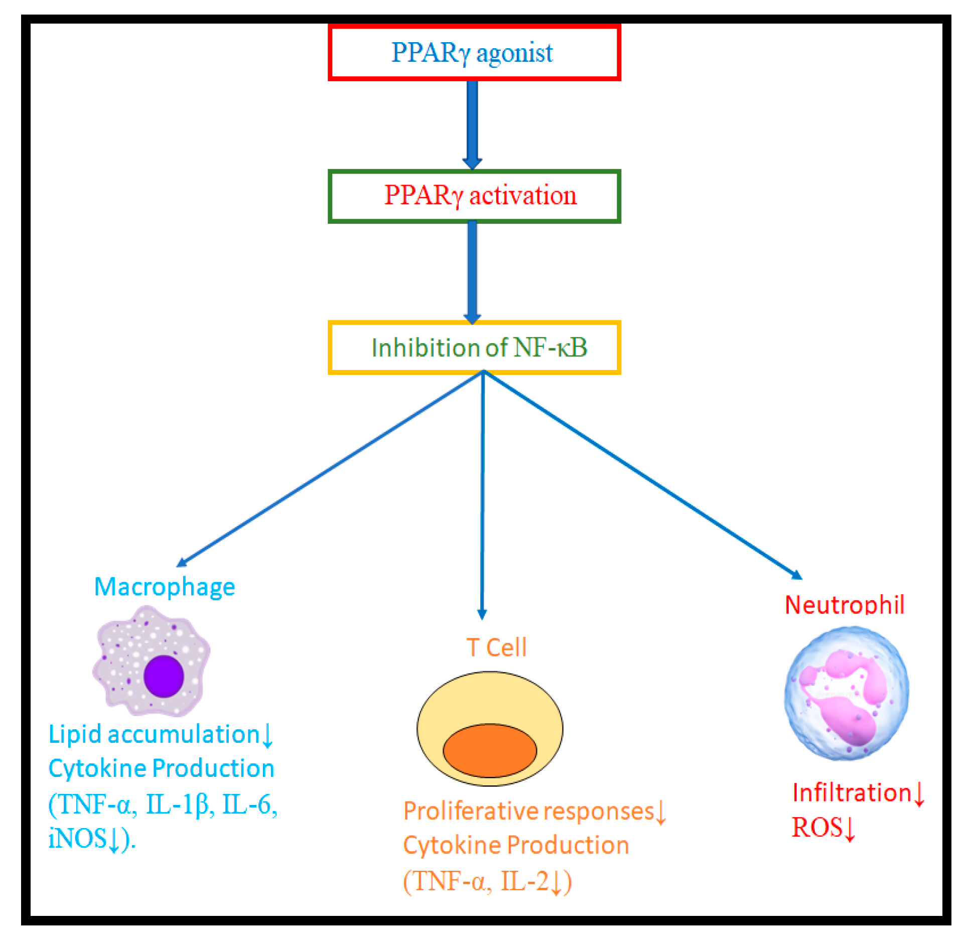
Figure 1. NF-κB activity that causes inflammatory responses is inhibited by activated PPARγ.
Molecules 28 03899 g001
Figure 2. Inflammatory mediators and activated NF-κB are inhibited by eugenol.
Molecules 28 03899 g002
Figure 3. Rational design of eugenol derivatives as PPARγ agonists.
Molecules 28 03899 g003
Scheme 1. Synthesis of eugenol derivatives: (a) Triethylamine, chloroacetyl chloride, and dichloromethane were agitated at 0 to 5 °C for 10 h. (b) Eugenol, K2CO3, acetone, 45 °C stirred for 26 h.
Molecules 28 03899 sch001
Figure 4. 2D interactions images showing the ligands (1d and 1f) and std pioglitazone with the PPARγ target protein active site (PDB_ID: 2PRG).
Molecules 28 03899 g004
Figure 5. 2D interactions images showing the ligands (1e and 1f) and std diclofenac with the egg lysozyme target protein active site (PDB_ID: 3WXU).
Molecules 28 03899 g005
Figure 6. Pharmacophore characteristics of synthesized compounds (1d, 1e & 1f).
Molecules 28 03899 g006
Figure 7. (A,B) is the Root mean square deviation (RMSD) of PPARγ and lysozyme and (C,D) is the Radius of gyration (Rg) of PPAR-Gamma and lysozyme compared with standard.
Molecules 28 03899 g007
Figure 8. The Binding affinity of reference standard drug pioglitazone and synthesized ligands using the LanthaScreen TR-FRET Assay.
Molecules 28 03899 g008
Figure 9. Albumin denaturation were affected by both synthesized compounds and standard diclofenac; their effects were expressed as means standard deviations (n = 3); ** p < 0.05 vs. Std.
Molecules 28 03899 g009
Figure 10. Percentage protection of standard diclofenac and synthesized compounds (1a1f) against HRBC lysis at various concentrations.
Molecules 28 03899 g010
Table 1. ADMET and Toxic profile.
Compound NameSolubilityBBBCPY2D6HIANTP_RATAmes Mutagen
MaleFemale
1a31NI0NCNCNM
1b33NI0NCNCNM
1c21NI0NCNCNM
1d21NI0NCNCNM
1e21NI0NCNCNM
1f21NI0NCNCNM
Std21NI0NCNCNM
NI: Non-inhibitor, NC: Non-carcinogen, NM: Non-mutagen.
Table 2. Pfizer rule of 5 and set dosage range of rat model.
Compound NameAlogpMWHBAHBDRat_Oral
LD50
g/kg_Body_Weight
Rat_Inhalational
LC50
mg/m3/h
Carcinogenic
Potency
TD50_Rat
mg/kg_Body_Weight/day
1a3.44297.34313.819659.63329.70
1b3.33342.34712.635797.7154.64
1c3.92311.37411.5010,394.00118.84
1d4.38365.34412.1335,981.6098.84
1e4.10331.79412.566764.9190.62
1f4.10331.79413.337232.8390.62
Pioglitazone3.90356.43517.286054.5861.73
MW: Molecular weight, HBA: Hydrogen bond acceptor, HBD: Hydrogen bond donor.
Table 3. Binding energies are calculated using the implicit solvent model GBSA and CDOCKER docked energy.
Compound Name—CDOCKER EnergyBinding Energy
PPAR-GammaAnti-InflammatoryPPAR-GammaAnti-Inflammatory
1a19.5615.14−1.01−0.92
1b19.3914.37−1.59−789
1c22.5015.51−1.32−0.95
1d23.8116.39−15.97−1.00
1e21.9818.14−5.94−1.32
1f24.1318.32−20.88−2.67
Std37.1523.11−12.58−2.01
Table 4. Energy parameter analysis average values and their deviations.
Energy ParametersPPARγInflammatory
1f1dPioglitazone1f1eDiclofenac
Potential−16,722 ± 160.70−16,424 ± 110.10−16,681 ± 128.70−7490 ± 104.10−7309 ± 82.44−7454 ± 39.93
Kinetic3324 ± 33.473324 ± 32.073326 ± 34.971498 ± 21.841499 ± 21.921488 ± 20.59
Electrostatic−20,068 ± 174.00−19,731 ± 120.70−19,930 ± 121.60−8988 ± 123.10−8803 ± 89.41−8909 ± 39.20
Van der Waals−1666 ± 26.59−1644 ± 27.00−1623 ± 26.76−721.2 ± 17.58−719.7 ± 18.73−734.7 ± 17.77
Table 5. IC50 values of bound ligands and reference standard using Lanthascreen TR- FRET assay.
Sl. No.Compound NameIC50 (µM)
1Pioglitazone1.77
21a29.83
31b42.37
41c8.20
51d6.47
61e11.45
71f5.15
Table 6. Percentage inhibition of denaturation of albumin at various concentrations.
Concentration in [µM]Standard Diclofenac1a1b1c1d1e1f
6.2510.25 ± 0.551.67 ± 0.372.06 ± 0.171.77 ± 0.374.21 ± 0.375.487 ± 0.437.74 ± 0.34
12.515.35 ± 0.435.32 ± 0.374.44 ± 0.365.32 ± 0.378.79 ± 0.329.47 ± 0.3913.59 ± 0.23
2528.22 ± 0.6110.19 ± 0.248.42 ± 0.248.42 ± 0.2414.04 ± 0.3115.73 ± 0.2424.42 ± 0.48
5038.93 ± 0.3714.54 ± 0.4313.37 ± 0.2413.79 ± 0.4318.75 ± 0.3922.35 ± 0.1831.04 ± 0.55
10049.46 ± 0.5119.20 ± 0.2417.62 ± 0.1819.20 ± 0.2432.46 ± 0.3238.61 ± 0.3043.08 ± 0.43
20060.29 ± 0.3625.07 ± 0.3121.19 ± 0.3027.26 ± 0.2438.49 ± 0.3748.21 ± 0.1850.22 ± 0.60
40068.54 ± 0.4332.34 ± 0.3732.24 ± 0.4034.03 ± 0.3149.09 ± 0.1856.62 ± 0.3762.41 ± 0.42
Values are expressed as means ± SD (n = 3).
Table 7. IC50 values of synthesized derivatives of eugenol and standard diclofenac using an albumin denaturation assay.
CompoundsIC50 Values in [µM]
Standard Diclofenac56.03
1a134.00
1b294.31
1c123.10
1d106.81
1e96.23
1f67.02
Table 8. HRBC membrane stabilization activities of synthesized compounds standard diclofenac at various concentrations.
Concentration [µM]Standard Diclofenac1a1b1c1d1e1f
6.2515.99 ± 0.644.79 ± 0.483.19 ± 0.286.21 ± 0.388.85 ± 0.3612.36 ± 0.4815.17 ± 0.64
12.526.93 ± 0.5510.45 ± 0.389.10 ± 0.2814.26 ± 0.3817.77 ± 0.4619.68 ± 0.3825.09 ± 0.66
2543.54 ± 0.4821.40 ± 0.3615.55 ± 0.2824.78 ± 0.3828.72 ± 0.4634.50 ± 0.3641.88 ± 0.55
5052.58 ± 0.4828.78 ± 0.6627.49 ± 0.3629.08 ± 0.4634.74 ± 0.5642.00 ± 0.5649.50 ± 0.46
10062.30 ± 0.5937.88 ± 0.2836.16 ± 0.4840.65 ± 0.2845.51 ± 0.2851.04 ± 0.4658.61 ± 0.38
20076.87 ± 0.6447.23 ± 0.4845.07 ± 0.2850.92 ± 0.4856.45 ± 0.3166.05 ± 0.3673.43 ± 0.36
40090.77 ± 0.3660.88 ± 0.3658.36 ± 0.2862.54 ± 0.3667.40 ± 0.2875.09 ± 0.3685.79 ± 0.18
Values are expressed as means ± SD (n = 3).
Table 9. IC50 values of synthesized derivatives of eugenol and standard diclofenac using HRBC membrane stabilization method.
CompoundsIC50 Values in [µM]
Standard Diclofenac48.14
1a79.50
1b83.15
1c79.70
1d66.30
1e58.15
1f54.97
Table 10. Physicochemical characteristics of synthesized compounds.
CompoundChemical StructureMol. wtMol. FormulaRf Value% Yield
1aMolecules 28 03899 i001297.35C18H19NO30.7280.00
1bMolecules 28 03899 i002342.35C18H18N2O50.6575.30
1cMolecules 28 03899 i003311.38C19H21NO30.6080.50
1dMolecules 28 03899 i004365.35C19H18F3NO30.6270.80
1eMolecules 28 03899 i005331.79C18H18ClNO30.6577.60
1fMolecules 28 03899 i006331.79C18H18ClNO30.6487.90
TLC mobile phase: n-Hexane & Ethyl acetate (9:1).
Disclaimer/Publisher’s Note: The statements, opinions and data contained in all publications are solely those of the individual author(s) and contributor(s) and not of MDPI and/or the editor(s). MDPI and/or the editor(s) disclaim responsibility for any injury to people or property resulting from any ideas, methods, instructions or products referred to in the content.