Article

Rapid Detection of Cd2+ Ions in the Aqueous Medium Using a Highly Sensitive and Selective Turn-On Fluorescent Chemosensor

1
Department of Chemistry, University of Malakand, Chakdara 18800, Pakistan
2
Department of Electrical Engineering, Kwangwoon University Seoul, Seoul 01897, Republic of Korea
3
Department of Biochemistry, University of Malakand, Chakdara 18800, Pakistan
4
Department of Pharmacognosy, College of Pharmacy, King Saud University, Riyadh 11495, Saudi Arabia
5
Department of Pharmaceutical Chemistry, College of Pharmacy, King Saud University, Riyadh 11495, Saudi Arabia
*
Author to whom correspondence should be addressed.
Molecules 2023, 28(8), 3635; https://doi.org/10.3390/molecules28083635
Submission received: 23 February 2023 / Revised: 19 April 2023 / Accepted: 20 April 2023 / Published: 21 April 2023
(This article belongs to the Section Analytical Chemistry)

Abstract

:
Herein, a novel optical chemosensor, (CM1 = 2, 6-di((E)-benzylidene)-4-methylcyclohexan-1-one), was designed/synthesized and characterized by 1H-NMR and FT-IR spectroscopy. The experimental observations indicated that CM1 is an efficient and selective chemosensor towards Cd2+, even in the presence of other metal ions, such as Mn2+, Cu2+, Co2+, Ce3+, K+, Hg2+,, and Zn2+ in the aqueous medium. The newly synthesized chemosensor, CM1, showed a significant change in the fluorescence emission spectrum upon coordination with Cd2+. The formation of the Cd2+ complex with CM1 was confirmed from the fluorometric response. The 1:2 combination of Cd2+ with CM1 was found optimum for the desired optical properties, which was confirmed through fluorescent titration, Job’s plot, and DFT calculation. Moreover, CM1 showed high sensitivity towards Cd2+ with a very low detection limit (19.25 nM). Additionally, the CM1 was recovered and recycled by the addition of EDTA solution that combines with Cd2+ ion and, hence, frees up the chemosensor.

1. Introduction

Cadmium (Cd2+) is one of the most hazardous and carcinogenic heavy metals, and it is widely employed in various industrial applications, such as the fabrication of metal alloys, batteries, electroplating films, and nuclear reactor control rods [1]. The effect of Cd2+ pollution should not be underestimated [2]. As a heavy metal ion with a biological half-life of 20 to 30 years, Cd2+ accumulates in the human body through contaminated water, air, soil, or other sources, leading to a variety of disorders of the kidney, liver, heart, lung, or other organs. Even if the accumulated Cd2+ content in the body is very low, it can still result in several health problems, including potentially fatal diseases, such as diabetes, cancer, and chondropathy [3]. Currently, there are several efficient methods to detect Cd2+: atomic absorption spectrometry (AAS) [4], inductively coupled plasma mass spectrometry (ICP-MS) [5], atomic fluorescence spectrophotometry (AFS) [6], the electrochemical method [7,8], and the fluorescence probe method [9,10,11]. Compared to the fluorescent probe technique, AAS, AFS, and ICP-MS need complex and expensive instruments, complicated sample preparation, and the electrochemical method, which has the disadvantage of poor selectivity.
The main advantage of the fluorescent probe technique is its high sensitivity, visibility, and rapid response. In addition, simple operation, low cost, and a wide linear range for heavy metal ions are also clear advantages. Therefore, the development of highly selective colorimetric/fluorescent chemosensors for the detection of harmful metal ions from environmental samples has received considerable interest due to their high sensitivity, selectivity, quick detection time, and cost-effectiveness [12,13,14]. In recent years, a large number of fluorescent probes with diverse scaffolds, such as quinoline, rhodamine, pyrene, boron-dipyrromethene (BODIPY), and anthraquinone, etc. have been developed with sensitivity, selectivity, and real-time detection of Cd2+ [12,15,16,17,18,19,20,20]. However, only a few examples of chemosensors have been reported where a considerable fluorescence enhancement is observed upon Cd2+ ion binding in aqueous medium [21,22,23]. The major difficulty in detecting Cd2+ is due to the relatively similar binding characteristics of Cd2+ with Zn2+ and Hg2+ cations, as they are in the same periodic table group. It is challenging to develop a probe for Cd2+ that does not exhibit the interference of Zn2+ and Hg2+ cations. Although a large number of fluorescent sensors have already been reported with success to differentiate Cd2+ from Zn2+ and Hg2+ cations, the majority of them have low water solubility with a lack of sensitivity and selectivity [24,25,26,27,28]. Therefore, designing and developing fluorescent probes with high water solubility and selectivity is a great challenge for the researcher. Herein, we report the design and synthesis of a water-soluble and simple fluorescent turn-on chemosensor comprising a CM1 fluorophore, selectively detecting Cd2+ over other relevant metal ions in aqueous samples. Due to possessing the best coordination sites, it was capable of forming stable complexes with Cd2+ ions. The CM1 showed a fast response, high sensitivity, and excellent selectivity for Cd2+ ion detection in an aqueous medium. The sensing mechanism of CM1 was based on competitive binding with Cd2+ ions among different metal ions. Ethylenediaminetetraacetic acid (EDTA) was used as a chelating agent in reversibility studies.

2. Results and Discussion

2.1. Spectroscopic Studies

The UV-Vis absorption spectra were investigated, and maximum absorption wavelength of CM1 appeared at 390 nm, which was attributed to the π–π* transition (Figure 1A). Similarly, fluorescence investigation of chemosensor CM1 was also carried out. When the chemosensor was excited at 390 nm, it gave an emission spectrum of low intensity at 610 nm, which could be attributed to the delocalization of oxygen lone pair electrons towards the aromatic ring, thus causing the quenching of fluorescence via the photoinduced electron transfer (PET) phenomenon (Figure 1B). The IR spectrum of the chemosensor CM1 is depicted in (Figure S1). The IR spectrum gave the characteristic carbonyl (C=O) peak at 1710 cm−1, while the appearance of peaks at 1628–1646 cm−1 and 3000–2960 cm−1, respectively, corresponded to a C=C aromatic and C-H stretching.

2.2. UV–Visible and Fluorescence Study

The selective chemosensing ability of CM1 towards Cd2+ was investigated through a detailed optical study. The UV–Vis absorption spectra of the CM1 and CM1-Cd2+ complex are shown in (Figure 2). The chemosensor CM1 gave maximum absorbance at 390 nm due to π–π* transition. Upon the addition of Cd2+, enhancement was observed at the absorption intensity at 390 nm. Similarly, the CM1 fluorescence spectrum and its response to various metal ions was monitored through its excitation at a maximum wavelength (390 nm) in the aqueous medium. The addition of CM1 to metal ions, such as Cd2+, Mn2+, Cu2+, Co2+, Ce3+, K+, Hg2+, and Zn2+, showed very low enhancement in the fluorescence intensity, but maximum enhancement was observed for Cd2+ (Figure 3). This enhancement in fluorescence intensity was probably due to large rigidity in conjugation and better coplanarity upon complexation with the Cd2+ ion. In the case of other metal ions, the same phenomenon could not take place, thus inhibiting a significant enhancement in fluorescence intensity [29,30,31]. Potassium (K) and cerium (Ce) also showed a low fluorescence enhancement, and it may be possible that factors beyond the MLCT and LMCT effects, such as the shape and size of the metal ion, and its ability to accommodate the ligand, could be contributing to this similarity.
Figure 1. (A) UV-Visible absorption spectrum of chemosensor CM1. (B) Fluorescence emission spectrum of the chemosensor CM1 when excited at 390 nm.
Molecules 28 03635 g001

2.3. Stokes Shift

The reason for the observed large Stokes shifts was intra-molecular charge-transfer excitation of an electron from the HOMO to the LUMO of the chromophore, accompanied by elongation of the bond and considerable solvent reorganization due to hydrogen bonding to the solvent. The chemosensor CM1 showed a prominent Stokes shift, and it can be used for practical application for the detection of Cd2+ [32].

2.4. Detection Limit and Quantum Yield

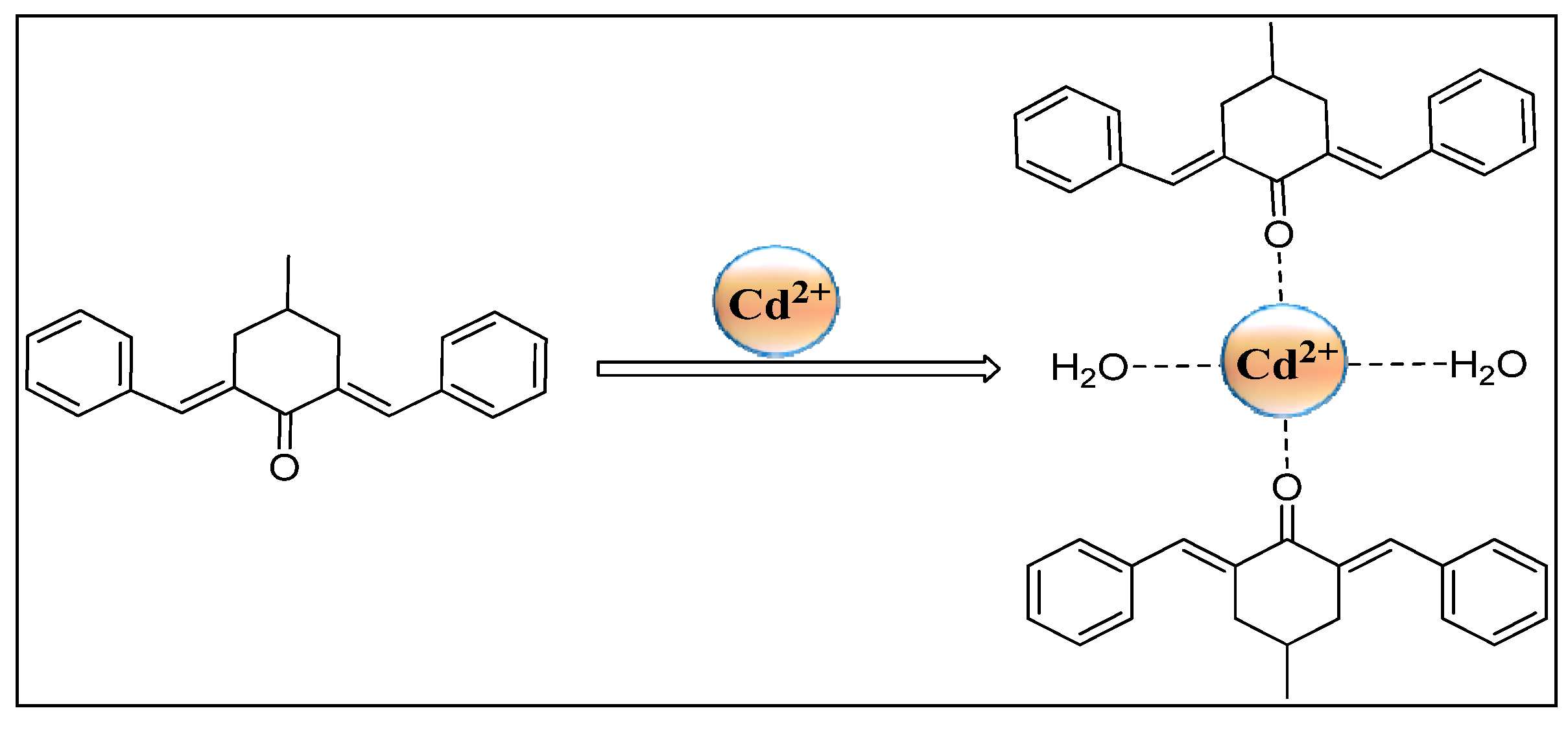
The LOD for the current chemosensor CM1 was calculated to be 19.25 nM, as shown in Figure 4 [33]. The predictable mechanism of complex formation is given in Scheme 1.
The quantum yield calculated using Equation (2) for the current chemosensor CM1 for Cd2+ detection was found to be 74%. Rhodamin 6G was chosen as the standard for quantum yield calculation because it is readily available and easily dissolvable in acetonitrile. Additionally, Rhodamone 6G has previously served as a standard for fluorescent chemosensors over a wide range of 340–540 [34].

2.5. Reuseability Study

The reuseability of the chemosensor CM1 in the detection of Cd2+ ion is important for its practical applications. To check the reuseability of chemosensor CM1, a reversibility test was carried out using EDTA as chelating agent in aqueous medium at room temperature. The CM1 can be freed and regenerated by the addition of EDTA to the Cd2+-CM1 complex solution, which was observed through diminished fluorescence intensity. The enhanced fluorescence intensity was resumed by the addition of Cd2+ solution, which again combined with the CM1 chemosensor. These results are a reflection of CM1 as a reusable chemosensor in the presence of an EDTA solution (Figure 5). The results indicated that the same chemosensor, CM1, could be used for Cd2+ detection up to five times, with consistent and satisfactory outcomes.
Figure 4. Calibration curve from fluorescence titration of Cd2+ (1–10 µM) range and CM1 (10 µM) at λex = 390 nm and λem = 610 nm.
Molecules 28 03635 g004

2.6. Effect of pH

The dependence of pH variation on the fluorescence intensity of free CM1 and its Cd2+ complex was investigated in the pH range of 2.0–12.0 (Figure 6). The pH of the test solution was adjusted with the help of a universal buffer. In case of a free chemosensor, fluorescence intensity remained constant from 2–12 pH, with weak fluorescence intensity. The low fluorescence intensity of CM1 could be due to the photoinduced electron transfer (PET) phenomenon. However, for the Cd2+ (18 μM) complex with CM1, the fluorescence intensity gradually increased with pH. Comparatively low fluorescence intensity of the Cd2+ complex at lower pH was observed due to the protonation of carbonyl oxygen, which blocked the complexing ability of CM1. The maximum fluorescence intensity for the Cd2+ complex was obtained at pH 7 due to the restriction of PET. The fluorescence intensity decreased again with pH increase beyond 8 due to possible formation of sparingly soluble hydroxides complex. These results indicate that chemosensor CM1 can be efficiently employed for Cd2+ detection at pH 7 [28].
The decreased fluorescence of the mixture of ligand and Cd2+ at both pH 3 and pH 12, as well as the lower fluorescence of the free ligand in the presence of excess cadmium, may be attributed to the formation of a non-fluorescent complex between the ligand and the metal ion. This complex may possess different structural and electronic properties compared to the free ligand, leading to a reduction in its fluorescence intensity. Additionally, factors, such as quenching, energy transfer, or changes in the solvent environment, may also play a role in the observed decrease in fluorescence. The decreased fluorescence of the mixture of ligand and Cd2+ at both pH 3 and pH 12, as well as the lower fluorescence of the free ligand in the presence of excess cadmium, may be attributed to the formation of a non-fluorescent complex between the ligand and the metal ion. This complex may possess different structural and electronic properties compared to the free ligand, leading to a reduction in its fluorescence intensity. Additionally, factors, such as quenching, energy transfer, or changes in the solvent environment, may also play a role in the observed decrease in fluorescence.
Figure 6. Effect of pH on fluorescence intensity of CM1 and CM1 (10 µM) with Cd2+ (18 µM) at λex = 390 nm and λem = 610 nm.
Molecules 28 03635 g006

2.7. Anti-Interference Studies

Anti-interference or competitive studies were carried out to verify the selectivity and specificity of the CM1 for Cd2+ in the presence of other metal cations, such as Mn2+, Cu2+, Co2+, Ce3+, K+, Hg2+, and Zn2+. The results (Figure 7) showed that these interfering metal ions do not affect the fluorescence intensity of chemosensor CM1-Cd2+ complex even in the presence of higher concentration of interfering ions. Thus, this indicated that chemosensor CM1 can be useful as a highly selective fluorescence-on sensor for trace determination of Cd2+ ions in different water samples [35,36,37].

2.8. Natural Water Samples Analysis by Spiking

Chemosensor CM1 was applied for the selective detection of Cd2+ in lake water, tap water, and river water. Because, in these water samples, no Cd2+ was present, therefore a cadmium (2–10 μM) concentration range was added, and its fluorescence intensity was recorded at optimized parameters. From the calibration curve, respective Cd2+ concentrations were determined, and then % recovery was calculated. The results are shown in Table 1. Accordingly, these results demonstrated an excellent recovery of Cd2+ from these studied water samples. Thus, here we present a highly sensitive, selective, and cost-effective method for determination of Cd2+ in an aqueous medium with good % recovery [38].

2.9. DFT Studies

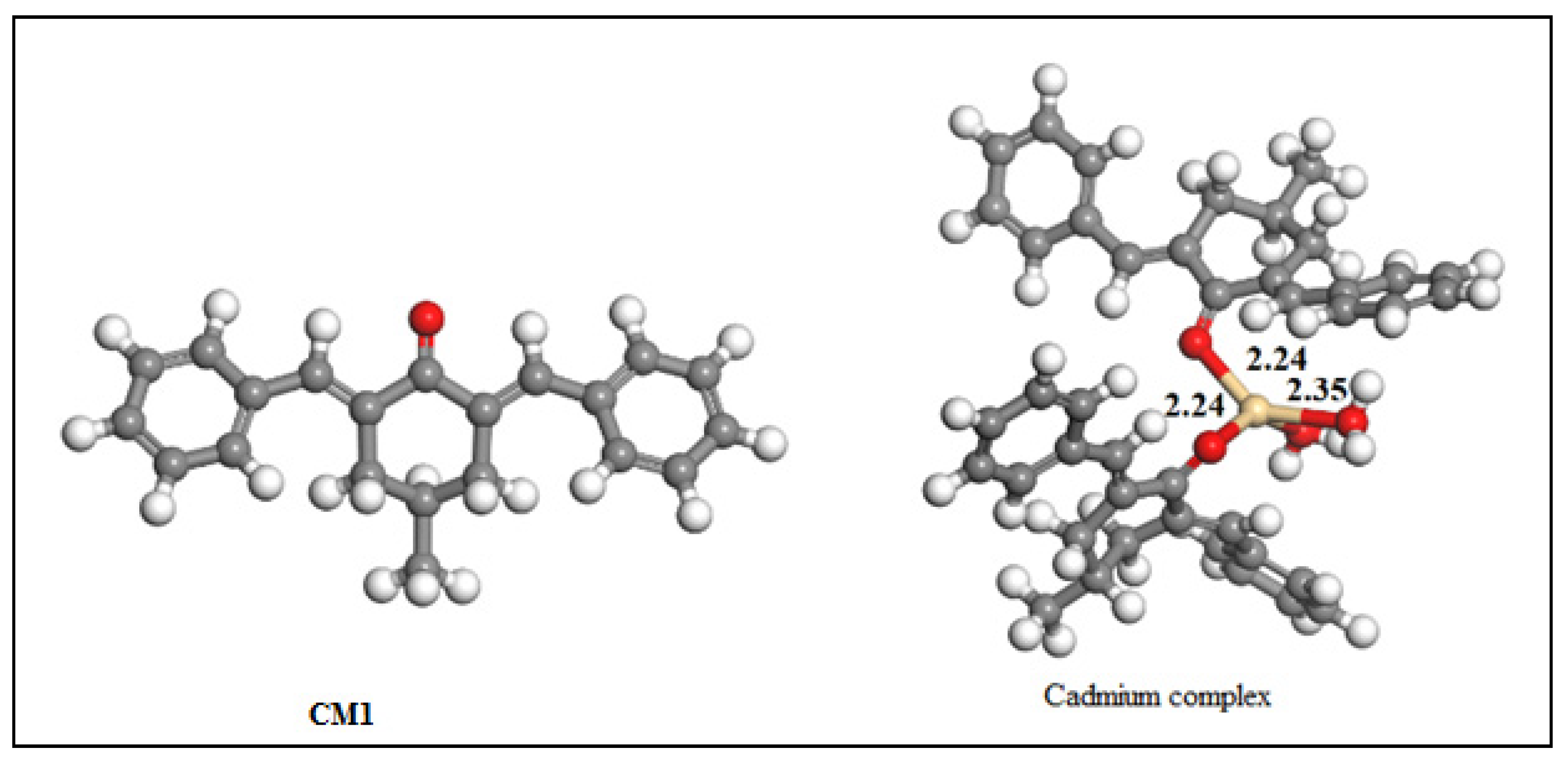
The detection of Cd2+ with the help of chemosensor CM1 was validated through computational investigation concerning the stability, orbital overlapping of Cd2+ and CM1, bond angles, bond lengths, and optimized geometry of the resulting cadmium complex. The optimized geometry of the CM1 and its cadmium complex are shown in Figure 8. The CM1 acted as a monodentate ligand attached through an oxygen atom to the Cd2+ ion, forming a stable distorted tetrahedral geometry.
The observed bond length, bond angles, metal–ligand interaction energy, charges on oxygen and cadmium, and highest occupied molecular orbital (HOMO) and lowest unoccupied molecular orbital (LUMO) energies of the cadmium complex are listed in Table 2. The observed Cd-O bond lengths with CM1 were 2.24 Å and Cd-O bond lengths with a water value of 2.35 Å. The shorter Cd-O bond lengths with CM1, as compared to water oxygen, was due to the extensive delocalization of electrons on chemosensor CM1 aromatic rings that made their donor oxygen atom more electron-rich and, hence, caused strong interaction with an electron-deficient metal center. The observed OCM-1Cd-OCM-1 and OCM-1-Cd-Owater bond angles were 103.08 and 99.24°, respectively, suggesting a distorted tetrahedral complex. The tetrahedral geometry of the complex could also be explained based on orbital hybridization and their involvement in bond formation, since, in Cd2+, all the d orbitals are filled (d10 system). Therefore, no orbital in the “d” subshell was available for overlapping with chemosensor CM1 filled orbital. Hence, the outer s and p subshells underwent sp3 hybridization and, consequently, formed tetrahedral structures. However, the small deviation from the regular tetrahedral structure was due to the bulky nature of the chemosensor CM1. The stability of the resulting Cd2+ complex was computationally calculated to be −14.35 eV, and the negative sign indicated the thermodynamic stability of the complex. It is worth noting that MLCT and LMCT effects, while useful for explaining metal–ligand interactions, are not the only contributing factors. Factors, such as the electronic configuration of the metal ion and the ligand, steric effects, and solvent effects, can also play a role in complexation.
The interaction between Cd2+ and chemosensor CM1 in the synthesized cadmium complex was also verified from the electron density difference (EDD) analysis. The EDD contour map shown in (Figure 9) indicated the concentration of a large electron cloud along the cadmium–chemosensor CM1 axis. The HOMO and LUMO band gap gave information about the charge transfer interaction in a molecule. The electron transfer was efficient from the HOMO to LUMO, as the energy gap between these two orbitals became small. The negative value of HOMO and LUMO orbitals depicted in (Table 2) indicated the thermodynamic stability of the complex. The HOMO contained mostly the electron clouds on chemosensor CM1, while the Cd2+ ion contained a small electron density in LUMO, suggesting the transfer of an electron from chemosensor CM1 to the cadmium ion upon complex formation. The smaller band gap (1.74) value suggested the utilization of the complex in semiconductor materials [39]. The formation of the cadmium complex was also supported by the extent of electron density transfer from both the oxygen atom of chemosensor CM1 and water attached to Cd+2. As is obvious from Table 2, the formation of the complex caused a reduction in the electron density on the donor atoms of CM1, suggesting the formation of the complex. The absorption and fluorescence analysis showed that almost every metal atom was capable of forming a complex with the ligand. However, the highest enhancement was observed in the case of cadmium, indicating a high selectivity for this particular metal ion. Thus, we proceeded to investigate only the complexation reaction between the ligand and cadmium through DFT calculations. Our experimental results were supported by the DFT calculations, which confirmed the stability of the complex.

2.10. Comparison with REPORTED Chemosensors

The sensitivity of CM1 was compared with previously reported chemosensors for the determination of Cd2+ ions. The result demonstrated that chemosensor CM1 showed excellent sensitivity, as compared to the reported chemosensors (Table 3). The literature study also showed the lower limit of detection for chemosensor CM1, as compared to most of the recently reported similar work, indicating its superiority in terms of selectivity and practical applicability.

3. Materials and Methods

The chemicals and solvents used in the present study were analytical grade and utilized without further purification; the water employed in the experiment was double distilled. The metal salts of NiSO6H2O, Ca(OH)2·4H2O, Mn(OH3)2·4H2O, ZnSO4·6H2O, Cd(SO4)2·3H2O, Cu(SO4)2·5H2O), Co(OH3)·6H2O, CrCl36H2O, KNO3, Hg(NO3)2, Pb(NO3)2, and Ce(NO3)3·6H2O were purchased from BDH chemical, England. As Ca(OH)2 was easily available in our laboratory, we utilized it as a source of calcium. Similarly, nitrate and sulphate salts of other metals were used. Sodium hydroxide, 4-methyl cyclohexanone, benzaldehyde, methanol, and acetonitrile were purchased from Sigma Aldrich and Merck. The UV-Visible spectra were obtained using a double-beam UV/VIS spectrophotometer (Shimadzu, Japan) model 1601. The fluorescent measurement was obtained through spectrophotofluorometer model no RF5301 PC (Shimadzu, Japan), containing a Xenon lamp as a source of excitation. The infra-red spectra were obtained through FT-IR spectrophotometer model 1601 (Shimadzu, Japan). The 1H NMR spectra were obtained on Prestige 21 (Shimadzu, Japan) using a Bruker Advance 400 MHz spectrometer. The spectrum was obtained in CDCl3 solution using tetramethyl silane (TMS) as an internal standard.

3.1. Synthesis of Chemosensor CM1

The chemosensor CM1/[(2,6-di((E)-benzylidene)-4-methylcyclohexan-1-one)] was synthesized as shown in (Scheme 2) [48,49,50]. Ethanolic solution of benzaldehyde (1.06 g, 10 mmole) and 4-methyl cyclohexanone (0.49 g, 5 mmole) were mixed with continuous addition of sodium hydroxide (40%). The resulting mixture was refluxed for 4 h. The mixture was cooled to room temperature and evaporated to afford the desired product, CM1, as a yellow-colored solid. The crude products were purified by recrystallization from ethanol. Melting point (M. P): 159 °C. Yield: 78%. IR (KBr, cm−1): 1710 cm−1 (C=O stretching), 1628–1646 cm−1 (C=C stretching), 3000–2960 cm−1 (C-H stretching) [51], 1H-NMR (300 MHz, ppm): δ 7.82 (bs, 2H, CH=C6H5), 7.34–7.50 (m, 10H, ArH), 3.07 (dd, JAB = 15.9, J = 3.6 Hz, 2H, CH2), 2.53 (m, 2H, 2CH2), 1.89 (m, 1H, CH), 1.08 (d, 3H J = 6.6 Hz, 3H, CHCH3) [52].

3.2. Solutions Preparation for Spectroscopic Measurements

Stock solution of chemosensor CM1 in acetonitrile, and metal solutions using respective salts were prepared in distilled water. Stock solutions of metal (0.1 mM) were prepared in 100 mL solvent/water. Then working solutions were prepared by taking 5 mL from this stock metal solution and diluting it to 100 mL. Each time, a fresh working solutions of both chemosensor CM1 and metal ion was prepared.

3.3. General UV–Vis and Fluorescence Spectra Measurements

A stock solution of CM1 (4 mM) was prepared in acetonitrile. Similarly, the same concentration solutions of the corresponding salts of copper, cerium, cadmium, zinc, mercury, cobalt, and manganese were prepared in double-distilled water. The tested solutions were prepared by mixing 50 μL stock solution of CM1 with the appropriate amount of each metal ion in a tube. Absorption spectra were taken from the 250–500 nm range. Based on UV-Vis analysis, the fluorescent measurements were set at 390 nm. Each spectrum was taken three times, and their mean value was considered as final data. The chemosensing utility of CM1 for Cd2+ in the presence of other metal ions was also investigated in natural water samples.

3.4. Excitation and Emission Spectra of Chemosensor CM1

Upon exciting chemosensor CM1 at λex = 390 nm, its fluorescence emission spectrum was recorded in the range of 230–800 nm. A weak fluorescence emission spectrum was observed at λem = 610 nm. Thus, at these wavelengths of excitation and emission, further fluorometric analyses were carried out.

3.5. Limit of Detection and Quantum Yield Calculation

The limit of detection (LOD) was calculated to be 19.25 nM from the calibration curve using Equation (1).
LOD = 3 δ S
In Equation (1) “𝛿” represents the standard deviation, and “S” is the slope of the fluorescence intensity vs. sample concentration curve.
Rhodamine 6G dye was used as a standard possessing a quantum yield (φ) 95 % for enumerating the quantum yield of the chemosensor CM1 because Rhodamine 6G dye is readily available and easily dissolvable in acetonitrile. Additionally, Rhodamone 6G can be used as a standard for fluorescent chemosensors over a wide range of 340–540 [34,53]. For the experiment, Rhodamine 6G solution was prepared, and its spectroscopic analysis (UV-visible and fluorescence) were performed. The quantum yield was determined with the help of Equation (2).
φ C   = φ R   ( I C I R ) ( η c 2 η R 2 ) ( A R Ac )
where “C” and “R” represent chemosensor CM1 and dye, respectively. “I” represents the integrated fluorescence intensity, “η” corresponds to the refractive index of the solvent, and “A” corresponds to absorbance.
The values of absorbance in the equation were determined and used. For Equation (2),   φ R   = 0.95, I R = 620 ,   η R = 1.33 ,   A R   = 0.209, I C = 40 ,   η C = 1.34 ,   and   A C = 1.5 .

3.6. Spiked Water Samples Analysis

Natural water samples were collected from River Swat at Chakdara point, Malakand Lake at Dargai, and from the University of Malakand campus, respectively. Since no cadmium was found in these samples, the applicability of the new chemosensor CM1 was determined by the spiking technique. A known amount of Cd2+ was added to each water sample, and the fluorescence intensity of spiked water samples was recorded. The selected water samples were spiked with different concentrations of Cd2+ (5–10) µM in the presence of chemosensor CM1 (10 µM). The fluorescence response was measured at specific wavelengths of excitation and emission. For verification of the test results, each spectral analysis was carried out three times.

3.7. Theoretical Calculation

The density functional theory (DFT) calculations were employed utilizing DMol3 simulation code [54,55] The geometry and energy of the synthesized chemosensor CM1 were optimized through Perdew Burke Ernzerhoffuncational (PBE) in the domain of generalized gradient approximation (GGA) [56,57]. The double numerical plus polarization (DNP) (Liu and Rodriguez 2005) was used for the adjustment of the basis set for all types of computation. Hessian calculations showed the absence of any imaginary frequency that reflects the stability of the relaxed structure. The thermal smearing parameters and basis set cut-off were adjusted, respectively, to 0.005 au and 4.6 Å. Grimme’s scheme DFT-D2 empirical dispersion correction was employed for van der Waals intermolecular interactions [58,59]. Convergence tolerance of 10−5 Ha for energy, 0.001 H/Å for force, and 0.005 Å for displacement were set during the geometries’ relaxation. The adsorption energies (Eint) of complexes were evaluated using the following equation.
E int = E complex ( E CMI + E Cd ( II ) )
where, Ecomplex, Elignd, and ECd(II) are the total-electronic energies of the Cd(II)-CM1 (complex), CM1 and Cd(II) ion), respectively.

4. Conclusions

In summary, we have presented, here, the design and development of a novel optical chemosensor based on a curcumin derivative CMI, which can detect Cd2+ over other competitive metal ions in an aqueous medium. CMI exhibited good photostability and enhanced fluorescence in an aqueous medium upon the formation of the Cd2+ complex due to the presence of a carbonyl oxygen group. The interaction ratio between chemosensor CMI and Cd2+ ion was determined by fluorescence titration, Job’s plot, and computational studies, and it was found to be 2:1. The competitive binding experiment revealed no interference from the other metal ions (Mn2+, Ni2+, Cu2+, Co2+, Ce3+, K+, Hg2+, and Zn2+), thus showing selectivity of CM1 towards Cd2+. Moreover, CM1 showed high sensitivity at physiological pH, with low detection limits (19.25 nM). The CM1-based chemosensor can be recycled and reused multiple times with the addition of an EDTA solution. This sensing approach certainly provides a basis that can be used for the design of various curcumin-based optical chemosensors to monitor other environmentally concerned hazardous heavy metal ions in aqueous medium.

Supplementary Materials

The following supporting information can be downloaded at: https://www.mdpi.com/article/10.3390/molecules28083635/s1, Figure S1 [60,61]: FT-IR spectrum of chemosensor CM1; Figure S2: 1H-NMR spectrum of the chemosensor CM1; Figure S3: Job’s plot analysis of CM1 shows 2:1 binding ratio between chemosensor CM1 and Cd2+.

Author Contributions

Conceptualization, M.S. and J.K.; methodology, R.K. and J.K.; validation, M.Z., M.S., A.W.K., A.B., E.A.A. and R.U.; formal analysis, A.W.K. and A.B.; investigation, A.B. and R.U.; data curation, J.K. and M.S; writing—original draft preparation J.K., M.Z., M.S. and R.K.; writing—review and editing, M.Z., R.U., R.K., E.A.A. and A.B. All authors have read and agreed to the published version of the manuscript.

Funding

The research work was supported by researchers supporting project number (RSP2023R45) at King Saud University, Riyadh, Saudi Arabia.

Institutional Review Board Statement

Not applicable.

Informed Consent Statement

Not applicable.

Data Availability Statement

Not applicable.

Acknowledgments

The authors extend their appreciation to the researchers supporting Project number (RSP2023R45) King Saud University, Riyadh, Saudi Arabia, for financial support.

Conflicts of Interest

The authors declare no conflict of interest.

Sample Availability Statement

Samples of the compounds are available from the authors.

References

  1. Wang, J.; Xia, T.; Zhang, X.; Zhang, Q.; Cui, Y.; Yang, Y.; Qian, G. A Turn-on Fluorescent Probe for Cd2+ Detection in Aqueous Environments Based on an Imine Functionalized Nanoscale Metal–Organic Framework. RSC Adv. 2017, 7, 54892–54897. [Google Scholar] [CrossRef]
  2. El-Saadani, Z.; Mingqi, W.; He, Z.; Hamukwaya, S.L.; Abdel Wahed, M.S.M.; Abu Khatita, A. Environmental Geochemistry and Fractionation of Cadmium Metal in Surficial Bottom Sediments and Water of the Nile River, Egypt. Toxics 2022, 10, 221. [Google Scholar] [CrossRef]
  3. Barone, G.; Storelli, A.; Garofalo, R.; Mallamaci, R.; Storelli, M.M. Residual Levels of Mercury, Cadmium, Lead and Arsenic in Some Commercially Key Species from Italian Coasts (Adriatic Sea): Focus on Human Health. Toxics 2022, 10, 223. [Google Scholar] [CrossRef]
  4. Smichowski, P.; Londonio, A. The Role of Analytical Techniques in the Determination of Metals and Metalloids in Dietary Supplements: A Review. Microchem. J. 2018, 136, 113–120. [Google Scholar] [CrossRef]
  5. He, D.; Zhu, Z.; Miao, X.; Zheng, H.; Li, X.; Belshaw, N.S.; Hu, S. Determination of Trace Cadmium in Geological Samples by Membrane Desolvation Inductively Coupled Plasma Mass Spectrometry. Microchem. J. 2019, 148, 561–567. [Google Scholar] [CrossRef]
  6. Haribala; Hu, B.; Wang, C.; Gerilemandahu; Xu, X.; Zhang, S.; Bao, S.; Li, Y. Assessment of Radioactive Materials and Heavy Metals in the Surface Soil around Uranium Mining Area of Tongliao, China. Ecotoxicol. Env. Saf. 2016, 130, 185–192. [Google Scholar] [CrossRef]
  7. Lee, W.; Kim, H.; Kang, Y.; Lee, Y.; Yoon, Y. A Biosensor Platform for Metal Detection Based on Enhanced Green Fluorescent Protein. Sensors 2019, 19, E1846. [Google Scholar] [CrossRef]
  8. Yao, Y.; Wu, H.; Ping, J. Simultaneous Determination of Cd(II) and Pb(II) Ions in Honey and Milk Samples Using a Single-Walled Carbon Nanohorns Modified Screen-Printed Electrochemical Sensor. Food Chem. 2019, 274, 8–15. [Google Scholar] [CrossRef]
  9. Lin, L.; Wang, Y.; Xiao, Y.; Liu, W. Hydrothermal Synthesis of Carbon Dots Codoped with Nitrogen and Phosphorus as a Turn-on Fluorescent Probe for Cadmium(II). Microchim. Acta 2019, 186, 147. [Google Scholar] [CrossRef]
  10. Pan, Y.; Xiao, W.; Fan, M.; Chen, W.; Xu, S. Detection of Cd(II) Ions by a Self-Tuning Method Using Quantum Dot Fluorescence Sensing. Mater. Res. Express 2017, 4, 105008. [Google Scholar] [CrossRef]
  11. Petryayeva, E.; Algar, W.R.; Medintz, I.L. Quantum Dots in Bioanalysis: A Review of Applications across Various Platforms for Fluorescence Spectroscopy and Imaging. Appl. Spectrosc. 2013, 67, 215–252. [Google Scholar] [CrossRef]
  12. Carter, K.P.; Young, A.M.; Palmer, A.E. Fluorescent Sensors for Measuring Metal Ions in Living Systems. Chem. Rev. 2014, 114, 4564–4601. [Google Scholar] [CrossRef]
  13. Li, J.; Yim, D.; Jang, W.-D.; Yoon, J. Recent Progress in the Design and Applications of Fluorescence Probes Containing Crown Ethers. Chem. Soc. Rev. 2017, 46, 2437–2458. [Google Scholar] [CrossRef]
  14. Zhu, H.; Fan, J.; Wang, B.; Peng, X. Fluorescent, MRI, and Colorimetric Chemical Sensors for the First-Row d-Block Metal Ions. Chem. Soc. Rev. 2015, 44, 4337–4366. [Google Scholar] [CrossRef]
  15. Ding, X.; Zhang, F.; Bai, Y.; Zhao, J.; Chen, X.; Ge, M.; Sun, W. Quinoline-Based Highly Selective and Sensitive Fluorescent Probe Specific for Cd2+ Detection in Mixed Aqueous Media. Tetrahedron Lett. 2017, 58, 3868–3874. [Google Scholar] [CrossRef]
  16. Kim, H.N.; Ren, W.X.; Kim, J.S.; Yoon, J. Fluorescent and Colorimetric Sensors for Detection of Lead, Cadmium, and Mercury Ions. Chem. Soc. Rev. 2012, 41, 3210–3244. [Google Scholar] [CrossRef]
  17. Qian, J.; Wang, K.; Wang, C.; Ren, C.; Liu, Q.; Hao, N.; Wang, K. Ratiometric Fluorescence Nanosensor for Selective and Visual Detection of Cadmium Ions Using Quencher Displacement-Induced Fluorescence Recovery of CdTe Quantum Dots-Based Hybrid Probe. Sens. Actuators B Chem. 2017, 241, 1153–1160. [Google Scholar] [CrossRef]
  18. Shi, C.; Huang, Z.; Wu, A.; Hu, Y.; Wang, N.; Zhang, Y.; Shu, W.; Yu, W. Recent Progress in Cadmium Fluorescent and Colorimetric Probes. RSC Adv. 2021, 11, 29632–29660. [Google Scholar] [CrossRef]
  19. Su, W.; Yuan, S.; Wang, E. A Rhodamine-Based Fluorescent Chemosensor for the Detection of Pb2+, Hg2+ and Cd2+. J. Fluoresc. 2017, 27, 1871–1875. [Google Scholar] [CrossRef]
  20. Zhang, Y.; Guo, X.; Zheng, M.; Yang, R.; Yang, H.; Jia, L.; Yang, M. A 4,5-Quinolimide-Based Fluorescent Sensor for the Turn-on Detection of Cd2+ with Live-Cell Imaging. Org. Biomol. Chem. 2017, 15, 2211–2216. [Google Scholar] [CrossRef]
  21. Liu, Z.; Zhang, C.; He, W.; Yang, Z.; Gao, X.; Guo, Z. A Highly Sensitive Ratiometric Fluorescent Probe for Cd2+ Detection in Aqueous Solution and Living Cells. Chem. Commun. 2010, 46, 6138–6140. [Google Scholar] [CrossRef] [PubMed]
  22. Wang, Y.; Hu, X.; Wang, L.; Shang, Z.; Chao, J.; Jin, W. A New Acridine Derivative as a Highly Selective ‘off–on’ Fluorescence Chemosensor for Cd2+ in Aqueous Media. Sens. Actuators B Chem. 2011, 156, 126–131. [Google Scholar] [CrossRef]
  23. Yang, Y.; Cheng, T.; Zhu, W.; Xu, Y.; Qian, X. Highly Selective and Sensitive Near-Infrared Fluorescent Sensors for Cadmium in Aqueous Solution. Org. Lett. 2011, 13, 264–267. [Google Scholar] [CrossRef] [PubMed]
  24. Jiang, X.-J.; Li, M.; Lu, H.-L.; Xu, L.-H.; Xu, H.; Zang, S.-Q.; Tang, M.-S.; Hou, H.-W.; Mak, T.C.W. A Highly Sensitive C3-Symmetric Schiff-Base Fluorescent Probe for Cd2+. Inorg. Chem. 2014, 53, 12665–12667. [Google Scholar] [CrossRef] [PubMed]
  25. Khani, R.; Ghiamati, E.; Boroujerdi, R.; Rezaeifard, A.; Zaryabi, M.H. A New and Highly Selective Turn-on Fluorescent Sensor with Fast Response Time for the Monitoring of Cadmium Ions in Cosmetic, and Health Product Samples. Spectrochim. Acta Part. A Mol. Biomol. Spectrosc. 2016, 163, 120–126. [Google Scholar] [CrossRef]
  26. Liu, D.; Qi, J.; Liu, X.; Cui, Z.; Chang, H.; Chen, J.; Yang, G. 4-Amino-1,8-Naphthalimide-Based Fluorescent Cd2+ Sensor with High Selectivity against Zn2+ and Its Imaging in Living Cells. Sens. Actuators B Chem. 2014, 204, 655–658. [Google Scholar] [CrossRef]
  27. Mikata, Y.; Kizu, A.; Konno, H. TQPHEN (N,N,N′,N′-Tetrakis(2-Quinolylmethyl)-1,2-Phenylenediamine) Derivatives as Highly Selective Fluorescent Probes for Cd2+. Dalton Trans. 2014, 44, 104–109. [Google Scholar] [CrossRef]
  28. Shaily; Kumar, A.; Ahmed, N. A Coumarin–Chalcone Hybrid Used as a Selective and Sensitive Colorimetric and Turn-on Fluorometric Sensor for Cd2+ Detection. New J. Chem. 2017, 41, 14746–14753. [Google Scholar] [CrossRef]
  29. Cai, H.; Zou, J.; Lin, J.; Li, J.; Huang, Y.; Zhang, S.; Yuan, B.; Ma, J. Sodium Hydroxide-Enhanced Acetaminophen Elimination in Heat/Peroxymonosulfate System: Production of Singlet Oxygen and Hydroxyl Radical. Chem. Eng. J. 2022, 429, 132438. [Google Scholar] [CrossRef]
  30. Li, J.; Zou, J.; Zhang, S.; Cai, H.; Huang, Y.; Lin, J.; Li, Q.; Yuan, B.; Ma, J. Sodium Tetraborate Simultaneously Enhances the Degradation of Acetaminophen and Reduces the Formation Potential of Chlorinated By-Products with Heat-Activated Peroxymonosulfate Oxidation. Water Res. 2022, 224, 119095. [Google Scholar] [CrossRef]
  31. Huang, Y.; Zou, J.; Lin, J.; Yang, H.; Wang, M.; Li, J.; Cao, W.; Yuan, B.; Ma, J. ABTS as Both Activator and Electron Shuttle to Activate Persulfate for Diclofenac Degradation: Formation and Contributions of ABTS•+, SO4•–, and •OH. Environ. Sci. Technol. 2022. [Google Scholar] [CrossRef]
  32. Bie, F.; Cao, H.; Yan, P.; Cui, H.; Shi, Y.; Ma, J.; Liu, X.; Han, Y. A Cyanobiphenyl-Based Ratiometric Fluorescent Sensor for Highly Selective and Sensitive Detection of Zn2+. Inorg. Chim. Acta 2020, 508, 119652. [Google Scholar] [CrossRef]
  33. Shaji, L.K.; Selva Kumar, R.; Jose, J.; Bhaskar, R.; Vetriarasu, V.; Bhat, S.G.; Ashok Kumar, S.K. Selective Chromogenic and Fluorogenic Signalling of Hg2+ Ions Using a Benzothiazole-Quinolinyl Acrylate Conjugate and Its Applications in the Environmental Water Samples and Living Cells. J. Photochem. Photobiol. A Chem. 2023, 434, 114220. [Google Scholar] [CrossRef]
  34. Bhasin, A.K.K.; Chauhan, P.; Chaudhary, S. A Novel Coumarin-Tagged Ditopic Scaffold as a Selectively Sensitive Fluorogenic Receptor of Zinc (II) Ion. Sens. Actuators B Chem. 2021, 330, 129328. [Google Scholar] [CrossRef]
  35. Desai, M.L.; Basu, H.; Saha, S.; Singhal, R.K.; Kailasa, S.K. One Pot Synthesis of Fluorescent Gold Nanoclusters from Curcuma Longa Extract for Independent Detection of Cd2+, Zn2+ and Cu2+ Ions with High Sensitivity. J. Mol. Liq. 2020, 304, 112697. [Google Scholar] [CrossRef]
  36. Maity, A.; Ghosh, U.; Giri, D.; Mukherjee, D.; Maiti, T.K.; Patra, S.K. A Water-Soluble BODIPY Based ‘OFF/ON’ Fluorescent Probe for the Detection of Cd2+ Ions with High Selectivity and Sensitivity. Dalton Trans. 2019, 48, 2108–2117. [Google Scholar] [CrossRef]
  37. Wang, P.; Zhou, D.; Chen, B. A Fluorescent Dansyl-Based Peptide Probe for Highly Selective and Sensitive Detect Cd2+ Ions and Its Application in Living Cell Imaging. Spectrochim. Acta A Mol. Biomol. Spectrosc. 2019, 207, 276–283. [Google Scholar] [CrossRef]
  38. Kumar, R.S.; Kumar, S.K.A.; Vijayakrishna, K.; Sivaramakrishna, A.; Paira, P.; Rao, C.V.S.B.; Sivaraman, N.; Sahoo, S.K. Bipyridine Bisphosphonate-Based Fluorescent Optical Sensor and Optode for Selective Detection of Zn2+ Ions and Its Applications. New J. Chem. 2018, 42, 8494–8502. [Google Scholar] [CrossRef]
  39. Ismail, B.A.; Nassar, D.A.; Abd El–Wahab, Z.H.; Ali, O.A.M. Synthesis, Characterization, Thermal, DFT Computational Studies and Anticancer Activity of Furfural-Type Schiff Base Complexes. J. Mol. Struct. 2021, 1227, 129393. [Google Scholar] [CrossRef]
  40. Lv, Y.; Wu, L.; Shen, W.; Wang, J.; Xuan, G.; Sun, X. A Porphyrin-Based Chemosensor for Colorimetric and Fluorometric Detection of Cadmium(II) with High Selectivity. J. Porphyr. Phthalocyanines 2015, 19, 769–774. [Google Scholar] [CrossRef]
  41. Pham, T.C.; Kim, Y.K.; Park, J.B.; Jeon, S.; Ahn, J.; Yim, Y.; Yoon, J.; Lee, S. A Selective Colorimetric and Fluorometric Chemosensor Based on Conjugated Polydiacetylenes for Cadmium Ion Detection. ChemPhotoChem 2019, 3, 1133–1137. [Google Scholar] [CrossRef]
  42. Kim, Y.K.; Pham, T.C.; Kim, J.; Bae, C.; Choi, Y.; Jo, M.H.; Lee, S. Polydiacetylenes Containing 2-Picolylamide Chemosensor for Colorimetric Detection of Cadmium Ions. Bull. Korean Chem. Soc. 2021, 42, 265–269. [Google Scholar] [CrossRef]
  43. Yadav, N.; Singh, A.K. Colorimetric and Fluorometric Detection of Heavy Metal Ions in Pure Aqueous Medium with Logic Gate Application. J. Electrochem. Soc. 2019, 166, B644. [Google Scholar] [CrossRef]
  44. Liu, Y.; Tang, X.; Deng, M.; Zhu, T.; Edman, L.; Wang, J. Hydrophilic AgInZnS Quantum Dots as a Fluorescent Turn-on Probe for Cd2+ Detection. J. Alloys Compd. 2021, 864, 158109. [Google Scholar] [CrossRef]
  45. Farahani, Y.D.; Safarifard, V. Highly Selective Detection of Fe3+, Cd2+ and CH2Cl2 Based on a Fluorescent Zn-MOF with Azine-Decorated Pores. J. Solid State Chem. 2019, 275, 131–140. [Google Scholar] [CrossRef]
  46. Zhou, B.; Yang, X.-Y.; Wang, Y.-S.; Yi, J.-C.; Zeng, Z.; Zhang, H.; Chen, Y.-T.; Hu, X.-J.; Suo, Q.-L. Label-Free Fluorescent Aptasensor of Cd2+ Detection Based on the Conformational Switching of Aptamer Probe and SYBR Green I. Microchem. J. 2019, 144, 377–382. [Google Scholar] [CrossRef]
  47. Jiang, H.; Chen, L.; Li, Z.; Li, J.; Ma, H.; Ning, L.; Li, N.; Liu, X. A Facile AIE Fluorescent Probe with Large Stokes Shift for the Detection of Cd2+ in Real Water Samples and Living Cells. J. Lumin. 2022, 243, 118672. [Google Scholar] [CrossRef]
  48. Carmona-Vargas, C.C.; Alves, L.d.C.; Brocksom, T.J.; De Oliveira, K.T. Combining Batch and Continuous Flow Setups in the End-to-End Synthesis of Naturally Occurring Curcuminoids. React. Chem. Eng. 2017, 2, 366–374. [Google Scholar] [CrossRef]
  49. Han, J.; Tang, X.; Wang, Y.; Liu, R.; Wang, L.; Ni, L. A Quinoline-Based Fluorescence “on-off-on” Probe for Relay Identification of Cu2+ and Cd2+ Ions. Spectrochim. Acta Part A Mol. Biomol. Spectrosc. 2018, 205, 597–602. [Google Scholar] [CrossRef]
  50. Khan, J.; Sadia, M.; Wadood Ali Shah, S.; Zahoor, M.; Alsharif, K.F.; Al-Joufi, F.A. Development of [(2E,6E)-2,6-Bis(4-(Dimethylamino)Benzylidene)Cyclohexanone] as Fluorescence-on Probe for Hg2+ Ion Detection: Computational Aided Experimental Studies. Arab. J. Chem. 2022, 15, 103710. [Google Scholar] [CrossRef]
  51. Mohammadi Ziarani, G.; Badiei, A.; Abbasi, A.; Farahani, Z. Cross-Aldol Condensation of Cycloalkanones and Aromatic Aldehydes in the Presence of Nanoporous Silica-Based Sulfonic Acid (SiO2-Pr-SO3H) under Solvent Free Conditions. Chin. J. Chem. 2009, 27, 1537–1542. [Google Scholar] [CrossRef]
  52. Tamang, N.; Ramamoorthy, G.; Joshi, M.; Choudury, A.R.; Siva Kumar, B.; Golakoti, N.R.; Doble, M. Diarylidenecyclopentanone Derivatives as Potent Anti-Inflammatory and Anticancer Agents. Med. Chem. Res. 2020, 29, 1579–1589. [Google Scholar] [CrossRef]
  53. Kubin, R.F.; Fletcher, A.N. Fluorescence Quantum Yields of Some Rhodamine Dyes. J. Lumin. 1982, 27, 455–462. [Google Scholar] [CrossRef]
  54. Delley, B. An All-electron Numerical Method for Solving the Local Density Functional for Polyatomic Molecules. J. Chem. Phys. 1990, 92, 508–517. [Google Scholar] [CrossRef]
  55. Delley, B. From Molecules to Solids with the DMol3 Approach. J. Chem. Phys. 2000, 113, 7756–7764. [Google Scholar] [CrossRef]
  56. Perdew, J.P.; Burke, K.; Ernzerhof, M. Generalized Gradient Approximation Made Simple. Phys. Rev. Lett. 1996, 77, 3865–3868. [Google Scholar] [CrossRef]
  57. Liu, P.; Rodriguez, J.A. Catalysts for Hydrogen Evolution from the [NiFe] Hydrogenase to the Ni2P(001) Surface:  The Importance of Ensemble Effect. J. Am. Chem. Soc. 2005, 127, 14871–14878. [Google Scholar] [CrossRef]
  58. Grimme, S. Accurate Description of van Der Waals Complexes by Density Functional Theory Including Empirical Corrections. J. Comput. Chem. 2004, 25, 1463–1473. [Google Scholar] [CrossRef]
  59. Grimme, S. Semiempirical GGA-type density functional constructed with a long-range dispersion correction. J. Comput. Chem. 2006, 27, 1787–1799. [Google Scholar] [CrossRef]
  60. Mehta, P.K.; Hwang, G.W.; Park, J.; Lee, K.H. Highly sensitive ratiometric fluorescent detection of indium (III) using fluorescent probe based on phosphoserine as a receptor. Anal. Chem. 2018, 90, 11256–11264. [Google Scholar] [CrossRef]
  61. Wu, X.; Niu, Q.; Li, T. A novel urea-based “turn-on” fluorescent sensor for detection of Fe3+/F ions with high selectivity and sensitivity. Sens. Actuators B Chem. 2016, 222, 714–720. [Google Scholar] [CrossRef]
Figure 2. Absorption study of CM1 (10 µM) and CM1 complex with Cd2+ (18 µM).
Molecules 28 03635 g002
Figure 3. Initial fluorescence study CM1 (10 µM) and metal ions (200 µM), at λex = 390 nm and λem= 610 nm.
Molecules 28 03635 g003
Scheme 1. With a Cd2+ ion.
Molecules 28 03635 sch001
Figure 5. Reusablity study of CM1 (10 µM) with Cd2+ (18 µM) and EDTA (18 µM).
Molecules 28 03635 g005
Figure 7. Interference study of CM1 (10 µM) and Cd2+ (18 µM) and interfering metal ions (200 µM) at λex = 390 nm and λem = 610 nm.
Molecules 28 03635 g007
Figure 8. Optimized geometry of the chemosensor CM1 and its cadmium complex.
Molecules 28 03635 g008
Figure 9. EDD map and the HOMO–LUMO orbitals of the cadmium complex.
Molecules 28 03635 g009
Scheme 2. Synthetic route of chemosensor CM1.
Molecules 28 03635 sch002
Table 1. Natural water samples spiking analysis using CM1 (10 μM) and Cd2+ ion (2–10 μM) range.
Type of Water Sample Amount of Cd2+ Added (μM)Amount of Cd2+ Found (μM) % Recovery
Tap water22.794
44.696
65.496
87.597
109.696
River water24.895
42.696
64.597
87.497
109.496
Lake water22.695
43.495
65.396
87.697
109.397
Table 2. Binding energies of CM1 and its Cd2+ complex.
Bond Length Bond AnglesMetal CM1 Interaction EnergyHOMO
Energy
LUMO
Energy
Band GapCharges on CM1Charges after
Complexation
2.35 Å (Cd-Owater)103.08°
(OCM-1Cd-OCM-1)
−14.3559 eV−10.37eV−8.63 eV1.74Owater = −0.309Owater = −0.208
OCM−1 = −0.235OCM-1 = −0.215
2.24 Å Cd-OCM-199.24°
(OCM-1-Cd-Owater)
Cd = 0.6352
Table 3. Comparison of sensitivity (LOD) of CM1 with other reported chemosensors.
ChemosensorsAnalytesLOD
(nM)
pHDetection MatrixesReferences
5-(4-Aminophenyl)-10,15,20-triphenylporphyrinCd2+736–8.5Aqueous media [40]
Conjugated PolydiacetylenesCd2+1857.4Aqueous media [41]
(E)-4-hydroxy-3-(3-(4-methoxyphenyl)acryloyl)-2H-chromen-2-oneCd2+58.47.0Mixed aqueous–organic media[28]
4-((pyridin-2-ylmethyl)carbamoyl)phenyl pentacosa-10,12-diynoateCd2+2000NAAqueous media [42]
Bis((indol-3-yl)methylene)oxalohydrazonamideHg2+, Cu2+, Cd2+110NAAqueous media [43]
ZnS quantum dotsCd2+37.85.6 to 11.5 Aqueous media [44]
Zn-based azine-functionalized TMU-16 MOFCd2+, Fe3+500NAAqueous media[45]
aptasensorCd2+3407.0Aqueous media[46]
AIE fluorescent probeCd2+5008.0Aqueous media [47]
2,6-di((E)-benzylidene)-4-methylcyclohexan-1-oneCd2+19.257.0Aqueous media Present work
Disclaimer/Publisher’s Note: The statements, opinions and data contained in all publications are solely those of the individual author(s) and contributor(s) and not of MDPI and/or the editor(s). MDPI and/or the editor(s) disclaim responsibility for any injury to people or property resulting from any ideas, methods, instructions or products referred to in the content.