Article

Preparation and Anti-Lung Cancer Activity Analysis of Guaiacyl-Type Dehydrogenation Polymer

1
Research Institute of Pulp & Paper Engineering, Hubei University of Technology, Wuhan 430068, China
2
Hubei Provincial Key Laboratory of Green Materials for Light Industry, Hubei University of Technology, Wuhan 430068, China
*
Author to whom correspondence should be addressed.
Molecules 2023, 28(8), 3589; https://doi.org/10.3390/molecules28083589
Submission received: 24 March 2023 / Revised: 11 April 2023 / Accepted: 18 April 2023 / Published: 20 April 2023

Abstract

:
In this paper, guaiacyl dehydrogenated lignin polymer (G-DHP) was synthesized using coniferin as a substrate in the presence of β-glucosidase and laccase. Carbon-13 nuclear magnetic resonance (13C-NMR) determination revealed that the structure of G-DHP was relatively similar to that of ginkgo milled wood lignin (MWL), with both containing β-O-4, β-5, β-1, β-β, and 5-5 substructures. G-DHP fractions with different molecular weights were obtained by classification with different polar solvents. The bioactivity assay indicated that the ether-soluble fraction (DC2) showed the strongest inhibition of A549 lung cancer cells, with an IC50 of 181.46 ± 28.01 μg/mL. The DC2 fraction was further purified using medium-pressure liquid chromatography. Anti-cancer analysis revealed that the D4 and D5 compounds from DC2 had better anti-tumor activity, with IC50 values of 61.54 ± 17.10 μg/mL and 28.61 ± 8.52 μg/mL, respectively. Heating electrospray ionization tandem mass spectrometry (HESI-MS) results showed that both the D4 and D5 were β-5-linked dimers of coniferyl aldehyde, and the 13C-NMR and 1H-NMR analyses confirmed the structure of the D5. Together, these results indicate that the presence of an aldehyde group on the side chain of the phenylpropane unit of G-DHP enhances its anticancer activity.

1. Introduction

In 2020, there were approximately 19.3 million cancer patients worldwide, and 10 million people died of cancer. At present, lung cancer remains the most lethal cancer [1,2]. Chemotherapy is the most common therapeutic approach used to fight lung cancer. However, it has several limitations, including low bioavailability, poor water solubility, a low therapeutic index, high dose requirements, and poor targeting [3]. In addition, chemotherapy has severe adverse effects on the human body. Therefore, there is a critical need to identify natural drugs that can be applied in the treatment of lung cancer.
Lu et al. [4] found that alkali-treated lignin inhibited enzymatic and non-enzymatic lipid peroxidation, hindered the activity of glucose-6-phosphate dehydrogenase (G6PD), which is an enzyme implicated in the generation of superoxide anion radicals, and reduced the growth of HeLa S3 cervical cancer cells. Zhang et al. [5] showed that lignin macromolecules can promote cancer cell apoptosis and inhibit the expression of the transcription factor nuclear factor-k gene binding (NF-κB) in cancer cells. Lábaj et al. [6] separated lignin from the waste liquor of kraft pulping and determined the anti-tumor activity of lignin in mouse lymphocytes. As compared with the control group, the DNA strand damage induced by H2O2 was significantly reduced, i.e., the resistance to H2O2-induced oxidative stress was enhanced. Geies et al. [7] used the in situ sodium hydroxide-sodium bisulphate method to separate lignin from three different agricultural and industrial wastes, i.e., sweet sorghum, rice straw, and bagasse. The results indicated that all lignin samples had high cytotoxicity potential on A549 lung cancer cells (IC50 = 12~17 µg/mL).
Lignin also has good biocompatibility. Sakagami et al. [8,9] found that after oral administration of labelled natural lignin and synthetic lignin to mice, lignin was absorbed through the digestive system and excreted through the urine and feces, and did not remain in the body. Ugartondo et al. [10] found that bagasse lignin had a high antioxidant capacity within a certain concentration range and was harmless to normal human cells. Barapatre et al. [11] used pressurized solvent extraction (PSE) and continuous solvent extraction (SSE) to extract different lignin components from acacia wood. The authors found that these components had high cytotoxicity potential on MCF-7 human breast cancer cells (IC50: 2~15 µg/mL), but only had significant inhibitory effects on normal primary human hepatic stellate cells (HHSteCs) when the concentration was greater than 100 µg/mL. A low-molecular-weight lignin sample was found to have stronger anti-human immunodeficiency virus type 1 (HIV-1) performance than high-molecular-weight lignin [12]. Further, synthetic lignin dimer-like compounds with a β-5′ structure displayed stronger inhibitory activities [12]. Together, findings in the literature demonstrate that lignin has anti-tumor, antibacterial, antiviral, antioxidant, and other physiological activities [8,9,10,11,12,13,14,15]. However, due to its complex structure, the source of lignin’s anticancer activity has not yet been fully identified. Studies have examined the effects of lignin dehydrogenated polymer (DHP) on breast cancer cells (MCF7), normal fetal lung fibroblasts (MRC5) [16], and cervical cancer HeLa cells [17], and DHP has been found to contain β-5. Dimer and trimer DHP with a β-5 structure can inhibit the growth of cervical cancer cells; the β-O-4 ether bond does not participate in this anticancer activity, while the carboxyl group helps to increase the inhibition effect of DHP on cervical cancer cells [17]. Further, low-molecular-weight DHP can inhibit the growth of MRC5 human embryonic lung cells [16]. Therefore, human malignant tumor cells are highly sensitive to DHP, which further demonstrates the potential value of DHP as an anticancer drug. However, there remains a paucity of studies on the anti-lung cancer activities of DHP, especially low-molecular-weight DHP with a guaiacyl substructure.
In this study, guaiacyl-type DHP was synthesized and extracted with different solvents. The phenolic compounds with good anti-lung cancer effects were separated by medium-pressure chromatography. Their anti-tumor activities against A549 lung cancer cells were analyzed. Furthermore, their chemical structures were analyzed by mass spectrometry (MS), carbon-13 nuclear magnetic resonance (13C-NMR), and proton nuclear magnetic resonance (1H-NMR), and the structure–activity relationship of lignin’s biological activity of lignin was investigated. These findings offer novel ideas for exploring the commercial application of the DHP.

2. Results and Discussion

2.1. 13C-NMR Spectral Analysis of DHP

The 13C-NMR spectrum of G-DHP is shown in Figure 1. A weak γ-CHO signal of cinnamaldehyde can be observed at 194.2 ppm (No. 1), indicating that a small amount of oxidation of G-DHP occurs during the polymerization process [18]. At 172.1 ppm (No. 2), the signal of ferulic acid at the γ-position of ferulic acid is observed, indicating that the hydroxymethyl group at the γ-position is oxidized to form cinnamic acid [19,20]. The signals at 169.5 ppm (No. 3), 149.9 ppm to 132.3 ppm (No. 4 to No. 9), 118.7 ppm (No. 15), and 115.0 ppm (No. 16) are of carbon atoms on the guaiacyl aromatic ring. The signals at 85.3 ppm (No. 19), 53.7 ppm (No. 28), and 67.3 ppm (No. 24) are of Cα, Cβ, and Cγ in the β-5 structure, respectively [21,22,23]. A peak at 87.3 ppm (No. 18) mainly reflects Cβ in the β-O-4 structure and Cα in the β-β structure. The signals at 73.6 ppm to 70.2 ppm (No. 21 to No.23) and 61.7 ppm (No. 26) are of Cα and Cγ in the β-O-4 structure [24], while the signal at 63.0 ppm (No. 25) is of Cα in the β-1 structure. The signal at 45.2 ppm (No. 29) is of C-β in the β-β structure [22]. These results are in general agreement with those of previous studies [17,25]. Together, these findings suggest that the synthesized G-DHP contained mainly β-O-4 and β-5 structures, but also β-β, β-1 and 5-5 structures and a small amount of cinnamic aldehyde and ferulic acid.

2.2. Analysis of Molecular Weights of the DHP Fractions

The molecular weights of each classified fraction of G-DHP are shown in Table 1. It can be found that the molecular weights of the classified fractions of the G-DHP increased with the increasing polarity and solubility of the solvent. The molecular weights of the G-DHP ranged from 250 Da to 3500 Da.

2.3. Analysis of the Total Phenolic Content (TPC) of DHP-Classified Fractions

The phenolic hydroxyl groups in phenolic compounds under alkaline conditions will change the Folin-Phenol reagent from yellow to blue. By measuring the absorbance of the standard sample at different concentrations, a standard curve was established, and the TPC of the experimental samples relative to the standard was calculated by substituting the absorbance of each sample into the standard curve equation.
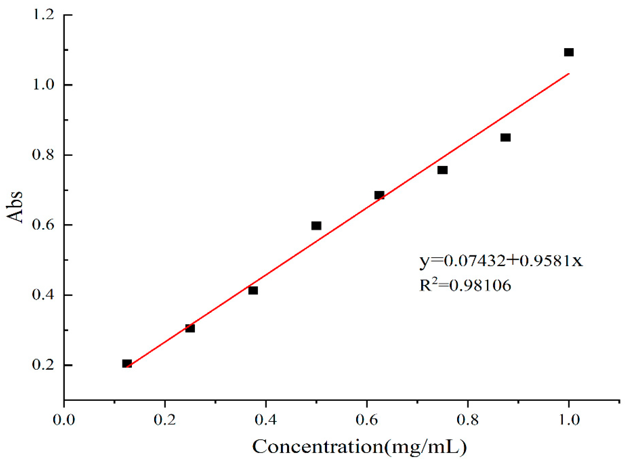
The relationship between the concentration of gallic acid and absorbance at 760 nm is shown in Figure 2. The TPC is expressed as mg of gallic acid per g of sample. The concentration of gallic acid was linearly related to the absorbance, and the equation of the standard curve was as follows: y = 0.9581x + 0.07432, R2 = 0.98106. The absorbance of each classified component of the G-DHP was substituted into the equation to obtain the concentration. The TPC of each classified component relative to gallic acid is presented in Table 2. The TPC of each classified component decreased with increasing molecular weight.

2.4. Analysis of the Anti-Tumor Activities of the G-DHP Classified Fractions

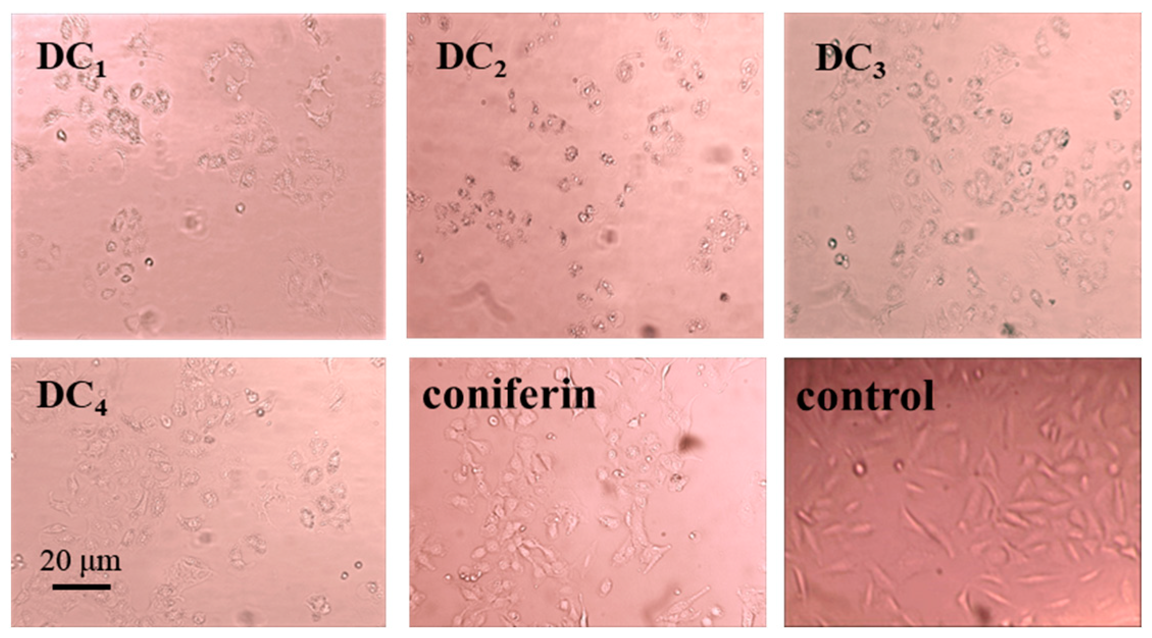
The effects of the G-DHP classified fractions and coniferin, at a concentration of 800 μg/mL, on the growth of A549 lung cancer cells are shown in Figure 3. It can be found that the lignin precursor coniferin did not have any obvious inhibitory effect on lung cancer cells. Meanwhile, the cell growth status of the control group, using the solvent only, was also normal, indicating that the solvent had no effect on the growth of A549 lung cancer cells. The inhibitory effects of the classified fractions of the G-DHP on A549 lung cancer cells were significant, especially for the ether-soluble fraction DC2. Specifically, the cells became round and contracted and no longer adhered to the wall, and some even broke into small fragments. Furthermore, many apoptotic cells died.
The relationships between the concentration of the classified fractions and the inhibition rate on A549 lung cancer cells are shown in Figure 4. The IC50 values are shown in Table 3. It can be found that all classified fractions inhibited A549 lung cancer cells. Furthermore, the relationships between the concentration and the inhibition rate were approximately logarithmic. The ether-soluble fraction (DC2) showed the strongest inhibitory effect on A549 lung cancer cells, with an IC50 of 181.46 ± 28.01 μg/mL. According to the standards of the National Cancer Institute. IC50 ≤ 20 μg/mL is highly cytotoxic; ranging from 21 to 200 μg/mL is moderately cytotoxic; ranging from 201 to 500 μg/mL is weakly cytotoxic; and an IC50 > 501 μg/mL is noncytotoxic [26,27,28].

2.5. Analysis of the Anti-Tumor Activity of the Purified Substances of G-DHP Ether-Soluble Fraction

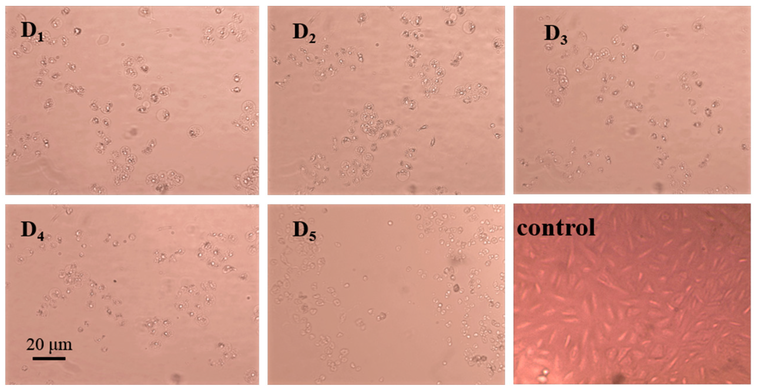
The effects of the purified substances from the DC2 fraction, at a concentration of 800 μg/mL, the growth state of A549 lung cancer cells are shown in Figure 5. It can be found that the A549 lung cancer cells were apoptotic in the experimental groups with the addition of each purified substance. The cells became smaller and rounder, the space between the cells became larger, and the cells no longer adhered to the wall. Some of the apoptotic cells broke off into small fragments. The purified substances exhibited different degrees of inhibitory effects on A549 lung cancer cells, with compound D5 exhibiting the best inhibitory effect.
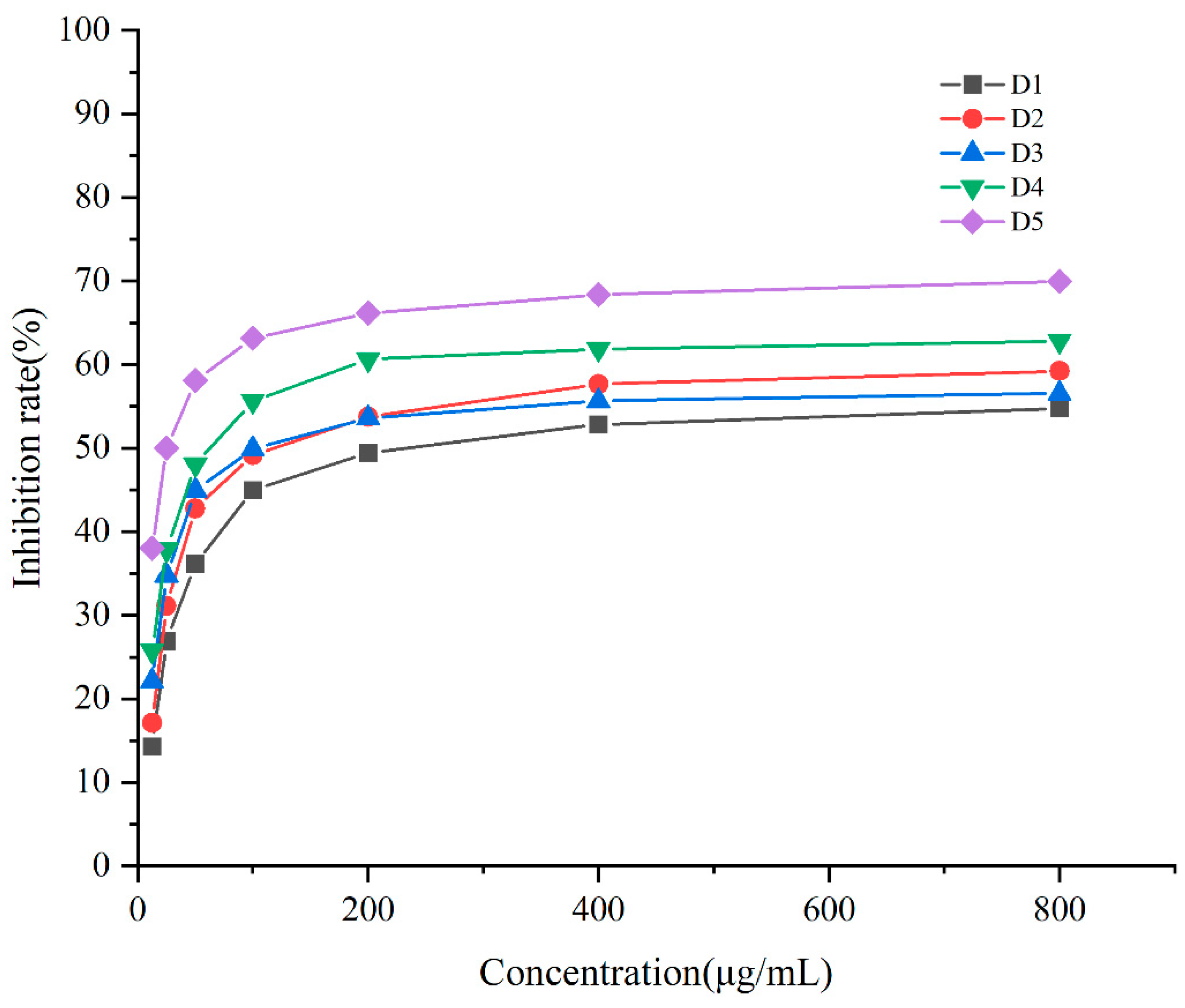
The relationships between the different concentrations of purified compounds from the DC2 fraction and the inhibition rate on A549 lung cancer cells are shown in Figure 6. The IC50 values are shown in Table 4. The results revealed that all purified compounds had inhibitory effects on A549 lung cancer cells, while the concentrations and inhibition rates exhibited roughly logarithmic relationships. Obvious inhibitory effects of D4 and D5 on A549 lung cancer cells were observed, with IC50 values of 61.54 ± 17.10 μg/mL and 28.61 ± 8.52 μg/mL, respectively. As compared with the original DC2 fraction, the purified substances showed significantly enhanced inhibitory effects on A549 lung cancer cells, indicating that the purification by medium-pressure liquid preparative chromatography was successful for the isolation of the bioactive substances. In our previous research, some compounds isolated from DHP had a certain inhibitory effect on the HeLa cervical cancer cells. The inhibitory property of a compound, i.e., 4-[3-Hydroxymethyl-5-(3-hydroxy-propenyl)-7-methoxy-2,3-dihydro-benzofuran-2-yl]-2-methoxy-phenol from ether-soluble fractions, were relatively strong, with IC50 values of 15.1 ± 2.3 μg/mL. This compound was a typical (β-5) G-type dimer, and very similar to compounds D4 and D5 in structure in the present work. This also meant that the D4 and D5 compounds are relatively effective in anti-cancer treatments [17]. We have conducted a large number of experiments on the metabolic activity of hepatocytes cultured in vitro in the lignin and lignin-carbohydrate complexes. The growth of normal hepatocytes cells was enhanced by the addition of lignin and linin-carbohydrate complexes, and the inhibition effect could be ignored [29,30].

2.6. HESI-MS Mass Spectrometry Analysis of the Purified Substances

Electrospray ionization mass spectrometry (ESI-MS) is appropriate for investigating the primary and fragment structure of biopolymers. Several previous studies on lignin that used ESI-MS have shown the utility of this analytical method for estimating the average molecular mass of lignin macromolecules, distinguishing their biological origins, and exploring the reactions occurring in solutions [31,32]. Given the high contents of hydroxyl and carboxyl groups in lignin, mass spectrometric information in the negative ion mode (M − H+) will produce better signals than in the positive ion mode (M + H+). Therefore, ESI under negative ionization conditions is the most widely used method for determination of lignin structure [33]. Coniferin was enzymatically hydrolyzed to coniferyl alcohol before polymerization to DHP. The molecular weight of the monomer coniferyl alcohol was about 180 g/mol. In addition, functional groups such as carboxyl and aldehyde groups may be generated under the catalytic action of laccase, thus, the molecular weight distributions of DHP with different degrees of polymerization were as follows: monomer (130–190 g/mol), dimer (190–380 g/mol), trimer (380–570 g/mol) [34,35,36].
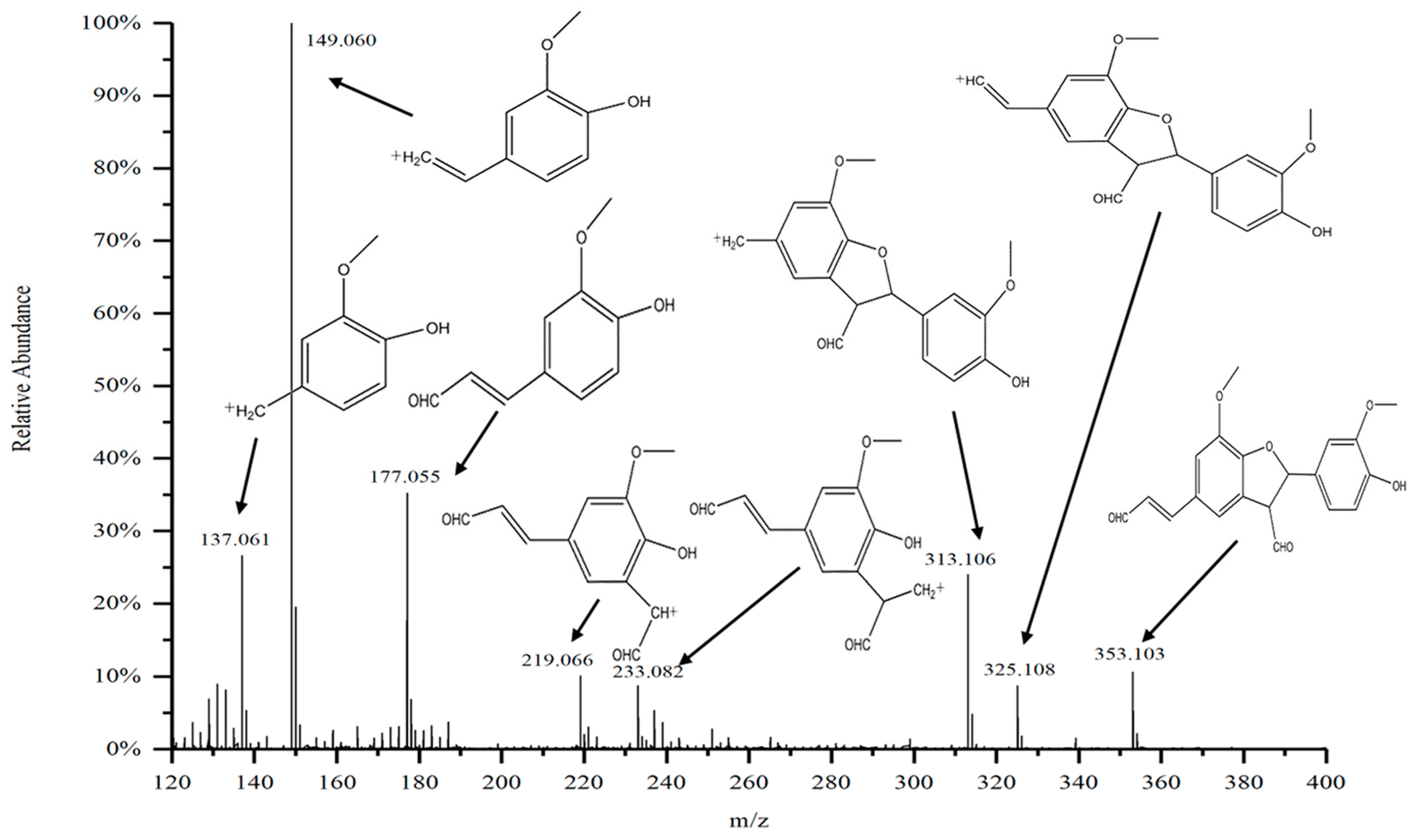
The mass spectral information of the purified substance D5 from the DC2 fraction of G-DHP is shown in Figure 7. The signal at m/z 353.103 is of the molecule D5. The molecular structure formula of D5 was calculated to be C20H18O6 based on the other fragments The most abundant ion fragment is at m/z 149.060, which is the ion peak of 2-methoxy-4-vinyl coniferyl alcohol. This ionic peak originates from the cleavage of the coumarin structure, indicating that the Cγ on the side chain of the phenyl propane monomer in guaiacyl lignin is unstable as compared to other positions. The signal at m/z 137.0 is of the fragment peak of 2-methoxy-4-methylphenol formed by the elimination of Cβ on the side chain of the fragment at m/z 149.060. This is consistent with previous findings, where m/z 177.0 was found to be the fragment peak of 2-methoxy-4-methylphenol [37]. The ionic peak of the coniferyl alcohol monomer at m/z 177.0 is also consistent with previous findings [38]. The peak at m/z 353.1 is of the β-5-linked dimer of coniferyl aldehyde (β-5, γ-CHO, γ’-CHO), while the peak at m/z 325.1 differs from m/z 353.1 by 28, indicating that m/z 325.1 is a fragment peak formed by the elimination of the aldehyde group on Cγ of the molecular ion at m/z 353.1. These results indicate that the oxidation of coniferin catalyzed by laccase and β-glucosidase led to the formation of the β-5 structure of the coniferyl aldehyde dimer. Similar findings have been reported in previous studies that have synthesized DHP [39].
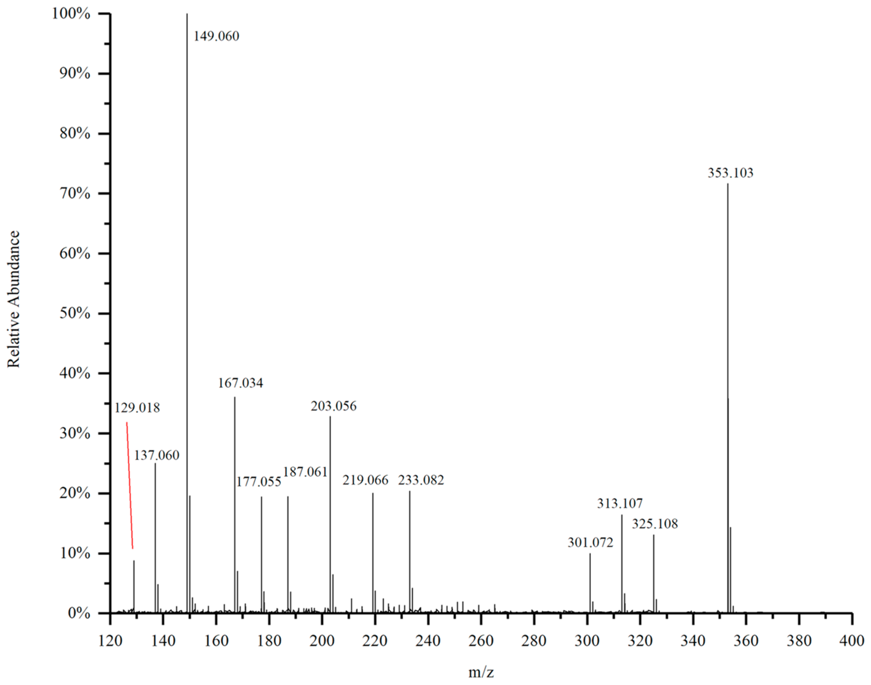
The mass spectrum of the purified substance D4 from the DC2 fraction of G-DHP is shown in Figure 8. Similarly to D5, the molecular ion of D4 is also found at m/z 353.103. Considering the other fragments, the molecular structure formula of D4 should also be C20H18O6. All of the fragment ion peaks in D5 can be found in the spectrum of compound D4, while D4 has additional peaks at m/z 129.018, m/z 167.034, m/z 187.061, m/z 203.056, and m/z 301.072, indicating that D4 contains some impurities. These results indicate that D4 and D5 are generally the same substance, but D4 contains more impurities. This analysis is also confirmed by the slightly lower inhibitory effect of the compound D4 on A549 cells, as compared to the compound D5.
The mass spectrum of the purified substance D4 from the DC2 fraction of G-DHP is shown in Figure 8. Similar to that of D5, the molecular ion of D4 is also found at m/z 353.103. Considering the other fragments, the molecular structure formula of D4 should also be C20H18O6, and all of the fragment ion peaks in D5 can be found in the spectrum of compound D4, while D4 has additional peaks at m/z 129.018, m/z 167.034, m/z 187.061, m/z 203.056, and m/z 301.072, indicating that D4 contains some impurities. The results indicate that D4 and D5 are mainly the same substance, but D4 contains more impurities. This is also confirmed by the slightly less inhibitory effect of D4 on A549 cells than D5.

2.7. 13C-NMR and 1H-NMR Analysis of the Purified Compound

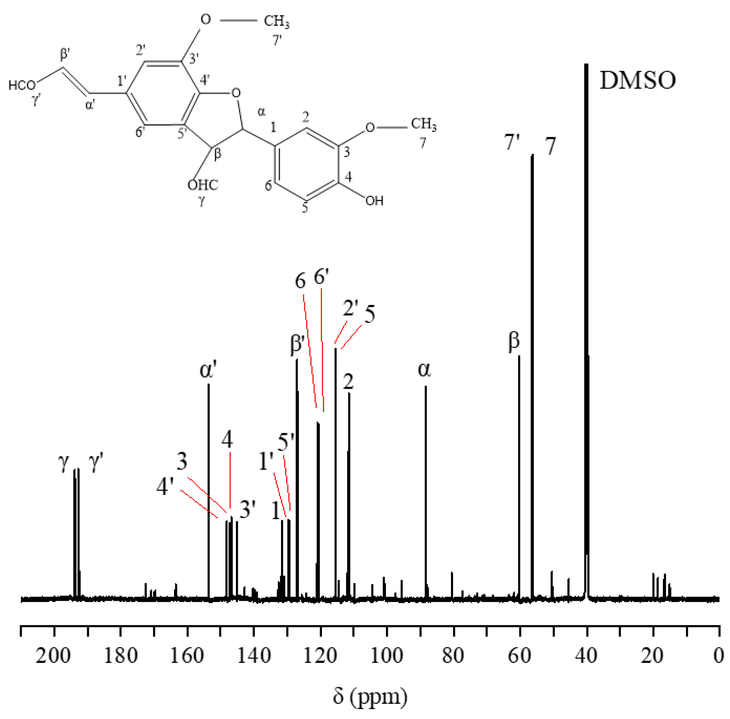
The 13C-NMR spectrum of the purified compound D5 from the DC2 fraction of G-DHP is shown in Figure 9. The major resonance signals are assigned as follows: δ 193.81 (Cγ), δ 192.81 (Cγ′), δ 153.60 (Cα′), δ 148.13 (C4′), δ 147.32 (C3), δ 146.75 (C4), δ 145.12 (C3′), δ 131.61 (C1), δ 129.72 (C1′), δ 129.43 (C5′), δ 127.03 (Cβ′), δ 120.94 (C6), δ 120.67 (C6′), δ 115.57 (C2′), δ 115.39 (C5), δ 111.51 (C2), δ 88.30 (Cα), δ 60.32 (Cβ), δ 56.52 (C7′), δ 56.16 (C7), δ 40.05 (DMSO).
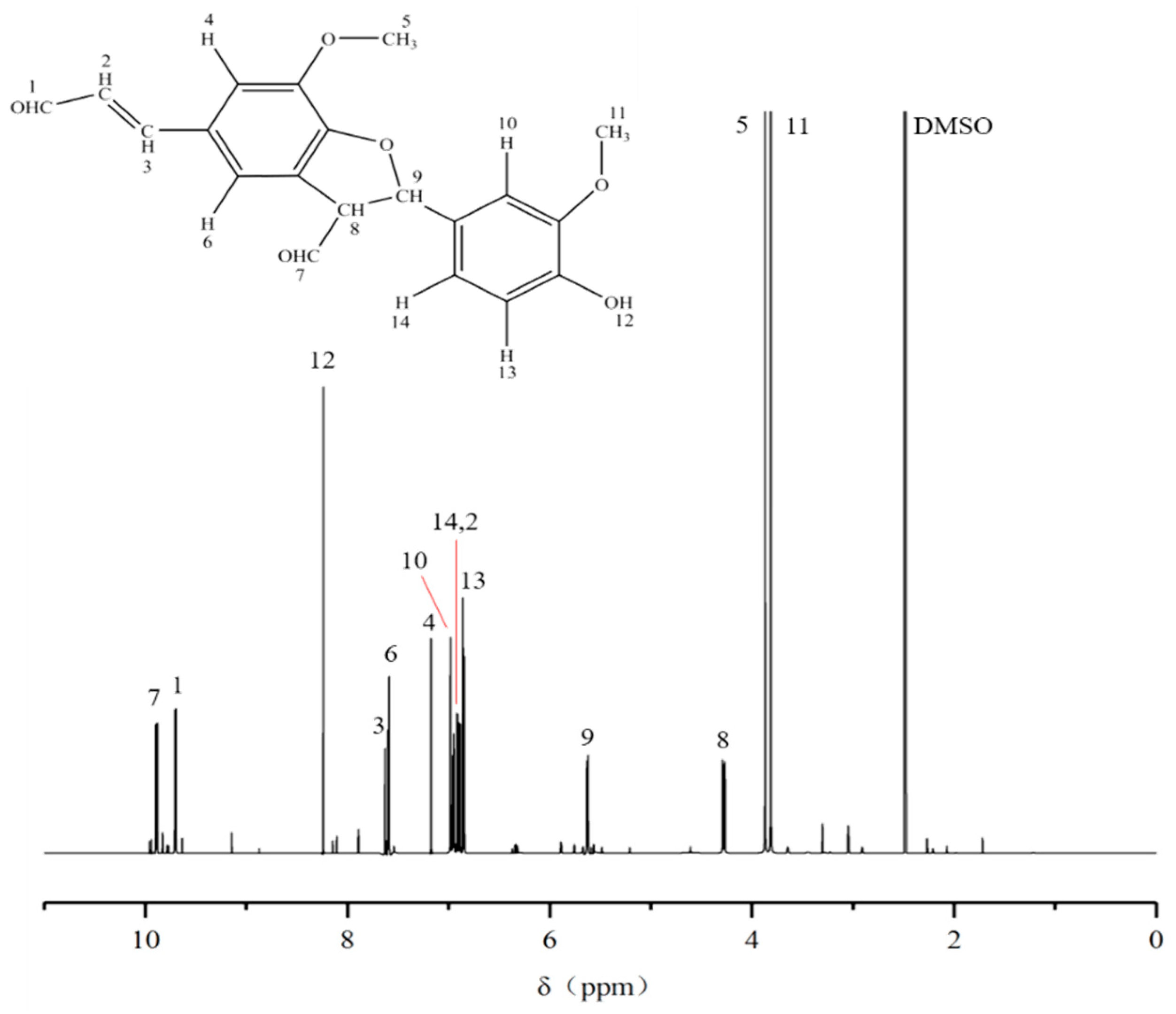
The 1H-NMR spectrum of the purified compound D5 from the DC2 fraction of G-DHP is shown in Figure 10. The major resonance signals are assigned as follows: δ 9.88 (7), δ 9.70 (1), δ 8.24 (12), δ 7.61 (3), δ 7.59 (6), δ 7.17 (4), δ 6.98 (10), δ 6.96 (14), δ 6.91 (2), δ 6.86 (13), δ 5.63 (9), δ 4.28 (8), δ 3.87 (5), δ 3.81 (11), δ 2.49 (DMSO). Thus, considering the mass spectrum, 13C-NMR spectrum, and 1H-NMR spectrum analyses, it can be concluded that compound D5 is a β-5-linked coniferyl aldehyde dimer.

2.8. Discussion of the Structure–Effect Relationships of the Purified Substances

The HESI-MS, 13C-NMR, and 1H-NMR results indicated that compound D5 is a coniferyl aldehyde dimer with a β-5 linkage. The compounds D4 and D5 are the same substance, although the compound D4 contains more impurities, leading to a reduction in its anti-tumor activity. Zemek et al. [40] studied the biological activity of lignin model substances and found that those containing Cα=Cβ on the side chain of the benzene ring and with methyl on Cγ were the most effective. In addition, increases in hydroxyl, carbonyl, and carboxyl groups on the side chain reduced the biological activity of the compound. Xie et al. [17,41] synthesized DHP from isoeugenol and found that the presence of an aldehyde group on the side chain of the phenylpropane structure weakened the antioxidant and antibacterial properties of DHP. However, the results of the present study indicated that compound D5 contains aldehyde groups on the side chain, yet it had strong anti-tumor activity. Gładkowski et al. [42] used aromatic aldehydes as raw materials to synthesize 20 aromatic aldehydes. The authors found that some of the obtained compounds had significant cytotoxic effects on two cancer cells: human leukemia cells (Jurka cells) and canine osteosarcoma cells (D17 cells). These results suggest that aldehydes have potential anti-tumor applications.
Previous studies have shown that β-5-linked oligomeric lignins have good inhibitory effects on cervical cancer HeLa cells, with IC50 values as low as 15.1 μg/mL [17], while the present study found that β-5-linked oligomeric lignin had an inhibitory effect on A549 lung cancer cells, indicating that β-5 structured oligomeric lignins have broad anticancer activity.

3. Experiment

3.1. Materials

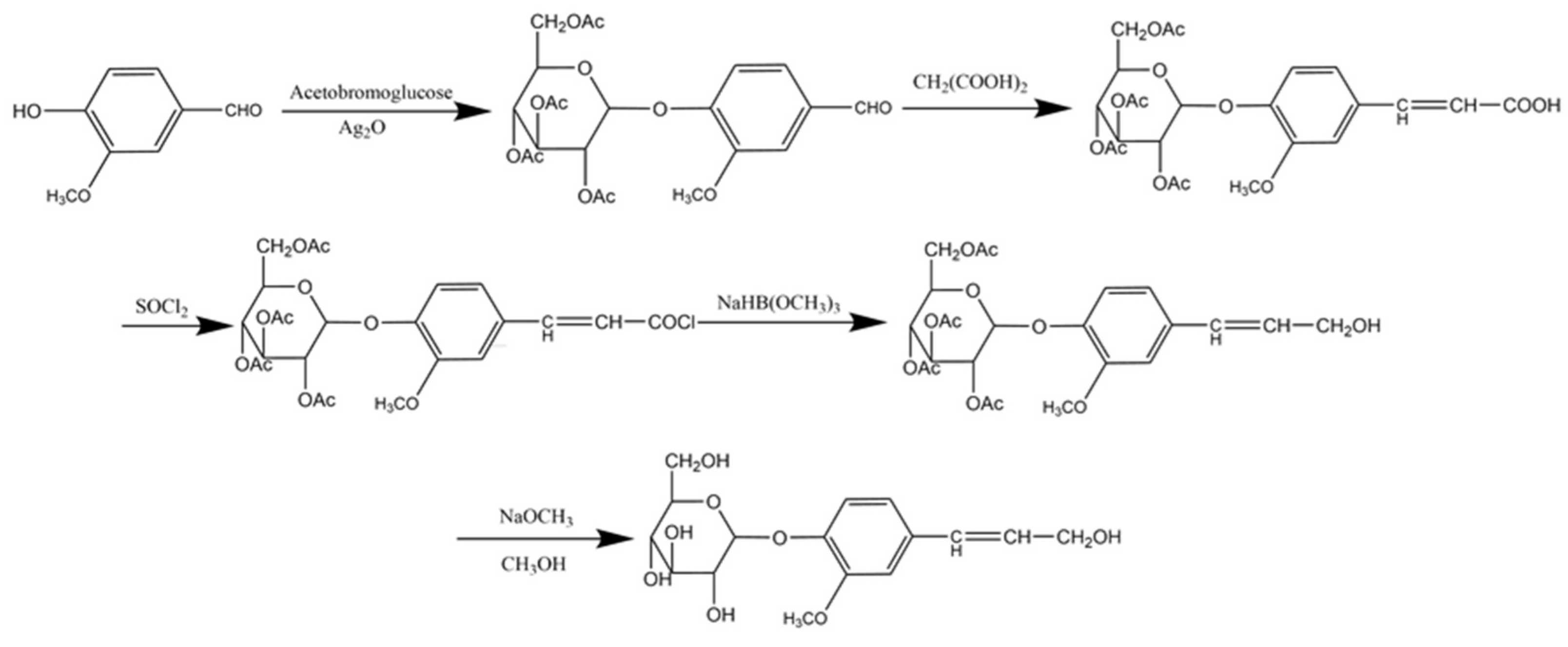
With reference to the method of Xie et al. [22], coniferin was synthesized from vanillin. The synthetic route is shown in Figure 11. β-Glucosidase was purchased from Sigma Co., Ltd. (Sigma, St. Louis, MO, USA) and laccase (No. 51003) was purchased from Novazyme Co., Ltd. (Tianjin, China). Complete Medium (CM2-1) was composed of 89% Roswell Park Memorial Institute (RPMI) 1640 basal medium (Gibco, Grand Island, NY, USA), 10% fetal bovine serum (FBS) (Tianhang, China), and 1% penicillin-streptomycin mixture (Gibco, USA). Other chemicals were of analytical grade and were purchased from Sinopharm Chemical Reagent Co., Ltd. (Shanghai, China). Human A549 lung cancer cells were purchased from Beijing Beina Biotechnology Co., Ltd. (BNCC, Henan Xinyang, China).

3.2. Methods

3.2.1. Synthesis of DHP

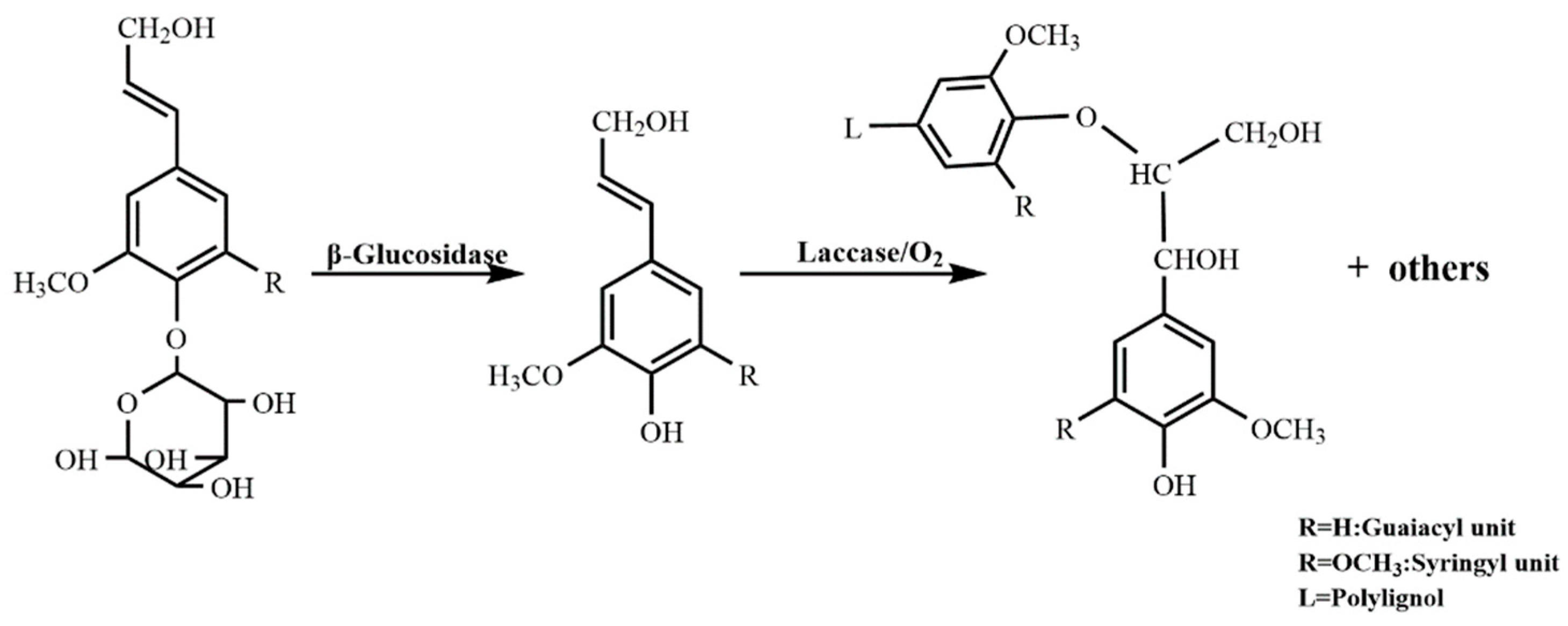
The DHP synthesis mechanism is shown in Figure 12. In 150 mL of 0.2 mol/L sterile acetic acid/sodium acetate buffer solution with pH 4.83, 5 g of coniferin, 50 mg of β-glucosidase (6.4 U/mL), and 3 mL of laccase (648 U/mL) were added with the continuous blowing of sterile air filtered by activated carbon with an air pump. Then, the mixture was allowed to continue reacting in a water bath at 30 °C for 25 min. After the reaction was completed, 150 mL of distilled water was added and then heated to 70 °C to stop the reaction. The mixture was then centrifuged, the precipitate collected and washed with distilled water more than 5 times to remove the unreacted coniferin, and some enzymes freeze-dried to obtain crude DHP. The mixture was dissolved in dichloroethane/ethanol (v/v = 2:1), and the precipitate of the residual enzyme was removed by centrifugation. The solvent was then removed in vacuo. The DHP was obtained by vacuum drying with a yield of 38.36%.

3.2.2. 13C-NMR Measurement of G-DHP

An 80 mg DHP sample was placed into φ 5 mm NMR tubes and dissolved with 0.6 mL of DMSO-d6. A 600-dd2 NMR spectrometer (Agilent Technologies, Santa Clara, CA, USA) was used to scan the solution at 150.90 MHz to obtain the corresponding 13C-NMR spectrum. The parameters of the instrument were as follows: test temperature: 25 °C; pulse delay: 2.0000 s; acquisition time: 0.6900 s; and number of scans: 3000.

3.2.3. Classification of G-DHP

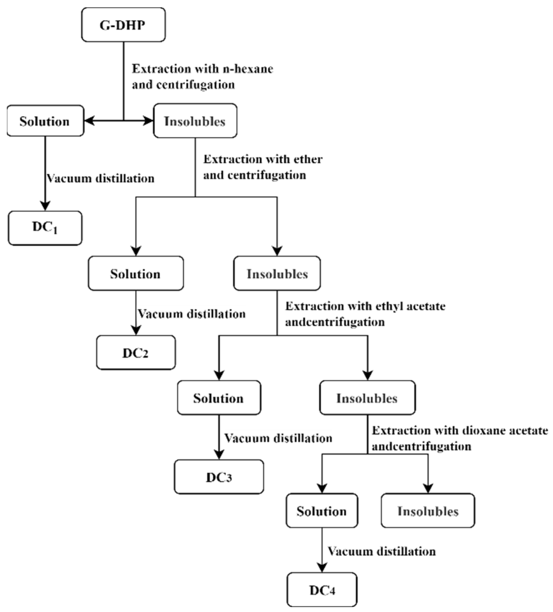
G-DHP was classified according to the polarity and solubility of different organic solvents, with reference to the method of Li et al. [43]. G-DHP was graded using n-hexane, ether, ethyl acetate, and dioxane. The classification process is shown in Figure 13. G-DHP was placed into a Soxhlet extractor together with n-hexane and heated at boiling point to reflux for 6 h. The extracted solution was distilled in vacuo to remove the solvent and then dried under a vacuum to obtain the n-hexane-soluble component (DC1). The remaining solid in the Soxhlet extractor was heated and refluxed with ether at 40 °C for 6 h. The extracted solution was distilled in vacuo to remove the solvent and then dried under a vacuum to obtain the ether-soluble component (DC2). The ethyl acetate-soluble component (DC3) and dioxane-soluble component (DC4) were obtained by repeating the above procedures with ethyl acetate and dioxane, respectively. Finally, the yields of the n-hexane-soluble component (DC1), ether-soluble component (DC2), ethyl acetate-soluble component (DC3), and dioxane-soluble component (DC4) were 6.8%, 11.7%, 43.6%, and 32.3%, respectively.

3.2.4. Determination of Molecular Weight of the G-DHP Fractions

The relative molecular weights of individual fractions were determined by gel permeation chromatography (GPC). The molecular weight standard curves were prepared with EasiVial Polystyrene PS-M. Each DHP fraction (2 mg) was dissolved in 2 mL N,N-dimethylformamide (DMF) filtered through a 0.22 μm microporous membrane, and then injected into the GPC system (20AT, Shimadzu, Osaka, Japan) for molecular weight measurement. The instrument parameters were as follows: column: Stirrage HR 4E DMF (7.8 × 300 mm) with a guide column of Styrage DMF (4.6 × 30 mm); detector: RID-10A differential refraction detector; mobile phase: DMF (containing 0.1 mol/L LiCl); flow rate: 1.0 mL/min; column temperature: 50 °C; injection volume: 20 μL.

3.2.5. Determination of Total Phenol Content (TPC)

The Folin–Ciocalteu colorimetric method [44] was used to determine the TPC of the various fractions. For the quantification of these extractives, a standard curve was established from the analysis of different concentrations of gallic acid varying from 125–1000 μg/mL. Then, 100 µL methanol extractive was added to test tubes with 500 µL Folin–Ciocalteu reagent. Six milliliters of distilled water was then added. After shaking for 1 min, 2 mL of 15% Na2CO3 solution was transferred to the test tubes within 0.5~8 min. Finally, distilled water was added to the tubes to make a total volume of 10 mL. After being kept in the dark at room temperature for 2 h, the test tubes were homogenized with ultrasonic agitation. The spectrophotometer (Shimadzu 2550, Nakagyo-ku, Kyoto, Japan) was set at a wavelength of 760 nm, and the absorbance reading was measured. The TPC of each sample was calculated according to the following formula:
TPC [mg (gallic acid)/g(sample)] = GAE × FV × DF/SW
Legend: GAE is the concentration of gallic acid calculated from the standard curve (mg/mL); FV is the final volume of the sample extract (mL); DF is the dilution multiple; SW is the sample mass (g).

3.2.6. Determination of the Anti-Tumor Activities of DHP Fractions and Purified Compounds

According to the method of Ahamed, Le et al. [45,46], the antitumor activities of the G-DHP fractions and purified compounds were determined using the 3-(4,5-Dimethylthiazol-2-yl)-2,5-diphenyltetrazolium bromide (MTT) method. First, samples were dissolved in acetone: CM2-1 medium (0.99:99.1 v/v, 0.1% polysorbate 80 as dispersant) to obtain a series of solutions with concentration in the range from 12.5 μg/mL to 800 μg/mL. Second, 5 × 10³ exponentially growing tumor cells were suspended in 100 μL of the CM2-1 medium and plated per experimental well on 96-well plates. Blank wells were also filled with 200 µL of complete medium. Third, the cells were cultured in a carbon dioxide cell incubator (37 °C, 5% CO2). After 16–48 h, 100-μL sample solutions with different concentrations (12.5~800 μg/mL) were transferred to every experimental well, which was different from the sample solvent added to the control wells. After incubation for 48 h, 20 μL MTT solution (5 mg/mL) was added to each well. Four hours later, the liquid in each well was discharged with a sterile syringe, and then 150 μL of dimethyl sulfoxide (DMSO) was added to each well. Finally, the plate was placed into a microplate reader after shaking at a low speed for 10 min. The absorbance of each well was measured at 490 nm. The inhibition rate of each sample was calculated using the following equation:
IR = (ACONTROL − AEXPERIMENTAL)/(ACONTROL − ABLANK) × 100%;
CONTROL = medium without sample containing cells, solvent, MTT and DMSO;
BLANK = medium without cells and sample containing, solvent, MTT and DMSO.
Then, SPSS 26 software was used to calculate the semi-inhibition concentration (IC50) of each sample [47].

3.2.7. Purification of the Ether-Soluble Fraction of DHP with Preparative Column Chromatography

The ether-soluble component DC2 of DHP was gradient eluted and repeatedly purified using a medium-pressure liquid preparation chromatograph (Buchi C-615, Zurich, Switzerland) equipped with an ultraviolet (UV) detector at a wavelength of 280 nm. The stationary phase was Sephadex LH-20 gel (100~200 mesh), and the mobile phase was a chloroform-methanol mixed eluent (the concentration gradient of methanol was in the range of 0% to 50%), with a flow rate of 2.5 mL/min.
First, the samples were dissolved in a 50% chloroform-methanol solution, and then the sample solution was injected into the dosing ring using a glass syringe. After eluting the separation column with 50% chloroform-methanol, the concentration of methanol was gradually increased, and finally, the column was completely washed with methanol. During the separation process, a UV 230 II ultraviolet-visible detector was applied. The ether-soluble component DC2 of DHP was purified to obtain compounds D1, D2, D3, D4, and D5, with yields of 7.47%, 41.67%, 29.91%, 10.83%, and 10.12%, respectively.

3.2.8. Mass Spectrometry Analysis of the Structure of the DHP Purified Compound

The mass spectrum information of D5 was obtained using electrostatic field orbital hydrazine mass spectrometry (Thermo Fisher Scientific, Waltham, MA, USA). The ion source was HESI. The determination conditions were as follows: dry gas: N2; spray voltage: 3200 V; temperature of the ion transmission capillary: 300 °C; auxiliary gas temperature: 280 °C; scanning mode: full MS/dd-MS2; scanning range m/z: 50–750.

3.2.9. 13C-NMR and 1H-NMR Measurement of the Purified Substance D5

13C-NMR Measurement of the Purified Substance D5

An 80 mg purified substance D5 sample was placed into φ 5 mm NMR tubes and dissolved with 0.6 mL of DMSO-d6. A 600-dd2 NMR spectrometer (Agilent Technologies, Santa Clara, CA, USA) was used to scan the solution at 150.90 MHz to obtain the corresponding 13C-NMR spectrum. The parameters of the instrument were as follows: test temperature: 25 °C; pulse delay: 2.0000 s; acquisition time: 0.6900 s; and number of scans: 3000.

1H-NMR Measurement of the Purified Substance D5

A 15 mg volume of the purified substance D5 sample was placed into φ 5 mm NMR tubes and dissolved with 0.6 mL of DMSO-d6. A 600-dd2 NMR spectrometer (Agilent Technologies, Santa Clara, CA, USA) was used to scan the above sample at 600 MHz to obtain the corresponding 1H-NMR spectrum. The parameters of the instrument were as follows: test temperature: 25 °C; pulse delay: 5.0000 s; acquisition time: 1.4500 s; and number of scans: 256.

4. Conclusions

(1)
13C-NMR determination revealed that the structure of G-DHP was relatively similar to that of the gymnosperm ginkgo MWL, which was dominated by β-O-4, β-5, β-1, β-β, and 5-5 substructures.
(2)
G-DHP was classified with different polar solvents to obtain a hexane-soluble fraction (DC1), ether-soluble fraction (DC2), ethyl acetate-soluble fraction (DC3), and dioxane-soluble fraction (DC4). The GPC results and total phenolic content measurements showed that the total phenolic content of fractions decreased with an increase in molecular weight. The bioactivity assay showed that the ether-soluble fraction (DC2) had the strongest inhibitory effect on lung cancer A549 with an IC50 of 181.46 ± 28.01 μg/mL.
(3)
Further purification of the DC2 fraction revealed that the obtained compounds D4 and D5 had significant anti-tumor activities, with IC50 values of 61.54 ± 17.10 μg/mL and 28.61 ± 8.52 μg/mL against lung cancer A549, respectively. HESI-MS measurements indicated that D5 was a coniferyl aldehyde dimer linked by β-5. The compounds D4 and D5 were similar substance, but D4 contained more impurities. 13C-NMR and 1H-NMR results also confirmed the chemical structure of D5.
In combination with the results of the anti-tumor assay, for the small molecule compound of guaiacyl type DHP, it was found that the presence of an aldehyde group on the side chain of the phenylpropane subunit can enhance the anti-cancer activity.

Author Contributions

Conceptualization, J.Z., Y.Y. and Y.X.; methodology, J.Z., X.W. and Y.X.; writing—original draft preparation, J.Z.; writing—review and editing, Y.Y. and Y.X.; visualization, J.Z.; supervision, Y.X.; project administration, Y.X.; funding acquisition, Y.X. All authors have read and agreed to the published version of the manuscript.

Funding

This research was supported by the National Natural Science Foundation of China (Grant No. 21878070) and Outstanding Young and Middle-aged Technological Innovation Team Project of Hubei Provincial Universities (Grant No. T201205).

Institutional Review Board Statement

Not applicable.

Informed Consent Statement

Not applicable.

Data Availability Statement

The data presented in this study are available in the manuscript.

Conflicts of Interest

The authors declare no conflict of interest.

Sample Availability

Samples of the compounds are not available from the authors.

References

  1. Thai, A.A.; Solomon, B.J.; Sequist, L.V.; Gainor, J.F.; Heist, R.S. Lung cancer. Lancet 2021, 398, 535–554. [Google Scholar] [CrossRef] [PubMed]
  2. Sung, H.; Ferlay, J.; Siegel, R.L.; Laversanne, M.; Soerjomataram, I.; Jemal, A.; Bray, F. Global cancer statistics 2020: GLOBOCAN estimates of incidence and mortality worldwide for 36 cancers in 185 countries. CA Cancer J. Clin. 2021, 71, 209–249. [Google Scholar] [CrossRef]
  3. Garg, J.; Chiu, M.N.; Krishnan, S.; Tripathi, L.K.; Pandit, S.; Far, B.F.; Jha, N.K.; Kesari, K.K.; Tripathi, V.; Pandey, S.; et al. Applications of lignin nanoparticles for cancer drug delivery: An update. Mater. Lett. 2022, 311, 131573. [Google Scholar] [CrossRef]
  4. Lu, F.J.; Chu, L.H.; Gau, R.J. Free radical-scavenging properties of lignin. Nutr. Cancer 1998, 30, 31–38. [Google Scholar] [CrossRef]
  5. Zhang, L.; Zhang, W.; Wang, Q.; Wang, D.; Dong, D.; Mu, H.; Ye, X.S.; Duan, J. Purification, antioxidant and immunological activities of polysaccharides from Actinidia Chinensis roots. Int. J. Biol. Macromol. 2015, 72, 975–983. [Google Scholar] [CrossRef] [PubMed]
  6. Lábaj, B.K.J. Antigenotoxic effect of lignin preparations from wastes of pulp and paper industry-in vitro and ex vivo activivity. Acta Fac. Xylologiae Zvolen 2010, 53, 69–76. [Google Scholar]
  7. Geies, A.; Abdelazim, M.; Sayed, A.M.; Ibrahim, S. Thermal, Morphological and Cytotoxicity Characterization of Hardwood Lignins Isolated by In-Situ Sodium Hydroxide-Sodium Bisulfate Method. Nat. Resour. 2020, 11, 427–438. [Google Scholar] [CrossRef]
  8. Sakagami, H.; Asano, K.; Yoshida, T.; Kawazoe, Y. Organ distribution and toxicity of lignin. In Vivo 1999, 13, 41–44. [Google Scholar] [PubMed]
  9. Sakagami, H.; Kushida, T.; Oizumi, T.; Nakashima, H.; Makino, T. Distribution of lignin-carbohydrate complex in plant kingdom and its functionality as alternative medicine. Pharm. Ther. 2010, 128, 91–105. [Google Scholar] [CrossRef]
  10. Ugartondo, V.; Mitjans, M.; Vinardell, M.P. Applicability of lignins from different sources as antioxidants based on the protective effects on lipid peroxidation induced by oxygen radicals. Ind. Crop. Prod. 2009, 30, 184–187. [Google Scholar] [CrossRef]
  11. Barapatre, A.; Meena, A.S.; Mekala, S.; Das, A.; Jha, H. In vitro evaluation of antioxidant and cytotoxic activities of lignin fractions extracted from Acacia nilotica. Int. J. Biol. Macromol. 2016, 86, 443–453. [Google Scholar] [CrossRef]
  12. Mitsuhashi, S.; Kishimoto, T.; Uraki, Y.; Okamato, T.; Ubukata, M. Low molecular weight lignin suppresses activation of NF-κB and HIV-1 promoter. Bioorg. Med. Chem. 2008, 16, 2645–2650. [Google Scholar] [CrossRef]
  13. Turliuc, M.D.; Cucu, A.I.; Carauleanu, A.; Costea, C. Efficiency and safety of microporous polysaccharide hemispheres from potato starch in brain surgery. Cellul. Chem. Technol. 2018, 52, 505–513. [Google Scholar]
  14. Spiridon, I.; Poni, P.; Ghica, G. Biological and pharmaceutical applications of lignin and its derivatives: A mini-review. Cellul. Chem. Technol. 2018, 52, 543–550. [Google Scholar]
  15. Shu, F.; Jiang, B.; Yuan, Y.; Li, M.; Wu, W.; Jin, Y.; Xiao, H. Biological activities and emerging roles of lignin and lignin-based products—A review. Biomacromolecules 2021, 22, 4905–4918. [Google Scholar] [CrossRef] [PubMed]
  16. Andrijevic, L.; Radotic, K.; Bogdanovic, J.; Mutavdzic, D.; Bogdanovic, G. Antiproliferative effect of synthetic lignin against human breast cancer and normal fetal lung cell lines. Potency of low molecular weight fractions. J. BUON 2008, 13, 241–244. [Google Scholar] [PubMed]
  17. Xie, Y.; Jiang, C.; Chen, X.; Wu, H.; Bi, S. Preparation of Oligomeric Phenolic Compounds (DHPs) from Coniferin and Syringin and Characterization of their Anticancer Properties. BioResources 2020, 15, 1791–1809. [Google Scholar] [CrossRef]
  18. Xie, Y.; Yasuda, S.; Wu, H.; Liu, H. Analysis of the structure of lignin-carbohydrate complexes by the specific 13C tracer method. J. Wood Sci. 2000, 46, 130–136. [Google Scholar] [CrossRef]
  19. Holtman, K.M.; Chang, H.M.; Kadla, J.F. Solution-state nuclear magnetic resonance study of the similarities between milled wood lignin and cellulolytic enzyme lignin. J. Agric. Food Chem. 2004, 52, 720–726. [Google Scholar] [CrossRef]
  20. Hage, R.E.; Brosse, N.; Chrusciel, L.; Sanchez, C.; Sannigrahi, P.; Ragauskas, A. Characterization of milled wood lignin and ethanol organosolv lignin from miscanthus. Polym. Degrad. Stab. 2009, 94, 1632–1638. [Google Scholar] [CrossRef]
  21. Lüdemann, H.D.; Nimz, H. Carbon-13 nuclear magnetic resonance spectra of lignins. Biochem. Biophys. Res. Commun. 1973, 52, 1162–1169. [Google Scholar] [CrossRef] [PubMed]
  22. Xie, Y.; Robert, D.R.; Terashima, N. Selective carbon 13 enrichment of side chain carbons of ginkgolignin traced by carbon 13 nuclear magnetic resonance. Plant Physiol. Biochem. 1994, 32, 243–249. [Google Scholar]
  23. Terashima, N.; Atalla, R.H.; Ralph, S.A.; Landucci, L.L.; Lapierre, C.; Monties, B. New Preparations of Lignin Polymer Models under Conditions that Approximate Cell Wall Lignification. I. Synthesis of Novel Lignin Polymer Models and their Structural Characterization by 13C NMR. Holzforschung 1995, 49, 521–527. [Google Scholar] [CrossRef]
  24. Harman-Ware, A.E.; Happs, R.M.; Davison, B.H.; Davis, M.F. The effect of coumaryl alcohol incorporation on the structure and composition of lignin dehydrogenation polymers. Biotechnol. Biofiuels 2017, 10, 281. [Google Scholar] [CrossRef] [PubMed]
  25. Wei, X.; Cui, S.; Xie, Y. Synthesis and Antibacterial Properties of Oligomeric Dehydrogenation Polymer from Lignin Precursors. Molecules 2022, 27, 1466. [Google Scholar] [CrossRef]
  26. Grever, M.R.; Schepartz, S.A.; Chabner, B.A. The National Cancer Institute: Cancer drug discovery and development program. Semin. Oncol. 1992, 19, 622–638. [Google Scholar]
  27. Goldin, A.; Venditti, J.M.; Macdonald, J.S.; Muggia, F.M.; Henney, J.E.; Devita, V.T., Jr. Current results of the screening program at the Division of Cancer Treatment, National Cancer Institute. Eur. J. Cancer 1981, 17, 129–142. [Google Scholar] [CrossRef]
  28. Geran, R.I. Protocols for screening chemical agents and natural products against animal tumors and other biological systems: Drug Evaluation Branch. Nat. Cancer Inst. 1962, 3, 1–107. [Google Scholar]
  29. Li, J.; Wang, P.; Yang, J.; Chen, F.; Xie, Y. Preparation of Porous Biological Carrier with Gingko LCC and Application in Culture of Human Hepatocytes. Chem. Ind. For. Prod. 2014, 34, 81–87. [Google Scholar]
  30. Zhao, H.; Feng, Q.; Xie, Y.; Li, J.; Chen, X. Preparation of Biocompatible Hydrogel from Lignin Carbohydrate Complex (LCC) as Cell Carriers. Bioresources 2017, 12, 8490–8504. [Google Scholar] [CrossRef]
  31. Cui, C.; Chen, X.; Liu, C.; Zhu, Y.; Ouyang, J.; Shen, Y.; Zhou, Z.; Qi, F. In Situ Reactor-Integrated Electrospray Ionization Mass Spectrometry for Heterogeneous Catalytic Reactions and Its Application in the Process Analysis of High-Pressure Liquid-Phase Lignin Depolymerization. Anal. Chem. 2021, 93, 12987–12994. [Google Scholar] [CrossRef]
  32. Dean, K.R.; Novak, B.; Moradipour, M.; Tong, X.; Moldovan, D.; Knutson, B.L.; Rankin, S.E.; Lynn, B.C. Complexation of Lignin Dimers with β-Cyclodextrin and Binding Stability Analysis by ESI-MS, Isothermal Titration Calorimetry, and Molecular Dynamics Simulations. J. Phys. Chem. B 2022, 126, 1655–1667. [Google Scholar] [CrossRef]
  33. Qi, Y.; Hempelmann, R.; Volmer, D.A. Shedding light on the structures of lignin compounds: Photo-oxidation under artificial UV light and characterization by high resolution mass spectrometry. Anal. Bioanal. Chem. 2016, 408, 8203–8210. [Google Scholar] [CrossRef] [PubMed]
  34. Terashima, N.; Atalla, R.H.; Ralph, S.A.; Landucci, L.L.; Lapierre, C.; Monties, B. New preparations of lignin polymer models under conditions that approximate cell wall lignification. II. Structural characterization of the models by thioacidolysis. Holzforschung 1996, 50, 9–14. [Google Scholar] [CrossRef]
  35. Ye, Z.Z.; Xie, Y.M.; Wu, C.X.; Wang, P.; Le, X. Dehydrogenation polymerization of isoeugenol and formation of lignin-carbohydrate complexes with presence of polysaccharide. Chem. Ind. For. Prod. 2016, 36, 45–50. [Google Scholar]
  36. Chen, X.K.; Zhao, H.K.; Wu, H.F.; Ye, Z.Z.; Xie, Y.M. Preparation and antioxidant properties of dehydrogenation polymer of isoeugenol. Chem. Ind. For. Prod. 2018, 38, 87–92. [Google Scholar]
  37. Niu, Y.; Zhang, X.; Xiao, Z.; Song, S.; Eric, K.; Jia, C.; Yu, H.; Zhu, J. Characterization of odor-active compounds of various cherry wines by gas chromatography–mass spectrometry, gas chromatography–olfactometry and their correlation with sensory attributes. J. Chromatogr. B 2011, 879, 2287–2293. [Google Scholar] [CrossRef] [PubMed]
  38. Kosyakov, D.S.; Pikovskoi, I.I.; Ul’yanovskii, N.V. Dopant-assisted atmospheric pressure photoionization Orbitrap mass spectrometry–An approach to molecular characterization of lignin oligomers. Anal. Chim. Acta 2021, 1179, 338836. [Google Scholar] [CrossRef]
  39. Ito, T.; Hayase, R.; Kawai, S.; Ohashi, H.; Higuchi, T. Coniferyl aldehyde dimers in dehydrogenative polymerization: Model of abnormal lignin formation in cinnamyl alcohol dehydrogenase-deficient plants. J. Wood Sci. 2002, 48, 216–221. [Google Scholar] [CrossRef]
  40. Zemek, J.; Košíková, B.; Augustin, J.; Joniak, D. Antibiotic properties of lignin components. Folia Microbiol. 1979, 24, 483–486. [Google Scholar] [CrossRef]
  41. Xie, Y.; Chen, X.; Jiang, C.; Wu, H.; Ye, Z. Preparation of oligomeric dehydrogenation polymer and characterization of its antibacterial properties. Bioresources 2019, 14, 2842–2860. [Google Scholar] [CrossRef]
  42. Gładkowski, W.; Skrobiszewski, A.; Mazur, M.; Siepka, M.; Pawlak, A.; Obminska-Mrukowicz, B.; Bialonska, A.; Poradowski, D.; Drynda, A.; Urbaniak, M. Synthesis and anticancer activity of novel halolactones with β-aryl substituents from simple aromatic aldehydes. Tetrahedron 2013, 69, 10414–10423. [Google Scholar] [CrossRef]
  43. Li, M.F.; Sun, S.N.; Xu, F.; Sun, R.C. Sequential solvent fractionation of heterogeneous bamboo organosolv lignin for value-added application. Sep. Purif. Technol. 2012, 101, 18–25. [Google Scholar] [CrossRef]
  44. Bonoli, M.; Verardo, V.; Marconi, E.; Caboni, M.F. Antioxidant phenols in barley (Hordeum vulgare L.) flour: Comparative spectrophotometric study among extraction methods of free and bound phenolic compounds. J. Agric. Food Chem. 2004, 52, 5195–5200. [Google Scholar] [CrossRef]
  45. Ahamed, M.; Ali, D.; Alhadlaq, H.A.; Akhtar, M.J. Nickel oxide nanoparticles exert cytotoxicity via oxidative stress and induce apoptotic response in human liver cells (HepG2). Chemosphere 2013, 93, 2514–2522. [Google Scholar] [CrossRef] [PubMed]
  46. Le, N.T.; Donadu, M.G.; Ho, D.V.; Doan, T.Q.; Le, A.T.; Raal, A.; Usai, D.; Sanna, G.; Marchetti, M.; Usai, M.; et al. Biological activities of essential oil extracted from leaves of Atalantia sessiflora Guillauminin Vietnam. J. Infect. Dev. Ctries. 2020, 14, 1054–1064. [Google Scholar] [CrossRef] [PubMed]
  47. Nga, N.T.H.; Ngoc, T.T.B.; Trinh, N.T.M.; Trinh, N.T.M.; Thuoc, T.L.; Thao, D.T.P. Optimization and application of MTT assay in determining density of suspension cells. Anal. Biochem. 2020, 610, 113937. [Google Scholar] [CrossRef] [PubMed]
Figure 1. 13C-NMR spectrum of G-DHP.
Molecules 28 03589 g001
Figure 2. Relationship between gallic acid concentration and absorbance at 760 nm.
Molecules 28 03589 g002
Figure 3. Effects of the classified G-DHP fractions and coniferin on the growth of A549 lung cancer cells at a concentration of 800 μg/mL. Legend: DC1–DC4: compounds classified from the G-DHP.
Molecules 28 03589 g003
Figure 4. Relationship between different concentrations of classified fractions and coniferin and the inhibition rate of A549 lung cancer cells.
Molecules 28 03589 g004
Figure 5. Effects of the purified compounds from the DC2 fraction, at a concentration of 800 μg/m, on the growth status of A549 lung cancer cells. Legend: D1–D5: compounds purified from the ether fraction of G-DHP by preparative chromatography; control: control group without sample and solvent.
Molecules 28 03589 g005
Figure 6. Relationship between different concentrations of purified compounds and the inhibition rate of A549 lung cancer cells.
Molecules 28 03589 g006
Figure 7. Mass spectrum of purified substance D5.
Molecules 28 03589 g007
Figure 8. Mass spectrum of purified substance D4.
Molecules 28 03589 g008
Figure 9. 13C-NMR spectrum of the purified compound D5.
Molecules 28 03589 g009
Figure 10. 1H-NMR spectrum of the purified substance D5.
Molecules 28 03589 g010
Figure 11. Synthesis route of coniferin.
Molecules 28 03589 g011
Figure 12. The polymerization of DHP.
Molecules 28 03589 g012
Figure 13. Classification flow chart of the G-DHP.
Molecules 28 03589 g013
Table 1. Average molecular weights of the DHP fractions.
DHP Fractions MwMnPDI
DC11882771.47
DC24026431.60
DC3120316951.41
DC4185334861.88
Legend: Mw: weight weighted average molecular weight; Mn: number average molecular weight; PDI: polymer dispersity index.
Table 2. Total phenol contents of the classified fractions of the G-DHP.
DHP FractionsMwTPC
DC1277228.66
DC2643186.57
DC3169592.21
DC4348670.53
Table 3. IC50 values of classified G-DHP fractions on A549 lung cancer cells.
Classified ComponentIC50 (μg/mL)
DC1318.55 ± 53.49
DC2181.46 ± 28.01
DC3401.71 ± 64.27
DC4461.03 ± 79.53
Table 4. The IC50 values of the purified substances from the DC2 fraction on lung cancer A549 cells.
Purified SubstancesIC50 (μg/mL)
D1286.76 ± 47.16
D2179.25 ± 27.96
D3113.70 ± 23.55
D461.54 ± 17.10
D528.61 ± 8.52
Disclaimer/Publisher’s Note: The statements, opinions and data contained in all publications are solely those of the individual author(s) and contributor(s) and not of MDPI and/or the editor(s). MDPI and/or the editor(s) disclaim responsibility for any injury to people or property resulting from any ideas, methods, instructions or products referred to in the content.