Communication

The Isolation and Structure Elucidation of Spirotetronate Lobophorins A, B, and H8 from Streptomyces sp. CB09030 and Their Biosynthetic Gene Cluster

1
Xiangya International Academy of Translational Medicine, Central South University, Changsha 410013, China
2
School of Pharmaceutical Sciences, Changsha Health Vocational College, Changsha 410100, China
3
South China Sea Institute of Oceanology, Chinese Academy of Sciences, Guangzhou 510301, China
4
Hunan Engineering Research Center of Combinatorial Biosynthesis and Natural Product Drug Discovery, Changsha 410011, China
5
National Engineering Research Center of Combinatorial Biosynthesis for Drug Discovery, Changsha 410011, China
6
Institute of Health and Medicine, Hefei Comprehensive National Science Center, Hefei 230093, China
*
Authors to whom correspondence should be addressed.
Molecules 2023, 28(8), 3597; https://doi.org/10.3390/molecules28083597
Submission received: 24 February 2023 / Revised: 6 April 2023 / Accepted: 12 April 2023 / Published: 20 April 2023

Abstract

:
Lobophorins (LOBs) are a growing family of spirotetronate natural products with significant cytotoxicity, anti-inflammatory, and antibacterial activities. Herein, we report the transwell-based discovery of Streptomyces sp. CB09030 from a panel of 16 in-house Streptomyces strains, which has significant anti-mycobacterial activity and produces LOB A (1), LOB B (2), and LOB H8 (3). Genome sequencing and bioinformatic analyses revealed the potential biosynthetic gene cluster (BGC) for 13, which is highly homologous with the reported BGCs for LOBs. However, the glycosyltransferase LobG1 in S. sp. CB09030 has certain point mutations compared to the reported LobG1. Finally, LOB analogue 4 (O-β-D-kijanosyl-(1→17)-kijanolide) was obtained through an acid-catalyzed hydrolysis of 2. Compounds 14 showed different antibacterial activities against Mycobacterium smegmatis and Bacillus subtilis, which revealed the varying roles of different sugars in their antibacterial activities.

Graphical Abstract

1. Introduction

Spirotetronate polyketides have displayed a variety of impressive biological activities, such as a significant cytotoxicity against tested tumor cell lines (spirohexenolide A (a) [1], okilactomycin (b) [2], and chrolactomycin (c) [3,4]), as well as anti-inflammatory and antibacterial activities (abyssomicin C (d), kijanimicin (e), lobophorin B (f), and MM46115 (g)) (Figure 1) [5,6,7,8]. Structurally, the over 70-membered spirotetronates are classified into class I, which features the spirotetronate moiety within a macrocycle with different sizes, or class II with an additional decalin. Lobophorins (LOBs) belong to class II spirotetronates and share several structural characteristics with kijanimicin, such as a decalin-containing pentacyclic aglycon, a D-kijanose or D-kijanose-like sugar, and an oligosaccharide moiety. LOB A and B, the first two members of LOBs, were reported by Fenical and co-workers from Actinomyces sp. CNB-837, a marine bacterium living in symbiosis with the Caribbean brown alga Lobophora variegate in 1999 [8]. To our knowledge, 16 LOBs have been isolated, including LOBs A–L (1, 2, 30, 31, and 512) [8,9,10,11,12,13,14,15,16] and LOBs CR1-CR4 (13, 14, 32, and 33) (Figure 1) [17,18]. Furthermore, 16 LOB derivatives, including LOBs H1-H15 (3 and 1528) and LOB G2-1 (29) (Figure 1), were subsequently produced by the heterologous expression of the LOB biosynthetic gene cluster (BGC) from the marine-derived Streptomyces pactum SCSIO 02999 by Zhang and colleagues [19]. Recently, three derivative LOBs, N1–N3 (3436), were isolated from the mutant by the disruption of the P450 monooxygenase LobP1 gene from Streptomyces sp. SCSIO 01127 [20].
Transwell-based microbial culturing systems have recently been used to co-culture and discover new natural products from several bacterial strains. In transwell, bacteria grow on the upper agar layer, which draws nutrients from the liquid media in the bottom layer and also allows for the diffusion of metabolites from the agar. For example, Clardy [21] and co-workers co-cultured nine actinomycetes in transwell plates, which allowed for the continuous monitoring of the metabolite production without disturbing the co-culturing community and led to the isolation of a new antibiotic, amycomicin [22]. Similarly, conocandins and dentigerumycin were discovered [23].
There is a great interest in discovering natural products to use against Mycobacterium ulcerans, the causative agent of Buruli ulcer [24,25]. This is the third most common mycobacterium disease after tuberculosis and leprosy [26]. M. ulcerans causes a serious skin infection and then induces a serious inflammation by secreting a macrolide toxin called mycolactone [24,27]. In this study, we hypothesize that the transwell assay system may allow for the screening of our in-house actinomycete strain collection and discover the strains with promising anti-mycobacterial activity. Streptomyces sp. CB09030 was thus discovered from a panel of 16 in-house Streptomyces strains, which produces LOB A (1), LOB B (2), and LOB H8 (3). A bioinformatic analysis of the LOB BGC revealed a glycosyltransferase LobG1, with several point mutations that were different from the previously reported glycosyltransferase responsible for D-kijanose attachment. The LOB analogue 4 (O-β-d-kijanosyl-(1→17)-kijanolide) was also obtained through an acid-catalyzed hydrolysis of 2. The study of 14 revealed the essential role of the oligosaccharide moiety in the antibacterial activities of 1 and 2 against Mycobacterium smegmatis MC2 155, the fast-growing model strain of mycobacterium, and Bacillus subtilis 62305.

2. Results and Discussion

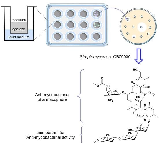
2.1. A Transwell-Based Culture Bioassay to Discover Streptomyces sp. CB09030 with Anti-Mycobacterial Potential

Due to the simplicity of performing a bioassay on the secreted secondary metabolites from Streptomyces strains growing on the solid agar, we cultured 16 Streptomyces species using the liquid medium from the transwell, with Mycobacterium smegmatis MC2 155 as the indicating strain for anti-mycobacterial activity. Among them, S. sp. CB0657, S. sp. CB01580, S. sp. CB02366, and S. sp. CB09030 showed certain inhibitory activity against M. smegmatis MC2 155 (Figure S1A). Further HPLC analyses of the culture extracts of these four strains in 250 mL shaking flasks revealed that S. sp. CB09030 produced more secondary metabolites than the other strains (Figure S1B). Therefore, it was used for a scaled-up fermentation to isolate the potential natural products with significant anti-mycobacterial activities.

2.2. Isolation and Structure Elucidation of 13 from S. sp. CB09030

A large scale fermentation of S. sp. CB09030 (27 L) led to the production and isolation of compounds 13 (Figure 2A). Compound 1 was isolated as a white powder. HR-ESI-MS revealed its molecular formula to be C61H92N2O19, based on the molecular ions of [M + H]+ 1157.6359 (calcd 1157.6370) and [M + Na]+ 1179.6178 (calcd 1179.6190) (Figure S7). The NMR data of 1 are almost identical to LOB A, which was previously isolated from Actinomyces sp. CNB-837, deep-sea derived Streptomcyes sp. SCSIO 01127, and marine-derived Streptomyces sp. MS100061 (Table S1 and Figures S11 and S12) [8,11,19]. Further HR-ESI-MS/MS analyses revealed the fragment ions belonging to the LOB A aglycon ([M + H − H2O]+ 517.2952 calcd 517.2954), the aglycon with A and D sugars ([M + H]+ 883.4941, calcd 883.4935), and the aglycon with sugar D ([M + H]+ 753.4308, calcd 753.4327), which further confirmed that 1 is LOB A.
Compound 2 was isolated as a white powder with a similar UV absorption to 1, suggesting that 2 might have a similar molecular skeleton to 1. HR-ESI-MS analyses revealed its molecular formula to be C61H90N2O21, based on the molecular ions of [M + Na]+ 1209.5921 (calcd 1209.5934) and [M + NH4]+ 1204.6367 (calcd 1204.6380) (Figure S8). Further HR-ESI-MS/MS analyses revealed the fragment ions belonging to the identical LOB A aglycon ([M + H − H2O]+ 517.2945 calcd 517.2954) and the different sugar attachments with the fragment ions of [M + H]+ 913.4707 (calcd 913.4698) and [M + H]+ 783.4075 (calcd 783.4068), respectively. The 1D and 2D NMR spectra of 2 further revealed that 2 is the previously isolated LOB B with an unusual D-kijanose sugar, which was usually isolated with LOB A (Table S2 and Figures S13–S20) [8,11,19].
Compound 3 was also isolated as a white powder. HR-ESI-MS revealed its molecular formula to be C33H44O6, based on the molecular ion of [M − H] 535.3206 (calcd 535.3060) (Figure S9). Its NMR spectra showed that 3 is the previously isolated LOB H8 via the heterologous expression of the LOB BGC (Table S3 and Figures S21–S28) [19].

2.3. Acid Hydrolysis of 2 Leads to the Generation of 4

In order to obtain more LOB derivatives, 2 was further hydrolyzed in 0.5 M-methanolic HCl to produce 4 (Figure 3A). The molecular formula of 4 was deduced to be C42H58N2O12, based on the molecular ion of 783.4076 [M + H]+ (calcd 783.4068) (Figure S10). The structure of 4 was established by analyses of its NMR data (Table S4 and Figures S29–S36) to be the previously isolated hydrolysis product of kijanimicin [28]. Based on the comparison of the 13C NMR spectra of 2, 3, and 4, only the D-kijanose attached to the aglycon in 4 had a characteristic methine signal at δC = 99.0 ppm (Figure 3B). Compound 4 has a hydroxymethyl group attached to its C-22, while a similar LOB derivative, LOB H11, has a methyl group attached to its C-22 from a heterologous host [19].

2.4. Bioinformatic Analysis of A Putative LOB BGC from S. sp. CB09030

The co-production of 13 in S. sp. CB09030 is unique among the isolated LOBs-producing Streptomyces strains, which usually co-produce 1 and 2. This suggests that the transfer of the D-kijanose by the putative glycosyltransferase LobG1 to the aglycon 3 is not efficient in this strain under the current culturing conditions. In order to understand the genetic basis, the whole genome sequencing of S. sp. CB09030 was used to identify the LOB BGC. The antiSMASH analysis of the S. sp. CB09030’s assembled genome sequence revealed that there are dozens of BGCs for the biosynthesis of putative olimycin and xiamycin A, et al. (Table S5, Figure S3). The putative LOB BGC spans a DNA region of 102 kb, which is highly similar to that of the reported LOB BGC from S. sp. SCSIO 01127, with 39 open reading frames (ORFs) (Figure 4A). For example, the five polyketide synthases (PKSs) LobA1–A5 share an ~84% to ~97% sequence identity with those from the LOB BGC in S. sp. SCSIO 01127, which was proposed for the assembly of the linear spirotetronate polyketide chain by utilizing six malonyl-CoA, six methylmalonyl-CoA, and a 3-carbon glycerol unit (Figure 4B) [19,29]. A further analysis of the LOB BGC suggested that it is also highly similar to the LOB BGC in S. olivaceus SCSIO T05 and the putative LOB BGC in S. sp. FXJ 7.023 (Table S6).
A phylogenetic analysis showed that the LobG1 from S. sp. CB09030 is more closely related to the LobG1 in S. sp. FXJ 7.023 than those of S. sp. SCSIO 01127 and S. olivaceus SCSIO T05 (Figure S4). A further amino acid sequence alignment identified several amino acid differences in these glycosyltransferases, among which the LobG1 in S. sp. CB09030 had three unique amino acids, S38T, D250G, and V284M (Figure 4C). The importance of these amino acids for the activity of LobG1 in LOB biosynthesis remains to be established, since it is the essential step to decorating the LOB aglycon.

2.5. The Antibacterial Activity of 14

Although 14 have been separately reported in several previous publications, the availability of these compounds in the current study provides an exciting opportunity to study their antibacterial activity. Compound 2 showed a remarkable antibacterial activity against M. smegmatis MC2 155, with a minimum inhibitory concentration (MIC) of 4 μg/mL, which is comparable to the clinically used antibiotic rifampicin (Table 1 and Figure S5). Interestingly, compounds 1, 3, and 4 only displayed slightly attenuated antibacterial activities against M. smegmatis MC2 155, with MICs of 8 μg/mL. In contrast, 2 had an MIC of 1 μg/mL against B. subtilis 62305, while 1, 3, and 4 only showed weak antibacterial activities against B. subtilis 62305, with MICs of 8, 32, and 32 μg/mL, respectively. These obvious differences are consistent with a previous study of 1 and 2 against M. tuberculosis H37Rv and B. subtilis, which showed MICs of 32 and 16 μg/mL or 12.5 and 1.56 μg/mL, respectively [11,19]. In addition, 14 showed no antibacterial activities against the tested Straptococcus aureus strains.
These data suggest that the oligosaccharide chains in 1 and 2 are critical for the anti-B. subtilis activity, while they are almost negligible for their anti-mycobacterial activities. In addition, the presence of D-kijanose has no effect on the antibacterial activities of 3 and 4 against M. smegmatis MC2 155 or B. subtilis. Since the many sugar moieties present in natural products are responsible for cell recognition, D-kijanose and the oligosaccharide chain in 2 likely play important roles in their interactions with B. subtilis, while the anti-mycobacterial activities of 14 should mainly benefit from the spirotetronate aglycon.

3. Materials and Methods

3.1. A Transwell-Based Culture Bioassay

In total, sixteen Streptomyces strains were used in the current study and were collected and maintained by the National Engineering Research Center of Combinatorial Biosynthesis for Drug Discovery (Changsha, Hunan 410011, PR China). S. sp. CB00316, CB00657, CB01249, CB01580, CB01883, CB01950, CB02009rt, CB02056, CB02058, CB02115, CB02130, CB02366, CB02414, CB02460, CB02488rt, and CB09030 were used. Their spores at ~106 CFU/mL were cultured in the upper chamber of transwell plates (400 μL 0.75% agar medium). Gauze’s medium (G1 medium, 2% soluble starch, 0.1% KNO3, 0.05% K2HPO4, 0.05% MgSO4·7H2O, 0.05% NaCl, and 0.001% FeSO4·7H2O) (1.5 mL, 10% v/v) was added to the lower chamber of the transwell for static fermentation. The fermentation broth (10 μL) was taken periodically (3rd to 27th day) from the lower chamber to measure the antibacterial activities against M. smegmatis MC2 155 with a paper disk assay.

3.2. General Experimental Procedures

The NMR experiments were conducted on a Bruker spectrometer (400 MHz–600 MHz) and the chemical shifts were reported in ppm, relative to CD3OD (δH = 3.31 ppm) for 1H NMR and CD3OD (δC = 49.03 ppm) for the 13C NMR spectroscopy. Column chromatography (CC) was carried out on silica gel (Yantai Jiangyou Silica Gel Development Co., Ltd., Yantai, China). Semipreparative reversed phase-high-performance liquid chromatography (RP—HPLC) was performed using a Waters 1525 Binary HPLC Pump equipped with a Waters 2489 UV/Visible detector, using a Welch Ultimate AQ—C18 column (250 × 10 mm, 5 μm).

3.3. Streptomyces Fermentation in Shaking Flasks and Extraction

The Streptomyces spores of S. sp. CB0657, S. sp. CB001580, S. sp. CB02366, and S. sp. CB09030 were inoculated into 250 mL shaking flasks that contained 50 mL of TSB liquid medium, which was next cultured for 48 h (30 °C, 200 rpm). The obtained seed cultures were inoculated into 2 L shaking flasks containing 500 mL of G1 medium (containing 3% macroporous resin HP—20) and grown for 7 days (30 °C, 200 rpm). The fermentation cultures were centrifuged (4000 rpm, 10 min) to collect the mycelia and resins.

3.4. Isolation and Acid Hydrolysis of 2

The mycelia cakes and resin were extracted with MeOH (3 L × 3). A large-scale fermentation (27 L) of the S. sp. CB09030 led to the isolation of 14. The fatty acid compounds in the crude extract were removed with an n-hexane extraction. The methanol layer was further concentrated under reduced pressure to obtain the crude extract (20 g). The crude extract was subjected to silica gel CC using DCM-EtOAc-MeOH (1:0:0, 9:1:0, 7:3:0, 5:1:0, 3:1:0, 1:1:0, 0:1:0, 0:10:1, 0:9:1, and 0:1:1) to provide eighteen fractions (Fr.1–18). Fr.16 (2.0 g) was separated by silica gel CC eluted with DCM-MeOH (20:1, 15:1, 10:1, 5:1, and 1:1) to yield six subfractions (Fr.16-1–Fr.16-6). Fr.16-1 was further purified by semipreparative RP-HPLC with a flow rate of 3 mL/min and a gradient elution of CH3CN/H2O (containing 0.1% formic acid) for 25 min (10–95% CH3CN for 12 min, followed by 95% for 3 min, and 95–5% for 2 min, followed by 5% for 8 min) to afford compound 1 (9.0 mg). Fr.16-4 was further purified by semipreparative RP-HPLC to afford compounds 2 (22.2 mg) and 3 (3.8 mg). Compound 2 (9.0 mg) was hydrolyzed in 128 µL of 0.5 M-methanolic HCl at 25 °C for 18 h. The reaction system was concentrated by rotary evaporation and then dissolved with an appropriate amount of methanol. Finally, the hydrolytic product 4 (3.4 mg) was separated by semipreparative RP-HPLC.

3.5. Genome Sequencing and Analysis

S. sp. CB09030 was cultured at 30 °C for 48 h in 50 mL of TSB liquid medium, which contained 0.5% glycine for genomic DNA. A total of 8.4 Mb was generated by Biomarker Technologies (Beijing, China). The biosynthetic gene clusters were predicted using antiSMASH version 6.0.1. In addition, the sequence homology searches method (Blastp) was also used to identify the genes related to biosynthesis. The LOB BGC was confirmed to be in region 22 by a comparison with the BGC in S. sp. SCSIO 01127.

3.6. Antibacterial Assay

The antibacterial activities of compounds 14 against M. smegmatis MC2 155 and B. subtilis 62305 were evaluated using a broth dilution assay [30]. The individual compounds were dissolved and diluted with MeOH to obtain the initial concentrations. The positive control antibiotics, including rifampin and ampicillin, were diluted with DMSO to obtain the initial concentrations. Bacillus subtilis 62305 was grown in an LB medium overnight until the OD600 reached 0.5–0.6, which were then diluted to 106 CFU/mL. First, the tested compounds or positive controls (100 μL) were mixed with a bacterial solution (100 μL) and stepwise diluted. The plates were incubated at 37 °C overnight. Finally, 50 μL of resazurin was added into each well to visualize the results. Similarly, M. smegmatis MC2 155 was cultured at 37 °C for 36 h and diluted to 106 CFU/mL in a Nutrient-Bertani broth (1% tryptone, 0.3% beef extract powder, 0.5% NaCl, and 0.05% Tween-80) at 37 °C for 36 h.

3.7. In Vitro Cytotoxicity

The cytotoxicity of 1 was evaluated by an MTT assay (Figure S6). In brief, the two cell lines were seeded at a density of ~5000 cells per well in 96-well plates. After 24–72 h, the cells were treated with different concentrations of the tested compounds. After further incubation for 72 h, the cell survival was determined by an addition of Cell Counting Kit-8 solution (10 μL/well). The absorbance was measured at 450 nm.

4. Conclusions

In this study, we established a transwell bioassay to discover LOB A (1), LOB B (2), and LOB H8 (3) from S. sp. CB09030, while a bioinformatic analysis revealed the putative LOB BGC in S. sp. CB09030, with a slightly different glycosyltransferase to the previously reported LobG1. LOB H8 (3) was directly isolated from the S. sp. CB09030 wild-type strain, which was previously obtained only through its heterologous expression in S. pactum SCSIO 02999. Our study also established, for the first time, the antibacterial activities of 4 against M. smegmatis and B. subtilis, which allow us to study the structure–activity relationships of 14. The oligosaccharide moieties in 1 and 2 are essential for the anti-B. subtilis activity, while the spirotetronate aglycon is more important in the observed anti-mycobacterial activity. Since both 1 and 2 exhibited stronger anti-inflammatory activities than the clinically used indomethacin, by reducing the edema by 84–86% in the phorbol-myristate-acetate-induced mouse ear mode [8], these compounds may hold great promise against M. ulcerans, since they have both anti-inflammatory and anti-mycobacterial activities. The evaluation of the anti-inflammatory activities of 3 and 4 and a mechanistic study on this anti-inflammatory activity will be reported in due course.

Supplementary Materials

The following supporting information can be downloaded at: https://www.mdpi.com/article/10.3390/molecules28083597/s1, The original spectral data (Figures S1–S36 and Tables S1–S7).

Author Contributions

Conceptualization, data acquisition, formal analysis, writing—original draft preparation, J.S.; formal analysis, writing—review and editing, D.P., F.-F.P. and Q.-B.Z.; conceptualization, funding acquisition, Y.-W.D.; conceptualization, formal analysis, funding acquisition, writing—original draft, writing—review and editing, Y.H. All authors have read and agreed to the published version of the manuscript.

Funding

This research was funded by NSFC grants 82173688 (to Y.H.); Hunan Provincial Innovation Foundation for Postgraduate CX20210342 (to J.S.) and Fundamental Research Funds for the Central Universities of Central South University (CSU) 2021zzts0331 (to J.S.).

Institutional Review Board Statement

Not applicable.

Data Availability Statement

Data generated in the process of this research are available in the Supplementary Materials.

Acknowledgments

We thank the Center for Advanced Research in Central South University for NMR experiments.

Conflicts of Interest

The authors declare no conflict of interest.

References

  1. Kang, M.J.; Jones, B.D.; Mandel, A.L.; Hammons, J.C.; DiPasquale, A.G.; Rheingold, A.L.; La Clair, J.J.; Burkart, M.D. Isolation, structure elucidation, and antitumor activity of spirohexenolides A and B. J. Org. Chem. 2009, 74, 9054–9061. [Google Scholar] [CrossRef]
  2. Imai, H.S.; Suzuki, K.I.; Morioka, M.; Numasaki, Y.; Kadota, S.; Nagai, K.; Iwanami, M.; Saito, T. Okilactomycin, a novel antibiotic produced by a Streptomyces species I. Taxonomy, fermentation, isolation and characterization. J. Antibiot. 1987, 40, 1475–1482. [Google Scholar] [CrossRef]
  3. Nakai, R.; Kakita, S.; Asai, A.; Chiba, S.; Akinaga, S.; Mizukami, T.; Yamashita, Y. Chrolactomycin, a novel antitumor antibiotic produced by Streptomyces sp. J. Antibiot. 2001, 54, 836–839. [Google Scholar] [CrossRef]
  4. Nakai, R.; Ishida, H.; Asai, A.; Ogawa, H.; Yamamoto, Y.; Kawasaki, H.; Akinaga, S.; Mizukami, T.; Yamashita, Y. Telomerase inhibitors identified by a forward chemical genetics approach using a yeast strain with shortened telomere length. Cell Chem. Biol. 2006, 13, 183–190. [Google Scholar] [CrossRef] [PubMed]
  5. Waitz, J.A.; Horan, A.C.; Kalyanpur, M.; Lee, B.K.; Loebenberg, D.; Marquez, J.A.; Miller, G.; Patel, M.G. Kijanimicin (sch 25663), a novel antibiotic produced by actinomad ura kijaniata scc 1256. J. Antibiot. 1981, 34, 1101–1106. [Google Scholar] [CrossRef]
  6. Ashton, R.J.; Kenig, M.D.; Luk, K.; Planterose, D.N.; Scott-Wood, G. MM 46115, a new antiviral antibiotic from Actinomadura pelletieri. J. Antibiot. 1990, 43, 1387–1393. [Google Scholar] [CrossRef]
  7. Bister, B.; Bischoff, D.; Strobele, M.; Riedlinger, J.; Reicke, A.; Wolter, F.; Bull, A.T.; Zahner, H.; Fiedler, H.P.; Sussmuth, R.D. Abyssomicin C—A polycyclic antibiotic from a marine Verrucosispora strain as an inhibitor of the p-aminobenzoic acid/tetrahydrofolate biosynthesis pathway. Angew. Chem. Int. Ed. 2004, 43, 2574–2576. [Google Scholar] [CrossRef] [PubMed]
  8. Jiang, Z.D.; Jensen, P.R.; Fenical, W. Lobophorins A and B, new antiinflammatory macrolides produced by a tropical marine bacterium. Bioorg. Med. Chem. Lett. 1999, 9, 2003–2006. [Google Scholar] [CrossRef]
  9. Niu, S.W.; Li, S.M.; Chen, Y.C.; Tian, X.P.; Zhang, H.B.; Zhang, G.T.; Zhang, W.M.; Yang, X.H.; Zhang, S.; Ju, J.H. Lobophorins E and F, new spirotetronate antibiotics from a South China Sea-derived Streptomyces sp. SCSIO 01127. J. Antibiot. 2011, 64, 711–716. [Google Scholar] [CrossRef]
  10. Wei, R.B.; Xi, T.; Li, J.; Wang, P.; Li, F.C.; Lin, Y.C.; Qin, S. Lobophorin C and D, new kijanimicin derivatives from a marine sponge-associated actinomycetal strain AZS17. Mar. Drugs 2011, 9, 359–368. [Google Scholar] [CrossRef] [PubMed]
  11. Chen, C.X.; Wang, J.; Guo, H.; Hou, W.Y.; Yang, N.; Ren, B.; Liu, M.; Dai, H.Q.; Liu, X.T.; Song, F.H.; et al. Three antimycobacterial metabolites identified from a marine-derived Streptomyces sp. MS100061. Appl. Microbiol. Biotechnol. 2013, 97, 3885–3892. [Google Scholar] [CrossRef]
  12. Pan, H.Q.; Zhang, S.Y.; Wang, N.; Li, Z.L.; Hua, H.M.; Hu, J.C.; Wang, S.J. New spirotetronate antibiotics, lobophorins H and I, from a south China sea-derived Streptomyces sp. 12A35. Mar. Drugs 2013, 11, 3891–3901. [Google Scholar] [CrossRef] [PubMed]
  13. Lin, Z.J.; Koch, M.; Pond, C.D.; Mabeza, G.; Seronay, R.A.; Concepcion, G.P.; Barrows, L.R.; Olivera, B.M.; Schmidt, E.W. Structure and activity of lobophorins from a turrid mollusk-associated Streptomyces sp. J. Antibiot. 2014, 67, 121–126. [Google Scholar] [CrossRef]
  14. Song, C.F.; Pan, H.Q.; Hu, J.C. Isolation and identification of a new antibiotic, lobophorin J, from a deep sea-derived Streptomyces sp. 12A35. Chin. J. Antibiot. 2015, 40, 721–727. [Google Scholar]
  15. Brana, A.F.; Sarmiento-Vizcaino, A.; Osset, M.; Perez-Victoria, I.; Martin, J.; de Pedro, N.; de la Cruz, M.; Diaz, C.; Vicente, F.; Reyes, F.; et al. Lobophorin K, a new natural product with cytotoxic activity produced by Streptomyces sp. M-207 associated with the deep-sea coral lophelia pertusa. Mar. Drugs 2017, 15, 144. [Google Scholar] [CrossRef]
  16. Luo, M.H.; Tang, L.J.; Dong, Y.L.; Huang, H.B.; Deng, Z.X.; Sun, Y.H. Antibacterial natural products lobophorin L and M from the marine-derived Streptomyces sp. 4506. Nat. Prod. Res. 2020, 35, 5581–5587. [Google Scholar] [CrossRef] [PubMed]
  17. Cruz, P.G.; Fribley, A.M.; Miller, J.R.; Larsen, M.J.; Schultz, P.J.; Jacob, R.T.; Tamayo-Castillo, G.; Kaufman, R.J.; Sherman, D.H. Novel lobophorins inhibit oral cancer cell growth and induce Atf4- and chop-dependent cell death in murine fibroblasts. ACS Med. Chem. Lett. 2015, 6, 877–881. [Google Scholar] [CrossRef] [PubMed]
  18. Zhang, C.Y.; Ding, W.J.; Qin, X.J.; Ju, J.H. Genome sequencing of Streptomyces olivaceus SCSIO T05 and activated production of lobophorin CR4 via metabolic engineering and genome mining. Mar. Drugs 2019, 17, 593. [Google Scholar] [CrossRef]
  19. Tan, B.; Chen, S.; Zhang, Q.; Chen, Y.; Zhu, Y.; Khan, I.; Zhang, W.; Zhang, C. Heterologous expression leads to discovery of diversified lobophorin analogues and a flexible glycosyltransferase. Org. Lett. 2020, 22, 1062–1066. [Google Scholar] [CrossRef]
  20. Tan, B.; Zhang, Q.B.; Xiao, J.; Yuan, C.S.; Chen, Y.C.; Chen, S.Q.; Zhang, W.M.; Zhu, Y.G.; Zhang, C.S. Characterization of the P450 monooxygenase LobP1 as C-32 hydroxylase in lobophorin biosynthesis. Chin. J. Chem. 2022, 40. [Google Scholar] [CrossRef]
  21. Seyedsayamdost, M.R.; Stallforth, P. Special issue in honor of professor Jon Clardy. J. Nat. Prod. 2020, 83, 565–568. [Google Scholar] [CrossRef] [PubMed]
  22. Pishchany, G.; Mevers, E.; Ndousse-Fetter, S.; Horvath, D.J.; Paludo, C.R.; Silva-Junior, E.A.; Koren, S.; Skaar, E.P.; Clardy, J.; Kolter, R. Amycomicin is a potent and specific antibiotic discovered with a targeted interaction screen. Proc. Natl. Acad. Sci. USA 2018, 115, 10124–10129. [Google Scholar] [CrossRef] [PubMed]
  23. Bae, M.; Mevers, E.; Pishchany, G.; Whaley, S.G.; Charles, O.R.; Andes, D.R.; Currie, C.R.; Pupo, M.T.; Clardy, J. Chemical exchanges between multilateral symbionts. Org. Lett. 2021, 23, 1648–1652. [Google Scholar] [CrossRef] [PubMed]
  24. George, K.M.; Chatterjee, D.; Gunawardana, G.; Welty, D.; Hayman, J.; Lee, R.; Small, P.L.C. Mycolactone: A polyketide toxin from Mycobacterium ulcerans required for virulence. Science 1999, 283, 854–857. [Google Scholar] [CrossRef]
  25. Chany, A.C.; Tresse, C.; Casarotto, V.; Blanchard, N. History, biology and chemistry of Mycobacterium ulcerans infections (Buruli ulcer disease). Nat. Prod. Rep. 2013, 30, 1527–1567. [Google Scholar] [CrossRef]
  26. Silva, M.T.; Portaels, F.; Pedrosa, J. Pathogenetic mechanisms of the intracellular parasite Mycobacterium ulcerans leading to Buruli ulcer. Lancet 2009, 9, 696–710. [Google Scholar] [CrossRef]
  27. Vale, A.D.; Cabanes, D.; Sousa, S. Bacterial toxins as pathogen weapons against phagocytes. Front. Microbiol. 2016, 7, 42. [Google Scholar] [PubMed]
  28. Mallams, A.K.; Puar, M.S.; Rossman, R.R.; McPhail, A.T.; Macfarlane, R.D.; Stephens, R.L. Kijanimicin. Part 3. structure and absolute stereochemistry of 1983. J. Chem. Soc. 1983, 1, 1497–1534. [Google Scholar]
  29. Li, S.M.; Xiao, J.; Zhu, Y.G.; Zhang, G.T.; Yang, C.F.; Zhang, H.B.; Ma, L.; Zhang, C.S. Dissecting glycosylation steps in lobophorin biosynthesis implies an iterative glycosyltransferase. Org. Lett. 2013, 15, 1374–1377. [Google Scholar] [CrossRef] [PubMed]
  30. Wiegand, I.; Hilpert, K.; Hancock, R.E. Agar and broth dilution methods to determine the minimal inhibitory concentration (MIC) of antimicrobial substances. Nat. Protoc. 2008, 3, 163–175. [Google Scholar] [CrossRef]
Figure 1. Selected representative spirotetronates and LOB derivatives.
Molecules 28 03597 g001
Figure 2. HPLC of the crude extract and chemical structure of the isolated LOBs 13. (A) HPLC analysis of S. sp. CB09030 culture extract. (B) The structures of isolated 1 (LOB A), 2 (LOB B), and 3 (LOB H8) from S. sp. CB09030.
Molecules 28 03597 g002
Figure 3. Generation of 4 from LOB B (2). (A) The hydrolysis of 2 in 0.5 M-methanolic HCl. (B) The comparison of 13C NMR spectra of 4 to 2 and 3 in CD3OD. ◾: The carbon signal (δC = 100.7 ppm) from sugar C; ▼: The carbon signal (δC = 99.7 ppm) from sugar A; ★: The carbon signal (δC = 99.0 ppm) from D-kijanose; and ●: The carbon signal (δC = 93.1 ppm) from sugar B.
Molecules 28 03597 g003
Figure 4. Bioinformatic analysis of LOB BGC in S. sp. CB09030. (A) Genetic organization of LOB BGC in S. sp. CB09030. (B) Proposed biosynthesis for 13. (C) Sequence alignment of LobG1 from S. sp. CB09030, S. sp. SCSIO 01127, S. olivaceus SCSIO T05, and S. sp. FXJ 7.023 involved in the transferase of D-kijanose, with the varying amino acids boxed.
Molecules 28 03597 g004
Table 1. The antimicrobial activities of 14 (MIC, µg/mL).
CompoundsM. smegmatis MC2 155B. subtilis 62305
188
241
3832
4832
ampicillin-0.5
rifampicin2-
-: No cytotoxicity observed.
Disclaimer/Publisher’s Note: The statements, opinions and data contained in all publications are solely those of the individual author(s) and contributor(s) and not of MDPI and/or the editor(s). MDPI and/or the editor(s) disclaim responsibility for any injury to people or property resulting from any ideas, methods, instructions or products referred to in the content.

