Article

The Microwave Rotational Electric Resonance (RER) Spectrum of Benzothiazole

1
Laboratory of Spectroscopy, Molecular Modeling, Materials, Nanomaterials, Water, and Environment, Faculty of Sciences, Mohammed V University, Av Ibn Battouta, Rabat B.P. 1014, Morocco
2
Univ Paris Est Creteil and Université Paris Cité, CNRS, LISA, 94010 Créteil, France
3
Université Paris Cité and Univ Paris Est Creteil, CNRS, LISA, 75013 Paris, France
4
Institut Universitaire de France (IUF), 75231 Paris, France
*
Authors to whom correspondence should be addressed.
Molecules 2023, 28(8), 3419; https://doi.org/10.3390/molecules28083419
Submission received: 14 March 2023 / Revised: 4 April 2023 / Accepted: 6 April 2023 / Published: 13 April 2023
(This article belongs to the Section Molecular Structure)

Abstract

:
The microwave spectra of benzothiazole were measured in the frequency range 2–26.5 GHz using a pulsed molecular jet Fourier transform microwave spectrometer. Hyperfine splittings arising from the quadrupole coupling of the 14N nucleus were fully resolved and analyzed simultaneously with the rotational frequencies. In total, 194 and 92 hyperfine components of the main species and the 34S isotopologue, respectively, were measured and fitted to measurement accuracy using a semi-rigid rotor model supplemented by a Hamiltonian accounting for the 14N nuclear quadrupole coupling effect. Highly accurate rotational constants, centrifugal distortion constants, and 14N nuclear quadrupole coupling constants were deduced. A large number of method and basis set combinations were used to optimize the molecular geometry of benzothiazole, and the calculated rotational constants were compared with the experimentally determined constants in the course of a benchmarking effort. The similar value of the χcc quadrupole coupling constant when compared to other thiazole derivatives indicates only very small changes of the electronic environment at the nitrogen nucleus in these compounds. The small negative inertial defect of −0.056 uÅ2 hints that low-frequency out-of-plane vibrations are present in benzothiazole, similar to the observation for some other planar aromatic molecules.

1. Introduction

Benzothiazole (BTA), see Figure 1, is a planar bicyclic compound in which a benzene ring is fused to a thiazole ring. It is a compound with wide industrial applications, such as in the synthetic rubber vulcanization, which exhibits an acute toxicity. BTA is also a respiratory irritant and dermal sensitizer [1]. Its derivative, 2-mercaptobenzothiazole, has been reported to be a rodent carcinogen [1]. BTA may volatilize from crumb rubber, resulting in inhalation exposure [2]. It is also present in clothing textiles, which are a potential source of environmental pollutants [3]. At the same time, its derivatives have attracted continuous interest due to their various biological activities, such as anticancer [4], antimicrobial [5], antimalarial [6], anti-inflammatory [7], and antidiabetic [8] activities. BTA derivatives were also evaluated as prospective diagnostic agents for amyloid binding in neurodegenerative diseases [9,10] and as histamine H3 antagonists [11]. The wide range of biological effects associated with this scaffold renders the structure of the core molecule BTA to be important and interesting. Among many molecular spectroscopic techniques, Fourier transform microwave spectroscopy is particularly useful for gas phase structural studies at the molecular level, allowing the determination of accurate geometry parameters without perturbation from matrix or solvent environments. The molecular structure, i.e., the mass distribution of atoms in the molecules, is directly linked to the microwave transition frequencies through the rotational constants.
Coupled with supersonic expansion through a pulsed nozzle with an inert carriergas such as helium, neon, or argon, a cooling of the vibrational and rotational states down to a rotational temperature of about 2 K is produced by the strong collisional regime during the adiabatic expansion, making only the rotational transitions in the vibrational ground state observable. The sensitivity of a Fourier transform microwave setup enables also the detection of minor isotopologues, such as 34S and 13C, in some cases, even 15N and 18O, in natural abundances of about 4.2%, 1.1%, 0.35%, and 0.21%, respectively. Since different isotopologues feature different total molecular masses, changes in isotopic substitution lead to different mass distributions and, as a consequence, distinct rotational constants. If the spectra of such minor isotopologues are available, the deduced rotational constants can be used to determine experimental atomic positions, bond lengths, and bond angles.
Nowadays, Fourier transform microwave spectroscopy is often integrated with quantum chemical calculations that serve as a powerful support for the spectral assignment. They deliver predicted values of the rotational constants to guide the assignment. For medium-sized or large molecules, or in cases where a sufficient number of minor isotopologue spectra cannot be observed, leading to the lack of an experimental microwave structure determination, indirect information on the molecular geometry through the rotational constants can be obtained by comparing them with the predicted ones. In the previous decades, not only more computational resources have become available during the information revolution, but also the accuracy of quantum chemical calculations has been significantly increased, and is still improving continuously with newer and more elaborate theoretical conceptualizations. Several benchmarking studies involving tight collaborations between microwave spectroscopic and theoretical groups have been successfully established, aiming to predict the experimental rotational constants with even higher accuracy [12,13,14,15,16,17]. On the one hand, this joint-venture allows experimentalists to efficiently assign the microwave spectra and to access detailed molecular geometries in the cases when minor isotopologue spectra are lacking. On the other hand, highly accurate constants obtained from the experiments can be utilized in return to benchmark results from quantum chemistry, helping theoreticians to identify methodological bottlenecks and limitations, and to push forward new methodology developments.
Using the combination of microwave spectroscopy and quantum chemical calculations, not only precise information about the molecular structures can be obtained. If an atom with a nuclear quadrupole moment is present in the molecule, information on the molecular electron density at this atom is also accessible. In a classical and idealized model, the nucleus of an atom is viewed as a monopole with a homogeneous charge distribution in a spherical symmetry. This is correct for nuclei with a spin of 0 and ½. For spins equal or higher than the absolute value of 1, the charge distribution is asymmetric in the shape of an ellipsoid, which creates an electric field gradient at the nucleus. BTA features a 14N nucleus with a nuclear spin I = 1. The nuclear quadrupole coupling with the molecular angular momentum J occurs for all nuclei with a nuclear spin I > ½. It is an interaction between the nuclear quadrupole moment arising from the non-spherical distribution of a nuclear charge and the electronic field gradient arising from the non-spherical distribution of the electronic charge around the nucleus. Although the effects of the molecular rotation towards the electric field gradients of the nuclei are minor, it is sufficient to result in hyperfine splittings of the rotational levels observable in the high-resolution microwave spectrum from which the nuclear quadrupole coupling constants can be determined. By comparing their values with those obtained for other thiazole derivatives, we can estimate the influence of substituents on the nuclear quadrupole coupling constants.
We present here the experimental results from a microwave spectroscopic investigation on BTA which aims at determining and understanding its molecular structure and the 14N nuclear quadrupole coupling effects. State-of-the-art quantum chemical calculations with an extensive benchmarking of several ab initio and density functional theory methods as well as Pople and Dunning basis sets were performed to support the experimental finding.

2. Quantum Chemical Calculations

2.1. Geometry Optimizations

Similar to thiazole and benzene, BTA possesses a planar, aromatic structure due to the delocalization of the π-electrons. Therefore, a single conformer is possible, as illustrated in Figure 1. To obtain the molecular geometry of BTA and to benchmark the rotational constants, full geometry optimizations were performed with the Gaussian 16 package [18] using various combinations of methods and basis sets in a systematic way. We employed two ab initio methods MP2 [19] and coupled cluster single and double excitation CCSD [20] as well as several exchange–correlation functionals from density functional theory method, such as B3LYP [21,22], Truhlar’s M06-2X [23], Head-Gordon’s ωB97X-D [24], Perdew–Burke–Ernzerhof PBE [25], and Yu’s Minnesota MN15 [26]. Furthermore, the B3LYP functional was combined with D3 [27], Becke–Johnson damping (D3BJ) [28], and with the Coulomb attenuation method (CAM-B3LYP) [29]. A total of 21 basis sets have been associated, such as the Pople’s split-valence double-zeta 6-31G and split-valence triple-zeta 6-311G basis sets [30] incorporating or not diffuse functions (+ or ++). Polarization functions of the types (d,p), (df,dp), (2d,2p), (2df,2pd), and (3df,3dp) are always included. In addition, Dunning’s double zeta polarization (cc-pVDZ) and triple zeta polarization (cc-pVTZ) basis sets are used [31] with or without diffuse functions (aug). Note that the basis set variation is not performed with the computationally expensive CCSD method, and we retain here with only the cc-pVDZ basis set. Vibrational harmonic frequencies were calculated on the optimized geometries to verify whether the stationary point is a saddle point or a true minimum geometry. The obtained rotational constants are presented in Table S1 of the Supplementary Materials. A selection of the results at three levels of theory, MP2/6-311++G(d,p), MP2/6-31G(d,p), and B3LYP-D3BJ/6-311++G(d,p) can be found in Table 1. The molecular structure optimized at the MP2/6-311++G(d,p) level is shown in Figure 1; the atomic Cartesian coordinates of this structure are available in Table S2.

2.2. 14N Nuclear Quadrupole Coupling Constants

The relation between the components of the electric field gradient tensor qij (i, j = a, b, c) with those of the nuclear quadrupole coupling tensor χij is described by the following equation:
χij = (eQ/h) · qij
where e is the fundamental electric charge, Q is the electric quadrupole moment of the nucleus, and h is the Planck’s constant. Calculated nuclear quadrupole coupling constants can be obtained directly from geometry optimizations, as shown exemplarily for the diagonal elements χaa, χbb, and χcc at three levels of theory in Table 1.
Several studies have indicated that the cost-efficient, semi-experimental method proposed by Bailey [32] for the calculations of 14N nuclear quadrupole coupling constants is reliable, especially for molecules containing conjugated double bonds [33,34,35,36,37,38]. Therefore, we applied this method to calculate the nuclear quadrupole coupling constants of BTA. With Bailey’s method, experimental data are calibrated at a chosen level of molecular orbital calculation. The experimental nuclear quadrupole coupling constants χij of 150 molecules are plotted against the electric field gradients qij, showing a clear linear regression where the slope, eQ/h (see Equation (1), is 4.599(12) MHz/a.u. For BTA, after the molecular geometry had been optimized at a given level of theory (the MP2/6-311++G(d,p) level is often chosen, but other levels can be used also), the electric field gradient tensor at the 14N nucleus site was calculated at the B3PW91/6-311+G(d,p) level, which is recommended for modeling π-electron conjugate systems [38]. The quadrupole coupling tensor is directly proportional to the electric field gradient tensor by the calibration factor eQ/h = −4.599 MHz/a.u. determined from the linear regression. The nuclear quadrupole coupling constant values obtained with this method are χaa = 1.727 MHz, χbb = −4.036 MHz, χcc = 2.309 MHz, χab = 0.078 MHz, χac = 0.000 MHz, and χbc = 0.000 MHz. Note that for a planar molecule, the values of χac and χbc should be zero.

3. Microwave Spectroscopy

The microwave spectra of BTA were measured using a molecular jet Fourier transform microwave spectrometer with a Fabry–Perot type resonator [39]. It operates between 2–26.5 GHz and is a modified version of that described in Ref. [40]. Therefore, details of the spectrometer will not be repeated here. BTA was purchased from Lancaster at a purity of 97% and was used as received. The sample was placed into a heated reservoir [41] at a temperature of 150 °C under helium gas stream. The total helium pressure was set to 2 bar. The helium-BTA mixture was expanded into the cavity at a rotational temperature of 2 K through a pulsed nozzle which reached a temperature of 140 °C. During the adiabatic expansion, the Doppler line and collisional broadening are simultaneously reduced by velocity equilibration, inducing strong rovibrational cooling. As a consequence, only the lowest rovibrational levels are populated which simplifies considerably the observed spectra. All spectra are measured with reference to a GPS disciplined rubidium frequency standard. The experimental uncertainty of the instrument is estimated to be 2 kHz for unblended lines [42].
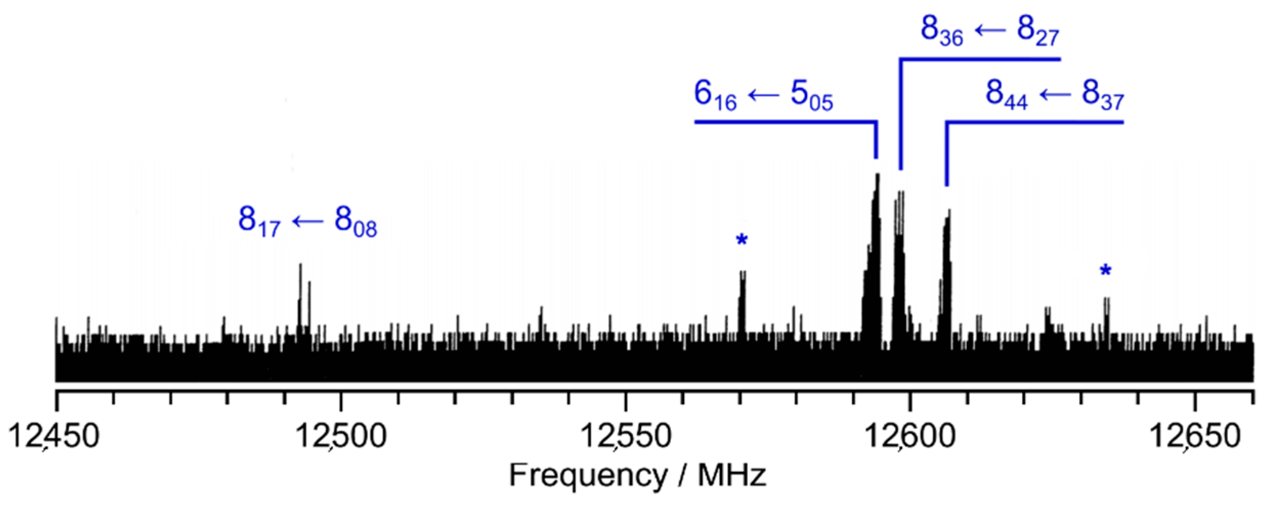
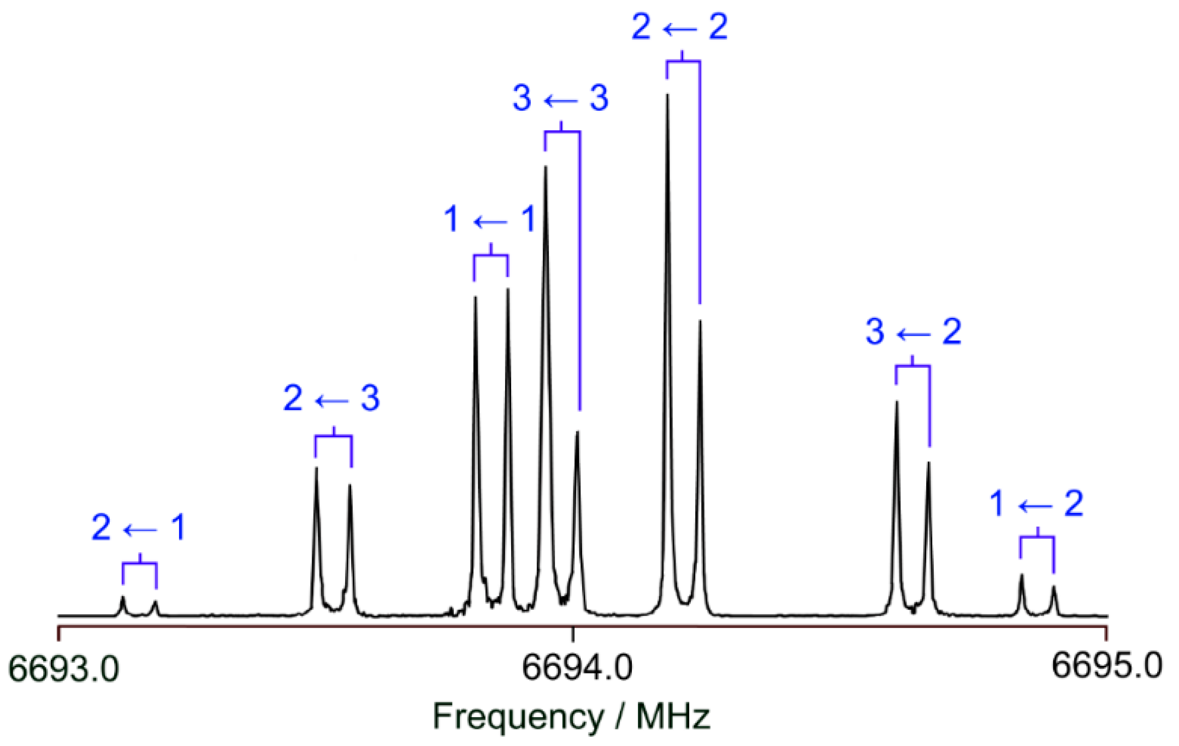
At the beginning, a survey spectrum was recorded. A portion of the spectrum in the frequency range from 12,450 to 12,660 MHz is illustrated Figure 2. High-resolution measurements were recorded for each signal found in the survey spectrum to determine the exact frequencies. Due to the coaxial arrangement of the resonators and the molecular beam, all rotational transitions appear as doublets arising from the Doppler effect. The expected hyperfine splittings due to the 14N nuclear quadrupole coupling could be observed. Due to this hyperfine structure, most rotational transitions appear as a triplet (the three strongest F-components). For some transitions, i.e., the transition shown in Figure 3, it was possible to measure all theoretically possible quadrupole F-components. A total of 194 lines with J ≤ 12 and Ka ≤ 4 were measured and fitted using the program XIAM [43] to precisely determine the rotational constants A, B, and C, the quartic centrifugal distortion constants in the A-reduced form ΔJ, ΔK, ΔJK, δJ, and δK of Watson’s asymmetric rotor Hamiltonian, as well as the diagonal elements χaa, χbb, and χcc of the nuclear quadrupole coupling tensor of the 14N nucleus. The off-diagonal quadrupole coupling constants could not be determined, which is often the case for 14N nuclear quadrupole coupling due to the relatively small quadrupole moment of the 14N nucleus.
To obtain information on the structure of BTA, we first searched for the spectra of the 34S isotopologue in natural abundance of about 4%. The search was straightforward and we could measure and fit the frequencies of 92 transitions also using the rotational constants A, B, and C, the quartic centrifugal distortion constants ΔJ, ΔK, ΔJK, δJ, and δK, and the nuclear quadrupole coupling constants χaa, χbb, and χcc of the 14N nucleus. The obtained parameters are summarized in Table 2 along with those of the parent species fit. The assigned frequencies along with their residues are listed in Table S3 in the Supplementary Materials for both the parent and the 34S species. We then searched for the spectra of the 13C isotopologues but their transitions were too weak to be observed. Because the natural abundance of 15N is even lower than that of 13C, the spectrum of the 15N isotopologue should be even weaker and, therefore, was not searched.

4. Results and Discussion

The XIAM fit containing 194 hyperfine components of 52 rotational transitions reached a standard deviation of 1.6 kHz, which is within the experimental accuracy of 2 kHz of the spectrometer in use. All fitted molecular parameters such as the rotational constants A, B, and C, the quartic centrifugal distortion constants ΔJ, ΔK, ΔJK, δJ, and δK, and the 14N nuclear quadrupole coupling constants χaa, χbb, and χcc are determined with high accuracy. The experimentally deduced values are close to the calculated ones. The quartic centrifugal distortion constants are extremely well predicted, as can be seen in Table 2. This is probably due to the rigidity of the aromatic ring. Bailey’s method using the electric field gradient calibration at the B3PW91/6-311+G(d,p) level maintains its reliability in calculating nuclear quadrupole coupling constant values for aromatic ring containing molecules. The χaa, χbb, and χcc values obtained with this method are significantly closer to the experimental values than those calculated directly from geometry optimizations shown in Table 1. For the rotational constants, the values obtained from the equilibrium structure Ae, Be, and Ce are closer to the experimental values A0, B0, and C0 obtained at the vibrational ground state than the calculated A0, B0, and C0 values from anharmonic frequency calculations (see Table 1). This is most probably due to error compensation. From theoretical perspective, calculated ground state rotational constants should be more accurate, and it is not physically meaningful to compare experimental A0, B0, and C0 constants with calculated Ae, Be, and Ce constants. Practically, cost-efficient geometry optimizations compared to resource-intense anharmonic frequency calculations, in combination with accidental accuracy from error compensation, renders the use of Ae, Be, and Ce constants to be helpful in assigning microwave spectra. With the continuous increase in computational capacity, benchmarking has become a core activity in the development of quantum chemical methods. A number of structure optimizations with different methods and basis sets described in Section 2.1. were performed for benchmarking purposes by comparing the calculated rotational constants Ae, Be, and Ce of BTA to the accurately determined experimental constants A0 = 3174.630260(98) MHz, B0 = 1341.764826(67) MHz, and C0 = 943.232720(28) MHz. We found that for all levels of theory, the calculated equilibrium rotational constants are quite close to those obtained experimentally with a deviation of about 1% or less. Although some levels performed better than some others, there is no clue to conclude a “golden level” for BTA. We note that the three levels mentioned in Table 1 all yielded good results, and among them the MP2/6-31G(d,p) level performed best, as it did in the cases of many other molecules containing aromatic rings such as coumarin [44], quinoline and isoquinoline [45], 2-ethylfuran [46], o-methylanisole [47], and a series of acetylmethylthiophene [13,48,49].
Though the number of lines assigned for the 34S isotopologue is only half as many as for the parent species, the 34S fit is also very satisfactory with standard deviation close to the measurement accuracy of 2 kHz [42] and the fitted parameters are well determined. Since only rotational constants of the 34S isotopologue are available, a complete structure determination cannot be performed but the position of the sulfur atom can be calculated utilizing Kraitchman’s equations [50] with errors estimated with Costain’s rule [51]. The sulfur coordinates with respect to the center of mass are |a0| = 1.74823(86) Å, |b0| = 0.9055(17) Å, and c = 0 Å (fixed value due to molecular symmetry), which are in very good agreement with the values obtained from the geometry calculated at the MP2/6-31G(d,p) level (ae = 1.757 Å, be = 0.903 Å, and ce = 0.000 Å, see Table S2 of the Supplementary Materials). The signs of the coordinates cannot be obtained from Kraitchman’s equations and should be taken from ab initio according to the axis orientations shown in Figure 1. The very small differences between the experimental and the theoretical coordinates can be, again, explained by the fact that the experimental values a0 and b0 refer to the vibrational ground state and the theoretical values ae and be to the equilibrium structure. We then corrected the experimental A0, B0, and C0 rotational constants of the parent species and the 34S isotopologue with the rovibrational corrections obtained from the cubic force field calculated at the MP2/6-31G(d,p) level. The resulted “semi-experimental equilibrium substitution” sulfur coordinates are a e SE = 1.74656(86) Å and b e SE = 0.9024(17) Å, in excellent agreement to the equilibrium values.
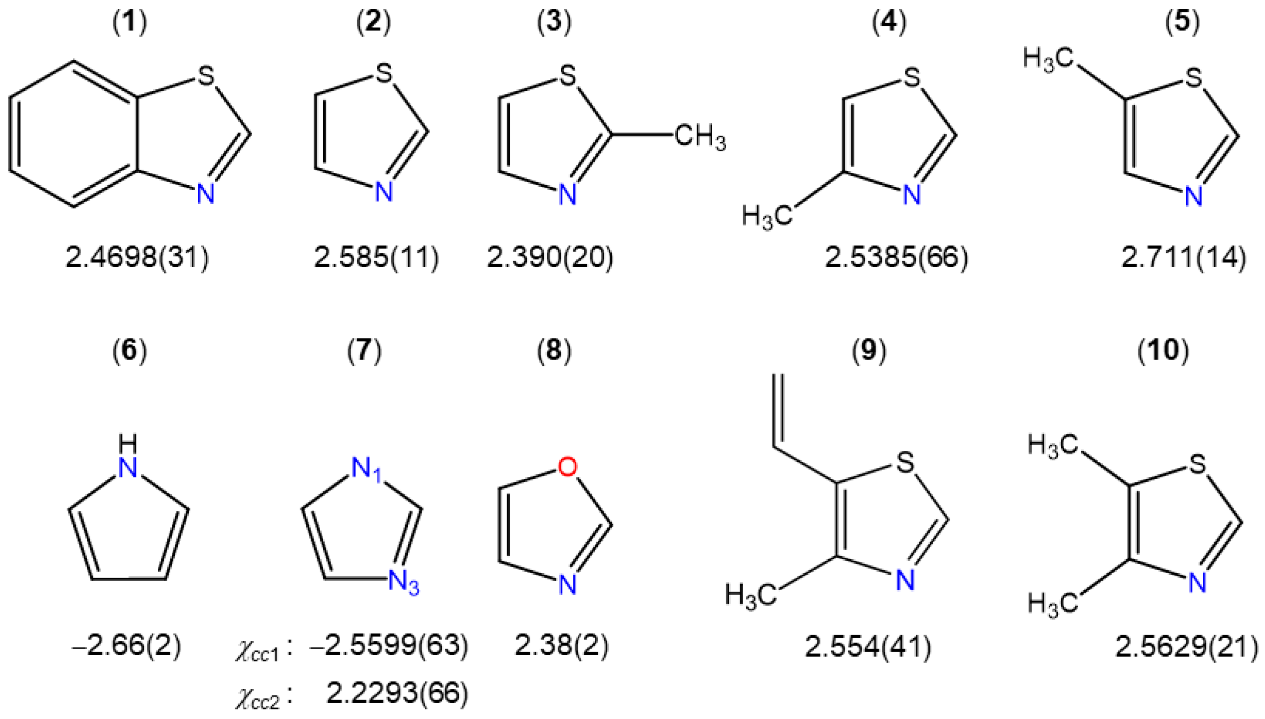
To compare the experimental 14N nuclear quadrupole coupling constant values of BTA and, consequently, the field gradient tensor at the site of the nitrogen nucleus with other thiazole derivatives, we selected some planar molecules, as illustrated in Figure 4. Due to planarity, the c-principal axis and one principal axis of the nitrogen coupling tensor are collinear, since they are both perpendicular to the plane of symmetry (the ring plane). Therefore, we can directly compare the χcc values.
Among all thiazole derivatives, i.e., BTA (1) (this work), thiazole (2) [52], 2-methylthiazole (3) [53], 4-methylthiazole (4) [54], 5-methylthiazole (5) [55], 4-methyl-5-vinylthiazole (9) [37], and 4,5-dimethylthiazole (10) [59], though all χcc values are similar, a small variation from 2.390 MHz in 2-methylthiazole (3) to 2.711 MHz in 5-methylthiazole (5) can be observed, showing that the electronic environment at the nitrogen nucleus changes slightly despite the fact that the molecules are very similar. It is also interesting that (i) the χcc value found for oxazole (8) [58] is almost the same as that of 2-methylthiazole (3) [53], and (ii) the value of the nitrogen atom at the 1-position of pyrrole (6) [56] and imidazole (7) [57] is negative due to a different bond situation at the 14N atom. To form one N–H bond and two N–C bonds, three of five 14N electrons are involved in three sp2 orbitals, and the other two electrons occupy the p orbital and contribute to the π electron system. Altogether, the nitrogen atoms donate two electrons to the aromatic system, while the others featuring positive χcc values provide only one electron. There, two 14N electrons are used in two sp2 orbitals to form two bonds, while the electron lone pair occupies the third sp2 orbital, leaving only one electron in the p orbital. If we consider that χcc is related to the unbalanced electrons in a pz orbital, the positive values found for BTA as well as other thiazole derivatives illustrated in Figure 4 also show that for these molecules there is an excess of electron density along the z axis of the 14N nucleus [60].
As an aromatic molecule with a π-conjugated system fused by thiazole and benzene, there is no doubt on the planarity of BTA, even though a complete experimental structure determination of the heavy atom backbone is not possible due to the lack of all 13C isotopologue spectra and the 15N isotopologue spectrum. A well-known judgement for the planarity of a molecule is its small positive inertial defect value, as observed for, e.g., thiazole (0.074 uÅ2) [52], oxazole (0.056 uÅ2) [58], pyrrole (0.017 uÅ2) [56], and 2-thiophenecarboxaldehyde (0.008 uÅ2) [61]. The inertial defect Δc = IcIaIb of –0.056 uÅ2 found for BTA is very close to zero, but negative. In many molecules whose molecular structures were experimentally determined to be planar such as coumarin [44] and 1,4-naphthoquinone [62], a negative value of the inertial defect was found due to low-frequency out-of-plane vibrations. This was discussed in detail by Oka [63]. The negative value varies, and the largest ones ranging from –0.6 uÅ2 to –0.8 uÅ2 were found for styrene and its halogenated derivatives. The very small, but negative inertial defect of BTA might hint that such vibrations are also present in the molecule. The value of –0.082 uÅ2 obtained from rotational constants calculated with the anharmonic force field is also negative and very close to the experimental value.

5. Conclusions

The microwave spectrum of BTA was measured in the frequency range between 2 and 26.5 GHz, aiming at determining precisely its molecular geometry parameters. All rotational transitions of the rigid rotor feature hyperfine splittings arising from the quadrupole coupling of the 14N nucleus. A large data set with 194 hyperfine components was included in a fit where geometry parameters (the A, B, and C rotational constants and the ΔJ, ΔK, ΔJK, δJ, and δK quartic centrifugal distortion constants) are obtained together with the 14N diagonal nuclear quadrupole coupling constants χaa, χbb, and χcc. The spectrum of the 34S minor isotopologue was also observed, including 92 transitions. By comparing the χcc value of 2.4698(31) MHz of BTA to those of other thiazole derivatives, we found that though all χcc values are similar, there is a small variation from 2.390 MHz to 2.711 MHz, showing that the electronic environment at the nitrogen nucleus is slightly different in those molecules. The very small, but negative inertial defect of −0.056 uÅ2 proposes that low-frequency out-of-plane vibrations are present in BTA.

Supplementary Materials

The following supporting information can be downloaded at: https://www.mdpi.com/article/10.3390/molecules28083419/s1, Table S1: The rotational constants A, B, C of BTA calculated at different levels of theory and their deviations to the experimental values; Table S2: Geometry parameters in the principal axes of inertia of BTA calculated at the MP2/6-31G(d,p), MP2/6-311++G(d,p), and B3LYP-D3BJ/6-311++G(d,p) levels of theory; Table S3: Calculated and observed frequencies of the main species and the 34S isotopologue of BTA.

Author Contributions

Conceptualization, H.V.L.N.; methodology, H.V.L.N.; validation, H.V.L.N. and I.K.; formal analysis, H.E.H., K.J.K. and H.V.L.N.; investigation, H.E.H., K.J.K. and H.V.L.N.; resources, H.V.L.N.; data curation, H.E.H. and H.V.L.N.; writing—original draft preparation, H.E.H. and H.V.L.N.; writing—review and editing, H.E.H., K.J.K., O.K.K., N.K., I.K. and H.V.L.N.; visualization, H.E.H., K.J.K. and H.V.L.N.; supervision, O.K.K., N.K., I.K. and H.V.L.N.; project administration, I.K. and H.V.L.N.; and funding acquisition, I.K. and H.V.L.N. All authors have read and agreed to the published version of the manuscript.

Funding

This work was supported by the European project “International Network for a Research and Innovation Staff Exchange” (RISE), Marie Curie Action (call: H2020-MSCA-RISE-2019), entitled “Pollutants and Greenhouse Gases in the Atmosphere—Understanding Gas–Gas and Gas–Solid Interactions Towards a Cleaner Atmosphere (ATMOS)”, and the European Union (ERC 101040480-LACRIDO). Views and opinions expressed are however those of the author(s) only and do not necessarily reflect those of the European Union or the European Research Council. Neither the European Union nor the granting authority can be held responsible for them. Part of the calculations were performed using HPC resources from GENCI-CINES (grant no. 2021-096175).

Institutional Review Board Statement

Not applicable.

Informed Consent Statement

Not applicable.

Data Availability Statement

Data are contained within the article and Supplementary Materials.

Acknowledgments

We thank Alexandra Welzel for the results from her thesis entitled “Bau einer heizbaren Ultraschalldüse zur mikrowellenspektroskopischen Untersuchung schwerflüchtiger Substanz im Molekularstrahl”, supervised by Wolfgang Stahl at the RWTH Aachen University. K.J.K. thanks the University Paris-Est Créteil for a post-doctoral fellowship.

Conflicts of Interest

The authors declare no conflict of interest.

References

  1. Liao, C.; Kim, U.-J.; Kannan, K. A Review of Environmental Occurrence, Fate, Exposure, and Toxicity of Benzothiazoles. Environ. Sci. Technol. 2018, 52, 5007–5026. [Google Scholar] [CrossRef] [PubMed]
  2. Ginsberg, G.; Toal, B.; Kurland, T. Benzothiazole Toxicity Assessment in Support of Synthetic Turf Field Human Health Risk Assessment. J. Toxicol. Environ. Health Part A 2011, 74, 1175–1183. [Google Scholar] [CrossRef] [PubMed]
  3. Avagyan, R.; Luongo, G.; Thorsén, G.; Östman, C. Benzothiazole, Benzotriazole, and their Derivates in Clothing Textiles—A Potential Source of Environmental Pollutants and Human Exposure. Environ. Sci. Pollut. Res. 2014, 22, 5842–5849. [Google Scholar] [CrossRef]
  4. Hutchinson, I.; Jennings, S.A.; Vishnuvajjala, B.R.; Westwell, A.D.; Stevens, M.F.G. Antitumor Benzothiazoles. 16. Synthesis and Pharmaceutical Properties of Antitumor 2-(4-Aminophenyl)benzothiazole Amino Acid Prodrugs. J. Med. Chem. 2001, 45, 744–747. [Google Scholar] [CrossRef]
  5. Siddiqui, N.; Pandeya, S.N.; Khan, S.A.; Stables, J.; Rana, A.; Alam, M.; Arshad, F.; Bhat, M. Synthesis and anticonvulsant activity of sulfonamide derivatives-hydrophobic domain. Bioorg. Med. Chem. Lett. 2007, 17, 255–259. [Google Scholar] [CrossRef]
  6. Burger, A.; Sawhney, S.N. Antimalarials. III. Benzothiazole Amino Alcohols. J. Med. Chem. 1968, 11, 270–273. [Google Scholar] [CrossRef]
  7. Gopal, M.; Padmashali, B.; Manohara, Y.; Gurupadayya, B. Synthesis and Pharmacological Evaluation of Azetidin-2-ones and Thiazolidin-4-ones Encompassing Benzothiazole. Indian J. Pharm. Sci. 2008, 70, 572–5777. [Google Scholar] [CrossRef] [PubMed] [Green Version]
  8. Pattan, S.R.; Suresh, C.; Pujar, V.D.; Reddy, V.V.K.; Rasal, V.P.; Koti, B.C. Synthesis and Antidiabetic Activity of 2-Amino [5′(4-Sulfonylbenzylidine)-2,4-Thiazolidinedione]-7-Chloro-6-Fluorobenzothiazole. ChemInform 2006, 37. [Google Scholar] [CrossRef]
  9. Henriksen, G.; Hauser, A.I.; Westwell, A.D.; Yousefi, B.H.; Schwaiger, M.; Drzezga, A.; Wester, H.-J. Metabolically Stabilized Benzothiazoles for Imaging of Amyloid Plaques. J. Med. Chem. 2007, 50, 1087–1089. [Google Scholar] [CrossRef]
  10. Mathis, C.A.; Wang, Y.; Holt, D.P.; Huang, G.-F.; Debnath, M.L.; Klunk, W.E. Synthesis and Evaluation of 11C-Labeled 6-Substituted 2-Arylbenzothiazoles as Amyloid Imaging Agents. J. Med. Chem. 2003, 46, 2740–2754. [Google Scholar] [CrossRef]
  11. Apelt, J.; Grassmann, S.; Ligneau, X.; Pertz, H.H.; Ganellin, C.R.; Arrang, J.M.; Schwartz, J.C.; Schunack, W.; Stark, H. Search for Histamine H3 Receptor Antagonists with Combined Inhibitory Potency at N-tau-Methyltransferase: Ether Derivatives. Pharmazie 2005, 60, 97–106. [Google Scholar] [PubMed]
  12. Sutikdja, L.W.; Nguyen, H.V.L.; Jelisavac, D.; Stahl, W.; Mouhib, H. Benchmarking Quantum Chemical Methods for Accurate Gas-Phase Structure Predictions of Carbonyl Compounds: The Case of Ethyl Butyrate. Phys. Chem. Chem. Phys. 2023, 25, 7688–7696. [Google Scholar] [CrossRef] [PubMed]
  13. Dindić, C.; Nguyen, H.V.L. Benchmarking Acetylthiophene Derivatives: Methyl Internal Rotations in the Microwave Spectrum of 2-Acetyl-5-Methylthiophene. Phys. Chem. Chem. Phys. 2023, 25, 509–519. [Google Scholar] [CrossRef]
  14. Puzzarini, C.; Stanton, J.F. Connections between the Accuracy of Rotational Constants and Equilibrium Molecular Structures. Phys. Chem. Chem. Phys. 2023, 25, 1421–1429. [Google Scholar] [CrossRef] [PubMed]
  15. Gottschalk, H.C.; Poblotzki, A.; Fatima, M.; Obenchain, D.A.; Pérez, C.; Antony, J.; Auer, A.A.; Baptista, L.; Benoit, D.M.; Bistoni, G.; et al. The First Microsolvation Step for Furans: New Experiments and Benchmarking Strategies. J. Chem. Phys. 2020, 152, 164303. [Google Scholar] [CrossRef]
  16. Spaniol, J.-T.; Lee, K.L.K.; Pirali, O.; Puzzarini, C.; Martin-Drumel, M.-A. A Rotational Investigation of the Three Isomeric Forms of Cyanoethynylbenzene (HCC-C6H4-CN): Benchmarking Experiments and Calculations Using the “Lego Brick” Approach. Phys. Chem. Chem. Phys. 2023, 25, 6397–6405. [Google Scholar] [CrossRef]
  17. Fischer, T.L.; Bödecker, M.; Zehnacker-Rentien, A.; Mata, R.A.; Suhm, M.A. Setting up the HyDRA Blind Challenge for the Microhydration of Organic Molecules. Phys. Chem. Chem. Phys. 2022, 24, 11442–11454. [Google Scholar] [CrossRef]
  18. Gaussian 16, Revision B.01; Gaussian Inc.: Wallingford, CT, USA, 2016.
  19. Møller, C.; Plesset, M.S. Note on an Approximation Treatment for Many-Electron Systems. Phys. Rev. 1934, 46, 618. [Google Scholar] [CrossRef] [Green Version]
  20. Scuseria, G.E.; Scheiner, A.C.; Lee, T.J.; Rice, J.E.; Schaefer, H.F. The Closed-Shell Coupled Cluster Single and Double Excitation (CCSD) Model for the Description of Electron Correlation. A Comparison with Configuration Interaction (CISD) Results. J. Chem. Phys. 1987, 86, 2881. [Google Scholar] [CrossRef]
  21. Becke, A.D. Density-Functional Thermochemistry. III. The Role of Exact Exchange. J. Chem. Phys. 1993, 98, 5648. [Google Scholar] [CrossRef] [Green Version]
  22. Lee, C.; Yang, W.; Parr, R.G. Development of the Colle-Salvetti Correlation-Energy Formula into a Functional of the Electron Density. Phys. Rev. B 1988, 37, 785. [Google Scholar] [CrossRef] [Green Version]
  23. Zhao, Y.; Truhlar, D.G. The M06 Suite of Density Functionals for Main Group Thermochemistry, Thermochemical Kinetics, Noncovalent Interactions, Excited States, and Transition Elements: Two New Functionals and Systematic Testing of Four M06-Class Functionals and 12 Other Functionals. Theor. Chem. Acc. 2008, 120, 215–241. [Google Scholar] [CrossRef] [Green Version]
  24. Chai, J.-D.; Head-Gordon, M. Long-Range Corrected Hybrid Density Functionals with Damped Atom–Atom Dispersion Corrections. Phys. Chem. Chem. Phys. 2008, 10, 6615–6620. [Google Scholar] [CrossRef] [PubMed] [Green Version]
  25. Ernzerhof, M.; Scuseria, G.E. Assessment of the Perdew–Burke–Ernzerhof Exchange-Correlation Functional. J. Chem. Phys. 1999, 110, 5029. [Google Scholar] [CrossRef] [Green Version]
  26. Yu, H.S.; He, X.; Li, S.L.; Truhlar, D.G. MN15: A Kohn–Sham Global-Hybrid Exchange–Correlation Density Functional with Broad Accuracy for Multi-Reference and Single-Reference Systems and Noncovalent Interactions. Chem. Sci. 2016, 7, 5032–5051. [Google Scholar] [CrossRef] [PubMed] [Green Version]
  27. Grimme, S.; Antony, J.; Ehrlich, S.; Krieg, H. A Consistent and Accurate Ab Initio Parametrization of Density Functional Dispersion Correction (DFT-D) for the 94 Elements H-Pu. J. Chem. Phys. 2010, 132, 154104–154119. [Google Scholar] [CrossRef] [Green Version]
  28. Grimme, S.; Ehrlich, S.; Goerigk, L. Effect of the Damping Function in Dispersion Corrected Density Functional Theory. J. Comput. Chem. 2011, 32, 1456–1465. [Google Scholar] [CrossRef]
  29. Yanai, T.; Tew, D.; Handy, N.C. A New Hybrid Exchange–Correlation Functional Using the Coulomb-Attenuating Method (CAM-B3LYP). Chem. Phys. Lett. 2004, 393, 51–57. [Google Scholar] [CrossRef] [Green Version]
  30. Frisch, M.J.; Pople, J.A.; Binkley, J.S. Self-Consistent Molecular Orbital Methods 25. Supplementary Functions for Gaussian Basis Sets. J. Chem. Phys. 1984, 80, 3265–3269. [Google Scholar] [CrossRef]
  31. Dunning, T.H., Jr. Gaussian Basis Sets for Use in Correlated Molecular Calculations. I. The Atoms Boron through Neon and Hydrogen. J. Chem. Phys. 1989, 90, 1007. [Google Scholar] [CrossRef]
  32. Bailey, W.C. DFT and HF–DFT Calculations of 14N Quadrupole Coupling Constants in Molecules. Chem. Phys. 2000, 252, 57–66. [Google Scholar] [CrossRef]
  33. Nguyen, T.; Dindic, C.; Stahl, W.; Nguyen, H.V.L.; Kleiner, I. 14N Nuclear Quadrupole Coupling and Methyl Internal Rotation in the Microwave Spectrum of 2-Methylpyrrole. Mol. Phys. 2020, 118, 1668572. [Google Scholar] [CrossRef]
  34. Nguyen, T.; Stahl, W.; Nguyen, H.V.L.; Kleiner, I. 14N Nuclear Quadrupole Coupling and Methyl Internal Rotation in 3-Methylpyrrole Investigated by Microwave Spectroscopy. J. Mol. Spectrosc. 2020, 372, 111351. [Google Scholar] [CrossRef]
  35. Nguyen, T.; Stahl, W.; Nguyen, H.V.L.; Kleiner, I. Local vs Global Approaches to Treat Two Equivalent Methyl Internal Rotations and 14N Nuclear Quadrupole Coupling of 2,5-Dimethylpyrrole. J. Chem. Phys. 2021, 154, 204304. [Google Scholar] [CrossRef] [PubMed]
  36. Tran, Q.T.; Errouane, A.; Condon, S.; Barreteau, C.; Nguyen, H.V.L.; Pichon, C. On the Planarity of Benzyl Cyanide. J. Mol. Spectrosc. 2022, 388, 111685. [Google Scholar] [CrossRef]
  37. Khemissi, S.; Schwell, M.; Kleiner, I.; Nguyen, H.V.L. Influence of π-Electron Conjugation Outside the Aromatic Ring on the Methyl Internal Rotation of 4-Methyl-5-Vinylthiazole. Mol. Phys. 2022, 120, e2052372. [Google Scholar] [CrossRef]
  38. Kannengießer, R.; Stahl, W.; Nguyen, H.V.L.; Bailey, W.C. 14N Quadrupole Coupling in the Microwave Spectra of N-Vinylformamide. J. Mol. Spectrosc. 2015, 317, 50–53. [Google Scholar] [CrossRef]
  39. Welzel, A. Bau Einer Heizbaren Ultraschalldüse zur Mikrowellenspektroskopischen Untersuchung Schwerflüchtiger Substanzen im Molekularstrahl. Ph.D. Thesis, RWTH Aachen University, Aachen, Germany, 2000. [Google Scholar]
  40. Grabow, J.; Stahl, W.; Dreizler, H. A Multioctave Coaxially Oriented Beam-Resonator Arrangement Fourier-Transform Microwave Spectrometer. Rev. Sci. Instrum. 1996, 67, 4072–4084. [Google Scholar] [CrossRef]
  41. Welzel, A.; Stahl, W. The FT Microwave Spectrum of Benzo[b]thiophene: First Application of a New Heatable Beam Nozzle. Phys. Chem. Chem. Phys. 1999, 1, 5109–5112. [Google Scholar] [CrossRef]
  42. Grabow, J.-U.; Stahl, W. A Pulsed Molecular Beam Microwave Fourier Transform Spectrometer with Parallel Molecular Beam and Resonator Axes. Z. Naturforsch. 1990, 45, 1043–1044. [Google Scholar] [CrossRef]
  43. Hartwig, H.; Dreizler, H. The Microwave Spectrum of trans-2,3-Dimethyloxirane in Torsional Excited States. Z. Naturforsch. 1996, 51, 923–932. [Google Scholar] [CrossRef]
  44. Nguyen, H.V.L.; Grabow, J.-U. The Scent of Maibowle–π Electron Localization in Coumarin from Its Microwave-Determined Structure. ChemPhysChem 2020, 21, 1243–1248. [Google Scholar] [CrossRef] [PubMed]
  45. Kisiel, Z.; Desyatnyk, O.; Pszczółkowski, L.; Charnley, S.; Ehrenfreund, P. Rotational Spectra of Quinoline and of Isoquinoline: Spectroscopic Constants and Electric Dipole Moments. J. Mol. Spectrosc. 2003, 217, 115–122. [Google Scholar] [CrossRef]
  46. Nguyen, H.V.L. The Heavy Atom Substitution and Semi-Experimental Equilibrium Structures of 2-Ethylfuran Obtained by Microwave Spectroscopy. J. Mol. Struct. 2020, 1208, 127909. [Google Scholar] [CrossRef]
  47. Ferres, L.; Mouhib, H.; Stahl, W.; Nguyen, H.V.L. Methyl Internal Rotation in the Microwave Spectrum of o-Methyl Anisole. ChemPhysChem 2017, 18, 1855–1859. [Google Scholar] [CrossRef] [PubMed]
  48. Dindić, C.; Nguyen, H.V.L. Microwave Spectrum of Two-Top Molecule: 2-Acetyl-3-Methylthiophene. ChemPhysChem 2021, 22, 2420–2428. [Google Scholar] [CrossRef] [PubMed]
  49. Dindić, C.; Barth, M.; Nguyen, H.V.L. Two Methyl Internal Rotations of 2-Acetyl-4-Methylthiophene Explored by Microwave Spectroscopy and Quantum Chemistry. Spectrochim. Acta A 2022, 280, 121505. [Google Scholar] [CrossRef]
  50. Kraitchman, J. Determination of Molecular Structure from Microwave Spectroscopic Data. Am. J. Phys. 1953, 21, 17–24. [Google Scholar] [CrossRef]
  51. Costain, C.C. Further Comments on the Accuracy of rs Substitution Structures. Trans. Am. Crystallogr. Assoc. 1966, 2, 157. [Google Scholar]
  52. Nygaard, L.; Asmussen, E.; Høg, J.H.; Maheshwari, R.; Nielsen, C.; Petersen, I.B.; Rastrup-Andersen, J.; Sørensen, G. Microwave Spectra of Isotopic Thiazoles. Molecular Structure and 14N Quadrupole Coupling Constants of Thiazole. J. Mol. Struct. 1971, 8, 225–233. [Google Scholar] [CrossRef]
  53. Nguyen, T.; Van, V.; Gutlé, C.; Stahl, W.; Schwell, M.; Kleiner, I.; Nguyen, H.V.L. The Microwave Spectrum of 2-Methylthiazole: 14N Nuclear Quadrupole Coupling and Methyl Internal Rotation. J. Chem. Phys. 2020, 152, 134306. [Google Scholar] [CrossRef]
  54. Jäger, W.; Mäder, H. The Microwave Spectrum of 4-Methylthiazole: Methyl Internal Rotation, 14N Nuclear Quadrupole Coupling and Electric Dipole Moment. Z. Naturforsch. 1987, 42, 1405–1409. [Google Scholar] [CrossRef]
  55. Jäger, W.; Mäder, H. The Microwave Spectrum of 5-Methylthiazole: Methyl Internal Rotation, 14N Nuclear Quadrupole Coupling and Electric Dipole Moment. J. Mol. Struct. 1988, 190, 295–305. [Google Scholar] [CrossRef]
  56. Nygaard, U.; Nielsen, J.; Kirchheiner, J.; Maltesen, G.; Rastrup-Andersen, J.; Sørensen, G. Microwave Spectra of Isotopic Pyrroles. Molecular Structure, Dipole Moment, and 14N Quadrupole Coupling Constants of Pyrrole. J. Mol. Struct. 1969, 3, 491–506. [Google Scholar] [CrossRef]
  57. Giuliano, B.M.; Bizzocchi, L.; Charmet, A.P.; Arenas, B.; Steber, A.L.; Schnell, M.; Caselli, P.; Harris, B.J.; Pate, B.H.; Guillemin, J.-C.; et al. Rotational Spectroscopy of Imidazole: Improved Rest Frequencies for Astrophysical Searches. Astron. Astrophys. 2019, 628, A53. [Google Scholar] [CrossRef]
  58. Kumar, A.; Sheridan, J.; Stiefvater, O.L. The Microwave Spectrum of Oxazole II. Dipole Moment and Quadrupole Coupling Constants. Z. Naturforsch. 1978, 33, 549–558. [Google Scholar] [CrossRef] [Green Version]
  59. Van, V.; Nguyen, T.; Stahl, W.; Nguyen, H.V.L.; Kleiner, I. Coupled Large Amplitude Motions: The Effects of Two Methyl Internal Rotations and 14N Quadrupole Coupling in 4,5-Dimethylthiazole Investigated by Microwave Spectroscopy. J. Mol. Struct. 2020, 1207, 127787. [Google Scholar] [CrossRef]
  60. Pinacho, P.; Obenchain, D.A.; Schnell, M. New Findings from Old Data: A Semi-Experimental Value for the eQq of the Nitrogen Atom. J. Chem. Phys. 2020, 153, 234307. [Google Scholar] [CrossRef]
  61. Hakiri, R.; Derbel, N.; Bailey, W.C.; Nguyen, H.V.L.; Mouhib, H. The Heavy Atom Structures and 33S Quadrupole Coupling Constants of 2-Thiophenecarboxaldehyde: Insights from Microwave Spectroscopy. Mol. Phys. 2020, 118, e1728406. [Google Scholar] [CrossRef]
  62. Saxena, S.; Panchagnula, S.; Sanz, M.E.; Pérez, C.; Evangelisti, L.; Pate, B.H. Structural Changes Induced by Quinones: High-Resolution Microwave Study of 1,4-Naphthoquinone. ChemPhysChem 2020, 21, 2579–2584. [Google Scholar] [CrossRef]
  63. Oka, T. On Negative Inertial Defect. J. Mol. Struct. 1995, 352–353, 225–233. [Google Scholar] [CrossRef]
Disclaimer/Publisher’s Note: The statements, opinions, and data contained in all publications are solely those of the individual author(s) and contributor(s) and not of MDPI and/or the editor(s). MDPI and/or the editor(s) disclaim responsibility for any injury to people or property resulting from any ideas, methods, instructions, or products referred to in the content.
Figure 1. Molecular structure of benzothiazole optimized at the MP2/6-311++G(d,p) level of theory in the principal axes of inertia (a, b, and c), with a planar geometry. The c-axis (not shown) is perpendicular to the ab-plane. The nitrogen atom is blue and the sulfur atom is yellow.
Molecules 28 03419 g001
Figure 2. A portion of the survey spectrum of BTA in the frequency range from 12,450 to 12,660 MHz, showing hyperfine splittings arising from the 14N nuclear quadrupole coupling. Assigned lines are labelled by their rotational quantum numbers J K a K c J K a K c and all belong to the main isotopologue of BTA. Lines marked with an asterisk are from impurities, as they disappeared when being remeasured later, probably due to a higher vapor pressure than that of BTA.
Molecules 28 03419 g002
Figure 3. A typical spectrum of the b-type 221 ← 212 transition of BTA, featuring the 14N quadrupole hyperfine structure with the hyperfine components indicated as F″F′. The Doppler pairs arising from the coaxial arrangement between the resonators and the molecular beam are connected by brackets. The intensities are not reliable since hyperfine components close to the polarization frequency (at 6694.0 MHz) appear to be more intense.
Molecules 28 03419 g003
Figure 4. χcc values in some thiazole derivatives and aromatic five-membered rings (in MHz). (1) Benzothiazole (this work), (2) thiazole [52], (3) 2-methylthiazole [53], (4) 4-methylthiazole [54], (5) 5-methylthiazole [55], (6) pyrrole [56], (7) imidazole [57], (8) oxazole [58], (9) 4-methyl-5-vinylthiazole [37], and (10) 4,5-dimethylthiazole [59].
Molecules 28 03419 g004
Table 1. The equilibrium Ae, Be, and Ce and ground vibrational state A0, B0, and C0 rotational constants, 14N diagonal nuclear quadrupole coupling constants χaa, χbb, and χcc, and dipole moment components μa, μb, and μc of BTA obtained at the MP2/6-311++G(d,p), MP2/6-31G(d,p), and B3LYP-D3BJ/6-311++G(d,p) levels of theory.
MP2/6-31G(d,p)MP2/6-311++G(d,p)B3LYP-D3BJ
AeMHz3166.63160.13168.6
BeMHz1338.71335.91334.2
CeMHz940.9939.0938.8
A0MHz3145.73136.43146.7
B0MHz1332.11328.21327.0
C0MHz936.0933.1933.5
χaaMHz1.6401.7171.785
χbbMHz−3.598−3.922−4.395
χccMHz1.9672.2052.611
|μa|D0.370.240.33
|μb|D1.331.561.36
|μc|D0.000.000.00
Table 2. Molecular parameters of the main species and the 34S isotopologue of BTA obtained with the XIAM program by fitting the rotational constants A, B, and C, the quartic centrifugal distortion constants ΔJ, ΔK, ΔJK, δJ, and δK, as well as the diagonal nuclear quadrupole coupling constants χaa, χbb, χcc.
Par. aUnit32S XIAM32S Calc b34S XIAM
AMHz3174.630260(98)3160.132393143.66433(21)
BMHz1341.764826(67)1335.936701320.53713(11)
CMHz943.232720(28)938.98407930.00014(6)
ΔJkHz0.02681(42)0.0254790.02678(49)
ΔJKkHz0.0472(22)0.0450330.0432(38)
ΔKkHz0.175(10)0.175890.185(14)
δJkHz0.00799(25)0.0076780.00754(30)
δKkHz0.0696(39)0.068220.0643(62)
χaaMHz1.60208(92)1.727 c1.5975(31)
χbb dMHz−4.0719(13)−4.036 c−4.0686(34)
χcc dMHz2.4698(31)2.309 c2.4710(71)
N e 19492
σ fkHz1.62.4
a All parameters refer to the principal axis system. Watson’s A reduction and Ir representation were used. Standard errors in parentheses are in the units of the last significant digits. b Calculated at the MP2/6-311++G(d,p) level of theory. The rotational constants A, B, and C are the equilibrium ones from geometry optimizations (see Table 1); the centrifugal distortion constants ΔJ, ΔK, ΔJK, δJ, and δK are from harmonic force field calculations. c Obtained with Bailey’s calibration [32] from electric field gradient calculations at the B3PW91/6-311+G(d,p) level of theory on the MP2/6-311++G(d,p) optimized structure with a correction factor eQ/h = −4.599 MHz/a.u. recommended for a molecule containing a conjugated double bond system [38]. d Derived from the fitted parameters χaa and χbbχcc = −6.5417(17) MHz. e Number of lines. f Standard deviation of the fit.
Disclaimer/Publisher’s Note: The statements, opinions and data contained in all publications are solely those of the individual author(s) and contributor(s) and not of MDPI and/or the editor(s). MDPI and/or the editor(s) disclaim responsibility for any injury to people or property resulting from any ideas, methods, instructions or products referred to in the content.