Article

Molecular Characterization of Some Bacillus Species from Vegetables and Evaluation of Their Antimicrobial and Antibiotic Potency

by
Moldir Koilybayeva
1,*,
Zhanserik Shynykul
2,*,
Gulbaram Ustenova
1,
Symbat Abzaliyeva
2,
Mereke Alimzhanova
3,
Akerke Amirkhanova
1,
Aknur Turgumbayeva
2,
Kamilya Mustafina
4,
Gulnur Yeleken
1,
Karlygash Raganina
1 and
Elmira Kapsalyamova
1
1
School of Pharmacy, S.D. Asfendiyarov Kazakh National Medical University, Tole-bi 94, Almaty 050012, Kazakhstan
2
Higher School of Medicine, Al-Farabi Kazakh National University, Tole-bi 96, Almaty 050040, Kazakhstan
3
Center of Physical Chemical Methods of Research and Analysis, Al-Farabi Kazakh National University, Tole-bi 96, Almaty 050012, Kazakhstan
4
School of Medicine, S.D. Asfendiyarov Kazakh National Medical University, Tole-bi 94, Almaty 050012, Kazakhstan
*
Authors to whom correspondence should be addressed.
Molecules 2023, 28(7), 3210; https://doi.org/10.3390/molecules28073210
Submission received: 16 February 2023 / Revised: 24 March 2023 / Accepted: 31 March 2023 / Published: 4 April 2023
(This article belongs to the Section Natural Products Chemistry)

Abstract

:
Numerous natural habitats, such as soil, air, fermented foods, and human stomachs, are home to different Bacillus strains. Some Bacillus strains have a distinctive predominance and are widely recognized among other microbial communities, as a result of their varied habitation and physiologically active metabolites. The present study collected vegetable products (potato, carrot, and tomato) from local markets in Almaty, Kazakhstan. The bacterial isolates were identified using biochemical and phylogenetic analyses after culturing. Our phylogenetic analysis revealed three Gram-positive bacterial isolates BSS11, BSS17, and BSS19 showing 99% nucleotide sequence similarities with Bacillus subtilis O-3, Bacillus subtilis Md1-42, and Bacillus subtilis Khozestan2. The crude extract was prepared from bacterial isolates to assess the antibiotic resistance potency and the antimicrobial potential against various targeted multidrug-resistant strains, including Staphylococcus aureus, Staphylococcus epidermidis, Streptococcus group B, Streptococcus mutans, Candida albicans, Candida krusei, Pseudomonas aeruginosa, Shigella sonnei, Klebsiella pneumoniae, Salmonella enteritidis, Klebsiella aerogenes, Enterococcus hirae, Escherichia coli, Serratia marcescens, and Proteus vulgaris. This study found that the species that were identified have the ability to produce antibiotic chemicals. Additionally, the GC–MS analysis of three bacterial extracts revealed the presence of many antibiotic substances including phenol, benzoic acid, 1,2-benzenedicarboxylic acid and bis(2-methylpropyl), methoxyphenyl-oxime, and benzaldehyde. This work sheds light on the potential of Bacillus to be employed as an antimicrobial agent to target different multidrug-resistant bacterial strains. The results indicate that market vegetables may be a useful source of strains displaying a range of advantageous characteristics that can be used in the creation of biological antibiotics.

1. Introduction

The prevalence of antibiotic resistance in bacterial strains poses a severe threat to public health and calls for urgent research into novel antibiotics or antimicrobial chemicals [1]. Numerous studies on the development of novel antibiotics from various microbe and plant strains have been published over the past few decades [2,3,4,5]. Antibiotics are chemicals that bacteria create and use in their natural environments for protection from the invasion of other bacterial species. In addition to serving as a kind of protection, these antibiotics are essential signaling molecules that allow the cells of the bacterial population to communicate with one another [6,7,8]. Their importance is enhanced when a bacterium is considered a probiotic which is administered in sufficient amounts and provides health benefits to the host. Cases of affirmed health profits of probiotics may comprise the support of immune health, gastrointestinal health, and so on. More precisely, the probiotic Bacillus subtilis has clinically demonstrated its effectiveness in dietary protein digestion [9]. Thus, it is impossible not to notice the growing number of benefits associated with the use of bacteria in various fields of activity. However, it is unfair not to consider the side effects of antibiotic compounds synthesized by bacteria. It was found that antibiotics influence food deterioration. Thus, spoilage of food is caused by a change in the chemical or physical properties of food caused by antibiotic-like substances of foodborne pathogens [10].
From the historical evidence, it is assumed that natural products are essential for the discovery and advancement of antibiotics [11]. It is imperative to investigate innovative antimicrobial substances with a high potential to eradicate or control a variety of microorganisms. Hence, one of the fundamental pillars of modern medicine is the antibiotic. Nevertheless, it is regrettable that some pathogenic strains render commonly-used antibiotics ineffective, and there is a requirement for novel antibiotics to take their place [12,13]. Microorganisms that may create bioactive secondary metabolites have unique structural features and biological activities. These bioactive compounds are produced by a few types of microflora found in vegetables and are employed as antibiotics. Several other notable types of investigation have also been reported to identify bacteria from vegetables with new antimicrobial agents [14,15]. Multidrug-resistant infections are more hazardous than infections caused by bacterial pathogens that are not resistant to multiple drugs because public health practitioners have recently had tremendous difficulty in treating these organisms.
Particularly, the prevalence of resistance developed in bacterial pathogens functions as a secondary infection in a number of life-threatening disorders, such as cancer, surgical procedures, transplantation, etc., and affects the effectiveness of contemporary treatments in treating these conditions [16,17]. It is evident that there are very few therapeutic drugs available to successfully treat these infections given the rapid evolution of multidrug-resistant strains due to the availability of relatively few effective treatments [18,19]. In order to evaluate the antibacterial properties against the majority of common human diseases, this study concentrated on extracting possible bacterial species from vegetable sources (potato, carrot, and tomato) and creating a crude extract from isolated bacterial strains. The main goal of this study is to provide insights into bacterial isolate composition and to provide a connection with the antimicrobial activity of investigating bacterial strains. The bioactive components in the crude extract were also identified using GC–MS. This research will aid in the creation of new drugs to combat multidrug-resistant bacterial strains.

2. Results

2.1. Isolation and Identification

A total of n = 19 bacteria strains were isolated and identified based on colonial morphology, microscopy, biochemical characteristics, and sugar fermentation. Among all, Gram-positive, rod-shaped, mycelial, and spore-forming bacterial strains were selected for further confirmatory tests. The molecular analysis further validated the bacterial strains (BSS11, BSS17, and BSS19) as Bacillus subtilis O-3, Bacillus subtilis Md1-42, and Bacillus subtilis Khozestan2.

2.2. Morphological Characterization

The morphology of each colony from the different bacterial isolates showed regular, irregular, slightly raised, flat, white, and cream-colored colonies. A motility test determined that the bacterial isolates were motile and possessed terminal and subterminal spores (Table 1).

2.3. Antimicrobial Activity Assessment

All tested isolates of BSS11, BSS17, and BSS19 showed antagonistic activity against most bacterial pathogens, such as Klebsiella aerogenes ATCC 13048, Staphylococcus aureus ATCC 29213, Staphylococcus epidermidis ATCC 12228, Candida krusei ATCC 14243, and Candida albicans ATCC 2091, and had less activity against other pathogens, such as Proteus vulgaris ATCC 6380, Klebsiella pneumoniae ATCC 13883, Shigella sonnei ATCC 25931, and Salmonella enterica ATCC 35664 (Figure 1).
The nine extracts showed antibacterial activity against all the bacterial pathogens except Shigella sonnei ATCC 25931, Klebsiella pneumonia ATCC 13883, Salmonella enterica ATCC 35664, and Enterococcus hirae ATCC 10541 (Table 2). Additionally, three extracts such as EAE (C), EAE (BC), and EAE (SC) from Bacillus subtilis Khozestan2 (BSS19) did not also demonstrate antibacterial activity. The nine extracts showed antibacterial activity against all the bacterial pathogens except Shigella sonnei ATCC 25931, Klebsiella pneumonia ATCC 13883, Salmonella enterica ATCC 35664, and Enterococcus hirae ATCC 10541. Additionally, three extracts such as EAE (C), EAE (BC), and EAE (SC) from Bacillus subtilis Khozestan2 (BSS19) have not demonstrated any antibacterial activity, too. The EAE (SC) preparation of Bacillus subtilis O-3 (BSS11), showed a better zone of inhibition for Staphylococcus epidermidis ATCC 12228 (25 ± 1.20 mm), Streptococcus group B (19 ± 1.20 mm), Candida krusei ATCC 14243 (29 ± 2.35 mm), Klebsiella aerogenes ATCC 13048 (17 ± 1.82 mm), and Proteus Vulgaris ATCC 6380 (18 ± 1.34 mm) when compared with the other pathogens, while the EAE (SC) preparation of Bacillus subtilis Md1-42 (BSS17) was effective against the pathogens Staphylococcus aureus ATCC 29213 (30 ± 2.50 mm) and Serratia marcescens ATCC 13880 (18 ± 1.64 mm). Additionally, the EAE (SC) preparation of Bacillus subtilis Khozestan2 (BSS19) was effective against the pathogens Streptococcus mutans ATCC 25175 (20 ± 1.32 mm), Candida albicans ATCC 2091 (40 ± 1.22 mm), and Pseudomonas aeruginosa ATCC 9027 (22 ± 1.81 mm). It was found assumingly the same potency meaning of the EAE (SC) preparations of Bacillus subtilis Md1-42 (BSS17) and Bacillus subtilis Khozestan2 (BSS19) against the pathogen Escherichia coli ATCC 25922.

2.4. Antibiotic Susceptibility Profile of the Isolates

The study of the antibiogram revealed that all three tested Bacillus subtilis subspecies were resistant to all antibiotics except for bacitromycin (B, 10), polymyxin (PB, 300), and cloxacillin (CX, 5) (Table 3). BSS11, BSS17, and BSS17 strains showed the highest vulnerability to gentamicin (CN, 120) with 40 ± 0.28 mm, 40 ± 0.28 mm, and 38 ± 0.28 sensitivity diameters, respectively, while strain BSS17 showed the lowest sensitivity to carbenicillin (10 ± 0.28) and to amoxycillin (12 ± 0.29) with high significant differences (p < 0.0001).

2.5. Analysis of the Isolates Using GC–MS

The crude extracts from several Bacillus bacterium species contained a number of chemicals, according to the results of the GC–MS analysis. Table 4, Table 5 and Table 6 explain the most significant and abundant components found in the crude extracts that were subjected to the GC–MS analysis, as well as information about where the chemicals found in this study had previously been identified. These substances exhibited similarities to natural products of bacterial and plant origin. According to the study of the GC–MS data, the majority derived from volatile substances, such as alkaloids, esters, ethers, and phenolic chemicals.
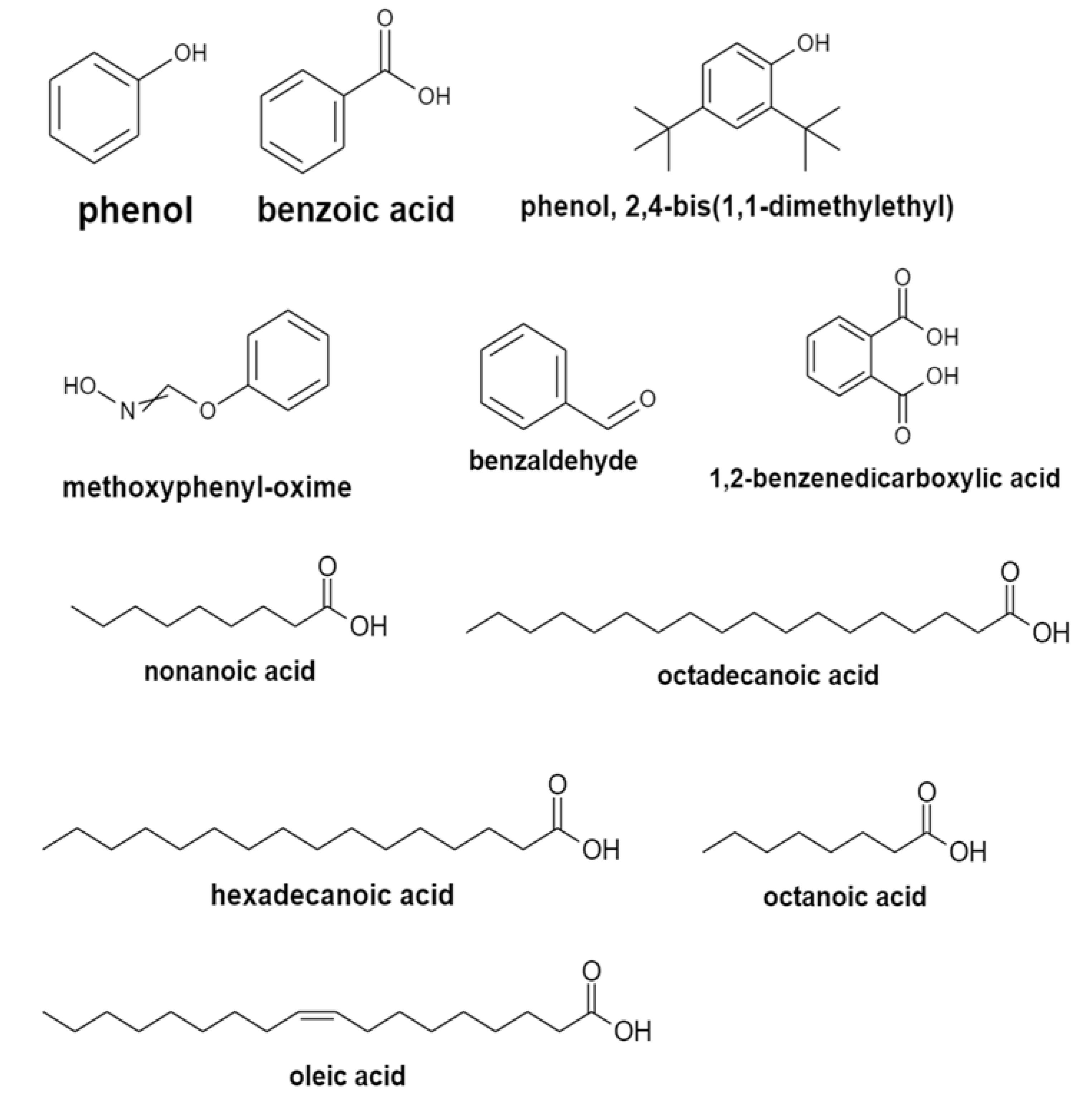
The GC–MS analysis of the ethyl acetate extracts of the three isolates detected a total of 106 compounds. Based on the analysis of bacterial isolates, BSS11 and BSS17 were found to share a similar composition of volatile organic components, while bacterial isolate BSS19 was found with fewer quantities of them. For isolate BSS11, the solvent with metabolites was ethyl acetate with 36 compounds (Table 4). Phenol, benzoic acid, phenol, 2,4-bis(1,1-dimethylethyl), 1,2-benzenedicarboxylic acid, methoxyphenyl-oxime, and benzaldehyde (Figure 2) were identified in the BSS11 extract with important concentrations of 4.246424%, 0.94121%, 1.242429%, 2.287847%, 2.008104%, and 7.155098%, respectively. In the extract, the major compounds were acetamide at 11.58042% and 2-butanone at 9.68622%, while minor compounds were fatty acids and their derivatives. In strain BSS17, ethyl acetate extraction showed the presence of 39 compounds (Table 5) in comparison to 32 compounds arising out with the same extraction strain BSS19 (Table 6). GC–MS analysis for two bacterial (BSS17 and BSS19) analyses also confirmed the presence of the same volatile organic compounds but with fewer amounts compared with BSS11. The BSS19 ethyl acetate extract did not reveal the presence of some fatty acids (octanoic acid, nonanoic acid, hexadecanoic acid, octadecanoic acid, oleic acid, and 9,12-octadecanoic acid (z,z)), although they are present in the extracts of BSS11 and BSS17.

2.6. Molecular Characterization

From various samples, three bacterial isolates with enhanced antibacterial activity were discovered. Phylogenetic analysis of the 16S rRNA gene sequences revealed that all three candidate bacterial isolates, BSS11, BSS17, and BSS19, belong to three different Bacillus species, respectively (Figure 3), as they group together in the evolutionary tree with the aforementioned bacterial species.
Bacillus subtilis O-3, Bacillus subtilis Md1-42, and Bacillus subtilis Khozestan2 were identified as having the highest hit sequence similarity for these bacterial isolates (Table 7). High bootstrap values were obtained following a phylogenetic analysis and tree topology both served to confirm the presumably described taxonomy.

3. Discussion

Extreme microbial diversity, abundance, and structure are also correlated with a wide range of metabolic processes, which generate a large number of metabolites with a variety of functions, including antimicrobial, anti-parasitic, anti-cancerous, and anti-pesticidal functions. The current study sought to examine the potential for specific vegetable microbial populations to exhibit antibacterial activities. Nineteen distinct bacterial isolates were discovered, as a result of a number of isolation stages, the identification of different general purposes with the selection of bacterial growth media, and biochemical studies. In recent years, a rise in the likelihood of discovering new antibiotics to combat or control untreated infectious diseases has been seen, thanks to a number of microorganisms that can produce antibiotics when grown in proper cultures [20,21]. Indeed, the development of resistance genes in bacteria through the use of mobile genetic elements or their inherent characteristics (natural phenotypic traits) are the two main causes of their antibiotic resistance [22]. All three Bacillus spp. strains were susceptible to almost all antibiotics, except for bacitracin, polymyxin, and cloxacillin, for which they were all resistant (Table 3). Similar findings on the susceptibility of various antibiotic-resistant Bacillus species were observed [23,24,25,26]. Our results also agree with those on the resistance of Bacillus strains to bacitracin, published by Adimpong et al. (2012) [27]. According to Adimpong et al. (2012) and Compaoré et al. (2013) [28], the resistance of specific Bacillus strains to particular antibiotics may be inherent or acquired and associated with the existence of resistance genes implicated in the production of resistance enzymes to these antibiotics. Conversely, the probability of passing on resistance genes to other dangerous bacteria is lower because of a natural resistance, rather than an acquired resistance. Since resistant bacteria can spread from the food chain to humans, antibiotic resistance has really become a serious global concern [29]. Although isolated Bacillus strains do not seem to harbor antibiotic resistance genes that can be passed on to dangerous germs, it is possible that they do not respond to a wide variety of antibiotics. Further research into these strains as prospective probiotic starter cultures could improve and maximize the production of high-quality, medicinal, and functional or health-promoting substances.
The antibacterial characteristic plays a crucial role in therapeutic activities. In the present study, the perpendicular streak method was used to determine the antibacterial properties of the bacterial isolates (BSS11, BSS17, and BSS19) against the selected human bacterial pathogens. This approach is regarded as a first-pass qualitative screening technique for the antimicrobial activity. The research demonstrated the strongest antagonistic action against human pathogen and as a result all tested isolates of BSS11, BSS17, and BSS19 showed antagonistic activity against most bacterial pathogens, such as Klebsiella aerogenes ATCC 13048, Staphylococcus aureus ATCC 29213, Staphylococcus epidermidis ATCC 12228, Candida krusei ATCC 14243, and Candida albicans ATCC 2091.
Our findings demonstrated that the growth of multidrug-resistant bacterial strains is inhibited by Bacillus subtilis O-3, Bacillus subtilis Md1-42, and Bacillus subtilis Khozestan2, which has previously been published [30,31]. Previous research found that the pH of the growing medium or the generation of volatile chemicals are what cause Bacillus to have an inhibitory impact. Bacillus is known to produce polypeptide antibiotic substances, such as bacitracin, polymyxin, gramicidin S., and tyrothricin, according to a number of other investigations. These substances work well against a variety of bacteria, including Gram-positive and Gram-negative bacteria [32].
GC-MS made it possible to detect markers in the studied samples of biological material—components of a microbial cell and its metabolites (fatty acids, aldehydes, phenolic compounds). Additionally, using GC-MS was beneficial in the case of both endogenous and exogenous microflora, without preliminary isolation of a pure culture of microorganisms, which is especially important when considering the difficulties in cultivating anaerobes. The distinctive advantages of the method were the speed of analysis and the ability to quantify the content of the marker. According to the GC–MS analysis, the Bacillus species produce a variety of antifungal chemicals. The synthesis of antibiotic substances by Bacillus subtilis O-3, Bacillus subtilis Md1-42, and Bacillus subtilis Khozestan2 strains was determined by the GC–MS analysis of their crude metabolites. The most important antifungal compounds detected from all three strains were phenol, benzoic acid, phenol, 2,4-bis(1,1-dimethylethyl), 1,2-benzenedicarboxylic acid, and bis(2-methylpropyl) (Table 4, Table 5 and Table 6 and Figure 2). Additionally, the GC–MS analysis showed that all three bacterial isolates contained methoxyphenyl-oxime and benzaldehyde. Previously, methoxyphenyl-oxime was reported as a true specific antibacterial agent that controlled some bacteria [33]. Another study proved that benzaldehyde generated by Photorhabdus temperata has insecticidal, great antibacterial, and antioxidant properties [34]. Two bacterial isolates of Bacillus subtilis O-3 and Bacillus subtilis Md1-42 share common fatty acids, such as octanoic acid, nonanoic acid, hexadecanoic acid, octadecanoic acid, oleic acid, and 9,12-octadecanoic acid (z,z). It is well known that fatty acids and their derivatives have powerful antibacterial and antifungal activities [35]. Because of their great biodegradability, low toxicity, and strong resistance to extremes in pH, salinity, and temperature, they are more environmentally friendly. As food additives, they are accepted. Antifungal fatty acids are less likely to make pathogenic fungi resistant to them [36]. Most of the chemicals identified from three different Bacillus species were derived from volatile compounds, such as esters, alkaloids, ethers, and phenolics, and shared structural similarities with natural products of bacterial and plant origin. Many volatile organic compounds are major constituents of the bacterial strain and have been shown to have properties against phytopathogens [37,38]. Our results prove that Bacillus spp. share common volatile compounds and complements previous findings related to the study of chemical composition of bacterial strains using GC–MS [39,40,41,42]. Moreover, bacterial strains of Bacillus subtilis O-3 and Bacillus subtilis Md1-42 containing mentioned above volatile organic compounds (phenol, benzoic acid, phenol, 2,4-bis(1,1-dimethylethyl), 1,2-benzenedicarboxylic acid, methoxyphenyl-oxime, and benzaldehyde) and fatty acid derivatives (octanoic acid, nonanoic acid, hexadecanoic acid, octadecanoic acid, oleic acid, and 9,12-octadecanoic acid (z,z)) showed high anti-microbial activity on different pathogens. However, the bacterial strain Bacillus subtilis Khozestan2 demonstrated low antimicrobial potency, and it may be caused by due to the absenteeism of fatty acid derivatives and comparingly low concentrations of volatile organic compounds (phenol (1.456046%), benzoic acid (1.3117335%), phenol, 2,4-bis(1,1-dimethylethyl) (1.085735%), 1,2-benzenedicarboxylic acid (0.986222%), methoxyphenyl-oxime (1.54511%), and benzaldehyde (3.066211%)).
There are many purposes for bacterial extract investigation. For instance, Bacillus spp. produce a variety of compounds involved in the biocontrol of plant pathogens and the promotion of plant growth, which makes them potential candidates for most agricultural and biotechnological applications. Moreover, the Bacillus strains as a form of probiotics are not generally pathogenic to mammals and appear to have significant potential for clinical use. Nevertheless, Bacillus probiotics may also generate toxins and biogenic amines; consequently, their safety is a concern.
A molecular investigation revealed the taxonomy of three different isolated species belonging to three Bacillus spp., such as Bacillus subtilis O-3, Bacillus subtilis Md1-42, and Bacillus subtilis Khozestan2. It was tentatively determined that the three most viable candidates of bacterial isolates BSS11, BSS17, and BSS19 belong to Bacillus subtilis O-3 (99%), Bacillus subtilis Md1-42 (99%), and Bacillus subtilis Khozestan2 (99%), respectively, based on a phylogenetic analysis and top hit sequence similarity results, which were supported by a high bootstrap value. In the future, the microbial screening and the isolation of active metabolites against multidrug-resistant strains could be carried out more easily by the identification of the three separate bacterial strains and their antibacterial activity.

4. Materials and Methods

4.1. Isolation of Potential Strains of the Genus Bacillus spp.

Bacillus spp. strains have been isolated from different vegetable sources (potato, carrot, and tomato). A 15 g vegetable sample was homogenized in 100 mL of 0.85% NaCl by shaking at 150 rpm for 15 min. Then, the sample was diluted step-wise and incubated in a water bath for 10 min at 90 °C. The sample was cooled to room temperature and then 0.1 mL samples were loaded onto nutrient agar/meat peptone agar (NA/MPA) plates, which is a nutrient medium for the cultivation of non-fastidious microorganisms. NA/MPA plates were composed of gelatin peptone (5 g/L), bacteriological agar (15 g/L), and meat extract (3 g/L). The plates were incubated for 48 h at 37 °C. The isolated pure strains were refrigerated at −20 °C in nutrient broth (NB) media supplemented with 20% (v/v) glycerin. Then, the fresh culture was subjected to morphological identification and the slightly raised, flat, white, and cream-colored colonies were selected for further identification. Strain isolates in NB media were useful in further studies, particularly in the preparation of ethyl acetate extract for subjecting GC–MS analysis.

4.2. Screening for Antagonistic Activity of Isolated Bacteria against Potent Bacterial Pathogens

A preliminary antibacterial analysis of the isolates was conducted on Mueller–Hinton agar (MHA) plates using the perpendicular streak method against powerful human pathogens. Staphylococcus aureus ATCC 29213, Staphylococcus epidermidis ATCC 12228, Streptococcus group B, Streptococcus mutans ATCC 25175, Candida albicans ATCC 2091, Candida krusei ATCC 14243, Pseudomonas aeruginosa ATCC 9027, Shigella sonnei ATCC 25931, Klebsiella pneumoniae ATCC 13883, Salmonella enterica ATCC 35664, Klebsiella aerogenes ATCC 13048, Enterococcus hirae ATCC 10541, Escherichia coli ATCC 25922, Serratia marcescens ATCC 13880, and Proteus vulgaris ATCC 6380 were employed in this investigation as bacterial pathogens. According to the widely-used method of perpendicular streaks, an exponential culture of the studied antagonist strains was streaked on the surface of an agar medium and incubated at 30 ± 4 °C for 24 h [43]. Then, an exponential culture of the test strain was inoculated perpendicularly from the edge of the cup to the stroke of the grown culture of the antagonist with a stroke by slightly touching the stroke of the antagonist strain. The plate was again incubated under conditions favorable for the growth of the test culture.
The cellular preparations were held relying on the procedure elaborated by Beiranvand et al. (2017) [44]. The chosen isolates underwent 14 days of incubation at 30 ± 4 °C while being cultivated in a nutritional broth. Then, three different techniques were used to extract the cultures. The cultures were used further in preparation of the ethyl acetate extract of culture EAE (C), the ethyl acetate extract of boiled and cooled culture EAE (BC), and the ethyl acetate extract of the sonicated culture EAE (SC). For the creation of the three different extracts, the culture broth was divided into three equal portions. In a separate flask, one part of the culture broth was combined with an equivalent amount of ethyl acetate to create EAE (C). Once separated, the ethyl acetate extract was centrifuged at 8000 rpm for 10 min. The ethyl acetate supernatant was poured into a spotless flask and heated to 50 °C for drying. Two (2) ml of DMSO were used to dissolve the dry extract. Another portion of the media was incubated in boiling water for 5 min, and then cooled for 5 min, to prepare the EAE (BC). This mixture was then diluted 1:1 with ethyl acetate. The samples were then processed using the first technique. The culture was sonicated for three minutes at 130 W to prepare EAE (SC), and extraction was then carried out as instructed in the first procedure. Three extracts were examined for their antibacterial efficacy using the well diffusion method against pathogenic bacteria, including Staphylococcus aureus ATCC 29213, Staphylococcus epidermidis ATCC 12228, Streptococcus group B, Streptococcus mutans ATCC 25175, Candida albicans ATCC 2091, Candida krusei ATCC 14243, Pseudomonas aeruginosa ATCC 9027, Shigella sonnei ATCC 25931, Klebsiella pneumoniae ATCC 13883, Salmonella enterica ATCC 35664, Klebsiella aerogenes ATCC 13048, Enterococcus hirae ATCC 10541, Escherichia coli ATCC 25922, Serratia marcescens ATCC 13880, and Proteus vulgaris ATCC 6380. Sterile saline (0.85% NaCl) with an optical density of 0.5 McFarland standard scale (5 × 10⁶ CFU/mL (CFU—Colony Forming Units/mL) for yeasts and 1.5 × 10⁸ CFU/mL for bacteria) was used to prepare microbial suspensions. Each bacterial pathogen’s zone of inhibition was evaluated and control wells containing 20 μL of streptomycin (1 mg/mL) were used.

4.3. Antibiotic Susceptibility of the Bacillus Strains

Using the disk diffusion method, the antibiotic susceptibility of isolated Bacillus strains (BSS11, BSS17, and BSS19) was assessed in accordance with the guidelines of the European Committee on Antimicrobial Susceptibility Testing (EUCAST, 2019). Bacillus strains were spread-plated using sterile beads on Mueller–Hinton (MH) agar using an aliquot of 1 mL each, at a concentration of 106 CFU/mL (0.5 McFarland, Hi-media, India). The plates were then left to dry for an hour. Then, antibiotic disks were inserted into the agar plates containing an inoculated Bacillus strain.
The widths of the inhibition zones surrounding the antibiotic disks were measured using an electronic digital vernier caliper micrometer measuring tool caliber digital ruler (ZHHRHC LCD) following a 24 h incubation period at 37 °C (Hardened, China). This made it possible to identify the strain’s antibiotic susceptibility (S), intermediate resistance (I), or resistance (R) according to the CLSI guidelines (2012) [44,45]. Eighteen antibiotic disks contained a sample each of penicillin G (PEN, 10), ampicillin (AMP, 10), amoxycillin (AMOX, 30), amoxycillin-clavulanic acid (AMC, 30), carbenicillin (CAR, 100), cloxacillin (CX, 5), erythromycin (ERO, 15), azithromycin (AZM, 15), cefepime (FEP, 30), cefepime/clavulanic acid FEC-40, cephalatin (KF, 30), cefotaxime (CTX, 30), gentamicin (CN, 120), streptomycin (STR, 10), tobramycin (TOB, 10), tetracycline (TET, 30), polymyxin (PB, 300), and bacitromycin (B, 10).

4.4. Gas Chromatography–Mass Spectrum Analysis of the Metabolites

The volatile compounds extraction for each bacterial strain (Bacillus subtilis O-3, Bacillus subtilis Md1-42, and Bacillus subtilis Khozestan2) was done separately two times from 50 mL of the culture broth with 25 mL ethyl acetate (Sigma-Aldrich, Germany) for 30 min and two extracts were combined. Thereafter, the extract with a volume of 1.5 mL was taken into plastic vials with a volume of 2 mL and placed onto the autosampler tray for analysis using GC–MS. Thermo Scientific GC Focus Series DSQ was used to perform a GC–MS analysis on bacterial secondary metabolites. A steady flow of 1 mL of helium gas per minute was employed as the carrier gas, and an infection volume of 1 L was used. The injector and hot oven were kept at 250 °C and 110 °C, respectively, with the temperature increasing by 10 °C per minute up to 200 °C, 5 °C per minute up to 280 °C, and shutting down after 9 min at a temperature of 280 °C. The GC column was used to elute peaks of various chemicals, and the retention times of these peaks were noted. The database was searched for compounds with similar molecular masses and retention times after the data were matched with the compounds’ mass spectra. The bioactivities of previously investigated natural substances were also investigated, and the current study found a comparable correlation between the bioactivities of the bacterial extracts and their constituent parts.

4.5. Molecular Characterization of the Bacterial Isolates

Based on 16S rRNA conserved gene sequences and universal bacterial primers, isolated bacterial strains were molecularly characterized. The targeted gene sequence was amplified using the standard PCR procedure, and the final product was run through 1% gel electrophoresis to examine the size of the amplified fragments. The amplified samples and the relevant sequencing fragments were sent for sequencing, and MEGA software was used to phylogenetically analyze the nucleotide sequences that were recovered (MEGA-11). Using GenBank NCBI’s BLAST search, the bacterial isolates were further verified and classified at the species level (National Center for Biotechnology Information). With the accession numbers GQ870259, MF581448, and MH036316, 16S rRNA gene sequences for these probiotic strains were uploaded to the GenBank database (www.ncbi.nlm.nih.gov/projects/genome/clone/, accessed on 9 December 2022).

4.6. Statistical Analyses

The XLSAT software version 2016.02.27444 was used to conduct the analysis of variance (one-factor ANOVA) at the significance level (α = 0.05). The Newman–Keuls test was used to rank the means when there was a significant difference between the studied parameters.

5. Conclusions

The growth of multidrug-resistant bacterial strains could be inhibited by vegetable bacterial isolates from three different species of Bacillus, according to the current study. When examined using well diffusion and the perpendicular streak method, the crude extracts from three isolated bacterial strains were effective against bacterial strains. The potent isolates BSS11, BSS17, and BSS19 with broad-spectrum antibacterial activities were identified through this screening. The metabolic diversity within isolates was highlighted by a comparative GC–MS analysis, despite the fact that they are all members of the same Bacillus subspecies. In particular, it is evident in the case of Bacillus subtilis O-3 and Bacillus subtilis Md1-42. This study discovered a number of volatile inhibitory substances, including esters, phenolics, and ethers that may be involved in antimicrobial activity. It was found how they differ in chemical composition and how it may influence antimicrobial activity and antibiotic potency. For decades, it has been known that strains of the B. subtilis group are capable of producing a variety of secondary metabolites that mediate their antimicrobial characteristics. Along with volatile organic compounds, the presence of bacteriocins, polyketides, peptides, etc., were known. Hence, it can be concluded that the discovered organic volatile substances enhance the antimicrobial properties of Bacillus spp. together with the above substances. Bacterial strains Bacillus subtilis O-3, Bacillus subtilis Md1-42, and Bacillus subtilis Khozestan2 exhibit a tremendous metabolic capacity and adaptive biochemistry that could be employed in a variety of commercial and biotechnological activities by generating a wide range of bioactive chemical substances. Additionally, bacterial extracts, including chemicals, could be employed as antimicrobial agents to target different multidrug-resistant bacterial strains. It is anticipated that a thorough investigation of a similar kind could investigate new microbiological possibilities with undiscovered substances or metabolites that have a strong antibacterial potential. As a result, it might be a viable strategy for lowering the burden and danger posed by bacterial strains that are resistant to many drugs.

Author Contributions

Conceptualization: M.K., A.A. and A.T.; methodology: M.K., Z.S., M.A., A.A., G.Y. and E.K.; software: M.K., Z.S. and A.A.; validation: M.K., Z.S. and M.A.; formal analysis: S.A., M.A. and K.R.; investigation: M.K. and Z.S.; resources: M.K. and A.A.; data curation: M.K. and A.A.; writing—original draft: M.K. and Z.S.; writing—review and editing: M.K. and Z.S.; visualization: S.A. and K.M.; supervision: G.U.; project administration: G.U.; funding acquisition: G.U. All authors have read and agreed to the published version of the manuscript.

Funding

This research received no external funding.

Institutional Review Board Statement

Not applicable.

Informed Consent Statement

Not applicable.

Data Availability Statement

The authors confirm that the data supporting the findings of this study are available within the article.

Conflicts of Interest

The authors declare no conflict of interest.

Sample Availability

Samples of the compounds are not available from the authors.

References

  1. Serwecińska, L. Antimicrobials and Antibiotic-Resistant Bacteria: A Risk to the Environment and to Public Health. Water 2020, 12, 3313. [Google Scholar] [CrossRef]
  2. Sonnenbichler, J.; Dietrich, J.; Peipp, H. Secondary fungal metabolites and their biological activities, V. Investigations concerning the induction of the biosynthesis of toxic secondary metabolites in basidiomycetes. Biol. Chem. Hoppe-Seyler 1994, 375, 71–79. [Google Scholar] [CrossRef]
  3. Amaning Danquah, C.; Minkah, P.A.B.; Osei Duah Junior, I.; Amankwah, K.B.; Somuah, S.O. Antimicrobial Compounds from Microorganisms. Antibiotics 2022, 11, 285. [Google Scholar] [CrossRef]
  4. Vaou, N.; Stavropoulou, E.; Voidarou, C.; Tsigalou, C.; Bezirtzoglou, E. Towards Advances in Medicinal Plant Antimicrobial Activity: A Review Study on Challenges and Future Perspectives. Microorganisms 2021, 9, 2041. [Google Scholar] [CrossRef]
  5. Bakal, S.N.; Bereswill, S.; Heimesaat, M.M. Finding Novel Antibiotic Substances from Medicinal Plants—Antimicrobial Properties of Nigella Sativa Directed against Multidrug-resistant Bacteria. Eur. J. Microbiol. Immunol. 2017, 7, 92–98. [Google Scholar] [CrossRef] [Green Version]
  6. Li, Y.; Feng, T.; Wang, Y. The role of bacterial signaling networks in antibiotics response and resistance regulation. Mar. Life Sci. Technol. 2022, 4, 163–178. [Google Scholar] [CrossRef]
  7. Romero, D.; Traxler, M.F.; López, D.; Kolter, R. Antibiotics as signal molecules. Chem. Rev. 2011, 111, 5492–5505. [Google Scholar] [CrossRef] [Green Version]
  8. Demain, A.; Sanchez, S. Microbial drug discovery: 80 years of progress. J. Antibiot. 2009, 62, 5–16. [Google Scholar] [CrossRef] [Green Version]
  9. Spears, J.L.; Kramer, R.; Nikiforov, A.I.; Rihner, M.O.; Lambert, E.A. Safety Assessment of Bacillus subtilis MB40 for Use in Foods and Dietary Supplements. Nutrients 2021, 13, 733. [Google Scholar] [CrossRef]
  10. Wu, Y.; Liang, S.; Zhang, M.; Wang, Z.; Wang, Z.; Ren, X. The Effect of Chlorogenic Acid on Bacillus subtilis Based on Metabolomics. Molecules 2020, 25, 4038. [Google Scholar] [CrossRef]
  11. Jackson, N.; Czaplewski, L.; Piddock, L.J. Discovery and development of new antibacterial drugs: Learning from experience? J. Antimicrob. Chemother. 2018, 73, 1452–1459. [Google Scholar] [CrossRef] [Green Version]
  12. Ventola, C.L. The antibiotic resistance crisis: Part 1: Causes and threats. P T A Peer-Rev. J. Formul. Manag. 2015, 40, 277–283. Available online: https://pubmed.ncbi.nlm.nih.gov/25859123/ (accessed on 19 May 2022).
  13. Terreni, M.; Taccani, M.; Pregnolato, M. New Antibiotics for Multidrug-Resistant Bacterial Strains: Latest Research Developments and Future Perspectives. Molecules 2021, 26, 2671. [Google Scholar] [CrossRef]
  14. Hölzel, C.S.; Tetens, J.L.; Schwaiger, K. Unraveling the Role of Vegetables in Spreading Antimicrobial-Resistant Bacteria: A Need for Quantitative Risk Assessment. Foodborne Pathog. Dis. 2018, 15, 671–688. [Google Scholar] [CrossRef] [Green Version]
  15. Allydice-Francis, K.; Brown, P.D. Diversity of Antimicrobial Resistance and Virulence Determinants in Pseudomonas aeruginosa Associated with Fresh Vegetables. Int. J. Microbiol. 2012, 2012, 426241. [Google Scholar] [CrossRef] [Green Version]
  16. Ali, F.; Niaz, Z.; Shah, P.T.; Shakeela, Q.; Uzma, B.; Ahmed, S. Antibiogram of ESBL and MBL producing Pseudomonas aeruginosa among the population of Hazara division. J. Pak. Med. Assoc. 2020, 70, 1979–1984. [Google Scholar] [CrossRef]
  17. Perez, F.; Van Duin, D. Carbapenem-resistant Enterobacteriaceae: A menace to our most vulnerable patients. Clevel. Clin. J. Med. 2013, 80, 225–233. [Google Scholar] [CrossRef] [Green Version]
  18. Ali, F.; Kamal, S.; Shakeela, Q.; Ahmed, S. Extended-spectrum and Metallo-beta lactamase enzymes mediated resistance in Pseudomonas aeruginosa in clinically isolated specimens. Kuwait J. Sci. 2021, 48, 1–9. [Google Scholar] [CrossRef]
  19. Saqib, S.; Munis, M.F.H.; Zaman, W.; Ullah, F.; Shah, S.N.; Ayaz, A.; Farooq, M.; Bahadur, S. Synthesis, characterization and use of iron oxide nano particles for antibacterial activity. Microsc. Res. Tech. 2019, 82, 415–420. [Google Scholar] [CrossRef]
  20. Ueda, K.; Beppu, T. Antibiotics in microbial coculture. J. Antibiot. 2017, 70, 361–365. [Google Scholar] [CrossRef]
  21. Yu, Y.; Gui, Y.; Li, Z.; Jiang, C.; Guo, J.; Niu, D. Induced Systemic Resistance for Improving Plant Immunity by Beneficial Microbes. Plants 2022, 11, 386. Available online: https://www.ncbi.nlm.nih.gov/pmc/articles/PMC8839143/ (accessed on 7 January 2023).
  22. Jagannath, S.; Matthew, W.; Alan, M.; Batter, H.; Spears, I.R. Gait Retraining and Incidence of Medial Tibial Stress Syndrome in Army Recruits. Off. J. Am. Coll. Sport. Med. 2014, 46, 1684–1692. [Google Scholar] [CrossRef] [Green Version]
  23. Sharma, P.; Kumar, S.; Goswami, P.; Sangwan, V.; Singh, R. Antibiotic resistance among commercially available probiotics. Food Res. Int. J. 2014, 57, 176–195. [Google Scholar] [CrossRef]
  24. Citron, D.M.; Appleman, M.D. In vitro activities of daptomycin, ciprofloxacin, and other antimicrobial agents against the cells and spores of clinical isolates of Bacillus species. J. Clin. Microbiol. 2006, 44, 3814–3818. Available online: https://pubmed.ncbi.nlm.nih.gov/17021118/ (accessed on 15 September 2022).
  25. Jensen, L.B.; Baloda, S.; Boye, M.; Aarestrup, F.M. Antimicrobial resistance among Pseudomonas spp. and the Bacillus cereus group isolated from Danish agricultural soil. Environ. Int. 2001, 26, 581–587. Available online: https://pubmed.ncbi.nlm.nih.gov/11485227/ (accessed on 27 June 2022).
  26. Fiedler, G.; Schneider, C.; Igbinosa, E.O.; Kabisch, J.; Brinks, E.; Becker, B.; Stoll, D.A.; Cho, G.-S.; Huch, M.; Franz, C.M.A.P. Antibiotics resistance and toxin profiles of Bacillus cereus-group isolates from fresh vegetables from German retail markets. BMC Microbiol. 2019, 19, 250. [Google Scholar] [CrossRef]
  27. Adimpong, D.B.; Sørensen, K.I.; Thorsen, L.; Stuer-Lauridsen, B.; Abdelgadir, W.S.; Nielsen, D.S.; Derkx, P.M.; Jespersen, L. Antimicrobial susceptibility of Bacillus strains isolated from primary starters for African traditional bread production and characterization of the bacitracin operon and bacitracin biosynthesis. Appl. Environ. Microbiol. 2012, 78, 7903–7914. Available online: https://pubmed.ncbi.nlm.nih.gov/22941078/ (accessed on 28 December 2022).
  28. Compaore, C.S.; Jensen, L.B.; Diawara, B.; Ouedraogo, G.A.; Jakobsen, M.; Ouoba, L.I. Resistance to antimicrobials and acid and bile tolerance of Bacillus spp isolated from Bikalga, fermented seeds of Hibiscus sabdariffa. Afr. J. Food Sci. 2013, 7, 408–414. [Google Scholar] [CrossRef] [Green Version]
  29. Bell, V.; Ferrão, J.; Pimentel, L.; Pintado, M.; Fernandes, T. One Health, Fermented Foods, and Gut Microbiota. Foods 2018, 7, 195. [Google Scholar] [CrossRef] [Green Version]
  30. Prasanna, L.; Eijsink, V.G.; Meadow, R.; Gåseidnes, S. A novel strain of Brevibacillus laterosporus produces chitinases that contribute to its biocontrol potential. Appl. Microbiol. Biotechnol. 2013, 97, 1601–1611. [Google Scholar] [CrossRef]
  31. Asghar, M.; Habib, S.; Zaman, W.; Hussain, S.; Ali, H.; Saqib, S. Synthesis and characterization of microbial mediated cadmium oxide nanoparticles. Microsc. Res. Tech. 2020, 83, 1574–1584. Available online: https://pubmed.ncbi.nlm.nih.gov/32757348/ (accessed on 20 December 2022).
  32. Balcázar, J.L.; Rojas-Luna, T. Inhibitory activity of probiotic Bacillus subtilis UTM 126 against Vibrio species confers protection against vibriosis in juvenile shrimp (Litopenaeus vannamei). Curr. Microbiol. 2007, 55, 409–412. Available online: https://pubmed.ncbi.nlm.nih.gov/17680306/ (accessed on 18 December 2022).
  33. Barghouthi, S.A.; Ayyad, I.; Ayesh, M.; Abu-Lafi, S. Isolation, identification, and characterization of the novel antibacterial agent methoxyphenyl-oxime from Streptomyces pratensis QUBC97 isolate. J. Antibiot. Res. 2017, 1, 105. [Google Scholar] [CrossRef] [Green Version]
  34. Ullah, I.; Khan, A.L.; Ali, L.; Khan, A.R.; Waqas, M.; Hussain, J.; Lee, I.-J.; Shin, J.-H. Benzaldehyde as an insecticidal, antimicrobial, and antioxidant compound produced by Photorhabdus temperata M1021. J. Microbiol. 2015, 53, 127–133. [Google Scholar] [CrossRef]
  35. Guimarães, A.; Venâncio, A. The Potential of Fatty Acids and Their Derivatives as Antifungal Agents: A Review. Toxins 2022, 14, 188. [Google Scholar] [CrossRef]
  36. Desbois, A.P.; Smith, V.J. Antibacterial free fatty acids: Activities, mechanisms of action and biotechnological potential. Appl. Microbiol. Biotechnol. 2010, 85, 1629–1642. Available online: https://pubmed.ncbi.nlm.nih.gov/19956944/ (accessed on 11 January 2023).
  37. Ghazala, I.; Chiab, N.; Najib, S.M.; Gargouri-Bouzid, R. Volatile organic compounds from Bacillus mojavensis I4 promote plant growth and inhibit phytopathogens. Physiol. Mol. Plant Pathol. 2022, 121, 101887. [Google Scholar] [CrossRef]
  38. Massawe, V.C.; Hanif, A.; Farzand, A.; Mburu, D.K.; Ochola, S.O.; Wu, L.; Tahir, H.A.S.; Gu, Q.; Wu, H.; Gao, X. Volatile Compounds of Endophytic Bacillus spp. have Biocontrol Activity Against Sclerotinia sclerotiorum. Phytopathology 2018, 108, 1373–1385. [Google Scholar] [CrossRef] [Green Version]
  39. Dongkun, W.; Yichi, L.; Yuan, Y.; Depeng, C.h.; Jianmin, C.; Guangjun, S.; Yongfeng, A.; Zhiyan, C.; Yongfeng, Z.h.; Fenglong, W.; et al. Identification of non-volatile and volatile organic compounds produced by Bacillus siamensis LZ88 and their antifungal activity against Alternaria alternata. Biol. Control 2022, 169, 104901. [Google Scholar] [CrossRef]
  40. Raj, A.; Krishna Reddy, M.M.; Chandra, R. Identification of low molecular weight aromatic compounds by gas chromatography–mass spectrometry (GC–MS) from kraft lignin degradation by three Bacillus sp. Int. Biodeterior. Biodegrad. 2007, 59, 292–296. [Google Scholar] [CrossRef]
  41. Surya, M.; Thiruvudainambi, S.; Ebenezar, E.G.; Vanniarajan, C.; Kumutha, K.; Vellaikumar, S. GC-MS Analysis of antimicrobial compounds produced by Bacillus spp. against rice sheath rot pathogen Sarocladium oryzae. J. Entomol. Zool. Stud. 2020, 8, 1417–1423. [Google Scholar] [CrossRef]
  42. Nas, F.; Aissaoui, N.; Mahjoubi, M.; Mosbah, A.; Arab, M.; Abdelwahed, S.; Klouche-Khelil, N. A comparative GC–MS analysis of bioactive secondary metabolites produced by halotolerant Bacillus spp. isolated from the Great Sebkha of Oran. Int. Microbiol. 2021, 24, 455–470. Available online: https://sci-hub.ru/10.1007/s10123-021-00185-x (accessed on 1 January 2023).
  43. Sabu, R.; Radhakrishnan, E.K. Bioprospecting of endophytic bacteria from zingiber officinale with antibacterial activities. Int. J. Curr. Microbiol. Appl. Sci. 2016, 5, 462–467. [Google Scholar] [CrossRef] [Green Version]
  44. Beiranvand, M.; Amin, M.; Hashemi-Shahraki, A.; Romani, B.; Yaghoubi, S.; Sadeghi, P. Antimicrobial activity of endophytic bacterial populations isolated from medical plants of Iran. Iran J. Microbiol. 2017, 9, 11–18. Available online: https://www.ncbi.nlm.nih.gov/pmc/articles/PMC5533999/ (accessed on 22 June 2022).
  45. Khan, Z.A.; Siddiqui, M.F.; Park, S. Current and Emerging Methods of Antibiotic Susceptibility Testing. Diagnostics 2019, 9, 49. Available online: https://pubmed.ncbi.nlm.nih.gov/31058811/ (accessed on 15 May 2021).
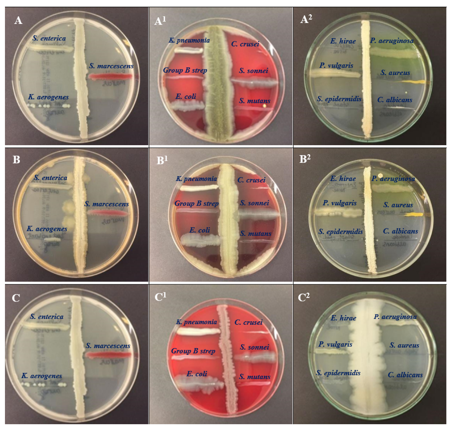
Figure 1. Antagonistic activity of the bacteria of the genus Bacillus against pathogens. Antagonistic efficacy of all three isolates was examined against pathogenic bacteria, such as Salmonella enterica ATCC 35664, Serratia marcescens ATCC 13880, Klebsiella aerogenes ATCC 13048, Candida krusei ATCC 14243, Shigella sonnei ATCC 25931, Streptococcus mutans ATCC 25175, Klebseiella pneumoniae ATCC 13883, Group B Streptococcus, Escherichia coli ATCC 25922, Pseudomonas aeruginosa ATCC 9027, Staphylococcus aureus ATCC 29213, Candida albicans ATCC 2091, Enterococcus hirae ATCC 10541, Proteus vulgaris ATCC 6380, and Staphylococcus epidermidis ATCC 12228. (AA2)—BSS11, (BB2)—BSS17, (CC2)—BSS19.
Molecules 28 03210 g001
Figure 2. Structure of the components identified from Bacillus spp. isolates.
Molecules 28 03210 g002
Figure 3. The phylogenetic tree using the neighbor-joining model was constructed based on 16S rRNA gene sequences representing different Bacillus subtilis subspecies, i.e., Bacillus subtilis O-3, Bacillus subtilis Md1-42, and Bacillus subtilis Khozestan2, respectively. As an outgroup, E. coli JCM 1649 (AB242910) was used.
Molecules 28 03210 g003
Table 1. Colony morphology and microscopic presentation of the isolated bacterial species.
Bacterial SpeciesMediaColony Color and TextureMicroscopic Presentation
Bacillus subtilis O-3 (BSS11)bacillus mediumwhite, irregular, flatgram positive, spore-forming, rod.
Bacillus subtilis Md1-42 (BSS17)bacillus mediumwhite, irregular, flatgram positive, spore-forming, rod.
Bacillus subtilis Khozestan2 (BSS19)bacillus mediumwhite, irregular, flatgram positive, spore-forming, rod.
Table 2. Antibacterial activity of the bacterial culture extracts against pathogenic strains.
Species of MicroorganismBSS11
(C), mm
BSS11 (BC), mmBSS11 (SC), mmBSS17
(C), mm
BSS17
(BC), mm
BSS17
(SC), mm
BSS19
(C), mm
BSS19
(BC), mm
BSS19
(SC), mm
Control (Streptomycin)
Staphylococcus aureus
ATCC 29213
19 ± 1.33 *21 ± 1.33 *25 ± 1.53 *22 ± 0.50 *26 ± 0.44 *30 ± 2.50 *22 ± 1.5526 ± 1.2429 ± 1.5429 ± 0.33 ***
Staphylococcus epidermidis
ATCC 12228
18 ± 1.20 *20 ± 1.33 *25 ± 1.20 *19 ± 1.0020 ± 1.2622 ± 1.5018 ± 1.42 *18 ± 1.44 *19 ± 1.54 *22 ± 0.33 ***
Streptococcus group B16 ± 1.2015 ± 1.0019 ± 1.2013 ± 1.16 *15 ± 1.14 *17 ± 1.15 *15 ± 1.33 *14 ± 1.17 *16 ± 1.33 *22 ± 0.33 ***
Streptococcus mutans
ATCC 25175
16 ± 1.22 *17 ± 1.22 *17 ± 1.82 *14 ± 1.3316 ± 1.3319 ± 1.3318 ± 0.31 *19 ± 1.00 *20 ± 1.32 *16 ± 0.33 ***
Candida albicans
ATCC 2091
27 ± 2.00 *25 ± 1.50 *30 ± 2.50 *29 ± 1.0031 ± 1.2235 ± 1.2638 ± 1.4935 ± 1.6240 ± 1.2239 ± 0.33 ***
Candida krusei
ATCC 14243
25 ± 2.33 *27 ± 2.33 *29 ± 2.35 *23 ± 1.3825 ± 0.3325 ± 1.3423 ± 2.00 *26 ± 1.66 *27 ± 2.00 *35 ± 0.33 ***
Pseudomonas aeruginosa
ATCC 9027
14 ± 1.2217 ± 0.3317 ± 1.5113 ± 0.4412 ± 1.4413 ± 1.1020 ± 1.27 *20 ± 1.53 *22 ± 1.81 *23 ***
Shigella sonnei
ATCC 25931
00000000019 ± 0.33 ***
Klebsiella pneumonia
ATCC 13883
00000000015 ± 0.33 ***
Salmonella enterica
ATCC 35664
00000000019 ± 0.33 ***
Klebsiella aerogenes
ATCC 13048
14 ± 1.82 *17 ± 1.57 *17 ± 1.82 *9 ± 1.33 *12 ± 1.22 *13 ± 1.63 *16 ± 0.53 *13 ± 1.53 *16 ± 1.53 *17 ± 0.33 ***
Enterococcus hirae
ATCC 10541
00000000020 ± 0.33 ***
Escherichia coli
ATCC 25922
18 ± 1.22 *19 ± 1.57 *20 ± 1.24 *22 ± 1.44 *20 ± 1.31 *22 ± 1.54 *19 ± 0.4922 ± 0.4622 ± 1.1723 ± 0.33 ***
Serratia marcescens
ATCC 13880
13 ± 1.22 *15 ± 0.33 *15 ± 1.56 *17 ± 1.2218 ± 1.5518 ± 1.6400024 ± 0.33 ***
Proteus Vulgaris
ATCC 6380
17 ± 1.33 *16 ± 0.33 *18 ± 1.34 *11 ± 1.33 *11 ± 1.54 *13 ± 1.53 *00022 ± 0.33 ***
* Data are represented as the means ± SE (n = 3). Values with same superscript symbols are not statistically different. Significance level * < ***.
Table 3. Antibiotic resistance profile of the Bacillus strains.
Antibiotic (AB, Charge in μg) UsedBacillus Strains
BSS11BSS17BSS19
Diameter (mm)S/RDiameter (mm)S/RDiameter (mm)S/R
PenicillinsPenicillin G (PEN, 10)30 ± 0.98 bcS24 ± 0.56 abS23 ± 0.29 aS
Ampicillin (AMP, 10)30 ± 1.43 abS27 ± 1.34 abS27± 0.38 aS
Amoxycillin (AMOX, 30)32 ± 0.98 abcS30 ± 1.30 abcS12 ± 0.29 abS
Amoxycillin-clavulanic acid (AMC, 30)28 ± 1.05 bcdS23 ± 0.33 abcS0 ± 0.00 bS
Carbenicillin (CAR, 100)38 ± 0.28 abcS28 ± 0.35 abS10 ± 0.28 abcS
Cloxacillin (CX, 5)0 ± 0.00 bR0 ± 0.00 bR0 ± 0.00 bR
MacrolidesErythromycin (ERO, 15)35 ± 0.31 abS32 ± 1.41 abcS30 ± 0.23 aS
Azithromycin (AZM, 15)36 ± 0.28 aS35 ± 1.43 abS27 ± 1.33 abS
CephalosporinsCefepime (FEP, 30)35 ± 0.51 abS25 ± 0.57 aS38 ± 0.86 abS
Cefepime/clavulanic acid FEC-4036 ± 0.98 aS30 ± 0.36 aS39 ± 0.67 aS
Cephalatin (KF, 30)32 ± 0.33 abS40 ± 0.37 abS26 ± 1.32 abcS
Cefotaxime (CTX, 30)26 ± 0.98 aS40 ± 0.52 aS24 ± 0.98 abS
AminoglycosidesGentamicin (CN, 120)40 ± 0.28 abS40 ± 0.27 abS38 ± 0.28 abS
Streptomycin (STR, 10)26 ± 0.19 abS25 ± 0.48 abS22 ± 0.20 abS
Tobramycin (TOB, 10)33 ± 0.98 aS39± 1.18 aS22 ± 0.23 abcS
TetracyclinesTetracycline (TET, 30)35 ± 0.28 aS30 ± 0.33 aS20 ± 1.18 abS
PolypeptidesPolymyxin (PB, 300)0 ± 0.00 bR0 ± 0.00 bR0 ± 0.00 bR
Bacitromycin (B, 10)0 ± 0.00 bR0 ± 0.00 bR0 ± 0.00 bR
The Newmann–Keuls test shows that the averages affected by the different superscript letters in the row and column are significantly different at the 5% level. Values are the means ± standard error; legend: D = dimension, S/R = sensible/resistant.
Table 4. The main constituents of bacterial extract BSS11 identified through a GC–MS analysis.
Bacillus subtilis O-3 (BSS11)
NoNameMolecular FormulaMolecular Mass, g/molRetention Time (min)Pubchem
Compound CID
SimilaritiesArea, %
12-ButanoneC4H8O72.112.1116569759.68622
2Acetic acid ethenyl esterC4H6O286.092.637904633.553507
32-Pentanone, 3-methyl-C6H12O100.162.99311262803.452664
4Disulfide, dimethylC2H6S294.23.57812232812.579829
52-Heptanone, 6-methyl-C8H16O128.215.60213572852.012434
6Pyrazine, methyl-C5H6N294.115.957976934.364266
7Pyrazine, 2,5-dimethyl-C6H8N2108.146.72231252924.600645
81-HexanolC6H14O102.177.0558103741.171664
9Pyrazine, trimethyl-C7H10N2122.177.82426808751.113828
10Pyrazine, 3-ethyl-2,5-dimethyl-C8H12N2136.198.35525916802.579326
111-Hexanol, 2-ethyl-C8H18O130.2298.8647720840.5058
12PyrroleC4H5N67.099.1668027900.639997
13BenzaldehydeC7H6O106.129.374240827.155098
14Acetic acid, trifluoro-, nonyl esterC11H19F3O2240.2610.3446428483730.416699
15(S)-(+)-6-Methyl-1-octanolC9H20O144.2510.60513548104790.560066
16Oxime-, methoxy-phenyl-_C8H9NO2151.1612.0259602988702.008104
17AcetamideC2H5NO59.0712.11789611.58042
18PropanamideC3H7NO73.0912.6126578710.730287
192,4-Decadienal, (E,E)-C10H16O152.2312.8255283349680.893933
20Hexanoic acidC6H12O2116.1613.0438892600.301542
212-TetradecanoneC14H28O212.3713.44175364861.671165
22(R)-(−)-4-Methylhexanoic acidC7H14O2130.1813.91312600623700.496031
23Hexanoic acid, 2-ethyl-C8H16O2144.2114.1728697660.587733
24PhenolC6H6O94.1114.738996964.246424
25Octanoic acidC8H16O2144.2115.283379670.803864
262,4,7,9-Tetramethyl-5-decyn-4,7-diolC14H26O2226.3515.65831362690.481144
27Nonanoic acidC9H18O2158.2416.3318158821.003712
282-Octyl benzoateC15H22O2234.3317.33243800660.967435
29Phenol, 2,4-bis(1,1-dimethylethyl)-C14H22O206.3217.7847311871.242429
30Benzoic acid, pentyl esterC12H16O2192.2518.34516296680.712256
31Benzoic acidC7H6O2122.1218.705243850.94121
321,2-Benzenedicarboxylic acid, bis(2-methylpropyl) esterC16H22O4278.3419.8226782932.287847
33Dibutyl phthalateC16H22O4278.3421.0723026742.75918
34Hexadecanoic acidC16H32O2256.4222.573985716.166636
35Oleic AcidC18H34O2282.524.497445639828.522779
369,12-Octadecadienoic acid (Z,Z)-C18H32O2280.425.0115280450713.023523
Table 5. The main constituents of bacterial extract BSS17 identified through a GC–MS analysis.
Bacillus subtilis Md1-42 (BSS17)
NoNameMolecular FormulaMolecular Mass, g/molRetention Time (min)Pubchem
Compound CID
SimilaritiesArea, %
1(2-Aziridinylethyl)amineC4H10N286.141.15797697780.440368
21-Propen-2-ol, acetateC5H8O2100.121.6647916760.91485
32,3-ButanedioneC4H6O286.092.6496509317.04656
43-Penten-1-olC5H10O86.135.699510370830.269452
5AcetoinC4H8O288.116.2471797637.17943
63-Pentanol, 2-methyl-C6H14O102.177.00911264801.154491
72-Nonen-1-olC9H18O142.247.10861896680.420964
82-Hydroxy-3-pentanoneC5H10O2102.137.215521790811.137636
9Ethane-1,1-diol dibutanoateC10H18O4202.258.244551339770.624527
10Acetic acidC2H4O260.058.355176930.854423
111-Hexanol, 2-ethyl-C8H18O130.2298.8897720840.206645
12BenzaldehydeC7H6O106.129.399240922.022607
132,3-ButanediolC4H10O290.129.483262783.407863
141,6-Octadien-3-ol, 3,7-dimethyl-C10H18O154.259.6326549830.530564
15Propanoic acid, 2-methyl-C4H8O288.119.8336590823.007214
162,3-Butanediol, [R-(R*,R*)]-C4H10O290.129.927225936750.516676
171-NonanolC9H20O144.2510.3678914790.235951
18(S)-(+)-6-Methyl-1-octanolC9H20O144.2510.6313548104860.668355
19Butanoic acid, 2-methyl-C5H10O2102.1311.0768314812.487502
20Oxime-, methoxy-phenyl-_C8H9NO2151.1612.051151.16700.59943
212,4-DecadienalC10H16O152.2312.8555283349700.310428
222,2,4-Trimethyl-1,3-pentanediol diisobutyrateC16H30O4286.4113.61523284730.259067
23(R)-(−)-4-Methylhexanoic acidC7H14O2130.1813.94212600623740.173808
24PhenolC6H6O94.1114.772996940.427543
25Octanoic acidC8H16O2144.2115.313379680.235055
26Nonanoic acidC9H18O2158.2416.3588158870.268447
27Hexadecanoic acid, methyl esterC17H34O2270.517.0118181890.296456
282-Octyl benzoateC15H22O2234.3317.353243800670.301277
29Benzoic acid, heptyl esterC14H20O2220.3118.07681591750.189386
30Benzoic acid, undecyl esterC18H28O2276.418.368229159700.218367
31Benzoic acidC7H6O2122.1218.538243840.270263
321,2-Benzenedicarboxylic acid, bis(2-methylpropyl) esterC16H22O4C16H22O419.2386782910.666223
33Phenol, 2,4-bis(1,1-dimethylethyl)-C14H22O206.3219.8497311860.746427
34Oleic AcidC18H34O2282.520.98445639680.295063
35Dibutyl phthalateC16H22O4278.3421.1133026821.029155
36Hexadecanoic acidC16H32O2256.4222.592985853.51103
37Octadecanoic acidC18H36O2284.524.2425281683.107501
38Oleic AcidC18H34O2282.524.525445639845.557709
399,12-Octadecadienoic acid (Z,Z)-C18H32O2280.425.0475280450849.157715
Table 6. The main constituents of bacterial extract BSS19 identified through a GC–MS analysis.
Bacillus subtilis Khozestan2 (BSS19)
NoNameMolecular FormulaMolecular Mass, g/molRetention Time (min)Pubchem
Compound CID
SimilaritiesArea, %
1Carbamic acid, monoammonium saltCH6N2O278.0711.134517232881.700997
2EthanolC2H6O46.072.234702810.46569
31-ButanolC4H10O74.124.321263916.044015
42-Heptanone, 6-methyl-C8H16O128.215.59913572851.508914
52-Heptanone, 5-methyl-C8H16O128.215.83928965870.899369
6Pyrazine, 2,5-dimethyl-C6H8N2108.146.71731252946.575729
7Acetic acidC2H4O260.058.318176934.27265
82-DecanoneC10H20O156.268.39912741763.555438
91-Hexanol, 2-ethyl-C8H18O130.2298.8597720851.019178
10BenzaldehydeC7H6O106.129.361240913.066211
111-Octene, 6-methyl-C9H18126.249.587518716770.767838
12Propanoic acid, 2-methyl-C4H8O288.119.7966590858.705907
131-Octanol, 2-methyl-C9H20O144.259.896102495820.837655
141-NonanolC9H20O144.2510.3428914811.748035
15(S)-(+)-6-Methyl-1-octanolC9H20O144.2510.60413548104903.260679
16BenzeneacetaldehydeC8H8O120.1510.821998692.227017
172-FuranmethanolC5H6O298.110.9187361871.948174
18Hexanoic acid, 2-methyl-C7H14O2130.1811.036206538025.50356
192-DodecanoneC12H24O184.3211.19822556771.361537
20Oxime-, methoxy-phenyl-_C8H9NO2151.1612.0189602988651.54511
212(5H)-FuranoneC4H4O284.0712.11410341772.40381
22FormamideCH3NO45.04112.32713770.972164
232-TetradecanoneC14H28O212.3713.4475364801.097307
24MaltolC6H6O3126.1114.3758369934.442068
25Ethanone, 1-(1H-pyrrol-2-yl)-C6H7NO109.1314.42914079671.456046
26PhenolC6H6O94.1114.739996966.642077
274H-Pyran-4-one, 2,3-dihydro-3,5-dihydroxy-6-methyl-C6H8O4144.1217.293119838881.650456
28Benzoic acid, hept-2-yl esterC14H20O2220.3118.053243678750.421796
29Benzoic acidC7H6O2122.1218.698243911.317335
305-Hydroxymethyldihydrofuran-2-oneC5H8O3116.1119.19698431730.384416
311,2-Benzenedicarboxylic acid, bis(2-methylpropyl) esterC16H22O4C16H22O419.3566782910.986222
32Phenol, 2,4-bis(1,1-dimethylethyl)-C14H22O206.3219.7867311851.085735
Table 7. Identification of the bacterial species based on the sequence similarities.
NoIsolates16S rRNA Amplified Region Length% SimilarityNCBI Accession No
1BSS111443 bp99% with Bacillus subtilis O-3GQ870259
2BSS171454 bp99% with Bacillus subtilis Md1-42MF581448
3BSS191450 bp99% with Bacillus subtilis Khozestan2MH036316
Disclaimer/Publisher’s Note: The statements, opinions and data contained in all publications are solely those of the individual author(s) and contributor(s) and not of MDPI and/or the editor(s). MDPI and/or the editor(s) disclaim responsibility for any injury to people or property resulting from any ideas, methods, instructions or products referred to in the content.