

Recent Advances in Mycotoxin Determination in Fish Feed Ingredients

 ,
, 
Abstract
1. Introduction
2. Sampling and Sample Preparation Methods
2.1. Sampling and Sample Preparation
2.2. Sampling Error
2.3. Sample Pre-Treatment (Extraction and/or Clean-Up)
2.3.1. Solid–Liquid Extraction (SLE)
2.3.2. Dispersive-Solid Phase Extraction (d-SPE)
2.3.3. Clean Up by Immunoaffinity Column (IAC)
2.3.4. Solid Phase Extraction (SPE)
2.3.5. Molecular Imprinted Polymer (MIP)
2.3.6. Ultrasonic Solvent Extraction (USE)
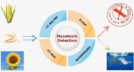
3. Instrumental Analysis
3.1. Chromatographic Methods—Detection Systems
3.2. Immunological Methods (Enzyme-Linked Immunosorbent Assay-ELISA)
3.3. Biosensors
3.4. Spectroscopic Methods FT-NIR
4. Conclusions and Outlook
Funding
Data Availability Statement
Conflicts of Interest
References
- FAO. The State of World Fisheries and Aquaculture 2022. Towards Blue Transformation; FAO: Rome, Italy, 2022. [Google Scholar]
- Edwards, P.; Zhang, W.; Belton, B.; Little, D.C. Misunderstandings, myths and mantras in aquaculture: Its contribution to world food supplies has been systematically over reported. Mar. Policy 2019, 106, 103547. [Google Scholar] [CrossRef]
- Ottinger, M.; Clauss, K.; Kuenzer, C. Aquaculture: Relevance, distribution, impacts and spatial assessments–A review. Ocean Coast. Manag. 2016, 119, 244–266. [Google Scholar] [CrossRef]
- Springmann, M.; Clark, M.; Mason-D’Croz, D.; Wiebe, K.; Bodirsky, B.L.; Lassaletta, L.; De Vries, W.; Vermeulen, S.J.; Herrero, M.; Carlson, K.M. Options for keeping the food system within environmental limits. Nature 2018, 562, 519–525. [Google Scholar] [CrossRef]
- Willett, W.; Rockström, J.; Loken, B.; Springmann, M.; Lang, T.; Vermeulen, S.; Garnett, T.; Tilman, D.; DeClerck, F.; Wood, A. Food in the Anthropocene: The EAT–Lancet Commission on healthy diets from sustainable food systems. Lancet 2019, 393, 447–492. [Google Scholar] [CrossRef]
- Newton, R.; Maiolo, S.; Malcorps, W.; Little, D. Life cycle inventories of marine ingredients. Aquaculture 2023, 565, 739096. [Google Scholar] [CrossRef]
- Miles, R.D.; Chapman, F.A. The benefits of fish meal in aquaculture diets. FA122/FA122, 5/2006. EDIS 2006, 12, 1–7. [Google Scholar]
- Naylor, R.L.; Hardy, R.W.; Buschmann, A.H.; Bush, S.R.; Cao, L.; Klinger, D.H.; Little, D.C.; Lubchenco, J.; Shumway, S.E.; Troell, M. A 20-year retrospective review of global aquaculture. Nature 2021, 591, 551–563. [Google Scholar] [CrossRef] [PubMed]
- IndexMundi—Country Facts. Available online: https://www.indexmundi.com/agriculture/?commodity=fish-meal&graph=production (accessed on 23 December 2022).
- Maiolo, S.; Parisi, G.; Biondi, N.; Lunelli, F.; Tibaldi, E.; Pastres, R. Fishmeal partial substitution within aquafeed formulations: Life cycle assessment of four alternative protein sources. Int. J. Life Cycle Assess. 2020, 25, 1455–1471. [Google Scholar] [CrossRef]
- Elumalai, P.; Kurian, A.; Lakshmi, S.; Faggio, C.; Esteban, M.A.; Ringø, E. Herbal immunomodulators in aquaculture. Rev. Fish. Sci. Aquac. 2020, 29, 33–57. [Google Scholar] [CrossRef]
- Gatlin, D.M., III; Barrows, F.T.; Brown, P.; Dabrowski, K.; Gaylord, T.G.; Hardy, R.W.; Herman, E.; Hu, G.; Krogdahl, Å.; Nelson, R. Expanding the utilization of sustainable plant products in aquafeeds: A review. Aquac. Res. 2007, 38, 551–579. [Google Scholar] [CrossRef]
- Hardy, R.W. Utilization of plant proteins in fish diets: Effects of global demand and supplies of fishmeal. Aquac. Res. 2010, 41, 770–776. [Google Scholar] [CrossRef]
- Sales, J. The effect of fish meal replacement by soyabean products on fish growth: A meta-analysis. Br. J. Nutr. 2009, 102, 1709–1722. [Google Scholar] [CrossRef]
- Hixson, S.M. Fish nutrition and current issues in aquaculture: The balance in providing safe and nutritious seafood, in an environmentally sustainable manner. J. Aquac. Res. Dev. 2014, 5, 1–10. [Google Scholar] [CrossRef]
- Samuelsen, T.A.; Hillestad, M.; Jacobsen, H.J.; Hjertnes, T.J.; Sixten, H.J. Physical feed quality and starch content causes a biological response in Atlantic salmon (Salmo salar L.). Aquac. Rep. 2021, 21, 100791. [Google Scholar] [CrossRef]
- Troell, M.; Naylor, R.L.; Metian, M.; Beveridge, M.; Tyedmers, P.H.; Folke, C.; Arrow, K.J.; Barrett, S.; Crépin, A.-S.; Ehrlich, P.R. Does aquaculture add resilience to the global food system? Proc. Natl. Acad. Sci. USA 2014, 111, 13257–13263. [Google Scholar] [CrossRef]
- Sicuro, B.; Gai, F.; Daprà, F.; Palmegiano, G.B. Hybrid sturgeon ‘AL’(Acipenser naccarii× Acipenser baeri) diets: The use of alternative plant protein sources. Aquac. Res. 2012, 43, 161–166. [Google Scholar] [CrossRef]
- Bonaldo, A.; Parma, L.; Mandrioli, L.; Sirri, R.; Fontanillas, R.; Badiani, A.; Gatta, P.P. Increasing dietary plant proteins affects growth performance and ammonia excretion but not digestibility and gut histology in turbot (Psetta maxima) juveniles. Aquaculture 2011, 318, 101–108. [Google Scholar] [CrossRef]
- Nogales-Mérida, S.; Tomás-Vidal, A.; Moñino-López, A.; Jover-Cerdá, M.; Martínez-Llorens, S. Pea protein concentrate in diets for sharpsnout sea bream (Diplodus puntazzo): Effects on growth and health status. Arch. Anim. Nutr. 2016, 70, 488–502. [Google Scholar] [CrossRef] [PubMed]
- Parma, L.; Candela, M.; Soverini, M.; Turroni, S.; Consolandi, C.; Brigidi, P.; Mandrioli, L.; Sirri, R.; Fontanillas, R.; Gatta, P.P. Next-generation sequencing characterization of the gut bacterial community of gilthead sea bream (Sparus aurata, L.) fed low fishmeal based diets with increasing soybean meal levels. Anim. Feed Sci. Technol. 2016, 222, 204–216. [Google Scholar] [CrossRef]
- Krogdahl, Å.; Penn, M.; Thorsen, J.; Refstie, S.; Bakke, A.M. Important antinutrients in plant feedstuffs for aquaculture: An update on recent findings regarding responses in salmonids. Aquac. Res. 2010, 41, 333–344. [Google Scholar] [CrossRef]
- Oliva-Teles, A. Nutrition and health of aquaculture fish. J. Fish Dis. 2012, 35, 83–108. [Google Scholar] [CrossRef]
- Oliva-Teles, A.; Enes, P.; Peres, H. Replacing fishmeal and fish oil in industrial aquafeeds for carnivorous fish. In Feed and Feeding Practices in Aquaculture; Woodhead Publishing Series in Food Science, Technology and Nutrition; Woodhead Publishing: Sawston, CA, USA, 2015; pp. 203–233. [Google Scholar]
- Kokou, F.; Fountoulaki, E. Aquaculture waste production associated with antinutrient presence in common fish feed plant ingredients. Aquaculture 2018, 495, 295–310. [Google Scholar] [CrossRef]
- Gonçalves, R.A.; Schatzmayr, D.; Albalat, A.; Mackenzie, S. Mycotoxins in aquaculture: Feed and food. Rev. Aquac. 2020, 12, 145–175. [Google Scholar] [CrossRef]
- Haque, M.A.; Wang, Y.; Shen, Z.; Li, X.; Saleemi, M.K.; He, C. Mycotoxin contamination and control strategy in human, domestic animal and poultry: A review. Microb. Pathog. 2020, 142, 104095. [Google Scholar] [CrossRef]
- Palumbo, R.; Crisci, A.; Venâncio, A.; Cortiñas Abrahantes, J.; Dorne, J.-L.; Battilani, P.; Toscano, P. Occurrence and co-occurrence of mycotoxins in cereal-based feed and food. Microorganisms 2020, 8, 74. [Google Scholar] [CrossRef]
- Streit, E.; Naehrer, K.; Rodrigues, I.; Schatzmayr, G. Mycotoxin occurrence in feed and feed raw materials worldwide: Long-term analysis with special focus on Europe and Asia. J. Sci. Food Agric. 2013, 93, 2892–2899. [Google Scholar] [CrossRef]
- International Agency for Research on Cancer. A Review of Human Carcinogens. F. Chemical Agents and Related Occupations: IARC Monographs on the Evaluation of Carcinogenic Risks to Humans; International Agency for Research on Cancer: Lyon, France, 2012; p. 100F. [Google Scholar]
- World Health Organization; International Agency for Research on Cancer. Some naturally occurring substances: Food items and constituents, heterocyclic aromatic amines and mycotoxins. In IARC Monographs on the Evaluation of the Carcinogenic Risk of Chemicals to Humans; World Health Organization: Geneva, Switzerland, 1993; Volume 56. [Google Scholar]
- Tomatis, L.; Wilbourn, J. Evaluation of carcinogenic risk to humans: The experience of IARC. In New Frontiers in Cancer Causation; CRC Press: Boca Raton, FL, USA, 1993; pp. 371–387. [Google Scholar]
- Alassane-Kpembi, I.; Schatzmayr, G.; Taranu, I.; Marin, D.; Puel, O.; Oswald, I.P. Mycotoxins co-contamination: Methodological aspects and biological relevance of combined toxicity studies. Crit. Rev. Food Sci. Nutr. 2017, 57, 3489–3507. [Google Scholar] [CrossRef]
- Lee, H.J.; Ryu, D. Worldwide occurrence of mycotoxins in cereals and cereal-derived food products: Public health perspectives of their co-occurrence. J. Agric. Food Chem. 2017, 65, 7034–7051. [Google Scholar] [CrossRef] [PubMed]
- Santos Pereira, C.; Cunha, S.C.; Fernandes, J.O. Prevalent mycotoxins in animal feed: Occurrence and analytical methods. Toxins 2019, 11, 290. [Google Scholar] [CrossRef] [PubMed]
- Marijani, E.; Kigadye, E.; Okoth, S. Occurrence of fungi and mycotoxins in fish feeds and their impact on fish health. Int. J. Microbiol. 2019, 2019, 1–17. [Google Scholar] [CrossRef]
- Oliveira, M.; Vasconcelos, V. Occurrence of mycotoxins in fish feed and its effects: A review. Toxins 2020, 12, 160. [Google Scholar] [CrossRef]
- Zain, M.E. Impact of mycotoxins on humans and animals. J. Saudi Chem. Soc. 2011, 15, 129–144. [Google Scholar] [CrossRef]
- Koletsi, P.; Schrama, J.W.; Graat, E.A.M.; Wiegertjes, G.F.; Lyons, P.; Pietsch, C. The Occurrence of Mycotoxins in Raw Materials and Fish Feeds in Europe and the Potential Effects of Deoxynivalenol (DON) on the Health and Growth of Farmed Fish Species—A Review. Toxins 2021, 13, 403. [Google Scholar] [CrossRef]
- Da Rocha, M.E.B.; Freire, F.d.C.O.; Maia, F.E.F.; Guedes, M.I.F.; Rondina, D. Mycotoxins and their effects on human and animal health. Food Control 2014, 36, 159–165. [Google Scholar] [CrossRef]
- Directive 2002/32/EC of the European Parliament and of the Council of 7 May 2002 on Undesirable Substances in Animal Feed. Available online: https://webarchive.nationalarchives.gov.uk/eu-exit/https://eur-lex.europa.eu/legal-content/EN/TXT/?uri=CELEX:02002L0032-20191128 (accessed on 28 December 2022).
- Alexander, J.; Benford, D.; Boobis, A.; Ceccatelli, S.; Cottrill, B.; Cravedi, J.-P.; Di Domenico, A.; Doerge, D.; Dogliotti, E.; Edler, L.; et al. Scientific Opinion on Ergot alkaloids in food and feed. EFSA J. 2012, 10, 2798. [Google Scholar] [CrossRef]
- Commission recommendation on the presence of deoxynivalenol, zearalenone, ochratoxin A, T-2 and HT-2 and fumonisins in products intended for animal feeding (Text with EEA relevance) At the request of the Commission the European Food Safety Authority (EFSA) adopted opinions on the myco-toxins deoxynivalenol. Off. J. Eur. Union 2006, 229, 7–9.
- EFSA Panel on Contaminants in the Food Chain (CONTAM). Scientific Opinion on the risks for animal and public health related to the presence of T-2 and HT-2 toxin in food and feed. EFSA J. 2011, 9, 2481. [Google Scholar] [CrossRef]
- Singh, J.; Mehta, A. Rapid and sensitive detection of mycotoxins by advanced and emerging analytical methods: A review. Food Sci. Nutr. 2020, 8, 2183–2204. [Google Scholar] [CrossRef]
- Janik, E.; Niemcewicz, M.; Podogrocki, M.; Ceremuga, M.; Gorniak, L.; Stela, M.; Bijak, M. The existing methods and novel approaches in mycotoxins’ detection. Molecules 2021, 26, 3981. [Google Scholar] [CrossRef]
- Wagner, C. Critical practicalities in sampling for mycotoxins in feed. J. AOAC Int. 2015, 98, 301–308. [Google Scholar] [CrossRef] [PubMed]
- Xie, L.; Chen, M.; Ying, Y. Development of methods for determination of aflatoxins. Crit. Rev. Food Sci. Nutr. 2016, 56, 2642–2664. [Google Scholar] [CrossRef]
- The European Commission Commission Regulation (EC) No 691/2013 of 19 July 2013 amending Regulation (EC) No 152/2009 as regards methods of sampling and analysis. Off. J. Eur. Union 2013, 197, 1–12.
- Turner, N.W.; Bramhmbhatt, H.; Szabo-Vezse, M.; Poma, A.; Coker, R.; Piletsky, S.A. Analytical methods for determination of mycotoxins: An update (2009–2014). Anal. Chim. Acta 2015, 901, 12–33. [Google Scholar] [CrossRef]
- European Commision. Commission Regulation (EC) No 401/2006 of 23 February 2006 laying down the methods of sampling and analysis for the official control of the levels of mycotoxins in foodstuffs. Off. J. Eur. Union 2006, 70, 12–34.
- Maestroni, B.; Cannavan, A. Sampling strategies to control mycotoxins. In Determining Mycotoxins and Mycotoxigenic Fungi in Food and Feed; Woodhead Publishing: Sawston, CA, USA, 2011; pp. 3–36. [Google Scholar]
- Turner, N.W.; Subrahmanyam, S.; Piletsky, S.A. Analytical methods for determination of mycotoxins: A review. Anal. Chim. Acta 2009, 632, 168–180. [Google Scholar] [CrossRef]
- Whitaker, T.B. Standardisation of mycotoxin sampling procedures: An urgent necessity. Food Control 2003, 14, 233–237. [Google Scholar] [CrossRef]
- Miraglia, M.; De Santis, B.; Minardi, V.; Debegnach, F.; Brera, C. The role of sampling in mycotoxin contamination: An holistic view. Food Addit. Contam. 2005, 22, 31–36. [Google Scholar] [CrossRef] [PubMed]
- Krska, R.; Welzig, E.; Berthiller, F.; Molinelli, A.; Mizaikoff, B. Advances in the analysis of mycotoxins and its quality assurance. Food Addit. Contam. 2005, 22, 345–353. [Google Scholar] [CrossRef]
- Razzazi-Fazeli, E.; Reiter, E.V. Sample preparation and clean up in mycotoxin analysis: Principles, applications and recent developments. In Determining Mycotoxins and Mycotoxigenic Fungi in Food and Feed; Woodhead Publishing: Sawston, CA, USA, 2011; pp. 37–70. [Google Scholar]
- Kafouris, D.; Christofidou, M.; Christodoulou, M.; Christou, E.; Ioannou-Kakouri, E. A validated UPLC-MS/MS multi-mycotoxin method for nuts and cereals: Results of the official control in Cyprus within the EU requirements. Food Agric. Immunol. 2017, 28, 90–108. [Google Scholar] [CrossRef]
- Kovač, M.; Nevistić, A.; Kovač, T.; Babić, J.; Šarić, A.; Miličević, B.; Panjičko, M.; Šarkanj, B. Development and Validation of an UHPLC-MS/MS Method for the Simultaneous Determination of 11 EU-Regulated Mycotoxins in Selected Cereals. J. Fungi 2022, 8, 665. [Google Scholar] [CrossRef] [PubMed]
- Nathanail, A.V.; Syvähuoko, J.; Malachová, A.; Jestoi, M.; Varga, E.; Michlmayr, H.; Adam, G.; Sieviläinen, E.; Berthiller, F.; Peltonen, K. Simultaneous determination of major type A and B trichothecenes, zearalenone and certain modified metabolites in Finnish cereal grains with a novel liquid chromatography-tandem mass spectrometric method. Anal. Bioanal. Chem. 2015, 407, 4745–4755. [Google Scholar] [CrossRef]
- Silva, A.S.; Brites, C.; Pouca, A.V.; Barbosa, J.; Freitas, A. UHPLC-ToF-MS method for determination of multi-mycotoxins in maize: Development and validation. Curr. Res. Food Sci. 2019, 1, 1–7. [Google Scholar] [CrossRef] [PubMed]
- Cortese, M.; Gigliobianco, M.R.; Magnoni, F.; Censi, R.; Di Martino, P. Compensate for or minimize matrix effects? Strategies for overcoming matrix effects in liquid chromatography-mass spectrometry technique: A tutorial review. Molecules 2020, 25, 3047. [Google Scholar] [CrossRef]
- Furey, A.; Moriarty, M.; Bane, V.; Kinsella, B.; Lehane, M. Ion suppression; a critical review on causes, evaluation, prevention and applications. Talanta 2013, 115, 104–122. [Google Scholar] [CrossRef] [PubMed]
- Varzakas, T.; Tzia, C. Food Engineering Handbook: Food Process Engineering; CRC Press: Boca Raton, FL, USA, 2014; ISBN 1482261669. [Google Scholar]
- Prado, J.; Rostagno, M. Natural Product Extraction: Principles and Applications; Royal Society of Chemistry: London, UK, 2022; ISBN 1839165901. [Google Scholar]
- Annunziata, L.; Stramenga, A.; Visciano, P.; Schirone, M.; De Colli, L.; Colagrande, M.N.; Campana, G.; Scortichini, G. Simultaneous determination of aflatoxins, T-2 and HT-2 toxins, and fumonisins in cereal-derived products by QuEChERS extraction coupled with LC-MS/MS. Anal. Bioanal. Chem. 2017, 409, 5143–5155. [Google Scholar] [CrossRef]
- Bolechová, M.; Benešová, K.; Běláková, S.; Čáslavský, J.; Pospíchalová, M.; Mikulíková, R. Determination of seventeen mycotoxins in barley and malt in the Czech Republic. Food Control 2015, 47, 108–113. [Google Scholar] [CrossRef]
- González-Jartín, J.M.; Alfonso, A.; Sainz, M.J.; Vieytes, M.R.; Botana, L.M. Multi-detection method for mycotoxins with a modified QuEChERS extraction in feed and development of a simple detoxification procedure. Anim. Feed Sci. Technol. 2021, 272, 114745. [Google Scholar] [CrossRef]
- Kim, H.; Baek, E.J.; Shin, B.G.; Kim, H.J.; Kim, J.-E. In-house validation of an efficient and rapid procedure for the simultaneous determination and monitoring of 23 mycotoxins in grains in Korea. Toxins 2022, 14, 457. [Google Scholar] [CrossRef]
- Rausch, A.-K.; Brockmeyer, R.; Schwerdtle, T. Development and validation of a QuEChERS-based liquid chromatography tandem mass spectrometry multi-method for the determination of 38 native and modified mycotoxins in cereals. J. Agric. Food Chem. 2020, 68, 4657–4669. [Google Scholar] [CrossRef]
- Tamura, M.; Mochizuki, N.; Nagatomi, Y.; Harayama, K.; Toriba, A.; Hayakawa, K. A method for simultaneous determination of 20 Fusarium toxins in cereals by high-resolution liquid chromatography-orbitrap mass spectrometry with a pentafluorophenyl column. Toxins 2015, 7, 1664–1682. [Google Scholar] [CrossRef]
- Zhou, Q.; Li, F.; Chen, L.; Jiang, D. Quantitative analysis of 10 mycotoxins in wheat flour by ultrahigh performance liquid chromatography-tandem mass spectrometry with a modified QuEChERS strategy. J. Food Sci. 2016, 81, T2886–T2890. [Google Scholar] [CrossRef] [PubMed]
- Scott, P.M.; Trucksess, M.W. Application of immunoaffinity columns to mycotoxin analysis. J. AOAC Int. 1997, 80, 941–950. [Google Scholar] [CrossRef]
- Gab-Allah, M.A.; Tahoun, I.F.; Yamani, R.N.; Rend, E.A.; Shehata, A.B. Eco-friendly and sensitive analytical method for determination of T-2 toxin and HT-2 toxin in cereal products using UPLC-MS/MS. J. Food Compos. Anal. 2022, 107, 104395. [Google Scholar] [CrossRef]
- Göbel, R.; Lusky, K. Simultaneous determination of aflatoxins, ochratoxin A, and zearalenone in grains by new immunoaffinity column/liquid chromatography. J. AOAC Int. 2004, 87, 411–416. [Google Scholar] [CrossRef] [PubMed]
- Pascale, M.; Panzarini, G.; Visconti, A. Determination of HT-2 and T-2 toxins in oats and wheat by ultra-performance liquid chromatography with photodiode array detection. Talanta 2012, 89, 231–236. [Google Scholar] [CrossRef]
- De Boevre, M.; Di Mavungu, J.D.; Maene, P.; Audenaert, K.; Deforce, D.; Haesaert, G.; Eeckhout, M.; Callebaut, A.; Berthiller, F.; Van Peteghem, C. Development and validation of an LC-MS/MS method for the simultaneous determination of deoxynivalenol, zearalenone, T-2-toxin and some masked metabolites in different cereals and cereal-derived food. Food Addit. Contam. Part A 2012, 29, 819–835. [Google Scholar] [CrossRef]
- Boulard, L.; Parrhysius, P.; Jacobs, B.; Dierkes, G.; Wick, A.; Buchmeier, G.; Koschorreck, J.; Ternes, T.A. Development of an analytical method to quantify pharmaceuticals in fish tissues by liquid chromatography-tandem mass spectrometry detection and application to environmental samples. J. Chromatogr. A 2020, 1633, 461612. [Google Scholar] [CrossRef]
- Rios, A.; Zougagh, M.; Bouri, M. Magnetic (nano) materials as an useful tool for sample preparation in analytical methods. A review. Anal. Methods 2013, 5, 4558–4573. [Google Scholar] [CrossRef]
- Mashhadizadeh, M.H.; Amoli-Diva, M.; Pourghazi, K. Magnetic nanoparticles solid phase extraction for determination of ochratoxin A in cereals using high-performance liquid chromatography with fluorescence detection. J. Chromatogr. A 2013, 1320, 17–26. [Google Scholar] [CrossRef]
- La Barbera, G.; Capriotti, A.L.; Cavaliere, C.; Foglia, P.; Montone, C.M.; Chiozzi, R.Z.; Laganà, A. A Rapid Magnetic Solid Phase Extraction Method Followed by Liquid Chromatography-Tandem Mass Spectrometry Analysis for the Determination of Mycotoxins in Cereals. Toxins 2017, 9, 147. [Google Scholar] [CrossRef]
- Lucci, P.; Derrien, D.; Alix, F.; Pérollier, C.; Bayoudh, S. Molecularly imprinted polymer solid-phase extraction for detection of zearalenone in cereal sample extracts. Anal. Chim. Acta 2010, 672, 15–19. [Google Scholar] [CrossRef] [PubMed]
- Cavaliere, C.; Antonelli, M.; Cerrato, A.; La Barbera, G.; Laganà, A.; Laus, M.; Piovesana, S.; Capriotti, A.L. A novel magnetic molecular imprinted polymer for selective extraction of zearalenone from cereal flours before liquid chromatography-tandem mass spectrometry determination. Toxins 2019, 11, 493. [Google Scholar] [CrossRef] [PubMed]
- Xie, Z.; Lu, G.; Yan, Z.; Liu, J.; Wang, P.; Wang, Y. Bioaccumulation and trophic transfer of pharmaceuticals in food webs from a large freshwater lake. Environ. Pollut. 2017, 222, 356–366. [Google Scholar] [CrossRef]
- Krska, R.; Stubbings, G.; Macarthur, R.; Crews, C. Simultaneous determination of six major ergot alkaloids and their epimers in cereals and foodstuffs by LC–MS–MS. Anal. Bioanal. Chem. 2008, 391, 563–576. [Google Scholar] [CrossRef]
- Soleimany, F.; Jinap, S.; Faridah, A.; Khatib, A. A UPLC–MS/MS for simultaneous determination of aflatoxins, ochratoxin A, zearalenone, DON, fumonisins, T-2 toxin and HT-2 toxin, in cereals. Food Control 2012, 25, 647–653. [Google Scholar] [CrossRef]
- Agriopoulou, S.; Stamatelopoulou, E.; Varzakas, T. Advances in occurrence, importance, and mycotoxin control strategies: Prevention and detoxification in foods. Foods 2020, 9, 137. [Google Scholar] [CrossRef]
- Bi, S.; Xu, J.; Yang, X.; Zhang, P.; Lian, K.; Ma, L. An HPLC-MS/MS Method Using a Multitoxin Clean up Column for Analysis of Seven Mycotoxins in Aquafeeds. J. AOAC Int. 2022, 105, 107–114. [Google Scholar] [CrossRef]
- Moreno-González, D.; Huertas-Pérez, J.F.; García-Campaña, A.M.; Gámiz-Gracia, L. Determination of carbamates in edible vegetable oils by ultra-high performance liquid chromatography–tandem mass spectrometry using a new clean-up based on zirconia for QuEChERS methodology. Talanta 2014, 128, 299–304. [Google Scholar] [CrossRef]
- Kim, D.-B.; Jung, Y.S.; Nam, T.G.; Lee, S.; Yoo, M. Simultaneous determination of trichothecene mycotoxins in cereals by LC-MS/MS. Food Sci. Biotechnol. 2022, 31, 165–174. [Google Scholar] [CrossRef]
- Lijalem, Y.G.; Gab-Allah, M.A.; Choi, K.; Kim, B. Development of isotope dilution-liquid chromatography/tandem mass spectrometry for the accurate determination of zearalenone and its metabolites in corn. Food Chem. 2022, 384, 132483. [Google Scholar] [CrossRef]
- Gab-Allah, M.A.; Choi, K.; Kim, B. Development of isotope dilution-liquid chromatography/tandem mass spectrometry for the accurate determination of type-A trichothecenes in grains. Food Chem. 2021, 344, 128698. [Google Scholar] [CrossRef] [PubMed]
- Meerpoel, C.; Vidal, A.; di Mavungu, J.D.; Huybrechts, B.; Tangni, E.K.; Devreese, M.; Croubels, S.; De Saeger, S. Development and validation of an LC–MS/MS method for the simultaneous determination of citrinin and ochratoxin a in a variety of feed and foodstuffs. J. Chromatogr. A 2018, 1580, 100–109. [Google Scholar] [CrossRef] [PubMed]
- Liu, Z.; Wu, H.-L.; Xie, L.-X.; Hu, Y.; Fang, H.; Sun, X.-D.; Wang, T.; Xiao, R.; Yu, R.-Q. Chemometrics-enhanced liquid chromatography-full scan-mass spectrometry for interference-free analysis of multi-class mycotoxins in complex cereal samples. Chemom. Intell. Lab. Syst. 2017, 160, 125–138. [Google Scholar] [CrossRef]
- Skendi, A.; Irakli, M.N.; Papageorgiou, M.D. Optimized and validated high-performance liquid chromatography method for the determination of deoxynivalenol and aflatoxins in cereals. J. Sep. Sci. 2016, 39, 1425–1432. [Google Scholar] [CrossRef] [PubMed]
- Vidal, J.C.; Duato, P.; Bonel, L.; Castillo, J.R. Molecularly imprinted on-line solid-phase extraction coupled with fluorescence detection for the determination of ochratoxin A in wheat samples. Anal. Lett. 2012, 45, 51–62. [Google Scholar] [CrossRef]
- Tahoun, I.F.; Gab-Allah, M.A.; Yamani, R.N.; Shehata, A.B. Development and validation of a reliable LC-MS/MS method for simultaneous determination of deoxynivalenol and T-2 toxin in maize and oats. Microchem. J. 2021, 169, 106599. [Google Scholar] [CrossRef]
- Arroyo-Manzanares, N.; De Ruyck, K.; Uka, V.; Gámiz-Gracia, L.; García-Campaña, A.M.; De Saeger, S.; Diana Di Mavungu, J. In-house validation of a rapid and efficient procedure for simultaneous determination of ergot alkaloids and other mycotoxins in wheat and maize. Anal. Bioanal. Chem. 2018, 410, 5567–5581. [Google Scholar] [CrossRef]
- Rahmani, A.; Jinap, S.; Khatib, A.; Tan, C.P. Simultaneous determination of aflatoxins, ochratoxin A, and zearalenone in cereals using a validated RP-HPLC method and PHRED derivatization system. J. Liq. Chromatogr. Relat. Technol. 2013, 36, 600–617. [Google Scholar] [CrossRef]
- Pietsch, C.; Kersten, S.; Burkhardt-Holm, P.; Valenta, H.; Dänicke, S. Occurrence of deoxynivalenol and zearalenone in commercial fish feed: An initial study. Toxins 2013, 5, 184–192. [Google Scholar] [CrossRef]
- Albero, B.; Fernández-Cruz, M.L.; Pérez, R.A. Simultaneous Determination of 15 Mycotoxins in Aquaculture Feed by Liquid Chromatography–Tandem Mass Spectrometry. Toxins 2022, 14, 316. [Google Scholar] [CrossRef]
- Li, P.; Zhang, Q.; Zhang, W. Immunoassays for aflatoxins. TrAC Trends Anal. Chem. 2009, 28, 1115–1126. [Google Scholar] [CrossRef]
- Meulenberg, E.P. Immunochemical methods for ochratoxin A detection: A review. Toxins 2012, 4, 244–266. [Google Scholar] [CrossRef]
- Sanders, M.; Guo, Y.; Iyer, A.; Ruiz García, Y.; Galvita, A.; Heyerick, A.; Deforce, D.; Risseeuw, M.D.P.; Van Calenbergh, S.; Bracke, M. An immunogen synthesis strategy for the development of specific anti-deoxynivalenol monoclonal antibodies. Food Addit. Contam. Part A 2014, 31, 1751–1759. [Google Scholar] [CrossRef]
- Li, S.; Wang, J.; Sheng, W.; Wen, W.; Gu, Y.; Wang, S. Fluorometric lateral flow immunochromatographic zearalenone assay by exploiting a quencher system composed of carbon dots and silver nanoparticles. Microchim. Acta 2018, 185, 388. [Google Scholar] [CrossRef]
- Ezquerra, A.; Vidal, J.C.; Bonel, L.; Castillo, J.R. A validated multi-channel electrochemical immunoassay for rapid fumonisin B1 determination in cereal samples. Anal. Methods 2015, 7, 3742–3749. [Google Scholar] [CrossRef]
- Oswald, S.; Karsunke, X.Y.Z.; Dietrich, R.; Märtlbauer, E.; Niessner, R.; Knopp, D. Automated regenerable microarray-based immunoassay for rapid parallel quantification of mycotoxins in cereals. Anal. Bioanal. Chem. 2013, 405, 6405–6415. [Google Scholar] [CrossRef]
- McNamee, S.E.; Bravin, F.; Rosar, G.; Elliott, C.T.; Campbell, K. Development of a nanoarray capable of the rapid and simultaneous detection of zearalenone, T2-toxin and fumonisin. Talanta 2017, 164, 368–376. [Google Scholar] [CrossRef]
- D’Agnello, P.; Vita, V.; Franchino, C.; Urbano, L.; Curiale, A.; Debegnach, F.; Iammarino, M.; Marchesani, G.; Chiaravalle, A.E.; De Pace, R. ELISA and UPLC/FLD as Screening and Confirmatory Techniques for T-2/HT-2 Mycotoxin Determination in Cereals. Appl. Sci. 2021, 11, 1688. [Google Scholar] [CrossRef]
- Peltomaa, R.; Abbas, A.; Yli-Mattila, T.; Lamminmäki, U. Single-step noncompetitive immunocomplex immunoassay for rapid aflatoxin detection. Food Chem. 2022, 392, 133287. [Google Scholar] [CrossRef]
- Wang, Y.-K.; Yan, Y.-X.; Li, S.-Q.; Wang, H.; Ji, W.-H.; Sun, J.-H. Simultaneous quantitative determination of multiple mycotoxins in cereal and feedstuff samples by a suspension array immunoassay. J. Agric. Food Chem. 2013, 61, 10948–10953. [Google Scholar] [CrossRef]
- Vidal, J.C.; Bertolín, J.R.; Ezquerra, A.; Hernández, S.; Castillo, J.R. Rapid simultaneous extraction and magnetic particle-based enzyme immunoassay for the parallel determination of ochratoxin A, fumonisin B1 and deoxynivalenol mycotoxins in cereal samples. Anal. Methods 2017, 9, 3602–3611. [Google Scholar] [CrossRef]
- Bian, Y.; Huang, X.; Ren, J. Sensitive and homogenous immunoassay of fumonisin in foods using single molecule fluorescence correlation spectroscopy. Anal. Methods 2016, 8, 1333–1338. [Google Scholar] [CrossRef]
- Beloglazova, N.V.; Foubert, A.; Gordienko, A.; Tessier, M.D.; Aubert, T.; Drijvers, E.; Goryacheva, I.; Hens, Z.; De Saeger, S. Sensitive QD@ SiO2-based immunoassay for triplex determination of cereal-borne mycotoxins. Talanta 2016, 160, 66–71. [Google Scholar] [CrossRef] [PubMed]
- Beloglazova, N.V.; Graniczkowska, K.; Njumbe Ediage, E.; Averkieva, O.; De Saeger, S. Sensitive flow-through immunoassay for rapid multiplex determination of cereal-borne mycotoxins in feed and feed ingredients. J. Agric. Food Chem. 2017, 65, 7131–7137. [Google Scholar] [CrossRef]
- He, Q.-H.; Xu, Y.; Wang, D.; Kang, M.; Huang, Z.-B.; Li, Y.-P. Simultaneous multiresidue determination of mycotoxins in cereal samples by polyvinylidene fluoride membrane based dot immunoassay. Food Chem. 2012, 134, 507–512. [Google Scholar] [CrossRef]
- Wang, X.; He, Q.; Xu, Y.; Liu, X.; Shu, M.; Tu, Z.; Li, Y.; Wang, W.; Cao, D. Anti-idiotypic VHH phage display-mediated immuno-PCR for ultrasensitive determination of mycotoxin zearalenone in cereals. Talanta 2016, 147, 410–415. [Google Scholar] [CrossRef]
- Beloglazova, N.; Lenain, P.; Tessier, M.; Goryacheva, I.; Hens, Z.; De Saeger, S. Bioimprinting for multiplex luminescent detection of deoxynivalenol and zearalenone. Talanta 2019, 192, 169–174. [Google Scholar]
- Ye, J.; Zheng, M.; Ma, H.; Xuan, Z.; Tian, W.; Liu, H.; Wang, S.; Zhang, Y. Development and Validation of an Automated Magneto-Controlled Pretreatment for Chromatography-Free Detection of Aflatoxin B1 in Cereals and Oils through Atomic Absorption Spectroscopy. Toxins 2022, 14, 454. [Google Scholar] [CrossRef]
- Li, L.; Ren, S.; Shao, M.; De Saeger, S.; Song, S.; Yan, L. A competitive immunoassay for zearalenone with integrated poly (dimethysiloxane) based microarray assay. Anal. Methods 2018, 10, 4036–4043. [Google Scholar] [CrossRef]
- Beloglazova, N.V.; Speranskaya, E.S.; Wu, A.; Wang, Z.; Sanders, M.; Goftman, V.V.; Zhang, D.; Goryacheva, I.Y.; De Saeger, S. Novel multiplex fluorescent immunoassays based on quantum dot nanolabels for mycotoxins determination. Biosens. Bioelectron. 2014, 62, 59–65. [Google Scholar] [CrossRef]
- Xu, L.; Zhang, Z.; Zhang, Q.; Zhang, W.; Yu, L.; Wang, D.; Li, H.; Li, P. An on-site simultaneous semi-quantification of aflatoxin B1, zearalenone, and T-2 toxin in maize-and cereal-based feed via multicolor immunochromatographic assay. Toxins 2018, 10, 87. [Google Scholar] [CrossRef]
- Li, S.; Sheng, W.; Wen, W.; Gu, Y.; Wang, J.; Wang, S. Three kinds of lateral flow immunochromatographic assays based on the use of nanoparticle labels for fluorometric determination of zearalenone. Microchim. Acta 2018, 185, 238. [Google Scholar] [CrossRef]
- Majer-Baranyi, K.; Adányi, N.; Székács, A. Biosensors for Deoxynivalenol and Zearalenone Determination in Feed Quality Control. Toxins 2021, 13, 499. [Google Scholar] [CrossRef]
- Li, R.; Wen, Y.; Wang, F.; He, P. Recent advances in immunoassays and biosensors for mycotoxins detection in feedstuffs and foods. J. Anim. Sci. Biotechnol. 2021, 12, 108. [Google Scholar] [CrossRef]
- Maragos, C. Biosensors for mycotoxin analysis: Recent developments and future prospects. World Mycotoxin J. 2009, 2, 221–238. [Google Scholar] [CrossRef]
- Tan, H.; Guo, T.; Zhou, H.; Dai, H.; Yu, Y.; Zhu, H.; Wang, H.; Fu, Y.; Zhang, Y.; Ma, L. A simple mesoporous silica nanoparticle-based fluorescence aptasensor for the detection of zearalenone in grain and cereal products. Anal. Bioanal. Chem. 2020, 412, 5627–5635. [Google Scholar] [CrossRef]
- Llorent-Martínez, E.J.; Fernández-Poyatos, M.P.; Ruiz-Medina, A. Automated fluorimetric sensor for the determination of zearalenone mycotoxin in maize and cereals feedstuff. Talanta 2019, 191, 89–93. [Google Scholar] [CrossRef]
- Anastasiadis, V.; Raptis, I.; Economou, A.; Kakabakos, S.E.; Petrou, P.S. Fast Deoxynivalenol Determination in Cereals Using a White Light Reflectance Spectroscopy Immunosensor. Biosensors 2020, 10, 154. [Google Scholar] [CrossRef]
- Yin, L.; You, T.; El-Seedi, H.R.; El-Garawani, I.M.; Guo, Z.; Zou, X.; Cai, J. Rapid and sensitive detection of zearalenone in corn using SERS-based lateral flow immunosensor. Food Chem. 2022, 396, 133707. [Google Scholar] [CrossRef]
- Qing, Y.; Li, X.; Chen, S.; Zhou, X.; Luo, M.; Xu, X.; Li, C.; Qiu, J. Differential pulse voltammetric ochratoxin A assay based on the use of an aptamer and hybridization chain reaction. Microchim. Acta 2017, 184, 863–870. [Google Scholar] [CrossRef]
- Liu, Y.; Chen, Y.; Xu, W.; Song, D.; Han, X.; Long, F. Rapid, Sensitive On-Site Detection of Deoxynivalenol in Cereals Using Portable and Reusable Evanescent Wave Optofluidic Immunosensor. Int. J. Environ. Res. Public Health 2022, 19, 3759. [Google Scholar] [CrossRef] [PubMed]
- McMullin, D.; Mizaikoff, B.; Krska, R. Advancements in IR spectroscopic approaches for the determination of fungal derived contaminations in food crops. Anal. Bioanal. Chem. 2015, 407, 653–660. [Google Scholar] [CrossRef] [PubMed]
- Pojić, M.M.; Mastilović, J.S. Near infrared spectroscopy—Advanced analytical tool in wheat breeding, trade, and processing. Food Bioprocess Technol. 2013, 6, 330–352. [Google Scholar] [CrossRef]
- Cen, H.; He, Y. Theory and application of near infrared reflectance spectroscopy in determination of food quality. Trends Food Sci. Technol. 2007, 18, 72–83. [Google Scholar] [CrossRef]
- Deng, J.; Jiang, H.; Chen, Q. Characteristic wavelengths optimization improved the predictive performance of near-infrared spectroscopy models for determination of aflatoxin B1 in maize. J. Cereal Sci. 2022, 105, 103474. [Google Scholar] [CrossRef]
- Ning, H.; Wang, J.; Jiang, H.; Chen, Q. Quantitative detection of zearalenone in wheat grains based on near-infrared spectroscopy. Spectrochim. Acta Part A Mol. Biomol. Spectrosc. 2022, 280, 121545. [Google Scholar] [CrossRef]
- Tyska, D.; Mallmann, A.O.; Vidal, J.K.; de Almeida, C.A.A.; Gressler, L.T.; Mallmann, C.A. Multivariate method for prediction of fumonisins B1 and B2 and zearalenone in Brazilian maize using Near Infrared Spectroscopy (NIR). PLoS ONE 2021, 16, e0244957. [Google Scholar] [CrossRef]
- Tyska, D.; Mallmann, A.; Gressler, L.T.; Mallmann, C.A. Near-infrared spectroscopy as a tool for rapid screening of deoxynivalenol in wheat flour and its applicability in the industry. Food Addit. Contam. Part A 2021, 38, 1958–1968. [Google Scholar] [CrossRef]

| Type of Cereal | Mycotoxins | Extraction Process—Clean-up | Analytical Technique | Recovery % | Limit of Detection (LOD) | Ref. |
|---|---|---|---|---|---|---|
| Maize, Wheat, Barley | 11 mycotoxins: | SLE acetonitrile/water/formic acid (79/20/1, v/v/v) | UHPLC-MS/MS | 63.2–111.2% | 0.15–61 μg/kg | [59] |
| Barley, Wheat, Oat | 10 mycotoxins | SLE: 84% (v/v) aqueous acetonitrile with 1% (v/v) formic acid Clean-up: d-SPE (mixture octadecyl silica and primary-secondary amine) | UPLC-MS/MS | 83.3–92.8% | 0.13–3.56 μg/kg | [90] |
| Barley, Wheat, Oat | 23 mycotoxins | QuEChERS Extraction: Acetonitrile 5% formic acid Clean-up: QuEChERS (MgSO4 and NaCl) | LC-MS/MS | 70.1–109.3% | 0.03–2.17 µg/kg | [69] |
| Corn, Oat | T-2 and HT-2 | Extraction: ethanol-water (80:20; v/v)- Clean-up: IAC | UPLC-MS/MS | 78.6–98.6 % | 0.02–0.08 μg/kg | [74] |
| Corn | ZEN, α-zearalenol (α-ZEL), β-zearalenol (β-ZEL), α-zearalanol (α-ZAL), β-zearalanol (β-ZAL), zearalanone (ZAN) | Extraction: acetonitrile/water (90/10; v/v). Clean-up: SPE using a MycoSep 226 column | Isotope dilution-liquid chromatography/tandem mass spectrometry (ID-UPLC-MS/MS) | 96.7−103.6%. | 0.14–0.33 µg/kg | [91] |
| Corn, wheat | T-2, HT-2, diacetoxyscirpenol (DAS) and neosolaniol (NEO) | Extraction: acetonitrile/water, 84/16; (v/v) Clean-up: SPE withMycoSep 227 column | ID-UPLC-MS/MS | 97–103% | 0.01–0.12 μg/kg | [92] |
| Maize, Oat | DON and T-2 | Extraction: acetonitrile/water mixture Clean-up: SPE by MycoSep 227 columns | UPLC-MS/MS | 85.0–95.3% | 0.13–0.38 µg/kg | [93] |
| Wheat, Corn Rice, Barley | 38 (modified) mycotoxins | QuEChERS Extraction: acetonitrile/water/formic acid (75:20:5, v/v/v) d-SPE: anhydrous MgSO4, NaCl, Na2H-citrate·1.5H2O, Na3-citrate·2H2O | LC-MS/MS | 61−120% | LOQ: 0.05−80.0 μg/kg for wheat, 0.07−120 μg/kg for corn, 0.05−150 μg/kg for rice, and 0.10−150 μg/kg for barley | [70] |
| Maize, Wheat, Rice | ZEN | Extraction: acetonitrile/water, 80:20 (v/v) with 0.2% HCOOH Clean-up: mMIPs | UHPLC-MS/MS | >95% | 0.044 μg/kg | [83] |
| Maize | AFB1, AFB2, AFG1, AFG2, OTA, ZEN, T2, FB1, FB2 | Extraction: 2 SLE steps with acetonitrile 80% (v/v) | UHPLC-ToF-MS | 77.8–110.4% | 0.5–62.5 μg/kg | [61] |
| Corn meal, Durum, wheat flour | AFB1, AFB2, AFG1, AFG2, OTA, ZEN | Extraction: acetonitrile/water/formic acid 80:19.8:0.2 (v/v/v) Clean-up: mSPE | LS-MS/MS | >60% | 0.05–2.2 μg/kg | [81] |
| Wheat flours, Corn meal and other cereal- derived products | AFB1, AFB2, AFG1, AFG2, T-2, HT, FB1, FB2 | QuEChERS Extraction: H2O 0.1% formic acid, Clean-up: Acetonitrile d-SPE: MgSO4 and NaCl | LC-MS/MS | 83.6–102.9% | 0.5–100 μg/kg | [66] |
| Maize, Wheat, Sunflower, Soybean, Barley, Feeds, Feedstuffs | 22 mycotoxins | QuEChERS Extraction: 2% acetic acid solution, Clean-up: Acetonitrile d-SPE: MgSO4 and NaCl | UHPLC-MS/MS | 67–94% | 0.064–119.04 μg/kg | [68] |
| Maize, Wheat | 11 mycotoxins | SLE extraction: acetonitrile/water mixture | UPLC-MS/MS | 52.8–113.9%. | 0.08–30.0 μg/kg | [58] |
| Maize, Rice | 10 mycotoxins | USE extraction after the addition of MeOH/H2O/CHCl3 (75:20:5, v/v/v) and NaCl | LC-MS second-order calibration method based on alternating trilinear decomposition (ATLD) algorithm | 93.8–109% | 0.01–1.17 μg/kg | [94] |
| Wheat | 10 mycotoxins | QuEChERS Extraction (acetonitrile–water (84/16)) d-SPE: QuEChERS (PSA and C18) | UHPLC-MS/MS | 70–116% | LOQ < 7 μg/kg | [72] |
| Maize, Oat, Rice, Rye, Barley, Wheat | AFB1, AFB2, AFG1, AFG2, DON | Extraction: 2 extractions with water and a mixture of methanol/water clean-up: SPE | HPLC–DAD–FLD | 90–112% | 0.02–16.2 μg/kg | [95] |
| Corn, Wheat, Barley | 20 Fusarium toxins | Extraction: 2% acetic acid aqueous solution/acetonitrile (1:1, v/v) clean-up: QuEChERS | LC-Orbitrap MS | 71–106% | LOQ: 5 μg/kg | [71] |
| Barley, Malt | 17 mycotoxins | Extraction: (0.1% HCOOH/cetonitrile (1:1, v/v) Clean-up: QuEChERS (MgSO4 + NaCl) | UPLC-MS/MS | 75–124% Except of Nivalenol 50–51% | 0.3–24 μg/kg | [67] |
| Rice, Wheat, Corn | OTA | Extraction: SLE Clean-up: mSPE | LC-FLD | 87–93% | 0.03–0.06 μg/kg | [80] |
| Rice, Wheat, Oat, Maize, Barley | 11 mycotoxins | SLE extraction acetonitrile: water: acetic acid, 79:20:1 | UPLC-MS/MS | 83.5–107.3% | 0.01–25 μg/kg | [86] |
| Oats, Wheat | HT-2 and T-2 toxins | Extraction: methanol/water (90:10, v/v) clean-up: immunoaffinity columns | UPLC-PDA | 87–103% | 8 μg/kg | [76] |
| Maize, Wheat, Oats, Cornflakes, Bread | 14 mycotoxins | Extraction: acetonitrile/water/acetic acid (79/20/1, v/v/v) followed by a hexane defatting step | LC-MS/MS | 70–110% | 5–13 μg/kg | [77] |
| Wheat | OTA | Extraction: methanol/3% aqueous sodium bicarbonate (3/7, v/v) Clean-up: MIP spe column | Automated SPE system with on-line fluorescence detection MISPE-FLD | 84–102% | 1.2 ng/mL | [96] |
| Maize, Oats | DON and T-2 | Extraction: acetonitrile/water (84:16; v/v) Clean-up: SPE column | UPLC-MS/MS | 85.0–95.3% | 0.04–0.12 µg/kg | [97] |
| Wheat, Maize | 35 mycotoxins | QuEChERS: extraction/partition process) of 5% formic acid in acetonitile (MgSO4 and NaCl) | UPLC-MS/MS | 60–103% | 0.13–23.99 µg/kg | [98] |
| Wheat, Corn, Oat, Barley, Rice | AFB1, AFB2, AFG1, AFG2, OTA, and ZEN | Extraction: 80% methanol Clean-up: multifunctional immunoaffinity column | HPLC-FLD Using a photochemical reactor enhance derivatization system (PHRED) | 77–104% | 0.004–0.5 μg/kg | [99] |
| Barley, Oat, Wheat | 16 mycotoxins | Extraction: SLE acetonitrile:water:acetic acid (79:20:1, v/v/v) | LC-MS/MS | 84–116% | 0.1–4.3 μg/kg | [60] |
| Fish feed and shrimp feed | AFB1, AFM1, T-2, HT-2, DON, OTA, and ZEN | acetonitrile–water (3 + 1, v/v) saturated hexane clean-up by multitoxin column | HPLC-MS/MS | 80.5 to 116.5% | 1.83–12.63 lg/kg | [88] |
| Fish feed | DON and ZEN | SLE Clean-up: IAC column | HPLC-DAD | 79–90% | 2–30 μg/kg | [100] |
| Fish feed | 15 mycotoxins | USE extraction Clean-up: Captiva EMR Lipid cartridge | LC-MS/MS | 25–109% | 0.05–54 μg/kg | [101] |
| Type of Cereal | Mycotoxins | Method | Detection Method | LOD | Ref. |
|---|---|---|---|---|---|
| Maize, wheat, vegetable oil samples | ZEN | Fluorescence quenchometric lateral flow immunochromatographic assay | UV-absorbance | 1–2.5 μg/kg | [105] |
| Maize | FB1 | Direct competitive multi-channel immunoassay | Electrochemical | 0.58 μg/L | [106] |
| Oat, wheat, rye, and maize | OTA, DON, FB1 and FB2 | Competitive indirect immunoassay | Chemiluminescence | 0.9–159 μg/kg | [107] |
| Wheat and maize | ZEN, T2 and FB1 | Competitive assay format | Colorimetric | N/A | [108] |
| Wheat, Durum wheat, Barley, Maize, Oats | T-2 and HT-2 toxins | Competitive ELISA | Colorimetric | 75 µg/kg | [109] |
| Maize, Rice, Hazelnut | AFB1 | Non-competitive immunoassay | Fluorescence | 70 pg/mL | [110] |
| Corn, Wheat, Feedstuff | ZEN, FB1, DON, AFB1 | Suspension array immunoassay | Luminex 200 suspension array analyzer | 0.51–6.0 ng/mL | [111] |
| Wheat and corn flours | DON, FB1 and OTA | Magnetic particle-based enzyme immunoassay | Colorimetric | 0.1–5 ng/mL | [112] |
| Maize | FB1 | Competitive immunoassay fluorescence correlation spectroscopy (FCS) | Fluorescence | 1.0 mg/L | [113] |
| Maize and wheat | DON, ZEN, AFB1 | QD@SiO2-based immunoassay | Colorimetric | 1.9–5.4 μg/kg | [114] |
| Wheat, Barley, Soybean, Rice, Maize, Rapeseed meal, Sunflower meal, Complete feeds | ZEN, DON, AFB1 and OTA | Enzyme-linked immunosorbent assay | Colorimetric | 1.4–28 μg/kg | [115] |
| Wheat, Corn, Peanut, Feedstuff | AFB1, ZEN, DON, OTA, and FB1 | Polyvinylidene fluoride (PVDF) membrane-based dot immunoassay | Densitometric analysis | 20–1000 μg/kg | [116] |
| Corn, Wheat, Rice | ZEN | Indirect competitive phage ELISA anti-idiotypic VHH phage particles were applied to PD-IPCR | Colorimetric | 6.5 pg/mL | [117] |
| Wheat and maize | DON and ZEN | Multiplex immunosorbent assay | Fluorescence | ZEN:100 μg/kg DON: 700 μg/kg | [118] |
| Wheat, Maize, Peanut Oil, Husked Rice | AFB1 | Quantum dots and immunomagnetic beads | Atomic absorption spectroscopy (AAS) | 0.04 µg/kg | [119] |
| Corn, wheat | ZEN | Competitive immunoassay integrated poly(dimethylsiloxane) (iPDMS) | Chemiluminescence | 0.53 μg/kg | [120] |
| Wheat, Maize |
| Fluorescent immunosorbent assay (FLISA)
| Fluorescence | a. 0.4–10 μg/kg b. 1–1.8 μg/kg | [121] |
| Maize and cereal-based animal feeds | AFB1, ZEN, T-2 toxin | Multicolor-based immunochromatographic strip (ICS) | Optical | Visible detection limit: 0.5–30 ng/mL, | [122] |
| Maize, Wheat Rice | ZEN | Three kinds of lateral flow immunochromatographic assays (ICAs) | Colorimetric | 6–60 μg/kg | [123] |
| Type of Cereal | Mycotoxins | Method | Detection Method | LOD | Ref. |
|---|---|---|---|---|---|
| Maize, Rice | ZEN | Direct binding surface of MSNs-NH2 and the aptamer-FAM (molecular recognition probe) | Fluorescence | 0.012 ng/mL | [127] |
| Maize and cereals feedstuff | ZEN | Flow-through fluorescence sensor | Fluorescence | 15 μg/kg | [128] |
| Wheat and maize samples | DON | Competitive immunoassay | Optical immunosensor White Light Reflectance Spectroscopy (WLRS) | 62.5 µg/kg | [129] |
| Corn | ZEN | SERS-based test strip bimetallic core–shell Au@AgNPs with embedded reporter molecules (4-MBA) as the SERS nanoprobe | Raman spectrometry | 3.6 μg/kg | [130] |
| Corn | OTA | Differential pulse voltammetric aptasensor based on hybridization chain reaction | Electrochemical | 2 pg·/mL | [131] |
| Corn, Wheat | DON | Indirect competitive immunoassay s | Fluorescence | 0.16 µg/L | [132] |
Disclaimer/Publisher’s Note: The statements, opinions and data contained in all publications are solely those of the individual author(s) and contributor(s) and not of MDPI and/or the editor(s). MDPI and/or the editor(s) disclaim responsibility for any injury to people or property resulting from any ideas, methods, instructions or products referred to in the content. |
© 2023 by the authors. Licensee MDPI, Basel, Switzerland. This article is an open access article distributed under the terms and conditions of the Creative Commons Attribution (CC BY) license (https://creativecommons.org/licenses/by/4.0/).
Share and Cite
Vardali, S.; Papadouli, C.; Rigos, G.; Nengas, I.; Panagiotaki, P.; Golomazou, E. Recent Advances in Mycotoxin Determination in Fish Feed Ingredients. Molecules 2023, 28, 2519. https://doi.org/10.3390/molecules28062519
Vardali S, Papadouli C, Rigos G, Nengas I, Panagiotaki P, Golomazou E. Recent Advances in Mycotoxin Determination in Fish Feed Ingredients. Molecules. 2023; 28(6):2519. https://doi.org/10.3390/molecules28062519
Chicago/Turabian StyleVardali, Sofia, Christina Papadouli, George Rigos, Ioannis Nengas, Panagiota Panagiotaki, and Eleni Golomazou. 2023. "Recent Advances in Mycotoxin Determination in Fish Feed Ingredients" Molecules 28, no. 6: 2519. https://doi.org/10.3390/molecules28062519
APA StyleVardali, S., Papadouli, C., Rigos, G., Nengas, I., Panagiotaki, P., & Golomazou, E. (2023). Recent Advances in Mycotoxin Determination in Fish Feed Ingredients. Molecules, 28(6), 2519. https://doi.org/10.3390/molecules28062519