
Metal Complexes with Naphthalene-Based Acetic Acids as Ligands: Structure and Biological Activity


Abstract
1. Introduction
2. Information on the Acids, Co-Ligands and Metal Ions
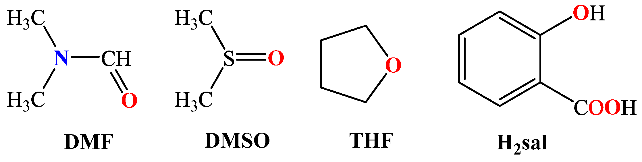
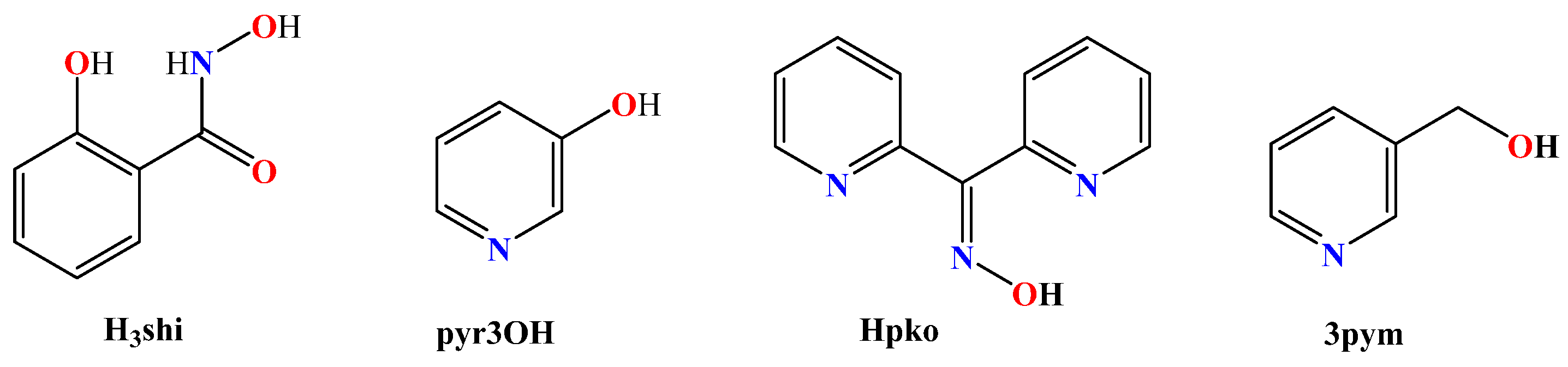
2.1. General Considerations for the Acids
2.2. The Co-Ligands
- (i)
- Imidazole derivatives, including imidazole (Himi) and compounds containing a 5-membered heterocyclic aromatic ring, such as 1,2–dimethylimidazole (1,2–dmimid), 1H–benzimidazole (Hbzmd), caffeine (caf) and 3,5–dimethylpyrazole (Hdmpz) (Figure 5);
- (ii)
- Pyridine derivatives, including pyridine (py) and compounds containing a 6-membered heterocyclic aromatic ring, such as 2–picoline (2pic), 3–picoline (3pic), 4–picoline (4pic), 2–aminopyridine (2ampy) and 2,2′–bipyridylamine (bipyam) (Figure 6), or 2,2′–bipyridine derivatives including 2,2′–bipyridine (bipy), 4,4′–bipyridine (4,4′–bipy), 5,5′–dimethyl–2,2′–bipyridine (5,5′–Me2–bipy) and 1,3–bis(4–pyridyl)propane (bpp) (Figure 7), as well as 1,3–dipyridin–3–ylurea (3U), 1,3–dipyridin–4–ylurea (4U), 2,4–diamine–6–phenyl–1,3,5–triazine (phdat) and tris(2–pyridyl)amine (TPA) (Figure 8);
- (iii)
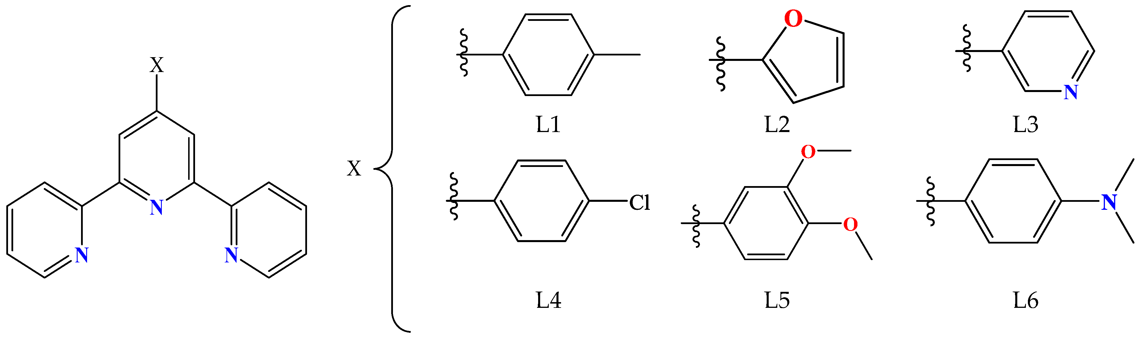
- 2,2′:6′,2″–terpyridine derivatives, including 4′–(4–tolyl)–2,2′:6′,2″–terpyridine (L1), 4′–(furan–2–yl)–2,2′:6′,2″–terpyridine (L2), 4′–(pyridin–3–yl)–2,2′:6′, 2″–terpyridine (L3), 4′–(4–chlorophenyl)–2,2′:6′,2″–terpyridine (L4), 4′–(3,4–dimethoxyphenyl)–2,2′:6′,2″–terpyridine (L5) and 4′–(4–dimethylaminophenyl)–2,2′:6′,2″–terpyridine (L6) (Figure 9);
- (iv)
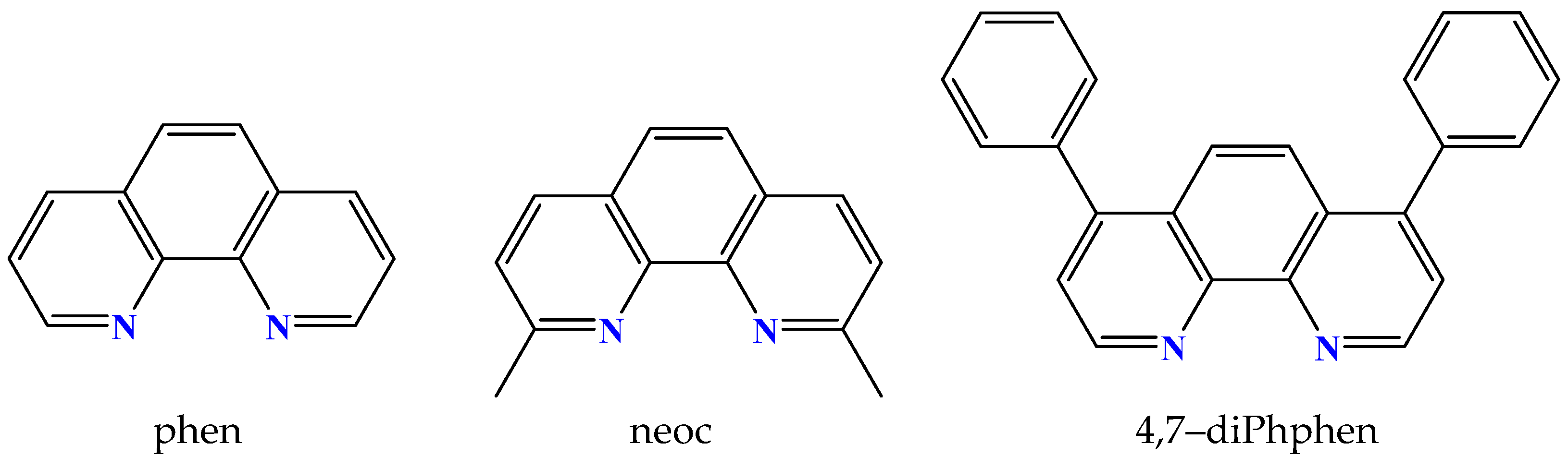
- Phenanthroline derivatives, including 1,10–phenanthroline (phen) and its derivatives 2,9–dimethyl–1,10–phenanthroline (neocuproine, neoc) and 4,7–diphenyl–1,10–phenanthroline (4,7–diPhphen) (Figure 10);
- (v)
- Alicyclic or aliphatic nitrogen–donors, such as 1,4,8,11–tetraazacyclotetradecane (cyclam), 1,4,7–trimethyl–1,4,7–triazacyclononane (TACN–Me3), tris(2–aminoethyl)amine (tren) and N,N–dimethylethane–1,2–diamine (EDA) (Figure 11).
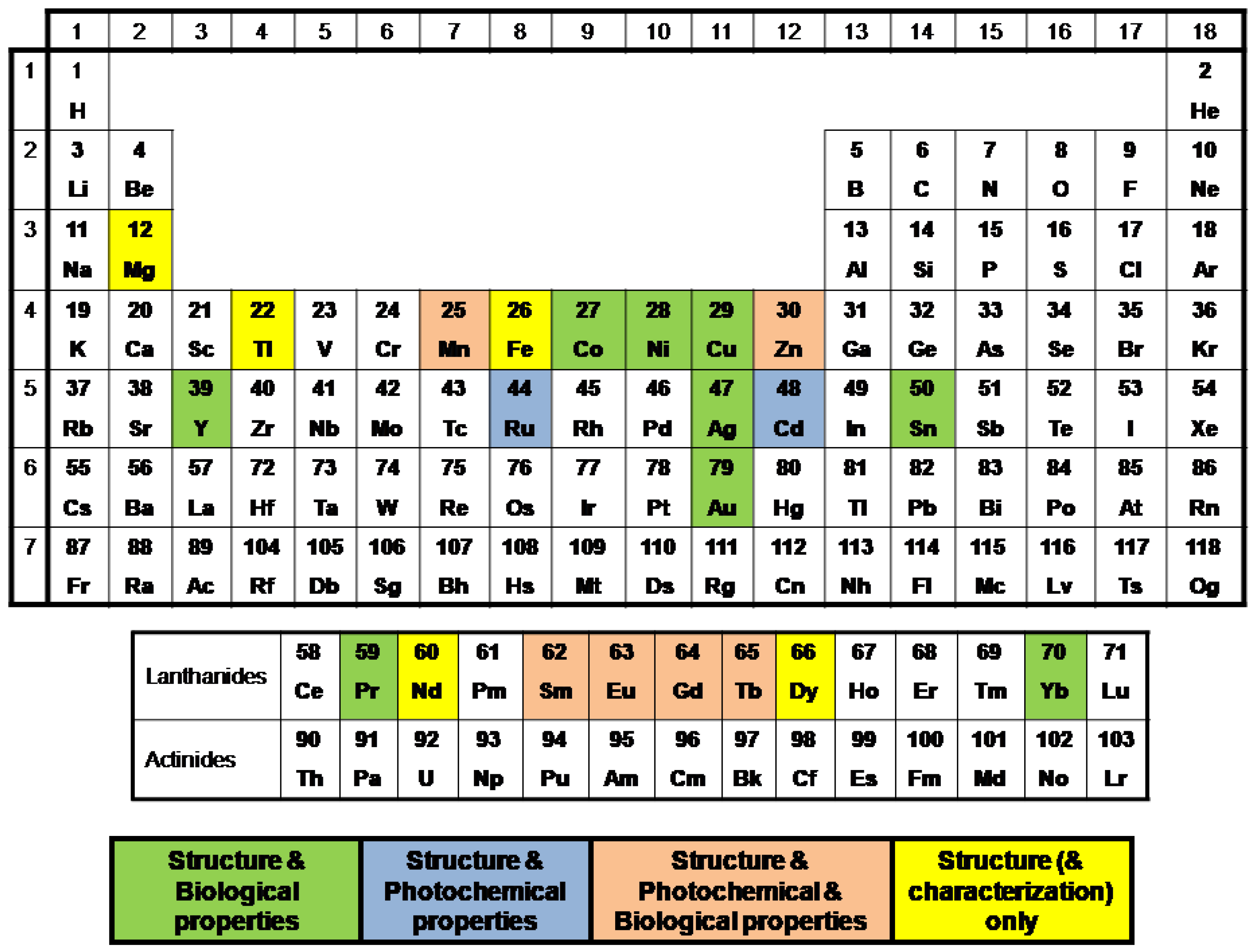
2.3. Metal Ions of the Reported Complexes
3. Structures of the Complexes
3.1. Coordination of the Carboxylato Ligands
3.2. Mononuclear Complexes
3.3. Dinuclear Complexes
3.4. Polynuclear Complexes
3.5. Polymeric Complexes
4. Spectroscopic and Physicochemical Characterization of the Metal Complexes
4.1. IR Spectroscopy
4.2. NMR Spectroscopy
4.3. Photochemical Properties
4.4. EPR Spectroscopy
4.5. Mössbauer Spectroscopy
4.6. Thermal Behavior
4.7. Magnetic Measurements
4.8. Electrochemical Behavior
5. Biological Activity
5.1. Anticancer Activity of the Complexes
5.2. Antibacterial Activity of the Complexes
5.3. Antioxidant Activity of the Complexes
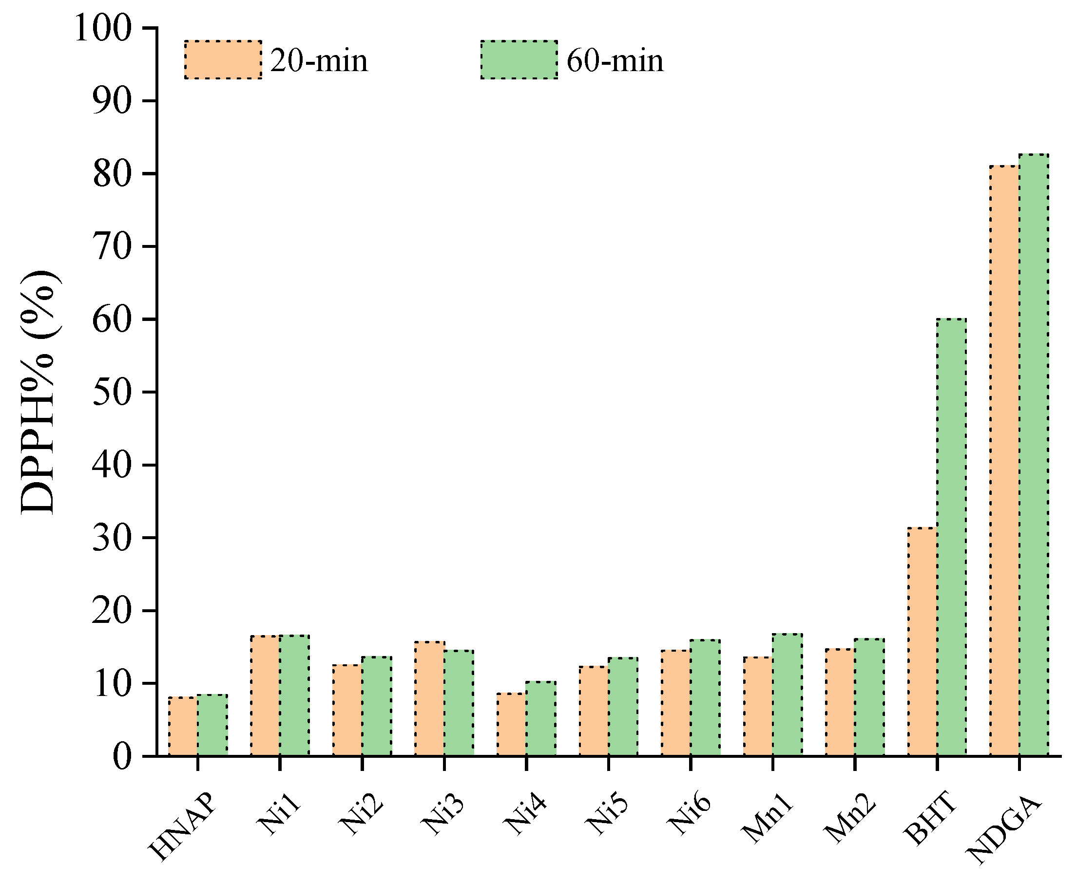
5.3.1. Scavenging of DPPH Radicals
5.3.2. Scavenging of Hydroxyl Radicals
5.3.3. ABTS Radical Scavenging
5.3.4. LOX Inhibitory Activity of the Complexes
5.3.5. SOD-like Activity of the Complexes
5.4. Other Biological Activities
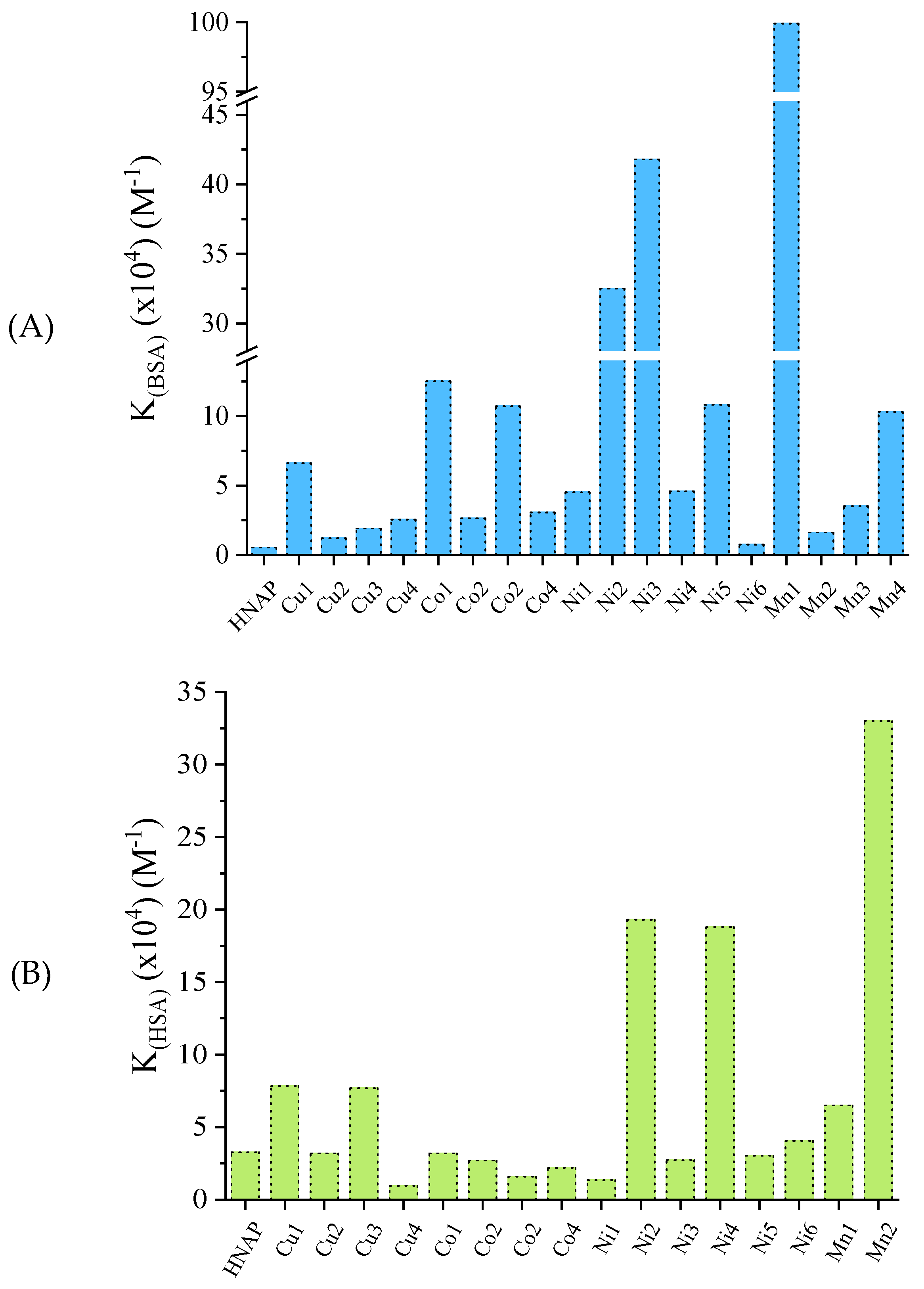
5.5. Interaction of the Compounds with Biomacromolecules
5.5.1. Interaction of the Reported Complexes with DNA
5.5.2. Interaction of the Reported Complexes with Albumins
6. Conclusions and Perspectives
Supplementary Materials
Author Contributions
Funding
Institutional Review Board Statement
Informed Consent Statement
Data Availability Statement
Conflicts of Interest
Abbreviations
References
- Janicki, R.; Mondry, A.; Starynowicz, P. Carboxylates of rare earth elements. Coord. Chem. Rev. 2017, 340, 98–133. [Google Scholar] [CrossRef]
- Loiseau, T.; Mihalcea, I.; Henry, N.; Volkringer, C. The crystal chemistry of uranium carboxylates. Coord. Chem. Rev. 2014, 266–267, 69–109. [Google Scholar] [CrossRef]
- Belousov, Y.A.; Drozdov, A.A.; Taydakov, I.V.; Marchetti, F.; Pettinari, R.; Pettinari, C. Lanthanide azolecarboxylate compounds: Structure, luminescent properties and applications. Coord. Chem. Rev. 2021, 445, 214084. [Google Scholar] [CrossRef]
- Ahmad, N.; Chughtai, A.H.; Younus, H.A.; Verpoort, F. Discrete metal-carboxylate self-assembled cages: Design, synthesis and applications. Coord. Chem. Rev. 2014, 280, 1–27. [Google Scholar] [CrossRef]
- Hietsoi, O.; Filatov, A.S.; Dubceac, C.; Petrukhina, M.A. Structural diversity and photoluminescence of copper(I) carboxylates: From discrete complexes to infinite metal-based wires and helices. Coord. Chem. Rev. 2015, 295, 125–138. [Google Scholar] [CrossRef]
- Ye, B.; Tong, M.; Chen, X. Metal-organic molecular architectures with 2,2′-bipyridyl-like and carboxylate ligands. Coord. Chem. Rev. 2005, 249, 545–565. [Google Scholar] [CrossRef]
- Teo, P.; Hor, T.S.A. Spacer directed metallo-supramolecular assemblies of pyridine carboxylates. Coord. Chem. Rev. 2011, 255, 273–289. [Google Scholar] [CrossRef]
- Lampropoulos, C.; Thuijs, A.E.; Mitchell, K.J.; Abboud, K.A.; Christou, G. Manganese/Cerium Clusters Spanning a Range of Oxidation Levels and CeMn8, Ce2Mn4, and Ce6Mn4 Nuclearities: Structural, Magnetic, and EPR Properties. Inorg. Chem. 2014, 53, 6805–6816. [Google Scholar] [CrossRef]
- Baruah, J.B. Naphthalenedicarboxylate based metal organic frameworks: Multifaceted material. Coord. Chem. Rev. 2021, 437, 213862. [Google Scholar] [CrossRef]
- Psomas, G.; Kessissoglou, D.P. Quinolones and non-steroidal anti-inflammatory drugs interacting with copper(II), nickel(II), cobalt(II) and zinc(II): Structural features, biological evaluation and perspectives. Dalton Trans. 2013, 42, 6252–6276. [Google Scholar] [CrossRef]
- Psomas, G. Copper(II) and zinc(II) coordination compounds of non-steroidal anti-inflammatory drugs: Structural features and antioxidant activity. Coord. Chem. Rev. 2020, 412, 213259. [Google Scholar] [CrossRef]
- Cambridge Crystallographic Data Centre. Available online: https://www.ccdc.cam.ac.uk/structures/ (accessed on 25 January 2023).
- Basuchaudhuri, P. 1-Naphthaleneacetic acid in rice cultivation. Curr. Sci. 2016, 110, 52–56. [Google Scholar] [CrossRef]
- Sourati, R.; Sharifi, P.; Poorghasemi, M.; Vieira, E.A.; Seidavi, A.; Anjum, N.A.; Sehar, Z.; Sofo, A. Effects of Naphthaleneacetic Acid, Indole-3-Butyric Acid and Zinc Sulfate on the Rooting and Growth of Mulberry Cuttings. Int. J. Plant Biol. 2022, 13, 245–256. [Google Scholar] [CrossRef]
- Chanu, K.D.; Singh, G. Effects Of 1-Naphthaleneacetic Acid, a Plant Hormone, on Invertebrates and Saccharomyces Cerevisiae. Eur. J. Mol. Clin. Med. 2020, 7, 4239–4244. [Google Scholar]
- Smulders, M.J.M.; van de Ven, E.T.W.M.; Croes, A.F.; Wullems, G.J. Metabolism of 1-Naphthaleneacetic Acid in Explants of Tobacco: Evidence for Release of Free Hormone from Conjugates. J. Plant Growth Regul. 1990, 9, 27–34. [Google Scholar] [CrossRef]
- Rehman, M.U.; Rather, G.H.; Gull, Y.; Mir, M.M.; Umar, I. Effect of 1-naphthaleneacetic acid and calcium chloride on preharvest drop and quality of ‘Red Delicious’ apples. Can. J. Plant Sci. 2017, 87, 902–905. [Google Scholar]
- Hill, R.J.; King, D.R.; Zollinger, R.; Moretti, M.L. 1-Naphthaleneacetic Acid (NAA) Reduces Sucker Growth in European Hazelnut (Corylus avellana L.). Hortscience 2021, 56, 1594–1598. [Google Scholar] [CrossRef]
- Pontikis, C.A. Effects of 2-naphthaleneacetic acid on alternate bearing of pistachio. Fruits 1990, 45, 281–285. [Google Scholar]
- Ohlenbusch, G.; Zwiener, C.; Meckenstock, R.U.; Frimmel, F.H. Identification and quantification of polar naphthalene derivatives in contaminated groundwater of a former gas plant site by liquid chromatography-electrospray ionization tandem mass spectrometry. J. Chromatogr. A 2002, 967, 201–207. [Google Scholar] [CrossRef]
- Todd, P.A.; Clissold, S.P. Naproxen. Drugs 1990, 40, 91–137. [Google Scholar] [CrossRef]
- French, L. Dysmenorrhea. Am. Fam. Physician 2005, 71, 285–291. [Google Scholar]
- Sharma, J.; Singla, A.K.; Dhawan, S. Zinc-naproxen complex: Synthesis, physicochemical and biological evaluation. Int. J. Pharm. 2003, 260, 217–227. [Google Scholar] [CrossRef]
- Ruschitzka, F.; Borer, J.S.; Krum, H.; Flammer, A.J.; Yeomans, N.D.; Libby, P.; Luscher, T.F.; Solomon, D.H.; Husni, M.E.; Graham, D.Y.; et al. Differential blood pressure effects of ibuprofen, naproxen, and celecoxib in patients with arthritis: The PRECISION–ABPM (Prospective Randomized Evaluation of Celecoxib Integrated Safety Versus Ibuprofen or Naproxen Ambulatory Blood Pressure Measurement) Trial. Eur. Heart J. 2017, 38, 3282–3292. [Google Scholar]
- Nissen, S.E.; Yeomans, N.D.; Solomon, D.H.; Lüscher, T.F.; Libby, P.; Husni, M.E.; Graham, D.Y.; Borer, J.S.; Wisniewski, L.M.; Wolski, K.E.; et al. Cardiovascular Safety of Celecoxib, Naproxen, or Ibuprofen for Arthritis. N. Engl. J. Med. 2016, 375, 2519–2529. [Google Scholar] [CrossRef]
- Richy, F.; Bruyere, O.; Ethgen, O.; Rabenda, V.; Bouvenot, G.; Audran, M.; Herrero-Beaumont, G.; Moore, A.; Eliakim, R.; Haim, M.; et al. Time dependent risk of gastrointestinal complications induced by non-steroidal anti-inflammatory drug use: A consensus statement using a meta-analytic approach. Ann. Rheum. Dis. 2004, 63, 759–766. [Google Scholar] [CrossRef]
- Mellemkjaer, L.; Blot, W.J.; Sorensen, H.T.; Thomassen, L.; McLaughlin, J.K.; Nielsen, G.L.; Olsen, J.H. Upper gastrointestinal bleeding among users of NSAIDs: A population-based cohort study in Denmark. Br. J. Clin. Pharmacol. 2002, 53, 173–181. [Google Scholar] [CrossRef]
- Duggan, K.C.; Walters, M.J.; Musee, J.; Harp, J.M.; Kiefer, J.R.; Oates, J.A.; Marnett, L.J. Molecular Basis for Cyclooxygenase Inhibition by the Non-steroidal Anti-inflammatory Drug Naproxen. J. Biol. Chem. 2010, 285, 34950–34959. [Google Scholar] [CrossRef]
- Wongrakpanich, S.; Wongrakpanich; Melhado, A.K.; Rangaswami, J. A Comprehensive Review of Non-Steroidal Anti-Inflammatory Drug Use in The Elderly. Aging Dis. 2018, 9, 143–150. [Google Scholar] [CrossRef]
- Davies, D.M.; Anderson, K.E. Clinical pharmacokinetics of naproxen. Clin. Pharmacokinet. 1997, 32, 268–293. [Google Scholar] [CrossRef]
- Tangoulis, V.; Lalioti, N.; Parthenios, J.; Langford, N.; Valsami-Jones, E.; Kakoulidou, C.; Psomas, G.; Bekiari, V. A Facile Method to Prepare pH-sensitive PEI-Functionalized Carbon Nanotubes As Rationally Designed Vehicles For Non-Steroidal Anti-Inflammatory Drugs (NSAIDs) Delivery. C—J. Carbon Res. 2020, 6, 62. [Google Scholar] [CrossRef]
- Wu, J.I.; Dobrowolski, M.A.; Cyranski, M.K.; Merner, B.L.; Bodwell, G.J.; Mo, Y.; Schleyer, P.V.R. On the aromatic stabilization energy of the 4N π electron pyrene. Mol. Phys. 2009, 107, 1177–1186. [Google Scholar] [CrossRef]
- Randic, M. Aromaticity of Polycyclic Conjugated Hydrocarbons. Chem. Rev. 2003, 103, 3449–3606. [Google Scholar] [CrossRef]
- Baba, M.; Saitoh, M.; Kowaka, Y.; Taguma, K.; Yoshida, K.; Semba, Y.; Kasahara, S.; Yamanaka, T.; Ohshima, Y.; Hsu, Y.; et al. Vibrational and rotational structure and excited-state dynamics of pyrene. J. Chem. Phys. 2009, 131, 224318. [Google Scholar] [CrossRef]
- Johnpeter, J.P.; Therrien, B. Sawhorse-type diruthenium tetracarbonyl complexes derived from pyrenyl-carboxylic acids. Inorg. Chim. Acta 2013, 405, 437–443. [Google Scholar] [CrossRef]
- Ahn, S.Y.; Kim, S.; Baek, K.; Eom, M.S.; Kang, S.; Han, M.S. Thioether Amide Based-Fluorescent Chemosensors for Pd2+ with High Selectivity over Pd0. Bull. Kor. Chem. Soc. 2014, 35, 2189–2192. [Google Scholar] [CrossRef]
- Jang, J.; Ko, S.; Kim, Y. Dual-Functionalized Polymer Nanotubes as Substrates for Molecular-Probe and DNA-Carrier Applications. Adv. Funct. Mater. 2006, 16, 754–759. [Google Scholar] [CrossRef]
- Alizadeh, V. Preparation a novel 1-pyreneacetic acid functionalized graphene/self-assembled monolayer modified gold electrode to immobilize and study interfacial electron transfer of cytochrome c by electrochemical approaches. Chem. Phys. Lett. 2022, 787, 139187. [Google Scholar] [CrossRef]
- Cabezas, J.A. A Facile Method for the Titration of Organolithium Reagents Using Naphtylamine or Naphtylmethylamine as Indicators. Int. J. Multidiscip. Curr. Res. 2019, 7, 30–32. [Google Scholar]
- Kiljunen, H.; Hase, T.A. Titration of Organolithiums and Grignards with 1-Pyreneacetic Acid. J. Org. Chem. 1991, 56, 6950–6952. [Google Scholar] [CrossRef]
- Friscic, T.; Halasz, I.; Strobridge, F.C.; Dinnebier, R.E.; Stein, R.S.; Fabian, L.; Curfs, C. A rational approach to screen for hydrated forms of the pharmaceutical derivative magnesium naproxen using liquid-assisted grinding. CrystEngComm 2011, 13, 3125–3129. [Google Scholar] [CrossRef]
- Dimiza, F.; Raptopoulou, C.P.; Psycharis, V.; Papadopoulos, A.N.; Psomas, G. Manganese(II) complexes with the non-steroidal anti-inflammatory drugs naproxen and mefenamic acid. Synthesis, structure, antioxidant capacity, interaction with albumins and DNA. New J. Chem. 2018, 42, 16666–16681. [Google Scholar] [CrossRef]
- Dimiza, F.; Papadopoulos, A.N.; Tangoulis, V.; Psycharis, V.; Raptopoulou, C.P.; Kessissoglou, D.P.; Psomas, G. Biological evaluation of cobalt(II) complexes with non-steroidal anti-inflammatory drug naproxen. J. Inorg. Biochem. 2012, 107, 54–64. [Google Scholar] [CrossRef]
- Cressey, P.B.; Eskandari, A.; Bruno, P.M.; Lu, C.; Hemann, M.T.; Suntharalingam, K. The Potent Inhibitory Effect of a Naproxen-Appended Cobalt(III)-Cyclam Complex on Cancer Stem Cells. ChemBioChem 2016, 17, 1713–1718. [Google Scholar] [CrossRef]
- Jin, S.; Ye, X.; Jin, L.; Zheng, L.; Li, J.; Jin, B.; Wang, D. Syntheses and structural characterization of nine coordination compounds assembled from copper acetate, 3,5-dimethylpyrazole and carboxylates. Polyhedron 2014, 81, 382–395. [Google Scholar] [CrossRef]
- Wang, Y.; Tang, G.; Wan, W.; Wu, Y.; Tian, T.; Wang, J.; He, C.; Long, X.; Wang, J.; Ng, S.W. New homochiral ferroelectric supramolecular networks of complexes constructed by chiral S-naproxen ligand. CrystEngComm 2012, 14, 3802–3812. [Google Scholar] [CrossRef]
- Sheng, G.; Cheng, X.; You, Z.; Zhu, H. Syntheses, Crystal Structures, and Characterization of Copper(II) and Zinc(II) Complexes Derived from N,N-Dimethylethane-1,2-diamine and Phenylacetic Acid Derivatives. Synth. React. Inorg. Metal-Org. Nano-Met. Chem. 2015, 45, 1273–1277. [Google Scholar] [CrossRef]
- Caglar, S.; Adiguzel, E.; Sariboga, B.; Temel, E.; Buyukgungor, O. Mono and dinuclear copper(II) naproxenato complexes containing 3-picoline and 4-picoline: Synthesis, structure, properties, catechol oxidase, and antimicrobial activities. J. Coord. Chem. 2014, 67, 670–683. [Google Scholar] [CrossRef]
- Mahendiran, D.; Gurumoorthy, P.; Gunasekaran, K.; Kumar, R.S.; Rahiman, A.K. Structural modeling, in vitro antiproliferative activity, and the effect of substituents on the DNA fastening and scission actions of heteroleptic copper(II) complexes with terpyridines and naproxen. New J. Chem. 2015, 39, 7895–7911. [Google Scholar] [CrossRef]
- Banti, C.N.; Giannoulis, A.D.; Kourkoumelis, N.; Owczarzak, A.M.; Kubicki, M.; Hadjikakou, S.K. Novel metallo-therapeutics of the NSAID naproxen. Interaction with intracellular components that leads the cells to apoptosis. Dalton Trans. 2014, 43, 6848–6863. [Google Scholar] [CrossRef]
- Johnson, A.; Olelewe, C.; Kim, J.H.; Northcote-Smith, J.; Mertens, R.T.; Passeri, G.; Singh, K.; Awuah, S.G.; Suntharalingam, K. The anti-breast cancer stem cell properties of gold(I)-non-steroidal anti-inflammatory drug complexes. Chem. Sci. 2023, 14, 557–565. [Google Scholar] [CrossRef]
- Xia, H.; Rong, D.; Liu, Y.; Wang, D. Crystal Structure and Luminescent Properties of Complex [Eu0.5Gd0.5(C12H9O2)2(C12H8N2)2(H2O)2]·(C12H9O2)·H2O. Adv. Mater. Res. 2012, 54, 691–694. [Google Scholar] [CrossRef]
- Zhang, Z.; Yin, F.; Li, S.; Wang, L. Bis(1H-benzimidazole-κN3)bis [2-(naphthalen-1-yl)acetato-κ2O,O′]manganese(II) monohydrate. Acta Crystallogr. Sect. E 2012, 68, m328. [Google Scholar] [CrossRef] [PubMed]
- Yin, F.; Han, L.; Hong, Z.; Xu, X.; Ren, L. Bis(1H-benzimidazole-κN3)bis[2-(naphthalen-1-yl)acetato-κ2O,O′]nickel(II) monohydrate. Acta Crystallogr. Sect. E 2012, 68, m416. [Google Scholar] [CrossRef] [PubMed]
- Zheng, P.; Eskandari, A.; Lu, C.; Laws, K.; Aldous, L.; Suntharalingam, K. Biophysical analysis of cancer stem cell-potent copper(II) coordination complexes. Dalton Trans. 2019, 48, 5892–5896. [Google Scholar] [CrossRef] [PubMed]
- Dimiza, F.; Perdih, F.; Tangoulis, V.; Turel, I.; Kessissoglou, D.P.; Psomas, G. Interaction of copper(II) with the non-steroidal anti-inflammatory drugs naproxen and diclofenac: Synthesis, Structure, DNA- and albumin-binding. J. Inorg. Biochem. 2011, 105, 476–489. [Google Scholar] [CrossRef] [PubMed]
- Liu, Y.; Xia, H.; Yang, S.; Wang, D. Bis(1-naphthylacetato-κ2O,O)(1,10-phenanthroline-κ2N,N)zinc(II). Acta Crystallogr. Sect. E 2006, 62, m2753–m2755. [Google Scholar] [CrossRef]
- Tang, D.; Feng, L.; Zhang, X. Synthesis, structure and antibacterial activity of the complex of zinc with phenanthroline and naphthylacetate. Wuji Huaxue Xuebao (Chin. J. Inorg. Chem.) 2006, 22, 1891–1894. [Google Scholar]
- Ma, C.; Zhang, B.; Zhang, S.; Zhang, R. Chiral organotin (IV) carboxylates complexes: Syntheses, characterization, and crystal structures with chiral (S)-(+)-6-methoxy-α-methyl-2-naphthaleneaceto acid ligand. J. Organomet. Chem. 2011, 696, 2165–2171. [Google Scholar] [CrossRef]
- Banti, C.N.; Gkaniatsou, E.I.; Kourkoumelis, N.; Manos, M.J.; Tasiopoulos, A.J.; Bakas, T.; Hadjikakou, S.K. Assessment of organotins against the linoleic acid, glutathione and CT-DNA. Inorg. Chim. Acta 2014, 423, 98–106. [Google Scholar] [CrossRef]
- Totta, X.; Hatzidimitriou, A.G.; Papadopoulos, A.N.; Psomas, G. Nickel(II)-naproxen mixed-ligand complexes: Synthesis, structure, antioxidant activity and interaction with albumins and calf-thymus DNA. New J. Chem. 2017, 41, 4478–4492. [Google Scholar] [CrossRef]
- Mirzaei-Kalar, Z.; Khandar, A.A.; White, J.M.; Abolhasani, H.; Movahhed, T.K.; Best, S.P.; Jouyban, A. Investigation of biological activity of nickel(II) complex with naproxen and 1,10-phenanthroline ligands. J. Biomol. Struct. Dyn. 2021, 39, 6939–6954. [Google Scholar] [CrossRef] [PubMed]
- Ali, H.A.; Fares, H.; Darawsheh, M.; Rappocciolo, E.; . Akkawi, M.; Jaber, S. Synthesis, characterization and biological activity of new mixed ligand complexes of Zn(II) naproxen with nitrogen based ligands. Eur. J. Med. Chem. 2015, 89, 67–76. [Google Scholar] [CrossRef] [PubMed]
- Ji, L.; Liu, J.; Song, W. (5,5′-Dimethyl-2,2′-bipyridine-κ2N,N′)(1-naphthylacetato-κO)(1-naphthylacetato-κ2O,O′)zinc hemihydrates. Acta Crystallogr. Sect. E 2011, 67, m606. [Google Scholar] [CrossRef] [PubMed]
- Picraux, L.B.; Smeigh, A.L.; Guo, D.; McCusker, J.K. Intramolecular Energy Transfer Involving Heisenberg Spin-Coupled Dinuclear Iron-Oxo Complexes. Inorg. Chem. 2005, 44, 7846–7859. [Google Scholar] [CrossRef]
- Picraux, L.B.; Weldon, B.T. McCusker, J.K. Intramolecular Excimer Formation in a Naphthalene-Appended Dinuclear Iron-Oxo Complex. Inorg. Chem. 2003, 42, 273–282. [Google Scholar] [CrossRef]
- Chen, L.; Zhang, J.; Song, L.; Wang, W.; Ju, Z. Tetrakis(µ-α-naphthylacetato-κ2O:O′)bis[(dimethyl sulfoxide-κO)copper(II)]-α-naphthylacetic acid-dimethyl sulfoxide (1/2/2). Acta Crystallogr. Sect. E 2004, 60, m1032–m1034. [Google Scholar] [CrossRef]
- Yin, F.; Yu, G.; Zhao, H.; Bai, D. Tetrakis(µ-naphthalene-1-acetato-κ2O:O′)bis[(N,N-dimethylformamide-κO)copper(II)]. Acta Crystallogr. Sect. E 2012, 68, m350. [Google Scholar] [CrossRef]
- Koman, M.; Melnik, M.; Moncol, J.; Glowiak, T. Caffeine in copper(II) complexes: Crystal and molecular structure of di(caffeine)tetrakis(naproxenato)dicopper(II). Inorg. Chem. Commun. 2000, 3, 489–492. [Google Scholar] [CrossRef]
- Li, Y.; Xiao, C.; Feng, H.; Guo, S.; Wang, S. Two binuclear tetracarboxylate-bridged complexes: Syntheses, structures, and properties. J. Coord. Chem. 2010, 65, 2820–2829. [Google Scholar] [CrossRef]
- O’Rourke, N.F.; Ronaldson, M.; Cameron, T.S.; Wang, R.; Aquino, M.A.S. Equatorial π-stacking interactions in diruthenium (II,III) tetracarboxylate complexes containing extended π-systems. J. Mol. Struct. 2013, 1052, 17–23. [Google Scholar] [CrossRef]
- Nakai, M.; Funabiki, T.; Ohtsuki, C.; Harada, M.; Ichimura, A.; Tanaka, R.; Kinoshita, I.; Mikuriya, M.; Benten, H.; Ohkita, H.; et al. Structure and Photochemical Properties of (µ-Alkoxo)bis(µ-carboxylato)diruthenium Complexes with Naphthylacetate Ligands. Inorg. Chem. 2006, 45, 3048–3056. [Google Scholar] [CrossRef] [PubMed]
- Liu, Y.; Rong, D.; Xia, H.; Wang, D.; Liu, Y. Luminescence Properties and Antibacterial Activity of [Ln2(NNA)6(Phen)2]·nC3H7NO (Ln = Y, Eu, Gd, Pr, Sm, Tb, Yb, n = 0, 1, 2, NAA = 1-Naphthylacetic Acid, Phen = 1,10-Phenanthroline). Synth. React. Inorg. Met.-Org. Nano-Met. Chem. 2008, 38, 635–641. [Google Scholar] [CrossRef]
- Xia, H.; Liu, Y.; Wang, D.; Chen, L. Hexakis(µ-naphthalene-1-acetato)bis[(1,10-phenanthroline)praseodymium(III)] N,N-dimethylformamide solvate. Acta Crystallogr. Sect. E 2007, 63, m2610. [Google Scholar] [CrossRef]
- Xia, H.; Liu, Y.; Lu, G.; Wang, D. Crystal Structure and Interaction with Bovine Serum Albumin of Rare Earth Complex [Nd(α-C10H7CH2COO)3(C12H8N2)]2. Synth. React. Inorg. Met.-Org. Nano-Met. Chem. 2012, 42, 145–153. [Google Scholar] [CrossRef]
- Xia, H.; Liu, Y.; Wang, D.; Yang, S. Tetrakis(µ-naphthalene-1-acetato)bis[(naphthalene-1-acetato)(1,10-phenanthroline)samarium(III)] N,N-dimethylformamide disolvate. Acta Crystallogr. Sect. E 2007, 63, m2708–m2709. [Google Scholar] [CrossRef]
- Liu, Y.; Xia, H.; Wang, D.; Yang, S. Hexakis(µ-naphthalene-1-acetato)bis[(1,10-phenanthroline)europium(III)] N,N-dimethylformamide disolvate. Acta Crystallogr. Sect. E 2007, 63, m2608. [Google Scholar] [CrossRef]
- Liu, Y.; Rong, D.; Xia, H.; Wang, D.; Chen, L. Synthesis, crystal structure, and luminescence properties of [TbGd(NAA)6(phen)2] and [Tb2(NNA)6(phen)2]·2C3H7NO. J. Coord. Chem. 2009, 62, 1835–1845. [Google Scholar] [CrossRef]
- Liu, Y.; Xia, H.; Wang, D.; Yang, S. Hexakis(µ-naphthalene-1-acetato)bis[(1,10-phenanthroline)gadolinium(III)] N,N-dimethylformamide disolvate. Acta Crystallogr. Sect. E 2007, 63, m2625–m2626. [Google Scholar] [CrossRef]
- Xia, H.; Liu, Y.; Wang, D.; Yang, S. Hexakis(µ-naphthalene-1-acetato)bis[(1,10-phenanthroline)terbium(III)] N,N-dimethylformamide disolvate. Acta Crystallogr. Sect. E 2007, 63, m2624. [Google Scholar] [CrossRef]
- Xia, H.; Liu, Y.; Wang, D.; Yang, S. Tetrakis(µ-naphthalene–1-acetato)bis[(naphthalene-1-acetato)(1,10-phenanthroline)ytterbium(III)]. Acta Crystallogr. Sect. E 2007, 63, m2795–m2796. [Google Scholar] [CrossRef]
- Li, Y.; Liu, Q.; Liu, C.; Wang, Y.; Chen, L. Dinuclear Lanthanide-Carboxylate Compounds: Field-Induced Slow Relaxation of Magnetization for Dysprosium(III) Analogue. Aust. J. Chem. 2015, 68, 488–492. [Google Scholar] [CrossRef]
- Zhao, G.; Yin, F.; Ge, H.; Li, S. Synthesis, Structure and Antibacterial Properties of Bis-Imidazole-bis(naphthalene-1-yl-acetato)copper(II). Asian J. Chem. 2014, 26, 2550–2552. [Google Scholar] [CrossRef]
- Dai, P.; Yang, E.; Zhao, X. Three Naphthoate-Based Cadmium(II) Complexes with Discrete Binuclear, Cyclic Tetranuclear, and Polymeric Double-Chain Motifs. Russ. J. Coord. Chem. 2015, 41, 16–24. [Google Scholar] [CrossRef]
- Pan, J.; Ju, J.; Wei, Q.; Liu, B.; Jin, S.; Lin, Z.; Wang, D. Noncovalently-bonded 2D–3D Metal-organic Frameworks via Assembly of Zn(Ac)2 with 3,5-Dimethylpyrazole and Carboxylate Ligands. Z. Anorg. Allg. Chem. 2014, 640, 1745–1753. [Google Scholar] [CrossRef]
- Nakai, M.; Funabiki, T.; Ohtsuki, C.; Harada, M.; Ichimura, A.; Tanaka, R.; Nishioka, T.; Kinoshita, I.; Mikuriya, M.; Guo, J.; et al. Syntheses, structures, and photochemical properties of (µ3-O)tris{bis(µ-carboxylato)}trimanganese complexes with naphthylacetate ligands with relevance to artificial solar energy-harvesting systems. Inorg. Chim. Acta 2013, 406, 130–137. [Google Scholar] [CrossRef]
- Yin, F.; Zhao, H.; Xu, X.; Yang, X. Synthesis, Crystal Structure, and Properties of a Tetranuclear Copper(II) Complex with α-Naphthylacetic Acid. Synth. React. Inorg. Met.-Org. Nano-Met. Chem. 2014, 44, 101–105. [Google Scholar] [CrossRef]
- Feng, Y.; Yu, J.; Kuang, D.; Tan, Y.; Zhang, F.; Jiang, W.; Zhu, X.; Zheng, J. Syntheses, Crystal Structures, and Anti-tumor Activity of Tetra-nuclear Cluster and 1D Chain Butyltin α-Naphthaleneacetic Carboxylates. Wuji Huaxue Xuebao (Chin. J. Inorg. Chem.) 2015, 31, 710–716. [Google Scholar]
- Liu, J.; Gao, M.; Fang, W.; Zhang, L.; Zhang, J. Bandgap Engineering of Titanium-Oxo Clusters: Labile Surface Sites Used for Ligand Substitution and Metal Incorporation. Angew. Chem. Int. Ed. 2016, 55, 5160–5165. [Google Scholar] [CrossRef]
- Tarushi, A.; Zampakou, M.; Perontsis, S.; Lafazanis, K.; Pantazaki, A.A.; Hatzidimitriou, A.G.; Geromichalos, G.D.; Psomas, G. Manganese(II) complexes of tolfenamic acid or naproxen in polymeric structures or encapsulated in [15-MC-5] manganese(III) metallacrowns: Structure and biological activity. Inorg. Chim. Acta 2018, 483, 579–592. [Google Scholar] [CrossRef]
- Liu, J.; Wang, W.; Gu, C. A two-dimensional grid-like cobaltII coordination polymer: Poly[[diaquacobalt(II)]-di–µ2-naphthalene-1-acetato]. Acta Crystallogr. Sect. E 2006, 62, m3445–m3447. [Google Scholar] [CrossRef]
- Liu, Y.; Xia, H.; An, X.; Zhou, L.; Wang, D. The Synthesis, Structure and Antimicrobial Study of Poly[diaqua bis(1-naphthylacetic acid)Cobalt(II) Complex [Co(C12H9O2)2(H2O)2]n. Synth. React. Inorg. Met.-Org. Nano-Met. Chem. 2008, 38, 356–361. [Google Scholar] [CrossRef]
- Caglar, S.; Altay, A.; Harurluoglu, B.; Yeniceri, E.K.K.; Caglar, B.; Sahin, O. Synthesis, structural characterization and evaluation of anticancer activity of polymeric silver(I) complexes based on niflumic acid/naproxen and picoline derivatives. J. Coord. Chem. 2022, 75, 178–196. [Google Scholar] [CrossRef]
- Biswas, P.; Dastidar, P. Anchoring Drugs to a Zinc(II) Coordination Polymer Network: Exploiting Structural Rationale toward the Design of Metallogels for Drug-Delivery Applications. Inorg. Chem. 2021, 60, 3218–3231. [Google Scholar] [CrossRef] [PubMed]
- Wang, C.; Wang, P.; Guo, G. 3D sandwich-like frameworks constructed from silver chains: Synthesis and crystal structures of six silver(I) coordination complexes. Trans. Met. Chem. 2012, 37, 345–359. [Google Scholar] [CrossRef]
- Nakamoto, K. Infrared and Raman Spectra of Inorganic and Coordination Compounds, Part B: Applications in Coordination, Organometallic, and Bioinorganic Chemistry, 6th ed.; Wiley: Hoboken, NJ, USA, 2009. [Google Scholar]
- Szorcsik, A.; Nagy, L.; Sletten, J.; Szalontai, G.; Kamu, E.; Fiore, T.; Pellerito, L.; Kalman, E. Preparation and structural studies on dibutyltin(IV) complexes with pyridine mono- and dicarboxylic acids. J. Organomet. Chem. 2004, 689, 1145–1154. [Google Scholar] [CrossRef]
- Holeeek, J.; Nadvornik, M.; Handlie, K.; Lyeka, A. 13C and 119Sn NMR spectra of Di-n-butyltin(IV) compounds. J. Organomet. Chem. 1986, 315, 299–308. [Google Scholar] [CrossRef]
- Hathaway, B.J. Comprehensive Coordination Chemistry; Wilkinson, G., Ed.; Pergamon Press: Oxford, UK, 1987; Volume 5, pp. 533–773. [Google Scholar]
- Kunishita, A.; Ishimaru, H.; Nakashima, S.; Ogura, T.; Itoh, S. Reactivity of Mononuclear Alkylperoxo Copper(II) Complex. O-O Bond Cleavage and C-H Bond Activation. J. Am. Chem. Soc. 2008, 130, 4244–4245. [Google Scholar] [CrossRef]
- Cini, R.; Giorgi, G.; Cinquantini, A.; Rossi, C.; Sabat, M. Metal complexes of the antiinflammatory drug piroxicam. Inorg. Chem. 1990, 29, 5197–5200. [Google Scholar] [CrossRef]
- El-Gammal, O.A.; El-Reash, G.M.A.; Ghazy, S.E.; Radwan, A.H. Synthesis, characterization, molecular modeling and antioxidant activity of (1E,5E)-1,5-bis(1-(pyridin-2-yl)ethylidene)carbonohydrazide (H2APC) and its zinc(II), cadmium(II) and mercury(II) complexes. J. Mol. Struct. 2012, 1020, 6–15. [Google Scholar] [CrossRef]
- Gilbert, B.C.; Davies, M.J.; McLauchlan, K.A. Electron Paramagnetic Resonance; Royal Society of Chemistry: Cambridge, UK, 2000; Volume 17. [Google Scholar]
- Cowie, J.M.G.; Arrighi, V. Polymers: Chemistry and Physics of Modern Materials, 3rd ed.; CRC Press Inc.: Boca Raton, FL, USA, 2007. [Google Scholar]
- Kontogiorgis, C.; Hadjipavlou-Litina, D. Biological Evaluation of Several Coumarin Derivatives Designed as Possible Anti-inflammatory/Antioxidant Agents. J. Enzym. Inhib. Med. Chem. 2003, 180, 63–69. [Google Scholar] [CrossRef]
- Panagoulis, D.; Pontiki, E.; Skeva, E.; Raptopoulou, C.; Girousi, S.; Hadjipavlou-Litina, D.; Dendrinou-Samara, C. Synthesis and pharmacochemical study of new Cu(II) complexes with thiophen-2-yl saturated and α,β-unsaturated substituted carboxylic acids. J. Inorg. Biochem. 2007, 101, 623–634. [Google Scholar] [CrossRef]
- Walker, R.B.; Everette, J.D. Comparative Reaction Rates of Various Antioxidants with ABTS Radical Cation. J. Agric. Food Chem. 2009, 57, 1156–1161. [Google Scholar] [CrossRef]
- Huang, D.; Ou, B.; Prior, R.L. The Chemistry behind Antioxidant Capacity Assays. J. Agric. Food Chem. 2005, 53, 1841–1856. [Google Scholar] [CrossRef]
- Re, R.; Pellegrini, N.; Proteggente, A.; Pannala, A.; Yang, M.; Rice-Evans, C. Antioxidant activity applying an improved ABTS radical cation decolorization assay. Free Radic. Biol. Med. 1999, 26, 1231–1237. [Google Scholar] [CrossRef]
- Muller, K. 5-Lipoxygenase and 12-Lipoxygenase: Attractive Targets for the Development of Novel Antipsoriatic Drugs. Arch. Pharm. 1994, 327, 3–19. [Google Scholar] [CrossRef]
- Kemal, C.; Louis-Flamberg, P.; Krupinski-Olsen, R.; Shorter, A.L. Reductive inactivation of soybean lipoxygenase 1 by catechols: A possible mechanism for regulation of lipoxygenase activity. Biochemistry 1987, 26, 7064–7072. [Google Scholar] [CrossRef]
- Oyanagui, Y. Inhibition of superoxide anion production in macrophages by anti-inflammatory drugs. Biochem. Pharmacol. 1976, 25, 1473–1480. [Google Scholar]
- Wasil, M.; Halliwell, B.; Moorhouse, C.P.; Hutchinson, D.C.S.; Barum, H. Biologically-significant scavenging of the myeloperoxidase-derived oxidant hypochlorous acid by some anti-inflammatory drugs. Biochem. Pharmacol. 1987, 36, 3847–3850. [Google Scholar] [CrossRef] [PubMed]
- Abuhijleh, A.L.; Khalaf, J. Copper(II) complexes of the anti-inflammatory drug naproxen and 3-pyridylmethanol as auxiliary ligand. Characterization, superoxide dismutase and catecholase–mimetic activities. Eur. J. Med. Chem. 2010, 45, 3811–3817. [Google Scholar] [CrossRef] [PubMed]
- Gurova, K. New hopes from old drugs: Revisiting DNA-binding small molecules as anticancer agents. Future Oncol. 2009, 5, 1685–1704. [Google Scholar] [CrossRef] [PubMed]
- Hardman, J.G.; Limbird, L.E. (Eds.) Goodman and Gilman’s the Pharmacological Basis of Therapeutics, 10th ed.; McGraw Hill: New York, NY, USA, 2001. [Google Scholar]
- Zeglis, B.M.; Pierre, V.C.; Barton, J.K. Metallo-intercalators and metallo-insertors. Chem. Commun. 2007, 44, 4565–4576. [Google Scholar] [CrossRef] [PubMed]
- Boer, D.R.; Canals, A.; Coll, M. DNA-binding drugs caught in action: The latest 3D pictures of drug-DNA complexes. Dalton Trans. 2009, 399–414. [Google Scholar] [CrossRef] [PubMed]
- Brabec, V.; Kasparkova, J. Ruthenium coordination compounds of biological and biomedical significance. DNA binding agents. Coord. Chem. Rev. 2018, 376, 75–94. [Google Scholar] [CrossRef]
- Bhattacharyya, U.; Kumar, B.; Garai, A.; Bhattacharyya, A.; Kumar, A.; Banerjee, S.; Kondaiah, P.; Chakravarty, A.R. Curcumin “Drug” Stabilized in Oxidovanadium(IV)-BODIPY Conjugates for Mitochondria-Targeted Photocytotoxicity. Inorg. Chem. 2017, 56, 12457–12468. [Google Scholar] [CrossRef] [PubMed]
- Lucky, S.S.; Soo, K.C.; Zhang, Y. Nanoparticles in Photodynamic Therapy. Chem. Rev. 2015, 115, 1990–2042. [Google Scholar] [CrossRef] [PubMed]
- Farney, E.P.; Chapman, S.J.; Swords, W.B.; Torelli, M.D.; Hamers, R.J.; Yoon, T.P. Discovery and Elucidation of Counteranion Dependence in Photoredox Catalysis. J. Am. Chem. Soc. 2019, 141, 6385–6391. [Google Scholar] [CrossRef] [PubMed]
- Zhang, X.; Hou, Y.; Xiao, X.; Chen, X.; Hu, M.; Geng, X.; Wang, Z.; Zhao, J. Recent development of the transition metal complexes showing strong absorption of visible light and long-lived triplet excited state: From molecular structure design to photophysical properties and applications. Coord. Chem. Rev. 2020, 417, 213371. [Google Scholar] [CrossRef]
- Wolfe, A.; Shimer, G.; Meehan, T. Polycyclic Aromatic Hydrocarbons Physically Intercalate into Duplex Regions of Denatured DNA. Biochemistry 1987, 26, 6392–6396. [Google Scholar] [CrossRef]
- Dimitrakopoulou, A.; Dendrinou-Samara, C.; Pantazaki, A.A.; Alexiou, M.; Nordlander, E.; Kessissoglou, D.P. Synthesis, structure and interactions with DNA of novel tetranuclear, [Mn4(II/II/II/IV)] mixed valence complexes. J. Inorg. Biochem. 2008, 102, 618–628. [Google Scholar] [CrossRef]
- He, X.; Carter, D.C. Atomic structure and chemistry of human serum albumin. Nature 1992, 358, 209–215. [Google Scholar] [CrossRef]
- Olson, R.E.; Christ, D.D. Chapter 33. Plasma Protein Binding of Drugs. Ann. Rep. Med. Chem. 1996, 31, 327–336. [Google Scholar]
- Majorek, K.A.; Porebski, P.J.; Dayal, A.; Zimmerman, M.D.; Jablonska, K.; Stewart, A.J.; Chruszcz, M.; Minor, W. Structural and immunologic characterization of bovine, horse, and rabbit serum albumins. Mol. Immunol. 2012, 52, 174–182. [Google Scholar] [CrossRef] [PubMed]
- Gelamo, E.L.; Silva, C.; Imasato, H.; Tabak, M. Interaction of bovine (BSA) and human (HSA) serum albumins with ionic surfactants: Spectroscopy and modeling. Biochim. Biophys. Acta (BBA)-Protein Struct. Mol. Enzymol. 2002, 1594, 84–99. [Google Scholar] [CrossRef]
- Sudlow, G.; Birkett, D.J.; Wade, D.N. Further characterization of specific drug binding sites on human serum albumin. Mol. Pharmacol. 1976, 12, 1052–1061. [Google Scholar] [PubMed]
- Petitpas, I.; Grune, T.; Bhattacharya, A.A.; Twine, S.; East, M.; Curry, S. Crystal structures of human serum albumin complexed with monounsaturated and polyunsaturated fatty acids. J. Mol. Biol. 2001, 314, 955–960. [Google Scholar] [CrossRef] [PubMed]
- Rajendiran, V.; Karthik, R.; Palaniandavar, M.; Stoeckli-Evans, H.; Periasamy, V.S.; Akbarsha, M.A.; Srinag, B.S.; Krishnamurthy, H. Mixed-Ligand Copper(II)-phenolate Complexes: Effect of Coligand on Enhanced DNA and Protein Binding, DNA Cleavage, and Anticancer Activity. Inorg. Chem. 2007, 46, 8208–8221. [Google Scholar] [CrossRef] [PubMed]
- Lakowicz, J.R. Principles of Fluorescence Spectroscopy, 3rd ed.; Plenum Press: New York, NY, USA, 2006. [Google Scholar]
- Zhao, G.; Lin, H.; Zhu, S.; Sun, H.; Chen, Y. Dinuclear palladium (II) complexes containing two monofunctional [Pd(en)(pyridine) Cl]+ units bridged by Se or S. Synthesis, characterization, cytotoxicity and kinetic studies of DNA-binding. J. Inorg. Biochem. 1998, 70, 219–226. [Google Scholar] [CrossRef] [PubMed]
- Laitinen, O.H.; Hytönen, V.P.; Nordlund, H.R.; Kulomaa, M.S. Genetically engineered avidins and streptavidins. Cell. Mol. Life Sci. 2006, 63, 2992–3017. [Google Scholar] [CrossRef]


| Complex | CCDC Name | Coordination Sphere of Ion M | Geometry of Ion M a | Reference |
|---|---|---|---|---|
| I: Monodentate coordination of the ligands | ||||
| [Mg(NAP–O)2(H2O)4] | ANOMIA | MO6 | Oh a | [41] |
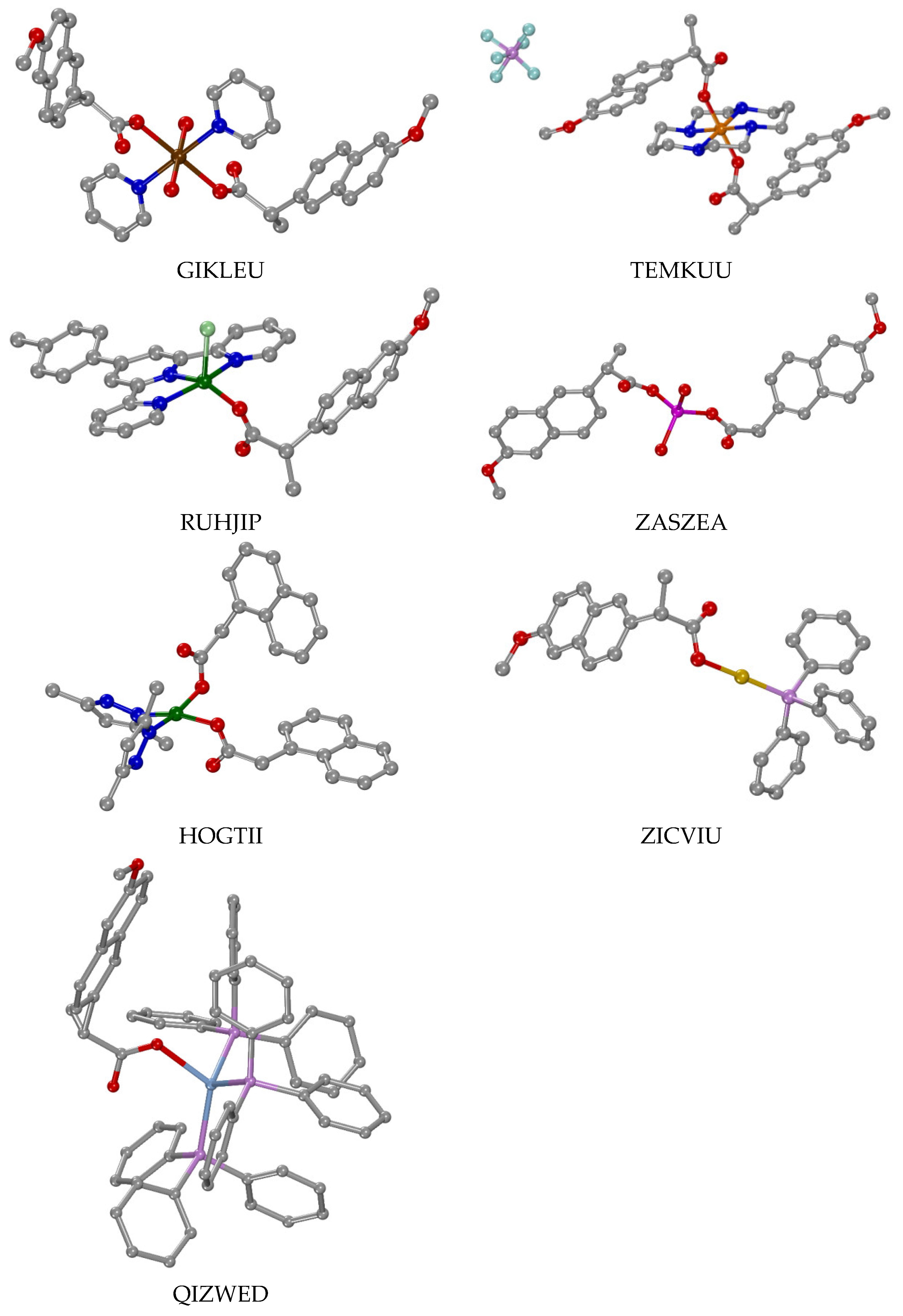
| [Mn(NAP–O)2(py)2(H2O)2] b | GIKLEU | MN2O4 | Oh | [42] |
| [Co(NAP–O)2(py)2(H2O)2] | KATSUV | MN2O4 | Oh | [43] |
| [Co(NAP–O)2(cyclam)] | TEMKUU | MN4O2 | Oh | [44] |
| [Cu(N1A–O)2(Hdmpz)2] | HOGTII | MN2O2 | Spl | [45] |
| [Cu(NAP–O)2(H2O)3]·H2O | ZASZAW | MO5 | Spy | [46] |
| [Cu(N1A–O)(EDA)2](ClO4) | TUFTUL | MN4O | Spy | [47] |
| [Cu(NAP–O)2(H2O)(4pic)2] | RANHOG | MN2O3 | Spy | [48] |
| [Cu(NAP–O)(L1)Cl] | RUHJIP | MN3OCl | Spy | [49] |
| [Cu(NAP–O)(L2)Cl] | BUSYEV | MN3OCl | Spy | [49] |
| [Zn(NAP–O)2(H2O)2]·H2O | ZASZEA | MO4 | Td | [46] |
| [Ag(NAP–O)(PPh3)3]·H2O | QIZWED | MOP3 | Td | [50] |
| [Au(NAP–O)(PPh3)] | ZICVIU | MOP | L | [51] |
| [Gd(N1A–O)2(phen)2(H2O)2]·[Eu(N1A–O)2(phen)2(H2O)2]·(N1A)2·2H2O | HOFMEW | Co-crystallized/MN4O4 | Dh | [52] |
| II: Bidentate chelating coordination of the ligand | ||||
| [Mn(N1A–O,O′)2(Hbzmd)2]·H2O | LANDEL | MN2O4 | Oh | [53] |
| [Ni(N1A–O,O′)2(Hbzmd–N3)2]·H2O | YAVLIS | MN2O4 | Oh | [54] |
| [Cu(NAP–O,O′)2(4,7–diPhphen)] | EYIKAA | MN2O4 | Oh | [55] |
| [Cu(NAP–O,O′)2(bipy)]·H2O | LEBVIZ | MN2O4 | Oh | [56] |
| [Cu(NAP–O,O′)2(phen)]·H2O | LEBVOF | MN2O4 | Oh | [56] |
| [Zn(N1A–O,O′)2(phen)] | NEQLIF, NEQLIF01 | MN2O4 | Oh | [57,58] |
| [Ag(NAP–O,O′)(tptp)2] | QIZWAZ | MO2P2 | Td | [50] |
| [Cd(NAP–O,O′)2(H2O)2]·H2O | ZASZUQ | MO6 | Oh | [46] |
| [(n–Bu)2Sn(NAP–O,O′)2] | XISCAF, XISCAF01 | MC2O4 | Oh | [59,60] |
| III: Monodentate + bidentate chelating coordination of the ligands | ||||
| [Mn(NAP–O)(NAP–O,O′)(phen)(H2O)] | GIHJEP | MN2O4 | Oh | [42] |
| [Ni(NAP–O)(NAP–O,O′)(bipy)(H2O)] | YAQJEI | MN2O4 | Oh | [61] |
| [Ni(NAP–O)(NAP–O,O′)(phen)(H2O)] | YAQJAE, GADYET | MN2O4 | Oh | [61,62] |
| [Zn(NAP–O,O′)2(neoc)] | NORBED | MN2O3 | Spy–TB | [63] |
| [Zn(N1A–O)(N1A–O,O′)(5,5′–Me2–bipy)] | UQUSAB | MN2O3 | Spy–TB | [64] |
| Complex | CCDC Name | Metal Ions | Coord.Sphere | Reference |
|---|---|---|---|---|
| I: Bidentate bridging (μ–O,O′) | ||||
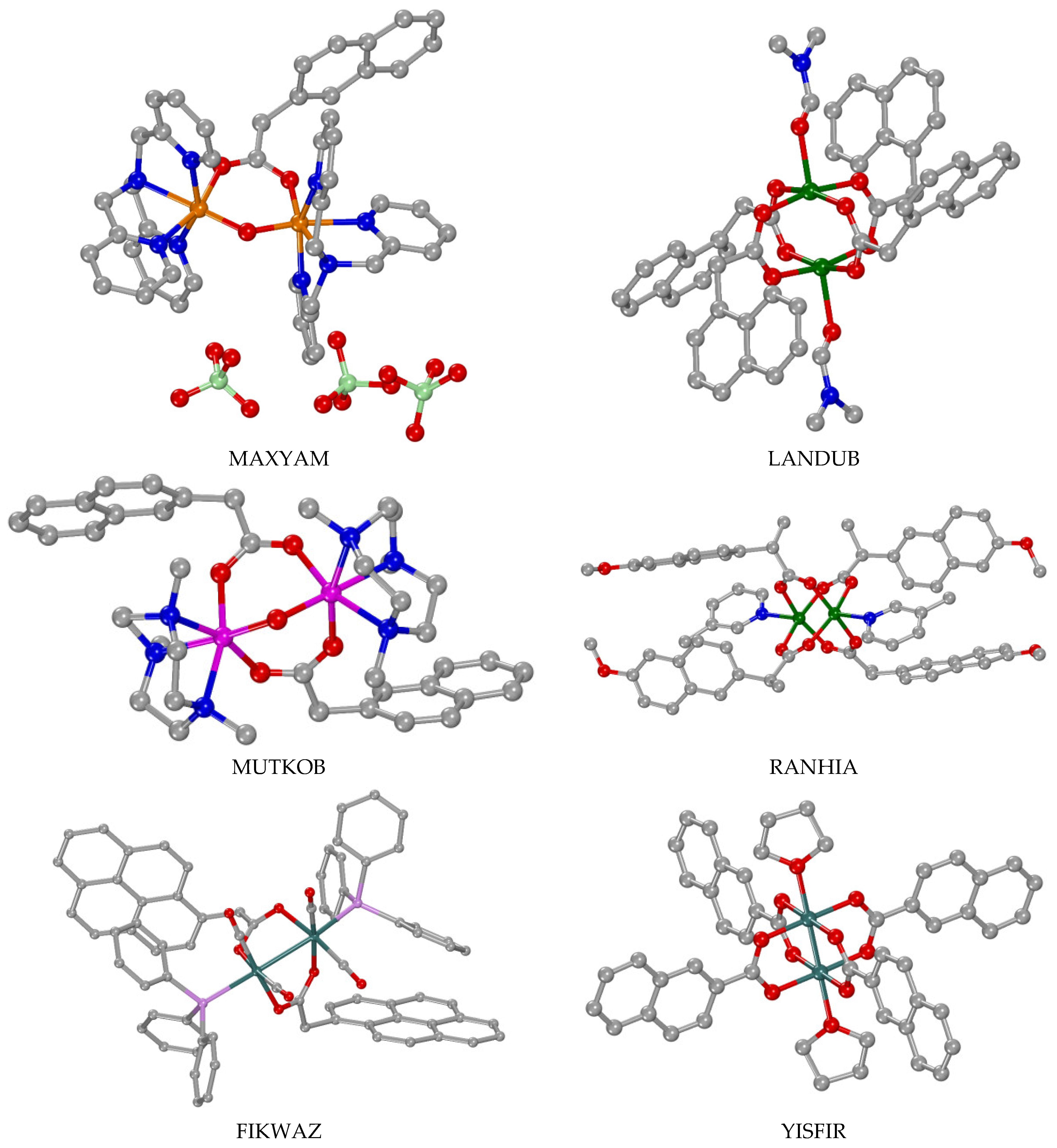
| [Fe2(µ2–O)(µ2–N2A–O,O′)(tren)2](BPh4)(NO3)2 | MAXXUF | Fe(III)2 | MN3O2 | [65] |
| [Fe2(µ2–O)(µ2–N2A–O,O′)(TPA)2](ClO4)3 | MAXYAM | Fe(III)2 | MN3O2 | [65] |
| [Fe2(µ2–O)(µ2–N2A–O,O′)2(Tp)2] | MAXYEQ | Fe(III)2 | MN3O3 | [65] |
| [Fe2(µ2–O)(µ2–N2A–O,O′)2(TACN–Me3)2](PF6)2 | MUTKIV | Fe(III)2 | MN3O3 | [66] |
| [Cu2(µ2–N2A–O,O′)4(DMSO)2]·2(HN2A)·2DMSO | IXAFOC | Cu(II)2 | MO5 | [67] |
| [Cu2(µ2–N2A–O,O′)4(DMF)2] | LANDUB | Cu(II)2 | MO5 | [68] |
| [Cu2(μ2–NAP–O,O′)4(3pic)2] | RANHIA | Cu(II)2 | MNO4 | [48] |
| [Cu2(μ2–NAP–O,O′)4(caf)2] | XIRCOQ | Cu(II)2 | MNO4 | [69] |
| [Zn2(µ2–OH)(µ2–N2A–O,O′)2(TACN–Me3)2](ClO4) | MUTKOB | Zn(II)2 | MN3O3 | [66] |
| [Zn2(µ2–N2A–O,O′)4(phdat)2] | TEWNEQ | Zn(II)2 | MNO4 | [70] |
| [Ru2(µ2–N2A–O,O′)4(H2O)2](PF6)·THF | YISFIR | Ru(II)/Ru(III) | MO5 | [71] |
| [Ru2(µ2–N1A–O,O′)4(THF)2](PF6)·THF | YISFUD | Ru(II)/Ru(III) | MO5 | [71] |
| K[Ru2(µ2–N2A–O,O′)2(dhpta)] | KEJKOA | Ru(III)2 | MNO5 | [72] |
| [Ru2(µ2–PYA–O,O′)2(CO)4(PPh3)2] | FIKWAZ | Ru(I)2 | MC2PO2 | [35] |
| II: Tridentate bridging (μ–O,O,O′) + bidentate bridging (μ–O,O′) + bidentate chelating (κ–O,O′) | ||||
| [Y2(µ2–N1A–O,O,O′)2(µ2–N1A–O,O′)2(κ–N1A–O,O′)2(phen)2]·DMF | LULCEB | Y(III)2 | MN2O7 | [73] |
| [Pr2(µ2–N1A–O,O,O′)2(µ2–N1A–O,O′)2(κ–N1A–O,O′)2(phen)2]·DMF | SILLOP | Pr(III)2 | MN2O7 | [73,74] |
| [Nd2(µ2–N1A–O,O,O′)2(µ2–N1A–O,O′)2(κ–N1A–O,O′)2(phen)2] | JOSJUZ | Nd(III)2 | MN2O7 | [75] |
| [Sm2(µ2–N1A–O,O,O′)2(µ2–N1A–O,O′)2(κ–N1A–O,O′)2(phen)2]·DMF | TIPBEA | Sm(III)2 | MN2O7 | [73,76] |
| [Eu2(µ2–N1A–O,O,O′)2(µ2–N1A–O,O′)2(κ–N1A–O,O′)2(phen)2]·DMF | SILLIJ | Eu(III)2 | MN2O7 | [73,77] |
| [GdTb(µ2–N1A–O,O,O′)2(µ2–N1A–O,O′)2(κ–N1A–O,O′)2(phen)2] | YUNFUJ | Gd(III)Tb(III) | MN2O7 | [78] |
| [Gd2(µ2–N1A–O,O,O′)2(µ2–N1A–O,O′)2(κ–N1A–O,O′)2(phen)2]·DMF | SILMIK | Gd(III)2 | MN2O7 | [73,79] |
| [Tb2(µ2–N1A–O,O,O′)2(µ2–N1A–O,O′)2(κ–N1A–O,O′)2(phen)2]·DMF | SILMEG | Tb(III)2 | MN2O7 | [73,78,80] |
| [Yb2(µ2–N1A–O,O,O′)2(µ2–N1A–O,O′)2(κ–N1A–O,O′)2(phen)2]·DMF | WINCIG | Yb(III)2 | MN2O7 | [73,81] |
| III: Bidentate bridging (μ–O,O′) + bidentate chelating (κ–O,O′) | ||||
| [Gd2(µ2–NAP–O,O′)4(NAP–O,O′)2(phen)2] | ZURGAW | Gd(III)2 | MN2O6 | [82] |
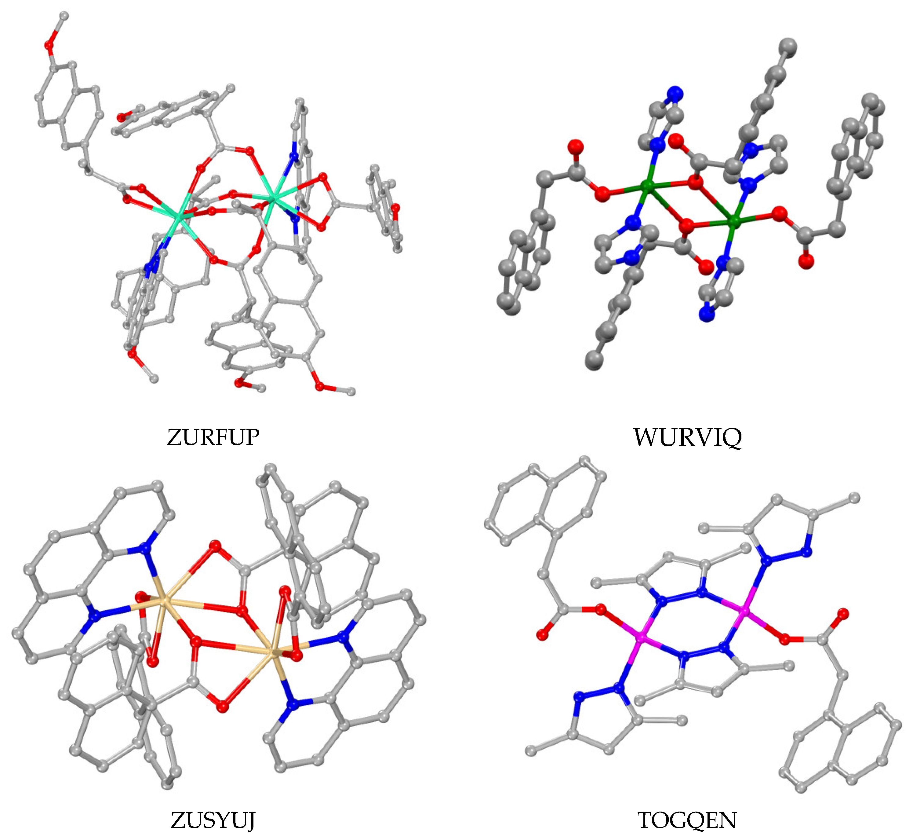
| [Dy2(µ2–NAP–O,O′)4(NAP–O,O′)2(phen)2] | ZURFUP | Dy(III)2 | MN2O6 | [82] |
| IV: Bidentate bridging (μ–O,O) + monodentate (κ–O) | ||||
| [Cu2(µ2–N1A–O,O)2(N1A–O)2(Himi)4] | WURVIQ | Cu(II)2 | MN2O3 | [83] |
| V: Tridentate bridging (μ–O,O,O′) + bidentate chelating (κ–O,O′) | ||||
| [Cd2(µ2–N1A–O,O,O′)2(κ–N1A–O,O′)2(phen)2] | ZUSYUJ | Cd(II)2 | MN2O4 | [84] |
| VI: Monodentate binding, bridging from other co-ligand | ||||
| [Zn2(μ–dmpz)2(Hdmpz)2(N1A–O)2] | TOGQEN | Zn(II) | MN3O | [85] |
| Complex | CCDC Name | Reference |
|---|---|---|
| [Mn3(µ3–O)(µ2–N1A–O,O′)6(py)3] | OFILAS | [86] |
| [Cu4(µ2–N1A–O,O′)6(µ2–N1A–O,O,O′)2(CH3CN)2] | QIJYOZ | [87] |
| [Cd4(µ2–N1A–O,O′)4(µ2–N1A–O,O,O′)2(N1A–O)2(µ2–H2O)2(bipy)2] | FUVZED | [84] |
| [(Me3Sn)4(µ2–NAP–O,O′)4] | COFTOJ | [59] |
| {[(n–Bu)2Sn]2(µ2–N1A–O,O′)(µ2–N1A–O,O)(µ3–O)}2 | OMOLIN | [88] |
| [Ti6(μ3–O)2(μ2–O)2(μ3–phenyl–phosphonato)2(μ2–isopropoxo)4(isopropoxo)6(μ2–N2A–O,O′)2] | EHOGOZ | [89] |
| [Μn6(µ3–NAP–O,O,O′)(µ2–Hsal–O,O′)(µ2–shi–N,O)5(py)6] | NIHVEI | [90] |
| Complex | CCDC Name | Polymerized via | Reference |
|---|---|---|---|
| Part I: As bridging ligands | |||
| [Mg(µ2–NAP–O,O)(µ2–NAP–O,O′)(µ2–H2O)]n | ANOMEW | µ2–O,O, µ2–O,O′, µ2–H2O | [41] |
| [Mn(µ2–NAP–O,O′)2(CH3OH)]n | NIHVOS | µ2–O,O′ | [90] |
| [Co(µ2–N1A–O,O′)2(H2O)2]n | MEQHAS | µ2–O,O′ | [91,92] |
| [Cd(NAP–O,O′)(µ2–NAP–O,O,O′)(H2O)]n | ZASZIE | µ2–O,O,O′ | [46] |
| [(Ph3Sn)(µ2–NAP–O,O′)]n | COFTID | µ2–O,O′ | [59] |
| [(n–Bu)3Sn(µ2–N1A–O,O,O′)]n | OMOLOT | µ2–O,O,O′ | [88] |
| [Ag4(µ2–NAP–O,O′)2(µ3–NAP–O,O,O′)2(2pic)2]n | CAVHOA | µ2–O,O,O′ | [93] |
| Part II: As coordinated ligands, other bridges | |||
| [Zn(NAP–O)2(µ–3U)]n | OMALIA | 3U | [94] |
| [Zn(NAP–O)2(µ–4U)]n | OMALEW | 4U | [94] |
| [Cd3(µ2–N1A–O,O′)2(µ2–N1A–O,O,O′)2(µ2–4,4′–bipy)2(κ–N1A–O,O′)2]n | FUVZIH | 4,4′–bipy | [84] |
| [Cd(µ3–pyr3O)(N1A–O,O′)(H2O)]n, | MIRSAI | pyr3O | [84] |
| [Ag(N1A–O)(µ–bpp–N,N′)]n | VIGDIA | bpp | [95] |
| Complex | λexcitation (nm) | λmax,emission (nm) (Transition) | Reference |
|---|---|---|---|
| [Eu2(N1A)6(phen)2]·2DMF | 322 | 581 (5D0→7F0), 593 (5D0→F1), 618 (5D0→7F2), 651 (5D0→7F3), 694 (5D0→7F4) | [73] |
| [Sm2(N1A)6(phen)2]·2DMF | 335 | 566 (4G5/2→6H5/2), 594 (4G5/2→6H7/2), 617 (4G5/2→6H7/2), 648 (4G5/2→6H9/2), 680 (4G5/2→6H11/2) | [73] |
| [Tb2(N1A)6(phen)2]·2DMF | 345 | 545 (5D4→7F5), 594 (5D4→7F4), 617 (5D4→7F3), 675 (5D4→7F2) | [73] |
| [Eu(N1A)2(phen)2(H2O)2](N1A)2·2H2O | 332 | 580 (5D0→7F0), 592 (5D0→7F1), 617 (5D0→7F2), 674 (5D0→7F3), 697 (5D0→7F4) | [52] |
| [Gd2(N1A)6(phen)2] | 351 | 490 (5D4→7F6), 593 (5D4→7F4), 615 (5D4→7F3), 645 (5D4→7F2) | [78] |
| [Tb2(N1A)6(phen)2].DMF | 359 | 490 (5D4→7F5), 594 (5D4→7F4), 615 (5D4→7F3) | [78] |
| Compounds | Stable Until (°C) | Steps | Temperature per Step | Reference |
|---|---|---|---|---|
| [Cu2(NAP)4(3pic)2] | 161 | two | I: 130–177 II: 177–455 | [48] |
| [Cu(NAP)2(H2O)(4pic)2] | 122 | two | I: 30–173 II: 173–461 | [48] |
| [Cu4(N1A)8(CH3CN)2] | 175.7 | three | I: 175.7–185.2 II: 252.8–266.9 III: 493.8–587.5 | [87] |
| [Cu(N1A)(EDA)2](ClO4) | Not given | two | I: 162–355 II: 355–616 | [47] |
| [Cu(N1A)2(Hdmpz)2] | 180 | two | I: 188.4–270.4 II: 290.7–433.1 | [45] |
| [Zn2(dmpz)2(Hdmpz)2(N1A)2] | 180 | three | I: 188.4–282.8 II: 285.4–422.6 III: 437.7–606.4 | [85] |
| [Zn2(N2A)4(phdat)2] | 208 | three | I + II + III: 208–586 | [70] |
| [Ag4(NAP)4(2pic)2]n | Not given | two | I: 30–204 II: 204–557 | [93] |
| [Cd2(N1A)4(phen)2] | 267 | two | I + II: 267–483 | [84] |
| [Cd4(N1A)8(bipy)2(H2O)2] | 130 | two | I: 130–190 II: 230–452 | [84] |
| [Cd3(N1A)6(4,4′–bipy)2]n | 294 | one | 294–449 | [84] |
| Complex | J (cm−1) | g | Reference |
|---|---|---|---|
| [Mn3(µ3–O)(µ2–N1A–O,O′)6(py)3] | −7.5 a, −5.0 b | 2.06 | [86] |
| [Mn3(µ3–O)(µ2–N2A–O,O′)6(py)3] | −7.0 a, −4.9 b | 2.06 | [86] |
| [Fe2(µ2–O)(µ2–N2A–O,O′)2(TACN–Me3)2](PF6)2 | −105 | [66] | |
| [Fe2(µ2–O)(µ2–N2A–O,O′)(tren)2](BPh4)(NO3)2 [Fe2(µ2–O)(µ2–N2A–O,O′)(TPA)2](ClO4)3 [Fe2(µ2–O)(µ2–N2A–O,O′)2(Tp)2] | −130 ± 10 | [66] | |
| [Cu4(N1A)8(CH3CN)] | 2J1 = −295 c, 2J2 = −38 c | 2.28 | [87] |
| K[Ru2(µ2–N1A–O,O′)2(dhpta)] | −581 | 2.1 | [72] |
| K[Ru2(µ2–N2A–O,O′)2(dhpta)] | −378 | 2.1 | [72] |
| Complex | Naphthalene Moiety | Metal-Centered | Reference | |||
|---|---|---|---|---|---|---|
| Solvent | Reduction (Epc) | Oxidation (Epa) | Reduction (Epc) | Oxidation (Epa) | ||
| [Fe2O(N2A)(tren)2](BPh4)(NO3)2 | CH3CN | −1.78 | +1.46 | −0.60 a | [65] | |
| [Fe2O(N2A)(TPA)2](ClO4)3 | CH3CN | −2.20, −2.40 | +1.50 | −1.20 a | +0.83 b | [65] |
| [Fe2O(N2A)2(Tp)2] | CH3CN | −2.05 | +1.60 | −1.07 a | +1.30 b | [65] |
| [Fe2O(N2A)2(TACN–Me3)2](PF6)2 | CH3CN | −1.9 | +1.65 | −0.75 a,c | [66] | |
| [Mn3O(N1A)6(py)3] | CH2Cl2 | +1.05 | −0.70 c,e | 0.02 c,d −0.61 | [86] | |
| [Mn3O(N2A)6(py)3] | CH2Cl2 | +1.03 | −0.61 c,e | 0.09 c,d −0.57 c,d | [86] | |
| K[Ru2(N1A)2(dhpta)] | CH3CN DMF | +1.29 | −1.05 f, −1.36 g −1.27 f, −1.73 g | 0.63 c,h 0.53 c,h | [72] | |
| K[Ru2(N2A)2(dhpta)] | CH3CN DMF | +1.30 | −1.05 f, −1.34 g −1.28 f, −1.71 g | 0.64 c,f 0.54 c,f | [72] | |
| Complex | Epc a | Epa b | Reference |
|---|---|---|---|
| [Co(NAP)2(MeOH)4] | –1109 a | –13 b | [43] |
| [Co(NAP)2(py)2(H2O)2] | –775 | –25 b | [43] |
| [Co(NAP)2(phen)(H2O)2] | –1296 | +53 b | [43] |
| [Co(NAP)2(bipy)(H2O)2] | –1065 | –56 b | [43] |
| [Ni(NAP)2(MeOH)4] | –607 | –384 | [61] |
| [Ni(NAP)2(bipy)(CH3OH)] | –544 | –304 | [61] |
| [Ni(NAP)2(phen)(H2O)] | –485 | –307 | [61] |
| [Ni(NAP)2(bipyam)] | –531 | –314 | [61] |
| [Ni(NAP)2(Hpko)2] | –473 | –362 | [61] |
| [Ni(NAP)2(py)2(H2O)2] | –524 | –314 | [61] |
| [Cu(NAP)2(4,7–dphphen)] | –355 | Not provided | [55] |
| Compound | Cell Lines | Reference | |
|---|---|---|---|
| HNAP | MCF–7: >160 HeLa: >160 3T3–L1: >250 | A549: >160 MDA–MB–453: >100 HT–29: >100 | [49,90,93] |
| [Μn6(NAP)(Hsal)(shi)6(py)6] | MCF–7: 9.6 ± 0.3 HeLa: 30.1 ± 1.3 | A549: 69.3 ± 4.0 | [90] |
| [Μn(NAP)2(CH3OH)]n | MCF–7: 62.0 ± 2.5 HeLa: >160 | A549: >160 | [90] |
| [Cu(NAP)(L1)Cl] | MCF–7: 1.51 ± 0.15 | [49] | |
| [Cu(NAP)(L2)Cl] | MCF–7: 31.03 ± 1.2 | [49] | |
| [Cu(NAP)(L3)Cl] | MCF–7: 10.40 ± 0.3 | [49] | |
| [Ag4(NAP)4(2pic)2]n | A549: 74.08 ± 1.05 3T3–L1: 224.87 ± 2.60 | MDA–MB–453: 39.77 ± 1.95 HT–29: 29.96 ± 0.84 | [49] |
| [Ag(NAP)(PPh3)3](H2O) | MCF–7: 0.7 ± 0.1 | [50] | |
| [Ag(NAP)(tptp)2] | MCF–7: 2.2 ± 0.2 | [50] | |
| [Ni(NAP)2(phen)(H2O)] | HepG2: >1000 HT 29: 35.50 ± 1.94 | HEK–293: 198.5 ± 35.45 (72h) | [62] |
| [Co(NAP)2(cyclam)] | HMLER: 0.43 ± 0.05 | HMLER–shEcad: 0.11 ± 0.03 | [44] |
| [Au(NAP)(PPh3)] | HMLER: 0.183 ± 0.001 MDA–MB–231: 7.77 ± 0.41 4T1: 10.08 ± 0.86 | HMLER–shEcad: 0.063 ± 0.006 MDA–MB–468: 6.48 ± 1.43 | [51] |
| {[(n–Bu)2Sn]2(N1A)2(O)}2 | MCF–7: 37.61 HeLa: 1.805 HepG2: 0.802 | Colo205: 0.100 NCI–H460: 67.29 | [88] |
| [(n–Bu)3Sn(N1A)]n | MCF–7: 0.301 HeLa: 0.361 HepG2: 0.127 | Colo205: 0.104 NCI–H460: 0.188 | [88] |
| H3shi | MCF–7: >160 HeLa: >160 | A549: >160 | [90] |
| PPh3 | MCF–7: 67.4 ± 13.9 | [50] | |
| tptp | MCF–7: 26.5 ± 2.8 | [50] | |
| 5–Fluorouracil | HMLER: 41.05 ± 5.30 | HMLER–shEcad: 49.10 ± 5.94 | [51] |
| Capecitabin | HMLER: >100 | HMLER–shEcad: >100 | [51] |
| Carboplatin | MCF–7: 26.83 HepG2: 0.613 A549: 39.43 ± 0.76 NCI–H460: 62.13 HT–29: 47.15 ± 2.80 HMLER: 67.31 ± 2.80 | HeLa: 24.78 Colo205: 0.531 MDA–MB–453: 56.73 ± 1.24 HMLER–shEcad: 72.39± 7.99 3T3–L1: 43.20 ± 1.35 | [3,51,88] |
| Cisplatin | MCF–7: 8.0 ± 0.7 HepG2: 23.71 ± 1.52 HT 29: 69.13 ± 1.88 | HEK–293: 46.81 ± 2.79 HMLER–shEcad: 5.64 ± 0.30 HMLER: 2.56 ± 0.02 | [49,50,51,62] |
| Doxorubicin | MCF–7: 10.90 | [49] | |
| Salinomycin | HMLER: 11.43 ± 0.42 | HMLER–shEcad: 4.23 ± 0.35 | [44,51] |
Disclaimer/Publisher’s Note: The statements, opinions and data contained in all publications are solely those of the individual author(s) and contributor(s) and not of MDPI and/or the editor(s). MDPI and/or the editor(s) disclaim responsibility for any injury to people or property resulting from any ideas, methods, instructions or products referred to in the content. |
© 2023 by the authors. Licensee MDPI, Basel, Switzerland. This article is an open access article distributed under the terms and conditions of the Creative Commons Attribution (CC BY) license (https://creativecommons.org/licenses/by/4.0/).
Share and Cite
Lazou, M.; Perontsis, S.; Psomas, G. Metal Complexes with Naphthalene-Based Acetic Acids as Ligands: Structure and Biological Activity. Molecules 2023, 28, 2171. https://doi.org/10.3390/molecules28052171
Lazou M, Perontsis S, Psomas G. Metal Complexes with Naphthalene-Based Acetic Acids as Ligands: Structure and Biological Activity. Molecules. 2023; 28(5):2171. https://doi.org/10.3390/molecules28052171
Chicago/Turabian StyleLazou, Marialena, Spyros Perontsis, and George Psomas. 2023. "Metal Complexes with Naphthalene-Based Acetic Acids as Ligands: Structure and Biological Activity" Molecules 28, no. 5: 2171. https://doi.org/10.3390/molecules28052171
APA StyleLazou, M., Perontsis, S., & Psomas, G. (2023). Metal Complexes with Naphthalene-Based Acetic Acids as Ligands: Structure and Biological Activity. Molecules, 28(5), 2171. https://doi.org/10.3390/molecules28052171