Article

Design, Synthesis and Structure-Activity Relationship Studies of Nicotinamide Derivatives as Potent Antifungal Agents by Disrupting Cell Wall

1
Department of Pharmacy, Shanghai Tenth People’s Hospital, School of Medicine, Tongji University, No. 1239 Siping Road, Shanghai 200092, China
2
School of Pharmacy, Naval Medical University, No. 325 Guohe Road, Shanghai 200433, China
3
School of Chinese Materia Medica, Shenyang Pharmaceutical University, Shenyang 110016, China
*
Authors to whom correspondence should be addressed.
These authors contributed equally to this work.
Molecules 2023, 28(3), 1135; https://doi.org/10.3390/molecules28031135
Submission received: 14 December 2022 / Revised: 16 January 2023 / Accepted: 20 January 2023 / Published: 23 January 2023

Abstract

:
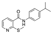
Fungal infections pose a serious challenge to human health due to the limited paucity of antifungal treatments. Starting as a hit compound screened from our compound library, a series of nicotinamide derivatives have been successfully synthesized via a facile one-step coupling reaction of aromatic carboxylic acid and amine. The synthesized compounds were evaluated for their antifungal activity against Candida albicans SC5314. Among the 37 nicotinamide derivatives screened, compound 16g was found to be the most active against C. albicans SC5314, with an MIC value of 0.25 μg/mL and without significant cytotoxicity. The rudimentary structure-activity relationships study revealed that the position of the amino and isopropyl groups of 16g was critical for its antifungal activity. In particular, compound 16g showed potent activity against six fluconazole-resistant C. albicans strains with MIC values ranging from 0.125–1 μg/mL and showed moderate activity against the other seven species of Candida, three strains of Cryptococcus neoformans, and three strains of Trichophyton. Furthermore, compound 16g showed fungicidal, anti-hyphal, and anti-biofilm activities in vitro, which were related to its ability to disrupt the cell wall of C. albicans. Taken together, 16g is a promising compound that is fungal-specific by targeting the cell wall and could be used as a lead compound for further investigation.

Graphical Abstract

1. Introduction

Invasive fungal infections (IFIs) are increasingly threatening the lives of immunocompromised patients (e.g., following organ transplantation, patients with HIV/AIDS, immunosuppression, or receiving chemotherapy for cancer) [1]. It was estimated that more than 300 million people suffered from serious fungal-related diseases, and over 1.6 million people were killed by IFIs annually [2]. Notably, the mortality rate of IFIs is frequently greater than 50%. Approximately 90% of mycotic deaths are caused by three major fungal species: Candida, Cryptococcus, and Aspergillus [3,4]. To date, the classes of available antifungal drugs for the treatment of invasive fungal infections are still limited to azoles, polyenes, flucytosine, echinocandins, and a triterpenoid. Moreover, the clinical application of these drugs is not only limited by their drawbacks but also by drug resistance, which is a commonly occurring treatment complication. Therefore, new antifungal drugs are urgently needed.
As an important class of heterocyclic derivatives, nicotinamide analogues play an important role in the development of antifungals [5,6,7,8,9,10,11]. Despite the development and commercialization of several nicotinamide analogues, most have been studied as pesticides (Figure 1). An example of such a fungicide is boscalid (1), which was discovered by BASF for the control of Alternaria late blight of pistachio [5,6]. In 2008, Queron et al. reported a 4-(3-chloro-5-(trifluoromethyl)pyridin-2-yl)butyl nicotinamide (2) possessing good fungicidal activities against Alternaria alternate [7]. In 2010, Nakamoto et al. described a nicotinamide analogue 5 possessing excellent antifungal activities against C. albicans, C. neoformans, and A. fumigatus [8]. In 2014, Ye et al. described that the compound N-(3-chloro-4-fluorophenyl)-2-(methylthio)nicotinamide (3) displayed moderate antifungal activity against two phytopathogenic fungi, Rhizoctonia solani and Sclerotinia sclerotiorum [9]. In 2019, our team reported that nicotinamide (4, Vitamin B3) showed moderate antifungal activity against C. albicans, including fluconazole-resistant isolates [10]. More recently, Wang and his co-workers also reported several nicotinamide derivatives (6) exhibiting good fungicidal activities after modification of the boscalid structure [11].
Through measuring the MIC of each compound in our in-house library, we discovered several new scaffolds with antifungal activities against C. albicans [12]. In this study, we focused on another moderately active antifungal hit compound, 7, which showed an MIC value of 16 μg/mL against C. albicans. Although the structure of compound 7 is very similar to that of compound 3 in Figure 1, compound 3 was demonstrated to be inactive against C. albicans (MIC > 64 μg/mL), which suggested a possible structure-activity relationship (SAR) divergence between activity against pathogenic fungi and phytopathogenic fungi. Herein, based on compound 7, the hit-to-lead optimization was preliminarily completed, and a series of 37 nicotinamide derivatives were designed, synthesized, and screened for their antifungal activity, which resulted in the identification of a potent and broad-spectrum lead nicotinamide derivative, namely 2-amino-N-(3-isopropylphenyl)nicotinamide (16g). Meanwhile, we assessed the cytotoxicity, anti-hyphae, and anti-biofilm activities of lead compound 16g against Candida albicans in vitro. The ADMET properties of 16g were also predicted by the SwissADME online tool to evaluate its theoretical druggability [13]. Our results indicated that the lead compound 16g warrants further in-depth investigation as a promising antifungal agent.

2. Results and Discussion

2.1. Screening and Hit Identification

More than 30,000 compounds were screened for their antifungal activities against three of the most common pathogenic fungi C. albicans, C. neoformans, and A. fumigatus. After filtering out pan-assay interference compounds (PAINS, by SwissADME) and cytotoxic compounds, several hits were obtained [12,14]. Among them, compound 7 attracted our attention, which possessed a simple structure and showed moderate antifungal activity against C. albicans with an MIC value of 16 μg/mL and without obvious cytotoxicity against human umbilical vein endothelial cells (HUVECs) with an IC50 value of > 64 μg/mL. Notably, neither compound 1 nor 3 (Figure 1) showed superior antifungal activities over compound 7 (Table 1). Encouraged by these results, we decided to conduct further structure-activity relationship studies on the hit compound 7.

2.2. Chemistry

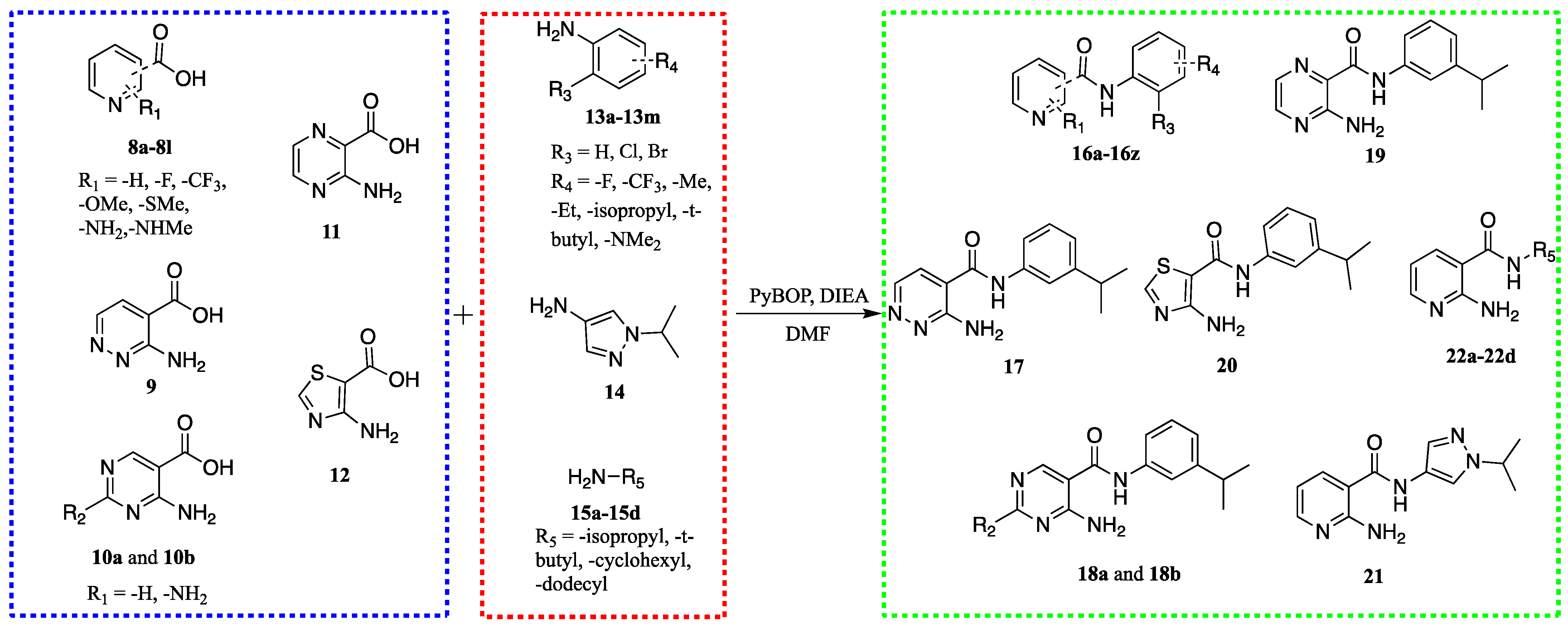
As depicted in Scheme 1, all compounds (16a-16z, 17, 18a-18d, 19, 20, 21, and 22a-22d) were synthesized facilely by a one-step coupling reaction of aromatic carboxylic acids (8a-8l, 9, 10a-10b, 11, and 12) and amines (13a-13m, 14, and 15a-15d) in the presence of PyBOP and TEA.

2.3. Structure-Activity Relationship

To analyze the structural determinants for antifungal activity of compound 7 against C. albicans, we initially replaced the meta-isopropyl group with meta-ethyl, meta-t-butyl, and para-isopropyl, giving 16a, 16b, and 16c, respectively. The results are shown in Table 1. These modifications led to a decrease in potency. Furthermore, we replaced -SMe with -Ome, -CF3, -NHMe, -NH2, and -H, giving 16d, 16e, 16f, 16g, and 16h, respectively. All these modifications were found unfavorable for antifungal activity with the exception of compound 16g, which showed excellent antifungal activity against C. albicans SC5314. The MIC value of 16g for C. albicans was 0.25 μg/mL, which was comparable to FLC.
Due to the high potency exhibited by 16g, we used it as a new starting point for further structural optimization. The results are shown in Table 2. Initially, modifications focused on the meta-isopropyl aniline moiety. Moving the meta-isopropyl group to the ortho- or para-position of the aniline moiety gave the compounds 16i and 16j, respectively. Para-substitution (16j) was relatively well-tolerated with a 2-fold decrease in potency (MIC = 0.5 μg/mL), whereas ortho-substitution (16i) resulted in a complete loss of activity (MIC > 64 μg/mL). In addition, the replacement of the isopropyl group with fluorine and trifluoromethyl, methyl, ethyl, tert-butyl, and dimethylamino groups to give compounds 16k-16r resulted in varying degrees of potency reduction. Moreover, the introduction of chlorine and bromine into aniline, giving 16s and 16t, respectively, proved unfavorable with a 32-fold and 16-fold reduction in activity. Subsequently, we investigated the effect of the 2-amino pyridine moiety of 16g by varying the position of the amino group and nitrogen atom while fixing meta-isopropyl aniline to give compounds 16u-16z. Unfortunately, these modifications appeared to be unhelpful. Furthermore, the incorporation of a second nitrogen atom into the pyridine ring gave the diazine analogue 17, the pyrimidine analogues 18a and 18b, and the pyrazine analogue 19, which also yielded disappointing results, despite 19 exhibiting moderate antifungal activity with an MIC value of 4 μg/mL. Replacement of the pyridine ring with it’s bioisosteric thiazole ring, giving 20, also demonstrated an unfavorable result. In a final study, we replaced the phenyl of the aniline moiety with pyrazole (21), isopropyl (22a), tert-butyl (22b), cyclohexyl (22c), and dodecyl (22d), all of which were found to be detrimental to antifungal activity.
Overall, the results discussed above shed light on 2-aminopyridine and meta-isopropyl as the essential moieties for the antifungal activity of this scaffold. Therefore, the most active compound, 16g, was selected for further investigation.

2.4. Compounds 16g and 16j Exhibit Low Toxicity to Mammalian Cells

To confirm the antifungal effects were not due to the cytotoxicities of compounds 16g and 16j, HUVEC cells were treated with various concentrations (2.5, 5, 10, and 20 μg/mL) of FLC, 16g, and 16j for 24 h. Cell viability was measured by the CCK8 assay. As shown in Figure 2, we found that FLC, 16g, and 16j did not cause significant cytotoxicity below 20 μg/mL, and no significant difference was observed between the two compounds and fluconazole.

2.5. Compound 16g Exhibits Broad-Spectrum Antifungal Activity

To investigate the antifungal effect of this chemotype, compound 16g was selected to screen for in vitro activity against two strains of fluconazole-sensitive C. albicans and six strains of fluconazole-resistant C. albicans (Table 3). The antifungal activity of 16g against the two fluconazole-sensitive strains was comparable to that of fluconazole, with MIC values ranging from 0.125–0.5 μg/mL. In contrast, the activity of 16g against the six fluconazole-resistant strains was significantly superior to fluconazole, with MIC values ranging from 0.125–1 μg/mL. In addition, we further evaluated the antifungal spectrum of 16g (Table 4). Our results demonstrated that the compound 16g had moderate activities against Candida, Cryptococcus, and Trichophyton, which were equivalent to FLC and ineffective against A. fumigatus.

2.6. Fungicidal Activity of 16g against C. albicans

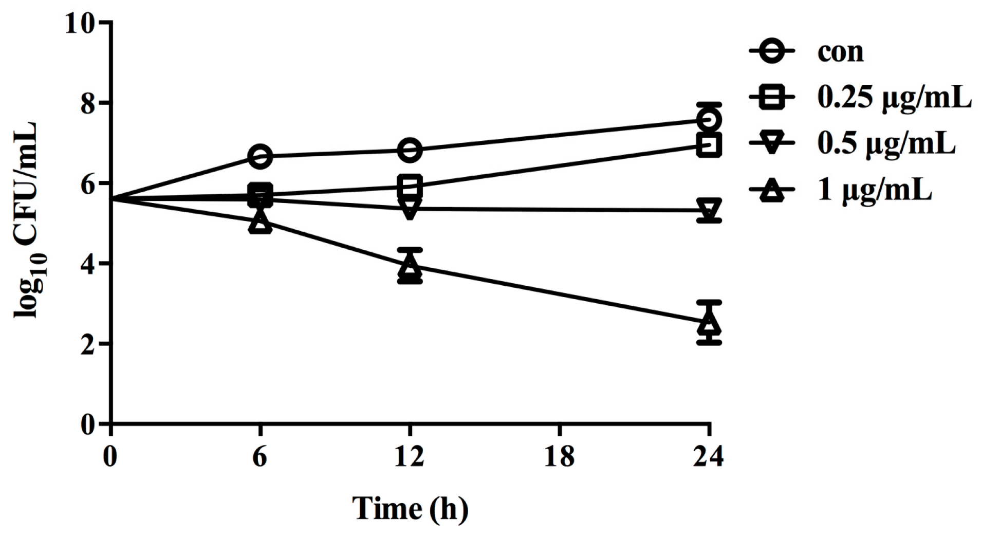
Time-kill curves showed that 16g at concentrations of 0.25 μg/mL and 0.5 μg/mL had a slight inhibitory effect on the growth of C. albicans (Figure 3). However, 1 μg/mL of 16g killed C. albicans after 24 h of treatment, which resulted in a log10 CFU/mL decrease from 5.5 to 2, approximately. Our results indicated that higher concentrations of 16g showed fungicidal activities.

2.7. Compound 16g Inhibits the Hyphae Formation of C. albicans

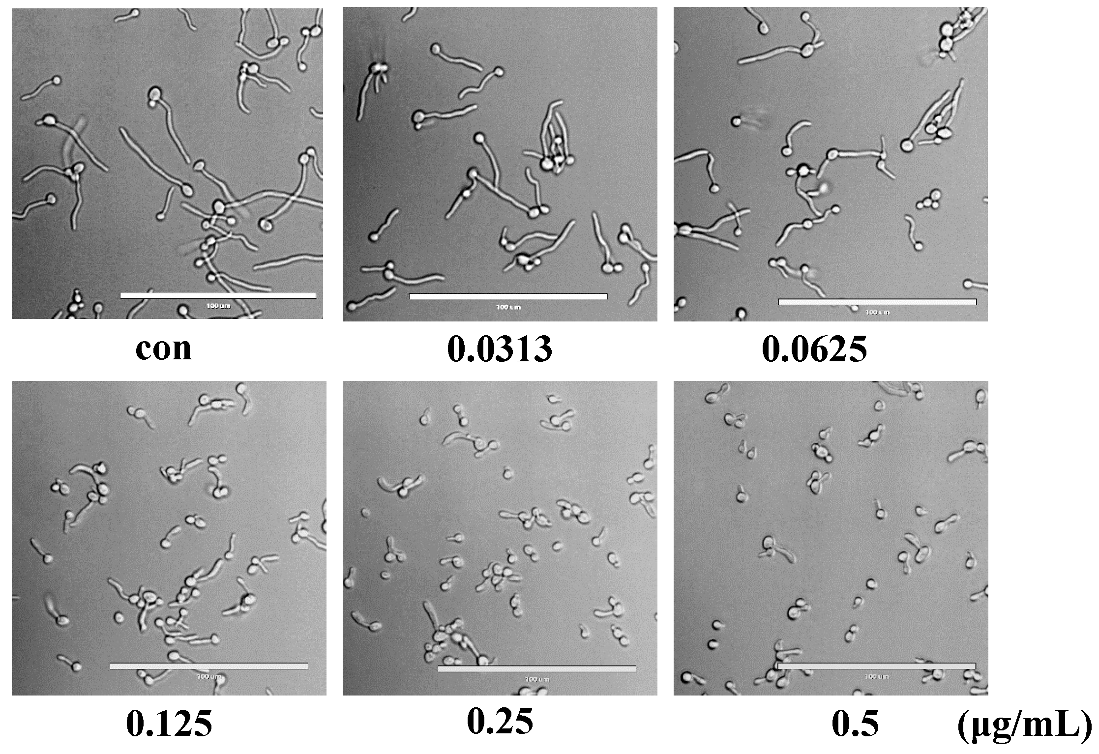
The morphological transition from yeast to hypha is the major contributor to the in vivo pathogenicity of C. albicans [15,16]. Therefore, we further investigated the activity of compound 16g against the yeast-to-hypha transition of C. albicans. As shown in Figure 4, 0.125 μg/mL or higher concentrations of compound 16g exhibited potent activity against C. albicans hypha formation, which showed fewer hyphae and more pseudohyphal cells.

2.8. Compound 16g Inhibits the Biofilm Formation in C. albicans

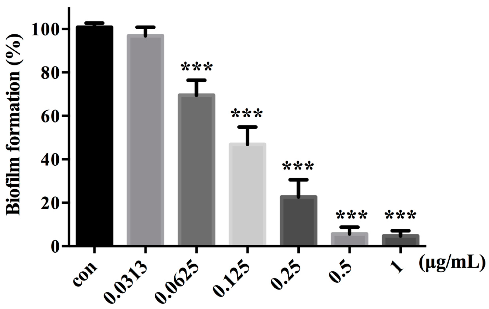
Biofilm formation is an important factor in the pathogenesis of C. albicans, which leads to high resistance to a wide range of antifungals [17]. In this study, we examined the effect of 16g on the biofilm formation of C. albicans. XTT reduction assays revealed that 16g showed an inhibitory effect on the biofilm formation in a dose-dependent manner (Figure 5). More specifically, 0.0625 μg/mL of 16g inhibited the biofilm formation by approximately 30%, and the inhibitory activity on the biofilm was enhanced as the concentrations of 16g increased. The addition of 0.125 μg/mL of 16g inhibited biofilm formation by 50%, while over 90% of the biofilms were inhibited in the presence of 0.5 μg/mL of 16g.

2.9. 16g Treatment Significantly Disrupted the Cell Wall Morphology of C. albicans

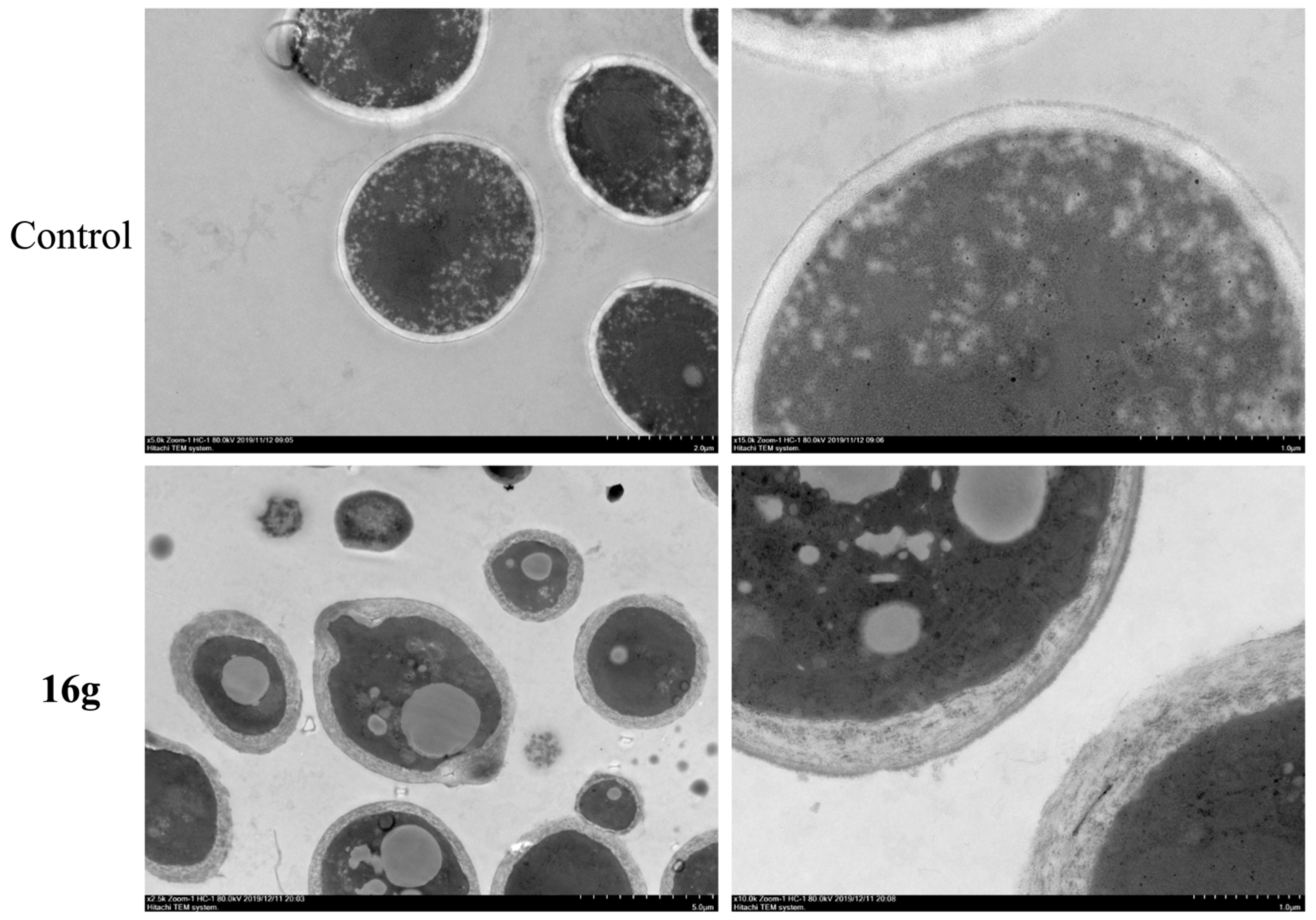
To further explore the antifungal mechanisms of nicotinamide derivatives, we investigated structural changes in cells treated with 1 μg/mL of 16g. As shown in Figure 6, the cell wall of 16g-treated cells displays broken edges and a thickened distance between the cell wall and cell membrane. Meanwhile, the gaps between the cell wall and cell membrane were filled with cytosolic fluid, which suggested the weakened protective effect of the cell wall was disrupted by 16g. Our results indicated that 16g could inhibit the growth of C. albicans by disrupting the cell walls.

2.10. ADMET Prediction

In silico ADMET prediction of compounds 16g and FLC was performed using the free SwissADME online tool [13]. The Brain Or IntestinaL EstimateD permeation (BOILED-Egg) method is a graphical model that works by calculating the polarity and lipophilicity of small molecules. According to Figure 7A, compound 16g was located in the yellow circle while FLC was in the white circle, representing that both compounds were highly absorbed through the gastrointestinal tract, but compound 16g penetrates the blood-brain barrier more readily than FLC. In addition, the compound 16g with the red spot is not a substrate for P-glycoprotein, which perhaps facilitated overcoming efflux pump-mediated resistance mechanisms in pathogenic fungi. Furthermore, as shown in the bioavailability radar (Figure 7B,C), the physicochemical characteristics, lipophilicity, solubility, pharmacokinetics, and drug-likeness properties of compound 16g and FLC were all located in the pink area (optimal range), indicating that compound 16g, such as FLC, is a good lead compound.

3. Materials and Methods

3.1. General Procedure for the Synthesis of Target Compounds

To a solution of aromatic carboxylic acid (8a-8l, 9, 10a, 10b, 11, and 12, 1 mmol), DIEA (2 mmol) and PyBOP (1.1 mmol) in DMF (5 mL) were added to various anilines (13a-13m and 14) or amines (15a-15d, 1 mmol) at room temperature. The mixture was stirred at this temperature for 2 h. The reaction was monitored by TLC. After the reaction was finished, the mixture was poured into water, and then the mixture was extracted with EtOAc (2 × 10 mL). After washing with brine (2 × 10 mL) and drying over anhydrous Na2SO4, the organic phase was evaporated in a vacuum. The crude product was purified by silica gel column chromatography using EtOAc/PE (1:1) as the eluent to give target compounds (16a-16z, 17, 18a, 18b, 19, 20, 21, and 22a-22d). The NMR spectra can be found in the Supplementary Materials.
16a: 238 mg, yield: 87%. White solid. 1H NMR (300 MHz, CDCl3-d1) δ 8.52 (d, J = 4.0 Hz, 1H), 8.24 (s, 1H), 7.88 (d, J = 7.5 Hz, 1H), 7.52 (s, 1H), 7.45 (d, J = 8.1 Hz, 1H), 7.28 (dd, J = 8.8, 6.8 Hz, 2H), 7.12 − 6.97 (m, 2H), 2.74 − 2.55 (m, 5H), 1.25 (t, J = 7.6 Hz, 3H). 13C NMR (151 MHz, DMSO-d6) δ 165.33, 157.91, 150.58, 144.78, 139.38, 135.79, 131.03, 129.09, 123.86, 119.65, 119.22, 117.76, 28.75, 16.00, 13.46.
16b: 273 mg, yield: 90%. White solid. 1H NMR (300 MHz, CDCl3-d1) δ 8.55 (dd, J = 4.8, 1.5 Hz, 1H), 8.17 (s, 1H), 7.92 (d, J = 7.3 Hz, 1H), 7.63 (s, 1H), 7.53 (d, J = 7.8 Hz, 1H), 7.31 (d, J = 7.9 Hz, 1H), 7.22 (d, J = 7.9 Hz, 1H), 7.10 (dd, J = 7.6, 4.9 Hz, 1H), 2.63 (s, 3H), 1.35 (s, 9H). 13C NMR (151 MHz, DMSO-d6) δ 165.31, 157.95, 151.74, 150.57, 139.17, 135.81, 131.00, 128.82, 121.31, 119.19, 117.49, 117.27, 34.92, 31.57, 13.47.
16c: 254 mg, yield: 89%. White solid. 1H NMR (300 MHz, CDCl3-d1) δ 8.56 (d, J = 3.1 Hz, 1H), 8.05 (s, 1H), 7.95 (d, J = 7.1 Hz, 1H), 7.57 (d, J = 8.2 Hz, 2H), 7.25 (d, J = 8.2 Hz, 2H), 7.17 − 7.09 (m, 1H), 2.96 − 2.87 (m, 1H), 2.64 (s, 3H), 1.25 (d, J = 6.9 Hz, 6H). 13C NMR (75 MHz, DMSO-d6) δ 165.18, 157.91, 150.56, 144.50, 137.12, 135.76, 131.04, 126.90, 120.39, 119.22, 33.40, 24.42, 13.46.
16d: 239 mg, yield: 88%. Yellow solid. 1H NMR (300 MHz, DMSO-d6) δ 10.34 (s, 1H), 9.56 (s, 1H), 8.78 (d, J = 1.4 Hz, 1H), 8.20 (d, J = 8.5 Hz, 1H), 8.10 (dd, J = 8.6, 1.8 Hz, 1H), 7.65 (d, J = 8.8 Hz, 2H), 7.27 (t, J = 7.8 Hz, 1H), 6.99 (d, J = 7.7 Hz, 1H), 2.96 − 2.79 (m, 1H), 1.21 (d, J = 6.9 Hz, 7H). 13C NMR (151 MHz, DMSO-d6) δ 165.40, 159.49, 155.27, 149.34, 139.62, 134.10, 132.69, 128.99, 126.24, 123.20, 122.95, 122.34, 118.74, 118.39, 33.99, 24.35.
16e: 263 mg, yield: 85%. Semi-solid. 1H NMR (300 MHz, DMSO-d6) δ 10.62 (s, 1H), 8.90 − 8.78 (m, 1H), 8.20 (dd, J = 7.8, 0.8 Hz, 1H), 7.84 (dd, J = 7.8, 4.8 Hz, 1H), 7.62 − 7.42 (m, 2H), 7.27 (t, J = 7.8 Hz, 1H), 7.02 (d, J = 7.7 Hz, 1H), 2.93 − 2.79 (m, 1H), 1.19 (d, J = 6.9 Hz, 6H). 13C NMR (151 MHz, DMSO-d6) δ 164.24, 150.49, 149.64, 142.96, 142.74, 139.11, 137.88, 132.88, 129.25, 127.67, 122.68, 118.01, 117.68, 33.95, 24.29.
16f: 231 mg, yield: 86%. White solid. 1H NMR (300 MHz, DMSO-d6) δ 10.10 (s, 1H), 8.21 (dd, J = 4.8, 1.7 Hz, 1H), 8.02 (dd, J = 7.7, 1.8 Hz, 1H), 7.85 (d, J = 4.7 Hz, 1H), 7.58 (d, J = 1.7 Hz, 1H), 7.55 − 7.46 (m, 1H), 7.24 (t, J = 7.8 Hz, 1H), 6.97 (d, J = 7.7 Hz, 1H), 6.61 (dd, J = 7.6, 4.8 Hz, 1H), 2.97 − 2.78 (m, 4H), 1.20 (d, J = 6.9 Hz, 6H). 13C NMR (151 MHz, DMSO-d6) δ 167.10, 158.39, 151.77, 149.25, 139.33, 137.27, 128.88, 122.32, 119.10, 118.73, 111.25, 110.65, 33.98, 28.19, 24.33.
16g: 204 mg, yield: 80%. White solid. Melting point:127.3 °C. 1H NMR (300 MHz, CDCl3-d1) δ 8.24 − 8.23 (d, J = 3 Hz, 1H), 7.81 − 7.78 (d, J = 9 Hz, 1H), 7.71 (s, 1H), 7.44 − 7.42 (t, J = 6.3 Hz, 2 H), 7.36 − 7.33 (d, J = 9 Hz, 1 H), 7.09 − 7.07 (d, J = 9 Hz, 1H), 6.72−6.68 (dd, J = 7.7, 4.9 Hz, 1H), 6.38 (s, 2H), 3.00 − 2.91 (m, 1H), 1.31 − 1.28 (d, J = 9 Hz, 6H). 13C NMR (75 MHz, DMSO-d6) δ 166.93, 159.15, 151.77, 149.26, 139.34, 137.72, 128.90, 122.31, 119.09, 118.74, 111.82, 110.77, 33.97, 24.34.
16h: 198 mg, yield: 82%. Semi-solid. 1H NMR (300 MHz, DMSO-d6) δ 10.37 (s, 1H), 9.09 (d, J = 1.7 Hz, 1H), 8.75 (dd, J = 4.8, 1.6 Hz, 1H), 8.33 – 8.23 (m, 1H), 7.66 − 7.50 (m, 3H), 7.26 (t, J = 7.8 Hz, 1H), 7.00 (d, J = 7.7 Hz, 1H), 2.92 − 2.82 (m, 1H), 1.20 (d, J = 6.9 Hz, 6H). 13C NMR (151 MHz, DMSO-d6) δ 164.41, 152.51, 149.39, 149.13, 139.29, 135.85, 131.11, 129.03, 123.92, 122.57, 118.77, 118.43, 33.96, 24.33.
16i: 214 mg, yield: 84%. White solid. 1H NMR (300 MHz, DMSO-d6) δ 9.86 (s, 1H), 8.14 − 8.09 (m, 2H), 7.40 − 7.16 (m, 4H), 7.05 (s, 2H), 6.68 − 6.64 (m, 1H), 3.21 − 3.03 (m, 1H), 1.15 (d, J = 6.9 Hz, 6H). 13C NMR (75 MHz, DMSO-d6) δ 167.74, 159.50, 152.07, 145.50, 137.38, 135.23, 128.73, 127.46, 126.31, 126.10, 111.90, 109.88, 28.02, 23.63.
16j: 203 mg, yield: 80%. White solid. Melting point: 180.0 °C. 1H NMR (300 MHz, DMSO-d6) δ 10.07 (s, 1H), 8.11 (dd, J = 4.8, 1.8 Hz, 1H), 8.01 (dd, J = 7.7, 1.8 Hz, 1H), 7.58 (d, J = 8.5 Hz, 2H), 7.20 (d, J = 8.5 Hz, 2H), 6.95 (s, 2H), 6.66 − 6.62 (m, 1H), 2.93 − 2.75 (m, 1H), 1.18 (d, J = 6.9 Hz, 6H). 13C NMR (75 MHz, DMSO-d6) δ 166.89, 159.26, 151.94, 144.34, 137.52, 137.09, 126.72, 121.29, 111.83, 110.68, 33.38, 24.42.
16k: 187 mg, yield: 81%. White solid. 1H NMR (300 MHz, CDCl3-d1) δ 8.28 (s, 1H), 8.13 − 8.12 (d, J =3 Hz, 1H), 7.95 − 7.92 (d, J = 9 Hz, 1H), 7.62 − 7.59 (d, J = 9 Hz, 1 H), 7.40 − 7.30 (m, 2H), 6.94 − 6.88 (t, J = 8.7 Hz, 1H), 6.77 − 6.73 (dd, J = 7.5, 5.1 Hz, 3H). 13C NMR (151 MHz, DMSO-d6) δ 167.25, 163.29, 161.69, 159.24, 152.33, 141.25, 141.18, 137.74, 130.66, 130.60, 116.69, 111.83, 110.63, 110.50, 110.25, 107.77, 107.60.
16l: 172 mg, yield: 76%. White solid. 1H NMR (300 MHz, CDCl3-d1) δ 8.24 − 8.22 (d, J = 6 Hz, 1H), 7.79 − 7.73 (t, J = 9.6Hz 2H), 7.44 (s, 1H), 7.37 − 7.26 (m, 2H), 7.03 − 7.01 (d, J = 6 Hz, 1H), 6.71 − 6.67 (dd, J = 7.5, 4.9 Hz, 1H), 6.36 (s, 2H), 2.40(s, 3H). 13C NMR (151 MHz, DMSO-d6) δ 167.00, 159.27, 151.99, 139.30, 138.17, 137.59, 128.85, 124.88, 121.69, 118.33, 111.83, 110.65, 21.65.
16m: 179 mg, yield: 79%. White solid. 1H NMR (300 MHz, CDCl3-d1) δ 8.26 − 8.24 (d, J = 6Hz, 1H), 7.82 − 7.77 (t, J = 8.5 Hz, 2H), 7.58 (s, 1H), 7.32 (s, 1H), 7.27 (s, 1H), 7.21 − 7.18 (t, J = 7.3 Hz, 1H), 6.73 − 6.69 (dd, J = 7.6, 4.9 Hz, 1H), 6.40 (s, 2H), 2.35 (s, 3H). 13C NMR (151 MHz, DMSO-d6) δ 167.04, 159.48, 152.07, 137.52, 136.68, 134.39, 130.74, 127.24, 126.51, 126.45, 111.87, 109.94, 46.33, 46.31, 26.39, 26.34, 18.34.
16n: 163 mg, yield: 72%. White solid. 1H NMR (300 MHz, CDCl3-d1) δ 8.23 − 8.21 (dd, J = 4.8, 1.4 Hz, 1H), 7.80 − 7.77 (m, 2H), 7.47 − 7.44 (d, J = 9 Hz, 2H), 7.22 − 7.19 (d, J = 8.2 Hz, 2H), 6.71 − 6.67 (dd, J = 7.7, 4.9 Hz, 1H), 6.36 (s, 2H), 2.37 (s, 3H). 13C NMR (151 MHz, DMSO) δ 166.88, 159.27, 151.93, 137.53, 136.83, 133.17, 129.41, 121.19, 111.83, 110.67, 20.95.
16o: 237 mg, yield: 84%. White solid. 1H NMR (300 MHz, CDCl3-d1) δ 8.23 − 8.21 (d, J = 6 Hz, 1H), 8.05 (s, 1H), 7.94 (s, 1H), 7.87 − 7.79 (dd, J = 16.7, 7.7 Hz, 2H), 7.56 − 7.44 (m, 2H), 6.75 − 6.71 (dd, J = 7.5, 4.9 Hz, 1H), 6.53 (s, 2H). 13C NMR (151 MHz, DMSO-d6) δ 167.39, 159.28, 152.45, 140.26, 137.81, 130.24, 124.48, 120.36, 117.05, 111.82, 110.03.
16p: 198 mg, yield: 82%. White solid. Melting point: 164.7 °C. 1H NMR (300 MHz, CDCl3-d1) δ 8.20 − 8.18 (dd, J = 4.9, 1.5 Hz, 1H), 7.85 − 7.82 (d, J = 9Hz, 2H), 7.45 − 7.32 (m, 3H), 7.07 − 7.04 (d, J = 9 Hz, 1H), 6.73 − 6.69 (dd, J = 7.7, 5.0 Hz, 1H), 6.52 (s, 2H), 2.74 − 2.66 (q, J = 7.6 Hz, 2H),1.31 − 1.26 (t, J = 7.6 Hz, 3H). 13C NMR (151 MHz, DMSO-d6) δ 167.00, 159.27, 151.99, 144.56, 139.37, 137.59, 128.91, 123.70, 120.53, 118.59, 111.82, 110.65, 28.74, 15.99.
16q: 221 mg, yield: 82%. White solid. Melting point: 117.5 °C. 1H NMR (300 MHz, CDCl3-d1) δ 8.23 − 8.21 (d, J = 6 Hz, 1H), 7.81 − 7.79 (d, J = 6 Hz, 2H), 7.52 − 7.46 (m, 2H), 7.37 − 7.32 (t, J = 15 Hz, 1H), 7.25 − 7.22 (d, J = 9 Hz, 1H), 6.71 − 6.66 (m, 1H), 6.36 (s, 2H), 1.36(s, 9H). 13C NMR (151 MHz, DMSO-d6) δ 167.00, 159.25, 151.97, 151.56, 139.11, 137.59, 128.63, 121.13, 118.38, 118.19, 111.81, 110.71, 34.91, 31.59.
16r: 201 mg, yield: 78%. White solid. Melting point:168.2 °C. 1H NMR (300 MHz, CDCl3-d1) δ 8.23 − 8.21 (d, J = 6 Hz, 1H), 7.80 − 7.77 (d, J = 9 Hz, 1H), 7.69 (s, 1H), 7.27 − 7.22 (t, J = 8.3 Hz, 1H), 7.06 (s, 1H), 6.86 − 6.83 (d, J = 9 Hz, 1H), 6.71 − 6.67 (m, 1H), 6.59 − 6.56 (d, J = 9 Hz, 1H), 6.33(s, 1H), 3.00(s, 6H). 13C NMR (151 MHz, DMSO-d6) δ 166.95, 159.25, 151.87, 151.21, 140.12, 137.53, 129.32, 111.80, 110.89, 109.42, 108.78, 105.32.
16s: 65 mg, yield: 22%. White solid. 1H NMR (300 MHz, DMSO-d6) δ 10.21 (s, 1H), 8.12 − 8.14 (dd, 1H, J = 1.5, 4.8 Hz), 8.02 − 8.05 (dd, 1H, J = 1.5, 4.8 Hz), 7.71 − 7.72 (d, 1H, J = 2.4 Hz), 7.62 − 7.66 (dd, 1H, J = 2.7, 8.7 Hz), 7.35 − 7.38 (d, 1H, J = 8.4 Hz), 6.98 (s, 2H), 6.64 − 6.68 (dd, 1H, J = 4.8, 7.8 Hz), 3.26 − 3.30 (m, 1H), 1.22 (s, 3H), 1.20 (s, 3H).
16t: 48 mg, yield: 14%. White solid. Melting point:198.1 °C. 1H NMR (300 MHz, CDCl3-d1) δ 8.19 − 8.21 (dd, 1H, J = 1.5, 4.8 Hz), 7.76 − 7.79 (dd, 2H, J = 1.5, 7.5 Hz), 7.50 − 7.53 (d, 1H, J = 8.7 Hz), 7.41 − 7.42 (d, 1H, J = 2.7 Hz), 7.34 − 7.37 (dd, 1H, J = 1.5, 8.4 Hz), 6.64 − 6.69 (dd, 1H, J = 1.8, 7.5 Hz), 6.37 (s, 2H), 3.31 − 3.41 (m, 1H), 1.26 (s, 3H), 1.24 (s,3H). 13C NMR (75 MHz, DMSO-d6) δ 167.07, 159.25, 152.23, 147.17, 139.44, 137.68, 132.88, 120.44, 119.42, 117.55, 111.79, 110.32, 33.00, 23.08.
16u: 195 mg, yield: 76%. White solid. 1H NMR (300 MHz, DMSO-d6) δ 10.15 (s, 1H), 8.16 (s, 1H), 7.80 (d, J = 5.1 Hz, 1H), 7.63 − 7.45 (m, 3H), 7.24 (t, J = 7.8 Hz, 1H), 6.99 (d, J = 7.7 Hz, 1H), 6.36 (s, 2H), 2.94−2.74 (m, 1H), 1.20 (d, J = 6.9 Hz, 6H). 13C NMR (75 MHz, DMSO-d6) δ 166.57, 149.30, 144.76, 140.48, 139.11, 136.05, 128.92, 122.49, 121.57, 120.36, 119.13, 118.76, 33.97, 24.33.
16v: 203 mg, yield: 80%. Semi-solid. 1H NMR (300 MHz, DMSO-d6) δ 10.14 (s, 1H), 7.69 − 7.49 (m, 3H), 7.35 − 7.18 (m, 2H), 6.98 (d, J = 7.7 Hz, 1H), 6.69 (dd, J = 8.3, 0.7 Hz, 1H), 6.28 (s, 2H), 2.96 − 2.75 (m, 1H), 1.20 (d, J = 6.9 Hz, 6H). 13C NMR (151 MHz, DMSO-d6) δ 162.97, 159.02, 149.68, 148.16, 138.95, 138.65, 129.25, 122.30, 117.80, 117.38, 112.28, 110.63, 33.96, 24.30.
16w: 189 mg, yield: 74%. Semi-solid. 1H NMR (300 MHz, DMSO-d6) δ 10.33 (s, 1H), 7.87 (dd, J = 4.1, 1.4 Hz, 1H), 7.76 − 7.58 (m, 2H), 7.41 – 7.14 (m, 3H), 7.05 − 6.78 (m, 3H), 2.92−2.79 (m, 1H), 1.20 (d, J = 6.9 Hz, 6H). 13C NMR (151 MHz, DMSO-d6) δ 166.22, 149.46, 147.35, 138.80, 136.05, 129.01, 128.59, 128.23, 125.43, 122.00, 118.32, 117.81, 33.99, 24.33.
16x: 207 mg, yield: 81%. White solid. 1H NMR (300 MHz, DMSO-d6) δ 10.20 (s, 1H), 8.04 (d, J = 5.2 Hz, 1H), 7.67 − 7.52 (m, 2H), 7.24 (t, J = 7.8 Hz, 1H), 6.98 (d, J = 7.7 Hz, 1H), 6.90 (dd, J = 5.3, 1.5 Hz, 1H), 6.85 (s, 1H), 6.19 (s, 2H), 2.94 − 2.76 (m, 1H), 1.19 (d, J = 6.9 Hz, 6H). 13C NMR (75 MHz, DMSO-d6) δ 165.32, 160.71, 149.34, 148.85, 143.96, 139.28, 128.97, 122.47, 118.79, 118.43, 109.86, 106.63, 33.95, 24.32.
16y: 214 mg, yield: 84%. White solid. 1H NMR (300 MHz, DMSO-d6) δ 9.82 (s, 1H), 8.58 (d, J = 2.2 Hz, 1H), 7.91 (dd, J = 8.7, 2.5 Hz, 1H), 7.66 − 7.50 (m, 2H), 7.21 (t, J = 7.8 Hz, 1H), 6.93 (d, J = 7.6 Hz, 1H), 6.59 (s, 2H), 6.46 (d, J = 8.8 Hz, 1H), 2.95 − 2.74 (m, 1H), 1.19 (d, J = 6.9 Hz, 6H). 13C NMR (75 MHz, DMSO-d6) δ 164.67, 162.11, 149.40, 149.19, 139.88, 136.99, 128.83, 121.76, 118.73, 118.62, 118.28, 107.25, 33.97, 24.34.
16z: 206 mg, yield: 81%. White solid. 1H NMR (300 MHz, DMSO-d6) δ 10.09 (s, 1H), 8.61 (s, 1H), 8.04 (d, J = 5.8 Hz, 1H), 7.59 (s, 1H), 7.52 (d, J = 8.1 Hz, 1H), 7.23 (t, J = 7.8 Hz, 1H), 7.10 − 6.91 (m, 3H), 6.64 (d, J = 5.8 Hz, 1H), 2.90 − 2.80 (m, 1H), 1.19 (d, J = 6.9 Hz, 6H). 13C NMR (151 MHz, DMSO-d6) δ 166.69, 154.99, 150.48, 149.70, 149.27, 139.32, 128.89, 122.25, 119.02, 118.65, 112.37, 110.98, 33.97, 24.33.
17: 213 mg, yield 83%. White solid. 1H NMR (300 MHz, DMSO-d6) δ 10.46 (s, 1H), 8.65 (d, J = 4.9 Hz, 1H), 7.69 (d, J = 4.9 Hz, 1H), 7.61 − 7.48 (m, 2H), 7.27 (t, J = 7.8 Hz, 1H), 7.10 − 6.88 (m, 3H), 2.96 − 2.77 (m, 1H), 1.20 (d, J = 6.9 Hz, 6H). 13C NMR (151 MHz, DMSO-d6) δ 165.13, 158.57, 149.43, 143.00, 138.78, 129.05, 125.59, 122.93, 119.04, 118.71, 115.60, 33.95, 24.31.
18a: 227 mg, yield: 84%. White solid. 1H NMR (300 MHz, DMSO-d6) δ 9.72 (s, 1H), 8.52 (s, 1H), 7.46 (dd, J = 21.3, 13.2 Hz, 4H), 7.20 (t, J = 7.8 Hz, 1H), 6.92 (d, J = 7.7 Hz, 1H), 6.61 (s, 2H), 2.92 − 2.74 (m, 1H), 1.19 (d, J = 6.9 Hz, 6H). 13C NMR (75 MHz, DMSO-d6) δ 166.23, 164.08, 163.79, 158.58, 149.16, 139.63, 128.79, 121.75, 118.97, 118.62, 99.57, 33.97, 24.34.
18b: 216 mg, yield: 84%. White solid. 1H NMR (300 MHz, DMSO-d6) δ 10.23 (s, 1H), 8.72 (s, 1H), 8.47 (s, 1H), 7.69 (s, 2H), 7.56 (s, 1H), 7.54 − 7.46 (m, 1H), 7.25 (t, J = 7.8 Hz, 1H), 6.99 (d, J = 7.7 Hz, 1H), 2.94 − 2.77 (m, 1H), 1.19 (d, J = 6.9 Hz, 6H). 13C NMR (75 MHz, DMSO-d6) δ 165.32, 162.23, 160.21, 155.85, 149.34, 139.03, 128.96, 122.56, 119.11, 118.76, 109.21, 33.96, 24.32.
19: 207 mg, yield: 81%. White solid. Melting point: 81.5 °C. 1H NMR (300 MHz, DMSO-d6) δ 10.39 (s, 1H), 8.27 (d, J = 2.3 Hz, 1H), 7.90 (d, J = 2.3 Hz, 1H), 7.75 − 7.44 (m, 4H), 7.24 (t, J = 7.8 Hz, 1H), 6.98 (d, J = 7.7 Hz, 1H), 2.96 − 2.76 (m, 1H), 1.20 (d, J = 6.9 Hz, 6H). 13C NMR (151 MHz, DMSO-d6) δ 164.87, 155.89, 149.42, 147.77, 138.56, 131.38, 128.98, 125.86, 122.46, 118.82, 118.30, 33.98, 24.31.
20: 218 mg, yield: 83%. White solid. 1H NMR (300 MHz, DMSO-d6) δ 9.43 (s, 1H), 8.90 (s, 1H), 7.52 − 7.44 (m, 2H), 7.23 − 7.14 (m, 1H), 7.02 (s, 2H), 6.92 (d, J = 7.7 Hz, 1H), 2.93 − 2.74 (m, 1H), 1.19 (d, J = 6.9 Hz, 6H). 13C NMR (75 MHz, DMSO-d6) δ 164.40, 162.94, 155.82, 149.12, 139.52, 128.74, 121.83, 119.18, 118.83, 93.85, 33.96, 24.34.
21: 192 mg, yield: 78%. White solid. 1H NMR (300 MHz, DMSO-d6) δ 10.30 (s, 1H), 8.09 (dd, J = 4.8, 1.7 Hz, 1H), 8.05 − 7.93 (m, 2H), 7.54 (s, 1H), 7.05 (s, 2H), 6.63 (dd, J = 7.7, 4.8 Hz, 1H), 4.53 − 4.40 (m, 1H), 1.39 (d, J = 6.7 Hz, 6H). 13C NMR (75 MHz, DMSO-d6) δ 165.20, 159.31, 151.92, 136.99, 130.39, 121.59, 119.01, 111.82, 109.89, 53.44, 23.09.
22a: 124 mg, yield: 69%. White solid. 1H NMR (300 MHz, CDCl3-d1) δ 8.11 − 8.14 (dd, 1H, J = 1.6, 4.9 Hz), 7.57 − 7.60 (dd, 1H, J = 1.7, 7.6 Hz), 6.58 − 6.62 (dd, 1H, J = 5.0, 7.8 Hz), 6.44 (s, 2H), 5.84 (s, 1H), 4.18 − 4.27 (m, 1H), 1.27 (s, 3H), 1.25 (s, 3H). 13C NMR (151 MHz, DMSO-d6) δ 166.91, 158.77, 150.46, 137.45, 111.66, 110.90, 41.23, 22.68.
22b: 132 mg, yield: 68%. White solid. 1H NMR (300 MHz, CDCl3-d1) δ 8.10 − 8.13 (dd, 1H, J = 1.7, 74.8 Hz), 7.52 − 7.55 (dd, 1H, J = 1.7, 7.6 Hz), 6.56 − 6.60 (dd, 1H, J = 5.0, 7.8 Hz), 6.28 (s, 2H), 5.82 (s, 1H), 1.45 (s, 9H). 13C NMR (151 MHz, DMSO-d6) δ 168.20, 159.05, 151.01, 137.36, 111.76, 111.66, 51.34, 29.01.
22c: 173 mg, yield: 79%. White solid. 1H NMR (300 MHz, CDCl3-d1) δ 8.12 − 8.14 (dd, 1H, J = 1.7, 5.1 Hz), 7.55 − 7.58 (dd, 1H, J = 1.6, 7.6 Hz), 6.57 − 6.61 (dd, 1H, J = 4.9, 7.6 Hz), 6.35 (s, 2H), 5.86 − 5.88 (d, 1H, J = 4.0 Hz), 3.85 − 3.97 (m, 1H), 1.92 − 2.03 (m, 2H), 1.72 − 1.79 (m, 2H), 1.62 − 1.69 (m, 2H), 1.15 − 1.49 (m, 4H). 13C NMR (75 MHz, DMSO-d6) δ 167.13, 159.24, 151.36, 136.93, 111.63, 110.50, 48.54, 32.79, 25.71, 25.39.
22d: 246 mg, yield: 81%. White solid. 1H NMR (300 MHz, CDCl3-d1) δ 8.12 − 8.14 (dd, 1H, J = 1.6, 4.8 Hz), 7.57 − 7.59 (d, 1H, J = 7.6 Hz), 6.57 − 6.62 (dd, 1H, J = 5.0, 7.6 Hz), 6.39 (s, 2H), 6.02 (s, 1H), 3.36 − 3.43 (m, 2H), 1.25 − 1.61 (m, 20H), 0.85 − 0.89 (m, 3H). 13C NMR (151 MHz, DMSO-d6) δ 167.85, 159.26, 151.45, 136.68, 111.67, 110.26, 31.76, 29.51, 29.46, 29.24, 29.18, 26.95, 22.56, 14.40.

3.2. Cytotoxicity Tests

The cytotoxic effect of compounds on HUVEC’s viability was assessed by the CCK-8 (Target Molecule Corp., Boston, MA, USA) assay as described previously [18,19]. Briefly, HUVECs were diluted with DMEM complete mediums to 5 × 10⁶ cells/mL, and 200 μL of cell suspension was added to the 96-well plates. After adhesion for 3 h, the cell supernatant was replaced with fresh 100 μL DMEM complete medium containing different concentrations of antifungal agents. Furthermore, the HUVECs were cultured for 24 h at 37 °C with 5% CO2. Finally, 10 μL of CCK-8 regent were added to the 96-well plates and incubated for another 2 h. The OD450 was measured by the microplate reader.

3.3. Drug Susceptibility Testing

MIC was determined in RPMI 1640 medium for 24 h as mentioned in CLSI M27-A [12,20]. Briefly, C. albicans or C. neoformans were cultured in a YPD medium for 18 h or overnight. The fungi were washed with PBS three times. The fungal suspension was adjusted to 5 × 10³ CFU/mL in RPMI 1640 medium. Furthermore, the fungal suspension was added to the 96 well plates. Antifungal compounds were dissolved in DMSO and added to the first column and diluted two-fold serially. Fungal cells were cultured at 30 °C for 24 h and OD630 was measured by the microplate reader.

3.4. Hyphae Formation Assay

The in vitro hyphae formation assay was determined as described previously [15,16]. C. albicans SC5314 was cultured in a YPD medium for 18 h. Exponentially growing cells were diluted with RPMI 1640 medium to 5 × 10⁵ CFU/mL and transferred to 24-well plates. Various concentrations of 16g were added to the fungal suspension. Finally, the cellular morphology was photographed after incubation at 37 °C for 3 h.

3.5. Biofilm Formation Assay

The in vitro biofilm formation assay was performed as described previously [21,22]. In brief, 100 μL of 1 × 10⁶ CFU/mL of C. albicans in RPMI 1640 medium were added to a 96-well tissue culture plate and incubated for 90 min at 37 °C. After adhesion, the suspension and non-adherent cells were removed. 150 μL of fresh RPMI 1640 or RPMI 1640 containing different concentrations of compounds 16g was added. The plate was further incubated at 37 °C for 24 h until the formation of mature biofilms. After incubation, each well was washed with PBS for three times. The formed biofilms were cultured at 37 °C for 3 h with 150 μL of XTT reagents, which contained 0.5 mg/mL of XTT and 1 μM menadione. After incubation, the OD490 was measured by the microplate reader.

3.6. Time-Kill Curve Studies

The time-kill studies were performed as previously described [23,24,25]. C. albicans SC5314 was cultured in a YPD medium for 16–20 h. Cells were adjusted to 1 × 10⁶ CFU/mL in RPMI 1640. Furthermore, 1 mL of the fungal suspension was added to 9 mL of fresh RPMI 1640 and incubated at 30 °C with shaking at 200 rpm. Compound 16g was dissolved in DMSO and added at each tube to the final concentrations of 0.25, 0.5, and 1 μg/mL. After incubation for 6, 12, 18, and 24 h, 100 μL of fungal suspensions from each tube were serially diluted in PBS and plated onto SDA agar. Finally, the plates were incubated at 30 °C for 48 h, and the C. albicans colonies were counted.

3.7. PAINS Screening and ADME/T Prediction

The Swiss ADME software (www.swissadme.ch, accessed on 25 May 2022) of the Swiss Institute of Bioinformatics was accessed through a web server displaying the Swiss ADME submission page and was used to evaluate the presence of chemical species belonging to the “Pan-Assay Interference Compounds” (PAINS) chemical class, implemented from the paper by Baell et al. and estimate individual ADME properties of compounds. The list contains one input for each molecule, as defined by the Simplified Molecular Input Line Input System (SMILES), and the results for each molecule are displayed in tables and Excel spreadsheets [13,26,27].

4. Conclusions

In summary, a hit-to-lead optimization was preliminarily performed in this study, and a series of 37 nicotinamide derivatives have been designed, synthesized, and their antifungal activities evaluated. Particularly, compound 16g exhibited excellent to moderate in vitro antifungal activity against species of Candida and Cryptococcus, including fluconazole-resistant C. albicans strains. In addition, potent anti-hyphal and anti-biofilm effects of compound 16g were also observed. Mechanistically, our results indicated that 16g inhibited the growth of C. albicans by disrupting the cell wall. In silico ADMET prediction suggests antifungal compound 16g is a good lead. Further structural optimization and the mechanisms of action of compound 16g are currently under investigation.

Supplementary Materials

The following supporting information can be downloaded at: https://www.mdpi.com/article/10.3390/molecules28031135/s1. NMR spectra of target compounds; HPLC spectra of representative compounds.

Author Contributions

Investigation, T.N., F.X. and L.L.; writing—original draft preparation, T.N. and Q.L.; methodology, Y.H. and X.C.; writing—review and editing, T.N. and Q.L.; supervision, L.Y.; funding acquisition, T.N., Y.J., D.Z. and Q.L. All authors have read and agreed to the published version of the manuscript.

Funding

This work was supported by the National Key Research and Development Program of China (No. 2021YFC2300400), National Natural Science Foundation of China (Nos. 82020108032; 82103991; 81830106; 81902039), Fundamental Research Funds for the Central Universities (No 22120210587).

Institutional Review Board Statement

Not applicable.

Informed Consent Statement

Not applicable.

Data Availability Statement

Not applicable.

Conflicts of Interest

These authors declare no conflict of interest.

Sample Availability

Samples of the compounds are not available from the authors.

References

  1. Perfect, J.R. The antifungal pipeline: A reality check. Nat. Rev. Drug Discov. 2017, 16, 603–616. [Google Scholar] [CrossRef] [PubMed] [Green Version]
  2. Jermy, A. Stop neglecting fungi. Nat. Microbiol. 2017, 2, 17120. [Google Scholar]
  3. Brown, G.D.; Denning, D.W.; Gow, N.A.; Levitz, S.M.; Netea, M.G.; White, T.C. Hidden killers: Human fungal infections. Sci. Transl. Med. 2012, 4, 165rv13. [Google Scholar] [CrossRef] [Green Version]
  4. Brown, G.D.; Denning, D.W.; Levitz, S.M. Tackling human fungal infections. Science 2012, 336, 647. [Google Scholar] [CrossRef] [PubMed] [Green Version]
  5. Wu, J.; Kang, S.; Luo, L.; Shi, Q.; Ma, J.; Yin, J.; Song, B.; Hu, D.; Yang, S. Synthesis and antifungal activities of novel nicotinamide derivatives containing 1,3,4-oxadiazole. Chem. Cent. J. 2013, 7, 64. [Google Scholar] [CrossRef] [Green Version]
  6. Avenot, H.F.; Michailides, T.J. Resistance to Boscalid Fungicide in Alternaria alternata Isolates from Pistachio in California. Plant Dis. 2007, 91, 1345–1350. [Google Scholar] [CrossRef] [PubMed] [Green Version]
  7. Queron, P.Y.; Grosjean-Cournoyer, M.C.; Desbordes, P.; Genix, P.; Hartmann, B.; Mattes, A.; Kunz, K.; Fischer, R.; Gaertzen, O.; Ort, O.; et al. N-[4-(pyridin-2-yl) butyl]-carboxamide derivatives as fungicides, their preparation, fungicidal compositions, and use for the control of phytopathogenic fungi. Chem. Abstr. 2008, 148, 121597. [Google Scholar]
  8. Nakamoto, K.; Tsukada, I.; Tanaka, K.; Matsukura, M.; Haneda, T.; Inoue, S.; Murai, N.; Abe, S.; Ueda, N.; Miyazaki, M.; et al. Synthesis and evaluation of novel antifungal agents-quinoline and pyridine amide derivatives. Bioorg. Med. Chem. Lett. 2010, 20, 4624–4626. [Google Scholar] [CrossRef]
  9. Ye, Y.H.; Ma, L.; Dai, Z.C.; Xiao, Y.; Zhang, Y.Y.; Li, D.D.; Wang, J.X.; Zhu, H.L. Synthesis and antifungal activity of nicotinamide derivatives as succinate dehydrogenase inhibitors. J. Agric. Food Chem. 2014, 62, 4063–4071. [Google Scholar] [CrossRef]
  10. Xing, X.; Liao, Z.; Tan, F.; Zhu, Z.; Jiang, Y.; Cao, Y. Effect of Nicotinamide Against Candida albicans. Front. Microbiol. 2019, 10, 595. [Google Scholar] [CrossRef]
  11. Wang, W.; Liu, X.J.; Lin, G.T.; Wu, J.P.; Xu, G.; Xu, D. Novel N-(1H-Pyrazol-5-yl)nicotinamide Derivatives: Design, Synthesis and Antifungal Activity. Chem. Biodivers. 2022, 19, e202101032. [Google Scholar] [CrossRef] [PubMed]
  12. Lv, Q.Z.; Ni, T.J.; Li, L.P.; Li, T.; Zhang, D.Z.; Jiang, Y.Y. A New Antifungal Agent (4-phenyl-1, 3-thiazol-2-yl) Hydrazine Induces Oxidative Damage in Candida albicans. Front. Cell. Infect. Microbiol. 2020, 10, 578956. [Google Scholar] [CrossRef] [PubMed]
  13. Daina, A.; Michielin, O.; Zoete, V. SwissADME: A free web tool to evaluate pharmacokinetics, drug-likeness and medicinal chemistry friendliness of small molecules. Sci. Rep. 2017, 7, 42717. [Google Scholar] [CrossRef] [PubMed] [Green Version]
  14. Li, L.; Wu, H.; Zhu, S.; Ji, Z.; Chi, X.; Xie, F.; Hao, Y.; Lu, H.; Yang, F.; Yan, L.; et al. Discovery of novel 7-hydroxy-5-oxo-4,5-dihydrothieno[3,2-b]pyridine-6-carboxamide derivatives with potent and selective antifungal activity against Cryptococcus species. J. Med. Chem. 2022, 65, 11257–11269. [Google Scholar] [CrossRef]
  15. Bohm, L.; Torsin, S.; Tint, S.H.; Eckstein, M.T.; Ludwig, T.; Perez, J.C. The yeast form of the fungus Candida albicans promotes persistence in the gut of gnotobiotic mice. PLoS Pathog. 2017, 13, e1006699. [Google Scholar] [CrossRef] [Green Version]
  16. Murad, A.M.; Leng, P.; Straffon, M.; Wishart, J.; Macaskill, S.; MacCallum, D.; Schnell, N.; Talibi, D.; Marechal, D.; Tekaia, F.; et al. NRG1 represses yeast-hypha morphogenesis and hypha-specific gene expression in Candida albicans. EMBO J. 2001, 20, 4742–4752. [Google Scholar] [CrossRef] [Green Version]
  17. Finkel, J.S.; Mitchell, A.P. Genetic control of Candida albicans biofilm development. Nat. Rev. Microbiol. 2011, 9, 109–118. [Google Scholar] [CrossRef]
  18. Han, S.B.; Shin, Y.J.; Hyon, J.Y.; Wee, W.R. Cytotoxicity of voriconazole on cultured human corneal endothelial cells. Antimicrob. Agents Chemother. 2011, 55, 4519–4523. [Google Scholar] [CrossRef] [Green Version]
  19. Beedie, S.L.; Mahony, C.; Walker, H.M.; Chau, C.H.; Figg, W.D.; Vargesson, N. Shared mechanism of teratogenicity of anti-angiogenic drugs identified in the chicken embryo model. Sci. Rep. 2016, 6, 30038. [Google Scholar]
  20. Clinical and Laboratory Standards Institute/National Committee for Clinical Laboratory Standards. M27ea3—Reference Method for Broth Dilution Antifungal Susceptibility Testing of Yeast, 3rd ed.; Clinical and Laboratory Standards Institute: Wayne, PA, USA, 2009. [Google Scholar]
  21. Zhong, H.; Hu, D.D.; Hu, G.H.; Su, J.; Bi, S.; Zhang, Z.E.; Wang, Z.; Zhang, R.L.; Xu, Z.; Jiang, Y.Y.; et al. Activity of Sanguinarine against Candida albicans Biofilms. Antimicrob. Agents Chemother. 2017, 61, e02259-16. [Google Scholar] [CrossRef] [Green Version]
  22. Ramage, G.; Saville, S.P.; Wickes, B.L.; Lopez-Ribot, J.L. Inhibition of Candida albicans biofilm formation by farnesol, a quorum-sensing molecule. Appl. Environ. Microbiol. 2002, 68, 5459–5463. [Google Scholar] [CrossRef] [PubMed] [Green Version]
  23. Li, D.D.; Zhao, L.X.; Mylonakis, E.; Hu, G.H.; Zou, Y.; Huang, T.K.; Yan, L.; Wang, Y.; Jiang, Y.Y. In vitro and in vivo activities of pterostilbene against Candida albicans biofilms. Antimicrob. Agents Chemother. 2014, 58, 2344–2355. [Google Scholar] [CrossRef] [PubMed] [Green Version]
  24. Canton, E.; Peman, J.; Hervas, D.; Espinel-Ingroff, A. Examination of the in vitro fungicidal activity of echinocandins against Candida lusitaniae by time-killing methods. J. Antimicrob. Chemother. 2013, 68, 864–868. [Google Scholar] [CrossRef] [PubMed]
  25. Gil-Alonso, S.; Jauregizar, N.; Canton, E.; Eraso, E.; Quindos, G. Comparison of the in vitro activity of echinocandins against Candida albicans, Candida dubliniensis, and Candida africana by time-kill curves. Diagn. Microbiol. Infect. Dis. 2015, 82, 57–61. [Google Scholar] [CrossRef] [PubMed]
  26. Egan, W.J.; Merz, K.M., Jr.; Baldwin, J.J. Prediction of drug absorption using multivariate statistics. J. Med. Chem. 2000, 43, 3867–3877. [Google Scholar] [CrossRef] [PubMed]
  27. Baell, J.B.; Holloway, G.A. New substructure filters for removal of pan assay interference compounds (PAINS) from screening libraries and for their exclusion in bioassays. J. Med. Chem. 2010, 53, 2719–2740. [Google Scholar] [CrossRef]
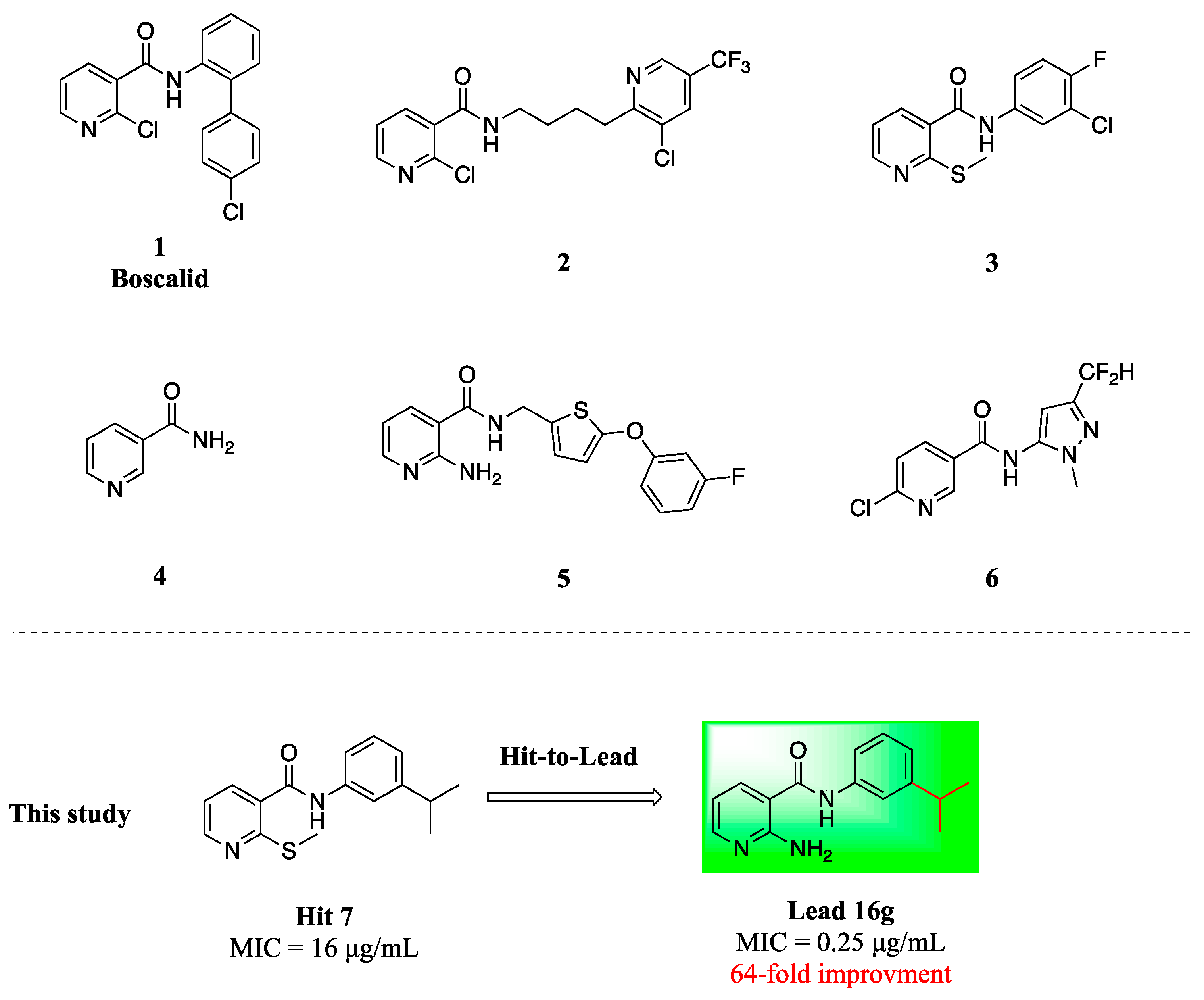
Figure 1. The structures of nicotinamide derivatives.
Molecules 28 01135 g001
Scheme 1. Synthesis of the target compounds. Reagents and conditions: PyBOP, DIEA, DMF, r.t, 1 h.
Molecules 28 01135 sch001
Figure 2. The in vitro toxicity evaluation of 16g, 16j and FLC. The cytotoxic effects of compounds 16g and 16j, compared to that of FLC, on HUVECs viability was assessed by the CCK-8 test following a 2 h treatment. FLC: fluconazole.
Molecules 28 01135 g002
Figure 3. Time-killed curves for 16g against the C. albicans. The concentrations of 16g were adjusted to 0.25 μg/mL (1 × MIC), 0.5 μg/mL (2 × MIC), 1 μg/mL (4 × MIC) in RPMI 1640 medium. con: control.
Molecules 28 01135 g003
Figure 4. Anti-hyphae effects of different concentrations of compound 16g. The inhibition of 16g on the hypha formation of C. albicans. C. albicans SC5314 were incubated at 37 °C for 3 h in RPMI 1640 medium. The concentrations of 16g were ranged from 0.0313–0.5 μg/mL. con: control.
Molecules 28 01135 g004
Figure 5. 16g inhibits C. albicans biofilm formation in vitro. Biofilm formation was evaluated by XTT reduction assay using C. albicans SC5314. The results were repeated for two times. *** p < 0.001, (t test).
Molecules 28 01135 g005
Figure 6. Cell wall damage caused by compound 16g in C. albicans. C. albicans SC5314 was treated with 1 μg/mL of 16g for 16 h. The structural changes of cell wall were observed by transmission electron microscope. con: control.
Molecules 28 01135 g006
Figure 7. (A)The BOILED-Egg model prediction of 16g and FLC; (B) Bioavailability radar for 16g; (C) Bioavailability radar for FLC; FLC: fluconazole.
Molecules 28 01135 g007
Table 1. In vitro antifungal activities of nicotinamide derivatives against C. albicans SC5314.
CompdStructureMIC (μg/mL)CompdStructureMIC (μg/mL)
C. alb SC5314C. alb SC5314
7Molecules 28 01135 i0011616fMolecules 28 01135 i002>16
16aMolecules 28 01135 i003>1616gMolecules 28 01135 i0040.25
16bMolecules 28 01135 i005>1616hMolecules 28 01135 i006>16
16cMolecules 28 01135 i007>16FLC-0.25
16dMolecules 28 01135 i008>161->16
16eMolecules 28 01135 i009>163->16
Abbreviations: MIC: minimum inhibitory concentration; C. alb: Candida albicans; FLC: fluconazole.
Table 2. In vitro antifungal activities of compounds against C. albicans SC5314.
Compd.StructureMIC (μg/mL)Compd.StructureMIC (μg/mL)
C. alb SC5314C. alb SC5314
16gMolecules 28 01135 i0100.2516wMolecules 28 01135 i01164
16iMolecules 28 01135 i012>6416xMolecules 28 01135 i013>64
16jMolecules 28 01135 i0140.516yMolecules 28 01135 i015>64
16kMolecules 28 01135 i016>6416zMolecules 28 01135 i01716
16lMolecules 28 01135 i0181617Molecules 28 01135 i01964
16mMolecules 28 01135 i020>6418aMolecules 28 01135 i02116
16nMolecules 28 01135 i0226418bMolecules 28 01135 i02364
16oMolecules 28 01135 i024>6419Molecules 28 01135 i0254
16pMolecules 28 01135 i026420Molecules 28 01135 i027>64
16qMolecules 28 01135 i028>6421Molecules 28 01135 i029>64
16rMolecules 28 01135 i0301622aMolecules 28 01135 i031>64
16sMolecules 28 01135 i032822bMolecules 28 01135 i033>64
16tMolecules 28 01135 i034422cMolecules 28 01135 i035>64
16uMolecules 28 01135 i0363222dMolecules 28 01135 i037>64
16vMolecules 28 01135 i038>64FLC-0.25
Abbreviations: MIC: minimum inhibitory concentration; C. alb: Candida albicans; FLC: fluconazole.
Table 3. In vitro antifungal activities against drug-resistant C. albicans of 16g and FLC.
C. albicansIsolateMIC (μg/mL)
16gFLC
fluconazole-sensitive (2)Y01090.1250.125
4650.50.125
fluconazole-resistant (6)8620.5>64
7860.5>64
1001>64
3850.125>64
8980.5>64
5040.25>64
Abbreviations: MIC: minimum inhibitory concentration; C. albicans, Candida albicans; FLC: fluconazole.
Table 4. In vitro antifungal activities of 16g and FLC.
SpeciesIsolateMIC (μg/mL)
16gFLC
C. parapsilosis2201941
66020.25
C. krusei46384
629162
C. glabrata53780.5
C. tropicalis7520.50.5
11293622
C. neoformans32609321
34877162
5699281
A. fumigatus7544>32>64
023-2>32>64
T.mentagrophytonT5A1616
T5B3216
T5E3216
Abbreviations: MIC: minimum inhibitory concentration; C. parapsilosis, Candida parapsilosis; C. krusei, Candida krusei; C. glabrata, Candida glabrata; C. tropicalis, Candida tropicalis; C. neoformans, Cryptococcus neoformans; A. fumigatus, Aspergillus fumigatus; T. mentagrophyton, Trichophyton mentagrophyton; FLC: fluconazole.
Disclaimer/Publisher’s Note: The statements, opinions and data contained in all publications are solely those of the individual author(s) and contributor(s) and not of MDPI and/or the editor(s). MDPI and/or the editor(s) disclaim responsibility for any injury to people or property resulting from any ideas, methods, instructions or products referred to in the content.