


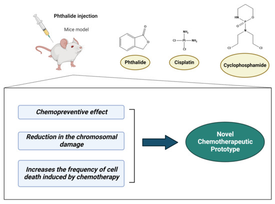
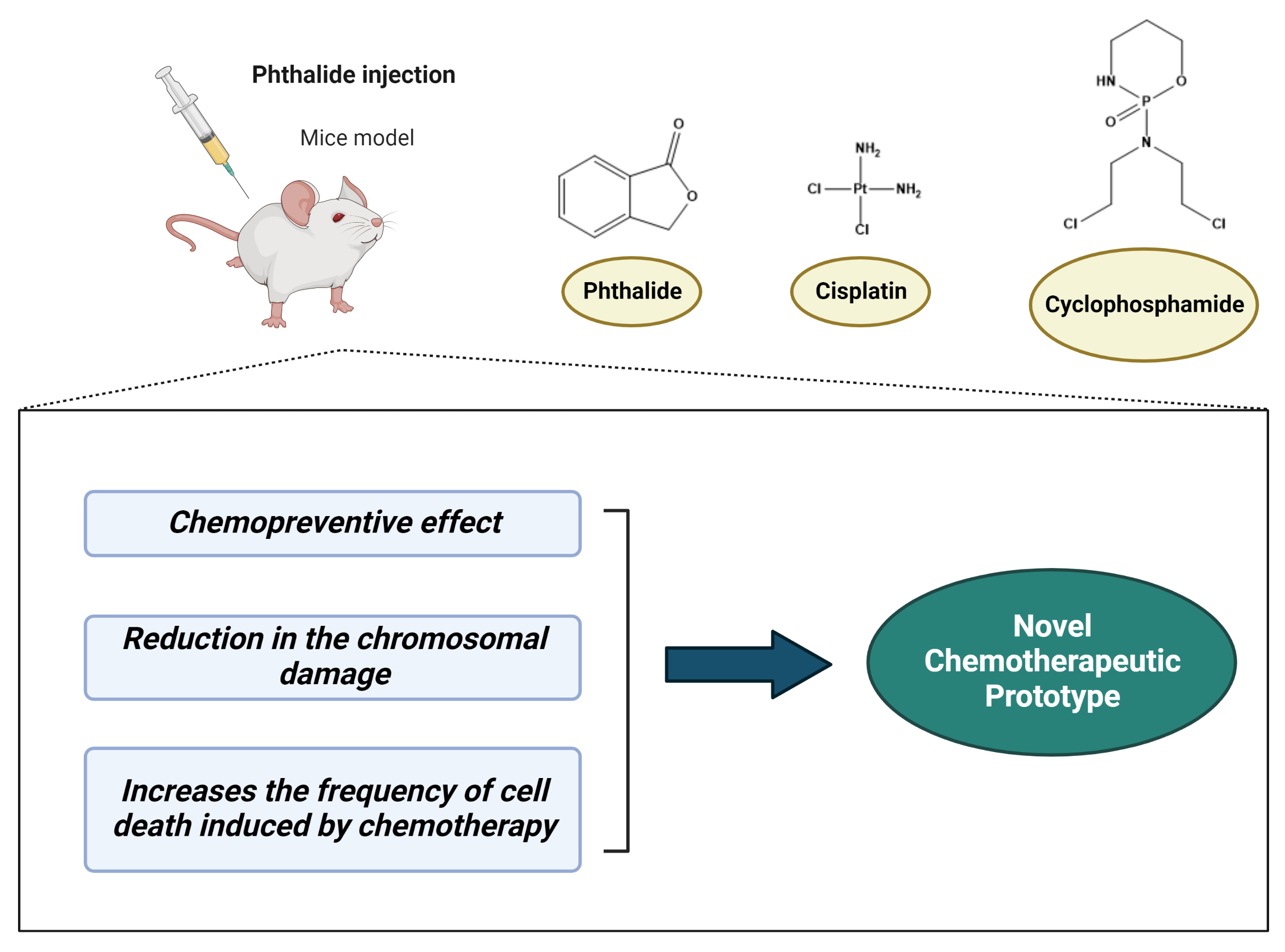
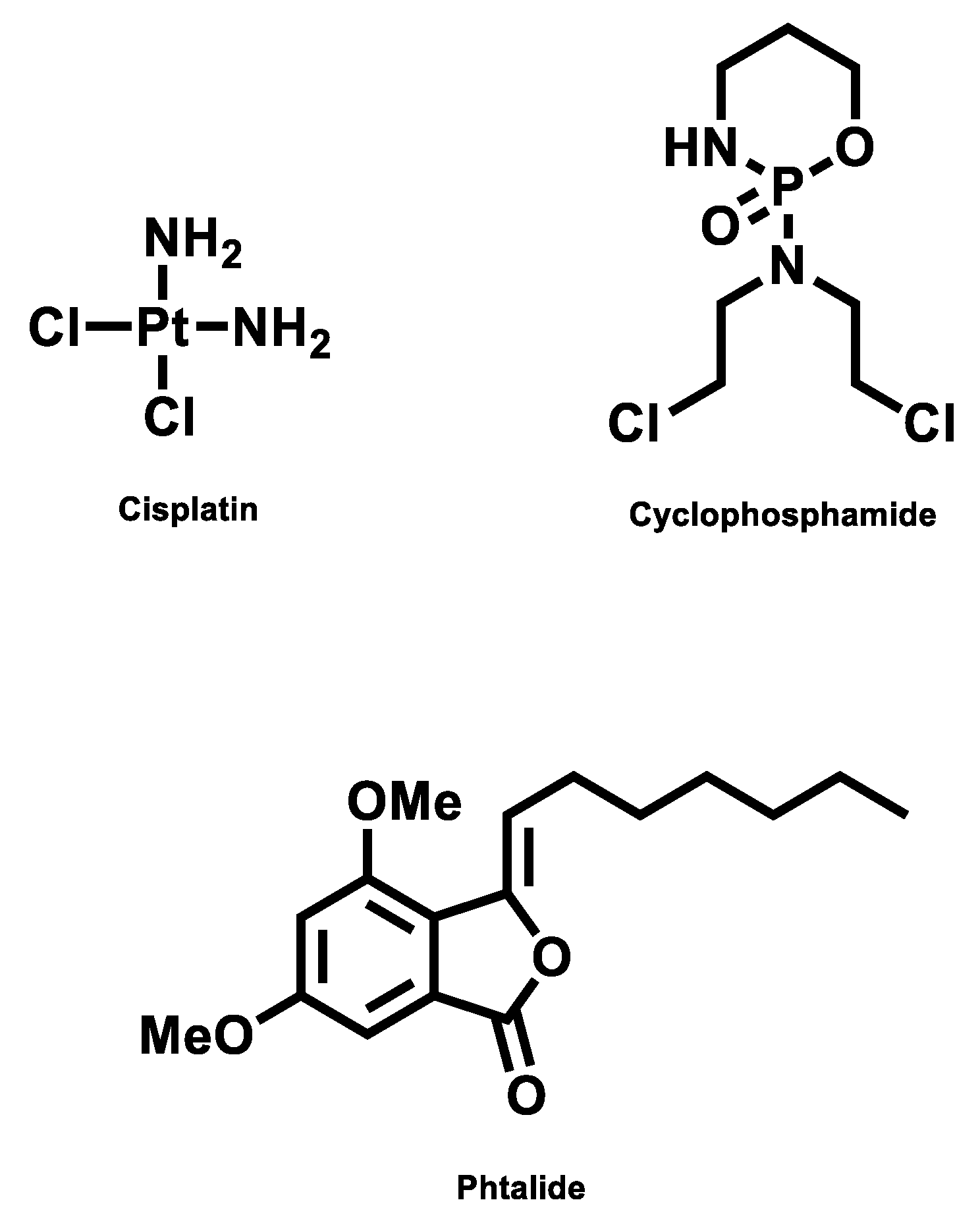
3-Heptylidene-4,6-Dimethoxy-3H-Isobenzofuran-1-One Is Genotoxic, Increases the Frequency of Cell Death, and Potentiates the Effects of Cyclophosphamide and Cisplatin

 , , ,
, , ,  , , , and
Abstract
1. Introduction
2. Results
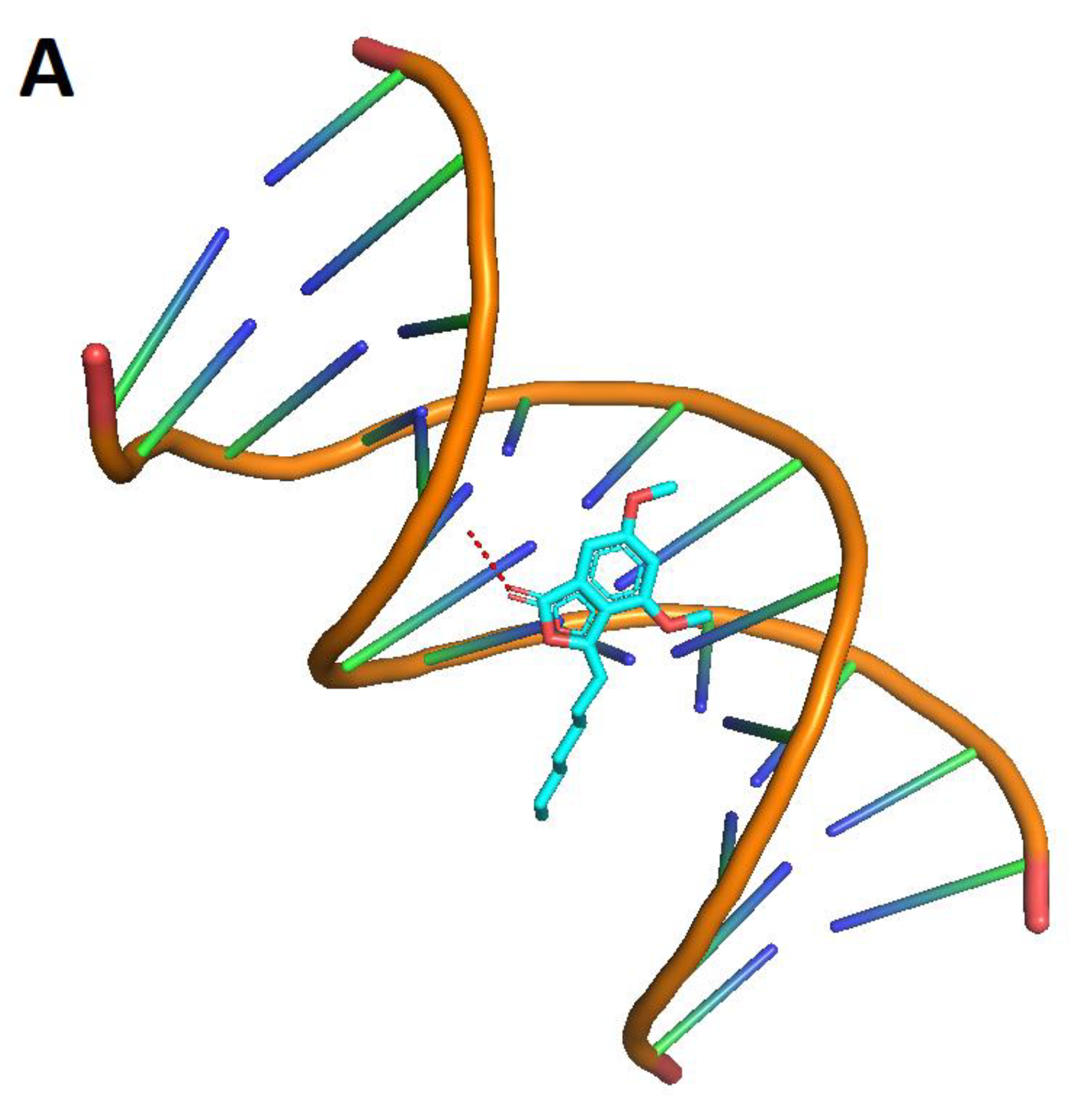
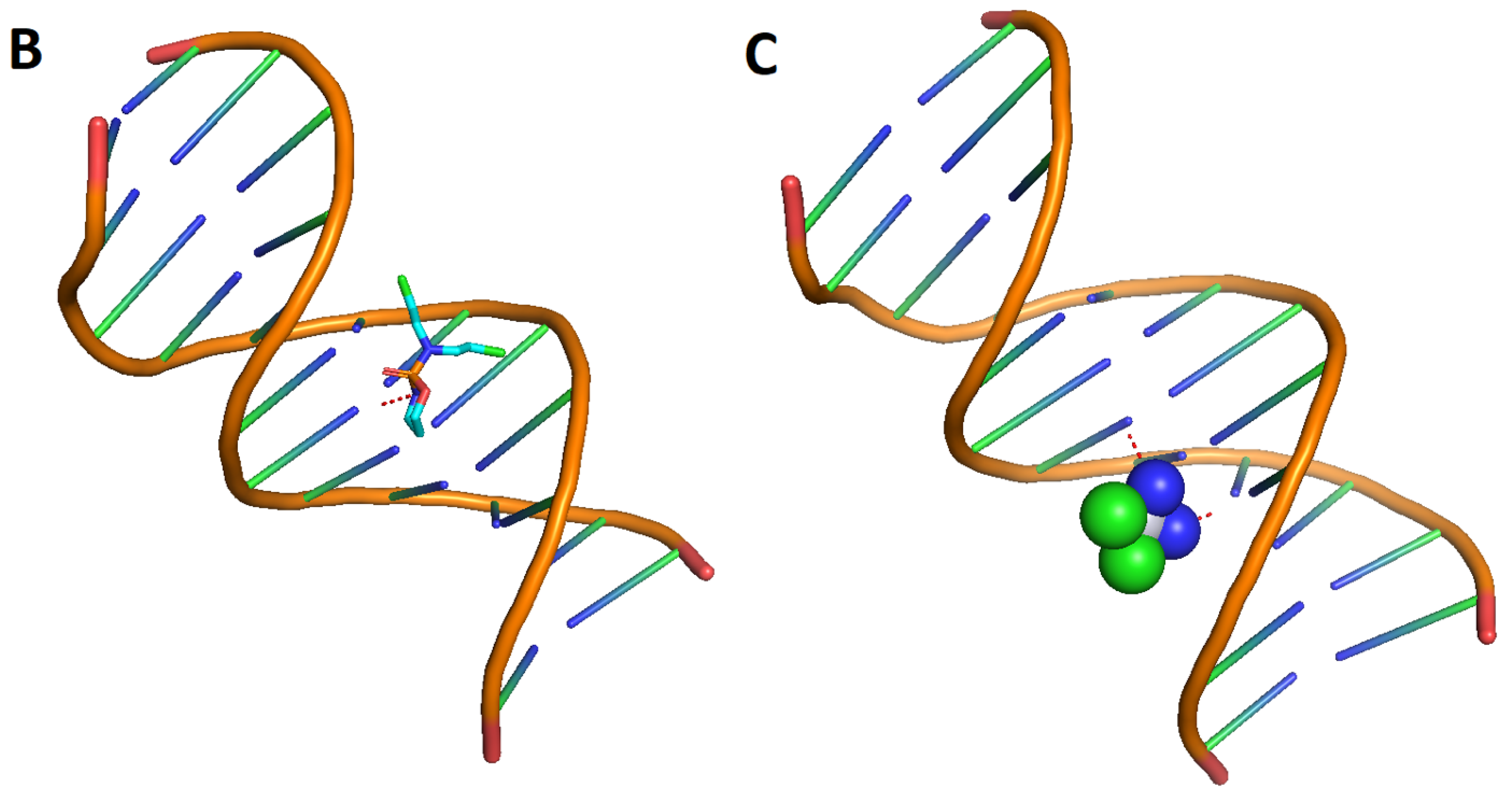
Molecular Modeling
3. Discussion
4. Material and Methods
4.1. Chemical Agents, Animals, and Experimental Design
4.2. Biological Assays
4.2.1. Micronucleus Assay in Peripheral Blood
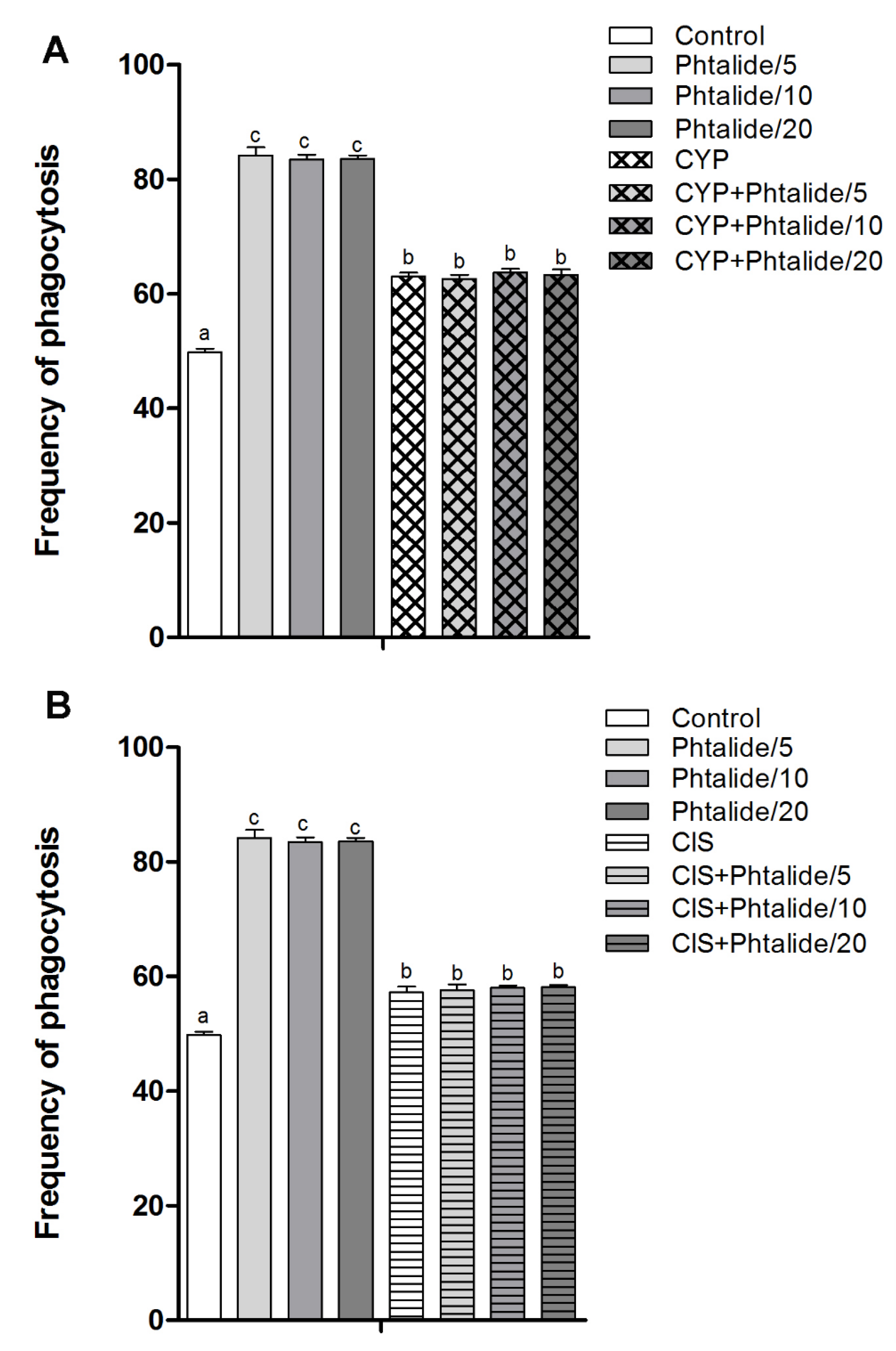
4.2.2. Splenic Phagocytosis Assay
4.2.3. Cell Death Assay
4.2.4. Calculation of Damage Reduction Percentage (DR%)
4.3. Statistical Analysis
4.4. Molecular Docking
4.4.1. Computational Details
4.4.2. Protein Preparation
4.4.3. Molecular Modeling
4.4.4. Target Selection and Molecular Docking Simulations
5. Conclusions
Author Contributions
Funding
Institutional Review Board Statement
Informed Consent Statement
Data Availability Statement
Acknowledgments
Conflicts of Interest
Abbreviation
| CIS | cisplatin |
| CP | cyclophosphamide |
| DNA | deoxyribonucleic acid |
| DMSO | dimethyl sulfoxide |
| RNA | ribonucleic acid |
| PDB | protein data bank |
References
- Vitor, N.; Meza, A.; Gomes, R.S.; Rafique, J.; DE Lima, D.P.; Beatriz, A. Straightforward synthesis of cytosporone analogs AMS35AA and AMS35BB. An. Acad. Bras. Cienc. 2021, 93, e20201347. [Google Scholar] [CrossRef]
- Navarro, S.D.; Pessatto, L.R.; Meza, A.; de Oliveira, E.J.T.; Auharek, S.A.; Vilela, L.C.; de Lima, D.P.; de Azevedo, R.B.; Kassuya, C.A.L.; Cáceres, O.I.A.; et al. Resorcinolic lipid 3-heptyl-3,4,6-trimethoxy-3H-isobenzofuran-1-one is a strategy for melanoma treatment. Life Sci. 2018, 209, 300–312. [Google Scholar] [CrossRef]
- Stepanenko, I.Y.; Strakhovskaia, M.G.; Belenikina, N.S.; Nikolaev, I.A.; Miliukin, A.L.; Kozlova, A.N.; Revina, A.A.; El’-Registan, G.I. Protection of Saccharomyces cerevisiae against oxidative and radiation-caused damage by alkyl hydroxybenzenes. Mikrobiologiia 2004, 73, 204–210. [Google Scholar]
- Rabacow, A.P.M.; Meza, A.; De Oliveira, E.J.T.; De David, N.; Vitor, N.; Antoniolli-Silva, A.C.M.B.; De Fátima Cepa Matos, M.; Perdomo, R.T.; Da Silva Gomes, R.; De Lima, D.P.; et al. Evaluation of the antitumor potential of the resorcinolic lipid 3-heptyl-3,4,6-trimethoxy-3H-isobenzofuran-1-one in breast cancer cells. Anticancer Res. 2018, 38, 4565–4576. [Google Scholar] [CrossRef]
- Navarro, S.D.; Beatriz, A.; Meza, A.; Pesarini, J.R.; Gomes, R.d.S.; Karaziack, C.B.; Cunha-Laura, A.L.; Monreal, A.C.; Romão, W.; Lacerda Júnior, V.; et al. A new synthetic resorcinolic lipid 3-heptyl-3,4,6-trimethoxy-3H-isobenzofuran-1-one: Evaluation of toxicology and ability to potentiate the mutagenic and apoptotic effects of cyclophosphamide. Eur. J. Med. Chem. 2014, 75, 132–142. [Google Scholar] [CrossRef]
- Oliveira, R.J.; Mantovani, M.S.; Pesarini, J.R.; Mauro, M.O.; da Silva, A.F.; Souza, T.R.; Ribeiro, L.R. 6-Dimethylaminopurine and cyclohexamide are mutagenic and alter reproductive performance and intrauterine development in vivo. Genet. Mol. Res. 2015, 14, 834–849. [Google Scholar] [CrossRef]
- OECD. Test No. 425: Acute Oral Toxicity: Up-and-Down Procedure; OECD Guidelines for the Testing of Chemicals, Section 4; OECD Publishing: Paris, France, 2022. [Google Scholar] [CrossRef]
- Cordelli, E.; Bignami, M.; Pacchierotti, F. Comet assay: A versatile but complex tool in genotoxicity testing. Toxicol. Res. 2021, 10, 68–78. [Google Scholar] [CrossRef]
- Sommer, S.; Buraczewska, I.; Kruszewski, M. Micronucleus assay: The state of art, and future directions. Int. J. Mol. Sci. 2020, 21, 1534. [Google Scholar] [CrossRef]
- Mehra, S.; Chadha, P. Naphthalene-2-sulfonate induced toxicity in blood cells of freshwater fish Channa punctatus using comet assay, micronucleus assay and ATIR-FTIR approach. Chemosphere 2021, 265, 129147. [Google Scholar] [CrossRef]
- Alhmoud, J.F.; Woolley, J.F.; Al Moustafa, A.E.; Malki, M.I. DNA Damage/Repair Management in Cancers. Cancers 2020, 12, 1050. [Google Scholar] [CrossRef]
- Carvalho, P.C.; Santos, E.A.; Schneider, B.U.; Matuo, R.; Pesarini, J.R.; Cunha-Laura, A.L.; Monreal, A.C.; Lima, D.P.; Antoniolli, A.C.; Oliveira, R.J. Diaryl sulfide analogs of combretastatin A-4: Toxicogenetic, immunomodulatory and apoptotic evaluations and prospects for use as a new chemotherapeutic drug. Environ. Toxicol. Pharmacol. 2015, 40, 715–721. [Google Scholar] [CrossRef] [PubMed]
- Ishii, P.L.; Prado, C.K.; Mauro, M.d.O.; Carreira, C.M.; Mantovani, M.S.; Ribeiro, L.R.; Dichi, J.B.; Oliveira, R.J. Evaluation of Agaricus blazei in vivo for antigenotoxic, anticarcinogenic, phagocytic and immunomodulatory activities. Regul. Toxicol. Pharmacol. 2011, 59, 412–422. [Google Scholar] [CrossRef] [PubMed]
- Oliveira, R.J.; da Cruz Leite Santos, N.; Pesarini, J.R.; de Oliveira, B.C.; Berno, C.R.; de Araújo, F.H.S.; da Silveira, I.O.M.F.; Nascimento, R.O.; Brochado Antoniolli-Silva, A.C.M.; Duenhas Monreal, A.C.; et al. Assessment of genetic integrity, splenic phagocytosis and cell death potential of (Z)-4-((1,5-dimethyl-3-oxo-2-phenyl-2,3dihydro-1H-pyrazol-4-yl) amino)-4-oxobut-2-enoic acid and its effect when combined with commercial chemotherapeutics. Genet. Mol. Biol. 2018, 41, 154–166. [Google Scholar] [CrossRef] [PubMed]
- Bazo, A.P.; Rodrigues, M.A.; Sforcin, J.M.; de Camargo, J.L.; Ribeiro, L.R.; Salvadori, D.M. Protective action of propolis on the rat colon carcinogenesis. Teratog. Carcinog. Mutagen. 2002, 22, 183–194. [Google Scholar] [CrossRef] [PubMed]
- Swan, D.; Gurney, M.; Krawczyk, J.; Ryan, A.E.; O’Dwyer, M. Beyond DNA damage: Exploring the immunomodulatory effects of cyclophosphamide in multiple myeloma. Hemasphere 2020, 4, e350. [Google Scholar] [CrossRef] [PubMed]
- Du, J.; Zhang, A.; Li, J.; Liu, X.; Wu, S.; Wang, B.; Wang, Y.; Jia, H. Doxorubicin-induced cognitive impairment: The mechanistic insights. Front. Oncol. 2021, 11, 673340. [Google Scholar] [CrossRef]
- Brown, A.; Kumar, S.; Tchounwou, P.B. Cisplatin-Based Chemotherapy of Human Cancers. J. Cancer Sci. Ther. 2019, 11, 97. [Google Scholar]
- Zhan, Y.; Du, X.; Chen, H.; Liu, J.; Zhao, B.; Huang, D.; Li, G.; Xu, Q.; Zhang, M.; Weimer, B.C.; et al. Cytosporone B is an agonist for nuclear orphan receptor Nur77. Nat. Chem. Biol. 2008, 4, 548–556. [Google Scholar] [CrossRef]
- Hao, L.; Wang, X.; Zhang, D.; Xu, Q.; Song, S.; Wang, F.; Li, C.; Guo, H.; Liu, Y.; Zheng, D.; et al. Studies on the preparation, characterization and pharmacokinetics of Amoitone B nanocrystals. Int. J. Pharm. 2012, 433, 157–164. [Google Scholar] [CrossRef]
- Maruyama, K.; Tsukada, T.; Ohkura, N.; Bandoh, S.; Hosono, T.; Yamaguchi, K. The NGFI-B subfamily of the nuclear receptor superfamily (review). Int. J. Oncol. 1998, 12, 1237–1243. [Google Scholar] [CrossRef]
- Li, Q.X.; Ke, N.; Sundaram, R.; Wong-Staal, F. NR4A1, 2, 3—An orphan nuclear hormone receptor family involved in cell apoptosis and carcinogenesis. Histol. Histopathol. 2006, 21, 533–540. [Google Scholar] [CrossRef] [PubMed]
- Winoto, A.; Littman, D.R. Nuclear hormone receptors in T lymphocytes. Cell 2002, 109, S57–S66. [Google Scholar] [CrossRef] [PubMed]
- Lin, B.; Kolluri, S.K.; Lin, F.; Liu, W.; Han, Y.H.; Cao, X.; Dawson, M.I.; Reed, J.C.; Zhang, X.K. Conversion of Bcl-2 from protector to killer by interaction with nuclear orphan receptor Nur77/TR3. Cell 2004, 116, 527–540. [Google Scholar] [CrossRef] [PubMed]
- Moll, U.M.; Marchenko, N.; Zhang, X.K. p53 and Nur77/TR3—Transcription factors that directly target mitochondria for cell death induction. Oncogene 2006, 25, 4725–4743. [Google Scholar] [CrossRef] [PubMed]
- Thompson, J.; Winoto, A. During negative selection, Nur77 family proteins translocate to mitochondria where they associate with Bcl-2 and expose its proapoptotic BH3 domain. J. Exp. Med. 2008, 205, 1029–1036. [Google Scholar] [CrossRef]
- Rohs, R.; Bloch, I.; Sklenar, H.; Shakked, Z. Molecular flexibility in ab initio drug docking to DNA: Binding-site and binding-mode transitions in all-atom Monte Carlo simulations. Nucleic Acids Res. 2005, 33, 7048–7057. [Google Scholar] [CrossRef]
- Jones, G.; Willett, P.; Glen, R.C. A genetic algorithm for flexible molecular overlay and pharmacophore elucidation. J. Comput.-Aided Mol. Des. 1995, 9, 532–549. [Google Scholar] [CrossRef]
- Han, X.; Gao, X. Sequence specific recognition of ligand-DNA complexes studied by NMR. Curr. Med. Chem. 2001, 8, 551–581. [Google Scholar] [CrossRef]
- Neidle, S.; Nunn, C.M. Crystal structures of nucleic acids and their drug complexes. Nat. Prod. Rep. 1998, 15, 1–15. [Google Scholar] [CrossRef]
- Ren, J.; Chaires, J.B. Sequence and structural selectivity of nucleic acid binding ligands. Biochemistry 1999, 38, 16067–16075. [Google Scholar] [CrossRef]
- Wemmer, D.E. Designed sequence-specific minor groove ligands. Annu. Rev. Biophys. Biomol. Struct. 2000, 29, 439–461. [Google Scholar] [CrossRef] [PubMed]
- Bischoff, G.; Hoffmann, S. DNA-binding of drugs used in medicinal therapies. Curr. Med. Chem. 2002, 9, 312–348. [Google Scholar] [CrossRef] [PubMed]
- Chaires, J.B. Energetics of drug-DNA interactions. Biopolymers 1997, 44, 201–215. [Google Scholar] [CrossRef]
- Haq, I.; Ladbury, J. Drug–DNA recognition: Energetics and implications for design. J. Mol. Recognit. 2000, 13, 188–197. [Google Scholar] [CrossRef]
- Korkmaz, A.; Topal, T.; Oter, S. Pathophysiological aspects of cyclophosphamide and ifosfamide induced hemorrhagic cystitis; implication of reactive oxygen and nitrogen species as well as PARP activation. Cell Biol. Toxicol. 2007, 23, 303–312. [Google Scholar] [CrossRef]
- Mills, K.A.; Chess-Williams, R.; McDermott, C. Novel insights into the mechanism of cyclophosphamide-induced bladder toxicity: Chloroacetaldehyde’s contribution to urothelial dysfunction in vitro. Arch. Toxicol. 2019, 93, 3291–3303. [Google Scholar] [CrossRef]
- Fuertes, M.A.; Castilla, J.; Alonso, C.; Pérez, J.M. Cisplatin biochemical mechanism of action: From cytotoxicity to induction of cell death through interconnections between apoptotic and necrotic pathways. Curr. Med. Chem. 2003, 10, 257–266. [Google Scholar] [CrossRef] [PubMed]
- Oliveira, R.J.; Navarro, S.D.; de Lima, D.P.; Meza, A.; Pesarini, J.R.; da Silva Gomes, R.; Karaziack, C.B.; de Oliveira Mauro, M.; Cunha-Laura, A.L.; Monreal, A.C.; et al. A novel cytosporone 3-heptyl-4,6-dihydroxy-3H-isobenzofuran-1-one: Synthesis; toxicological, apoptotic and immunomodulatory properties; and potentiation of mutagenic damage. BMC Cancer 2015, 15, 561. [Google Scholar] [CrossRef]
- Berno, C.R.; Rós, B.d.T.; da Silveira, I.O.; Coelho, H.R.; Antoniolli, A.C.; Beatriz, A.; de Lima, D.P.; Monreal, A.C.; Sousa, F.G.; da Silva Gomes, R.; et al. 4-Aminoantipyrine reduces toxic and genotoxic effects of doxorubicin, Cisplatin, and cyclophosphamide in male mice. Mutat. Res. Genet. Toxicol. Environ. Mutagen. 2016, 805, 19–24. [Google Scholar] [CrossRef] [PubMed]
- Araújo, F.H.S.; Figueiredo, D.R.; Auharek, S.A.; Pesarini, J.R.; Meza, A.; Gomes, R.S.; Monreal, A.C.D.; Antoniolli-Silva, A.C.M.B.; Lima, D.P.; Kassuya, C.A.L.; et al. In vivo chemotherapeutic insight of a novel isocoumarin (3-hexyl-5,7-dimethoxy-isochromen-1-one): Genotoxicity, cell death induction, leukometry and phagocytic evaluation. Genet. Mol. Biol. 2017, 40, 665–675. [Google Scholar] [CrossRef]
- Hayashi, M.; Morita, T.; Kodama, Y.; Sofuni, T.; Ishidate, M., Jr. The micronucleus assay with mouse peripheral blood reticulocytes using acridine orange-coated slides. Mutat. Res. Lett. 1990, 245, 245–249. [Google Scholar] [CrossRef] [PubMed]
- Oliveira, R.J.; Baise, E.; Mauro, M.d.O.; Pesarini, J.R.; Matuo, R.; Silva, A.F.; Ribeiro, L.R.; Mantovani, M.S. Evaluation of chemopreventive activity of glutamine by the comet and the micronucleus assay in mice’s peripheral blood. Environ. Toxicol. Pharmacol. 2009, 28, 120–124. [Google Scholar] [CrossRef] [PubMed]
- Manoharan, K.; Banerjee, M.R. β-Carotene reduces sister chromatid exchanges induced by chemical carcinogens in mouse mammary cells in organ culture. Cell Biol. Int. Rep. 1985, 9, 783–789. [Google Scholar] [CrossRef] [PubMed]
- Waters, M.D.; Brady, A.L.; Stack, H.F.; Brockman, H.E. Antimutagenicity profiles for some model compounds. Mutat. Res./Rev. Genet. Toxicol. 1990, 238, 57–85. [Google Scholar] [CrossRef]
- BIOVIA Discovery Studio Visualizer, version 20; Dassault Systèmes: San Diego, CA, USA, 2021.
- Yan, Y.; Zhang, D.; Zhou, P.; Li, B.; Huang, S.Y. HDOCK: A web server for protein-protein and protein-DNA/RNA docking based on a hybrid strategy. Nucleic Acids Res. 2017, 45, W365–W373. [Google Scholar] [CrossRef] [PubMed]


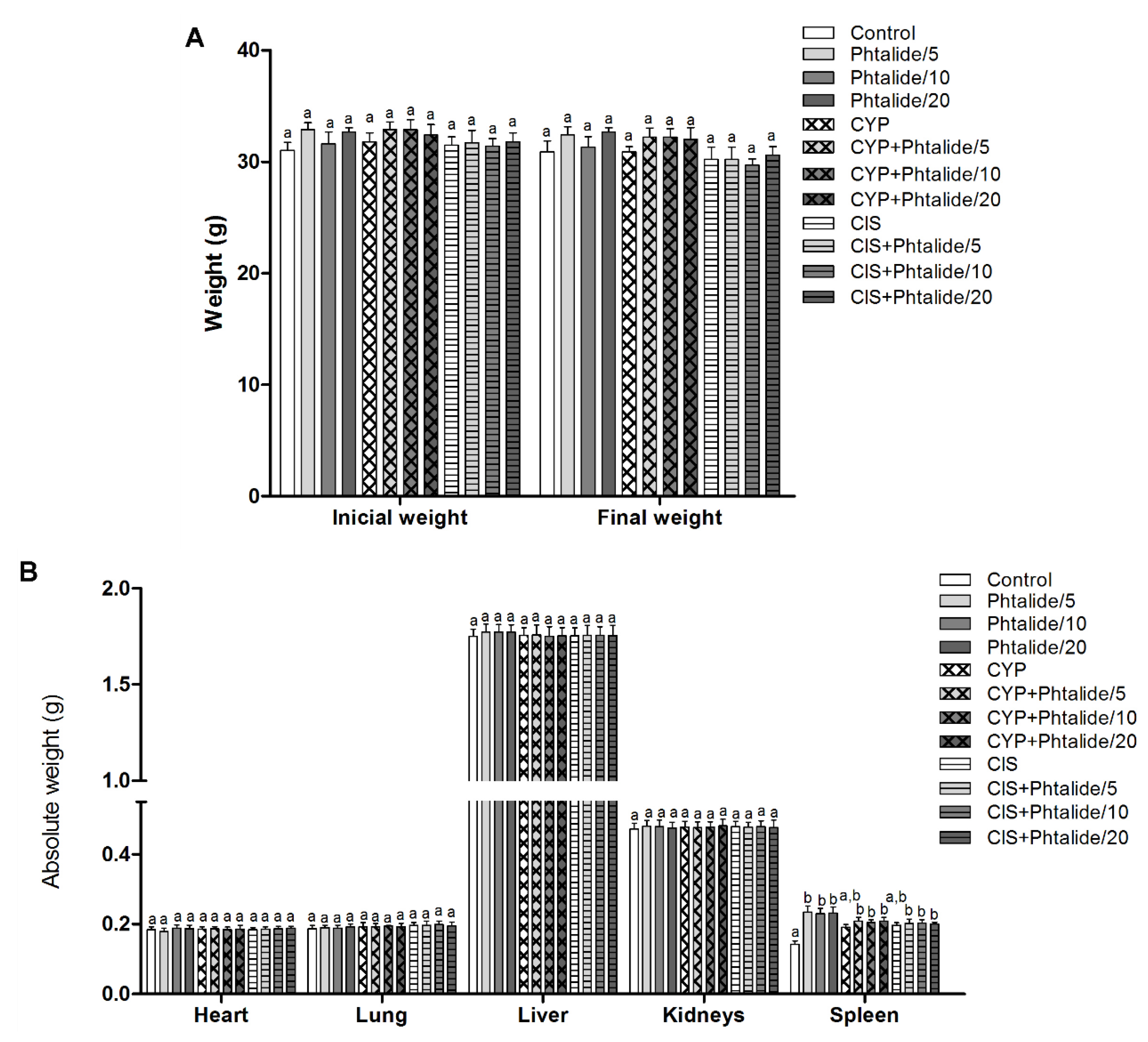
| Experimental Groups | Initial Weight | Final Weight |
|---|---|---|
| Control | 31.00 ± 0.75 a | 30.90 ± 0.97 a |
| 1 5 mg/kg | 32.90 ± 0.63 a | 32.40 ± 0.75 a |
| 1 10 mg/kg | 31.60 ± 1.10 a | 31.30 ± 0.95 a |
| 1 20 mg/kg | 32.70 ± 0.36 a | 32.70 ± 0.36 a |
| cyclophosphamide (CYP) | 31.80 ± 0.82 a | 30.90 ± 0.47 a |
| CYP + 1 5 mg/kg | 32.90 ± 0.68 a | 32.20 ± 0.83 a |
| CYP + 1 10 mg/kg | 32.90 ± 0.89 a | 32.20 ± 0.77 a |
| CYP + 1 20 mg/kg | 32.40 ± 0.95 a | 32.00 ± 1.07 a |
| cisplatin (CIS) | 31.50 ± 0.74 a | 30.20 ± 0.84 a |
| CIS + 1 5 mg/kg | 31.70 ± 1.12 a | 30.20 ± 1.12 a |
| CIS + 1 10 mg/kg | 31.40 ± 0.69 a | 29.70 ± 0.57 a |
| CIS + 1 20 mg/kg | 31.80 ± 0.82 a | 30.60 ± 0.76 a |
| Experimental Groups | Heart | Lung | Liver | Kidneys | Spleen |
|---|---|---|---|---|---|
| Control | 0.184 ± 0.009 a | 0.187 ± 0.009 a | 1.749 ± 0.039 a | 0.472 ± 0.017 a | 0.142 ± 0.010 a |
| 1 5 mg/kg | 0.179 ± 0.010 a | 0.190 ± 0.007 a | 1.772 ± 0.042 a | 0.480 ± 0.017 a | 0.234 ± 0.018 b |
| 1 10 mg/kg | 0.188 ± 0.010 a | 0.189 ± 0.008 a | 1.773 ± 0.038 a | 0.479 ± 0.019 a | 0.230 ± 0.015 b |
| 1 20 mg/kg | 0.187 ± 0.010 a | 0.192 ± 0.009 a | 1.772 ± 0.038 a | 0.475 ± 0.017 a | 0.231 ± 0.018 b |
| cyclophosphamide (CYP) | 0.186 ± 0.007 a | 0.192 ± 0.012 a | 1.754 ± 0.042 a | 0.478 ± 0.017 a | 0.191 ± 0.008 a,b |
| CYP + 1 5 mg/kg | 0.187 ± 0.006 a | 0.193 ± 0.009 | 1.757 ± 0.052 a | 0.476 ± 0.017 a | 0.208 ± 0.012 b |
| CYP + 1 10 mg/kg | 0.184 ± 0.008 a | 0.195 ± 0.012 a | 1.751 ± 0.049 a | 0.478 ± 0.015 a | 0.204 ± 0.008 |
| CYP + 1 20 mg/kg | 0.186 ± 0.011 a | 0.192 ± 0.010 a | 1.752 ± 0.050 a | 0.481 ± 0.020 a | 0.209 ± 0.010 b |
| cisplatin (CIS) | 0.184 ± 0.006 a | 0.196 ± 0.009 a | 1.752 ± 0.044 a | 0.479 ± 0.016 a | 0.196 ± 0.008 a,b |
| CIS + 1 5 mg/kg | 0.186 ± 0.010 a | 0.197 ± 0.012 a | 1.756 ± 0.051 a | 0.477 ± 0.014 a | 0.202 ± 0.012 b |
| CIS + 1 10mg/kg | 0.187 ± 0.007 a | 0.199 ± 0.010 a | 1.754 ± 0.047 a | 0.479 ± 0.017 a | 0.203 ± 0.010 b |
| CIS + 1 20mg/kg | 0.188 ± 0.006 a | 0.195 ± 0.011 a | 1.753 ± 0.055 a | 0.476 ± 0.022 a | 0.200 ± 0.005 b |
| Experimental Groups | Average Frequency of Micronuclei | Damage Reduction Percentage | ||||
|---|---|---|---|---|---|---|
| 24 h | 48 h | 72 h | 24 h | 48 h | 72 h | |
| Experiment 1 | ||||||
| Control | 8.40 ± 0.30 a | 7.50 ± 0.27 a | 6.80 ± 0.20 a | - | - | - |
| cyclophosphamide (CYP) | 103.00 ± 1.00 d | 110.80 ± 0.97 d | 111.80 ± 1.08 d | - | - | - |
| 1 5 mg/kg | 36.60 ± 0.54 b | 35.40 ± 0.67 b | 34.30 ± 0.67 b | - | - | - |
| 1 10 mg/kg | 37.50 ± 0.63 b | 35.90 ± 0.69 b | 34.60 ± 0.50 b | - | - | - |
| 1 20 mg/kg | 37.60 ± 0.45 b | 36.60 ± 0.45 b | 35.40 ± 0.67 b | - | - | - |
| CYP + 1 5 mg/kg | 64.80 ± 0.59 c | 66.70 ± 1.29 c | 66.90 ± 1.28 c | 41.43 | 42.69 | 42.76 |
| CYP + 1 10 mg/kg | 63.80 ± 0.78 c | 66.10 ± 0.50 c | 66.40 ± 0.47 c | 41.22 | 43.27 | 43.23 |
| CYP + 1 20 mg/kg | 64.00 ± 1.23 c | 67.80 ± 0.61 c | 67.00 ± 0.80 c | 41.22 | 41.62 | 42.66 |
| Experiment 2 | ||||||
| Control | 8.40 ± 0.30 a | 7.50 ± 0.27 a | 6.80 ± 0.20 a | - | - | - |
| cisplatin (CIS) | 93.30 ± 0.84 d | 103.10 ± 1.24 d | 102.40 ± 1.08 d | - | - | - |
| 1 5 mg/kg | 36.60 ± 0.54 b | 35.40 ± 0.67 b | 34.30 ± 0.67 b | - | - | - |
| 1 10 mg/kg | 37.50 ± 0.63 b | 35.90 ± 0.69 b | 34.60 ± 0.50 b | - | - | - |
| 1 20 mg/kg | 37.60 ± 0.45 b | 36.60 ± 0.45 b | 35.40 ± 0.67 b | - | - | - |
| CIS + 1 5 mg/kg | 75.10 ± 0.70 c | 74.90 ± 0.65 c | 74.10 ± 0.38 c | 21.44 | 29.50 | 29.60 |
| CIS + 1 10 mg/kg | 74.10 ± 0.31 c | 75.00 ± 0.74 c | 74.20 ± 0.61 c | 22.61 | 29.39 | 29.50 |
| CIS + 1 20 mg/kg | 75.40 ± 0.45 c | 75.20 ± 0.94 c | 75.10 ± 0.38 c | 21.08 | 29.18 | 28.56 |
| Experimental Group | Total Cells Analyzed | Total Cells without Evidence of Phagocytosis A.V Mean ± SE Percentage | Total Cells with Evidence of Phagocytosis A.V Mean ± SE Percentage | ||||
|---|---|---|---|---|---|---|---|
| Experiment 1 (CYP) | |||||||
| Control | 1000 | 502 | 50.20 ± 0.64 c | 50.2 | 498 | 49.80 ± 0.64 a | 49.8 |
| cyclophosphamide (CYP) | 1000 | 370 | 37.00 ± 0.71 b | 37.0 | 630 | 63.00 ± 0.71 b | 63.0 |
| 1 5 mg/kg | 1000 | 158 | 15.80 ± 1.40 a | 15.8 | 842 | 84.20 ± 1.40 c | 84.2 |
| 1 10 mg/kg | 1000 | 165 | 16.50 ± 0.80 a | 16.5 | 835 | 83.50 ± 0.80 c | 83.5 |
| 1 20 mg/kg | 1000 | 164 | 16.40 ± 0.62 a | 16.4 | 836 | 83.60 ± 0.62 c | 83.6 |
| CYP + 1 5 mg/kg | 1000 | 374 | 37.40 ± 0.72 b | 37.4 | 626 | 62.60 ± 0.72 b | 62.6 |
| CYP + 1 10 mg/kg | 1000 | 363 | 36.30 ± 0.70 b | 36.3 | 637 | 63.70 ± 0.70 b | 63.7 |
| CYP + 1 20 mg/kg | 1000 | 367 | 36.70 ± 0.96 b | 36.7 | 633 | 63.30 ± 0.96 b | 63.3 |
| Experiment 2 (CIS) | |||||||
| Control cisplatin (CIS) | 1000 | 502 | 50.20 ± 0.64 c | 50.2 | 498 | 49.80 ± 0.64 a | 49.8 |
| 1000 | 428 | 42.80 ± 1.00 b | 42.8 | 572 | 57.20 ± 1.00 b | 57.2 | |
| 1 5 mg/kg | 1000 | 158 | 15.80 ± 1.40 a | 15.8 | 842 | 84.20 ± 1.40 c | 84.2 |
| 1 10 mg/kg | 1000 | 165 | 16.50 ± 0.80 a | 16.5 | 835 | 83.50 ± 0.80 c | 83.5 |
| 1 20 mg/kg | 1000 | 164 | 16.40 ± 0.62 a | 16.4 | 836 | 83.60 ± 0.62 c | 83.6 |
| CIS + 1 5 mg/kg | 1000 | 424 | 42.40 ± 1.01 b | 42.4 | 576 | 57.60 ± 1.01 b | 57.6 |
| CIS + 1 10 mg/kg | 1000 | 420 | 42.00 ± 0.40 b | 42.0 | 580 | 58.00 ± 0.40 b | 58.0 |
| CIS + 1 20 mg/kg | 1000 | 419 | 41.90 ± 0.38 b | 41.9 | 581 | 58.10 ± 0.38 b | 58.1 |
| Experimental Groups | Total Cells Analyzed | Total Cells in Apoptosis in the Liver A.V Mean ± SE Percentage | Total Cells in Apoptosis in the Kidneys A.V Mean ± SE Percentage | ||||
|---|---|---|---|---|---|---|---|
| Experiment 1 (CYP) | |||||||
| Control | 1000 | 118 | 11.80 ± 0.88 a | 11.8 | 15.50 ± 0.69 a | 15.5 | |
| cyclophosphamide (CYP) | 1000 | 353 | 35.30 ± 0.93 c | 35.3 | 30.50 ± 0.56 c | 30.5 | |
| 1 5 mg/kg | 1000 | 235 | 23.50 ± 0.99 b | 23.5 | 20.40 ± 0.43 b | 20.4 | |
| 1 10 mg/kg | 1000 | 238 | 23.80 ± 0.75 b | 23.8 | 20.80 ± 0.66 b | 20.8 | |
| 1 20 mg/kg | 1000 | 242 | 24.20 ± 1.33 b | 24.2 | 20.90 ± 0.61 b | 20.9 | |
| CYP + 1 5 mg/kg | 1000 | 351 | 35.10 ± 0.91 c | 35.1 | 31.10 ± 0.86 c | 31.1 | |
| CYP + 1 10 mg/kg | 1000 | 356 | 35.60 ± 0.99 c | 35.6 | 31.50 ± 0.49 c | 31.5 | |
| CYP + 1 20 mg/kg | 1000 | 342 | 34.20 ± 0.94 c | 34.2 | 32.30 ± 1.28 c | 32.3 | |
| Experiment 2 (CIS) | |||||||
| Control cisplatin (CIS) | 1000 | 118 | 11.80 ± 0.88 a | 11.8 | 155 | 15.50 ± 0.69 a | 15.5 |
| 1000 | 300 | 30.00 ± 0.57 c | 30.0 | 296 | 29.60 ± 0.65 c | 29.6 | |
| 1 5 mg/kg | 1000 | 235 | 23.50 ± 0.99 b | 23.5 | 204 | 20.40 ± 0.43 b | 20.4 |
| 1 10 mg/kg | 1000 | 238 | 23.80 ± 0.75 b | 23.8 | 209 | 20.90 ± 0.61 b | 20.9 |
| 1 20 mg/kg | 1000 | 242 | 24.20 ± 1.33 b | 24.2 | 208 | 20.80 ± 0.66 b | 20.8 |
| CIS + 1 5 mg/kg | 1000 | 301 | 30.10 ± 0.61 c | 30.1 | 299 | 29.90 ± 0.39 c | 29.9 |
| CIS + 1 10 mg/kg | 1000 | 311 | 31.10 ± 0.55 c | 31.1 | 296 | 29.60 ± 0.47 c | 29.6 |
| CIS + 1 20 mg/kg | 1000 | 305 | 30.50 ± 0.56 c | 30.5 | 291 | 29.10 ± 0.66 c | 29.1 |
| Compound | Docking Score (kcal/mol) |
|---|---|
| 1 | −88.75 |
| cyclophosphamide | −90.39 |
| cisplatin | −69.81 |
Disclaimer/Publisher’s Note: The statements, opinions and data contained in all publications are solely those of the individual author(s) and contributor(s) and not of MDPI and/or the editor(s). MDPI and/or the editor(s) disclaim responsibility for any injury to people or property resulting from any ideas, methods, instructions or products referred to in the content. |
© 2023 by the authors. Licensee MDPI, Basel, Switzerland. This article is an open access article distributed under the terms and conditions of the Creative Commons Attribution (CC BY) license (https://creativecommons.org/licenses/by/4.0/).
Share and Cite
das Neves, S.C.; de Araújo, F.H.; Correa, W.A.; Martins, A.C.F.; Coelho, H.R.S.; Vilela, M.L.B.; do Nascimento, V.A.; Kassuya, C.A.L.; de Lima, D.P.; Beatriz, A.; et al. 3-Heptylidene-4,6-Dimethoxy-3H-Isobenzofuran-1-One Is Genotoxic, Increases the Frequency of Cell Death, and Potentiates the Effects of Cyclophosphamide and Cisplatin. Molecules 2023, 28, 1044. https://doi.org/10.3390/molecules28031044
das Neves SC, de Araújo FH, Correa WA, Martins ACF, Coelho HRS, Vilela MLB, do Nascimento VA, Kassuya CAL, de Lima DP, Beatriz A, et al. 3-Heptylidene-4,6-Dimethoxy-3H-Isobenzofuran-1-One Is Genotoxic, Increases the Frequency of Cell Death, and Potentiates the Effects of Cyclophosphamide and Cisplatin. Molecules. 2023; 28(3):1044. https://doi.org/10.3390/molecules28031044
Chicago/Turabian Styledas Neves, Silvia Cordeiro, Flavio Henrique de Araújo, Willian Ayala Correa, Allana Cristina Faustino Martins, Henrique Rodrigues Scherer Coelho, Marcelo Luiz Brandão Vilela, Valter Aragão do Nascimento, Candida Aparecida Leite Kassuya, Dênis Pires de Lima, Adilson Beatriz, and et al. 2023. "3-Heptylidene-4,6-Dimethoxy-3H-Isobenzofuran-1-One Is Genotoxic, Increases the Frequency of Cell Death, and Potentiates the Effects of Cyclophosphamide and Cisplatin" Molecules 28, no. 3: 1044. https://doi.org/10.3390/molecules28031044
APA Styledas Neves, S. C., de Araújo, F. H., Correa, W. A., Martins, A. C. F., Coelho, H. R. S., Vilela, M. L. B., do Nascimento, V. A., Kassuya, C. A. L., de Lima, D. P., Beatriz, A., Oliveira, R. J., & Gomes, R. d. S. (2023). 3-Heptylidene-4,6-Dimethoxy-3H-Isobenzofuran-1-One Is Genotoxic, Increases the Frequency of Cell Death, and Potentiates the Effects of Cyclophosphamide and Cisplatin. Molecules, 28(3), 1044. https://doi.org/10.3390/molecules28031044