Speculation of Sphingolipids in Capsanthin by Ultra-Performance Liquid Chromatography Coupled with Electrospray Ionization-Quadrupole—Time-of-Flight Mass Spectrometry
Abstract
1. Introduction
2. Results and Discussion
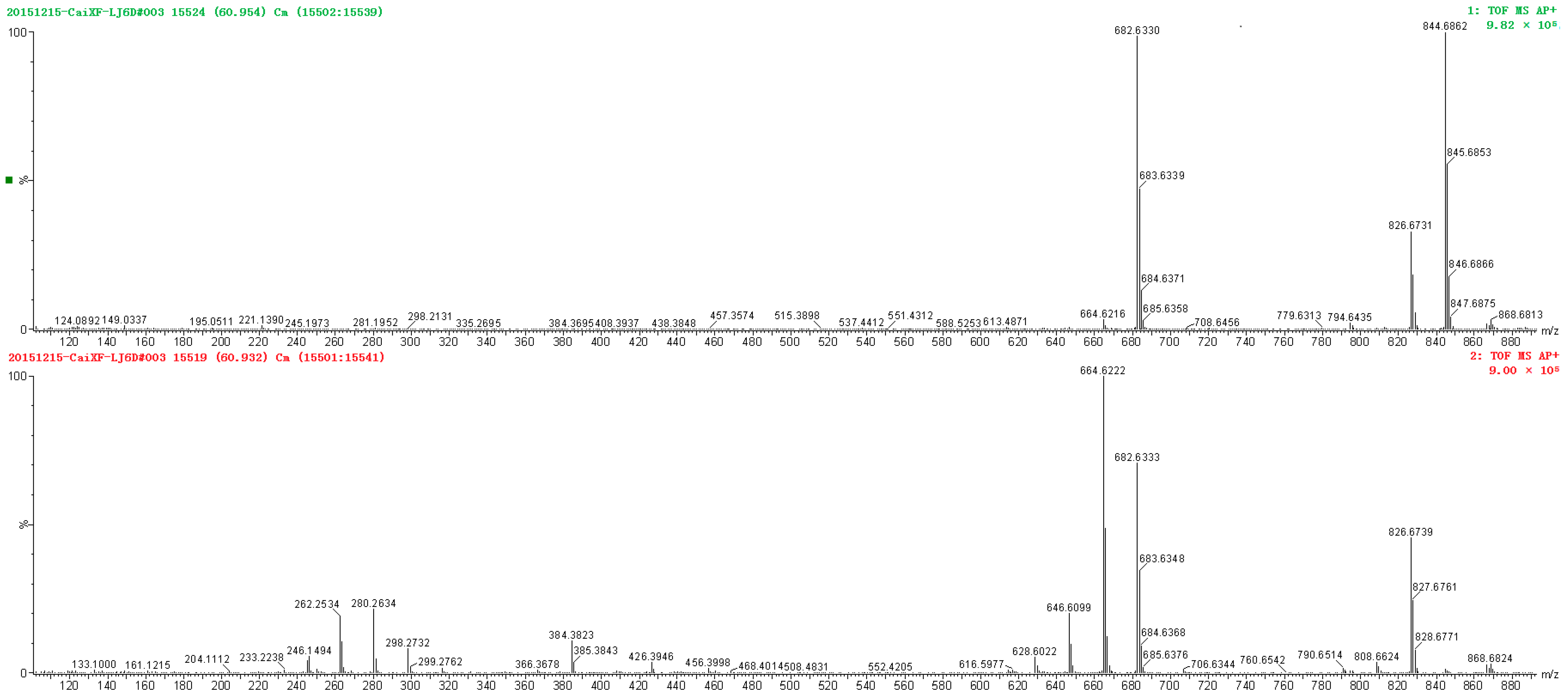
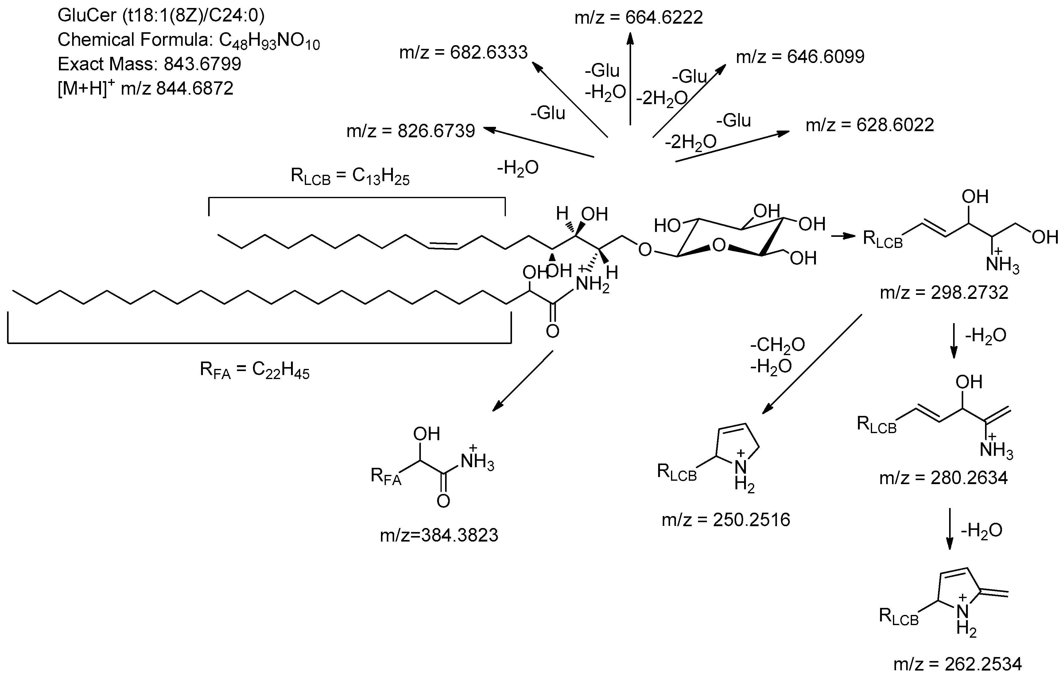
- Fragments formed by loss of small neutrals (m/z = 826.6739, 682.6333, 664.6222, 646.6099, 628.6022);
- Fragments referring to the LCB (m/z = 298.2732, 280.2634, 262.2534);
- Fragments referring to the acyl component (m/z = 384.3823).
3. Materials and Methods
3.1. Chemicals and Materials
3.2. Pretreatment of Sphingolipids
3.3. UPLC Conditions
3.4. Mass Spectrometric Conditions
Author Contributions
Funding
Institutional Review Board Statement
Informed Consent Statement
Data Availability Statement
Conflicts of Interest
References
- Haiqing, T.; Xiaokun, H. Shanshan, Regulation of the lysosome by sphingolipids: Potential role in aging. J. Biol. Chem. 2022, 298, 102118–102127. [Google Scholar] [CrossRef]
- Sugawara, T. Sphingolipids as functional food components: Benefits in skin improvement and disease prevention. J. Agric. Food Chem. 2022, 70, 9597–9609. [Google Scholar] [CrossRef] [PubMed]
- Hermier, D.; Lan, A.; Tellier, F.; Blais, A.; Culetto, M.G.; Mathe, V.; Bellec, Y.; Gissot, L.; Schmidely, P.; Faure, J.-D. Intestinal availability and metabolic effects of dietary camelina sphingolipids during the metabolic syndrome onset in mice. J. Agric. Food Chem. 2020, 68, 788–798. [Google Scholar] [CrossRef] [PubMed]
- Bachollet, S.P.J.T.; Vece, V.N.A. McCracken, synthetic sphingolipids with 1,2-Pyridazine appendages improve antiproliferative activity in human cancer cell lines. ACS Med. Chem. Lett. 2020, 11, 686–690. [Google Scholar] [CrossRef]
- García-Barros, M.; Coant, N.; Truman, J.P.; Snider, A.J.; Hannun, Y.A. Sphingolipids in colon cancer. Biochim. Biophys. Acta Mol. Cell Biol. Lipids 2014, 1841, 773–782. [Google Scholar] [CrossRef]
- Taniguchi, M.; Okazak, T. The role of sphingomyelin and sphingomyelin synthases in cell death, proliferation and migration-From cell and animal models to human disorders. Biochim. Biophys. Acta Mol. Cell Biol. Lipids 2014, 1841, 692–703. [Google Scholar] [CrossRef]
- Maceyka, M.; Spiegel, S. Sphingolipid metabolites in inflammatory disease. Nature 2014, 510, 58–67. [Google Scholar] [CrossRef]
- Lynch, D.V.; Dun, T.M. An introduction to plant sphingolipids and a review of recent advances in understanding their metabolism and function. New Phytol. 2004, 161, 677–702. [Google Scholar] [CrossRef]
- Kojima, M.; Ohnishi, M.; Ito, S. Composition and molecular species of ceramide and cerebroside in scarlet runner beans (Phaseolus coccineus L.) and kidney beans (Phaseolus vulgaris L.). J. Agric. Food Chem. 1991, 39, 1709–1714. [Google Scholar] [CrossRef]
- Bartke, N.; Fischbeck, A.; Humpf, H.U. Analysis of sphingolipids in potatoes (Solanumtuberosum L.) and sweet potatoes (Ipomoeabatatas (L.) Lam.) by reversed phase high-performance liquid chromatography electrospray ionization tandem mass spectrometry (HPLC−ESI−MS/MS). Mol. Nutr. Food Res. 2006, 50, 1201–1211. [Google Scholar] [CrossRef]
- Ohnishi, M.; Ito, S.; Fujino, T. Characterization of sphingolipidin spinach leaves. Biochim. Biophys. Acta. 1983, 752, 416–422. [Google Scholar] [CrossRef]
- Vesper, H.; Schmelz, E.M.; Nikolova-Karakashian, M.N.; Dillehay, D.L.; Lynch, D.V.; Merrill, A.H., Jr. Sphingolipids in food and the emerging importance of sphingolipids to nutrition. J. Nutr. 1999, 129, 1239–1250. [Google Scholar] [CrossRef] [PubMed]
- Valsecchi, M.; Mauri, L.; Casellato, R.; Ciampa, M.G.; Rizza, L.; Bonina, A.; Bonina, F.; Sonnino, S. Ceramides as possible nutraceutical compounds: Characterization of the Ceramides of the Moro Blood Orange (Citrus sinensis). J. Agric. Food Chem. 2012, 60, 10103–10110. [Google Scholar] [CrossRef] [PubMed]
- Rubino, F.; Zecca, L.; Sonnino, S. Characterization of a complex mixture of ceramides by fast atom bombardment and precursor and fragmnent analysis tandem mass spectrometry. Biol. Mass Spectrom. 1994, 23, 82–90. [Google Scholar] [CrossRef]
- Liebisch, G.; Drobnik, W.; Reil, M.; Trumbach, B.; Arnecke, R.; Olgemoller, B.; Roscher, A.; Schimitz, G. Quantitative measurement of different ceramide species from crude cellular extracts by electrospray ionization tandem mass spectrometry (ESI−MS/MS). J. Lipid Res. 1999, 40, 1539–1546. [Google Scholar] [CrossRef]
- Gu, M.; Kerwin, J.L.; Watts, J.D.; Aebersold, R. Ceramide profiling of complex lipid mixtures by electrospray ionization mass spectrometry. Anal. Biochem. 1997, 244, 347–356. [Google Scholar] [CrossRef]
- Hsu, F.F.; Turk, J.; Stewart, M.E.; Downing, D.T. Downing, Structural studies on ceramides as lithiated adducts by low energy collisional- activated dissociation tandem mass spectrometry with electrospray ionization. J. Am. Soc. Mass Spectrom. 2002, 13, 680–695. [Google Scholar] [CrossRef]
- Hsu, F.F.; Turk, J. Characterization of ceramides by low energy collisional-activated dissociation tandem mass spectrometry with negative-ion electrospray ionization. J. Am. Soc. Mass Spectrom. 2002, 13, 558–570. [Google Scholar] [CrossRef]
- Merrill, A.H., Jr.; Sullards, M.C.; Allegood, J.C.; Kelly, S.; Wang, E. Sphingolipidomics: High-throughput, structure-specific, and quantitative analysis of sphingolipids by liquid chromatography tandem mass spectrometry. Methods 2005, 36, 207–224. [Google Scholar] [CrossRef]
- Kyogashima, M.K.; Tadano-Aritomi, K.; Aoyama, T.; Yusa, A.; Goto, Y.; Tamiya-Koizumi, K.; Ito, H.; Murate, T.; Kannagi, R.; Hara, A. Chemical and apoptotic properties of hydroxy-ceramides containing long-chain bases with unusual alkyl chain lengths. J. Biochem. 2008, 144, 95–106. [Google Scholar] [CrossRef]
- Bijttebier, S.; Zhani, K.D.; Hond, E. Generic characterization of apolar metabolites in red chili peppers (Capsicum frutescens L.) by orbitrap mass spectrometry. J. Agric. Food Chem. 2014, 62, 4812–4831. [Google Scholar] [CrossRef] [PubMed]
- Yamauchi, R.; Aizawa, K.; Inakuma, T.; Kato, K. Analysis of molecular species of glycolipids in fruit pastes of red bell pepper (Capsicum annuum L.) by high-performance liquid chromatography-mass spectrometry. J. Agric. Food Chem. 2001, 49, 622–627. [Google Scholar] [CrossRef] [PubMed]
- Whitaker, B.D. Cerebrosides in mature-green and red-ripe bell pepper and tomato fruits. Phytochemistry 1996, 42, 627–632. [Google Scholar] [CrossRef]


| Peak NO. in Figure 4 | Identity | Mol Formula | Retention Time (min) | Precursor Ion [M + H]+ | Mass Deviation (ppm) | In-Source Fragments |
|---|---|---|---|---|---|---|
| 1 a | GlcCer t18:1(8Z)/C16:0 | C40H77NO10 | 39.240 | 732.5634 | 1.1 | 714.5470, 570.5038, 552.5008, 534.4952, 516.4750, 272.2622, 298.2640, 280.2631, 262.2523, 250.2230 |
| 2 a | GlcCer t18:1(8E)/C16:0 | C40H77NO10 | 39.762 | 732.5577 | −6.7 | 714.5466, 570.5014, 552.4971, 534.4833, 516.4709, 272.2516, 298.2710, 280.2632, 262.2488, 250.2545 |
| 3 a | GlcCer d18:2(4E,8Z)/C16:0 | C40H75NO9 | 41.180 | 714.5504 | −2.2 | 696.5394, 552.5018, 534.4865, 516.4765, 498.4711, 272.2555, 280.2615, 262.2527, 250.2527 |
| 4 a | GlcCer d18:2(4E,8E)/C16:0 | C40H75NO9 | 41.887 | 714.5492 | −3.9 | 696.5392, 552.4990, 534.4872, 516.4753, 498.4577, 272.2591, 280.2633, 262.2532, 250.2511 |
| 5 a | GlcCer t18:1(8Z)/C20:0 | C44H85NO10 | 50.302 | 788.6201 | −6.5 | 770.6164, 626.5701, 608.5527, 590.5428, 572.6311, 328.2327, 298.2710, 280.2494, 262.2310, 250.1502 |
| 6 | GlcCer t18:1(8E)/C20:0 | C44H85NO10 | 50.962 | 788.6236 | −2.0 | 770.6119, 626.5787, 608.5562, 590.5421, 572.5375, 328.3295, 298.2754, 280.2599, 262.2542, 250.1935 |
| 7 | GlcCer t18:1(8Z)/C21:0 | C45H87NO10 | 53.003 | 802.6357 | −6.4 | 784.6235, 640.5804, 622.5745, 604.5681, 586.5478, 342.3314, 298.2718, 280.2637, 262.2533, 250.2842 |
| 8 | GlcCer t18:1(8E)/C21:0 | C45H87NO10 | 53.706 | 802.6435 | 3.4 | 784.6340, 640.5870, 622.5740, 604.5605, 586.5483, 342.3280, 298.2476, 280.2622, 262.2554, 250.2282 |
| 9 a | GlcCer t18:1(8Z)/C22:0 | C46H89NO10 | 55.743 | 816.6546 | −2.3 | 798.6442, 654.6014, 636.5905, 618.5792, 600.5681, 356.3521, 298.2734, 280.2638, 262.2538, 250.2433 |
| 10 a | GlcCer t18:1(8E)/C22:0 | C46H89NO10 | 56.414 | 816.6538 | −3.3 | 798.6420, 654.6010, 636.5898, 618.5790, 600.5659, 356.3503, 298.2736, 280.2635, 262.2519, 250.2485 |
| 11 a | GlcCer t18:1(8Z)/C23:0 | C47H91NO10 | 58.393 | 830.6686 | −4.2 | 812.6581, 668.6169, 650.6047, 632.5930, 614.5784, 370.3656, 298.2737, 280.2629, 262.2523, 250.2486 |
| 12 a | GlcCer t18:1(8E)/C23:0 | C47H91NO10 | 59.022 | 830.6694 | −3.3 | 812.6564, 668.6173, 650.6068, 632.5977, 614.5822, 370.3662, 298.2727, 280.2620, 262.2531, 250.2401 |
| 13 a | GlcCer t18:1(8Z)/C24:0 | C48H93NO10 | 60.932 | 844.6862 | −1.9 | 826.6739, 682.6333, 664.6222, 646.6099, 628.6022, 384.3823, 298.2732, 280.2634, 262.2534, 250.2516 |
| 14 a | GlcCer t18:1(8E)/C24:0 | C48H93NO10 | 61.558 | 844.6882 | 0.5 | 826.6769, 682.6355, 664.6232, 646.6137, 628.6021, 384.3827, 298.2742, 280.2648, 262.2551, 250.2624 |
| 15 a | GlcCer t18:1(8Z)/C25:0 | C49H95NO10 | 63.338 | 858.7012 | −2.6 | 840.6909, 696.6468, 678.6390, 660.6227, 642.6226, 398.3954, 298.2740, 280.2657, 262.2558, 250.2472 |
| 16 | GlcCer t18:1(8Z)/C26:0 | C50H97NO10 | 65.662 | 872.7170 | −2.4 | 854.7078, 710.6650, 692.6543, 674.6449, 656.6318, 412.4139, 298.2817, 280.2620, 262.2531, 250.2545 |
| 17 | GlcCer d18:1(4E)/C16:0 | C40H77NO9 | 42.627 | 716.5623 | −7.5 | 698.5519, 554.5049, 536.5037, 518.3867, 500.4689, 272.2453, 300.2813, 282.2692, 264.2675, 252.2696 |
| 18 | GlcCer d18:1 (4Z)/C16:0 | C40H77NO9 | 43.065 | 716.5671 | −0.8 | 698.5558, 554.5139, 536.5027, 518.4926, 500.4810, 272.2585, 300.2824, 282.2780, 264.2684, 252.2589 |
| 19 a | GlcCer d18:1(8E)/C16:0 | C40H77NO9 | 43.824 | 716.5661 | −2.2 | 698.5563, 554.5127, 536.5024, 518.4901, 500.4734, 272.2595, 300.2853, 282.2785, 264.2689, 252.2610 |
| 20 a | GlcCer d18:1(8Z)/C16:0 | C40H77NO9 | 44.941 | 716.5644 | −4.6 | 698.5516, 554.5101, 536.5000, 518.4897, 500.4706, 272.2574, 300.2909, 282.2749, 264.2679, 252.2704 |
| Peak NO. in Figure 4 | Identity | Mol Formula | Retention Time (min) | Precursor Ion [M + H]+ | Mass Deviation (ppm) | In-Source Fragments |
|---|---|---|---|---|---|---|
| 21 | Cer t18:1(8E)/C21:0 | C39H77NO5 | 56.061 | 640.5842 | −5.9 | 622.5762, 604.5622, 586.5604, 342.3251, 298.2794, 280.2544, 262.2525, 250.2413 |
| 22 | Cer t18:1(8E)/C22:0 | C40H79NO5 | 58.731 | 654.6017 | −2.9 | 636.5911, 618.5797, 600.5630, 356.3488, 298.2737, 280.2621, 262.2532, 250.2483 |
| 23 | Cer t18:1(8E)/C23:0 | C41H81NO5 | 61.332 | 668.6201 | 1.2 | 650.6070, 632.5964, 614.5864, 370.3588, 298.2745, 280.2654, 262.2546, 250.2499 |
| 24 | Cer t18:1(8E)/C24:0 | C42H83NO5 | 63.794 | 682.6343 | −0.9 | 664.6221, 646.6118, 628.6007, 384.3823, 298.2739, 280.2634, 262.2530, 250.2474 |
| 25 | Cer t18:1(8E)/C25:0 | C43H85NO5 | 66.180 | 696.6473 | −4.7 | 678.6379, 660.6263, 642.6121, 398.3926, 298.2733, 280.2634, 262.2524, 250.2519 |
| 26 | Cer t18:1(8E)/C26:0 | C44H87NO5 | 68.398 | 710.6653 | 1.3 | 692.6543, 674.6428, 656.6281, 412.4193, 298.2742, 280.2641, 262.2527, 250.2606 |
| 27 | Cer t18:0/C22:0 | C40H81NO5 | 61.823 | 656.6196 | 0.5 | 638.6069, 620.5929, 602.5778, 356.3482, 300.2935, 282.2811, 264.2675, 252.2646 |
| 28 | Cer t18:0/C23:0 | C41H83NO5 | 64.323 | 670.6332 | −2.5 | 652.6213, 634.6117, 616.5888, 370.3677, 300.2863, 282.2784, 264.2646, 252.2683 |
| 29 | Cer t18:0/C24:0 | C42H85NO5 | 66.660 | 684.6489 | −2.5 | 666.6374, 648.6246, 630.6136, 384.3810, 300.2896, 282.2793, 264.2678, 252.2687 |
| 30 | Cer t18:0/C25:0 | C43H87NO5 | 68.893 | 698.6639 | −3.3 | 680.6516, 662.6420, 644.6274, 398.3915, 300.2901, 282.2774, 264.2668, 252.2619 |
| 31 | Cer t18:0/C26:0 | C44H89NO5 | 71.083 | 712.6754 | −9.1 | 694.6643, 676.6531, 658.6328, 412.3976, 300.2846, 282.2782, 264.2603, 252.2670 |
| Peak No. in Figure 4 | Identity | Mol Formula | Retention Time (min) | Precursor Ion [M + H]+ | Mass Deviation (ppm) | In-Source Fragments |
|---|---|---|---|---|---|---|
| 32 | Cer t18:0/C22:0 | C40H81NO6 | 57.956 | 672.6092 | −7.4 | 654.5990, 636.5912, 618.5792, 600.5424, 372.3345, 300.2885, 282.2762, 264.2688, 252.2687 |
| 33 | Cer t18:0/C23:0 | C41H83NO6 | 60.725 | 686.6282 | −2.5 | 668.6127, 650.6048, 632.5945, 614.5081, 386.3432, 300.2786, 282.2796, 264.2616, 252.2395 |
| 34 | Cer t18:0/C24:0 | C42H85NO6 | 63.192 | 700.6422 | −4.7 | 682.6349, 664.6192, 646.6100, 628.5762, 400.3618, 300.2859, 282.2801, 264.2687, 252.2649 |
| 35 | Cer t18:0/C26:0 | C44H89NO6 | 67.868 | 728.6722 | −6.3 | 710.6717, 692.6498, 674.5972, 656.6024, 428.3588, 300.3049, 282.2872, 264.2456, 252.2775 |
| 36 | Cer t18:1/C22:0 | C40H79NO6 | 54.852 | 670.5996 | 1.5 | 652.5925, 634.6226, 616.5663, 598.5677, 372.3235, 298.2316, 280.2595, 262.2215, 250.2215 |
| 37 | Cer t18:1/C23:0 | C41H81NO6 | 57.534 | 684.6113 | −4.2 | 666.6025, 648.6174, 630.6168, 612.3077, 386.3209, 298.2623, 280.2640, 262.2560, 250.2477 |
| 38 | Cer t18:1/C24:0 | C42H83NO6 | 60.227 | 698.6283 | −2.3 | 680.6160, 662.6033, 644.6029, 626.5577, 400.3712, 298.2746, 280.2657, 262.2518, 250.2541 |
| 39 | Cer t18:1/C25:0 | C43H85NO6 | 62.697 | 712.6404 | −7.2 | 694.6312, 676.6130, 658.6057, 640.5693, 414.3831, 298.2746, 280.2602, 262.2528, 250.2536 |
| 40 | Cer t18:1/C26:0 | C44H87NO6 | 65.076 | 726.6617 | 0.7 | 708.6427, 690.6297, 672.6454, 654.5604, 428.4056, 298.2639, 280.2538, 262.2497, 250.2513 |
Disclaimer/Publisher’s Note: The statements, opinions and data contained in all publications are solely those of the individual author(s) and contributor(s) and not of MDPI and/or the editor(s). MDPI and/or the editor(s) disclaim responsibility for any injury to people or property resulting from any ideas, methods, instructions or products referred to in the content. |
© 2023 by the authors. Licensee MDPI, Basel, Switzerland. This article is an open access article distributed under the terms and conditions of the Creative Commons Attribution (CC BY) license (https://creativecommons.org/licenses/by/4.0/).
Share and Cite
Xu, M.-L.; Qi, L.; Cai, X.; Cao, T.; Tang, R.; Cao, K.; Lian, Y. Speculation of Sphingolipids in Capsanthin by Ultra-Performance Liquid Chromatography Coupled with Electrospray Ionization-Quadrupole—Time-of-Flight Mass Spectrometry. Molecules 2023, 28, 1010. https://doi.org/10.3390/molecules28031010
Xu M-L, Qi L, Cai X, Cao T, Tang R, Cao K, Lian Y. Speculation of Sphingolipids in Capsanthin by Ultra-Performance Liquid Chromatography Coupled with Electrospray Ionization-Quadrupole—Time-of-Flight Mass Spectrometry. Molecules. 2023; 28(3):1010. https://doi.org/10.3390/molecules28031010
Chicago/Turabian StyleXu, Mei-Li, Lijun Qi, Xingfu Cai, Tong Cao, Rumeng Tang, Kaihang Cao, and Yunhe Lian. 2023. "Speculation of Sphingolipids in Capsanthin by Ultra-Performance Liquid Chromatography Coupled with Electrospray Ionization-Quadrupole—Time-of-Flight Mass Spectrometry" Molecules 28, no. 3: 1010. https://doi.org/10.3390/molecules28031010
APA StyleXu, M.-L., Qi, L., Cai, X., Cao, T., Tang, R., Cao, K., & Lian, Y. (2023). Speculation of Sphingolipids in Capsanthin by Ultra-Performance Liquid Chromatography Coupled with Electrospray Ionization-Quadrupole—Time-of-Flight Mass Spectrometry. Molecules, 28(3), 1010. https://doi.org/10.3390/molecules28031010


